abstract - clinicalneuropsychiatry.org · clinical neuropsychiatry (2017) 14, 6, 359-373 sensory...
TRANSCRIPT

Clinical Neuropsychiatry (2017) 14, 6, 359-373
SENSORY PROCESSING SENSITIVITY AND CHILDHOOD QUALITY’S EFFECTS ON NEURAL RESPONSES TO EMOTIONAL STIMULI
Bianca P. Acevedo, Jadzia Jagiellowicz, Elaine Aron, Robert Marhenke, Arthur Aron
AbstractObjective: This study examined the neural correlates of adult sensory processing sensitivity (SPS) and its interaction
with subjective ratings of quality of childhood parenting (QCP). Method: Fourteen women (ages 18-25) underwent fMRI while viewing positive, negative and neutral images from the
standard International Affective Picture System (IAPS) and completed the Highly Sensitive Person (HSP) Scale. (HSP) Scale, a neuroticism scale, and measures of quality of recalled childhood parenting.
Results: In response to emotional (versus neutral) IAPS images, the SPS x QCP interaction (and also of SPS directly controlling for neuroticism) showed significant positive neural correlations in the hippocampus, entorhinal area, hypothalamus, and temporal/parietal areas, which process emotional memory, learning, physiological homeostasis, awareness, reflective thinking, and integration of information. For positive stimuli only, SPS showed significant correlations with areas involved in reward processing (VTA, SN, caudate), self-other integration (insula and IFG), calm (PAG), and satiation (subcallosal AC); and to a greater extent with increasing QCP. For negative images, the SPS x QCP interaction showed significant activation in the amygdala and PFC (involved in emotion and self-control), without diminished reward activity.
Conclusions: SPS (and its interaction with childhood environment) is positively associated with activation of brain regions associated with depth of processing, memory, and physiological regulation in response to emotional stimuli. Results support differential susceptibility, vantage sensitivity and HSP models suggesting that SPS is associated with environmental sensitivity so that positive environments (such as high QCP) may provide benefits, such as adaptive responsivity (with awareness, arousal, self-control and calm) to emotionally evocative stimuli.
Key words: sensory processing sensitivity, fMRI, childhood environment, emotions, reward, self-regulation
Declaration of interest: all of the authors declare no financial interests or potential conflicts of interest.
Bianca P. Acevedo1, Jadzia Jagiellowicz2, Elaine Aron2, Robert Marhenke3, Arthur Aron2
1 University of California, Santa Barbara, Neuroscience Research Institute, Santa Barbara, CA, 93106-5060, USA. 2 Stony Brook University, Stony Brook, NY, 11794-2500, USA. 3 Leopold-Franzens University of Innsbruck, Innsbruck, Austria
Corresponding author Bianca Acevedo, University of California, Santa Barbara, Neuroscience Research Institute,Santa Barbara, CA 93106-5060.Contact: [email protected]
Submitted September 2017, Accepted November 2017© 2017 Giovanni Fioriti Editore s.r.l. 359
Sensory processing sensitivity (SPS) is thought to be a genetically-based trait found in humans and in over 100 other species (Wolf et al. 2008). It is characterized by greater environmental sensitivity (Pluess 2015), including social stimuli (Acevedo et al. 2014, Wolf et al. 2008), and behaviorally it is typically associated with reactivity to salient stimuli and greater cautiousness in approaching novel situations and objects (Aron et al. 2012; Gartstein et al. 2016, review). It appears to be a survival strategy found across species in only in a significant minority within each species, probably due to negative frequency dependence (Wolf et al. 2008). The trait bestows the advantage (as long as most individuals do not possess it) of being more aware than others of opportunities and threats. Interpersonal sensitivity would also allow them to select mates, parent, empathize and form alliances more effectively resulting in survival
advantages (Acevedo et al. 2017).SPS has a unique flavor in humans, and is generally
measured with the Highly Sensitive Person (HSP) scale (Aron et al. 1997). Its items include sensitivity to bright lights, loud noises, others’ moods, violent stimuli, caffeine, and hunger as well as having “a rich, complex inner life” greater conscientiousness, and “being deeply moved by arts or music. SPS is thought to be mediated by deep and connective cognitive processing (Mesulam 1998), as well as emotion responsivity, which enhances memory, attention and learning (Baumeister et al. 2007). Research on differential susceptibility (Belsky et al. 2009) suggests that greater sensitivity leads to more vulnerability to negative environments and greater vantage sensitivity in positive ones (Belsky et al. 2009, Boyce et al. 2005, Pluess et al. 2013). Similarly, studies of SPS in adults, using the standard HSP scale

Bianca P. Acevedo et al.
360 Clinical Neuropsychiatry (2017) 14, 6
including the 27-item HSP Scale (M = 4.26, SD = 0.99), Cronbach’s alpha = 0.87 (as in previous studies Aron et al. 2005, Benham 2006); and a measure of quality of childhood parenting (QCP), calculated as a weighted sum (computed from the larger sample initially surveyed) of 7 inter-correlated scales. The weights were based on contributions to the first principal component, positively or negatively assigned to represent high-quality parenting: (a-d) the care and overprotection subscales of the Parental Bonding Inventory (Parker et al. 1979), completed for both mother and father (Cronbach’s alphas: care, 0.94 and 0.92 for mother and father, respectively; overprotection, 0.88 and 0.90, respectively; weights: -.307, -265, .286, 301); (e and f) the abuse subscale of the Measure of Parenting Style (Parker et al. 1997): for mother, (Cronbach’s alpha =0.86; weight: 0.12); and for father (Cronbach’s alpha = 0.87, weight: 0.09); and (g) an eight-item scale (e.g., “Would you characterize your childhood as troubled?”) used in previous SPS studies (e.g. Aron EN et al. 2005; Cronbach’s alpha = 0.74, weight = 0.22). In addition a measure of neuroticism (M = 3.96; SD = 1.58) was included, with two items “Are you prone to depression?” and “Are you prone to fears?” (Cronbach’s alpha = 0.51). As in previous studies, SPS and neuroticism were highly correlated (r = 0.66, p = 0.01) thus we followed standard procedures (e.g. Aron A et al. 2005), and controlled for neuroticism for the basic correlations of SPS with brain responsivity (results are shown in tables 1 and 2). For the SPS X QCP interaction we did not control for neuroticism as the parenting interaction also predicts neuroticism.
Experimental ProcedureStimuli and MRI protocol. Stimuli consisted of
pictures from the IAPS, which were specifically selected for wide use and have been shown to correspond with the elicitation of emotions in normative and clinical samples (Bradley et al. 2007). The fMRI protocol (modified from Canli et al. 2001 and Ribeiro et al. 2007) consisted of an 8-minute session where participants viewed 6 alternating blocks of four pictures, each of the same valence (positive, negative, or neutral); and with 3 practice-trials at the beginning of the entire sequence. Each picture was presented for 6,000 ms, with an interstimulus interval (a fixation cross) of 1,125 ms. The initial block (after the practice trials) consisted of neutral pictures. The order of subsequent blocks was alternated across participants.
Post-scan anxiety ratings. Post-scan, participants were asked to indicate their level of anxiety while in the scanner on a Likert scale from 1 to 7, with 1 representing “not at all” and 7 representing “extremely.” The mean anxiety rating was 2.62.
Data Acquisition and AnalysisTo collect brain imaging data, we used a 3.0 T
MAGNETOM TrioTim magnetic resonance imaging scanner at the SCAN Center of Stony Brook University. A T2-weighted gradient-echo echo-planar sequence (repetition time 2,000 ms, echo time 30 ms, 80˚ flip angle, field of view 240 X 240 mm, 64 X 64 matrix) was used to acquire functional scans. The pictures consisted of 30 contiguous axial slices, with no gap between slices, voxel size was 3.8 X 3.8 X 4.0 mm. Anatomical scans were also acquired (axial T1-weighted scans; repetition time 300 ms, echo time ms, 256 X 256 matrix, 80˚ flip angle, 240 mm X 240 mm field of view, slice thickness 4 mm) in the same session. Voxel size for the anatomical scans was 0.9 X 0.9 X 4 mm.
(Aron et al. 1997), found that high SPS individuals with negative childhood environments had more anxiety and depression as adults compared to their less sensitive counterparts (Aron et al. 2005, Liss et al. 2005). Also in support of vantage sensitivity, another study found that high (vs. low) SPS adults with positive recalled childhoods showed especially greater arousal to positive (versus neutral) images (Jagiellowicz et al. 2016).
Although heightened emotional arousal may provide some explanation for the differential susceptibility associated with the trait, it has mostly been reported in behavioral studies. The three fMRI studies of SPS to date (all using the HSP scale) found that it is associated with overall greater expression in (a) visual areas associated with making fine visual distinctions (Jagiellowicz et al. 2011); (b) regions associated with attention and working memory in response to a task involving attending to context to visual scenery (Aron et al. 2010); and (c) regions involved in empathy, awareness, sensory integration, self-referential processing and action in response to others’ emotional expressions (Acevedo et al. 2014). Moreover, a review of the brain structures involved in SPS versus seemingly related clinical disorders (i.e., autism) suggests that it is largely differentiated by neural processing in regions associated with physiological homeostasis, self-regulation, self-other processing, empathy and awareness (Acevedo et al. 2017).
The present study used functional MRI (as did the three previous studies) to measure the neural correlates of SPS in response to standardized emotional pictures from the International Affective Picture System (IAPS). It also used seven well-established childhood measures of perceived quality of childhood environment. Our two questions were: (1) whether participants with high SPS would show evidence of greater emotional, memory, awareness and self-referential processing to affective images; and (2) whether this would vary with degree of positive childhood quality. Thus, we specifically focused on brain regions shown in previous neuroimaging studies of SPS as well as the amygdala – a main site of emotion processing, especially to aversive stimuli (Canli et al. 2000; Ochsner et al. 2004, 2009; Phan et al. 2004) – whose role in emotional SPS processing has been inconclusive.
MethodParticipants
Participants were undergraduate student females recruited from Stony Brook University, with scores at the top and bottom quartile of the HSP scale (eliminating the top and bottom 2.5% of scorers). Sample selection was consistent with conditions delineated by previous studies (Preacher et al. 2005), such as recruitment of only one gender (females), as studies have shown significant gender differences for the IAPS emotion task (Blair 2002, Velderman et al. 2006). Our resulting sample consisted of 14 right-handed females, ages 18-25 (M age = 19.00 years, SD = 1.84), with an ethnic composition of 50% Caucasian, 40% Asian and 11% reporting “other”. Of these 14, 7 were in the top SPS quartile and 7 in the bottom quartile. All participants met criteria for fMRI contraindications (e.g., no severe alcohol or drug use, claustrophobia, etc.).
QuestionnairesParticipants completed a battery of questionnaires

Table 1. Correlations of Adult Sensory Processing Sensitivity (controlling for Neuroticism) with Neural Response to Positive versus Neutral Images
Brain Region Left Rightx y z T p k x y z T p k
ROI ActivationsVTA/SN 8 -16 -16 1.91 0.04a 3Caudal cingulate -20 -8 36 2.55 0.01a 8 12 -20 28 3.11 0.004a 15Caudate tail -28 -60 12 2.87 0.02a 9Hippocampus/ Entorhinal area
-36 -8 -28 3.64 0.002abc 22 36 -8 -28 3.62 0.002abc 22
Hypothalamus -4 -8 -8 2.25 0.02abc 3Periacqueductal gray -4 -36 -32 2.05 0.03a 9 4 -36 -32 1.86 0.04a 9Anterior cingulate, subcallosal
8 32 4 3.21 .004a 3
Insula -40 12 12 2.77 0.01a 5Fusiform gyrus -36 -32 -16 2.65 0.01b 9Tempoparietal junction 47 -66 24 2.61 0.01a 24Precuneus/parietal area -16 -48 52 2.46 0.03a 21 7 -49 56 3.13 0.01 16Superior/middle temporal gyrus
68 -44 4 3.70 0.002abc 6
Inferior temporal gyrus -52 -4 -24 3.04 0.01 abc 16Medial PFC 21 40 -8 2.37 0.02a 8Whole-brain DeactivationsDorsomedial PFC 12 40 28 4.04 < .001 88Inferior parietal lobule 40 -76 44 4.20 < .001ac 22Inferior parietal cortex 52 -36 56 4.15 < .001ac 262Anterior cingulate cortex -12 48 -4 3.86 < .001 79 Note. Results are for regions showing significant brain response in association with the Highly Sensitive Person (HSP) Scale scores moderated by Positive Childhood scale scores. MNI coordinates (x,y,z) are at the maximum value for the cluster, which may be elongated in any direction. Legend: a overlapping area for Positive Conditions; b overlapping with the Negative condition (controlling for Neuroticism); and coverlapping with Negative condition x Childhood.
Sensory processing sensitivity, the brain, and emotions
Clinical Neuropsychiatry (2017) 14, 6 361
.05 (Genovese et al. 2002) to correct for multiple comparisons. ROIs were derived from previous fMRI studies of SPS (Aron et al. 2010, Jagiellowicz et al. 2011), a meta-analysis on human brain responses to emotional stimuli (Morelli et al. 2015), and close inspection of the amygdala (Costafreda et al. 2008, Phan et al. 2004). All ROIs occupied a 3-mm radius (minimum) and anatomic regions were confirmed with the Atlas of the Human Brain (Mai et al. 2008).
Results
SPS (controlling for Neuroticism) Correlations with Human Brain Activity
Positive versus Neutral Contrast. As shown in table 1, significant regional brain correlations were shown for SPS (controlling for Neuroticism) in response to positive (vs. neutral) IAPS images in the ventral tegmental area (VTA)/ substantia nigra (SN), caudate, hippocampus, periaqueductal gray (PAG), anterior cingulate (AC), insula, fusiform gyrus (FG), temporoparietal junction (TPJ), precuneus, temporal gyrus, and medial PFC.
Negative versus Neutral Contrast. As shown in table 2, significant regional brain correlations were shown for SPS (controlling for Neuroticism) in response to negative versus neutral IAPS images in the amygdala, hippocampus/entorhinal area, hypothalamus,
Stimuli were shown using E-Prime software (Version 2.0, Psychology Software Tools, Pittsburgh, PA) and were projected on a screen placed directly outside the MRI tube, and viewed via an angled mirror mounted on the RF coil of the scanner.
Data were analyzed using SPM5 (http://www.fil.ion.ucl.ac.uk/spm). For preprocessing, functional EPI volumes were realigned to the first volume, smoothed with a Gaussian kernel of 6mm, and then normalized to the T1.nii image template. No participant showed movement greater than 3 mm (whole-voxel). After preprocessing, contrasts were created (e.g., positive vs. neutral) followed by regression analyses examining the associations between each contrast (positive vs. neutral and negative vs. neutral). For SPS controlling for Neuroticism, first we calculated SPS residual scores controlling for the interaction of SPS with Neuroticism. The residuals were used to carry out a mixed-effects general linear model, with participants as the random-effects factor and conditions as the fixed effect. For the SPS x QCP interaction, regression analyses were conducted estimating group brain activity in association with SPS and QCP scores which produced an interaction term controlling for the independent contribution of each of the variables.
We conducted exploratory, whole-brain analyses using a threshold of p < .001 (uncorrected) and a spatial extent of > 15 contiguous voxels. We also examined, a priori regions of interest (ROIs) applying a standard false discovery rate (FDR) with a threshold of p <

Table 3. Correlations of Adult Sensory Processing Sensitivity and Childhood Environment with Neural Response to Positive versus Neutral Images
Brain RegionLeft Right
x y z T p k x y z T p k
VTA/SN -4 -8 -16 2.6 0.01 15 8 -16 -16 2.10 0.03a 43Caudate tail/posterior cingulate -28 -64 12 3.31 0.005a 95
Caudate cingulate -20 -8 36 3.24 0.01a 20 16 -20 28 3.20 0.02 a 35
Hippocampus/ entorhinal area -36 -8 -28 1.93 0.04abc
4536 -8 -28 4.38 0.01abc 23
Hypothalamus -4 -8 -12 3.09 0.01abc 15 4 -8 -12 2.68 0.02 15Periacqueductal gray -4 -36 -32 2.79 0.01a 11 4 -36 -32 2.50 0.02a 11Anterior cingulate, subcallosal 8 32 4 2.63 0.01a 4
Insula -40 12 12 3.77 < .001a 21Inferior frontal gyrus -44 24 4 3.29 0.01 24Fusiform gyrus 36 -24 -12 2.31 0.02bc 45Tempoparietal junction 48 -68 28 2.84 0.01a 114Precuneus/parietal area -12 -60 52 3.23 0.01 84Superior/middle temporal gyrus 68 -48 4 1.90 0.01abc 19
Inferior temporal gyrus -52 -4 -29 1.85 0.01abc 16
Medial PFC -20 32 -12 3.04 0.01 16 24 40 -8 3.12 0.01 a 6PFC -40 44 32 3.53 0.003 37DeactivationsInferior parietal area 40 -72 48 4.77 < .001ac 37
Note. Results are for regions showing significant brain response in association with the Highly Sensitive Person (HSP) Scale scores moderated by Positive Childhood scale scores. MNI coordinates (x,y,z) are at the maximum value for the cluster, which may be elongated in any direction. Legend: a overlapping area for Positive conditions; b overlapping with the Negative condition (controlling for Neuroticism); and c overlapping with Negative x Childhood.
Bianca P. Acevedo et al.
362 Clinical Neuropsychiatry (2017) 14, 6
that for positive (vs. neutral) pictures, the SPS x QCP interaction showed significant neural activations that were not shown for the SPS correlation in the left VTA, IFG, dorsomedial and ventromedial PFC; and the right hypothalamus. These areas are well-known for their role in reward, self-other processing, cognitive control, and physiological homeostasis.
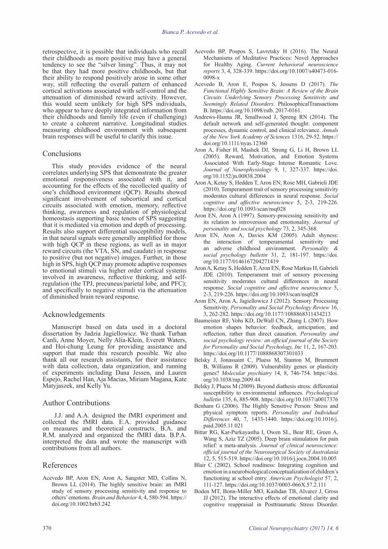
Negative versus Neutral Contrast. As shown in table 4, the interaction of SPS x QCP in response to negative (vs. neutral) images resulted in significant activation of the bilateral amygdala, hippocampus, precuneus/parietal area, temporal pole, middle frontal gyrus (MFG), ventromedial PFC, secondary somatosensory cortex (SII), and the supplementary motor area (SMA); the left hypothalamus, PC, TPJ, dorsomedial PFC, sensorimotor cortex; and the right STG, MTG, ITG, occipital/FG, precentral gyrus, and frontal pole.
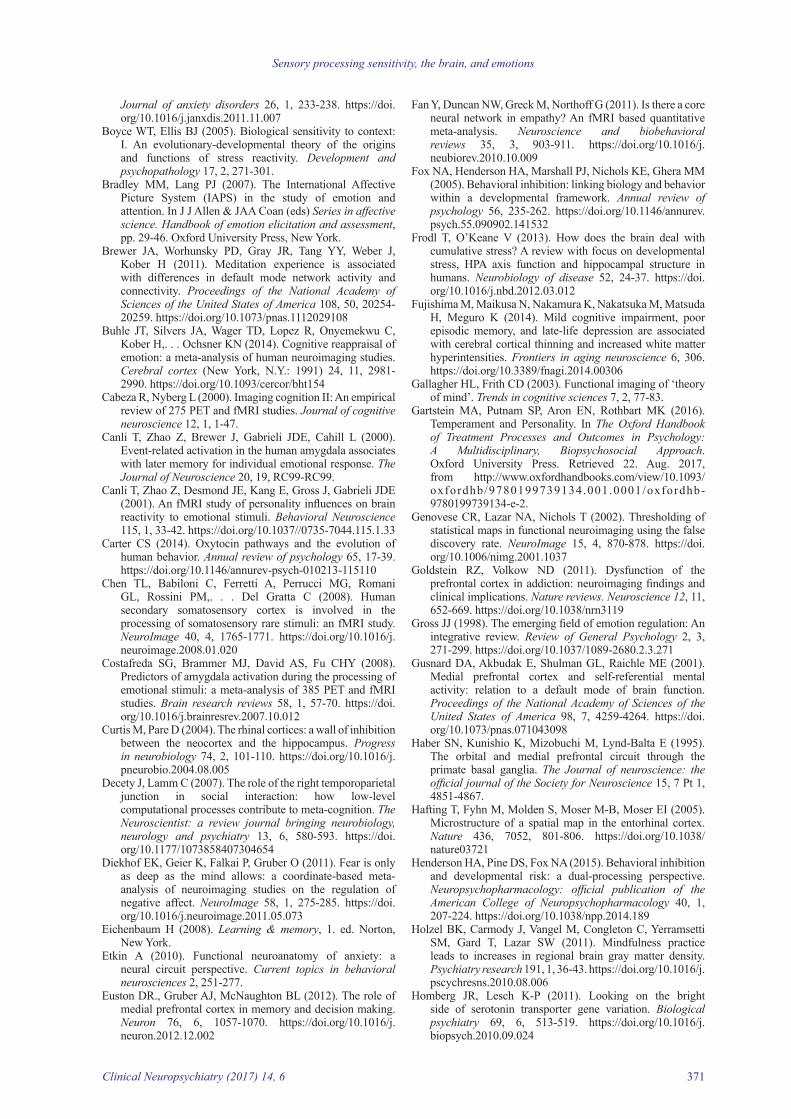
In general, the pattern of results for the SPS x QCP interaction in response to negative images was similar to those seen for the SPS correlation (denoted by superscript “a” in tables 2 and 4). However, a few important differences emerged. In response to negative stimuli, SPS showed significant deactivation in the VTA, SN, caudate (figure 3), and IFG (indicating less reward and self/other processing). This pattern did not emerge for the SPS x QCP interaction. In contrast, the SPS x QCP interaction for negative (versus neural) images showed significant brain activations in the dorsomedial
AC, posterior cingulate (PC), precuneus/parietal area, TPJ, temporal gyrus, FG, frontal gyri, ventromedial PFC, SII, and premotor cortex (PMC).
SPS x Quality of Childhood Parenting (QCP) Activations in the Human Brain
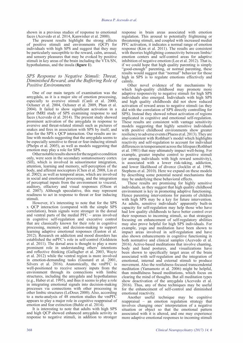
Positive versus Neutral Contrast. As shown in table 3, the interaction of SPS x QCP in response to positive (vs. neutral) IAPS images resulted in significant neural activity in the bilateral VTA/SN, caudal cingulate, hippocampus/entorhinal area, hypothalamus, PAG, and medial PFC; right AC, FG, TPJ, and superior/middle temporal gyrus (STG, MTG); and the left caudate tail/PC, insula, inferior frontal gyrus (IFG), insula, precuneus/parietal area, inferior temporal gyrus (ITG), and PFC. The pattern of the SPS x QCP interaction for positive (vs. neutral) images was such that the combination of greater HSP and QCP scores resulted in stronger brain activation for most of the regions, compared to lower HSP and QCP (figure 1). In other words, subjects with low SPS did not show large differences in the strength of neural signals as a function of QCP.
Many of the results shown for the SPS x QCP interaction (positive versus neutral condition) were also shown for the SPS correlation as denoted by superscripts “a”. However, it’s interesting to note

Sensory processing sensitivity, the brain, and emotions
Clinical Neuropsychiatry (2017) 14, 6 363
4
5
C
0
1
2
3
Caudate tail Insula
E
Hippocampus/Entorhinal area
0
1
2
3
4
5
A
0
1
2
3
4
5
VTA Hypothalamus
-0.125
0
0.125
0.25
0.375
0.5
NegPar PosPar
LoHSP HiHSP
-0.125
0
0.125
0.25
0.375
0.5
NegPar PosPar
LoHSP HiHSP
-0.125
0
0.125
0.25
0.375
0.5
NegPar PosPar
LoHSP HiHSP
D
F
B
Activation intensity
for the Caudate tail
(-29,-20,12)
Activation intensity
for VTA
(8,-16,-16)
Activation intensity
for the Hippocampus/Entorhinal area
(36,-8,-28)
Figure 1. Sensory processing sensitivity (SPS) and subjective quality of childhood parenting (QCP) interaction is associated with adults’ brain responsivity to positive (vs. neutral) images in the: A) ventral tegmental area (VTA)/substantia nigra (SN) and hypothalamus; C) the caudate tail and insula; and E) the hippocampus/entorhinal area. Plots show that subjective positive childhood moderates the response intensity in the B) R. VTA/SN, and D) the L. caudate tail, and F) the R. hippocampus/entorhinal area
4
5
C
0
1
2
3
Caudate tail Insula
E
Hippocampus/Entorhinal area
0
1
2
3
4
5
A
0
1
2
3
4
5
VTA Hypothalamus
-0.125
0
0.125
0.25
0.375
0.5
NegPar PosPar
LoHSP HiHSP
-0.125
0
0.125
0.25
0.375
0.5
NegPar PosPar
LoHSP HiHSP
-0.125
0
0.125
0.25
0.375
0.5
NegPar PosPar
LoHSP HiHSP
D
F
B
Activation intensity
for the Caudate tail
(-29,-20,12)
Activation intensity
for VTA
(8,-16,-16)
Activation intensity
for the Hippocampus/Entorhinal area
(36,-8,-28)
4
5
C
0
1
2
3
Caudate tail Insula
E
Hippocampus/Entorhinal area
0
1
2
3
4
5
A
0
1
2
3
4
5
VTA Hypothalamus
-0.125
0
0.125
0.25
0.375
0.5
NegPar PosPar
LoHSP HiHSP
-0.125
0
0.125
0.25
0.375
0.5
NegPar PosPar
LoHSP HiHSP
-0.125
0
0.125
0.25
0.375
0.5
NegPar PosPar
LoHSP HiHSP
D
F
B
Activation intensity
for the Caudate tail
(-29,-20,12)
Activation intensity
for VTA
(8,-16,-16)
Activation intensity
for the Hippocampus/Entorhinal area
(36,-8,-28)
B
4
5
C
0
1
2
3
Caudate tail Insula
E
Hippocampus/Entorhinal area
0
1
2
3
4
5
A
0
1
2
3
4
5
VTA Hypothalamus
-0.125
0
0.125
0.25
0.375
0.5
NegPar PosPar
LoHSP HiHSP
-0.125
0
0.125
0.25
0.375
0.5
NegPar PosPar
LoHSP HiHSP
-0.125
0
0.125
0.25
0.375
0.5
NegPar PosPar
LoHSP HiHSP
D
F
B
Activation intensity
for the Caudate tail
(-29,-20,12)
Activation intensity
for VTA
(8,-16,-16)
Activation intensity
for the Hippocampus/Entorhinal area
(36,-8,-28)
4
5
C
0
1
2
3
Caudate tail Insula
E
Hippocampus/Entorhinal area
0
1
2
3
4
5
A
0
1
2
3
4
5
VTA Hypothalamus
-0.125
0
0.125
0.25
0.375
0.5
NegPar PosPar
LoHSP HiHSP
-0.125
0
0.125
0.25
0.375
0.5
NegPar PosPar
LoHSP HiHSP
-0.125
0
0.125
0.25
0.375
0.5
NegPar PosPar
LoHSP HiHSP
D
F
B
Activation intensity
for the Caudate tail
(-29,-20,12)
Activation intensity
for VTA
(8,-16,-16)
Activation intensity
for the Hippocampus/Entorhinal area
(36,-8,-28)

Bianca P. Acevedo et al.
364 Clinical Neuropsychiatry (2017) 14, 6
DiscussionThis was the first neuroimaging study to investigate
the neural correlates of SPS in response to standard emotional images from the IAPS, with the addition of examination of the effects of self-reported quality of childhood parenting (QCP). Our results, along with those from previous empirical studies, suggest that in response to both positive and negative visual stimuli, SPS evokes brain activation in regions that mediate: (a) memory, attention, awareness, and reflective thinking in response to both positive and negative emotional stimuli; and (b) reward processing (VTA, SN, caudate), self-other integration (insula and IFG), calm (PAG), and satiation (subcallosal AC) to positive stimuli only.
This pattern of results was also shown for the SPS x QCP interaction. However, the interaction resulted in overall stronger brain activation of regions that mediate emotions, memory, physiological homeostasis, attention and cognitive processes ‒ specifically in the
PFC, occipital/FG, precentral gyrus, frontal pole, and sensorimotor cortex ‒ areas involved in cognitive emotion processing, visual processing, decision-making and self-regulation (Buhle et al. 2014, Sabatinelli et al. 2011); while these areas were not shown for the SPS correlation. These results highlight the role that QCP may play for SPS in adaptive response to negative stimuli ‒ namely through enhanced cognitive and self-regulatory processing without diminished reward.
Commonalities for All SPS Conditions Across all SPS conditions (that is, in response to
both positive and negative images, when controlling for Neuroticism, and for the SPS x QCP interaction), SPS showed significant positive correlations with activation in the hippocampus, entorhinal area, hypothalamus, and temporal gyri; and deactivation of the inferior parietal area.
Table 2. Correlations of Adult Sensory Processing Sensitivity (controlling for Neuroticism) with Neural Response to Negative versus Neutral Images
Brain RegionLeft Right
x y z T p k x y z T p k
ROI ActivationsAmygdala -20 -12 -24 2.41 0.02a 4Hippocampus/ entorhinal area -36 -8 -28 3.64 0.002abc 22 36 -8 -28 3.62 0.002abc 22
Hypothalamus 0 -4 -8 3.02 0.01abc 5Anterior cingulate 0 44 20 2.58 0.01 19
Posterior cingulate/ precuneus 8 -56 28 1.82 0.05a 292
Precuneus/parietal area -12 -68 28 2.77 0.01a 292 16 -56 36 4.53 0.004a 292Tempoparietal junction -40 -52 24 2.86 0.01a 36 36 -60 32 2.16 0.001 36Middle/inferior temporal gyrus -52 -24 -16 4.00 < .001abc 203 52 -32 -16 4.24 < .001
abc 69
MTG/temporal pole -52 -4 -12 4.04 < .001abc 203Fusiform gyrus -36 -32 -16 4.56 < .001b 203 36 -32 -16 2.29 0.02ac 31Superior/middle frontal gyrus -28 52 12 4.17 < .001a 53 24 52 4 2.45 0.01ac 87
Middle frontal gyrus 32 24 56 2.81 0.01a 50Ventromedial PFC 0 56 -4 2.67 0.01a 13SII -40 -24 16 2.23 0.05a 3Premotor cortex 8 -28 56 4.19 < .001a 52DeactivationsVTA 4 -12 -8 2.00 0.03 3SN 12 -8 -12 2.48 0.01 6Caudate, head 20 24 0 2.13 0.03 4Inferior frontal gyrus -30 28 -12 1.88 0.04 9Posterior orbital/frontomarginal gyrus -28 48 -12 2.33 0.02 6
Note. Results are for regions showing brain responses associated with the Highly Sensitive Person (HSP) Scale scores controlling for Neuroticism. MNI coordinates (x,y,z) are at the maximum value for the cluster, which may be elongated in any direction. Legend: a overlapping area for Negative conditions; b overlapping with the Positive condition (controlling for Neuroticism); and c overlapping with Positive condition (x Childhood)

Sensory processing sensitivity, the brain, and emotions
Clinical Neuropsychiatry (2017) 14, 6 365
at least one approach that may over-ride the effects of negative experiences and stress. Other techniques include behavioral interventions as shown by at least one study with pre adolescent females (Pluess et al. 2015), in which only the third of girls highest in SPS benefited one year later from the procedures designed to reduce adolescent depression.
Sensory Processing Sensitivity, Emotions, Memory and Homoeostasis
Across every condition examined in the present study, SPS, as well as the interaction of high SPS with QCP, was associated with significant neural response in regions associated with emotional memory (hippocampus/entorhinal area), and physiological homeostasis and energy balance (hypothalamus). These findings are in line with previous fMRI studies of SPS examining response to emotionally evocative social stimuli (Acevedo et al. 2014) and behavioral and self-reports of SPS suggesting its cardinal features of depth of processing, attention to detail, and awareness of subtleties in the environment and other people’s moods (Aron et al. 1997). Such processing would also require greater emotional memory, through activation of the hippocampus, in order to compare the meaning of present details with those observed in the past.
TPJ, precuneus/parietal lobe, and PFC ‒ areas that are involved in reflective thinking, present-moment awareness, and self-regulation in response to both positive and negative stimuli. For positive images only, the SPS x QCP interaction conferred increases activation in brain regions for reward and self-other processing (i.e., VTA, caudate, IFG, and FG) with better QCP. In response to negative (versus neutral) images, the interaction of SPS x QCP showed unique significant activations in ventromedial and dorsal parts of the PFC, but without the diminishment of reward signals (VTA, SN, caudate; table 2), that was seen for the SPS correlation for negative stimuli without the interaction with QCP (figures 2 and 3).
These results provide support for differential susceptibility models, in particular the positive effects of good environments, which propose that some individuals are highly sensitive to the effects of their environment (Belsky et al. 2009). These findings also elucidate the neural mechanisms by which SPS and environmental conditions (such as the quality of childhood parenting) affect long-term outcomes ‒ namely via circuits that mediate mood (reward), higher-order cognitive processing, self-regulation, reflective-thinking, self/other elaboration and awareness. Promisingly, these circuits are the main targets for mindfulness, yoga and meditative practices (for review see Acevedo et al. 2016, Tang et al. 2015), thus providing
Table 4. Correlations of Adult Sensory Processing Sensitivity and Childhood Environment with Neural Response to Negative versus Neutral Images
Brain RegionLeft Right
x y z T p k x y z T p kROI ActivationsAmygdala/hippocampus -28 -8 -28 2.19 0.02a 32 20 0 -24 3.09 0.01 32
Hippocampus/entorhinal -20 -12 -20 4.01 0.002a 32 32 -4 -24 2.55 0.02abc 32
Hypothalamus 0 -4 -8 4.91 .001abc 10Posterior cingulate -24 -64 12 4.91 < .001 23Precuneus/parietal area -12 -72 36 2.14 0.03a 15 16 -60 44 4.64 < .001a 154Tempoparietal junction -56 -64 24 3.33 0.001a 22Superior/middle/inferior temporal gyrus
-40 16 -28 7.14 0.001 abc 253 52 -32 -8 6.67 0.001abc 151
Occipital/fusfiform gyrus 40 -56 -12 2.08 0.01ac 5
Pre-central gyrus 44 0 44 3.22 0.001 21Middle frontal gyrus -28 52 32 4.34 < .001 a 11 24 44 48 4.28 < .001 40Frontal pole 32 60 -8 4.03 < .001 89Ventromedial PFC -12 56 -8 2.52 0.02 a 7 12 56 -8 3.51 0.02 89Dorsomedial PFC -8 48 40 2.75 0.01 4Sensorimotor cortex -16 -32 56 2.19 0.03 19SII -44 -24 28 1.90 0.04a 17 44 -24 28 1.85 0.02 16SMA -4 -16 72 2.29 < .001 5 4 -9 68 2.01 0.01a 4DeactivationsInferior parietal area 52 -40 60 4.20 < .001c 19Note. Results are for regions showing brain responses associated with the Highly Sensitive Person (HSP) Scale scores moderated by Positive Childhood. MNI coordinates (x,y,z) are at the maximum value for the cluster, which may be elongated in any direction. Legend: a overlapping area for Negative conditions; b overlapping with the Positive condition (controlling for Neuroticism); and c overlapping with Positive x Childhood.

Bianca P. Acevedo et al.
366 Clinical Neuropsychiatry (2017) 14, 6
integrate information from the limbic, visual, auditory, and somatosensory systems (van Overwalle et al. 2009). Several meta-analyses have suggested that the TPJ plays a major role in attention, inferring others’ intentions, making self/other distinctions, and detecting and reorienting attention to unexpected changes (Decety et al. 2007, Krall et al. 2015, Saxe et al. 2006, van Overwalle et al. 2009). In sum, it can be thought of as processing information from multisensory systems to “make sense” of the present moment and relevant stimuli.
Results from the present study of SPS x QCP response to emotional images also showed large activation clusters in temporal areas, which are associated with language, semantic memory processing, and visual perception (Cabeza et al. 2000, Jagiellowicz et al. 2011, Olson et al. 2013, Tek et al. 2002). In addition, the temporal, parietal, and TPJ regions are consistently found in a wide range of meditation studies of the human, including those with active-based meditation (that involve postures, breath-work, chanting) and mindful practices where the focus is on present-moment awareness (Acevedo et al. 2016, Brewer et al. 2011, Holzel et al. 2011, Yang et al. 2016).
Positive Environments and SPS: Reward, Calm, and Self-Control
The effects of positive environments and positive stimuli have been largely understudied in research on SPS, differential susceptibility and biological sensitivity to context. However, the present study examined the effects of perceived positive childhood environments on brain response to positive stimuli in association with SPS. Our findings showed greater reward response (namely in the VTA, SN, and caudate) as a function of SPS, and also with its interaction with QCP such that more postive childhoods showed stronger reward activation to positive images. These results are particularly striking because both the VTA and SN are major dopamine sites involved in reward and motivation (Ikemoto 2007), and that serve basic motivational drives for survival of the species such as feeding and mating, and that may also be used for pleasure such as addictive substances Robinson et al. 2016). Also, the caudate processes object-reward associations and mediates reward-related actions (White et al. 2016). These results add to the conjecture that SPS is one of several diverse strategies that may help to promote survival of the species by deeper processing of environmental stimuli, to learn and memorize associations, so that decisions and behaviors may be enacted readily upon subsequent presentations. Certainly in the case of positive stimuli this may be observed as greater approach behaviors and there is at least some evidence in the present study and a previous fMRI study of such markers in numerous motor and premotor areas (Acevedo et al. 2014). Moreover, a behavioral study showed that high (versus low) SPS individuals rated positive and negative pictures, considered together, more quickly (Jagiellowicz et al. 2016).
Additional results for positive conditions only were shown in the PAG, an area that is well-known for its role in pain-control and the regulation of anxiety (Bittar et al. 2005). It is also a major site of opioid release in the brain (Sims-Williams et al. 2016) and facilitates fear-conditioning/extinction to stimuli (McNally et al. 2004). Also, activation of the PAG for the interaction of SPS x QCP, was greater with increasing QCP. Again,
Hippocampal activation as a function of SPS is especially interesting because it plays a role in memory, associative learning (Nees et al. 2014), and is closely situated near the entorhinal cortex (EC), a region which plays a key role in cognitive processing of salient emotional information (Etkin 2010). The EC is the gateway between the hippocampus and the neocortex (Curtis et al. 2004) and has been associated with memory (Eichenbaum 2008) and spatial navigation (Hafting et al. 2005). Research has shown that patients with EC lesions experience greater spontaneous confabulations and greater defective memory retrieval (Schnider et al. 1999). Also, the EC is affected early in Alzheimer’s disease (AD) and mild cognitive impairment (Khan et al. 2014, Markesbery 2010).
The hypothalamus is also notable in the context of SPS processing as it is the center of autonomic and physiological response regulation; with its neurons playing essential roles in controlling stress, metabolism, growth, reproduction, sexual behaviors, immune response, as well as more traditional autonomic functions such as gastrointestinal functioning, breathing, and sleep (Carter 2014, Frodl et al. 2013). As part of its stress-control function, it releases cortisol to enhance emotional memory consolidation (Wingenfeld et al. 2014, Wolf 2009). The hypothalamus also shows increased connectivity with the hippocampus, thalamus, amygdala, and the striatum in response to joyful music (Koelsch 2014, Koelsch et al. 2014), as well as other emotionally evocative stimuli. These results support behavioral evidence that emotional arousal, in conjunction with memory, may facilitate deep processing of relevant incoming information, again, the cardinal features of SPS (Aron et al. 1997). Moreover, we see indications of how high-SPS is expressed neurally to emotional stimuli through areas that mediate calm, which may facilitate memory, and adaptive SPS responsivity to emotional stimuli.
Additionally, interaction results showed that SPS and QCP, together, evoked increased activation of memory, emotion, physiological regulatory areas (hippocampus, EC, and hypothalamus). Specifically, the pattern of the interaction was such that high SPS with high QCP showed the strongest activations in the hippocampus, EC and hypothalamus in response to both positive (figures 1A, E, and F) and negative (figures 2A and B) stimuli. These results substantiate behavioral evidence that positive environments (such as high QCP) may enhance the positive effects of SPS through greater memory, emotion, and physiological homeostasis.
Depth of Processing and Sensory SensitivityThe present fMRI study of SPS also showed
activation across all conditions in areas of the default mode network (DMN) ‒ including the precuneus, parietal, TPJ, and temporal regions ‒ which are involved in self/other elaboration, semantic representations, and perceptual and present-moment awareness (Andrews-Hanna et al. 2014, Schilbach et al. 2012, Spreng et al. 2009, Utevsky et al. 2014). It’s interesting to note that previous fMRI studies showed activation of the DMN when using non-emotional stimuli ‒ such as a change detection task using landscape photos and when making judgments about line lengths (Aron et al. 2010, Jagiellowicz et al. 2011) ‒ as well as in response to emotionally evocative face images (Acevedo et al. 2014). The TPJ is an area where the temporal and parietal lobes meet, and thus, it is well-situated to

Sensory processing sensitivity, the brain, and emotions
Clinical Neuropsychiatry (2017) 14, 6 367
awareness elicited both from internal (e.g., visceral sensations) and external/environmental inputs (Fan et al. 2011, Kandylaki et al. 2015, Kanwisher et al. 2000, Macefield et al. 2016, Simmons et al. 2013). In fact, the insula showed significant activation as a function of increasing QCP (figure1C), and it replicated activations
these results suggest some of the vehicles by which differential susceptibility exerts its effects on behavior in positive contexts.
Other results unique to the positive condition appeared in the insula, known for its role in self-other processing, awareness, theory of mind, and emotional
FIGURE 2A
Amygalda Hippocampus
0
1
2
3
4
B
Amygdala response intensity
at (28,-8,-12)
-0.15
-0.1
-0.05
0
0.05
0.1
0.15
0.2
0.25
0.3
0.35
NegPar PosPar
LoHSP HiHSP
-0.3
-0.2
0
0.6
NegPar PosPar
LoHSP HiHSP
D
mPFC response intensity
at (0, 50, 40)
0.5
0.4
0.3
0.2
0.1
-0.1
C
0
1
2
3
4
medial PFC
medial PFC
FIGURE 2A
Amygalda Hippocampus
0
1
2
3
4
B
Amygdala response intensity
at (28,-8,-12)
-0.15
-0.1
-0.05
0
0.05
0.1
0.15
0.2
0.25
0.3
0.35
NegPar PosPar
LoHSP HiHSP
-0.3
-0.2
0
0.6
NegPar PosPar
LoHSP HiHSP
DmPFC response intensity
at (0, 50, 40)
0.5
0.4
0.3
0.2
0.1
-0.1
C
0
1
2
3
4
medial PFC
medial PFC
FIGURE 2A
Amygalda Hippocampus
0
1
2
3
4
B
Amygdala response intensity
at (28,-8,-12)
-0.15
-0.1
-0.05
0
0.05
0.1
0.15
0.2
0.25
0.3
0.35
NegPar PosPar
LoHSP HiHSP
-0.3
-0.2
0
0.6
NegPar PosPar
LoHSP HiHSP
D
mPFC response intensity
at (0, 50, 40)
0.5
0.4
0.3
0.2
0.1
-0.1
C
0
1
2
3
4
medial PFC
medial PFC
FIGURE 2A
Amygalda Hippocampus
0
1
2
3
4
B
Amygdala response intensity
at (28,-8,-12)
-0.15
-0.1
-0.05
0
0.05
0.1
0.15
0.2
0.25
0.3
0.35
NegPar PosPar
LoHSP HiHSP
-0.3
-0.2
0
0.6
NegPar PosPar
LoHSP HiHSP
DmPFC response intensity
at (0, 50, 40)
0.5
0.4
0.3
0.2
0.1
-0.1
C
0
1
2
3
4
medial PFC
medial PFC
FIGURE 2A
Amygalda Hippocampus
0
1
2
3
4
B
Amygdala response intensity
at (28,-8,-12)
-0.15
-0.1
-0.05
0
0.05
0.1
0.15
0.2
0.25
0.3
0.35
NegPar PosPar
LoHSP HiHSP
-0.3
-0.2
0
0.6
NegPar PosPar
LoHSP HiHSP
D
mPFC response intensity
at (0, 50, 40)
0.5
0.4
0.3
0.2
0.1
-0.1
C
0
1
2
3
4
medial PFC
medial PFC
FIGURE 2A
Amygalda Hippocampus
0
1
2
3
4
B
Amygdala response intensity
at (28,-8,-12)
-0.15
-0.1
-0.05
0
0.05
0.1
0.15
0.2
0.25
0.3
0.35
NegPar PosPar
LoHSP HiHSP
-0.3
-0.2
0
0.6
NegPar PosPar
LoHSP HiHSP
D
mPFC response intensity
at (0, 50, 40)
0.5
0.4
0.3
0.2
0.1
-0.1
C
0
1
2
3
4
medial PFC
medial PFC
FIGURE 2A
Amygalda Hippocampus
0
1
2
3
4
B
Amygdala response intensity
at (28,-8,-12)
-0.15
-0.1
-0.05
0
0.05
0.1
0.15
0.2
0.25
0.3
0.35
NegPar PosPar
LoHSP HiHSP
-0.3
-0.2
0
0.6
NegPar PosPar
LoHSP HiHSP
D
mPFC response intensity
at (0, 50, 40)
0.5
0.4
0.3
0.2
0.1
-0.1
C
0
1
2
3
4
medial PFC
medial PFC
Figure 2. SPS X QCP interaction is associated with adults’ brain responsivity to negative (vs. neutral) images in the: A) amygdala and hippocampus and C) the medial PFC. Plots show that subjective positive childhood moderates the response intensity in the B) R. amygdala and D) the L. medial PFC
Bianca P. Acevedo et al.
368 Clinical Neuropsychiatry (2017) 14, 6
response in brain areas associated with emotion regulation. This arousal to potentially frightening or threatening stimuli, and coupled with increased medial PFC activation, it indicates a normal range of emotion response (Kim et al. 2011). The results are consistent with theories highlighting connectivity between limbic/emotion centers and self-control areas for adaptive inhibition of negative emotion (Lee et al. 2012). That is, if we could hope that high quality parenting is simply “good-enough” parenting, or normal parenting, these results would suggest that “normal” behavior for those high in SPS is to regulate emotions effectively and calmly.
Other novel evidence of the mechanisms by which high-quality childhood may promote more adaptive responsivity to negative stimuli for high SPS individuals also emerged. Individuals with high SPS and high quality childhoods did not show reduced activation of reward areas to negative stimuli (as they did with the correlation of SPS directly controlling for SPS). Instead they showed robust activation of regions implicated in cognitive and emotional self-regulation. These results are consistent with vantage sensitivity models suggesting that highly sensitive individuals with positive childhood environments show greater resiliency to adverse events (Pluess et al. 2013). They are also consistent with Rothbart’s model which highlights reactivity and self-regulation to account for individual differences in temperament across the lifespan (Rothbart et al. 1981) that may ultimately impact well-being. For example, greater impulse control to positive stimuli (or among individuals with high reward sensitivity), is associated with a lower risk-taking, addiction, and lower likelihood of divorce (Jocklin et al. 1996, Stephens et al. 2010). Here we expand on these models by describing some potential neural mechanisms that may be underlying these behavioral effects.
These results are promising for highly sensitive individuals, as they suggest that high quality childhood environment is key in promoting adaptive functioning. Hence parenting interventions for parents of children with high SPS may be a key for future intervention. As adults, sensitive individuals’ apparently built-in capacity for self-regulation may help those who have had low quality childhoods to have better control over their responses to incoming stimuli, so that strategies focusing on enhancement of self-regulatory abilities may also prove helpful for high SPS individuals. For example, yoga and meditation have been shown to impact areas involved in self-regulation and have also shown enhancements in cognitive functioning in both normative and clinical samples (Acevedo et al. 2016). Active-based meditations that involve chanting, body and hand postures, and visualizations have been shown to specifically target areas of the brain associated with self-regulation and the integration of emotional, internal and external stimuli to produce movement. Also the restfulness-focused transcendental meditation (Yamamoto et al. 2006) might be helpful, than mindfulness based meditations, which focus on clearing the mind of thoughts. But all meditation types show deactivation of the amygdala (Acevedo et al. 2016). Thus, any of these techniques may be useful for the enhancement of self-control and diminished emotional reactivity.
Another useful technique may be cognitive reappraisal ‒ an emotion regulation strategy that involves changing ones’ interpretation of a negative situation or object so that the emotional pattern associated with it is altered, and one may experience more adaptive emotional responses to incoming stimuli
shown in a previous studies of response to emotional faces (Acevedo et al. 2014, Kanwisher et al. 2000).
The present results highlight the strong effects of positive stimuli and environments (QCP) for individuals with high SPS and suggest that they may be particularly susceptible to the reward, calm, arousal, and sensory pleasures that may be evoked by positive stimuli in key areas of the brain including the VTA/SN, hypothalamus, and the insula (figure 1).
SPS Response to Negative Stimuli: Threat, Diminished Reward, and the Buffering Role of Positive Environments
One of our main targets of examination was the amygdala, as it is a major site of emotion processing, especially to aversive stimuli (Canli et al. 2000, Ochsner et al. 2004, Ochsner et al. 2009, Phan et al. 2004). It failed to show significant activation in a prior fMRI study of SPS examining response to sad faces (Acevedo et al. 2014). The present study showed prominent activation of the amygdala in response to aversive and threat-related stimuli such as pictures of snakes and fires in association with SPS by itself, and also for the SPS x QCP interaction. Our results are in-line with models suggesting that the amygdala seems to be especially sensitive to threat or fear-inducing stimuli (Phelps et al. 2005), as well as models suggesting that emotion may play a role for SPS.
Other notable results shown for the negative condition only, were seen in the secondary somatosensory cortex (SII), which is involved in sensorimotor integration, attention, learning and memory, self-perception of the body, and afferent nociceptors (Chen et al. 2008, Lin et al. 2002); as well as temporal areas, which are involved in social and emotional processing, and the integration of perceptual inputs from the environment to visceral, auditory, olfactory and visual responses (Olson et al. 2007). Although speculative, this may represent readiness to act in response to threat or fear inducing stimuli.
However, it’s interesting to note that for the SPS x QCP interaction (compared with the simple SPS correlation), brain signals were stronger in the dorsal and ventral parts of the medial PFC ‒ areas involved in cognitive self-regulation and executive control that are classically known for their role in cognitive processing, memory, and decision-making to support learning adaptive emotional responses (Euston et al. 2012). Research on addiction and mood disorders has established the mPFC’s role in self-control (Goldstein et al. 2011). The dorsal area is thought to play a more prominent role in understanding others’ intentions and reflective thinking (Gallagher et al. 2003, Waytz et al. 2012) while the ventral region is more involved in emotion-demanding tasks (Gusnard et al. 2001, Silvers et al. 2016). Anatomically, the vmPFC is well-positioned to receive sensory inputs from the environment through its connections with limbic structures, including the amygdala and hypothalamus (e.g., Haber et al. 1995), and thus it seems to play a role in integrating emotional signals into decision-making processes via connections with other processing in other limbic structures (LeDoux 2000). Also, according to a meta-analysis of 48 emotion studies the vmPFC appears to play a major role in cognitive reappraisal of emotion and fear extinction (Buhle et al. 2014).
It is interesting to note that adults with high SPS and high QCP showed enhanced amygdala activity in response to negative stimuli, in addition to stronger

Sensory processing sensitivity, the brain, and emotions
Clinical Neuropsychiatry (2017) 14, 6 369
Figure 3. SPS (controlling for Neuroticism) is associated with decreased reward response to negative (vs. neutral) images in the: A) VTA and C) caudate, head. Plots show decreased reward response with greater SPS in the: B) R. VTA and D) the R. caudate, headFIGURE 3A
VTA
0
1
2
3
B
−2 −1.5 −1 −0.5 0 0.5 1 1.5 2−0.4
−0.3
−0.2
−0.1
0
0.1
0.2
0.3
0.4
HSP (controlling for Neuroticism) scores
VTA response intensity
at (0,-16,-8)
C
0
1
2
3
4
Caudate head
DCaudate response intensity
at (20, 24, 0)
−2 −1.5 −1 −0.5 0 0.5 1 1.5 2−0.1
−0.08
−0.06
−0.04
−0.02
0
0.02
0.04
0.06
0.08
0.1
response at [20, 24, 0]
HSP (controlling for Neuroticism) scores
FIGURE 3A
VTA
0
1
2
3
B
−2 −1.5 −1 −0.5 0 0.5 1 1.5 2−0.4
−0.3
−0.2
−0.1
0
0.1
0.2
0.3
0.4
HSP (controlling for Neuroticism) scores
VTA response intensity
at (0,-16,-8)
C
0
1
2
3
4
Caudate head
D
Caudate response intensity
at (20, 24, 0)
−2 −1.5 −1 −0.5 0 0.5 1 1.5 2−0.1
−0.08
−0.06
−0.04
−0.02
0
0.02
0.04
0.06
0.08
0.1
response at [20, 24, 0]
HSP (controlling for Neuroticism) scores
(Boden et al. 2012, Gross 1998). A few meta-analyses to date have shown that cognitive reappraisal exerts its effects via brain regions associated with cognitive control (the dmPFC, vmPFC, dlPFC, and vlPFC), self-reflection (posterior parietal areas), and modulation of emotion in the bilateral amygdala (Buhle et al. 2014, Diekhof et al. 2011).
Finally, an intervention designed to develop resilience and thereby prevent depression in adolescent girls had a similar positive effect, but only on those high in SPS (Pluess et al. 2015). Thus it may be most important to continue to test whether highly sensitive individuals seem to respond particularly well to positive interventions in general.
Limitations and Future DirectionsThis is now the fourth study investigating the neural
correlates of SPS that may provide a foundation for determining the biological underpinnings of this trait. Although our sample size was small and comprised of women only, we implemented several techniques to increase effect sizes including selecting the top and bottom quartile HSP scorers and only women. The power was sufficient to reveal significant a-priori and meaningful unexpected findings. Nevertheless, it will be important to confirm these results with a larger and more diverse sample, including males, to examine potential gender differences. Also considering that measures of reported childhood environment were
Bianca P. Acevedo et al.
370 Clinical Neuropsychiatry (2017) 14, 6
Acevedo BP, Pospos S, Lavretsky H (2016). The Neural Mechanisms of Meditative Practices: Novel Approaches for Healthy Aging. Current behavioral neuroscience reports 3, 4, 328-339. https://doi.org/10.1007/s40473-016-0098-x
Acevedo B, Aron E, Pospos S, Jessens D (2017). The Functional Highly Sensitive Brain: A Review of the Brain Circuits Underlying Sensory Processing Sensitivity and Seemingly Related Disorders. PhilosophicalTransactions B. https://doi.org/10.1098/rstb. 2017-0161.
Andrews-Hanna JR, Smallwood J, Spreng RN (2014). The default network and self-generated thought: component processes, dynamic control, and clinical relevance. Annals of the New York Academy of Sciences 1316, 29-52. https://doi.org/10.1111/nyas.12360
Aron A, Fisher H, Mashek DJ, Strong G, Li H, Brown LL (2005). Reward, Motivation, and Emotion Systems Associated With Early-Stage Intense Romantic Love. Journal of Neurophysiology 9, 1, 327-337. https://doi.org/10.1152/jn.00838.2004
Aron A, Ketay S, Hedden T, Aron EN, Rose MH, Gabrieli JDE (2010). Temperament trait of sensory processing sensitivity moderates cultural differences in neural response. Social cognitive and affective neuroscience 5, 2-3, 219-226. https://doi.org/10.1093/scan/nsq028
Aron EN, Aron A (1997). Sensory-processing sensitivity and its relation to introversion and emotionality. Journal of personality and social psychology 73, 2, 345-368.
Aron EN, Aron A, Davies KM (2005). Adult shyness: the interaction of temperamental sensitivity and an adverse childhood environment. Personality & social psychology bulletin 31, 2, 181-197. https://doi.org/10.1177/0146167204271419
Aron A, Ketay S, Hedden T, Aron EN, Rose Markus H, Gabrieli JDE (2010). Temperament trait of sensory processing sensitivity moderates cultural differences in neural response. Social cognitive and affective neuroscience 5, 2-3, 219-226. https://doi.org/10.1093/scan/nsq028
Aron EN, Aron A, Jagiellowicz J (2012). Sensory Processing Sensitivity. Personality and Social Psychology Review 16, 3, 262-282. https://doi.org/10.1177/1088868311434213
Baumeister RF, Vohs KD, DeWall CN, Zhang L (2007). How emotion shapes behavior: feedback, anticipation, and reflection, rather than direct causation. Personality and social psychology review: an official journal of the Society for Personality and Social Psychology, Inc 11, 2, 167-203. https://doi.org/10.1177/1088868307301033
Belsky J, Jonassaint C, Pluess M, Stanton M, Brummett B, Williams R (2009). Vulnerability genes or plasticity genes? Molecular psychiatry 14, 8, 746-754. https://doi.org/10.1038/mp.2009.44
Belsky J, Pluess M (2009). Beyond diathesis stress: differential susceptibility to environmental influences. Psychological bulletin 135, 6, 885-908. https://doi.org/10.1037/a0017376
Benham G (2006). The Highly Sensitive Person: Stress and physical symptom reports. Personality and Individual Differences 40, 7, 1433-1440. https://doi.org/10.1016/j.paid.2005.11.021
Bittar RG, Kar-Purkayastha I, Owen SL, Bear RE, Green A, Wang S, Aziz TZ (2005). Deep brain stimulation for pain relief: a meta-analysis. Journal of clinical neuroscience: official journal of the Neurosurgical Society of Australasia 12, 5, 515-519. https://doi.org/10.1016/j.jocn.2004.10.005
Blair C (2002). School readiness: Integrating cognition and emotion in a neurobiological conceptualization of children’s functioning at school entry. American Psychologist 57, 2, 111-127. https://doi.org/10.1037/0003-066X.57.2.111
Boden MT, Bonn-Miller MO, Kashdan TB, Alvarez J, Gross JJ (2012). The interactive effects of emotional clarity and cognitive reappraisal in Posttraumatic Stress Disorder.
retrospective, it is possible that individuals who recall their childhoods as more positive may have a general tendency to see the “silver lining”. Thus, it may not be that they had more positive childhoods, but that their ability to respond positively arose in some other way, still reflecting the overall pattern of enhanced cortical activations associated with self-control and the attenuation of diminished reward activity. However, this would seem unlikely for high SPS individuals, who appear to have deeply integrated information from their childhoods and family life (even if challenging) to create a coherent narrative. Longitudinal studies measuring childhood environment with subsequent brain responses will be useful to clarify this issue.
ConclusionsThis study provides evidence of the neural
correlates underlying SPS that demonstrate the greater emotional responsiveness associated with it, and accounting for the effects of the recollected quality of one’s childhood environment (QCP). Results showed significant involvement of subcortical and cortical circuits associated with emotion, memory, reflective thinking, awareness and regulation of physiological homeostasis supporting basic tenets of SPS suggesting that it is mediated via emotion and depth of processing. Results also support differential susceptibility models, in that neural signals were generally amplified for those with high QCP in these regions, as well as in major reward circuits (the VTA, SN, and caudate) in response to positive (but not negative) images. Further, in those high in SPS, high QCP may promote adaptive responses to emotional stimuli via higher order cortical systems involved in awareness, reflective thinking, and self-regulation (the TPJ, precuneus/parietal lobe, and PFC); and specifically to negative stimuli via the attenuation of diminished brain reward response.
AcknowledgementsManuscript based on data used in a doctoral
dissertation by Jadzia Jagiellowicz. We thank Turhan Canli, Anne Moyer, Nelly Alia-Klein, Everett Waters, and Hoi-chung Leung for providing assistance and support that made this research possible. We also thank all our research assistants, for their assistance with data collection, data organization, and running of experiments including Dana Jessen, and Lauren Espejo, Rachel Han, Aja Macias, Miriam Magana, Kate Matyjaszek, and Kelly Yu.
Author Contributions J.J. and A.A. designed the fMRI experiment and
collected the fMRI data. E.A. provided guidance on measures and theoretical constructs. B.A. and R.M. analyzed and organized the fMRI data. B.P.A. interpreted the data and wrote the manuscript with contributions from all authors.
ReferencesAcevedo BP, Aron EN, Aron A, Sangster MD, Collins N,
Brown LL (2014). The highly sensitive brain: an fMRI study of sensory processing sensitivity and response to others’ emotions. Brain and Behavior 4, 4, 580-594. https://doi.org/10.1002/brb3.242
Sensory processing sensitivity, the brain, and emotions
Clinical Neuropsychiatry (2017) 14, 6 371
Fan Y, Duncan NW, Greck M, Northoff G (2011). Is there a core neural network in empathy? An fMRI based quantitative meta-analysis. Neuroscience and biobehavioral reviews 35, 3, 903-911. https://doi.org/10.1016/j.neubiorev.2010.10.009
Fox NA, Henderson HA, Marshall PJ, Nichols KE, Ghera MM (2005). Behavioral inhibition: linking biology and behavior within a developmental framework. Annual review of psychology 56, 235-262. https://doi.org/10.1146/annurev.psych.55.090902.141532
Frodl T, O’Keane V (2013). How does the brain deal with cumulative stress? A review with focus on developmental stress, HPA axis function and hippocampal structure in humans. Neurobiology of disease 52, 24-37. https://doi.org/10.1016/j.nbd.2012.03.012
Fujishima M, Maikusa N, Nakamura K, Nakatsuka M, Matsuda H, Meguro K (2014). Mild cognitive impairment, poor episodic memory, and late-life depression are associated with cerebral cortical thinning and increased white matter hyperintensities. Frontiers in aging neuroscience 6, 306. https://doi.org/10.3389/fnagi.2014.00306
Gallagher HL, Frith CD (2003). Functional imaging of ‘theory of mind’. Trends in cognitive sciences 7, 2, 77-83.
Gartstein MA, Putnam SP, Aron EN, Rothbart MK (2016). Temperament and Personality. In The Oxford Handbook of Treatment Processes and Outcomes in Psychology: A Multidisciplinary, Biopsychosocial Approach. Oxford University Press. Retrieved 22. Aug. 2017, from http://www.oxfordhandbooks.com/view/10.1093/oxfordhb/9780199739134.001.0001/oxfordhb-9780199739134-e-2.
Genovese CR, Lazar NA, Nichols T (2002). Thresholding of statistical maps in functional neuroimaging using the false discovery rate. NeuroImage 15, 4, 870-878. https://doi.org/10.1006/nimg.2001.1037
Goldstein RZ, Volkow ND (2011). Dysfunction of the prefrontal cortex in addiction: neuroimaging findings and clinical implications. Nature reviews. Neuroscience 12, 11, 652-669. https://doi.org/10.1038/nrn3119
Gross JJ (1998). The emerging field of emotion regulation: An integrative review. Review of General Psychology 2, 3, 271-299. https://doi.org/10.1037/1089-2680.2.3.271
Gusnard DA, Akbudak E, Shulman GL, Raichle ME (2001). Medial prefrontal cortex and self-referential mental activity: relation to a default mode of brain function. Proceedings of the National Academy of Sciences of the United States of America 98, 7, 4259-4264. https://doi.org/10.1073/pnas.071043098
Haber SN, Kunishio K, Mizobuchi M, Lynd-Balta E (1995). The orbital and medial prefrontal circuit through the primate basal ganglia. The Journal of neuroscience: the official journal of the Society for Neuroscience 15, 7 Pt 1, 4851-4867.
Hafting T, Fyhn M, Molden S, Moser M-B, Moser EI (2005). Microstructure of a spatial map in the entorhinal cortex. Nature 436, 7052, 801-806. https://doi.org/10.1038/nature03721
Henderson HA, Pine DS, Fox NA (2015). Behavioral inhibition and developmental risk: a dual-processing perspective. Neuropsychopharmacology: official publication of the American College of Neuropsychopharmacology 40, 1, 207-224. https://doi.org/10.1038/npp.2014.189
Holzel BK, Carmody J, Vangel M, Congleton C, Yerramsetti SM, Gard T, Lazar SW (2011). Mindfulness practice leads to increases in regional brain gray matter density. Psychiatry research 191, 1, 36-43. https://doi.org/10.1016/j.pscychresns.2010.08.006
Homberg JR, Lesch K-P (2011). Looking on the bright side of serotonin transporter gene variation. Biological psychiatry 69, 6, 513-519. https://doi.org/10.1016/j.biopsych.2010.09.024
Journal of anxiety disorders 26, 1, 233-238. https://doi.org/10.1016/j.janxdis.2011.11.007
Boyce WT, Ellis BJ (2005). Biological sensitivity to context: I. An evolutionary-developmental theory of the origins and functions of stress reactivity. Development and psychopathology 17, 2, 271-301.
Bradley MM, Lang PJ (2007). The International Affective Picture System (IAPS) in the study of emotion and attention. In J J Allen & JAA Coan (eds) Series in affective science. Handbook of emotion elicitation and assessment, pp. 29-46. Oxford University Press, New York.
Brewer JA, Worhunsky PD, Gray JR, Tang YY, Weber J, Kober H (2011). Meditation experience is associated with differences in default mode network activity and connectivity. Proceedings of the National Academy of Sciences of the United States of America 108, 50, 20254-20259. https://doi.org/10.1073/pnas.1112029108
Buhle JT, Silvers JA, Wager TD, Lopez R, Onyemekwu C, Kober H,. . . Ochsner KN (2014). Cognitive reappraisal of emotion: a meta-analysis of human neuroimaging studies. Cerebral cortex (New York, N.Y.: 1991) 24, 11, 2981-2990. https://doi.org/10.1093/cercor/bht154
Cabeza R, Nyberg L (2000). Imaging cognition II: An empirical review of 275 PET and fMRI studies. Journal of cognitive neuroscience 12, 1, 1-47.
Canli T, Zhao Z, Brewer J, Gabrieli JDE, Cahill L (2000). Event-related activation in the human amygdala associates with later memory for individual emotional response. The Journal of Neuroscience 20, 19, RC99-RC99.
Canli T, Zhao Z, Desmond JE, Kang E, Gross J, Gabrieli JDE (2001). An fMRI study of personality influences on brain reactivity to emotional stimuli. Behavioral Neuroscience 115, 1, 33-42. https://doi.org/10.1037//0735-7044.115.1.33
Carter CS (2014). Oxytocin pathways and the evolution of human behavior. Annual review of psychology 65, 17-39. https://doi.org/10.1146/annurev-psych-010213-115110
Chen TL, Babiloni C, Ferretti A, Perrucci MG, Romani GL, Rossini PM,. . . Del Gratta C (2008). Human secondary somatosensory cortex is involved in the processing of somatosensory rare stimuli: an fMRI study. NeuroImage 40, 4, 1765-1771. https://doi.org/10.1016/j.neuroimage.2008.01.020
Costafreda SG, Brammer MJ, David AS, Fu CHY (2008). Predictors of amygdala activation during the processing of emotional stimuli: a meta-analysis of 385 PET and fMRI studies. Brain research reviews 58, 1, 57-70. https://doi.org/10.1016/j.brainresrev.2007.10.012
Curtis M, Pare D (2004). The rhinal cortices: a wall of inhibition between the neocortex and the hippocampus. Progress in neurobiology 74, 2, 101-110. https://doi.org/10.1016/j.pneurobio.2004.08.005
Decety J, Lamm C (2007). The role of the right temporoparietal junction in social interaction: how low-level computational processes contribute to meta-cognition. The Neuroscientist: a review journal bringing neurobiology, neurology and psychiatry 13, 6, 580-593. https://doi.org/10.1177/1073858407304654
Diekhof EK, Geier K, Falkai P, Gruber O (2011). Fear is only as deep as the mind allows: a coordinate-based meta-analysis of neuroimaging studies on the regulation of negative affect. NeuroImage 58, 1, 275-285. https://doi.org/10.1016/j.neuroimage.2011.05.073
Eichenbaum H (2008). Learning & memory, 1. ed. Norton, New York.
Etkin A (2010). Functional neuroanatomy of anxiety: a neural circuit perspective. Current topics in behavioral neurosciences 2, 251-277.
Euston DR., Gruber AJ, McNaughton BL (2012). The role of medial prefrontal cortex in memory and decision making. Neuron 76, 6, 1057-1070. https://doi.org/10.1016/j.neuron.2012.12.002

Bianca P. Acevedo et al.
372 Clinical Neuropsychiatry (2017) 14, 6
cognitive impairment: a review. Journal of Alzheimer’s disease, JAD 19, 1, 221-228. https://doi.org/10.3233/JAD-2010-1220
McNally GP, Pigg M, Weidemann G (2004). Opioid receptors in the midbrain periaqueductal gray regulate extinction of pavlovian fear conditioning. The Journal of neuroscience: the official journal of the Society for Neuroscience 24, 31, 6912-6919. https://doi.org/10.1523/JNEUROSCI.1828-04.2004
Mesulam MM (1998). From sensation to cognition. Brain: a journal of neurology 121, Pt 6, 1013-1052.
Morelli SA, Sacchet MD, Zaki J (2015). Common and distinct neural correlates of personal and vicarious reward: A quantitative meta-analysis. NeuroImage 112, 244-253. https://doi.org/10.1016/j.neuroimage.2014.12.056
Nees F, Pohlack ST (2014). Functional MRI studies of the hippocampus. Frontiers of neurology and neuroscience 34, 85-94. https://doi.org/10.1159/000356427
Ochsner KN, Ray RD, Cooper JC, Robertson ER, Chopra S, Gabrieli JDE, Gross JJ (2004). For better or for worse: neural systems supporting the cognitive down- and up-regulation of negative emotion. NeuroImage 23, 2, 483-499. https://doi.org/10.1016/j.neuroimage.2004.06.030
Ochsner KN, Ray RR, Hughes B, McRae K, Cooper JC, Weber J,. . . Gross JJ (2009). Bottom-up and top-down processes in emotion generation: common and distinct neural mechanisms. Psychological Science 20, 11, 1322-1331. https://doi.org/10.1111/j.1467-9280.2009.02459.x
Olson IR, McCoy D, Klobusicky E, Ross LA (2013). Social cognition and the anterior temporal lobes: a review and theoretical framework. Social cognitive and affective neuroscience 8, 2, 123-133. https://doi.org/10.1093/scan/nss119
Olson IR, Plotzker A, & Ezzyat Y (2007). The Enigmatic temporal pole: a review of findings on social and emotional processing. Brain: a journal of neurology 130, Pt 7, 1718-1731. https://doi.org/10.1093/brain/awm052
Parker G, Roussos J, Hadzi-Pavlovic D, Mitchell P, Wilhelm K, Austin MP (1997). The development of a refined measure of dysfunctional parenting and assessment of its relevance in patients with affective disorders. Psychological medicine 27, 5, 1193-1203.
Parker G, Tupling H, Brown LB (1979). A Parental Bonding Instrument. British Journal of Medical Psychology 52, 1, 1-10. https://doi.org/10.1111/j.2044-8341.1979.tb02487.x
Phan KL, Taylor SF, Welsh RC, Ho S-H, Britton JC, Liberzon I (2004). Neural correlates of individual ratings of emotional salience: a trial-related fMRI study. NeuroImage 21, 2, 768-780. https://doi.org/10.1016/j.neuroimage.2003.09.072
Phelps EA, LeDoux JE. (2005). Contributions of the amygdala to emotion processing: from animal models to human behavior. Neuron 48, 2, 175-187. https://doi.org/10.1016/j.neuron.2005.09.025
Pluess M, Belsky J (2013). Vantage sensitivity: individual differences in response to positive experiences. Psychological bulletin 139, 4, 901-916. https://doi.org/10.1037/a0030196
Pluess M (2015). Individual Differences in Environmental Sensitivity. Child Development Perspectives 9, 3, 138-143. https://doi.org/10.1111/cdep.12120
Pluess M, Boniwell I (2015). Sensory-Processing Sensitivity predicts treatment response to a school-based depression prevention program: Evidence of Vantage Sensitivity. Personality and Individual Differences 82, 40-45. https://doi.org/10.1016/j.paid.2015.03.011
Preacher KJ, Rucker DD, MacCallum RC, Nicewander WA (2005). Use of the extreme groups approach: a critical reexamination and new recommendations. Psychological methods 10, 2, 178-192. https://doi.org/10.1037/1082-989X.10.2.178
Ribeiro PS, Kuranaga E, Tenev T, Leulier F, Miura M, Meier P
Ikemoto S (2007). Dopamine reward circuitry: two projection systems from the ventral midbrain to the nucleus accumbens-olfactory tubercle complex. Brain research reviews 56, 1, 27-78. https://doi.org/10.1016/j.brainresrev.2007.05.004
Jagiellowicz J, Aron A, Aron EN (2016). Relationship between the temperament trait of sensory processing sensitivity and emotional reactivity. Social Behavior and Personality: an international journal 44, 2, 185-199.
Jagiellowicz J, Xu X, Aron A, Aron E, Cao G, Feng T, Weng X (2011). The trait of sensory processing sensitivity and neural responses to changes in visual scenes. Social cognitive and affective neuroscience 6, 1, 38-47. https://doi.org/10.1093/scan/nsq001
Jocklin V, McGue M, Lykken DT (1996). Personality and divorce: a genetic analysis. Journal of personality and social psychology 71, 2, 288-299.
Kandylaki KD, Nagels A, Tune S, Wiese R, Bornkessel-Schlesewsky I, Kircher T (2015). Processing of false belief passages during natural story comprehension: An fMRI study. Human brain mapping 36, 11, 4231-4246.
Kanwisher N, Moscovitch M (2000). The cognitive neuroscience of face processing (Special issue of Cognitive neuropsychology). Psychology Press, Hove, East Sussex.
Khan UA, Liu L, Provenzano FA, Berman DE, Profaci CP, Sloan R,. . . Small SA (2014). Molecular drivers and cortical spread of lateral entorhinal cortex dysfunction in preclinical Alzheimer’s disease. Nature neuroscience 17, 2, 304-311. https://doi.org/10.1038/nn.3606
Kim MJ, Loucks RA, Palmer AL, Brown AC, Solomon KM, Marchante AN, Whalen PJ (2011). The structural and functional connectivity of the amygdala: from normal emotion to pathological anxiety. Behavioural brain research 223, 2, 403-410. https://doi.org/10.1016/j.bbr.2011.04.025
Koelsch S (2014). Brain correlates of music-evoked emotions. Nature reviews. Neuroscience 15, 3, 170-180. https://doi.org/10.1038/nrn3666
Koelsch S, Skouras S (2014). Functional centrality of amygdala, striatum and hypothalamus in a “small-world” network underlying joy: An fMRI study with music. Human Brain Mapping 35, 7, 3485-3498. https://doi.org/10.1002/hbm.22416
Krall SC, Rottschy C, Oberwelland E, Bzdok D, Fox PT, Eickhoff SB,. . . Konrad K (2015). The role of the right temporoparietal junction in attention and social interaction as revealed by ALE meta-analysis. Brain structure & function 220, 2, 587-604. https://doi.org/10.1007/s00429-014-0803-z
LeDoux JE (2000). Emotion circuits in the brain. Annual review of neuroscience 23, 155-184. https://doi.org/10.1146/annurev.neuro.23.1.155
Lee H, Heller AS, van Reekum CM, Nelson B, Davidson RJ (2012). Amygdala-prefrontal coupling underlies individual differences in emotion regulation. NeuroImage 62, 3, 1575-1581. https://doi.org/10.1016/j.neuroimage.2012.05.044
Lin YY, Forss N (2002). Functional characterization of human second somatosensory cortex by magnetoencephalography. Behavioural brain research 135, 1-2, 141-145.
Liss M, Timmel L, Baxley K, Killingsworth P (2005). Sensory processing sensitivity and its relation to parental bonding, anxiety, and depression. Personality and Individual Differences 39, 8, 1429-1439. https://doi.org/10.1016/j.paid.2005.05.007
Macefield VG, Henderson LA (2016). “Real-time” imaging of cortical and subcortical sites of cardiovascular control: concurrent recordings of sympathetic nerve activity and fMRI in awake subjects. Journal of Neurophysiology 116, 3, 1199-1207. https://doi.org/10.1152/jn.00783.2015
Mai JK, Paxinos G, Voss T (2008). Atlas of the human brain, 3. ed. Elsevier Acad. Press, Amsterdam.
Markesbery WR (2010). Neuropathologic alterations in mild

Sensory processing sensitivity, the brain, and emotions
Clinical Neuropsychiatry (2017) 14, 6 373
and animal phenotypes. Addiction biology 15, 2, 145-168. https://doi.org/10.1111/j.1369-1600.2009.00193.x
Szyf M, Bick J (2013). DNA methylation: a mechanism for embedding early life experiences in the genome. Child development 84, 49-57.
Tang Y-Y, Hölzel BK, Posner MI (2015). The neuroscience of mindfulness meditation. Nature reviews. Neuroscience 16, 213-225
Tek C, Gold J, Blaxton T, Wilk C, McMahon RP, Buchanan RW (2002). Visual Perceptual and Working Memory Impairments in Schizophrenia. Archives of General Psychiatry 59, 2, 146. https://doi.org/10.1001/archpsyc.59.2.146
Utevsky AV, Smith DV, Huettel SA (2014). Precuneus is a functional core of the default-mode network. The Journal of neuroscience: the official journal of the Society for Neuroscience 34, 3, 932-940. https://doi.org/10.1523/JNEUROSCI.4227-13.2014
van Overwalle F, Baetens K (2009). Understanding others’ actions and goals by mirror and mentalizing systems: a meta-analysis. NeuroImage 48, 3, 564-584. https://doi.org/10.1016/j.neuroimage.2009.06.009
Velderman MK, Bakermans-Kranenburg MJ, Juffer F, van IJzendoorn MH (2006). Effects of attachment-based interventions on maternal sensitivity and infant attachment: differential susceptibility of highly reactive infants. Journal of family psychology: JFP: journal of the Division of Family Psychology of the American Psychological Association (Division 43) 20, 2, 266-274. https://doi.org/10.1037/0893-3200.20.2.266
Waytz A, Zaki J, Mitchell JP (2012). Response of dorsomedial prefrontal cortex predicts altruistic behavior. The Journal of neuroscience: the official journal of the Society for Neuroscience 32, 22, 7646-7650. https://doi.org/10.1523/JNEUROSCI.6193-11.2012
White JK, Monosov IE (2016). Neurons in the primate dorsal striatum signal the uncertainty of object-reward associations. Nature communications 7, 12735. https://doi.org/10.1038/ncomms12735
Wingenfeld K, Wolf OT (2014). Stress, memory, and the hippocampus. Frontiers of neurology and neuroscience 34, 109-120. https://doi.org/10.1159/000356423
Wolf M, van Doorn GS, Weissing FJ (2008). Evolutionary emergence of responsive and unresponsive personalities. Proceedings of the National Academy of Sciences of the United States of America 105, 41, 15825-15830. https://doi.org/10.1073/pnas.0805473105
Wolf OT (2009). Stress and memory in humans: twelve years of progress? Brain research 1293, 142-154. https://doi.org/10.1016/j.brainres.2009.04.013.
Yamamoto S, Kitamura Y, Yamada N, Nakashima Y, Kuroda S (2006). Medial profrontal cortex and anterior cingulate cortex in the generation of alpha activity induced by transcendental meditation: a magnetoencephalographic study. Acta medica Okayama 60, 1, 51-58. https://doi.org/10.18926/AMO/30752
Yang CC, Barros-Loscertales A, Pinazo D, Ventura-Campos N, Borchardt V, Bustamante J-C,. . . Walter M (2016). State and Training Effects of Mindfulness Meditation on Brain Networks Reflect Neuronal Mechanisms of Its Antidepressant Effect. Neural plasticity 2016, 9504642. https://doi.org/10.1155/2016/9504642 plasticity, 2016, 9504642. https://doi.org/10.1155/2016/9504642
(2007). DIAP2 functions as a mechanism-based regulator of drICE that contributes to the caspase activity threshold in living cells. The Journal of cell biology 179, 7, 1467-1480. https://doi.org/10.1083/jcb.200706027
Roalf DR, Quarmley M, Calkins ME, Satterthwaite TD, Ruparel K, Elliott MA,. . . Turetsky BI (2016). Temporal Lobe Volume Decrements in Psychosis Spectrum Youths. Schizophrenia bulletin. Advance online publication. https://doi.org/10.1093/schbul/sbw112
Robinson MJF, Fischer AM, Ahuja A, Lesser EN, Maniates H (2016). Roles of “Wanting” and “Liking” in Motivating Behavior: Gambling, Food, and Drug Addictions. Current topics in behavioral neurosciences 27, 105-136. https://doi.org/10.1007/7854_2015_387
Rothbart MK, Derryberry D (1981). Development of individual differences in temperament. In M Lamb & A Brown (eds) Advances in Developmental Psychology, 1st ed. Erlbaum, Hillsdale, NJ.
Sabatinelli D, Fortune EE, Li Q, Siddiqui A, Krafft C, Oliver WT, . . . Jeffries J (2011). Emotional perception: meta-analyses of face and natural scene processing. NeuroImage 54, 3, 2524-2533. https://doi.org/10.1016/j.neuroimage.2010.10.011
Saxe R, Powell LJ (2006). It’s the thought that counts: specific brain regions for one component of theory of mind. Psychological Science 17, 8, 692-699. https://doi.org/10.1111/j.1467-9280.2006.01768.x
Schilbach L, Bzdok D, Timmermans B, Fox PT, Laird AR, Vogeley K, Eickhoff SB (2012). Introspective minds: using ALE meta-analyses to study commonalities in the neural correlates of emotional processing, social & unconstrained cognition. PloS one 7, 2, e30920. https://doi.org/10.1371/journal.pone.0030920
Schnider A, Ptak R (1999). Spontaneous confabulators fail to suppress currently irrelevant memory traces. Nature neuroscience 2, 7, 677-681.
Senn V, Wolff SBE, Herry C, Grenier F, Ehrlich I, Grundemann J,. . . Luthi A (2014). Long-range connectivity defines behavioral specificity of amygdala neurons. Neuron 81, 2, 428-437. https://doi.org/10.1016/j.neuron.2013.11.006
Silvers JA, Insel C, Powers A, Franz P, Helion C, Martin RE,. . . Ochsner KN (2016). vlPFC-vmPFC-Amygdala Interactions Underlie Age-Related Differences in Cognitive Regulation of Emotion. Cerebral cortex. Advance online publication. https://doi.org/10.1093/cercor/bhw073
Simmons WK, Avery JA, Barcalow JC, Bodurka J, Drevets WC, Bellgowan P (2013). Keeping the body in mind: insula functional organization and functional connectivity integrate interoceptive, exteroceptive, and emotional awareness. Human brain mapping 34, 11, 2944-2958. https://doi.org/10.1002/hbm.22113
Sims-Williams H, Matthews JC, Talbot PS, Love-Jones S, Brooks JC, Patel NK, Pickering AE (2016). Deep brain stimulation of the periaqueductal gray releases endogenous opioids in humans. NeuroImage. Advance online publication. https://doi.org/10.1016/j.neuroimage.2016.08.038
Spreng RN, Mar RA, Kim ASN (2009). The common neural basis of autobiographical memory, prospection, navigation, theory of mind, and the default mode: a quantitative meta-analysis. Journal of cognitive neuroscience 21, 489-510.
Stephens DN, Duka T, Crombag HS, Cunningham CL, Heilig M, Crabbe JC (2010). Reward sensitivity: issues of measurement, and achieving consilience between human