abstract - arts.units.it · studi funzionali mirati. ... type (wt) di actn1 o con i costrutti...
TRANSCRIPT

i

i
ABSTRACT
Inherited thrombocytopenias (IT) are a heterogeneous group of diseases characterized by platelet
count lower than 150x109/L. They are clinically and genetically heterogeneous diseases, with
mutations in at least 30 causative. However, these genes account for approximately 50% of the IT
patients, suggesting that novel forms are still to be characterized.
For this reason, in collaboration with Medical Genetic Unit of Policlinico Sant’Orsola Malpighi in
Bologna and the department of General Medicine 3 , IRCCS San Matteo in Pavia, our laboratory is
applying the Next Generation Sequencing (NGS) technologies to identify mutations and genes
responsible for the disease in thombocytopenic families. However, understanding the effects of the
thousands variants identified remains a major problem in diseases like ITs, which are mainly
autosomal dominant diseases caused by private, often missense, mutations. Whereas the deleterious
effect of nonsense or frameshift variants is clear, that of the amino acid substitutions, which are
classified as variants of unknown significance (VUS) is not always obvious. Therefore, it is
fundamental to develop assays to tackle the pathogenicity question with functional studies.
My PhD work fits into this project and is focused on the development of functional assays to test
the pathogenicity of the variants of the IT genes encoding for cytosckeleton components, such as
ACTN1 and TUBB1.
Thanks to Whole Exome Sequencing (WES) in a family suffering from an IT of unknown origin,
we identified a single-base substitution located within exon 19 of the ACTN1 gene (c.2305G>A),
which was predicted to result in a missense mutation, p.Glu769Lys. In order to identify ACTN1
mutations in our cohort of patients, we searched for ACTN1 mutations in 127 probands affected and
identified nine different heterozygous missense variants in ten families. Three were known amino
acid substitutions (p.Gly225Lys, p.Arg738Trp and p.Arg752Gln) that were previously reported
(Kunishima, al. 2013). The other six were not present in either the dbSNP or the 1000 Genomes
Project datasets and therefore regarded as novel variants of ACTN1.
For each of these novel variants, we evaluated their potential effect on protein function using three
different pathogenicity prediction tools: SIFT, PolyPhen2 and Mutation Taster. We also performed
segregation analysis which was confirmed in all but one (p.Asp666Val) cases.

ii
To confirm the genetic analysis, we performed an immunofluorescence analysis of human
fibroblasts overexpressing wild-type and mutated forms of ACTN1. Confocal microscopy analysis
revealed a well organized cytoskeleton in which α-actinin1 colocalizes with actin along the
filaments in cells transfected with the wild type construct. On the contrary, all but one (
p.Asp666Val) mutant constructs presented an abnormal distribution of actin, which was no longer
able to form filaments causing an apparent disruption in the cytoskeletal structure. Determination of
the pathogenicity of variants identified through screening of mutation has allowed clinicians to
define ACTN1-Related Thrombocytopenia as a mild form of thrombocytopenia without platelet
dysfunction. These data have been reported in a paper published on Blood (Bottega et al, 2015).
In the last year, NGS analysis detected ACTN1 variants in another 6 families. For the 3 novel amino
acid substitutions identified we performed immunofluorescence analysis. The deleterious effects on
cytoskeleton organization were confirmed only for 2 mutations. NGS analysis identified also
potential pathogenetic variants in TUBB1, another cytoskeleton component whose variants could be
tested functionally using the same strategy as for those affecting ACTN1. No family members are
available for segregation study. However, we generated constructs to perform functional studies and
confirm the molecular diagnosis in these families.
NGS is a revolutionary technique that over the past few years has been providing significant
breakthroughs in the knowledge of the molecular bases of Mendelian diseases, including ITs. Its
application in the diagnostic process of IT would allow us to screen directly at least all the known
IT genes. However, understanding the effects of the thousands variants identified remains a major
problem. For this reason, it is fundamental to develop further functional studies for other genes.

iii
RIASSUNTO
Le piastrinopenie ereditarie (IT) sono un gruppo di malattie rare caratterizzate da una bassa conta
piastrinica (inferiore a 150.000/mL) e da un elevato grado di eterogeneità sia dal punto di vista
clinico che genetico. Sebbene ad oggi siano state identificate mutazioni in almeno 30 geni diversi, il
50% dei pazienti rimane ancora privo di una diagnosi molecolare, in parte per la complessità
dell’inquadramento diagnostico e in parte perché molti pazienti sono probabilmente affetti da forme
di IT non ancora caratterizzate. Per questo motivo il nostro laboratorio, in collaborazione con
l’Unità di Genetica Medica del Policlinico Sant’Orsola-Malpighi di Bologna e La Medicina
Generale 3, IRCCS San Matteo di Pavia, è impegnato in un progetto volto ad analizzare i pazienti
affetti da piastrinopenia ereditaria mediante tecniche di “next-generation sequencing” (NGS) allo
scopo di identificare le mutazioni e i geni responsabili della malattia. Tuttavia, la comprensione
degli effetti delle migliaia di varianti identificate con queste tecnologie resta un grave problema in
disordini come le piastrinopenie, che sono principalmente malattie autosomiche dominanti causate
da mutazioni “private”, spesso missense. Mentre per le mutazioni “deleterie” come quelle nonsenso,
frameshift o determinate da grandi delezioni del gene la relazione variante/patogenicità è spesso
immediata a causa delle importanti conseguenze strutturali sulla composizione proteica,
determinare il ruolo patogenetico di una variante missense è un processo complesso che richiede
studi funzionali mirati.
Il mio progetto di dottorato si inserisce in questo contesto e ha come scopo lo sviluppo di saggi
funzionali per verificare la patogenicità delle varianti di ACTN1 e TUBB1, due geni del
citoscheletro che sono stati recentemente identificati come responsabili di IT.
In seguito all’analisi di WES su un paziente piastrinopenico risultato negativo allo screening per
tutti i geni allora noti, nel 2013 è stata identificata la sostituzione nucleotidica c.2305G>A
all'interno dell'esone 19 del gene ACTN1. Questa scoperta, unita alla volontà di fornire una diagnosi
molecolare ai molti pazienti della nostra coorte che ne erano ancora privi, ci ha spinto a condurre
uno screening di mutazioni su questo gene in altri 127 pazienti in cui le forme note di IT erano già
state escluse. Grazie a questo studio sono state identificate altre 9 varianti missense in 10 famiglie,
di queste 3 (p.Glu225Lys, p.Arg738Trp e p.Arg752Gln) erano già state descritte (Kunishima et

iv
al.,2013), mentre 6 erano nuove varianti non presenti nelle banche dati degli SNP (p.Asp22Asn,
p.Arg46Trp, p.Gly215Arg, p.Asp666Val, p.Thr737Asn e p.Gly764Ser). Per ciascuna delle varianti
identificate sono stati considerati gli effetti sulla struttura e la funzionalità della proteina mediante
l’utilizzo di programmi bioinformatici di predizione di patogenicità quali SIFT, PolyPhen2 e
Mutation Taster. E’ stata, inoltre, valutata la segregazione delle varianti nelle rispettive famiglie che
è stata confermata in tutti i casi, ad eccezione della sostituzione p.Asp666Val. Per confermare le
analisi genetiche effettuate su queste varianti, abbiamo sviluppato un saggio di immunofluorescenza
su una linea di fibroblasti immortalizzati transfettata con il plasmide codificante per la forma wild-
type (wt) di ACTN1 o con i costrutti codificanti per le proteine mutate generati mediante mutagenesi
sito-specifica del plasmide wt. L’analisi al microscopio confocale ha rivelato l’assenza di fasci di
actina e una completa disorganizzazione del citoscheletro in tutte le cellule trasfettate con tutte le
nuove varianti descritte ad eccezione di una (Asp666Val), confermando così la loro patogenicità. La
determinazione della patogenicità delle varianti identificate attraverso lo screening di mutazioni ha
permesso ai clinici di definire ACTN1-RT come una forma lieve di trombocitopenia, senza
disfunzione piastrinica. Questi dati sono stati oggetto di una pubblicazione sulla rivista scientifica
“Blood” (Bottega et al, 2015). Le analisi di next generation sequencing hanno portato in
quest’ultimo anno all’identificazione di altre 6 varianti nel gene ACTN1 di cui 3 nuove. Anche in
questo caso abbiamo effettuato analisi di immunofluorescenza confermando gli effetti deleteri
sull’organizzazione del citoscheletro solo per 2 mutazioni. Lo stesso approccio ha permesso anche
l’identificazione di potenziali varianti patogenetiche in TUBB1, un altro componente citoscheletrico
le cui sostituzioni nucleotidiche potrebbero essere verificate da punto di vista funzionale utilizzando
la stessa strategia messa a punto per α-actinina1. Sfortunatamente, ad oggi, non sono disponibili
parenti per lo studio di segregazione. Tuttavia, abbiamo generato i costrutti necessari per eseguire
gli studi funzionali e confermare la diagnosi molecolare di queste famiglie.
Le tecnologie di sequenziamento di nuova generazione sono uno strumento rivoluzionario che ha
permesso, nel corso degli ultimi anni, scoperte significative, le quali hanno migliorato la
comprensione delle basi molecolari delle malattie mendeliane, comprese le piastrinopenie. La loro
applicazione nel processo diagnostico delle IT potrebbe consentirci di “screenare” in un’unica
analisi tutti i geni causativi noti. Tuttavia, la comprensione degli effetti funzionali delle migliaia di
varianti identificate resta un grave problema e, per questo motivo, è di fondamentale importanza
sviluppare dei saggi che permettano di confermare o meno l’effetto patogenetico delle sostituzioni
nucleotidiche anche in altre classi di geni.

TABLE OF CONTENTS
1. INTRODUCTION ............................................................................................................................................. 6
1.1 Biogenesis of Platelets ....................................................................................................................... 7
1.2 Inherited Thrombocytopenias ........................................................................................................... 9
1.2.1 Classification of inherited thrombocytopenias ............................................................................... 9
1.2.2 ITs diagnosis ................................................................................................................................... 14
1.3 Next Generation Sequencing ........................................................................................................... 16
2. AIM OF THE STUDY .................................................................................................................................... 17
3. MATERIALS AND METHODS ........................................................................................................................ 19
3.1 Patients ............................................................................................................................................ 20
3.2 Whole Exome Sequencing ............................................................................................................... 20
3.3 Target Sequencing ........................................................................................................................... 20
3.4 Sanger Sequencing .......................................................................................................................... 21
3.5 Bioinformatic Analysis ..................................................................................................................... 21
3.7 Immunofluorescence Assay ............................................................................................................. 22
3.8 Platelet Studies ................................................................................................................................ 22
4. RESULTS AND DISCUSSION ......................................................................................................................... 23
4.1 Identification of c.2305G>A variant in ACTN1 gene by Whole Exome Sequencing ....................... 24
4.2 Identification of ACTN1 mutations in 10 families ............................................................................ 25
4.3 Evaluation of the effect of missense mutations on α-actinin1 structure ........................................ 27
4.4 Functional analysis of the novel missense mutations ..................................................................... 28
4.5 Clinical features of ACTN1-Related Thrombocytopenia (ACTN1-RT) .............................................. 30
4.6 Identification of ACTN1 and TUBB1 variants through NGS approach ............................................. 32
4.6.1 ACTN1 variants ........................................................................................................................ 33
4.6.2 TUBB1 variants ........................................................................................................................ 35
5. CONCLUSIONS ............................................................................................................................................. 36
6. BIBLIOGRAPHY ............................................................................................................................................. 38

1. INTRODUCTION

Introduction
7
1.1 Biogenesis of Platelets
Platelets are small, anucleate, discoid cells roughly 1-3 µm in diameter that play an essential role in
hemostasis, wound healing, angiogenesis, inflammation, and innate immunity.
Platelets are generated from the cytoplasm of precursor cells, megakaryocytes (MKs), through a
complex process named megakaryocitopoiesis (Figure 1).
In early phase, the megakaryocitopoiesis occurs within yolk sac and fetal liver and it consists of
hematopoietic stem cells (HSCs) differentiation and maturation into MKs [1]. The differentiation of
MKs in bone marrow is mediated by thrombopoietin (THPO), which binds to the c-MPL receptor
on the surface of the cell. This binding promotes endomitosis, a process implying DNA replication
(up to 64 fold) without cell division. Proportionately with diploidy, MK cytoplasmic volume
increases, becomes full of platelet’s specific organelles, granules, cytoskeletal proteins and develop
a highly tortuous invaginated membrane system (IMS) that acts as a membrane reservoir for
platelets production.
Figure 1. Schematic representation of megakaryopoiesis and platelets release. The cartoon represents the different
phases of platelets biogenesis, including differentiation and maturation of megakaryocytes, proplatelets production and
platelets release into blood stream. [2]

Introduction
8
During this phase, MKs migrate from the osteoblastic to the vascular niche of the bone marrow,
where they form pseudopodia. Pseudopodia elongate and branch generating long processes
(proplatelets) extending into the sinusoidal blood vessels. Proplatelets formation is mainly driven by
apoptotic pathways and remodeling of the microtubule cytoskeleton [3]. Microtubules of α/β-
tubulin dimers organize into thick bundles where the pseudopodia originate and elongate forming
increasingly narrower bundles as the proplatelets extend. At the distal end, the microtubule bundle
loops and generates a teardrop-shaped structure that is released into the blood stream as a
preplatelet, an intermediate form then converted into single platelets [4, 5]. Abnormalities in each
step of platelets biogenesis due to mutations affecting the different players involved can result in
clinical disorders known as inherited thrombocytopenias.

Introduction
9
1.2 Inherited Thrombocytopenias
Inherited thrombocytopenias (IT) are a heterogeneous group of diseases characterized by platelet
count lower than 150x109/L. ITs are considered exceedingly rare, though their prevalence is
unknown because no population-based study has been performed. The most important feature of ITs
is increased bleeding tendency, which consists of mucocutaneous haemorrhages, nose bleeds,
menorrhagia, and gastrointestinal bleeding. Patients with platelet counts lower than 20-30x109 /L
suffer from spontaneous often life-threating haemorrhages since birth; in most cases the degree of
thrombocytopenia is mild and bleeding is occasional and related to trauma or surgery. However,
some patients show a bleeding tendency disproportionate to the degree of thrombocytopenia
because of defects associated with platelet function [6]. Furthermore, approximately half of the ITs
are syndromic disorders, associated with additional defects that could affect nearly all organs and
system.
The large variability in clinical phenotype derives from a wide genetic heterogeneity, with
mutations in at least 20 genes responsible for the ITs identified so far. However, these genes
account for approximately 50% of the IT patients, suggesting that novel forms are still to be
characterized.
1.2.1 Classification of inherited thrombocytopenias
Based on the stage of platelets biogenesis in which the genetic defect occurs, we can classify ITs in
three major groups [2, 7] (Table 1):
Defects in MKs production. When the early phases of megakaryopoiesis are defective,
proliferation of the committed cells is impaired resulting in absence or reduction of MKs in
the bone marrow (BM). The best-known amegakaryocytic thrombocytopenia is CAMT, an
autosomal recessive disease caused by mutations in MPL, the gene encoding for the THPO
receptor [8].
There are at least another two ITs that could be included in this group, radioulnar synostosis
with amegakaryocytic thrombocytopenia (RUSAT) and thrombocytopenia-absent radius
syndrome (TAR), both associated with skeletal defects of the radius [9-11].
Because of the process involved, these forms of IT are usually severe from birth. Moreover,
CAMT and RUSAT even if less frequently evolve into BM aplasia within the first years of

Introduction
10
life and results in death whenever patients do not undergo successful haematopoietic stem
cell transplantation. On the contrary, the platelet count in patients with TAR ameliorates
during the first years of life and patients usually do not develop additional cytopenias.
Defects in MKs maturation. Defects of megakaryopoiesis may also appear later, during the
maturation of MKs. Interestingly, most of the ITs with defects in this phase are associated
with alterations of genes encoding for transcription factors. These constitute a complex
network of numerous elements including FLI1, a regulator of divergence of bipotential
megakaryocytic/erythroid progenitors into platelets. Loss of one copy of FLI1 is responsible
for Paris-Trousseau thrombocytopenia (TCPT) and Jacobsen Syndrome (JBS) [12]. Another
transcription factor governing the maturation of MKs and erythroid cells is GATA1.
Variations in this gene cause an X-linked thrombocytopenia with either dyserythropoietic
anemia (XLTDA) or β-thalassemia (XLTT) [13]. The phenotype of the GATA1-related
disease is similar to individuals affected by GFI1B-related thrombocytopenia, suggesting
that GFI1B and GATA1 are transcription factors that cooperate in the same cascade of
events controlling the expression of genes essential for platelet and red cell formation. In
these patients maturation of MKs is severely compromised and platelets are large with
reduced α-granule content [14]. When α-granules lack completely, individuals are affected
with gray platelet syndrome (GPS), which is due to mutations in NBEAL2 gene [15, 16].
Another group of ITs with defects in the MK maturation is characterized by an increased
risk of developing hematological malignancies :
1) the familial platelet disorder with propensity to acute myelogenous leukemia (FPD-
AML), which is caused by mutations of RUNX1 [17, 18];
2) ANKRD26-related thrombocytopenia (ANKRD26-RT), due to mutation in 5’UTR of
ANKRD26 [19] and
3) thrombocytopenia caused by mutations in ETV6 (ETV6-RT), a tumor suppressor whose
somatic alteration have been associated with leukemia or myelodysplastic syndromes [20,
21]

Introduction
11
Table 1. Main features of inherited thrombocytopenias classified according to the main mechanism of
defective platelet production [7].
Defects in proplatelet formation and platelet release. The largest group of ITs is
associated with defects in proplatelet formation and platelet release, whereas MK
differentiation and maturation are preserved. Mutations affecting the GPIb-IX-V complex
(Bernard-Soulier syndrome) or filamin 1 (FLNA-related thrombocytopenia), as well as the

Introduction
12
disruption of cytoskeleton structure induced by mutations in ACTN1 (ACTN1-related
thrombocytopenia) and TUBB1 (TUBB1-related thrombocytopenia) or the constitutive
activation of the GPIIb-IIIa complex induced by monoallelic mutations in ITGA2 or ITGB3
(ITGA2/ ITGB3-related thrombocytopenia) prevent proplatelets formation [22-28]. In
addition to impaired formation of proplatelets, thrombocytopenia might be related to
premature, ectopic release of platelets, as it occurs in MYH9-related thrombocytopenia,
CYCS-related thrombocytopenia, Wiskott–Aldrich syndrome and X-linked
thrombocytopenia [29-31].
In particular, my research has focused on two genes, ACTN1 and TUBB1, encoding for cytoskeleton
components, which have recently been identified as responsible for two different forms of IT.
1.2.1.1 ACTN1 and α-Actinin
ACTN1 is the gene encoding α-actinin 1, one of the two non-muscle isoforms of α-actinin mainly
expressed in megakaryocytes and platelets. Alpha-actinin is a cytoskeletal actin-binding protein and
a member of the spectrin superfamily, which comprises spectrin, dystrophin and their homologues
and isoforms. It is organized in an actin binding domain (ABD) constituted of two calponin
homology domains (CHD) at the N-terminus, four spectrin repeats and a calmodulin-like domain
(CaM) at the C-terminus. Antiparallel molecules dimerize resulting in rod-like structures that are
able to cross-link the actin filaments into bundles thanks to the presence of the ABD at each
boundary [32] (Figure 2).
A B
Figure 2. α-Actinin domain structure and interaction with Actinin. a) α-actinin-1 consists of an N-terminal actin-
binding domain (ABD), composed of two calponin homology domains (CH), four spectrin repeats (R1–R4), and a C-
terminal calmodulin-like domain (CaM). Two molecules form an antiparallel dimer; b) α-actinin-1 cross-links actin
filaments into actin-filament bundles (Adapted from Kunishima et al., 2013).
Introduction
13
1.2.1.2 TUBB1 and ß-tubulin
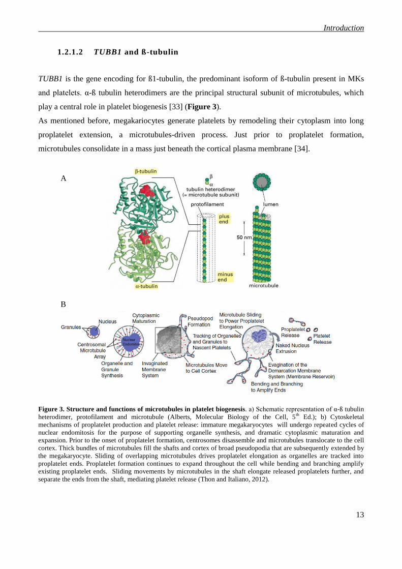
TUBB1 is the gene encoding for ß1-tubulin, the predominant isoform of ß-tubulin present in MKs
and platelets. α-ß tubulin heterodimers are the principal structural subunit of microtubules, which
play a central role in platelet biogenesis [33] (Figure 3).
As mentioned before, megakariocytes generate platelets by remodeling their cytoplasm into long
proplatelet extension, a microtubules-driven process. Just prior to proplatelet formation,
microtubules consolidate in a mass just beneath the cortical plasma membrane [34].
A
B
Figure 3. Structure and functions of microtubules in platelet biogenesis. a) Schematic representation of α-ß tubulin
heterodimer, protofilament and microtubule (Alberts, Molecular Biology of the Cell, 5th
Ed.); b) Cytoskeletal
mechanisms of proplatelet production and platelet release: immature megakaryocytes will undergo repeated cycles of
nuclear endomitosis for the purpose of supporting organelle synthesis, and dramatic cytoplasmic maturation and
expansion. Prior to the onset of proplatelet formation, centrosomes disassemble and microtubules translocate to the cell
cortex. Thick bundles of microtubules fill the shafts and cortex of broad pseudopodia that are subsequently extended by
the megakaryocyte. Sliding of overlapping microtubules drives proplatelet elongation as organelles are tracked into
proplatelet ends. Proplatelet formation continues to expand throughout the cell while bending and branching amplify
existing proplatelet ends. Sliding movements by microtubules in the shaft elongate released proplatelets further, and
separate the ends from the shaft, mediating platelet release (Thon and Italiano, 2012).

Introduction
14
Proplatelet formation begins when these microtubules align into bundles and fill the cortex of the
first blunt processes extended by megakaryocytes. Proplatelet shafts continue to become filled with
thick bundles of hundreds of microtubules that undergo a thinning phase (to ~20 microtubules) and
loop around within the proplatelet to reenter the shaft forming buds at the proplatelet tip [35].
Proplatelet extension is due to the continuous polymerization of tubulin bundles at their free plus
ends, and dynein-powered sliding of overlapping microtubules [1]. Because of the key role of
microtubules in platelet formation, mice deficient in ß1-tubulin develop moderate
thrombocytopenia as a result of reduced proplatelet formation and their spherocytic platelets carry a
structurally defective marginal band and reduced microtubule content [33]. More recently
Kunishima and colleagues reported TUBB1 mutations affecting microtubule assembly in the context
of inherited thrombocytopenia [25, 36]
1.2.2 ITs diagnosis
The recognition of genetic origin of ITs is often hampered by several difficulties. First of all, the
clinical recognition of ITs is often delayed because an overlapping between ITs and acquired forms
of thrombocytopenia. Thus, to distinguish these forms, it is important to carefully analyze the
family medical history of probands. Moreover, ITs could be often misdiagnosed because of
ambiguous platelet count that results both from the combination of ethnic-, age- and sex variables
and from errors in count measurement by cell counters, especially for patients with very large
platelets [37]. Once an IT is suspected, to avoid the problem of variable expressivity of clinical
features, the diagnostic process usually take advantage of the algorithm proposed by the Italian
Platelet Study Group in 2003 [38] (Figure 4).
The first step consists of discrimination of the syndromic forms from the nonsyndromic ones. The
latter are then classified according to the platelet size. Finally, to select potentially candidate genes,
different investigations tests, such as blood film and bone marrow evaluation, ristocetin response
analysis and immunofluorescence test, are performed. Despite of the application of diagnostic
algorithm, in about the 50% of the IT patients mutations are not identified and families remain
without a molecular diagnosis [6].

Introduction
15
Figure 4. Diagnostic algorithm for ITs. Medical history and physical examination are sufficient in most cases for
suspecting syndromic forms. For the nonsyndromic forms, further investigations of the platelets size and laboratory
tests are necessary (Adapted from Balduini et al, 2013).

Introduction
16
1.3 Next Generation Sequencing
DNA sequencing has come a long way since the days of two-dimensional chromatography in the
1970s. With the advent of capillary electrophoresis (CE)- based sequencing in 1977, scientists
gained the ability to sequence the full genome of any species in a reliable, reproducible manner
[39]. This technology, known as “Sanger Sequencing”, had dominated the industry for almost 30
years and led to a number of accomplishments, including the completion of the human genome
sequence. In recent years, sequence data have become more and more relevant for the assessment of
disease at the molecular level and the ever increasing need for large-scale analysis of large
quantities of samples has highlighted the limitations of automated Sanger sequencing, despite many
technical improvements occurs in this “era”. For this reason over the past ten years, there has been a
fundamental shift away from the application of automated Sanger sequencing for genome analysis
and the development of Next Generation Sequencing (NGS) technologies able of producing large
volumes of sequence data in a short time and at low cost [40]. Important applications include whole
genomes and transcriptomes sequencing, genome resequencing, DNA-protein interactions through
chromatin immunoprecipitation sequencing, discovering noncoding RNAs, metagenomics, and
other applications that will appear over the next few years.
Application of NGS strategies has provided new insights into the field of ITs with the identification
of novel IT genes, such as NBEAL2, GFI1B, RBM8A and PRKACG responsible for gray platelet
syndrome, thrombocytopenia and absent radii and PRKACG-related thrombocytopenia,
respectively [14, 15, 41, 42]. In 2015, mutations were identified also in FYB [43], SLFN14 [44] and
ETV6 [20, 21], driving up to 30 the IT genes currently known and further reducing the number of
families that remain without a molecular diagnosis.
Application of this technology will have a strong impact not only in cloning novel genes but also in
molecular genetic testing using target panels of all the genes responsible for ITs.

2. AIM OF THE STUDY

Aim of the study
18
Diagnosis of IT is a complex difficult process due to high grade of heterogeneity from both the
clinical and genetic point of view. Moreover, almost half of the IT patients still remain without
molecular diagnosis because they suffer from forms that are not identified yet. For this reason, in
collaboration with Medical Genetic Unit of Policlinico Sant’Orsola Malpighi in Bologna and the
Department of General Medicine 3, IRCCS San Matteo in Pavia, our laboratory is involved in a
project aimed to studying IT patients using NGS technologies. The purpose of this project is to:
1) simplify and shorten the time for the molecular diagnosis using a target sequencing panel
of known causative genes;
2) perform whole exome sequencing in patients without mutations identified by the target
sequencing panel in order to identify new candidate genes.
My PhD work fits into this project since it is focused on development of functional assays to test
pathogenicity of the variants identify by NGS. Indeed, whereas there is no doubt on the deleterious
effects of nonsense or frameshift, the pathogenic role of missense alterations is not always obvious.
In this condition, search of the disease-causing gene is further prevented when the model of
inheritance requires only mutated allele. Therefore, in diseases like ITs, which are mainly
autosomal dominant diseases caused by private, often missense, the development of targeted
functional studies is absolutely necessary to determine the pathogenicity of missense variants.

3. MATERIALS AND METHODS
Materials and Methods
20
3.1 Patients
Our cohort of patients included 239 consecutive probands with IT examined at the Istituto di
Ricovero e Cura a Carattere Scientifico (IRCCS) Policlinico San Matteo Foundation, in Pavia, Italy,
until 2013. For mutational screening of ACTN1 we enrolled 128 individuals without a certain
diagnosis after the IT diagnostic work-up. In particular, according to their clinical features, patients
were previously analyzed for mutations in MYH9, CYCS, GP1BB, GP1BA, GP9 and ANKRD26
genes without finding any causative mutation. Patients enrolled from 2014 and some cases of our
cohort still unsolved were analyzed during 2015 by means of next generation sequencing
technologies (N=100).
3.2 Whole Exome Sequencing
Whole exome DNA from patient's whole blood was captured using the solid-phase NimbleGen
SeqCap EZ Exome 44Mb array (Nimblegen Inc., Madison, WI, USA) and sequenced as 91/100 bp
paired-end reads on Illumina HiSeq2000 platform (Illumina Inc., San Diego, CA, USA).
Generated reads were checked with FastQC (http://www.bioinformatics.babraham.ac.uk/
publications.html) and aligned with BWA (Li and Durbin, 2010) to the reference genome hg19.
Aligned reads were treated for realignment and base quality score recalibration with GATK (De
Pristo et al., 2011), and for duplicate removal with PicardTools (http://picartools.sourceforge.net).
Alignment statistics were collected by SAMtools (http://samtools.sourceforge.net./) and GATK.
Coverage statistics over the targeted regions were calculated with GATK. Variant calling and
filtering by quality were performed by GATK. Variants passing quality filters were annotated using
ANNOVAR (Wang and Li, 2010) against NCBI RefGene (http://www.ncbi.nlm.nih.gov).
3.3 Target Sequencing
Target sequence analysis were performed using Ion Torrent Personal Genome Machine (IPGM)
platform. Sequencing primers were designed on the coding and intronic flanking regions of the IT
genes using the Ion Ampliseq Designer software (https://www. ampliseq.com/browse.action).
Following the manufacturer’s recommendations (Life Technologies), two multiplex PCRs were

Materials and Methods
21
carried out for each sample using Ion AmpliSeq library kit 2.0. Emulsion-PCR and enrichment
reactions were performed on the template using Ion One Touch 2 system and
the enriched-template quality was analyzed using Qubit 2.0 Fluorometer (Invitrogen Corporation).
Sequencing reactions were performed using Ion PGMTM Sequencing 200 Kit v2. Sequencing data
were analyzed using Ion Torrent Suite software (v 4.0). Using the plug-in Coverage Analysis
(TSCA v 4.0), we evaluated the quality of data sequencing. Data were aligned with hg19 human
genomic sequence using the plug-in Variant Caller (TSVC v.4.0). Functional annotations of all the
sequence variants were performed using the w-annovar software (http://wannovar.usc.edu/). The
exons containing variants were confirmed by Sanger sequencing using standard conditions in an
ABI 3100 automated sequencer (Applied Biosystems, Foster City, CA).
3.4 Sanger Sequencing
ACTN1 was screened for mutations using genomic DNA extracted from peripheral blood.
Mutational analysis was performed by polymerase chain reaction (PCR) amplification using
primers covering exons 1-8 and 17-21 of the gene and the relative exon/intron boundaries. PCR was
carried out in 35 μL of total reaction volume with 25 ng of genomic DNA, 10μM of each primer,
and Kapa 2G Fast Hot Start ReadyMix 2X (KapaBiosystems, Cape Town, South Africa). PCR
products were bidirectionally sequenced using the ABI PRISM BigDye v3.1 Terminator Cycle
Sequencing Ready Reaction Kit and ABI PRISM 3130xl sequencer (Applied Biosystems, Foster
City, CA).
3.5 Bioinformatic Analysis
Multiple species alignment of α-actinin 1 was generated using the Clustal Omega program
(https://www.ebi.ac.uk/Tools/msa/clustalw2/). Analysis of protein domains was performed using
the database Prosite (http://prosite.expasy.org). The effect of the missense variations was
evaluated using three pathogenicity prediction programs: PoliPhen-2
(http://genetics.bwh.harvard.edu/pph2/), Mutation Taster (http://www.mutationtaster.org/) and
SIFT (http://sift.jcvi.org).

Materials and Methods
22
3.6 Cloning Procedures
A full-length ACTN1 sequence was amplified from normal platelet cDNA and constructed into
mammalian expression vector pcDNA3.1 (Invitrogen, San Diego, CA, USA) with a 5’Myc tag
sequence. ACTN1 mutants were generated by PCR using specific mutagenesis primers (view table
below). The parental methylated DNA was eliminated by digesting with DpnI restriction enzyme.
The construct obtained was transformed in TOP10 competent cells. The full-length sequences,
frame, and orientation were confirmed by sequencing. The same stategy was used to cloning
pcDNA3.1-Myc-TUBB1.
3.7 Immunofluorescence Assay
For immunofluorescence assay human fibroblasts (PD220) were seeded on chamberslides and
transfected with myc-tagged wild type or mutant ACTN1 plasmids. After 16 hours cells were fixed
with 4% parafolmaldehyde for 20 minutes and then permeabilized with 0.1% Triton X-100. For
detection of ACTN1 primary antibody against c-myc (9E10, Santa Cruz Biotechnology) was used
with anti mouse FITC secondary antibody (F0479, DakoCytomation),while actin filaments were
stained with AlexaFluor594 conjugated phalloidin (Invitrogen).
Images were obtained with a Nikon C1si confocal microscope, containing 488nm, argon laser line
and 561 nm diode laser. Light was delivered to the sample with an 80/20 reflector. Electronic zoom
was kept at minimum values for measurements to reduce potential bleaching. The images were
acquired using a 60X Plan Apo objectives (with a corresponding NA of 1.4), collecting series of
optical images at 1 μm z resolution step size. The corresponding voxel size was 100x100x1000 nm
(X×Y×Z). Images were processed for z-projection (maximum intensity), brightness and contrast
regulation using ImageJ 1.45 (NIH,Bethesda, USA).
3.8 Platelet Studies
Platelet studies was performed by clinicians as reported in Bottega, Marconi, Faleschini et al., 2015

4. RESULTS AND DISCUSSION

Results and Discussion
24
4.1 Identification of c.2305G>A variant in ACTN1 gene by Whole Exome
Sequencing
In order to identify the causative gene in a family with an IT of unknown origin in which candidate
genes were previously excluded, we performed Whole Exome Sequencing (WES) in the proband of
Family 1 (F1). The WES experiments allowed us to identify a single-base substitution located
within exon 19 of the ACTN1 gene (c.2305G>A), which was predicted to result in a missense
mutation, p.Glu769Lys. The mutation was absent in the public dbSNP and the 1000 Genomes
Project dataset (http://www.ncbi.nlm.nih.gov/SNP/ ; http://www.1000genomes.org/category/dbsnp).
Moreover, it occurs in a very conserved position, according to PhyloP score. The variant was
predicted to be Benign from Polyphen2, while SIFT and Mutation Taster indicated it as deleterious.
The variant was confirmed by Sanger sequencing and was observed to correctly segregate with the
disease in family F1 (Figure 1). According to the putative causative role of this mutation, at the
same time Kunishima and colleagues identified ACTN1 as a novel gene responsible for
macrothrombocytopenia [26].
Figure 1. Identification of c.2305G>A variant in ACTN1 gene. A) Pedigree of Family 1; B) Chromatogram of
mutation confirmed in Sanger sequencing: arrow indicates the aminoacid substitution c.2305G>A; C) Aminoacid
conservation: arrow indicates the aminoacid substitution Glu769Lys .

Results and Discussion
25
4.2 Identification of ACTN1 mutations in 10 families
According to protein structure and mutations identified by Kunishima and colleagues, we decided to
restrict the mutational screening to exons that encode for functional domains. For this reason, we
performed direct Sanger sequencing of exons 1-8 and 17-21 of the ACTN1 gene in 127 probands.
We identified nine different heterozygous missense variants in ten families (Table1). Three were
known amino acid substitutions (p.Gly225Lys, p.Arg738Trp and p.Arg752Gln) that were
previously reported [26]. The other six were not present in either the dbSNP or the 1000 Genomes
Project datasets and therefore regarded as novel variants of ACTN1.
Table 1. Variants identified in ACTN1 gene. Nucleotide A of the ATG translation initiaton start site of the ACTN1
gene cDNA in GenBank sequence NM_001130004.1 is indicated as nucleotide +1.
Interestingly, the c.136C>T (p.Arg46Trp) variant affects the same residue as it does c.137G>A
(p.Arg46Gln), the mutation described by Kunishima and Guéguen [26, 45]. Although the two
Italian families carrying p.Arg46Trp were not aware of a relationship, both of them come from a
little village in the north of Italy, suggesting a possible founder effect for this mutation. All the
missense variants segregate with macrothrombocytopenia within the individual pedigrees when
family members were available, except for c.1997A>T (p.Asp666Val) (Figure 2). In Family 7 (F7),
Family Mutation
Exon Type of
mutation References
Nucleotide Protein
F2 c.64G>A p.Asp22Asn 1 Missense New mutation
F3 c.136C>T p.Arg46Trp 2 Missense New mutation
F4 c.136C>T p.Arg46Trp 2 Missense New mutation
F5 c.673G>A p.Glu225Arg 7 Missense Kunishima et al.,2013
F6 c.751G>A p.Gly251Arg 8 Missense New mutation
F7 c.1997A>T p.Asp666Val 17 Missense New mutation
F8 c.2210C>A p.Thr737Asn 18 Missense New mutation
F9 c.2212C<T p.Arg738Trp 18 Missense Kunishima et al.,2013
F10 c.2255G<A p.Arg752Gln 18 Missense Kunishima et al.,2013
F11 c.2290G>A p.Gly764Ser 19 Missense New mutation
Results and Discussion
26
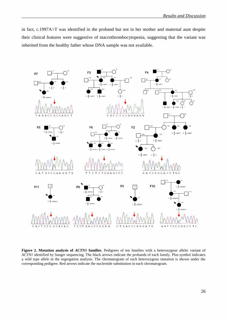
in fact, c.1997A>T was identified in the proband but not in her mother and maternal aunt despite
their clinical features were suggestive of macrothrombocytopenia, suggesting that the variant was
inherited from the healthy father whose DNA sample was not available.
Figure 2. Mutation analysis of ACTN1 families. Pedigrees of ten families with a heterozygous allelic variant of
ACTN1 identified by Sanger sequencing. The black arrows indicate the probands of each family. Plus symbol indicates
a wild type allele in the segregation analysis. The chromatogram of each heterozygous mutation is shown under the
corresponding pedigree. Red arrows indicate the nucleotide substitution in each chromatogram.

Results and Discussion
27
4.3 Evaluation of the effect of missense mutations on α-actinin1 structure
The multiple-sequence alignment indicated that all the six novel missense variants hit amino acid
residues highly conserved from zebrafish. Furthermore, their potential effect on protein function
was evaluated using three different pathogenicity prediction tools: SIFT, Polyphen and Mutation
Taster (Table 2). Regarding p.Asp22Asn, p.Arg46Trp, p.Gly251Arg, and p.Thr737Asn, all
programs predicted their deleterious effects on α-actinin1 function. In contrast, the p.Gly764Ser
substitution was tolerated using SIFT even if the variant occurs in a highly conserved functional
domain (calmodulin-like domain, CaM). Instead, the significance of p.Asp666Val was unclear:
although it does not segregate its pathogenicity scores were high.
Table 2. Bioinformatic analysis for predicting the functional effect of the novel ACTN1 variants. Effect of
variations was evaluated using three pathogenicity prediction programs: PolyPhen-2
(http://genetics.bwh.harvard.edu/pph2/), SIFT (http:// http://sift.jcvi.org/), and Mutation Taster
(http://www.mutationtaster.org/).
Mutation
PolyPhen-2 SIFT MutationTaster
Nucleotide Protein
c.64G>A p.Asp22Asn possibly damaging not tolerated disease causing
c.136C>T p.Arg46Trp probably damaging not tolerated disease causing
c.751G>A p.Gly251Arg possibly damaging not tolerated disease causing
c.1997A>T p.Asp666Val probably damaging not tolerated disease causing
c.2210C>A p.Thr737Asn possibly damaging not tolerated disease causing
c.2290G>A p.Gly764Ser possibly damaging tolerated disease causing
Results and Discussion
28
4.4 Functional analysis of the novel missense mutations
Whereas the pathogenetic role of nonsense and frameshift variants or large deletions is clear
(usually for RNA or protein degradation) that of other variants, such as missense or small in-frame
deletions/duplications is of uncertain significance (VUS). Unraveling the effect of the VUS is a
complex process that requires targeted functional studies to explore whether a single amino acid
substitution alters or not the protein function, for example modifying its folding and therefore
stability, or its functional domains.
For this reason, in order to determine the pathogenetic role of the seven novel missense variants
identified in this study, we performed an immunofluorescence analysis after transfection of wild
type or mutant ACTN1 cDNA cloned into mammalian expression vector pcDNA3.1 with a 5’ Myc
tag sequence [26] into human fibroblasts (Figure 3A). When cells were transfected with the wild
type construct, we observed a well-organized cytoskeleton in which α-actinin1 colocalizes in large
part with actin along the filaments.
On the contrary, cells transfected with mutant constructs presented an abnormal distribution of
actin, which was no longer able to form filaments causing an apparent disruption in the cytoskeletal
structure, as observed in other studies testing the effect of different ACTN1 missense mutations [26,
45]. This was particularly evident when we compared the actin staining in cells expressing and not
expressing the mutant constructs. In addition, when mutant α-actinin1 was expressed, the staining
appears widespread in the cytoplasm and the colocalization with actin was not specific. These
phenotypic features are shared by all but one (p.Asp666Val) mutation. Consistent with segregation
analysis, the correct organization of the cytoskeleton in cells expressing p.Asp666Val excluded this
variant as a disease-causing mutation. It is worth noting that p.Asp666Val is outside the actin
binding (ABD) and calmodulin-like (CaM) domains (Figure 3B), suggesting that only alterations of
these functional regions are compatible with the disease.
Results and Discussion
29
Figure 3. Functional studies of novel ACTN1 variants. (A) Immunofluorescence analysis in PD220 fibroblast cell
line transiently transfected according to standard procedures. Both wild type (top panel) or mutant (lower panels)
ACTN1 cDNAs were cloned into the pcDNA3.1-Myc tagged expression vector. The subcellular localization of
exogenous α-actinin1 (green) was examined using c-myc antibodies (9E10; Santa Cruz Biotechnology, Dallas, Texas,
U.S.A.) while the actin filaments were stained with AlexaFluor594 (red) conjugated phalloidin (Invitrogen, Marseille,
France). Images were obtained with a Nikon C1si confocal microscope using a 60X Plan Apo objectives. Images were
processed for z-projection (maximum intensity), brightness and contrast regulation using ImageJ 1.45 (NIH, Bethesda,
USA). The cells shown are representative of three independent experiments. Scale bar =50 µm. (B) Domain structure of
α-actinin and localization of ACTN1 mutations identified in Japanese families (arrowheads) and in this paper (arrows).
The p.Arg46Gln mutation was also identified in a French family.

Results and Discussion
30
4.5 Clinical features of ACTN1-Related Thrombocytopenia (ACTN1-RT)
Determination of the pathogenicity of variants identified through screening of mutation has allowed
clinicians to define the clinical and laboratory features of this new form of thrombocytopenia. As
reported in Table 3, in most cases thrombocytopenia was mild and both mean platelet volume and
diameter were significantly higher in patients than in healthy subjects. The mean reticulated platelet
count was significantly lower whereas the serum level of TPO was only slightly higher than in
controls (these data are consistent with normal bone marrow megakaryocyte concentration and
reduced platelet production). Regarding functional activity, in vitro platelet aggregation was within
the normal range in the 17 investigated patients. The GPIbα, GPIX, GPIIb and GPIIIa glycoproteins
were expressed on platelet surface at normal level, as indicated by flow cytometry of in the 24
investigated patients. Briefly, the study of affected individuals lead us to classify ACTN1-RT as a
mild form of macrothrombocytopenia without platelet dysfunction, confirming that
thrombocytopenia derives from defects of the phase of proplatelet formation and platelet release.
The data obtained in this part of my PhD work have been published at the beginning of 2015 on the
scientific journal “Blood” (Bottega, Marconi, Faleschini et al.,2015). One of immunofluorescence
included in this work was also selected for the cover of the journal.

______
______
________
________
______
________
_
R
esults a
nd D
iscussio
n
Table 3. Clinical and laboratory features of families with ACTN1 mutations. aWHO (World Health Organization) bleeding score: grade 0, no bleeding; grade 1,
only cutaneous bleeding; grade 2, mild blood loss; grade 3, gross blood loss, requiring transfusion; grade 4, debilitating blood loss, retinal or cerebral associated with
fatality. bIn vitro platelet aggregation after collagen (4 µg/mL), ADP (5 mM) and ristocetin (1.5 mg/mL);
cIndividuals were classified as thrombocytopenic based on the
reference range of platelet count (150-400 x 109/L) and the recently proposed age- and gender specific reference intervals (Savoia et al., 2001). According to these
criteria, two three-year-old girls from families 5 and 8 were enrolled despite their platelet count (in bold) was in the normal range; nd, not determined
Family
(N. of patients)
Mean age at
diagnosis Years
(range)
WHO bleeding
scorea
(N. of patients)
Mean platelet
count using cell
counterb
x109/L (range)
Mean platelet
volume
fL (range)
Mean platelet
diameter
µm (range)
In vitro platelet
aggregationc
(N. of patients)
Flow cytometry
of platelet
glycoproteins
(N. of patients)
F1 (4) 38
(3-66) 0 (1), 1 (2), 2 (1) 86 (46-120) 14-3 (12.6-15) 3.5 (2.9-3.9) normal (4) normal (4)
F2 (4) 43
(22-55) 0 (1), 1 (3) 107 (89-134) 11,1 (10,1-12) 2.8 (2.7-3) nd normal (4)
F3 (4) 46
(26-64) 0 (1), 1 (2), 2 (1) 103 (81-118) 12,5 (10.6-14.7) 3.3 (3-3.7) normal (4) normal (4)
F4 (4) 42
(14-72) 0 (1), 1 (1), 2 (2) 95 (66-124) 14.8 (14-15.6) 3.8 (3.5-4.1) normal (4) normal (3)
F5 (2) 30
(12-49) 0 (2) 103 (97-110) 11.8 (11.3-12.4) 3 (2.8-3.1) normal (2) normal (1)
F6 (6) 23
(7-44) 0 (1), 1 (3), 2 (3) 103 (78-154) 12.3 (10.4-14) 3.3 (2.6-4.3) normal (2) normal (5)
F8 (2) 58
(34-82) 2 (2) 58 (55-62) 10.5 (10.4-10.6) 3.2 (2.9-3.5) normal (1) nd
F9 (1) nd 0 (1) 110 12.1 2.5 nd nd
F10 (3) 25
(3-44) 0 (2), 1 (1) 112 (65-166) 12.5 (11.3-14.3) 2.8 (2.6-3) nd normal (2)
F11 (1) 48 0 (1) 117 14.4 3.3 normal (1) normal (1)
27

Results and Discussion
32
4.6 Identification of ACTN1 and TUBB1 variants through NGS approach
As mentioned before, ITs are characterized to high grade of clinical and genetic heterogeneity.
Despite of application of diagnostic algorithm described before, 50% of IT patients remains without
molecular diagnosis, suggesting that they are likely to suffer from IT not characterized yet.
For this reason, in collaboration with Medical Genetic Unit of Policlinico Sant’Orsola Malpighi in
Bologna and the department of General Medicine 3 , IRCCS San Matteo in Pavia, since last year
we have been analyzing these patients using next generation sequencing approaches based on :
- target sequencing (Ion Torrent) to identify rapidly variants in known IT genes;
- whole exome sequencing in patients negative after target sequencing to identify new genes
responsible for the disease.
At present, we have analyzed 120 patients. In 13 individuals, we have identified potential
pathogenetic variants of ACTN1 and TUBB1, another cytoskeleton component, whose variants
could be tested functionally using the same strategy as for those affecting ACTN1 (Table 4).
Table 4. Identification of ACTN1 and TUBB1 variants through NGS approach. Nucleotide A of the ATG
translation initiaton start site of the ACTN1 gene cDNA in GenBank sequence NM_001130004.1 is indicated as
nucleotide +1. Effect of variations was evaluated using the three pathogenicity prediction programs previously
mentioned, but only PolyPhen-2 score is reported in this table.

Results and Discussion
33
4.6.1 ACTN1 variants
The NGS analyses revealed 5 ACTN1 variants in 6 families. Whereas two of these are known
mutations (p.Arg46Trp and p.Glu225Lys), the others are novel variations (p.Trp128Cys,
p.Pro233Leu and p.Val328Met). In all these cases, the variants segregate with
macrothrombocytopenia within pedigrees when family members were available (Figure 4).
Notably, we found a homozygous variant in the proband of Family IT6 with a severe
thrombocytopenia. Her father, heterozygous for the variant, had a platelet count of 100-120 x109/L,
while the mother was reported as healthy, even though there are no laboratory tests that confirm her
condition.
Figure 4. Pedigrees of families with a variant of ACTN1 identified by NGS approach. Plus symbol indicates a wild
type allele in the segregation analysis. The chromatogram of each mutation is shown under the corresponding pedigree.
Arrows indicate the nucleotide substitution in each chromatogram.

Results and Discussion
34
To confirm the pathogenic role predicted by bioinformatical analysis (Table 4), we performed
immunofluorescence assay on the three novel variants identified (Figure 5).
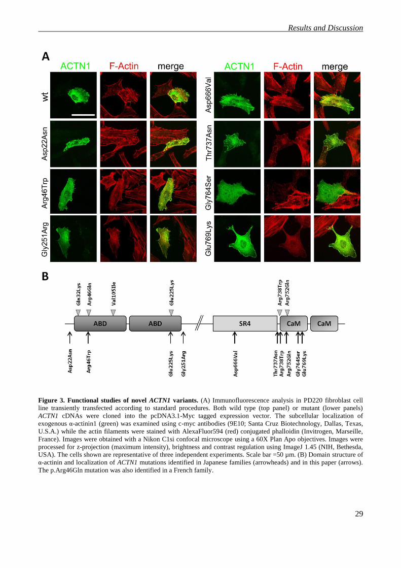
Figure 5. Functional studies of novel ACTN1 variants. Immunofluorescence analysis in PD220 fibroblast cell line
transiently transfected according to standard procedures. Both wild type (top panel) or mutant (lower panels) ACTN1
cDNAs were cloned into the pcDNA3.1-Myc tagged expression vector. The subcellular localization of exogenous α-
actinin1 (green) was examined using c-myc antibodies (9E10; Santa Cruz Biotechnology, Dallas, Texas, U.S.A.) while
the actin filaments were stained with AlexaFluor594 (red) conjugated phalloidin (Invitrogen, Marseille, France). Images
were obtained with a Nikon C1si confocal microscope using a 60X Plan Apo objectives. Images were processed for z-
projection (maximum intensity), brightness and contrast regulation using ImageJ 1.45 (NIH, Bethesda, USA).
Results and Discussion
35
While cells transfected with p.Trp128Cys and p.Pro233Leu showed a disorganized cytoskeleton,
those expressing p.Val328Met had alterations within cells but not in proximity of the membrane. In
these cells, there are no actin filaments in the cytoplasm and α-actinin1 is widespread. However,
they colocalize perfectly in the apical regions at the cell-to-cell junctions.
This is particularly interesting if we consider that ,unlike mutations previously reported which
reside in actin-binding domain (ABD) or calmodulin-like (CaM) domain, p.Val328Met hits the
spacer rod domain, suggesting a novel mechanism for the pathogenesis of ACTN1-related
macrothrombocytopenia that does not involve functional domain mutations.
4.6.2 TUBB1 variants
We detected 4 TUBB1 variants, including 3 missense (p.Gly109Glu, p.Gln191Pro, p.Val286Met)
and one nonsense (p.Tyr55 *), in 7 probands. All these variants were confirmed by Sanger
sequencing and had a "deleterious" prediction of pathogenicity (Table 4). At present, no family
members are available for segregation study. However, we are generating constructs cloning
TUBB1 cDNA into mammalian expression vector pcDNA3.1 with a 5’ Myc tag sequence on N-
terminus. Once transfected the wild-type and mutant constructs in a immortalized fibroblast cell
line, we will evaluate the incorporation of exogenous ß-tubulin at the microtubule level. What we
expect to see is a colocalization of endogenous α-tubulin and ß-tubulin exogenous only in cells
expressing the wild-type and not the mutated forms of TUBB1[25]. This would demonstrate the
inability of mutated ß-tubulin to be incorporated into rising microtubules and so the effect of
pathogenic variants.

5. CONCLUSIONS
Conclusions
37
The purpose of my PhD project was to develop functional assays to test the pathogenicity of
variants obtained by NGS. These analysis are absolutely necessary to determine the pathogenicity
of missense variants because, in contrast to nonsense or frameshift mutations, the pathogenic role of
them is not obvious. For this reason, we developed a functional assay on ACTN1, a novel gene
responsible for macrothrombocytopenia identified through NGS analysis. In particular, we
performed immunofluorescence on human fibroblasts overexpressing wild-type and mutated forms
of ACTN1, in order to discriminate pathogenetic missense variants from those having no effect on
actin cytoskeletal structure. Thanks to functional studies, we confirmed ACTN1-RT diagnosis
among 15 families of our cohort of patients, describing it as the fourth most frequent (6,3%) form of
IT in Italy after Bernard-Soulier syndrome (13%), MYH9-Related Disease (12%) and ANKRD26-
Related Thrombocytopenia (10%). The NGS analyses revealed also 4 potential pathogenetic
variants of TUBB1, another cytoskeleton component, which variants could be tested functionally
using the same strategy as for those affecting ACTN1.
In conclusion, NGS is a revolutionary technique that over the past few years has been providing
significant breakthroughs in the knowledge of the molecular bases of Mendelian diseases,
including ITs. Its application in the diagnostic process of IT would allow us to screen directly at
least all the known IT genes. However, understanding the effects of the thousands variants
identified remains a major problem. For this reason, it is fundamental to develop further functional
studies for other genes.

6. BIBLIOGRAPHY

Bibliography
39
1. Patel SR, Hartwig JH, Italiano JE, Jr.: The biogenesis of platelets from megakaryocyte proplatelets. J Clin
Invest 2005, 115(12):3348-3354.
2. Savoia A: Molecular basis of inherited thrombocytopenias. Clin Genet 2016, 89(2):154-162.
3. Kile BT: The role of apoptosis in megakaryocytes and platelets. Br J Haematol 2014, 165(2):217-226.
4. Machlus KR, Italiano JE, Jr.: The incredible journey: From megakaryocyte development to platelet
formation. J Cell Biol 2013, 201(6):785-796.
5. Thon JN, Italiano JE: Platelets: production, morphology and ultrastructure. Handb Exp Pharmacol
2012(210):3-22.
6. Balduini CL, Savoia A: Genetics of familial forms of thrombocytopenia. Hum Genet 2012, 131(12):1821-
1832.
7. Pecci A, Balduini CL: Lessons in platelet production from inherited thrombocytopenias. Br J Haematol
2014, 165(2):179-192.
8. Ballmaier M, Germeshausen M: Congenital amegakaryocytic thrombocytopenia: clinical presentation,
diagnosis, and treatment. Semin Thromb Hemost 2011, 37(6):673-681.
9. Thompson AA, Nguyen LT: Amegakaryocytic thrombocytopenia and radio-ulnar synostosis are
associated with HOXA11 mutation. Nat Genet 2000, 26(4):397-398.
10. Albers CA, Paul DS, Schulze H, Freson K, Stephens JC, Smethurst PA, Jolley JD, Cvejic A, Kostadima M,
Bertone P et al: Compound inheritance of a low-frequency regulatory SNP and a rare null mutation in
exon-junction complex subunit RBM8A causes TAR syndrome. Nat Genet 2012, 44(4):435-439, S431-432.
11. Klopocki E, Schulze H, Strauss G, Ott CE, Hall J, Trotier F, Fleischhauer S, Greenhalgh L, Newbury-Ecob
RA, Neumann LM et al: Complex inheritance pattern resembling autosomal recessive inheritance
involving a microdeletion in thrombocytopenia-absent radius syndrome. Am J Hum Genet 2007,
80(2):232-240.
12. Raslova H, Komura E, Le Couedic JP, Larbret F, Debili N, Feunteun J, Danos O, Albagli O, Vainchenker W,
Favier R: FLI1 monoallelic expression combined with its hemizygous loss underlies Paris-
Trousseau/Jacobsen thrombopenia. J Clin Invest 2004, 114(1):77-84.
13. Millikan PD, Balamohan SM, Raskind WH, Kacena MA: Inherited thrombocytopenia due to GATA-1
mutations. Semin Thromb Hemost 2011, 37(6):682-689.
14. Monteferrario D, Bolar NA, Marneth AE, Hebeda KM, Bergevoet SM, Veenstra H, Laros-van Gorkom BA,
MacKenzie MA, Khandanpour C, Botezatu L et al: A dominant-negative GFI1B mutation in the gray
platelet syndrome. N Engl J Med 2014, 370(3):245-253.
15. Albers CA, Cvejic A, Favier R, Bouwmans EE, Alessi MC, Bertone P, Jordan G, Kettleborough RN, Kiddle
G, Kostadima M et al: Exome sequencing identifies NBEAL2 as the causative gene for gray platelet
syndrome. Nat Genet 2011, 43(8):735-737.
16. Bottega R, Pecci A, De Candia E, Pujol-Moix N, Heller PG, Noris P, De Rocco D, Podda GM, Glembotsky
AC, Cattaneo M et al: Correlation between platelet phenotype and NBEAL2 genotype in patients with
congenital thrombocytopenia and alpha-granule deficiency. Haematologica 2013, 98(6):868-874.
17. Song WJ, Sullivan MG, Legare RD, Hutchings S, Tan X, Kufrin D, Ratajczak J, Resende IC, Haworth C,
Hock R et al: Haploinsufficiency of CBFA2 causes familial thrombocytopenia with propensity to develop
acute myelogenous leukaemia. Nat Genet 1999, 23(2):166-175.
18. Liew E, Owen C: Familial myelodysplastic syndromes: a review of the literature. Haematologica 2011,
96(10):1536-1542.
19. Pippucci T, Savoia A, Perrotta S, Pujol-Moix N, Noris P, Castegnaro G, Pecci A, Gnan C, Punzo F, Marconi C
et al: Mutations in the 5' UTR of ANKRD26, the ankirin repeat domain 26 gene, cause an autosomal-
dominant form of inherited thrombocytopenia, THC2. Am J Hum Genet 2011, 88(1):115-120.
20. Zhang MY, Churpek JE, Keel SB, Walsh T, Lee MK, Loeb KR, Gulsuner S, Pritchard CC, Sanchez-Bonilla
M, Delrow JJ et al: Germline ETV6 mutations in familial thrombocytopenia and hematologic
malignancy. Nat Genet 2015, 47(2):180-185.

Bibliography
40
21. Noetzli L, Lo RW, Lee-Sherick AB, Callaghan M, Noris P, Savoia A, Rajpurkar M, Jones K, Gowan K,
Balduini CL et al: Germline mutations in ETV6 are associated with thrombocytopenia, red cell
macrocytosis and predisposition to lymphoblastic leukemia. Nat Genet 2015, 47(5):535-538.
22. Berndt MC, Andrews RK: Bernard-Soulier syndrome. Haematologica 2011, 96(3):355-359.
23. Savoia A, Pastore A, De Rocco D, Civaschi E, Di Stazio M, Bottega R, Melazzini F, Bozzi V, Pecci A, Magrin
S et al: Clinical and genetic aspects of Bernard-Soulier syndrome: searching for genotype/phenotype
correlations. Haematologica 2011, 96(3):417-423.
24. Berrou E, Adam F, Lebret M, Fergelot P, Kauskot A, Coupry I, Jandrot-Perrus M, Nurden A, Favier R, Rosa
JP et al: Heterogeneity of platelet functional alterations in patients with filamin A mutations. Arterioscler
Thromb Vasc Biol 2013, 33(1):e11-18.
25. Kunishima S, Kobayashi R, Itoh TJ, Hamaguchi M, Saito H: Mutation of the beta1-tubulin gene associated
with congenital macrothrombocytopenia affecting microtubule assembly. Blood 2009, 113(2):458-461.
26. Kunishima S, Okuno Y, Yoshida K, Shiraishi Y, Sanada M, Muramatsu H, Chiba K, Tanaka H, Miyazaki K,
Sakai M et al: ACTN1 mutations cause congenital macrothrombocytopenia. Am J Hum Genet 2013,
92(3):431-438.
27. Bottega R, Marconi C, Faleschini M, Baj G, Cagioni C, Pecci A, Pippucci T, Ramenghi U, Pardini S, Ngu L et
al: ACTN1-related thrombocytopenia: identification of novel families for phenotypic characterization.
Blood 2015, 125(5):869-872.
28. Nurden AT, Pillois X, Wilcox DA: Glanzmann thrombasthenia: state of the art and future directions.
Semin Thromb Hemost 2013, 39(6):642-655.
29. Savoia A, De Rocco D, Panza E, Bozzi V, Scandellari R, Loffredo G, Mumford A, Heller PG, Noris P, De
Groot MR et al: Heavy chain myosin 9-related disease (MYH9 -RD): neutrophil inclusions of myosin-9 as
a pathognomonic sign of the disorder. Thromb Haemost 2010, 103(4):826-832.
30. Morison IM, Cramer Borde EM, Cheesman EJ, Cheong PL, Holyoake AJ, Fichelson S, Weeks RJ, Lo A,
Davies SM, Wilbanks SM et al: A mutation of human cytochrome c enhances the intrinsic apoptotic
pathway but causes only thrombocytopenia. Nat Genet 2008, 40(4):387-389.
31. Massaad MJ, Ramesh N, Geha RS: Wiskott-Aldrich syndrome: a comprehensive review. Ann N Y Acad Sci
2013, 1285:26-43.
32. Sjoblom B, Salmazo A, Djinovic-Carugo K: Alpha-actinin structure and regulation. Cell Mol Life Sci 2008,
65(17):2688-2701.
33. Schwer HD, Lecine P, Tiwari S, Italiano JE, Jr., Hartwig JH, Shivdasani RA: A lineage-restricted and
divergent beta-tubulin isoform is essential for the biogenesis, structure and function of blood platelets.
Curr Biol 2001, 11(8):579-586.
34. Italiano JE, Jr., Lecine P, Shivdasani RA, Hartwig JH: Blood platelets are assembled principally at the ends
of proplatelet processes produced by differentiated megakaryocytes. J Cell Biol 1999, 147(6):1299-1312.
35. Hartwig JH, Italiano JE, Jr.: Cytoskeletal mechanisms for platelet production. Blood Cells Mol Dis 2006,
36(2):99-103.
36. Kunishima S, Nishimura S, Suzuki H, Imaizumi M, Saito H: TUBB1 mutation disrupting microtubule
assembly impairs proplatelet formation and results in congenital macrothrombocytopenia. Eur J
Haematol 2014, 92(4):276-282.
37. Balduini CL, Pecci A, Noris P: Diagnosis and management of inherited thrombocytopenias. Semin Thromb
Hemost 2013, 39(2):161-171.
38. Balduini CL, Cattaneo M, Fabris F, Gresele P, Iolascon A, Pulcinelli FM, Savoia A, Italian Gruppo di Studio
delle P: Inherited thrombocytopenias: a proposed diagnostic algorithm from the Italian Gruppo di
Studio delle Piastrine. Haematologica 2003, 88(5):582-592.
39. Sanger F, Nicklen S, Coulson AR: DNA sequencing with chain-terminating inhibitors. Proc Natl Acad Sci
U S A 1977, 74(12):5463-5467.
40. Johnsen JM, Nickerson DA, Reiner AP: Massively parallel sequencing: the new frontier of hematologic
genomics. Blood 2013, 122(19):3268-3275.

Bibliography
41
41. Albers CA, Newbury-Ecob R, Ouwehand WH, Ghevaert C: New insights into the genetic basis of TAR
(thrombocytopenia-absent radii) syndrome. Curr Opin Genet Dev 2013, 23(3):316-323.
42. Manchev VT, Hilpert M, Berrou E, Elaib Z, Aouba A, Boukour S, Souquere S, Pierron G, Rameau P, Andrews
R et al: A new form of macrothrombocytopenia induced by a germ-line mutation in the PRKACG gene.
Blood 2014, 124(16):2554-2563.
43. Levin C, Koren A, Pretorius E, Rosenberg N, Shenkman B, Hauschner H, Zalman L, Khayat M, Salama I,
Elpeleg O et al: Deleterious mutation in the FYB gene is associated with congenital autosomal recessive
small-platelet thrombocytopenia. J Thromb Haemost 2015, 13(7):1285-1292.
44. Fletcher SJ, Johnson B, Lowe GC, Bem D, Drake S, Lordkipanidze M, Guiu IS, Dawood B, Rivera J, Simpson
MA et al: SLFN14 mutations underlie thrombocytopenia with excessive bleeding and platelet secretion
defects. J Clin Invest 2015, 125(9):3600-3605.
45. Gueguen P, Rouault K, Chen JM, Raguenes O, Fichou Y, Hardy E, Gobin E, Pan-Petesch B, Kerbiriou M,
Trouve P et al: A missense mutation in the alpha-actinin 1 gene (ACTN1) is the cause of autosomal
dominant macrothrombocytopenia in a large French family. PLoS One 2013, 8(9):e74728.