a successful pregnancy despite the presence of an
TRANSCRIPT
Case ReportA Successful Pregnancy Despite the Presence of an Intrauterine Fetal Bone Fragment
Evangelos Petrakis, Ioannis Chatzipapas, Ioannis K. Papapanagiotou , Panagiotis Fotinopoulos, Panagiota Siemou, Konstantinos Ntzeros, and Dimitrios Loutradis
1st Department of Obstetrics and Gynaecology, School of Medicine, National and Kapodistrian University of Athens, Alexandra General Hospital, Lourou and Vasilisis Soas Avenue, 11528 Athens, Greece
Correspondence should be addressed to Ioannis K. Papapanagiotou; [email protected]
Received 2 July 2019; Accepted 3 September 2019; Published 17 October 2019
Academic Editor: Maria Grazia Porpora
Copyright © 2019 Evangelos Petrakis et al. �is is an open access article distributed under the Creative Commons Attribution License, which permits unrestricted use, distribution, and reproduction in any medium, provided the original work is properly cited.
Objective. Intrauterine retention of fetal bones following a termination of a pregnancy is a rare complication. Among the few reported cases in literature, there has been no report describing the birth of a live fetus, despite the presence of an embryonic ossicle within the endometrial cavity. Case Report. A 28-year-old woman, with an obstetrical history of a miscarriage at the 19th week of gestation, underwent a diagnostic hysteroscopy for evaluation of pelvic pain and infertility, which revealed an intrauterine embryonic ossicle. �e patient did not comply with our recommendations to undergo a surgical hysteroscopy. �e patient’s next visit was during her 9th week of gestation. She was followed up regularly at our Obstetrics Department. Her gestation was uneventful, while an elective caesarean section at the 39th week of gestation was performed. Conclusion. �e present clinical case has demonstrated that achieving childbearing is possible, despite the requirement of removing such bone fragments.
1. Introduction
Intrauterine retention of fetal bones following a termination of a pregnancy in the second and third trimester of gestation is correlated with high rates of spontaneous miscarriages. It is a rare complication as it is observed in 0.15% [1] of the women undergoing diagnostic hysteroscopy for investigation of sec-ondary infertility and a history of a recent termination of a pregnancy. Bony fragments usually imitate the role of intrau-terine contraceptive devices (ICD) by stimulating the secretion of endometrial prostaglandins, thus resulting in secondary infertility. Once the bones are hysteroscopically removed, and if no coexisting infertility factors are present, normal fertility is restored in most of the cases. �us, hysteroscopy, in these cases, is both diagnostic and therapeutic. Among the few reported cases in the literature, there has been no report describing the achievement of a pregnancy and the birth of a live fetus, despite the presence of an embryonic ossicle within the endometrial cavity.
2. Case Presentation
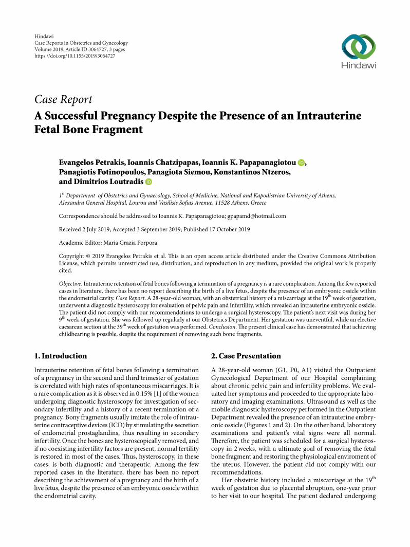
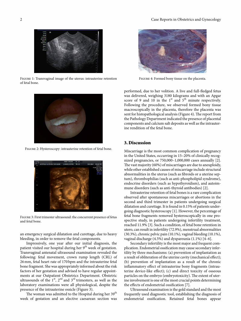
Α 28-year-old woman (G1, P0, A1) visited the Outpatient Gynecological Department of our Hospital complaining about chronic pelvic pain and infertility problems. We eval-uated her symptoms and proceeded to the appropriate labo-ratory and imaging examinations. Ultrasound as well as the mobile diagnostic hysteroscopy performed in the Outpatient Department revealed the presence of an intrauterine embry-onic ossicle (Figures 1 and 2). On the other hand, laboratory examinations and patient’s vital signs were all normal. �erefore, the patient was scheduled for a surgical hysteros-copy in 2 weeks, with a ultimate goal of removing the fetal bone fragment and restoring the physiological enviroment of the uterus. However, the patient did not comply with our recommendations.
Her obstetric history included a miscarriage at the 19th
week of gestation due to placental abruption, one-year prior to her visit to our hospital. �e patient declared undergoing
HindawiCase Reports in Obstetrics and GynecologyVolume 2019, Article ID 3064727, 3 pageshttps://doi.org/10.1155/2019/3064727
Case Reports in Obstetrics and Gynecology2
an emergency surgical dilatation and curettage, due to heavy bleeding, in order to remove the fetal components.
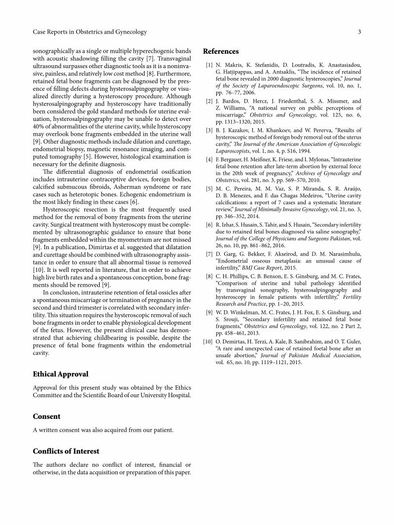
Impressively, one year a¡er our initial diagnosis, the patient visited our hospital during her 9th week of gestation. Transvaginal antenatal ultrasound examination revealed the following: fetal movement, crown rump length (CRL) of 26 mm, fetal heart rate of 170 bpm and the intrauterine fetal bone fragment. She was appropriately informed about the risk factors of her gestation and advised to have regular appoint-ments at our Outpatient Obstetrics Department. Obstetric ultrasounds of the 1st, 2nd and 3rd trimesters, as well as the laboratory examinations were all physiological, despite the presence of the intrauterine ossicle (Figure 3).
�e woman was admitted to the Hospital during her 39th
week of gestation and an elective caesarean section was
performed, due to her volition. A live and full-¥edged fetus was delivered, weighing 3180 kilograms and with an Apgar score of 9 and 10 in the 1st and 5th minute respectively. Following the procedure, we observed formed bony tissue macroscopically in the placenta, therefore the placenta was sent for histopathological analysis (Figure 4). �e report from the Pathology Department indicated the presence of placental components and calcium salt deposits as well as the intrauter-ine rendition of the fetal bone.
3. Discussion
Miscarriage is the most common complication of pregnancy in the United States, occurring in 15–20% of clinically recog-nized pregnancies, or 750,000–1,000,000 cases annually [2]. �e vast majority (60%) of miscarriages are due to aneuploidy, while other established causes of miscarriage include structural abnormalities in the uterus (such as ©broids or a uterine sep-tum), thrombophilias (such as anti-phospholipid syndrome), endocrine disorders (such as hypothyroidism), and autoim-mune disorders (such as anti-thyroid antibodies) [2].
Intrauterine retention of fetal bones is a rare complication observed a¡er spontaneous miscarriages or abortions in the second and third trimester in patients undergoing surgical dilatation and curettage. It is found in 0.15% of patients under-going diagnostic hysteroscopy [1]. However, the percentage of fetal bone fragments removed hysteroscopically in one pro-spective study, in patients undergoing infertility treatment, reached 11.9% [3]. Such a condition, of fetal bone retention in utero, can result in infertility (72.9%), menstrual abnormalities (30.3%), chronic pelvic pain (10.1%), vaginal bleeding (10.1%), vaginal discharge (4.5%) and dyspareunia (1.1%) [4–6].
Secondary infertility is the most major and frequent com-plication. Endometrial ossi©cation may cause secondary infer-tility by three mechanisms: (a) prevention of implantation as a result of obliteration of the uterine cavity (mechanical eªect); (b) prevention of implantation as a result of the chronic in¥ammatory eªect of intrauterine bony fragments (intrau-terine device-like eªect); (c) and direct toxicity of osseous particles on the embryo (embryotoxicity). �e extent of uter-ine involvement is one of the most crucial points determining the eªects of endometrial ossi©cation [7].
Ultrasound examination is the gold standard and the most frequently used diagnostic tool, establishing the diagnosis of endometrial ossi©cation. Retained fetal bones appear
Figure 1: Transvaginal image of the uterus: intrauterine retention of fetal bone.
Figure 2: Hysteroscopy: intrauterine retention of fetal bone.
Figure 3: First trimester ultrasound: the concurrent presence of fetus and fetal bone.
Figure 4: Formed bony tissue on the placenta.
3Case Reports in Obstetrics and Gynecology
sonographically as a single or multiple hyperechogenic bands with acoustic shadowing filling the cavity [7]. Transvaginal ultrasound surpasses other diagnostic tools as it is a noninva-sive, painless, and relatively low cost method [8]. Furthermore, retained fetal bone fragments can be diagnosed by the pres-ence of filling defects during hysterosalpingography or visu-alized directly during a hysteroscopy procedure. Although hysterosalpingography and hysteroscopy have traditionally been considered the gold standard methods for uterine eval-uation, hysterosalpingography may be unable to detect over 40% of abnormalities of the uterine cavity, while hysteroscopy may overlook bone fragments embedded in the uterine wall [9]. Other diagnostic methods include dilation and curettage, endometrial biopsy, magnetic resonance imaging, and com-puted tomography [5]. However, histological examination is necessary for the definite diagnosis.
�e differential diagnosis of endometrial ossification includes intrauterine contraceptive devices, foreign bodies, calcified submucous fibroids, Asherman syndrome or rare cases such as heterotopic bones. Echogenic endometrium is the most likely finding in these cases [6].
Hysteroscopic resection is the most frequently used method for the removal of bony fragments from the uterine cavity. Surgical treatment with hysteroscopy must be comple-mented by ultrasonographic guidance to ensure that bone fragments embedded within the myometrium are not missed [9]. In a publication, Dimirtas et al. suggested that dilatation and curettage should be combined with ultrasonography assis-tance in order to ensure that all abnormal tissue is removed [10]. It is well reported in literature, that in order to achieve high live birth rates and a spontaneous conception, bone frag-ments should be removed [9].
In conclusion, intrauterine retention of fetal ossicles a�er a spontaneous miscarriage or termination of pregnancy in the second and third trimester is correlated with secondary infer-tility. �is situation requires the hysteroscopic removal of such bone fragments in order to enable physiological development of the fetus. However, the present clinical case has demon-strated that achieving childbearing is possible, despite the presence of fetal bone fragments within the endometrial cavity.
Ethical Approval
Approval for this present study was obtained by the Ethics Committee and the Scientific Board of our University Hospital.
Consent
A written consent was also acquired from our patient.
Conflicts of Interest
�e authors declare no conflict of interest, financial or otherwise, in the data acquisition or preparation of this paper.
References
[1] N. Makris, K. Stefanidis, D. Loutradis, K. Anastasiadou, G. Hatjipappas, and A. Antsaklis, “�e incidence of retained fetal bone revealed in 2000 diagnostic hysteroscopies,” Journal of the Society of Laparoendoscopic Surgeons, vol. 10, no. 1, pp. 76–77, 2006.
[2] J. Bardos, D. Hercz, J. Friedenthal, S. A. Missmer, and Z. Williams, “A national survey on public perceptions of miscarriage,” Obstetrics and Gynecology, vol. 125, no. 6, pp. 1313–1320, 2015.
[3] B. J. Kazakov, I. M. Khankoev, and W. Pererva, “Results of hysteroscopic method of foreign body removal out of the uterus cavity,” �e Journal of the American Association of Gynecologic Laparoscopists, vol. 1, no. 4, p. S16, 1994.
[4] F. Bergauer, H. Meißner, K. Friese, and I. Mylonas, “Intrauterine fetal bone retention a�er late-term abortion by external force in the 20th week of pregnancy,” Archives of Gynecology and Obstetrics, vol. 281, no. 3, pp. 569–570, 2010.
[5] M. C. Pereira, M. M. Vaz, S. P. Miranda, S. R. Araújo, D. B. Menezes, and F. das Chagas Medeiros, “Uterine cavity calcifications: a report of 7 cases and a systematic literature review,” Journal of Minimally Invasive Gynecology, vol. 21, no. 3, pp. 346–352, 2014.
[6] R. Izhar, S. Husain, S. Tahir, and S. Husain, “Secondary infertility due to retained fetal bones diagnosed via saline sonography,” Journal of the College of Physicians and Surgeons Pakistan, vol. 26, no. 10, pp. 861–862, 2016.
[7] D. Garg, G. Bekker, F. Akseirod, and D. M. Narasimhulu, “Endometrial osseous metaplasia: an unusual cause of infertility,” BMJ Case Report, 2015.
[8] C. H. Phillips, C. B. Benson, E. S. Ginsburg, and M. C. Frates, “Comparison of uterine and tubal pathology identified by transvaginal sonography, hysterosalpingography and hysteroscopy in female patients with infertility,” Fertility Research and Practice, pp. 1–20, 2015.
[9] W. D. Winkelman, M. C. Frates, J. H. Fox, E. S. Ginsburg, and S. Srouji, “Secondary infertility and retained fetal bone fragments,” Obstetrics and Gynecology, vol. 122, no. 2 Part 2, pp. 458–461, 2013.
[10] O. Demirtas, H. Terzi, A. Kale, B. Sanibrahim, and O. T. Guler, “A rare and unexpected case of retained foetal bone a�er an unsafe abortion,” Journal of Pakistan Medical Association, vol. 65, no. 10, pp. 1119–1121, 2015.

Stem Cells International
Hindawiwww.hindawi.com Volume 2018
Hindawiwww.hindawi.com Volume 2018
MEDIATORSINFLAMMATION
of
EndocrinologyInternational Journal of
Hindawiwww.hindawi.com Volume 2018
Hindawiwww.hindawi.com Volume 2018
Disease Markers
Hindawiwww.hindawi.com Volume 2018
BioMed Research International
OncologyJournal of
Hindawiwww.hindawi.com Volume 2013
Hindawiwww.hindawi.com Volume 2018
Oxidative Medicine and Cellular Longevity
Hindawiwww.hindawi.com Volume 2018
PPAR Research
Hindawi Publishing Corporation http://www.hindawi.com Volume 2013Hindawiwww.hindawi.com
The Scientific World Journal
Volume 2018
Immunology ResearchHindawiwww.hindawi.com Volume 2018
Journal of
ObesityJournal of
Hindawiwww.hindawi.com Volume 2018
Hindawiwww.hindawi.com Volume 2018
Computational and Mathematical Methods in Medicine
Hindawiwww.hindawi.com Volume 2018
Behavioural Neurology
OphthalmologyJournal of
Hindawiwww.hindawi.com Volume 2018
Diabetes ResearchJournal of
Hindawiwww.hindawi.com Volume 2018
Hindawiwww.hindawi.com Volume 2018
Research and TreatmentAIDS
Hindawiwww.hindawi.com Volume 2018
Gastroenterology Research and Practice
Hindawiwww.hindawi.com Volume 2018
Parkinson’s Disease
Evidence-Based Complementary andAlternative Medicine
Volume 2018Hindawiwww.hindawi.com
Submit your manuscripts atwww.hindawi.com