a study to determine the efficacy of 0.3m sodium citrate
TRANSCRIPT
A STUDY TO DETERMINE THE EFFICACY OF
0.3M SODIUM CITRATE AS AN ANTACID
PROPHYLAXIS AGAINST ASPIRATION PNEUMONITIS
IN OBSTETRIC PATIENTS UNDERGOING ELECTIVE
CEASEREAN SECTION UNDER GENERAL ANESTHESIA
Dissertation submitted
In partial fulfillment for the award of
M.D DEGREE EXAMINATION
M.D ANESTHESIOLOGY& CRITICAL CARE-BRANCH X
KILPAUK MEDICAL COLLEGE & HOSPITAL, CHENNAI-10
SUBMITTED TO
THE TAMILNADU DR.MGR MEDICAL UNIVERSITY
CHENNAI - 32.
APRIL – 2013
CORE Metadata, citation and similar papers at core.ac.uk
Provided by ePrints@TNMGRM (Tamil Nadu Dr. M.G.R. Medical University)

CERTIFICATE
This is to certify that this dissertation titled “A STUDY TO
DETERMINE THE EFFICACY OF 0.3M SODIUM CITRATE AS AN
ANTACID PROPHYLAXIS AGAINST ASPIRATION
PNEUMONITIS IN OBSTETRIC PATIENTS UNDERGOING
ELECTIVE CEASEREAN SECTION UNDER GENERAL
ANESTHESIA” has been prepared by Dr. SUJARITHA. T, under my
supervision in the Department of Anesthesiology, Government Kilpauk
Medical College, Chennia-10 during the academic period 2010-2013 and is
being submitted to the Tamil Nadu Dr.MGR Medical University, Chennai-
32 in partial fulfillment of the University regulation for the award of Degree
of Doctor of Medicine ( M.D Anesthesiology) and her dissertation is a
bonafide work.
Prof. Dr. P. RAMAKRISHNAN,
M.D., DLO.,
Dean,
Govt. Kilpauk Medical College &
Hospital, Chennai -10
Prof.Dr.S.GUNASEKARAN,
MD.,D.A., DNB
Professor & HOD
Department of Anesthesiology
Govt. Kilpauk Medical College &
Hospital, Chennai-10

DECLARATION
I, Dr. SUJARITHA.T, solemnly declare that the dissertation ,
“A STUDY TO DETERMINE THE EFFICACY OF 0.3M SODIUM
CITRATE AS AN ANTACID PROPHYLAXIS AGAINST
ASPIRATION PNEUMONITIS IN OBSTETRIC PATIENTS
UNDERGOING ELECTIVE CEASEREAN SECTION UNDER
GENERAL ANESTHESIA” is a bonafide work done by me in the
Department of Anesthesiology and Critical care, Government Kilpauk
Medical College, Chennai-10 under the guidance of
Prof. S. GUNASEKARAN, M.D., D.A., D.N.B, Professor and HOD,
Department of Anesthesiology, Government Kilpauk Medical College,
Chennai-10.
Place: Chennai-10 Signature
Date: (SUJARITHA.T)

ACKNOWLEDGEMENT
I wish to express my sincere thanks to Dr. P. RAMAKRISHNAN,
M.D., D.L.O., Dean, Government Kilpauk Medical College, Chennai-10
for having kindly permitted me to utilize the facilities of the hospital for
the conduct of the study.
I am grateful to the Professor and Head of the Department of
Anesthesiology, Kilpauk Medical College and Hospital
Prof. S. GUNASEKARAN, M.D., D.A., D.N.B, for his motivation,
valuable suggestions, and constant supervision and for providing all
necessary arrangement for conducting the study.
I express my sincere thanks to Prof. VASANTHI
VIDYASAGARAN, M.D., D.A., DNB, former Professor & HOD,
Department of Anesthesiology, KMC/GRH, for her valuable suggestions
and supervision.
I sincerely thank Prof. P. S. SHANMUGAM, MD., DA., former
Professor & HOD, Department of Anesthesiology, KMCH, for his
valuable suggestions and constant support.
I also thank my other professors Prof. S. SOUNDARAPANDIYAN,
MD., DA., Professor of Anesthesiology Department, KMCH,

Prof. T. MURUGAN MD., DA., Professor of Anesthesiology
Department, GRH, Prof. R. LAKSHMI, MD., DA., Professor of
Anesthesiology Department, KMCH and Prof. G. R. RAJASHREE,
MD., Professor of Anesthesiology Department, KMCH for their guidance
and encouragement in carrying out this study.
I thank all the Assistant Professors and Tutors of Anesthesiology
KMCH and GRH for their keen interest and support without which this
study would not have been possible.
I thank Department of Obstetrics & Gynaecology, KMCH and their
faculty members for their kind cooperation and permitting me to use the
hospital facilities for the study.
I would like to thank Mr. V. Senthilvel, for his valuable
contribution to the analysis of data and statistics work.
I also thank my fellow Postgraduates for supporting me throughout
the study. I also thank the theatre personnel for their co-operation and
assistance. I also thank my family members for their constant
encouragement and help throughout the study.
I also extend my thanks to Ambar Healthcare (PVT) Ltd., for
their sample drugs.

I wish to thank all the patients whose willingness and patience
made this study possible.
I finally thank God Almighty for his blessings in successfully
complete the study.


CONTENTS
Sl. No. TITLE PAGE
NO.
1 INTRODUCTION 1
2 PHYSIOLOGY OF GASTRIC ACID SECRETION 3
3 GASTRIC pH ANALYSIS 5
4 PHYSIOLOGY OF NAUSEA AND VOMITTING 9
5 PATHOPHYSIOLOGY OF PULMONARY
ASPIRATION 12
6 PHARMACOLOGY OF 0.3M SODIUM CITRATE 32
7 PHARMACOLOGY OF RANITIDINE 36
8 PULMONARY ASPIRATION IN THE OBSTETRICS 39
9 REVIEW OF LITERATURE 44
10 AIM OF THE STUDY 56
11 METHODOLOGY 57
12 OBSERVATION AND RESULTS 66
13 DISCUSSION OF RESULTS 86
14 SUMMARY 91
15 CONCLUSION 93
16 BIBLIOGRAPHY 96
17 ANNEXURES 98

1
1. INTRODUCTION
Pulmonary aspiration of gastric contents in patients undergoing
surgical procedures under General anesthesia still remains one of the
common intra operative complications. This carries even more greater
significance in emergency scenarios where the preoperative fasting
guidelines are not met with. The obstetric subset of patients still carries even
more increased risk of pulmonary aspiration, since they have delayed gastric
emptying time and reduced LES tone and hence, they are considered always
as full stomach.
In UK, the recent maternal mortality auditing report shows that,
majority of deaths resulting from anesthetic events in the peri-operative
period are found to be associated at the time of induction of general
anesthesia. This is thought to result from two major causes, inhalation of
gastric contents (aspiration) and failure to intubate the trachea, resulting in
cardiac arrest. Aspiration occurs in 1 in every 3000 cases of anaesthesia and
accounts for 10% - 30% of the deaths related with anaesthesia. Studies
regarding perioperative aspiration in general surgical population in US
shows incidence of 1/3216, morbidity of 1/ 16576 & mortality of 1/71829 3.
As it has been shown that acid aspiration causes chemical pneumonitis,
various methods are used to reduce the pH and volume of the stomach
contents [1]
.

2
Particulate antacids, e.g Aluminium hydroxide and Magnesium trisilicate,
were used until they themselves were implicated in causing a chemical
reaction in the lungs of animals [1]
. Hence, particulate antacids should be
avoided in the perioperative setting. This led to the use of non-particulate
antacids. Of all the non particulate antacids, the most popular is 0.3 mol
sodium citrate. This drug is specially useful in neutralizing of gastric acid
especially during emergency surgical procedures under general anesthesia.
The risk of pulmonary aspiration is severe when the gastric content
has a pH< 2.5 and a volume > 25ml. It has been proven that, when
administered as a single dose before the induction of anesthesia 0.3 molar
sodium citrate is effective in elevating gastric pH above 3.5 in all patients2.
The risk of acid pneumonitis should aspiration occur, would therefore be
minimized. In Indian scenarios, not much of studies or reviews are there
regarding the administration and efficacy of sodium citrate as antacid
prophylaxis.
Hence this study was carried out with an aim to establish the efficacy
and to encourage the routine use of 0.3 molar sodium citrate, especially in
the obstetric population. In this study, pH of gastric content samples before
and after sodium citrate administration are measured with aid of a digital pH
meter and is used to determine the efficacy of 0.3M sodium citrate.

3
2. PHYSIOLOGY OF GASTRIC ACID SECRETION
Food is generally presented to the stomach in small soft boluses,
prepared in the mouth by chewing and moistened by saliva, containing
mucins and ptyalin. As a result of its large capacity, the stomach is capable
of accommodating a significant quantity of food without a large increase in
intragastric pressure. Its main function is to maintain an environment where
its digestive enzymes can commence protein digestion and to move food at
a controlled rate via the pyloric sphincter into the duodenum. The major
issues for gastric physiology are the nature and control of gastric secretion
and the methods of controlling motility and gastric emptying. Not
surprisingly, the system is integrated with considerable overlap in control of
both functions.
Gastric secretion:
Normal volume of gastric secretion is 2–3 L/day . There are three types
of cells:
1. Chief or peptic cells in the antrum, which secrete proteolytic proenzymes
called pepsinogens. To avoid cellular damage, they are inactive until
they enter the gastric lumen, where in the acid pH they are cleaved to
form active pepsins that hydrolyse proteins.
2. Parietal cells, which secrete hydrochloric acid and intrinsic factor. The
latter is important for the absorption of vitamin B12 in the terminal

4
ileum. Hydrochloric acid secretion requires the production of H2CO3 in
the cell interior, catalysed by carbonic anhydrase. The secretion of H+ is
an active process involving a proton pump working against a 3 million-
fold concentration gradient between the cell and gastric lumen and in
which K+ is exchanged. It produces a gastric pH of between 1 and 3,
which kills bacteria, allows the activation of pepsin, and is optimum for
its function (active at pH < 3.5). As acid secretion increases after eating,
it is accompanied by an increase in pH of gastric venous blood (alkaline
tide), with bicarbonate entering the blood in active exchange for chloride
ion. This is mirrored, however, by bicarbonate secretion in pancreatic
juices such that the body pH remains stable.
3. Mucous cells, which secrete mucin. This secretion is alkaline, has a
protective role for mucosal cells, and may lubricate the gastric lumen.
Inhibition of prostaglandin function disrupts mucin production, leaving
gastric cells vulnerable to gastric acids.
Secretions pH
Saliva 6-7
Gastric fluid 1.0-3.5
Bile 7-8
Pancreatic fluid 8.0-8.3
Small intestine 6.5-7.5
Colon 7.5-8.0

5
3. GASTRIC pH ANALYSIS30
The pH of a substance is a measure of its hydrogen ion activity which
determines whether it is acidic, neutral or alkaline.
Methods – Various methods have been used in measuring the pH of
body fluids.
1. Litmus Paper
Litmus paper is a small strip of specially chemical impregnated
paper. The paper strip is made by dipping and treating it in a combination of
dyes. So, while these strips are used for testing, the dyes change color
according to the pH of the medium in which they are tested in. On testing
the paper in Acidic liquids with a pH of less than 7, the paper turns red.
Alkaline liquids (pH more than 7) change it to blue or purple. Litmus paper
strips are used for estimating the relative pH of liquids roughly, but it does
not indicate accurate values. Method - Measurement is made by briefly
dipping the end of an unused strip in the testing liquid and allowing it to
dry. The color change is then noted based on the acidity or alkalinity.
2. Field Kit
A field kit consists of a empty, clean container into which a sample
liquid is placed, and a bottle of indicator solution. A few drops of the

6
indicator solution are placed in the sample, and the pH is determined by the
change in color of the liquid. Because different indicator solutions perform
better at certain pH levels, a variety of kits is available for different ranges.
The accuracy of the field kit depends on the narrowness of the indicator
solution's range.
3. Probe and Meter
This is the most accurate and widely used common means of
measuring pH. In this method, the pH is measured by a lab device called a
probe and meter, otherwise called, a pH meter. The probe consists of a
electrode made of glass, through which a small voltage is passed. The meter,
a voltmeter, measures the electronic impedance across the glass electrode
and displays pH in terms of units, by conversion of volts. Measurement is
made by submerging the probe in the liquid till the mark given, until a
reading is registered by the meter. A pH meter has to be calibrated with two
standard liquid solutions of known pH before testing the liquid every time.
As this method needs large volumes of 40-50ml of gastric aspirate, its
routine clinical use is not always feasible.
4. Digital pH meters:
Digital pH meters are used for measuring and display of the pH of
liquids and semi-solids. In this type of digital pH meters, a probe is

7
incorporated that reacts with the liquid being measured. Then, the internal
electronics to read the output signals from the probe and it displays the
result. These meters are more reliable and accurate than the other types like
test strips or liquid reagents. As this requires only15-20ml of gastric aspirate
and the results are displayed instantaneously, this method has been used
widely. Moreover it is of cheaper cost, portable, easily available.
Other non-invasive methods in common are by using electrical
impedance tomography and pH sensitive radio telemetric capsule.
The pH meter used in this study for evaluating the pH of gastric
aspirate is a hand held pen like digital PH meter shown below An useful
instrument to perform quick pH measurements - simply remove the black
protective cap (shown below), switch on and dip the probe into the liquid to
be tested and pH value is indicated in form of the LCD digital display. The
reading is calibrated with the buffer solutions, whose pH is known.

8
Picture of Digital pH meter
Model : Hanna HI-96106 Champ pH Tester- used in our study.

9
4. PHYSIOLOGY OF NAUSEA AND VOMITTING
Nausea is an unpleasant subjective sensation of impending vomiting
and is sometimes associated with epigastric discomfort. Vomiting is an
active process under the control of the vomiting centre, and involves the
active muscular expulsion of stomach contents in a reflex that, like
swallowing, involves carefully timed respiratory and peristaltic responses.
The vomiting centre lies in the dorsal part of the lateral reticular
formation in the medulla oblongata of the brainstem. It receives inputs from
a variety of sources, including the cerebral cortex, which can produce
vomiting associated with emotion and unpleasant somatic sensations. The
predominant receptor types are dopamine, serotonin (5-HT3), and
acetylcholine. The chemoreceptor trigger zone (CTZ) is located in what is
known as the area postrema in the floor of the fourth ventricle and relays to
the vomiting centre. It represents the major area of input into the vomiting
centre. Lying outside the bloodbrain barrier, it is sensitive to chemical
stimuli from drugs such as opioids and bloodborne toxins. The most
prevalent receptor subtypes in the CTZ are dopamine, acetylcholine, and
serotonin.
The act of vomiting, initiated by the vomiting centre, involves
integration of respiratory, peristaltic and vascular reflexes involving a

10
number of cranial nerves (5th, 9th, 10th, and llth) and spinal nerves
supplying the abdominal musculature. It is often preceded by pallor,
increased heart rate, salivation, and sweating. A deep inspiration
accompanies closure of the glottis and inhibition of further respiration.
Descent of the diaphragm and repeated contraction of abdominal muscles
raises intragastric pressure, and retrograde contractions of the stomach and
small intestine force gastric contents into the oesophagus as the lower
oesophageal sphincter relaxes. This retching manoeuvre precedes relaxation
of the upper oesophageal sphincter, which allows food to be expelled in the
act of vomiting.

11
PULMONARY ASPIRATION IN THE PERIOPERATIVE
SCENARIO
Pulmonary aspiration of gastric contents is considered to be one of
the most dreaded and worst complications of anesthesia. Pulmonary
aspiration is defined as a constellation of clinical features resulting from the
inhalation by the patient or the passive introduction of oropharyngeal or
gastric contents into the larynx and lower respiratory tract.5 Prevention of
aspiration by identification of patients at risk, preoperative fasting, drug
treatment and various anesthetic maneuvers are cornerstones of safe
anesthetic practice.

12
5. PATHOPHYSIOLOGY OF PULMONARY ASPIRATION
When gastric contents get aspirated into the lungs, the resultant
pulmonary damage will manifest based on the quantity and quality of the
contents aspirated. The pulmonary reactive injury after gastric aspiration
comes under 3 groups:
1. Particle related,
2. Acid related
3. Bacterial 8.
Pathophysiology of Aspiration4
1. Aspiration of particulate matter :
Obstruction of airway due to edema
Acute inflammation
Granuloma formation
2. Aspiration of acid :
Infiltration of neutrophils at that site
Pulmonary edema
Damage to alveolar mucosa
Depletion of type I pneumocytes
Reduced surfactant and alveolar collapse

13
3. Alveolar-capillary membrane disruption
4. Fluid leakage from capillaries in pulmonary bed
5. Bacterial infection- due to translocation of organisms from
oropharyngeal secretions
Food particles which are very miniscule that they enter the distal
airway to initiate a foreign body reaction characterized by acute or subacute
inflammation and eventually formation of granuloma in chronic period. The
aspiration of particulate antacids like aluminium or magnesium hydroxides
produces an adverse reaction similar to the above9.
A study by Kennedy et.al in rats showed a biphasic pattern of
pulmonary mucosa injury after aspiration of acid. 3 The peak in the phase
one occurs at around 1-2 hrs after aspiration and it is due to the direct,
caustic nature of the gastric contents and a low pH of the aspirated contents
on the alveolar–capillary cells. The second phase, which peaks at 4-6 hours,
is caused due to the inflammatory infiltration of polymorphic cells, across
the alveolar barrier into the alveolar space and into the interstitial area ,
suggesting features of acute inflammation.

14
MECHANISM OF BRONCHOALVEOLAR INJURY :
The mechanisms proposed by which the pulmonary injury occurs
after gastric aspiration, has been mediated by a variety of numerous
inflammatory cells, inflammatory mediators, cellular adhesion factors. It is
also aided by an array of enzymes cyclooxygenase , Tumor Necrosis
Factor- alpha, interleukin – 6, 8, and lipoxygenase enzyme products, and
various reactive oxygen species.11
RISK FACTORS FOR PULMONARY ASPIRATION 4 :
Patients likely to have gastric contents of increased volume or acidity,
elevated intragastric pressure, or decreased tone of the lower esophageal
sphincter (LES) are considered to be at increased risk for perioperative
pulmonary aspiration. These patients have dysphagia due to neurological
causes, gastroesophageal sphincter incompetency, or anatomical
abnormalities of the upper alimentary tract. The risk is higher in elderly
persons (dysphagia & gastroesophageal reflux). Also, in old age there is
poor oral care, resulting in colonization by pathogens, including
Pseudomonas aeruginosa, and Staphylococcus aureus. In 40-70% patients
with stroke,“silent aspiration” occurs.

15
1. Regurgitation or vomiting
During the period of hypotension
Increased intragastric volume and pressure
Decreased lower esophageal barrier pressure
Lower esophageal sphincter incompetency
2. Incompetent and ineffective protective reflexes of the larynx
Neurological disorders (lower cranial nerves palsy)
Depressants of the Central nervous system
Neuromuscular causes and myopathies
Debilitaed patients and critically ill.
Elderly patients due to debility or advanced age(obtunded airway
reflexes)
Among this, the pregnant patient is at increased risk of aspiration
because of increased frequency of gastro-oesophageal reflux and delayed
gastric motility and gastric emptying 5, 6
.
DETERMINANTS OF MORBIDITY:
The main factors which play a role in morbidity are the critical
volume and pH of gastric contents and the type of particulate content in the
aspirate.

16
1. CRITICAL VOLUME AND pH :
Initial stages of experimental studies in animals by Teabeaut
emphasized the importance of the pH of the gastric contents aspirated, the
severity of pneumonitis being related to increased acidity of aspirate .
Subsequently, Roberts and Shirley5 "arbitrarily defined the patient at risk of
aspiration as that patient with at least 25 mL of gastric juice of pH below 2.5
in the stomach at delivery" . A further study by James et al. demonstrates
mortality rate of 90% in rats after aspiration of gastric contents, 0.4 mL kg-'
at pH of 1.0.
Later studies by Rocke D A et al, suggests enough evidence to change
the "at risk" criteria to a pH less than 3.5 and gastric volume of more than
50 mL. They also tell that by using newer critical value criteria, it will allow
us to focus less on attempts at targeting to get small residual gastric volumes
and focus more on pH correction through H2 blockers and antacids.
2. PARTICULATE MATTER :
The volume and acidity of aspirated gastric contents are not
considered the only determinants of the clinical sequel when gastric
contents get aspirated into the trachea. Since the analysis of studies by
Bond and coworkers, it has been emphazised that aspiration of gastric fluid
containing particulate antacids can cause severe aspiration pneumonitis,

17
even when pH is at near 7.0. It can present pulmonary edema and
hypoxemia requiring mechanical ventilatory support in the immediate
postoperative period
CLINICAL FEATURES :
Patients who have aspirated the gastric might manifest with various
signs and symptoms. Most of the patients in this group present with only
cough or an inspiratory wheeze, and some persons may have what is called
as a „silent aspiration‟. It presents as a arterial hypoxia and desaturation
along with radiologic features of aspiration.
In the most extreme cases, they might present with intense wheezing
with bilateral rales, severe cough, shortness of breath (dyspnea). Sinus
tachycardia, cyanosis, hypoxemia and pulmonary edema, hypotension.
Finally, there is a rapid progression to severe acute respiratory distress
syndrome and death ensues.12
Warner et.al did an analysis on 67 patients who got accidentally
aspirated while under anesthesia.3
Among these, forty two (63%) patients
had no features of aspiration. Among the remaining twenty five who
manifested symptoms, 13 patients were given mechanical ventilatory
support for more than 6 hours duration. Four patients succumbed to death.

18
DIAGNOSIS OF PULMONARY ASPIRATION:
Asymptomatic aspiration of gastric contents can occur during sleep in
45% of individual and in 70 % persons who are unconscious. The risk factor
even more goes up in obese, obstructive sleep apnea and in pregnancy.
Clinical signs like dyspnea, tachycardia, low grade fever, wheezing, diffuse
rales suggest aspiration13
.
A chest radiograph can be useful in diagnosing aspiration
pneumonitis; however, in patients who aspirate and have an uncomplicated
clinical course, 8% may have normal chest radiographs throughout their
hospitalization. In almost one third of aspiration cases, the initial chest
radiograph does not represent the full extent of lung involvement, and the
findings on the chest film will worsen before improvement is seen.
No particular distribution of lung injury on the chest radiograph is
diagnostic of aspiration pneumonitis. Both the right and left lungs may be
affected, and any lobe of the lungs may be involved. Likewise, the
characteristics of the infiltrates noted on the chest film are not diagnostic.
Small, irregular lung infiltrates are generally observed; however, mixed
infiltrates are seen and may be misinterpreted as acute processes
superimposed upon chronic processes, or even as two distinct disease
processes.
19
The earliest clinical findings reflective of the pulmonary aspiration of
gastric contents are those of altered pulmonary function. Following
aspiration, reflex laryngospasm and bronchospasm result because of
chemical and physical irritation of the airways. Surfactant activity decreases
with the ensuing rapid development of airway and alveolar injury and fluid
exudation. Intrapulmonary shunting develops, and hypoxemia results. With
increasing damage to lung tissue, lung compliance decreases.Invasive
investigations may confirm aspiration, such as broncho alveolar lavage,
fiberoptic bronchoscopy.
Fiberoptic bronchoscopy is considered the gold standard for
diagnosing a suspected case of aspiration. Broncho alveolar lavage and
protected brush specimen are useful diagnosing ceses of nosocomial
pneumonia. Less invasive methods like chest X-ray and radio scintigraphy
are also helpful.
Percutaneous needle aspiration and open lung biopsy offer definitive
diagnosis but are associated with high complication rates. Bronchoscopy
examination after aspiration shows erythematous changes at the major
bronchial carina 18
. Diffuse infiltrates or consolidation of dependent
pulmonary segments is seen in the radiography.

20
Radiographically visible14
infiltrates are almost evident within several
hours and resolves by the next 48-72 hours. An increasing intensity of the
infiltrates denotes super added infection or retained secretions. Foreign body
aspiration in children can be diagnosed by ventilation –perfusion imaging.
The radiographic evidence of an infiltrate in specific
bronchopulmonary segment can vary depending on the patient position. In
patients in recumbent position, the posterior segment of upper lobes and the
apical segment of lower lobes are commonly involved, whereas in patients
who aspirate in semirecumbent or upright position, the basal segments of
lower lobes are affected.
Thus in short, unless the aspiration event is witnessed or the tracheal
suction yields gastric contents or enteral feeds, no modality is confirmatory
for diagnosing a case of aspiration.
SEQUELAE OF PULMONARY ASPIRATION:
1. Aspiration pneumonitis
2. Aspiration pnumonia.
3. Community acquired pneumonia.
4. Acute Respiratory Distress Syndrome.
5. Pulmonary edema.

21
Anteroposterior Radiograph of the Chest, Showing Air-Space
Consolidation (Arrows) in the Right Lower Lobe- suggestive of
aspiration pneumonitis.
DIFFERENTIATING ASPIRATION PNEMUONITIS AND
ASPIRATION PNEUMONIA :
Aspiration pneumonitis also well known by the term Mendelson‟s
syndrome is a chemical induced injury of the pulmonary mucosa caused by
the inhalation of sterile gastric contents which is acidic in nature.
Aspiration pneumonia is an infectious process caused by the
inhalation of oropharyngeal secretions that are colonized by pathogenic
bacteria.

22
Pulmonary aspiration of pharyngeal liquids is fairly common and
usually is without sequelae.13
However, when this aspiration exceeds a
certain frequency or volume (as mentioned above) and contains pathogenic
organisms, aspiration pneumonia results. Aspiration pneumonia is not to be
confused with aspiration pneumonitis, which results from chemically
induced damage to lung tissue. Aspiration pneumonia is caused by a
bacterial infection and is the cause of at least 10% of community-acquired
pneumonias.16
The infective organisms are Pseudomonas sp, Enterobacter
sp, Klebsiella sp, Actinobacter sp, and methacillin-resistant Staphylococcus
aureus.
MANAGEMENT OF PULMONARY ASPIRATION :
Aspiration Pneumonitis:
The upper airway including the oropharynx and hypopharynx needs
to be thoroughly suctioned after a witnessed aspiration. Endotracheal
intubation is considered as a protection for patients who are unable to
protect their airway from secretions16
. Antibiotic therapy should be
considered for patients with aspiration pneumonitis failing to clear within
next 48 hours after aspiration17
. Empirical antibiotics coverage is
appropriate for patients who aspirate gastric contents and in patients with
small intestinal obstruction or other causes which may be associated with

23
bacterial colonization in stomach. In this case, the sterile gastric contents
become infective17
.
Steroids have been in use since a long time for the management of
aspiration pneumonitis. But, on the other hand some of the controlled trials
on steroids did not demonstrate a special benefit of high-dose
corticosteroids in patients with the acute respiratory distress syndrome. This
implies that the administration of corticosteroids cannot be routinely
recommended in all patients with aspiration.18
Aspiration Pneumonia:
Antibiotic therapy is indicated in patients with aspiration pneumonia.
The choice of antibiotics should depend on the setting in which the
aspiration occurs as well as the patient‟s medical and surgical comorbid
illness. However, when indicated antibiotic agents acting against gram-
negative spectrum like fluoroquinolones, third-generation cephalosporins,
and piperacillin are used16,17
.
PREVENTION OF ASPIRATION:
GENERAL MEASURES:
Because diagnosis and treatment may be quite difficult, prevention of
aspiration pneumonitis is important. When intubation is required, the
duration of intubation and ventilation must be as brief as clinically possible.

24
Airway contamination should be minimized, and suctioning of the airway
must be conducted in a sterile manner. Antibiotic use should be minimized
to reduce the emergence of resistant strains. When tube feeding is
administered, gastric distension is avoided. Good oral hygiene is necessary,
and patients should be maintained in a semierect position (≥30 degrees),
with the head of bed elevated whenever possible to reduce passive
regurgitation 19
.
Methods22
to Reduce Risk of Regurgitation and Pulmonary Aspiration
1. Minimize Intake
a. Adequatepreoperative fasting
b. Clear liquids only if necessary
2. Increase gastric emptying
Prokinetics (e.g., metoclopramide)
3. Reduce gastric volume and acidity
a. Nasogastric tube aspiration
b. Nonparticulate antacid (e.g.,0.3.M sodium citrate)
c. H2-receptor antagonists (e.g., ranitidine)
4. Airway management and protection during anesthetic induction and
intubation :
a. Cricoid pressure( Sellick‟s maneuver)
b. Cuffed endotracheal intubation- provides better airway seal
25
c. ProSeal laryngeal mask airway- it has got a gastric drainage port
and the cuff provides better seal when compared to classic LMA
1. PREOPERATIVE FASTING GUIDELINES FOR ELECTIVE
SURGERY- ASA- 201120 :
Food Material Minimum Fasting Period required
Clear liquids 2 hours
Breast milk 4 hours
Nonhuman milk 6 hours
Infant formula 6 hours
Light meal 6 hours
Fatty, heavy meals 8 hours
These recommendations, as given in American Society of
Anesthesiologists- Fasting guidelines 2011, are applicable in all healthy
patients posted for elective surgical procedures.
2. PREINDUCTION NASOGASTRIC TUBE ASPIRATION :
When a patient who is at increased risk for periop aspiration comes
for surgery, the stomach can be emptied, by introducing and suctioning
through an orogastric or a nasogastric (NG) tube4.But, the presence of a
gastric tube interferes with the integrity and function of the lower
esophageal sphincter of the gastroesophageal junction and this is going to

26
augment the gastro esophageal reflux by its action as a “wick.”21
Further,
the presence of a foreign body (nasogastric tube) in the pharynx could also
interfere with laryngoscopy. These considerations supports the removal of
the gastric tube before induction.
Hardy and colleagues did a study in 24 patients, by measuring the
volume of gastric contents aspirated through an 18 F Salem Sump tube, then
came to a conclusion “that the amount of aspirated gastric fluid… is a very
reliable estimate of the volume of contents present in the stomach during the
time of induction” and that suctioning in the naso gastric tube “could also be
an effective method to empty the liquid contents of the stomach, prior to
giving anaesthesia.”
3. CRICOID PRESSURE :
It is given during anaesthetic induction and intubation. As described
by Sellick[23]
in 1961, “this maneuver results in the temporary passive
occlusion of the upper end of the oesophagus by giving backward pressure
of the cricoid cartilage(the only cartilage in the larynx which forms a
complete ring), against the bodies of the cervical vertebrae. Extension of
the neck and applying pressure over the cricoid cartilage obliterates the
oesophageal lumen at the level of the body of the fifth cervical vertebra.

27
This occlusive force or pressure is maintained until intubation of trachea and
inflation of the cuff of the endotracheal tube is completed.”
By following this maneuver , the lumen of the esophagus is nearly
occluded, but the patency of tracheal lumen is maintained by the completely
circular nature of the cricoid cartilage.22
Early cadaveric studies showed that
correctly applied cricoid pressure was effective in preventing gastric fluids
under 100 cm H2O pressure from leaking into the pharynx, thus preventing
aspiration22
.
4. PHARMACOLOGICAL METHODS OF REDUCING THE
GASTRIC VOLUME AND ACIDITY4 :
An wide and impressive range of pharmacologic interventions are
now implicated in promoting gastric emptying, inhibit GER, and reduce the
acid content of gastric fluids. These drugs have been in use since a long time
with an established record of safety and helps in converting the more acidic
gastric fluid to less damaging to the lungs. However, because of the limited
incidence of clinically significant perioperative cases of actual aspiration, it
may not be possible to demonstrate statistically that the use of these agents
actually improves patients' outcomes. In reference to gastric prokinetic
drugs, antacids, and inhibitors of acid secretion, the ASA task force used the
same phrasing, “the routine preoperative use of [such medications] … in

28
patients who have no apparent increased risk for pulmonary aspiration is not
recommended.” Chemoprophylaxis is only an adjunct to and not a
substitute for otherwise sound clinical practice4.
A. DRUGS TO INCREASE GASTRIC MOTILITY-
METOCLOPRAMIDE:
It increases rate of gastric emptying, also an antiemetic which
increases the lower oesophageal sphincter tone. Metoclopramide increases
the amount of acetylcholine released at post-ganglionic terminals. It is a
central dopamine antagonist and raises the threshold of the CTZ. It also
decreases the sensitivity of the visceral nerves that carry impulses from the
gut to the emetic centre. It is relatively ineffective in motion sickness and
other forms of centrally mediated vomiting.
After I.V. administration, it showed an accelerated gastric emptying
in elective cesasrean section and also in established labour. When prokinetic
drugs were compared alone with placebo in pregnant women, there was no
statistically significant difference identified in 'risk of aspiration' although it
reduces risk of aspiration when combined along with H2 receptor
antagonists. Adverse effects – Extrapyramidal effects (1%) consist of
dystonic effects including akathisia, oculogyric crises, trismus, torticollis
and opisthotonos.

29
B.REDUCTION OF GASTRIC ACIDITY & VOLUME:
1. Neutralization of secreted gastric acid
a) Particulate antacids-aluminium & magnesium hydroxide
b) Non particulate antacid-0.3 molar sodium citrate.
2. Inhibition of Gastric Acid Secretion
a. H2-Receptor Blockade
b. Proton Pump Inhibition
ANTACIDS:
Antacids are mainly divide into particulate and non-particulate
antacids. Particulate antacids are those containing magnesium or aluminum.
They are more commonly found to be associated with more severe
pnemonitis, should aspiration occur. With respect to aspiration prophylaxis,
clinical use is now confined to non-particulate antacids like 0.3 molar
sodium citrate. Particulate antacids are commercially freely available and
they are as effective as sodium citrate in buffering capacity. But, clear
antacids mix much more effectively with the gastric contents than
particulate ones25
. Laboratory evidence in studies 26
also indicates that
particulate antacids can produce significant pulmonary mucosal damage
when aspirated.

30
H 2 BLOCKERS:
This group of drugs act by reducing gastric acid secretion by H2
receptor antagonism. Ranitidine is a highly selective H2 blocker, which
when given orally, causes a sustained reduction in acid secretion24
. The
intravenous mode of administration has been more extensively studied and
it is found to have a faster onset of action. In emergency general anesthesia
for ceaserean section, Tripathi et al found all patients had a gastric pH >2.5
and volume < 25ml by 45min after 50mg of iv ranitidine. Ranitidinre will
not neutralize the already secreted gastric acid whereas 0.3M sodium citrate
is effective here. It is also shown that combining 0.3molar sodium citrate
and iv ranitidine is more effective than ranitidine alone.
PROTON PUMP INHIBITORS:
In this group of agents, Omeprazole is the drug which is most
elaborately studied. It is given orally or iv at 40mg, 80mg doses. When
given orally, omeprazole alone is not as much effective as it is when given
along with sodium citrate. In setting of emergency LSCS, a single dose iv
omeprazole 40mg results in same results of pH >2.5 and volume < 25ml as
like ranitidine. The intravenous formulations of esomeprazole, lanzoprazole
and pantoprazole have characteristics similar to those of the oral drugs.
When given to a fasting patient, they inactivate acid pumps that are actively
secreting.

31
ANTIEMETICS- 5HT3 RECEPTOR ANTAGONISTS :
They have potent antiemetic properties, mediated through central 5-
HT3-receptor blockade in the vomiting center and chemoreceptor trigger
zone. They also act by blockade of peripheral 5-HT3 receptors on extrinsic
intestinal vagal and spinal afferent nerves. Ondansetron, granisetron, and
dolasetron have a half-life of 4–9 hours, given oral,iv. Palonosetron is a
newer intravenous agent that has greater affinity for the 5-HT3 receptor and
a long serum half-life of 40 hours. These drugs are effective in controlling
PONV (postoperative nausea and vomiting).
32
6. PHARMACOLOGY OF NON-PARTICULATE
ANTACID- 0.3 MOLAR SODIUM CITRATE
Sodium citrate is considered to be one among the most effective
medications used for immediate neutralization of the acidic gastric
contents26
. Hence, this drug appears to be equally effective in emergency
and elective cases, done under either regional or general anesthesia27
.
Mechanism of action and dosage:
Sodium citrate is the salt of a weak acid. When given orally, it gets
mixed and combined in the stomach with hydrochloric acid, a strong acid,.
This reaction produces sodium chloride and citric acid, a weaker acid, which
acts as a buffer increasing the intragastric pH.
The formulation used in this study is AmbNPA®
- available as 30ml
solution containing sodium citrate IP 500mg, citric acid monohydrate IP
334mg per 5ml of solution. It is given as single dose of 30ml just 10-20
minutes prior to induction of anesthesia, is effective in increasing the gastric
fluid pH above 2.5. There is no „lag time‟ in the onset of action of sodium
citrate as seen with H2 blockers22
.
This drug is mainly exerts its effect by acting on the fluid already
present in the stomach. It has become a reasonable option in emergency

33
situations (assuming patients takes medication orally) 22
. In addition, they do
not produce pulmonary damage, should aspiration occur. Its effect usually
starts immediately after administration, lasting for about 60-180 minutes2, 26
.
Two studies were done using continuous measurement of intra gastric
pH in pregnant term woman, which showed the sodium citrate neutralizes
the gastric acid immediately, but the factor that influences the duration of
action is the gastric emptying. Sodium citrate in combination with
effervescent ranitidine cause a rapid increase in gastric pH and maintain the
pH >2.5 for about 14hr when given orally after induction general
anesthesia32
.
Side Effects:
Citrate when given together along with particulate antacids, increases
the intestinal absorption of aluminium salts by its reaction with them and
formation of aluminium citrate. Aluminium citrate is more absorbable and
soluble, thus resulting in increased serum concentrations of aluminium. This
may lead to features of aluminium accumulation such as encephalopathy
and toxicity, especially in chronic renal failure patients, where there is an
already existing abnormality in electrolyte handling.

34
Contra indications:
1. Patients who are advised on sodium restriction in diet.
2. Severe renal impairment.
Potassium citrate and citric acid oral solution are contraindicated in
patients with acute dehydration like diarrhea or vomiting, anuria,
hyperkalaemia, severe myocardial damage or heat cramps
Special precautions:
Patients with low urine output leading to aluminium retention,
congestive cardiac failure, hypertension, renal dysfunction, pulmonary
oedema, pedal and facial edema or hypertensive disorders and toxaemia of
pregnancy.
Drug interactions:
Concurrent usage of antacids with other drugs is common. The scope
for antacid-drug interaction is mainly dependent upon the physical and
chemical properties of antacid given. In particulate antacids, the intragastric
release of free magnesium and aluminum ions has high effects on
gastrointestinal function and on drug pharmacokinetics. Antacid-drug
interactions may also occur in accordance with the changes in
gastrointestinal motility or alterations in gastric acid pH.

35
Direct adsorption onto the gastric mucosa may cause reduced
bioavailability of the drug. The clinical evidences in the recent times, would
suggest that antacids do interact in a remarkable way with certain drugs of
the cephalosporin, fluoroquinolone, nonsteroidal anti-inflammatory drug
(NSAID) and group of drugs. Notable interactions are also seen with
ketoconazole, tetracycline, quinine and glucocorticoids. These interactions
are taken into serious account in patients with cardiac disease, sepsis, or
inflammatory syndromes.
36
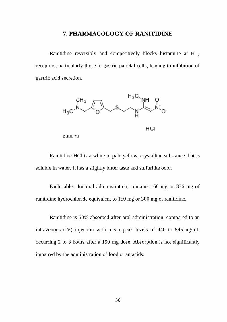
7. PHARMACOLOGY OF RANITIDINE
Ranitidine reversibly and competitively blocks histamine at H 2
receptors, particularly those in gastric parietal cells, leading to inhibition of
gastric acid secretion.
Ranitidine HCl is a white to pale yellow, crystalline substance that is
soluble in water. It has a slightly bitter taste and sulfurlike odor.
Each tablet, for oral administration, contains 168 mg or 336 mg of
ranitidine hydrochloride equivalent to 150 mg or 300 mg of ranitidine,
Ranitidine is 50% absorbed after oral administration, compared to an
intravenous (IV) injection with mean peak levels of 440 to 545 ng/mL
occurring 2 to 3 hours after a 150 mg dose. Absorption is not significantly
impaired by the administration of food or antacids.

37
The principal route of excretion is the urine, with approximately 30%
of the orally administered dose collected in the urine as unchanged drug in
24 hours. The elimination half-life is 2.5 to 3 hours
INDICATIONS AND USAGE :
Short-term treatment and maintenance therapy of active duodenal
ulcer. Short-term treatment of active, benign gastric ulcer. Most
patients heal within 6 weeks.
Treatment of GERD and endoscopically diagnosed erosive
esophagitis. Symptomatic relief commonly occurs within 24 hours
after starting therapy with ranitidine 150 mg bd
Prophylaxis and treatment of aspiration pneumonitis.
ADVERSE REACTIONS:
Malaise, dizziness, somnolence, insomnia, and vertigo, Constipation,
diarrhea, nausea/vomiting, abdominal discomfort/ pain, Rare cases of
hypersensitivity reactions (e.g., bronchospasm, fever, rash, eosinophilia),
anaphylaxis, angioneurotic edema, and small increases in serum creatinine.

38
DRUG INTERACTIONS AND CONTRAINDICATIONS:
Ranitidine has been reported to bind weakly to cytochrome P-450,
Increased or decreased prothrombin times have been reported during
concurrent use of ranitidine and warfarin. Ranitidine tablets are
contraindicated for patients known to have hypersensitivity to the drug or
any of the ingredients.
39
8. PULMONARY ASPIRATION RISK IN OBSTETRIC
PATIENTS
Mendelson was the first person to describe the entity of pulmonary
aspiration of gastric contents in obstetric subset of population. He described
the syndrome in 1946 and the pathogenesis associated with this syndrome29
.
He also stressed the importance of perioperative use of anti aspiration
prophylaxis.
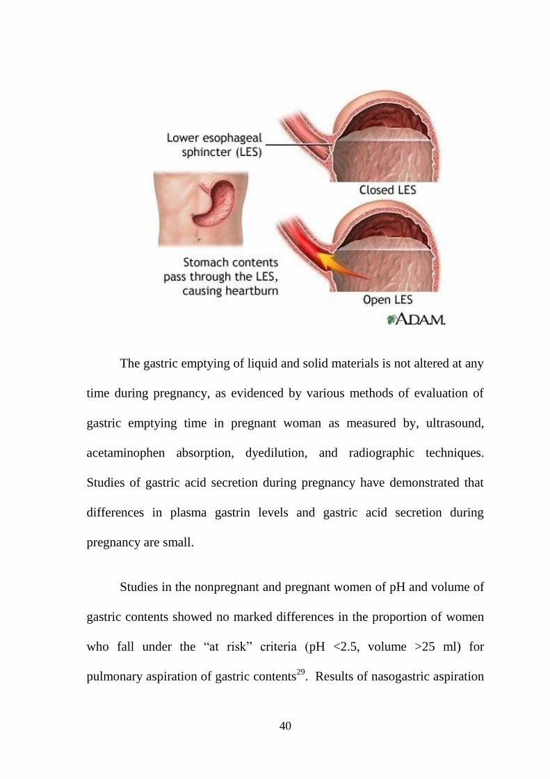
The gastrointestinal system in pregnancy undergoes various changes.
The stomach is displaced upward toward the left side of the diaphragm
during pregnancy. The altered position of the stomach tend to alter the
position of the intraabdominal segment of the esophagus, by displacing it
into the thorax. This causes a decrease in tone of the lower esophageal high-
pressure zone (LEHPZ), which normally prevents the reflux of gastric
contents. This displacement of the esophagus also prevents the rise in lower
esophageal tone that normally accompanies an increase in intragastric
pressure (IGP)28
. Progesterone also may contribute to a relaxation of the
LEHPZ.28
The lower esophageal sphincter changes occur as early as in the
first trimester.
40
The gastric emptying of liquid and solid materials is not altered at any
time during pregnancy, as evidenced by various methods of evaluation of
gastric emptying time in pregnant woman as measured by, ultrasound,
acetaminophen absorption, dyedilution, and radiographic techniques.
Studies of gastric acid secretion during pregnancy have demonstrated that
differences in plasma gastrin levels and gastric acid secretion during
pregnancy are small.
Studies in the nonpregnant and pregnant women of pH and volume of
gastric contents showed no marked differences in the proportion of women
who fall under the “at risk” criteria (pH <2.5, volume >25 ml) for
pulmonary aspiration of gastric contents29
. Results of nasogastric aspiration
41
of gastric contents in nonpregnant patients undergoing elective surgery and
in pregnant women undergoing elective cesarean section have shown that
patients who received no preoperative medication that would alter gastric
volume or pH, approximately 80% of individuals in each group (pregnant
and nonpregnant) had a gastric pH of 2.5 or less, approximately 50% had
gastric volumes of 25 mL or greater, and 40% to 50% exhibited both a low
pH and a volume of at least 25 Ml29
.
PHARMACOLOGICAL METHODS OF PREVENTING
ASPIRATION IN PREGNANCY31
:
A) DURING LABOUR
Food and fluids may be taken during labour at the women‟s
discretion.
Fluids only in cases of women at high risk of requiring an operative
birth.
Avoid the use of antacids containing magnesium or aluminium
(e.g. Mylanta) for symptoms of heartburn or indigestion during
labour – these medication are associated with severe pneumonitis
should aspiration occur.
Inj. RANITIDINE 50mg IV six hourly for high risk cases as
selected by anaesthetist in consultation with obstetric staff.

42
An additional dose if it is five hours since last dose when decision for
operative birth/procedure made.
B) ELECTIVE SURGERY DURING PREGNANCY INCLUDING
ELECTIVE CAESAREAN SECTION:
Either Tab. RANITIDINE 150mg orally the evening prior to surgery
plus 150mg orally at least one hour pre-operatively on the day of
surgery.
Or Tab. RANITIDINE 300mg orally at least one hour pre-
operatively on the day of surgery. Inj. METOCLOPRAMIDE 10mg
intravenously one hour pre-operatively.
30ml of SODIUM CITRATE mixture orally (0.3 molar solution)
shall be given 20minutes prior to induction of anaesthesia
C) EMERGENCY OPERATIONS (CAESAREAN, POSTPARTUM
PROCEDURES etc.)
Since the risk of aspiration in pregnancy starts from the third trimester
itself, any emergency procedure during the antenatal period and postpartum
period should have the following method of acid prophylaxis
Inj. RANITIDINE 50mg IV as soon as possible after notification if
not previously on oral ranitidine.

43
Inj. METOCLOPRAMIDE 10mg IV as soon as possible after
notification.
30ml of SODIUM CITRATE mixture orally (0.3 molar solution)
shall be given 20minutes prior to induction of anaesthesia.

44
9. REVIEW OF LITERATURE
STUDIES RELATED TO ASPIRATION IN THE PERIOPERATIVE
PERIOD:
1. MENDELSON STUDY: AMERICAN JOURNAL OF
OBSTETRICS AND GYNAECOLOGY, 1946
In this remarkable study, Curtis Lester Mendelson analyzed and
presented the remarkable article entitled "Aspiration of gastric contents into
the lungs under obstetric anaesthesia". He found out aspiration in 66 cases
out of 43,000 pregnancies. This equals to a notable incidence of about 1 in
660 pregnancies. Nowadays the occurrence is much lower, but it still
represents the most common cause of anaesthetic death in pregnant woman.
2. ROBERTS et al 1974:
He did a pioneering study on the usage of sodium citrate as antacid
prophylaxis in obstetric subset of patients and arrived at the conclusion that
it can be a effective regimen when compared with the existing anti-
aspiration pharmacological interventions. Base on their studies, they
formulated that Volumes of gastric aspirates in excess of 0.3 to 0.4 ml/kg or
20 to 25 ml may be potentially hazardous sand causes aspiration
pneumonitis, if inhaled.
45
3. F.M. MESSAHEL, A.S. AL-QAHTANI: PULMONARY
ASPIRATION OF GASTRIC CONTENTS IN ANESTHESIA: A
REVIEW OVER 5-YEAR PERIOD - The Internet Journal of
Anesthesiology. 2009 Volume 19 Number 1.
This study did an analysis of the incidence, morbidity and mortality
of pulmonary aspiration during administration of anesthesia in an institute.
The database of anesthetic related events were examined to collect the
details of 12828 patients who were administered general anesthesia during
the 5-year period following application and adoption of stringent guidelines
for the prevention of aspiration in the preoperative period.
It included details of patients who got regurgitation and aspiration of
gastric contents during the course of the anesthetic and in the immediate
recovery period. Among this, 451 patients suffered aspiration (3.5% of
total), out of them 95 (21.1%) were elective and 356 (78.9%) were
emergency. Out of these, 80 patients (17.7%) aspirated at induction and 371
(82.3%) at extubation.

46
STUDIES RELATED TO 0.3MOLAR SODIUM CITRATE
4. SODIUM CITRATE PRETREATMENT IN ELECTIVE
CESAREAN SECTION PATIENTS- Dewan Dm, Floyd H M,
Thistle wood J M, Bogard TD , Spielman FJ.
Term pregnant woman, 32 in number who underwent elective
ceaserean section randomly divided into 3 groups. Group1 got no antacid,
group 2 -30 ml of 0.3 molar sodium citrate <60mts preoperatively, group 3-
30 ml of 0.3 molar sodium citrate >60mt preoperatively. Immediately after
delivery, the stomach was emptied. Mean pH of the gastric aspirate was
measured in the three groups were 1.8± 2.7, 5.0± 1.5, and 2.7±1.2,
respectively. Gastric fluid pH was found to be markedly high in group 2,
compared with other two groups. All patients in group 1, 90% in group 2
and 50% in group 3 had a gastric pH <2.5. in group 2, none had both pH of
less than 2.5 and volume > 25 ml. They came to a conclusion that sodium
citrate effectively rises gastric pH when given < 60 minutes prior to
induction.
47
5. GASTRIC FLUID pH IN PATIENTS RECEIVING SODIUM
CITRATE Oscar J. Viegas, MD, Ram S. Ravindran, MD, and Carol
A. Shumacker,
In 30 patients undergoing elective surgery, they did an analysis of pH
of gastric fluid after giving sodium citrate. Following induction of
anaesthesia & intubation of these patients, the gastric fluid was aspirated
and the pH was measured. Out of them, 5 persons who had been given 5 ml
of sodium citrate 5 to 20 minutes before induction of anesthesia were found
to have a mean pH of 6.2 ± 0.8. In the control group of 5 patients, who did
not receive sodium citrate had a mean pH of 2.1 ± 1.4. The sodium citrate
given increases the gastric pH and this would result in decreased pulmonary
mucosal damage, should aspiration occur.
6. USE OF SINGLE DOSE OF SODIUM CITRATE AS A
PROPHYLAXIS AGAINST ACID ASPIRATION IN OBSTETRIC
PATIENTS UNDERGOING CAESAREAN SECTION. Lim SK,
Elegbe EO. Med J Malaysia. 1991 :
The effectiveness of sodium citrate as antacid prophylaxis was
studied in 3 groups of 20 patients each. Group I (control) received no
antacid. Group II( elective caesarean section) and Group III (emergency
caesarean section) were given 30ml of 0.3M sodium citrate immediately
after their entry into the operation theatre. The gastric content was aspirated

48
and pH analysis was done just after induction of anaesthesia and at the end
of surgery before extubating the patient. Sodium citrate was found to
increase the gastric fluid content pH to much higher range in Group II and
III patients as compared with the control group.
7. SODIUM CITRATE: AN ALTERNATIVE ANTACID FOR
PROPHYLAXIS AGAINST ASPIRATION PNEUMONITIS. By J
Wrobel, T C Koh, J M Saunders Anaesthesia and intensive care
(1982).
In this study, about 107 general surgical patients who underwent
elective and emergency procedures were divided into two groups. The test
group received 5 ml of either sodium citrate 0.3 M and the control got
placebo 10 minutes before the induction of anesthesia. Gastric contents were
aspirated soon after induction and intubation and the pH analysis of the
samples was done. The mean pH of the gastric contents in the sodium citrate
group was 5.67, and it was 3.21 for those given the placebo it (p less than
0.001). Of patients who were given sodium citrate 92% had a gastric pH
above 3.0 when compared with 37% in the placebo group.

49
8. THE EFFECTIVENESS OF SODIUM CITRATE AS AN
ANTACID- Charles P.Gibbs et al, Anesthesiology 1982 :
26 obstretric patients scheduled for emergency cesarean section were
given 30 ml of sodium citrate at interval of 10-20 min before induction of
anesthesia. Two gastric aspirate samples were collected- first sample at 12-
50min after ingestion of antacid and second at 60-180 min. The pH of all
samples were above 2.5 (mean pH- 5.7 in 1st and 5.2 in 2
nd sample). The
lowest pH were 3.2 and 1.8 respectively.
9. ASPIRATION PROPHYLAXIS FOR PREGNANT PATIENTS
REQUIRING ANESTHESIA- PUBLISHED NOV 2008.
This article analysed the incidence and various risk factors involving
the morbidity and mortality from aspiration. It recommends routine
antiaspiration measures to be taken in all women of >18-20wks gestation,
and upto 18 hours post partum. It states that particulate antacids when used
for aspiration prophylaxis causes severe pneumonitis should aspiration
occur. Sodium citrate 0.3M, 30ml given orally is the most efficient way of
immediate neutralization of gastric contents- acts within minutes and lasts
upto 1 hour. The combination of sodium citrate plus ranitidine is even more
synergistic in reducing gastric acidity.

50
STUDIES RELATED TO COMPARISON OF 0.3MOLAR SODIUM
CITRATE WITH OTHER DRUGS:
10. EFFECT OF SINGLE DOSE ORAL RANITIDINE AND SODIUM
CITRATE ON GASTRIC PH DURING AND AFTER GENERAL
ANESTHESIA (CANADIAN JOURNAL OF ANESTHESIA, 1995,
PETER ATANASSOFF et al)
In 25 patients scheduled for elective surgery,They analysed the effect
on gastric pH of the H2 blockers(R) with sodium citrate(SC) as a oral
effervescent and plain sodium citrate(SC). The drugs were given by
nasogastric tube placed after induction. A 24hr continuous gastric pH
monitoring was done by pH electrode. Mean baseline pH were 1.3 in R+SC
group and 1.2 in plain SC group. These values raised to 6.9(R+SC) and
4.9(SC) during emergence from anesthesia.. The pH remained above 2.5 for
14hrs in R+ SC group and for 6hrs in SC group. They concluded that both
the drugs are effective in neutralizing gastric acid when given orally after
induction. However, the action of plain SC is shortlived, and if maintenance
of gastric pH of >2.5 for more than 6hrs is needed, the R + SC combination
is recommended.
51
11. AN ORAL SODIUM CITRATE- CITRIC ACID NON-
PARTICULATE BUFFER IN HUMANS(J.J.HAUPTFLEISCH
AND K A PAYNE, BRITISH JOURNAL OF ANESTHESIA 1996)
This study investigated the effect on pH of the gastric pH of single
dose sodium citrate(antacid) and sodium citrate dehydrate with citric acid
monohydrate (buffer) in 30 neurosurgical patients for 5-7hrs duration. A
control group of 10 received no antacid. The mean baseline pH- 2.64. in
control group, pH increased to 4.4 at 5hr, returning to beaseline at 7 hr. In
antacid group, pH raised to 6.11 at 5min and decreased to 3.7 at 7hrs. In
buffer group, pH was stable at 3.80- 3.95 over 7hr.
12. BICITRA AS AN EFFECTIVE PREOPERATIVE ANTACID by
Charles. P. Gibbs and Tina et al :
In this study, the analyzers used Bicitra, a commercially available,
urine alkanizing solituion which contains the same amount of sodium
citrate, as that of 0.3 molar sodium citrate. They determined the efficacy of
Bicitra in elevating the pH of gastric contents above 2.5 in 26 patients
undergoing ceaserean section in general anesthesia. The pateients were
given 30ml of Bicitra just before induction, they were rotated side to side
for the effective mixing of the contents with the antacid. It was found,
Bicitra increased the pH in 88.5% patients. The buffering capacity (mean

52
pH), as determined with Bicitra is explained is less due to its low pH(4.8)
Vs 8.5 with 0.3M sod citrate. This is because of the low citric acid content
of Bicitra.
13. COCHRANE DATA BASE
This study did 16 meta-analyses on 23 studies that related to
interventions for reducing aspiration pneumonitis., involving 2658 women
undergoing cesarean section. The study reviewed the effectiveness of non-
pharmacological interventions and pharmacological drugs which are in
common practice to reduce aspiration pneumonitis for women who have
caesarean sections. They measured the primary outcome in terms of
1. Incidence of morbidity and mortality due to aspiration pneumonitis
2. Low intragastric pH of less than 2.5, measured after induction of
anaesthesia.
3. Increase of intragastric volume of more than 0.4 ml/kg or 25 ml,
measured after induction of anaesthesia.
They also analyzed secondary outcomes like:
1. Incidence of nausea and vomitting during caesarean section or the
postoperative period.
2. Intragastric pH above 2.5 and intragastric volume to less than 0.4
ml/kg measured prior to extubation.

53
They arrived at results suggesting that while administering a single
agent, antacids alone are proposed to have superior efficacy than H2
blockers, which are in turn more efficacious than proton pump inhibitors
for increasing gastric pH. The combination of antacids(0.3 molar sodium
citrate) plus H2 antagonists(ranitidine) was shown to be more effective than
in the patients who had received no intervention, and it was a superior
mode of treatment to antacids alone in the mode of rising the pH of gastric
contents. It was also stated that.
The influence of treatments on gastric volume are less analyzed in
studies and reported. These findings are can be applied for all term
parturients undergoing caesarean section, especially under general
anaesthesia. The need of antiaspiration prophylaxis in women undergoing
caesarean section under regional anaesthesia is a clinical judgement to be
decided on an individual patient basis. In general these treatments are
relatively inexpensive and well tolerated in pregnancy. Hence their routine
use is strongly considered in view of the potential benefits, as aspiration is
a cause of maternal mortality, even today.

54
14. GASTRIC FLUID VOLUME AND pH IN ELECTIVE SURGICAL
PATIENTS: TRIPLE PROPHYLAXIS IS NOT SUPERIOR TO
RANITIDINE ALONE Maltby JR et al Can J Anaesth. 1990 :
They compared the effect of oral ranitidine given as a sole drug
against the serial administration of metoclopramide, ranitidine, and sodium
citrate on gastric aspirate volume and pH in 196 healthy, elective surgical
patients. Each of the patients were randomly allotted to one of the four
groups. Patients in all groups got oral ranitidine 50 mg 2-3 hr before the
starting of surgery.
Those in Group 1 also received oral metaclopramide 10 mg, about
one hour before the start of surgery, and sodium citrate 0.3 M 30 ml on
arrival into the operating area; Group 2 received sodium citrate but no
metaclopramide; Group 3 received metaclopramide but no sodium citrate. In
Group 4, the patients received ranitidine drug alone.
In all groups, mean pH was greater than 5.8. Mean aspirate volumes
were significantly greater in patients who received citrate (Groups 1 and 2-
it was 22 and 19 ml respectively) than in patients who did not get sod citrate
(Groups 3 and 4- it was 10ml and 8 ml respectively). Moreover in groups 2
and 3, one patient each had a gastric aspirate pH of less than 2.5 with
volume greater than 25 ml. On arrival at these results, they concluded that

55
administration of single ranitidine alone has no greater significant advantage
than triple prophylaxis with other drugs.
15. ACID ASPIRATION PROPHYLAXIS FOR EMERGENCY
CAESAREAN SECTION by Stuart Et al, Anaesthesia. 1996:
384 patients requiring emergency Caesarean section under general
anaesthesia randomly received one of six anti aspiration prophylaxis
treatments. . They were given drugs-metoclopramide 10 mg, sodium citrate
administered orally 0.3 M, 30 ml, intravenous administration of ranitidine
50 mg, omeprazole 40 mg, alone and in various combinations of two of
these drugs. Compared with sodium citrate alone, the addition of either
omeprazole ,ranitidine, or metoclopramide alone did not reduce the aspirate
volume while smaller reduction in gastric volume was seen with the
addition of metoclopramide and either ranitidine or omeprazole.
56
10. AIM OF THE STUDY
The objective of this study is to establish the efficacy of 0.3m sodium
citrate, a non particulate antacid in neutralizing the secreted gastric acid- as
prophylaxis against aspiration pneumonitis in obstretic patients undergoing
elective lower segment cesarean section under general anesthesia.

57
11. MATERIALS AND METHODS
After obtaining approval from the institutional ethical committee of
Govt. Kilpauk Medical College and Hospital, Chennai-10 and written
informed consent, fifty term pregnant patients of ASA physical status I & II
undergoing elective lower segment cesarean section under standardized
general anesthesia were enrolled in the study. This study was conducted in
Govt. Kilpauk Medical College and Hospital, Chennai from May 2012-
August 2012.
STUDY DESIGN:
Our study was a double- blind prospective randomized control study.
DOUBLE BLINDING TECHNIQUE:
The solutions to be administered to the patients were prepared by
anesthesiology assistant who prepared the solutions in such a way that both
the solutions are stored in identical amber coloured bottles and labeled
accordingly. The testing solution, 30ml of 0.3 molar sodium citrate was
labeled SOLUTION A. The control solution, 30ml of distilled water was
labeled SOLUTION B.

58
STUDY PERIOD:
The study period was from the time of 30minutes before induction of
anesthesia up to 2 hours in the postoperative period.
OBSERVATION PERIOD:
Patients in both the groups were monitored and observed in the
PACU for 24 hours for any side effects and complications.
STUDY GROUPS:
The 50 selected and assessed patients were randomly divided into
two groups of 25 patients each.
GROUP A -25 patients received 30ml of testing solution A.
GROUP B-25 patients received 30ml of control solution B,
Both the solutions were kept in identical amber coloured bottles. So,
neither the patient who is receiving it nor the person giving it, did not know
what is contained inside the bottle.
The analyzer then allotted them into 2 groups. 25 patients who had
received 30ml 0.3 molar sodium citrate were assigned to group A or study
group. Remaining 25 patients who had received 30ml of distilled water were
allocated to group B or control group. At the end of the surgery, after
obtaining the gastric aspirate before extubation, all patients in both the

59
groups were given Inj. Ranitidine 50mg i.v. to protect them from the
aspiration risk.
PATIENT SELECTION:
All patients
INCLUSION CRITERIA (OBSTETRIC PATIENTS):
pts undergoing elective LSCS under general anesthesia
pts fasting for >= 8hrs
no use of any other particulate antacids in the preoperative period
EXCLUSION CRITERIA:
Patients with BMI > 30
patients with anticipated difficult airway
patients undergoing emergency surgery
h/o any drug use or disease which alters the gastric secretion
h/o any drug allergy
patient refusal for GA
MATERIALS USED IN OUR STUDY:
Testing solution A or control solution B- 30ml in amber color bottles
Digital pH meter
Nasogastric tube
20ml syringe
60
Xylocaine jelly and adhesive tapes
Stethoscope
PARAMETERS OBSERVED IN THE STUDY:
Baseline vital parameters- PR, BP, SpO2
Baseline pH of gastric aspirate.
pH of gastric aspirate – at 30 min following induction
pH of gastric aspirate – before extubation
Incidence of nausea and vomiting
Incidence of pulmonary aspiration in the post op period
Post op vital parameters.
MONITORING:
STANDARDISED GENERAL ANESTHESIA IN BOTH THE
GROUPS:
observation of baseline vital parameters
vital parameters monitoring
Pulse oximetry
Non invasive bood pressure
Electrocardiogram
End tidal CO2
Urine output monitoring

61
Temperature monitoring
pH measurement by digital pH meter
premedication – Inj. Glycopyrrolate 0.2mg iv
rapid sequence induction with Inj. Thiopentone and Inj. Succinyl choline
cricoid pressure (Sellick‟s manouvere )- released after inflating ET tube
cuff
intubation with 6.5 or 7.0mm cuffed oral ET tube.
Maintenance(along with IPPV) – before baby delivery : 50-50 of O2:
N2O , after baby delivery : 67 % O2 in N2O
Reversal of neuromuscular blockade- Inj.Neostigmine &
Inj.Glycopyrrolate
Post op monitoring and observation
CONDUCT OF STUDY:
Pre operative instructions:
All term pregnant patients were posted for elective lower segment
cesarean section, after a complete medical history and examination and a
proper preoperative assessment. They were explained about this study in
their own language and written informed consent was obtained from them
for inclusion into this study. Then, they were taken up for the study, after
satisfying the inclusion and exclusion criteria.

62
All patients were advised overnight fasting. Patients in both the study
and control groups received Tab. Ranitidine 300mg on the night before
surgery. Apart from this, the patients in control group received no other
non-pharmacological interventions or any form of medications for
aspiration prophylaxis in the pre- or intra- operative period.
Conduct of standardized general anesthesia :
On the day of surgery, the patients were shifted to the operating
theatre. In the premedication room, all the baseline vital parameters were
recorded. All patients were premedicated with Inj. Glcopyrrolate 0.2mg just
before induction. A good intravenous line was established with 18G
venflon.
A 16 gauge naso gastric tube was introduced gently after thorough
lubrication and secured, after confirming proper placement in the stomach.
The gastric aspirate was obtained, and pH of the sample was measured
using a hand-held pH meter (Hanna HI-96106 Champ pH Tester). It
was taken as the baseline pH. Patients in the study group were given 30ml
of 0.3 molar sodium citrate (Amb NPA) orally, about 20min prior to
induction of anesthesia. Patients in the control group received 30ml distilled
water, at around the same period. the study was double blinded since both

63
the patient and the person giving did not know which solution was present
inside the amber coloured bottle.
On shifting the patient to the operating table, routine monitors – pulse
oximetry, non invasive blood pressure, electrocardiogram, capnography,
temperature were connected. Patients were explained beforehand, about the
cricoid pressure that would be given and advised not to get panic. The
patient was preoxygenated for five minutes with 100% oxygen. During the
time of induction and intubation, NG tube is pulled out by 10-15cm so that
the tip of NG tube lies proximal to the lower esophageal sphincter. This is to
avoid aspiration risk caused by NG tube induced Lower Esophageal
Sphincter incompetency and also to prevent the regurgitation occuring
during Inj. Scoline administration.
Patients in both the groups were induced by Rapid Sequence
Induction using Inj. Thiopentone 3mg/kg. Once the patient loses
consciousness, the cricoid pressure (sellick‟s manouvere) was applied and
maintained by a trained personnel. Inj.Succinyl choline 1mg/ kg was given,
maintaining the cricoid pressure. They were intubated using 6.5 or 7mm ID
size endotracheal tube, under direct larnygoscopic vision of the glottis. The
cricoid pressure was released once the tracheal tube cuff is inflated.
The endotracheal tube was secured after confirming bilateral equal air entry.
64
Following intubation, the NG tube was reinserted to the same level
and secured. The gastric contents were aspirated at intervals of 5min,
30min after induction and prior to extubation. pH of samples were
analyzed using pH meter.
Anesthesia was maintained with 50%: 50% oxygen and nitrous oxide,
non-depolarizing muscle relaxant. After delivery of the baby, Inj.
Pentazoscine , Inj. Syntocinon were given and anesthesia maintained with
67% nitrous oxide in oxygen. All patients maintained hemodynamic
stability in the intra operative period.
The gastric aspirate was sampled before extubation and the pH ws
checked. Before extubation, all patients in both the groups were given Inj.
Ranitidine 50mg i.v. to protect them from the aspiration risk
The stomach contents were completely emptied before extubation.
After the patient showed spontaneous breathing efforts, the neuromuscular
blockade was reversed with Inj.Neostigmine and Inj.Glycopyrrolate
10µg/kg iv. All patients were extubated on table uneventful, after satisfying
the extubation criteria.
The NG tube was removed, after applying constant suctioning in the
recovery room after extubation.The patients were shifted to the recovery

65
room for monitoring for 2hrs.. Then they were moved to post anesthesia
care unit for further follow up, monitoring and observation for 24 hours.
During the study period, the following parameters are measured, analyzed
and compared in the test and control groups.
1. pH of gastric aspirate – Baseline( before induction of anesthesia)
2. pH of gastric aspirate – at 5 min after induction
3. pH of gastric aspirate – at 30 min following induction
4. pH of gastric aspirate – during extubation
5. The number of patients who are at high risk of aspiration (based on
pH of gastric aspirate – Baseline)
STATISTICAL ANALYSIS:
It is a randomized double blind clinical study
Variabls were analysed with student „t‟ test and Mann & Whitney „U‟
test
Sample size obtained according to previous background study.
„p‟ value less than 0.05 was taken as significant.

66
12. OBSERVATION AND RESULTS
Fifty (50) female patients in ASA I & II who are at term pregnancy,
undergoing elective lower segment caesarean section under general
anesthesia were selected for the study. The data & measurements obtained
from the study were analyzed & tabulated using SPSS. In this study, a „p‟
value of less than 0.05 was considered statistically significant and a „p‟
value of less than 0.001 was taken as highly statistically significant.
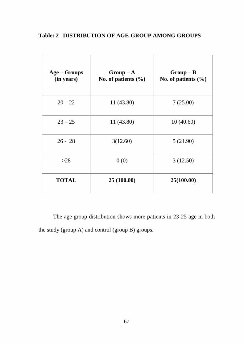
Table: 1 AGE DISTRIBUTION IN STUDY & CONTROL GROUPS
GROUP NO. OF
PATIENTS
MEAN
AGE
STD.
DEVIATION
„P‟
VALUE
STUDY
(GROUP A) 25 23.12 2.12 0.537*
CONTROL
(GROUP B) 25 24.78 2.94
*Not Significant (p<0.05) values are express in mean ± SD
The mean age in both the groups was around 25 years. Both the
groups were comparable with regard to age and there was no statistically
difference between the two groups.
67
Table: 2 DISTRIBUTION OF AGE-GROUP AMONG GROUPS
The age group distribution shows more patients in 23-25 age in both
the study (group A) and control (group B) groups.
Age – Groups
(in years)
Group – A
No. of patients (%)
Group – B
No. of patients (%)
20 – 22 11 (43.80) 7 (25.00)
23 – 25 11 (43.80) 10 (40.60)
26 - 28 3(12.60) 5 (21.90)
>28 0 (0) 3 (12.50)
TOTAL 25 (100.00) 25(100.00)

68
Figure: 1 DISTRIBUTION OF AGE-GROUP AMONG GROUPS
This graphical representation shows the age group distribution of
patients in both the groups.
0.00%
5.00%
10.00%
15.00%
20.00%
25.00%
30.00%
35.00%
40.00%
45.00%
20 - 22 23 - 25 26 - 28 >28
AGE-GROUP
43.80% 43.80%
12.60%
0%
25.00%
40.60%
21.90%
12.50%
GROUP - AGROUP - B

69
Table: 3 BODY WEIGHT IN STUDY AND CONTROL GROUPS
GROUP
NUMBER
OF
PATIENTS
MEAN
WEIGHT
IN KG
STD.
DEVIATION P VALUE
STUDY
(GROUP A)
25 59.53 11.45 0.950*
CONTROL
(GROUP B)
25 58.75 10.52
*Not Significant (p<0.05) values are express in mean ± SD
The mean weight in both the groups was around 59 kgs. Both the
groups were comparable with regard to weight. There was no statistical
difference in between the groups in terms of weight.

70
Table: 4 BODY MASS INDEX IN STUDY AND CONTROL GROUPS
GROUP
NUMBER
OF
PATIENTS
MEAN
WEIGHT
IN KG
STD.
DEVIATION P VALUE
STUDY
(GROUP A) 25 24.34 2.38 0.572
CONTROL
(GROUP B) 25 27.18 2.98
*Not Significant (p<0.05)
The values are expressed in mean ± SD
The mean Body Mass Index was around 25. There was no statistical
difference in between the groups in terms of BMI.

71
0
2
4
6
8
10
12
14
FIGURE :2 DISTRIBUTION OF INDICATIONS FOR LSCS IN TWO
GROUPS
In this column diagram, the indications for which the patients
underwent ceaserean section are shown. The other causes- post dated
pregnancy, precious pregnancy etc.
A – Previous LSCS
B – Primi with CPD
C – Other causes
A B C
GROUP – A GROUP - B

72
Table: 5 FASTING DURATION IN STUDY AND CONTROL
GROUPS
This table compares the fasting duration (in hours) in the pre
operative period, which is almost the same – 9 hours in both the groups.
GROUP
NUMBER
OF
PATIENTS
MEAN
FASTING
TIME(HRS)
STD.
DEVIATION P VALUE
STUDY
(GROUP
A)
25 9.41 0.76 0.589
CONTROL
(GROUP
B)
25 9.06 0.84

73
Table: 6 MEAN pH VALUES AT VARIOUS INTERVALS IN THE
STUDY AND CONTROL GROUPS
The values are expressed in mean ± SD.
The mean baseline pH in study and control groups are 2.94 and 2.84
respectively and there is no statistical difference in baseline pH values in
both. After administration of the test solution, the pH values in the study
group at 5min, 30min and extubation are all at a higher range than that of
control group, signifying the acid neutralizing effect of 0.3M sodium citrate
in the study group.
VARIABLES
STUDY
(GROUP A)
CONTROL
(GROUP B)
Baseline pH
2.94 ± 0.76
2.84 ± 0.73
pH after 5 mins.
4.46 ± 1.05
2.97 ± 0.73
pH after 30 mins.
4.53 ± 1.11
2.86 ± 0.72
pH before Extubation
4.64 ± 1.20
2.77 ± 0.67
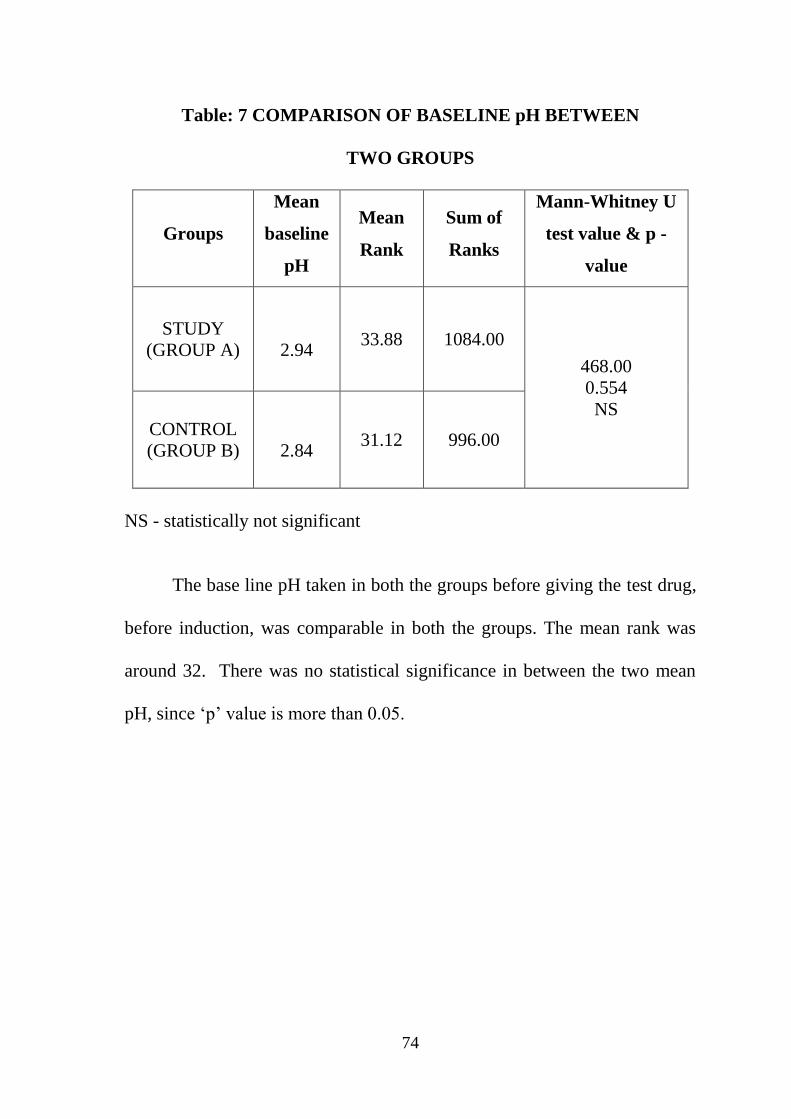
74
Table: 7 COMPARISON OF BASELINE pH BETWEEN
TWO GROUPS
Groups
Mean
baseline
pH
Mean
Rank
Sum of
Ranks
Mann-Whitney U
test value & p -
value
STUDY
(GROUP A)
2.94 33.88 1084.00
468.00
0.554
NS CONTROL
(GROUP B)
2.84 31.12 996.00
NS - statistically not significant
The base line pH taken in both the groups before giving the test drug,
before induction, was comparable in both the groups. The mean rank was
around 32. There was no statistical significance in between the two mean
pH, since „p‟ value is more than 0.05.

75
Table: 8 COMPARISON OF pH AT 5MIN OF INDUCTION
BETWEEN TWO GROUPS
HS: Highly Statistically Significant
The table 8 shown compares the pH values at 5 min after induction,
after the test drug is given in the study and control groups. A highly
statistical difference was observed in between the groups. This implies that
sodium citrate increases the pH of the gastric contents well above than the
pH in control group.
Groups Mean pH
at 5min
Mean
Rank
Sum of
Ranks
Mann-Whitney U
test value & p -
value
STUDY
(GROUP A) 4.46 44.75 1432.00 120.00
0.000
HS CONTROL
(GROUP B) 2.97 20.25 648.00

76
Table: 9 COMPARISON OF pH AT 30 MIN OF INDUCTION
BETWEEN TWO GROUPS
HS : Highly Statistically Significant
In table 9, the pH in the study group is higher than in the control
group, as seen by the difference in mean ranks in both the groups. A „p‟
value of <0.001 is observed in this table, implying high statistical difference
in pH between the groups.
Groups Mean pH
at 30min
Mean
Rank
Sum of
Ranks
Mann-Whitney U
test value &
p - value
STUDY
(GROUP A) 4.53 45.44 1454.00 98.00
0.000
HS CONTROL
(GROUP B) 2.86 19.56 626.00

77
TABLE: 10 COMPARISON OF pH AT EXTUBATION BETWEEN
TWO GROUPS
HS : Highly Statistically Significant
Table 10 shows the pH measured at extubation, which also states a
high statistically significant difference in the pH between the groups, as
seen in the mean rank and „p‟ value.
Groups Mean pH at
extubation
Mean
Rank
Sum of
Ranks
Mann-Whitney U
test value &
p - value
STUDY
(GROUP A) 4.64 45.78 1465.00 87.00
0.000
HS CONTROL
(GROUP B) 2.77 19.22 624.00

78
Table: 11 PATIENTS AT HIGH RISK (pH < 2.5) AT VARIOUS
INTERVALS:
SAMPLING INTERVALS
STUDY
GROUP – A
(N = 25)
CONTROL
GROUP – B
(N = 25)
Baseline 11 (44 %) 11 (44%)
5 minutes after induction 0 (0) 10 (40%)
30 minutes after induction 0 (0) 9 (36%)
Extubation 0 (0) 11 (44%)
Table 11 shows the number patients who are having a pH of less than
2.5 in both the groups at various time intervals after induction and at
extubation. They in turn fall under the high risk category for pulmonary
damage if aspiration occurs, as per criteria. From this table, it is evident that,
no patient in the study group came under high risk, after the drug is given.

79
Figure: 3 DISTRIBUTION OF PATIENTS AT HIGH RISK (pH < 2.5)
AT VARIOUS TIMING
FIGURE 3 is the pictorial representation of table 11. This also shows
no patient is in high risk in the study group.
0
2
4
6
8
10
12
Baseline 5 minutes 30 minutes Extubation
VARIOUS TIMINGS
11
0 0 0
11
10
9
11
NO
. OF
PA
TIE
NTS
GROUP - A
GROUP - B

80
Table : 12 DISTRIBUTION OF TIME AT WHICH pH WAS
MAXIMUM IN STUDY AND CONTROL GROUPS
Various Timing Group – A Group - B
5 min
after induction
6.8 4.5
30 min after
induction
6.7 4.6
Extubation 6.4 4.3
Table 12 helps to find out the maximum highest pH attained in the
two groups at various time intervals. It is obvious that the highest pH of
study group, A is higher than the pH in the control group at all times. In the
study group A itself, the5min pH value is the highest. This signifies the
protective effect of sodium citrate is maximal in about 30min after
administration, offering lower risk of pulmonary damage, should aspiration
occur during intubation. The figure 4 also mentions the same.
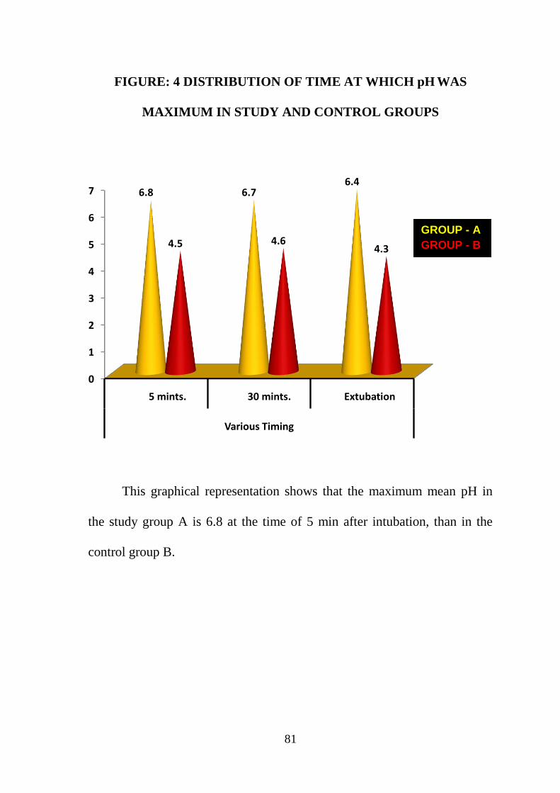
81
0
1
2
3
4
5
6
7
5 mints. 30 mints. Extubation
Various Timing
6.8 6.76.4
4.5 4.64.3
FIGURE: 4 DISTRIBUTION OF TIME AT WHICH pH WAS
MAXIMUM IN STUDY AND CONTROL GROUPS
This graphical representation shows that the maximum mean pH in
the study group A is 6.8 at the time of 5 min after intubation, than in the
control group B.
GROUP - A
GROUP - B

82
Table 13: DISTRIBUTION OF RANGE OF pH AT VARIOUS TIME
PERIOD AMONG STUDY (GROUP A)
In table 10, maximum number of patients have a pH range between
3.1- 5.0 at all times, well above the high risk (pH of less than 2.5). The pie-
chart in figure 5 denotes the same.
RANGE OF pH
STUDY GROUP A ( N=25 )
5 MIN
AFTER
INDUCTION
30 MIN
AFTER
INDUCTION
EXTUBATION
< 2.5 0(0.00) 0(0.00) 0(0.00)
2.6 – 3.0 2(8%) 1(4%) 3(12%)
3.1 – 4.0 9(36%) 7(28%) 8(32%)
4.1 – 5.0 7(28%) 7(28%) 6(24%)
5.1 – 6.0 4(16%) 6(24%) 4(16%)
6.1 – 7.0 3(12%) 4(16%) 4(16%)

83
FIGURE 5 : DISTRIBUTION OF pH IN STUDY GROUP A
FIGURE 5A- DISTRIBUTION OF pH AT 5 MIN
FIGURE 5B- DISTRIBUTION OF pH AT 30 MIN
FIGURE 5C - DISTRIBUTION OF pH AT EXTUBATION
< 2.5
2.6-3.0
3.1-4.0
4.1-5.0
5.1-6.0
6.1-7.0
pH RANGE
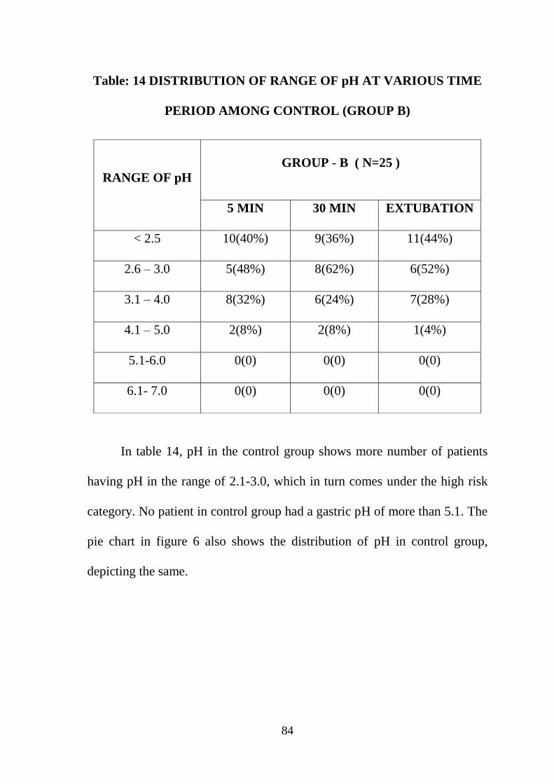
84
Table: 14 DISTRIBUTION OF RANGE OF pH AT VARIOUS TIME
PERIOD AMONG CONTROL (GROUP B)
In table 14, pH in the control group shows more number of patients
having pH in the range of 2.1-3.0, which in turn comes under the high risk
category. No patient in control group had a gastric pH of more than 5.1. The
pie chart in figure 6 also shows the distribution of pH in control group,
depicting the same.
RANGE OF pH
GROUP - B ( N=25 )
5 MIN 30 MIN EXTUBATION
< 2.5 10(40%) 9(36%) 11(44%)
2.6 – 3.0 5(48%) 8(62%) 6(52%)
3.1 – 4.0 8(32%) 6(24%) 7(28%)
4.1 – 5.0 2(8%) 2(8%) 1(4%)
5.1-6.0 0(0) 0(0) 0(0)
6.1- 7.0 0(0) 0(0) 0(0)

85
FIGURE 6 : DISTRIBUTION OF pH IN THE CONTROL GROUP B
FIGURE 6A: DISTRIBUTION OF pH AT 5 MIN OF INDUCTION
FIGURE 6B: DISTRIBUTION OF pH AT 30 MIN OF INDUCTION
FIGURE 6C: DISTRIBUTION OF pH AT EXTUBATION
< 2.5
2.6-3.0
3.1-4.0
4.1-5.0
5.1-6.0

86
13. DISCUSSION
Based on the observation and results obtained in our study involving
25 patients in each group are discussed in detail by comparing with the
available evidence in the literature.
In our study comparing the efficacy of 0.3 molar sodium citrate, a non
particulate antacid with control group, the mean age, weight, Body Mass
Index were comparable among the two groups(Tables 1,2,3,4 ). Our results
show that 0.3M sodium citrate is effective as a form of anti-aspiration
prophylaxis by increasing the pH of gastric contents than that of the control
group. It in turn lessens the degree of damage to pulmonary mucosa, if
aspiration of this less acidic gastric content occurs.
0.3 molar sodium citrate, 30ml when given in 20min before
induction, raises the pH of gastric contents to above 2.5, in the protective
range. The study group chosen was pregnant women undergoing elective
LSCS. This type of patients are considered to be „full stomach‟ even after
they are allowed adequate proper fasting time in the preoperative period.
Hence, they are always at a greater risk of aspiration during the peripartum
period. Procedures under general anesthesia in this group carries even more
higher risk, especially during times of intubation and extubation.

87
The dosage of sodium citrate was 30ml. In his study, Lahiri et al.
found that 5 ml of 0.3 molar sodium citrate increased th gastric pH above
3.0 in 21 of 22 parturients. Later, Heath and Hester, analyzed the same
volume and dosage of sodium citrate and they found no difference was there
in between treated and untreated groups. In the subsequent related studies,
they increased the volume of sodium citrate given to twice, and the
buffering capacity of antacid administered, and successfully brought the
gastric content ph to above 2.5. In our study also, 30ml was used to increase
the pH.
The timing of drug before is also accountable, since in their study,
DEWAN ET AL showed that the elevated mean ph in the short interval
group (sodium citrate given in less than 60min before induction) and the
increased incidence of low pH in the long interval group (sodium citrate
given in more than 60min before induction), when compared to the short
interval group, is due to the shorter duration of action of sodium citrate. In
our study, the mean timing of drug administration was 20 minutes (less than
half an hour) before induction.
Recently, O'Sullivan and Bullingham observed that gastric emptying
represents an important factor in determining the duration of action of
antacids (14). When gastric emptying occurs faster, the antacid rapidly

88
leaves the stomach and this results in a short duration of action. Factors that
slow gastric emptying cause extended duration of action of antacids. Our
patients received narcotics in the intraop period that might slow gastric
emptying.
The sampling interval was standardized in both the groups to find out
the time at which the drug action in elevating the gastric pH was maximal.
In our study, it was noted that ph elevation was maximal 6.8 in drug group
(table 10) during the time of intubation. Thus, it offers higher protection at
that time.
As per Roberts and Shirley risk criteria, no patients given sodium
citrate had a pH of less than 2.5 at any point of time during subsequent
samplings (table 9). This correlates well with the study done by Oscar and
Ravindran et al, where it was noted that nearly all patients given sodium
citrate had a gastric pH of more than 3.5, whereas in the control group only
two patients (13 %) had the pH above 2.5.
In our study, volume of gastric contents was not measured as it was
not considered as a parameter for comparison between the two groups. The
influence of gastric volume by antacids have been described in the
literature. An increased incidence of gastric volumes more than 20 ml after
the administration of antacids has been found in study of in

89
Stoelting R K et al. However, other reports (Detemir M D et al, Newson A J
et al ) found that the dosing of antacid does not affect the mean volume of
gastric contents.
Most commercial preparations has aluminum hydroxide which may
slow down the gastric emptying and therefore produce larger gastric
volumes ((Detemir M D et al). The estimation of gastric fluid volume by
aspiration of the stomach is less than ideal and may reflect an erroneously
low measurement, as full aspiration of all the gastric contents is not possible
even after repositioning and aspirating. Hence, the measurement of gastric
fluid volume is imprecise. So, in our study we elected not to measure gastric
volume in both the group of patients.
Moreover, as stated before, the critical volume and pH of gastric
contents needs to be revised as per the pioneering studies by Rocke DA et
al. this will make clinicians towards a more liberal use of antacids, which
are mainly avoided because of their inherent propery of increasing the
gastric content volume to above the critical limit.
The pH meter used for analyzing pH in our study, is a pen type pH
meter. Hence, the pH can be measured at the patient‟s bedside, which is
more time conserving and economical. The amount of aspirate required for

90
each sample is also less than 20ml when compared with the conventional
laboratory techniques requiring 50-60ml of aspirate for analysis.
The side effects and drug interaction of sodium citrate as discussed in
the literature was seldom seen in our study. This may be due to the reason
that all patients were well scrutinized in the preoperative for co-existing
medical illness and drug intake that interact with sodium citrate.
91
14. SUMMARY
A prospective randomized double blind study was designed to
establish the efficacy of 0.3molar sodium citrate in pregnant patients
undergoing elective lower segment caesarean section under general
anesthesia. Based on the analysis of the results and discussion in our study,
the conclusions arrived at are summarized as below.
In our study,
The demographic variables such as age, height, weight & body mass
are comparable between the groups
The preoperative fasting time was around 9 hours, which is also
comparable in both the groups.
We observed that the baseline pH was > 2.5 and were comparable
with no significant difference between the study and control groups.
The mean pH measured at 5 minutes, 30 minutes and extubation in
study group was statistically significantly higher in the study group
than in the control group. ( p value < 0.001- highly significant ).
The mean pH in the study group was maximally highest at 5min after
intubation, signifying the protective effect of 0.3M sodium citrate
during the time of induction, laryngoscopy and intubation.
No patient were at high risk zone (i.e pH <2.5) in the study and
control groups, at any time during the anesthesia.

92
No patients were observed to have complications like aspiration,
nausea or vomiting in the intra operative or postoperative period.
The side effects of general anesthesia like nausea, vomiting,
epigastric pain was found in few of our patients in both the groups
with not much statistical significance in our study.

93
15. CONCLUSION
We conclude that the non particulate antacid 0.3 molar sodium citrate
given orally about 20min before induction of anesthesia, is an effective and
safe antacid for anti-aspiration prophylaxis in all elective obstetric surgeries
without producing any side effects.

94
16. BIBLIOGRAPHY
1. Textbook of anesthesiology-AITKENHEAD 4th
ed
2. Gastric Fluid pH in Patients Receiving Sodium Citrate,Viegas et al 1981
;60:521-3.
3. Clinical significance of pulmonary aspiration during the perioperative
period Warner MA et al,Anesthesiology. 1993 Jan; 78(1):56-62.
4. BENUMOFS AIRWAY MANAGEMENT- 2nd
ed
5. Roberts RB, Shirley MA: Reducing the risk of acid aspiration during
cesarean section. Anesth Analg 1974; 53:859-868.
6. Ng A, Smith G: Gastroesophageal reflux and aspiration of gastric
contents in anesthetic practice. Anesth Analg 2001; 93:494-513.
7. Baum GL, et al, Textbook of pulmonary diseases. 6th ed. Vol. 1.
Philadelphia: Lippincott–Raven, 1998:645-55.
8. Aspiration pneumonitis and aspiration pneumonia -Paule Marik, N Engl
J Med, Vol. 344,2001
9. Gibbs CP, Banner TC: Effectiveness of Bicitra as a preoperative antacid.
Anesthesiology 1984; 61.
10. Kennedy TP, et al.Acute acid aspiration lung injury in the rat: biphasic
pathogenesis. Anesth Analg 1989; 69.
11. Nader et al. Reactive oxygen species contribute to oxygen-related lung
injury after acid aspiration. Anesth Analg 1998;87:127-33.

95
12. Gibbs CP, Modell JH. Pulmonary aspiration of gastric contents:
pathophysiology, prevention, and management. 1994:1437-64.
13. Pulmonary Aspiration Of Gastric Contents In Anesthesia , Eingelhardt
& Webster, B J A, 1999, 83,453-60
14. Goodmann LR, postoperative radiograph .I. alterations after abdominal
surgeries ,Am J of Roentgenol 1980 134, 533-41
15. Campinos L, Duval G- value of early fibreoptic bronchoscopy after
aspiration of gastric contents, B J A 1983, 55,1103-5.
16. Marrie TJ, Durant H, Yates L. Community-acquired pneumonia
requiring hospitalization: 5 year prospective study. Rev Infect Dis.
1989;11:586.
17. Bartlett JG, et al. Community-acquired pneumonia in adults: Guidelines
for management. Clin Infect Dis. 1998;26:811.
18. Bone RC et al. Early methylprednisolone treatment for septic syndrome
and ARDS. Chest 1987;92:1032-6. [Erratum, Chest 1988;94:448.]
19. Lobato, Emilio B.; Gravenstein, Nikolaus; Kirby, Robert R. Title:
Complications in Anesthesiology, 1st Edition.
20. Report by the ASA Task Force on Preoperative Fasting. Anesthesiology
1999.
96
21. Stone SB: Efficacy of cimetidine in decreasing the acidity and volume of
gastric contents: Inadequacy of nasogastric suction and endotracheal
intubation [letter]. J Trauma 1981; 21:996.
22. Barash, Paul G.; Cullen, Bruce F.; Stoelting, Robert K.; Cahalan,
Michael K.; Stock, M. Christine Title: Clinical Anesthesia, 6th Edition.
23. Sellick BA: Cricoid pressure to control regurgitation of stomach contents
during induction of anaesthesia. Lancet 1961; 2: 404.
24. Peden NR, saunders et al, Inhibition of pentagastrin stimulated and
nocturnal gastric secretion by Ranitidine, LANCET 1979, 1, 690-2.
25. Taylor G, acid pulmonary aspiration syndrome after antacids, B J A
1975, 65-7.
26. Gibbs CP, Spohr et al: the effectiveness of sodium citrate as an antacid.
ANESTHESIOLOGY 57: 44-46, 1982
27. Sodium citrate pretreatment in elective cesarean section patients- dewan
dm, floyd h m, thistle wood j m, bogard td , spielman fj.
28. Brock-Utne JG et al: Effect of domperidone on lower esophageal
sphincter tone in late pregnancy. Anesthesiology 1980.
29. Chestnut: Obstetric Anesthesia: Principles and Practice, 3rd ed.
30. Methods for Testing pH of Liquids | eHow.com.

97
31. Prevention of gastric aspiration in pregnant women, King Edward
Memorial Hospital Perth Western Australia , July 2009.
32. Effect of single dose oral ranitidine and sodium citrate on gastric pH
during & after general anesthesia, C J A 1995/42:5/ 382-6.

98
17. ANNEXURES
PROFORMA
Name of patient :
Group assigned :
Age / Sex :
IP number :
Weight/ height :
Preop assessment
History-
Examination-
Airway assessment-
Diagnosis and indication for LSCS-
ASA status –
Last oral intake :
Premedication :
Test solution number :
Time of drug administration before induction :
Duration of surgery :
Baseline pH :
pH at 5min : pH at 30min : pH at extubation:

99
ETHICAL COMMITTEE CERTIFICATE

100
PATIENT CONSENT FORM
“A Study To Determine The Efficacy Of 0.3 M Sodium Citrate As
An Antacid Prophylaxis Against Aspiration Pneumonitis In Obstetrical
Patients Undergoing Elective Ceasarean Section Under General
Anesthesia”
Study centre: Department of Anaesthesiology & Critical Care, Kilpauk.
Medical college:
Participant name: Age: Sex:
I.P. no:
I, confirm that I have understood the purpose of procedure for the
above study. I had the opportunity to ask the question and all my questions
and doubts have been answered to my satisfaction.
I have been explained about the pitfall in the procedure and the
management of it. I have been explained about the safety, advantages and
disadvantages of the techniques.
I understand that my participation in the study is voluntary and that I
am free to withdraw at anytime without giving any reason.

101
I understand that investigator, regulatory authorities and the ethics
committee will not need my permission to look at my health records both in
respect to current study and any further research that may be conducted in
relation to it, even if I withdraw from the study.
I understand that my identity will not be revealed in any information
released to third parties or published, unless as required under the law. I
agree not to restrict the use of any data or results that arise from the study.
I hereby consent to participate in this study of “A Study To
Determine The Efficacy Of 0.3M Sodium Citrate As An Antacid
Prophylaxis Against Aspiration Pneumonitis In Obstetrical Patients
Undergoing Elective Caesarian Section Under General Anesthesia”.
Name of the patient: Signature/thumb impression of patient:
Name of the witness: Signature:
Address: Contact Number:
Name of the investigator: Signature:
Time: Date:
Place:

102
MASTER CHART – STUDY GROUP - A
S.NO NAME AGE/SEX WT.
(KG) IP NO DRUG DIAGNOSIS
FASTING
(HRS)
BASELINE
PH
PH
5MIN
PH 30
MIN
PH
EXTUB
1 INBASELVI 25/F 80 13140
NA
CITRATE PREV LSCS &CPD 10 4.3 3.1 5.2 5.8
2 NIRMALA 24/F 90 10517 NA CIT PRIMI WITH CPD 9 2.2 3.8 5.2 5.1
3 SHOBANA 26/F 74 11863 NA CIT PREV LSCS &CPD 10 2.2 5.8 6.1 6
4 JAMUNA 24/F 82 17062 NA CIT PRIMI -POSTDATED PREG 10 4.3 6.2 6.7 6.1
5 UMA 25/F 75 13415 NA CIT PRIMI WITH CPD 8 2 5.8 4.2 6.4
6 KANIMOZHI 22/F 55 10338 NA CIT PREV LSCS &CPD 10 1.9 4.8 5.2 5.3
7 SIVASAKTHI 25/F 56 14087 NA CIT PREV LSCS &CPD 10 2.1 6.8 5.8 3.1
8 REVATHY 26/F 47 14090 NA CIT PRIMI- PRECIOUS PREG 10 3.8 5.2 6.4 6.2
9 KUMUDHA 20/F 50 18140 NA CIT PRIMI WITH CPD 8 3.2 4.1 4.3 3.8
10 PAULIN 21/F 45 18121 NA CIT PREV LSCS 10 4.2 4.5 4.6 4.2
11 SAMUNDEESWARI 20/F 55 17853 NA CIT PRIMI-POSTDATED PREG 9 3 3.6 4.5 4.6
12 LATHA 21/F 50 16812 NA CIT PREV LSCS &CPD 10 4.3 6.2 6.3 2.8
13 KAMALA DEVI 25/F 62 17179 NA CIT PREV LSCS &CPD 8 3.1 4.8 5.1 5.2
14 JHANSI RANI 21/F 65 17233 NA CIT PRIMI WITH CPD 10 2.3 3 3.2 3.3
15 JAYANTHI 22/F 45 17423 NA CIT PREV LSCS 10 3.2 5 5.1 4.9
16 KOMALA 23/F 60 17942 NA CIT PREV 2 LSCS 9 1.8 2.6 2.7 3
17 AROKIA MARY 22/F 54 17899 NA CIT PRIMI WITH CPD 8 2.3 3.8 3.8 3.7
18 SHAGIRA BEGUM 20/F 65 20045 NA CIT PREV LSCS 10 2.4 3.2 3.3 3.6
19 REKHA 26/F 66 20076 NA CIT PRIMI WITH CPD 9 2.4 3.2 3.3 3.4
20 PUSHPA 21/F 45 20013 NA CIT PREV LSCS 10 2.8 3.9 4.1 4.3
21 DURGA DEVI 24/F 68 18426 NA CIT PRIMI WITH CPD 10 4.1 5.2 3.8 2.8
22 PACHAIAMMAL 22/F 50 20043 NA CIT PREV LSCS 9 2.3 3.8 3.2 3.6
23 MARY 24/F 48 19986 NA CIT PREV 2 LSCS 10 2.8 3.8 3.6 3.6
24 ALLIRANI 24/F 56 18453 NA CIT PREV LSCS 10 3.5 4.6 4.7 4.8
25 SATHYA 23/F 58 20527 NACIT PREV LSCS 8 3.1 4.6 4.3 4.2

103
MASTER CHART – CONTROL GROUP - B
S.
NO NAME AGE SEX
WT
(KG) IP NO DRUG DIAGNOSIS FASTING
BASELINE
PH
PH
5MIN
PH
30
min
PH
EXTUB
1 NANDHINI 22/F 62 10517 PLACEBO PREV LSCS 9 HR S 2.6 2.8 2.6 3.1
2 MALLIGA 26/F 65 11223 PLACEBO PRIMI WITH CPD 9 HRS 2.1 2.1 2.4 2.5
3 KANNAKI 28/F 56 10113 PLACEBO PREV LSCS 10 HRS 2.5 2.8 2.5 2.6
4 KALAISELVI 21/F 55 18103 PLACEBO PRIMI WITH CPD 8 HRS 3.2 3.3 2.8 2.8
5 SUBHA 24/F 60 16800 PLACEBO PREV LSCS 10 HRS 2.9 3.1 2.6 2.6
6 DHANALAKSHMI 21/F 45 16870 PLACEBO PRIMI- POSTDATED PREG 9 HRS 3.2 3.4 3.1 2.7
7 SARALA 24/F 55 17176 PLACEBO PRIMI WITH CPD 8 HRS 1.7 2 1.6 1.7
8 KAVITHA 25/F 70 17236 PLACEBO PREV LSCS 10 HRS 2.1 2.2 2.4 1.9
9 NASEEMA 24/F 68 18423 PLACEBO PREV LSCS 7 HRS 3.4 2.9 3 3.1
10 DEVIKA DEVI 27/F 60 18446 PLACEBO PRIMI WITH CPD 10 HRS 2.7 2.8 2.3 2.3
11 AMUL 21/F 48 18502 PLACEBO PREV LSCS 8 HRS 4.2 4.5 4.5 3.9
12 KANCHANA 23/F 80 18842 PLACEBO PREV LSCS 9 HRS 2.6 2.7 3 2.5
13 SHAKILA BANU 23/F 75 18823 PLACEBO PRIMI -POSTDATED PREG 10 HRS 2.3 1.8 1.8 2
14 MUTHU SELVI 30/F 70 18876 PLACEBO PRECIOUS PREGNANCY 9 HRS 2.8 3.1 2.8 2.6
15 LALITHA 28/F 68 19277 PLACEBO PREV LSCS 8HRS 2.2 2.4 2.3 2.3
16 NATHIYA 22/F 53 19677 PLACEBO PRIMI WITH CPD 10 HRS 3.1 3.5 3.4 3
17 MARIAMMAL 29/F 55 19740 PLACEBO PREV LSCS 9 HRS 4.2 4.5 4.6 4.3
18 BAGHYALAKSHMI 24/F 65 19233 PLACEBO PREV LSCS WITH CPD 8 HRS 2.4 3.1 3.2 3.3
19 ANNAL 32/F 67 19824 PLACEBO PREV LSCS 10 HRS 2.7 2.9 2.7 2.3
20 DEVI 24/F 55 19199 PLACEBO PRIMI- POSTDATED PREG 9 HRS 2.1 2.4 2.3 2.3
21 INBASEELI 28/F 45 12537 PLACEBO PRIMI-SHORT STATURE 10 HRS 2.8 2.7 2.8 2.6
22 SUDHA 26/F 59 19124 PLACEBO PREV LSCS 9 HRS 2.2 2.4 2.6 2.1
23 RAJESWARI 29/F 65 19599 PLACEBO PREV LSCS 10 HRS 3.9 3.8 3.5 3.6
24 DEVAKI 24/F 56 19604 PLACEBO PRIMI WITH CPD 9 HRS 3.4 3.2 2.8 2.5
25 INDIRA 22/F 55 20807 PLACEBO PRIMI WITH MOBILE HEAD 8 HRS 2.8 3.9 4.2 4.3

Your digital receiptThis receipt acknowledges that Turnitin received your paper. Below you will find the receipt informationregarding your submission.
Paper ID 290455346Paper title THESIS SUBMISSION
Assignment title MedicalAuthor Sujaritha. 20103985E-mail [email protected]
Submission time 25-Dec-2012 07:52PMTotal words 12108
First 100 words of your submission
1 A STUDY TO DETERMINE THE EFFICACY OF 0.3M SODIUM CITRATE AS AN ANTACIDPROPHYLAXIS AGAINST ASPIRATION PNEUMONITIS IN OBSTRETIC PATIENTS UNDERGOINGELECTIVE CEASEREAN SECTION UNDER GENERAL ANESTHESIA Dissertation submitted Inpartial fulfillment for the award of M.D DEGREE EXAMINATION M.D ANESTHESIOLOGY &CRITICAL CARE-BRANCH X KILPAUK MEDICAL COLLEGE & HOSPITAL, CHENNAI-10SUBMITTED TO THE TAMILNADU DR.MGR MEDICAL UNIVERSITY CHENNAI APRIL-2013 2CERTIFICATE This is to certify that this dissertation titled “A STUDY TO DETERMINE THE EFFICACYOF 0.3M SODIUM CITRATE AS AN ANTACID PROPHYLAXIS AGAINST ASPIRATIONPNEUMONITIS IN OBSTRETIC PATIENTS UNDERGOING ELECTIVE CEASEREAN SECTIONUNDER...
Copyright 2012 Turnitin. All rights reserved.