a study on paediatric head injury patients under 12...
TRANSCRIPT

THE TAMILNADU DR.M.G.R MEDICAL UNIVERSITY
CHENNAI -600032.
A STUDY ON PAEDIATRIC HEAD INJURY PATIENTS
UNDER 12 YEARS
Dissertation submitted in partial fulfillment by the
requirements for the degree of
M.Ch BRANCH II NEUROSURGERY
EXAMINATIONS – AUGUST 2013
INSTITUTE OF NEUROLOGY
MADRAS MEDICAL COLLEGE & RAJIV GANDHI
GOVERNMENT GENERAL HOSPITAL
CHENNAI-600003.
AUGUST -2013

CERTIFICATE
This is to certify that this dissertation entitled “A Study on
Paediatric Head Injury Patients under 12 years” submitted by
Dr.M.A.Bose, appearing for M.Ch (Neurosurgery) degree examination in
August 2013 is a original bonafide record of work done from August 2010
to February 2013 by him under my guidance and supervision in partial
fulfillment of requirement of the Tamil Nadu Dr.M.G.R. Medical
University, Chennai. I forward this to the Tamil Nadu Dr.M.G.R. Medical
University, Chennai, Tamil Nadu, India.
Prof.Dr.K.Deiveegan, M.S.,M.ChProfessor of Neurosurgery and Head,Institute of Neurology,Madras Medical College &Rajiv Gandhi Govt. General Hospital,Chennai - 600 003.
Prof. Dr. V. Kanagasabai, M.D. Ph.D.,The DEANMadras Medical College &Rajiv Gandhi Govt. General Hospital,Chennai - 600 003.
DECLARATION
I, Dr. M.A. Bose, solemnly declare that this dissertation “A Study
on Paediatric Head Injury Patients under 12 years” was done by me at
the Institute of Neurology, Madras Medical College and Rajiv Gandhi
Government General Hospital, Chennai under the guidance and
supervision of the Professor of Neurosurgery, Institute of Neurology,
Madras Medical College and Rajiv Gandhi Government General Hospital,
Chennai-3, between 2010 and 2013.
This dissertation is submitted to the Tamil Nadu Dr.M.G.R. Medical
University, Chennai-600032 in partial fulfilment of the University
requirements for the award of the degree of M.Ch., Neurosurgery.
Place : Chennai
Date : (M.A. Bose)

ACKNOWLEDGEMENT
I owe my thanks to THE DEAN, Madras Medical College, Chennai, for
permitting me to utilize the facilities and conducting this study and the members
of Ethical Committee for their role.
I am extremely grateful to Prof. K.DEIVEEGAN, M.S., M.Ch., Professor
of Neurosurgery and Head of the Department, Institute of Neurology, Madras
Medical College and Rajiv Gandhi Government General Hospital, Chennai, for
his constant encouragement and guidance throughout the study and periodic
reviews.
I sincerely thank all the Professors of our department Prof.
K.MAHESHWAR, Prof. S.D.SUBBIAH, Prof. RANGANATHAN JOTHI, Prof.
G.S.JAGAN NARAYANA, Prof. S.SYAMALA and our former Professors Prof.
R.ARUNKUMAR, Prof. V.G.RAMESH, Prof. C.SEKAR, Prof. V.SUNDAR, Prof.
S.SUNDARAM, Prof. J.V.MAHENDRAN for helping me with their time and
advice during this study.
I am indebted to all my assistant professors for their support, guidance
and help without which it would have been difficult to carry out this study. I
wish to thank the Professors, Assistant Professors, Post Graduates, Interns,
Paramedics, Office Staff, Technicians and Workers of the department of Pathology
and Radiology for their cooperation which enormously helped me in this study.
I thank my patients and their relatives for participating in this study.
CONTENTS
S.No. Title Page No.
1. INTRODUCTION 1
2. AIM OF STUDY 10
3. REVIEW OF LITERATURE 11
4. MATERIALS & METHODS 18
5. RESULTS 20
6. DISCUSSION 45
7. CONCLUSION 61
8. BIBLIOGRAPHY 62

1
INTRODUCTION
There is probably no area in medicine in which the adage that
“Children are not Little Adults and Infants are not Little Children” has
greater applicability than in Craniocerebral trauma. Paediatric Head
injuries contribute approximately 30% of total head injury cases. There are
various mechanisms which contribute to injury in children and its
prognosis which are different from others. The mechanism of injury, the
response of the skull and the cranial contents to injury and the long term
prognosis are quite different in the paediatric age group than compared
with adults. Therefore paediatric head injury contributes a separate entity
for the treating Neurosurgeons.
Paediatric population can also be sub grouped as;
1. Infants below 1 year
2. Toddlers – 1-5 years
3. Older children – More than 5 years
The 3 groups differ from each other in the mode of injury,
susceptibility to skull fractures, brain swelling, ischemic brain damage,
tolerance to hypoxia and blood loss in the prognosis.

2
Causes of Head Injuries
In comparison to adults the common mode of injuries in paediatric
population are birth injuries and fall.
Mode of Head Injury
The commonest mode of head injury in neonate is due to birth
trauma. In children up to 5 years of age head injuries are commonly due to
accidental fall. In older children motor vehicle accidents contribute to
significant number.
The resulting insult in Paediatric head injury includes from the Scalp
to the Cranial vault and the brain parenchyma inside. In neonates “Caput
succedaneum” which means scalp hematoma is due to cephalo pelvic
disproportion and occurs during obstructed labour.
“Cephal haematoma” which is sub galeal hematoma is common in
new borns following head injury.
Fractures are less common in Paediatric age group compared to
adults because of elastic nature of the skull bone and the yielding nature of
the skull bones compared to adult bones which are quite rigid can result in
ping-pong fracture which is indentation without fracture is also specific to
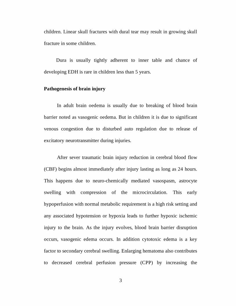
3
children. Linear skull fractures with dural tear may result in growing skull
fracture in some children.
Dura is usually tightly adherent to inner table and chance of
developing EDH is rare in children less than 5 years.
Pathogenesis of brain injury
In adult brain oedema is usually due to breaking of blood brain
barrier noted as vasogenic oedema. But in children it is due to significant
venous congestion due to disturbed auto regulation due to release of
excitatory neurotransmitter during injuries.
After sever traumatic brain injury reduction in cerebral blood flow
(CBF) begins almost immediately after injury lasting as long as 24 hours.
This happens due to neuro-chemically mediated vasospasm, astrocyte
swelling with compression of the microcirculation. This early
hypoperfusion with normal metabolic requirement is a high risk setting and
any associated hypotension or hypoxia leads to further hypoxic ischemic
injury to the brain. As the injury evolves, blood brain barrier disruption
occurs, vasogenic edema occurs. In addition cytotoxic edema is a key
factor to secondary cerebral swelling. Enlarging hematoma also contributes
to decreased cerebral perfusion pressure (CPP) by increasing the
4
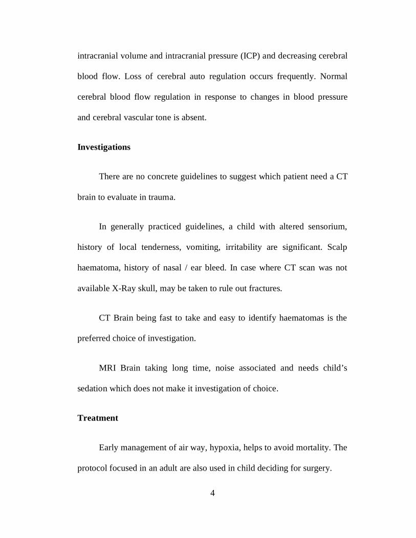
intracranial volume and intracranial pressure (ICP) and decreasing cerebral
blood flow. Loss of cerebral auto regulation occurs frequently. Normal
cerebral blood flow regulation in response to changes in blood pressure
and cerebral vascular tone is absent.
Investigations
There are no concrete guidelines to suggest which patient need a CT
brain to evaluate in trauma.
In generally practiced guidelines, a child with altered sensorium,
history of local tenderness, vomiting, irritability are significant. Scalp
haematoma, history of nasal / ear bleed. In case where CT scan was not
available X-Ray skull, may be taken to rule out fractures.
CT Brain being fast to take and easy to identify haematomas is the
preferred choice of investigation.
MRI Brain taking long time, noise associated and needs child’s
sedation which does not make it investigation of choice.
Treatment
Early management of air way, hypoxia, helps to avoid mortality. The
protocol focused in an adult are also used in child deciding for surgery.
5
As in adult, management of brain oedema, mannitol is advised but
hypertonic saline may also used. Anti convulsants should be given if
necessary care to avoid epilepsy.
Predictions for Mortality
Early low GCS Scale
Early post traumatic cranial nerve palsy, bulging fontenelle.
Hypovolemia at presentation
Age less than 1 year
All these features indicate severity of TBI and are significant
contribution to mortality.
Clinical evaluation and management in emergency
In any child with multiple trauma, a quick primary and secondary
survey is performed with prompt attention to airway, breathing and.
Pediatric patient with head injury may be brought unconscious, posturing
(decerebrate or decorticate), or actively convulsing. All patients should be
presumed to be full stomach and oxygen therapy should be initiated.
Comatose patients need to be intubated with rapid sequence intubation
technique, with due attention to cervical spine stabilization. Jaw thrust
6
maneuver can be performed during bag mask ventilation. Head tilt and
chin lift maneuvers should be avoided. A cervical spine collar should be
placed until cervical spine X-rays are obtained to rule out a fracture or
dislocation.
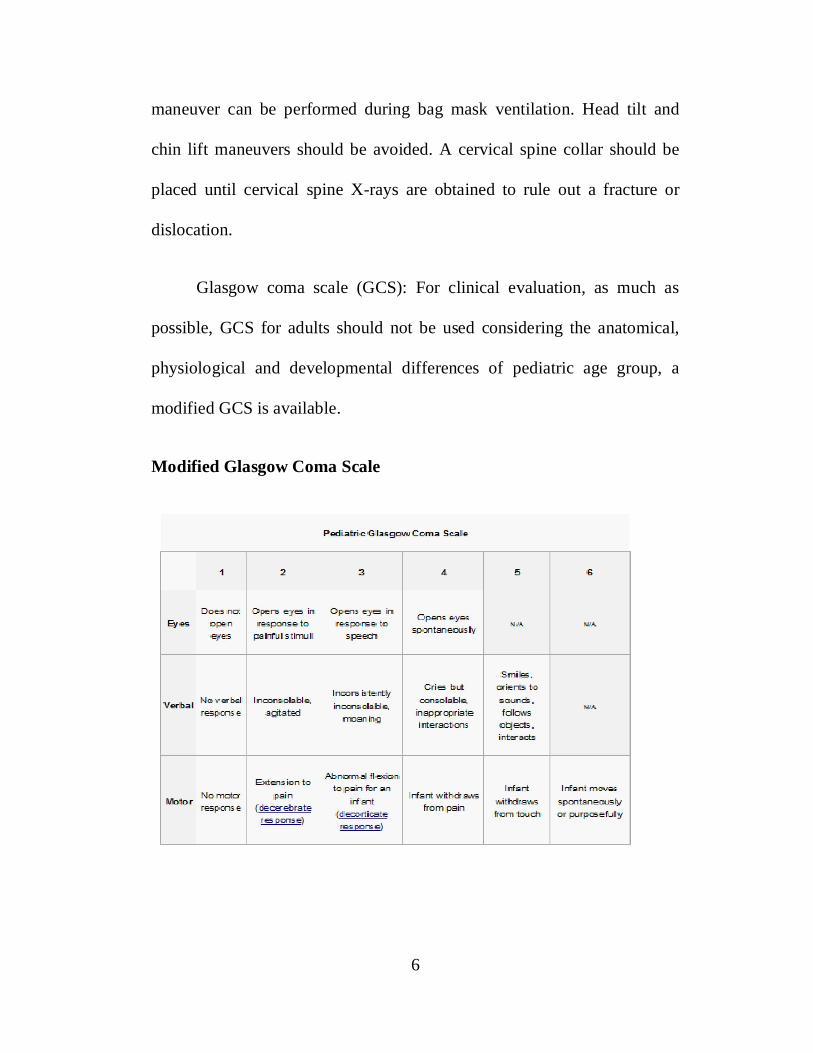
Glasgow coma scale (GCS): For clinical evaluation, as much as
possible, GCS for adults should not be used considering the anatomical,
physiological and developmental differences of pediatric age group, a
modified GCS is available.
Modified Glasgow Coma Scale

7
Indices of good outcome 9-12
Single most reliable examination for evaluating outcome in children
less than 3 years of age is ocular examination, as oculomotor functions are
fully developed by two months of age, while cortico spinal myelianation
and optic pathway myelination develop much later. Child with open
fontanel and ocular score of 3-4 generally has good outcome. Similarly
children with motor score of 4 and closed fontanel will have good
outcome. Those with closed fontenel and verbal score of 3 have good
outcome.
Indices of poor outcome
Evidence of retinal hemorrhage indicates poor outcome.
Radiographic evidence of post-traumatic splitting of suture indicates
poor outcome and high incidence of seizures. A bilateral linear skull
fracture correlates with poor outcome. Incidence of post-traumatic seizure
is 10%
8
Complication and sequealae of head injury
Early
Transient cortical blindness
Seizures
Cranial Nerve palsy
Diabetes insipidus
Syndrome of inappropriate secretion of ADH Cortical venous
occlusion
Hemiparesis
Late
Post traumatic epilepsy Post traumatic aneurysm
Meningitis Hydrocephalus Memory loss Disability
Muscle contractures
Outcome after paediatric head injury
Child outcome score has been described based on various
parameters such as neurological and cognitive deficit.

9
Child outcome score
I. Child Outcome Score Excellent recovery
II. Moderate but non-disabling deficit
III.Either a secure motor or cognitive deficit
IV.Vegetative
V. Death
I and II – Good outcome
III to V – Poor outcome
Indices of good outcome and poor outcome have already been
described earlier.

10
AIM OF THE STUDY
To analyse the incidence and the factors predicting the final outcome
in paediatric head injury patients.
To analyse the symptoms, CT findings in paediatric head injury
patients.
To analyse the mode of management in paediatric head injury
patients.

11
REVIEW OF LITERATURE
Berney et al.,1 in their study on children in the age group 0-3 years
sustained low energy trauma suffered more skull fractures and SDH and
early seizures compared to other ages of children between 3 and 9 years
had more high energy trauma with brain swelling & EDH were observed.
Children between 9 and 15 years more often found to have EDH when
compared to SDH.
According to Rivara et al.,2 in low fall like fall from table, bed etc.,
the common pathology encountered are concussion brain, fracture skull,
ICH.
Toft et al.,3 have analysed outcome in traumatic brain injury of
paediatric population in comparison with adult with respect to mode of
injury and found that in infants the common cause of nonfatal Traumatic
brain injury is fall (65%) and total traumatic brain injury is equally
distributed between motor vehicle accidents and fall (40%).
Greenes and Shutz,4 have analysed head injury in children less than
2 years and found that they are often (19%) asymptomatic, alert and
playful with occult intra cranial injuries. The CT Scan showed EDH /
SDH.

12
According to Durkin et al.,5 RTA are the leading cause of severe
head injuries in children. The common type of RTA in children are
pedestrian injuries, bicycle injuries, motor vehicle injuries, these being
common in their age groups 6-10, 9-15, 12-16 respectively.
According to Myhre Mc et al.,6 traumatic head injuries in infants
and toddlers - EDH, isolated skull fracture are more common in accidental
injuries.
Fundavo et al.,7 state that vomiting is the only significant symptom
noted in infant with head injury, loss of consciousness in other age group
children. The relation between swelling in the scalp CT abnormalities is
statistically significant, hence LOC and scalp swelling and vomiting
deserve CT scanning in children.
Homes et al.,8 have analysed, performance of paediatric Glasgow
coma scale children with blunt head trauma. They have concluded as
paediatric GCS is effective in assessing in head injury in children 2 years
or younger (preverbal) and standard GCS is effective in old children.
Al Habib et al. from annals of medicine Jan 2013 states that injury to
the head is the most commonly affected body parts in paediatric injuries
and is associated with serious consequences.

13
According to Durkin et al.,5 death secondary to paediatric head
injury represent a significant public health problem with cost burden
directly on Government Agencies. Pedestrian injuries are common among
6-10 years old children and bicycle injury are common in 9-15 years old
children of motor vehicle occupants injury were common from 12-16
years.
Fundavo et al., 7 vomiting is the only significant symptom noted in
infant with head injury, LOC in other children group. The relation
between, swelling in the scalp and CT abnormalities is statically
significant. Hence, LOC and scalp swelling and vomiting reserve CT.
Homes et al.,8 performance of paediatric Glasgow coma scale in
children with blunt head trauma. Paediatric GCS is effective in assessing
head injury in children 2 years or younger (Pre verbal) and standard GC, is
effective in older children.
Wang et al., 9 in a population based multi-center, prospective study
of pediatric trauma patients with mild alterations in consciousness (GCS
13-14), reported an incidence of 27.4% abnormal CT scans and 3.7%
required surgical intervention in a selected group of patients and concluded
that great majority of this patients will not require operative intervention,
but the implications of missing these hemorrhages can be severe for this

14
sub group of head injured patients. Because clinical criteria and cranial X-
rays are poor predictors of intracranial hemorrhage, it was recommended
that all children with a GCS score as 13 and 14 routinely undergo
screening via computer tomography.
Scalea et al.,10 in their retrospective study of selected pediatric
population noted an incidence of 14% as positive CT scan and 0.70% of
the study group underwent surgical intervention and concluded that a
normal neurologic exam and maintenance of consciousness does not
preclude significant rates of intracranial injury in pediatric trauma patients.
Contrary to convention, neither LOC nor mild altered mentation was a
sensitive indicator to select patients for CT scanning. Skull fractures and
superficial craniofacial injuries were similarly unreliable. Identification of
these patients was important for the occasional case requiring intervention
and for the tracking of complications. A liberal policy of CT scanning was
warranted for pediatric patients with a high-risk mechanism of injury
despite maintenance of normal neurologic status in the field and at hospital
screening.
Schutzman11 proposed various guidelines for evaluation and
management of children younger than 2 years old with apparently minor
head trauma but concluded that the effect of the proposed guidelines on

15
clinical outcomes and resource utilization should be evaluated.
Schunk et al.,12 in their retrospective analysis of the utility of head
computed tomographic scanning in pediatric patients with normal
neurologic examination in the emergency department, reported an
incidence <5% and need for neurosurgical intervention in 1% of the cases
and concluded that commonly used clinical variables viz., sleepiness,
vomiting, headache, LOC, irritability, amnesia and seizures, were not
associated with intracranial injuries in these children.
Aitken et al.,13 in their survey of current management practices of
pediatricians, emergency physicians and family physicians of minor
pediatric head trauma concluded that most physicians chose clinic or home
observation for initial management, and clinical management was more
varied when patients had sustained either loss of consciousness or seizures
and suggested further study of the appropriate management of head trauma
in children needed to guide physicians in their case.
Rattan et al., 14 in their prospective, selective study of pediatric head
injured patients, concluded that while a significant association was found
between the duration of consciousness and GCS, but no significant
association of either of these variable with CT scan findings was noted.

16
Murshid15 in his retrospective review of selective cases concluded
that the indications for CT scan were, an abnormal GCS, presence of
neurological deficit, signs of suspicion of basal or depressed fracture and
persistent or progressive head ache or vomiting and recommended that
infants with minor head injuries should be followed up atleast once after 2-
3 months for possible growing fractures.
Moran et al.,16 reported an incidence of 8.3% positive scan in their
prospective, selective population and concluded that LOC and skull
fracture are independent predictors of positive cerebral CT scans and
recommended immediate CT scan in all minor head injury patients with
LOC or a suspected skull fracture, to optimize the outcome of those
needing surgical intervention and those patients without LOC and GCS
score of 13-15 do not require CT scanning unless otherwise clinically
indicated.
Inamasu et al., 17 in their retrospective study, reported an incidence
of 0.5% deterioration of mild head injured patients and concluded that,
although routine use of CT scans in patients with mild head injury has been
controversial, CT scans should be taken if patients have experienced
transient LOC to prevent or reduce the occurrence of deterioration in the
emergency department.

17
Borzuck et al., 18 in his retrospective descriptive study reported an
incidence of 8.2% of abnormal CT scan and 0.76% neurosurgical
intervention and concluded that abnormalities on CT scans in patients with
mild head trauma are fairly common, although the need for neurosurgical
intervention was rare. Clinical decision rules can be used to identify these
patients with more serious intracranial pathology and recommended such
strategies should be validated prospectively in various emergency
department settings. He also defined that, loss of consciousness (LOC) was
a difficult variable to quantify because, qualified witness was usually not
available. Instead, LOC was coded as questionable LOC, brief LOC of
several seconds, or LOC of a minute or more.
Mikhail et al.,19 in their prospective study of 35 selected patients
reported, 22.86% incidence of intracranial injury and 8.57% required
surgery. One patient died following surgery and concluded that intracranial
injury does exist in patients suffering from minor head trauma with a GCS
score of 13 or above and further, age over 4 years and complaint of
headache were associated with an increased risk of intracranial injury.
Stein et al., 20 in their retrospective study reported an incidence of
18.4% intracranial lesions and 5.5% requirement of surgery and
recommended that any patient, who had suffered a loss of consciousness or
amnesia needs CT examination.
18
MATERIALS AND METHODS
HOSPITAL SETUP
This study was conducted at RAJIV GANDHI GOVERNMENT
GENERAL HOSPITAL, Chennai, Tamil Nadu, and our neurosurgical
department MADRAS INSTITUTE OF NEUROLOGY is one of the
pioneers in the establishment of neurosurgical centers in our nation.
Our government general hospital is a tertiary care referral centre
where all the trauma cases including the pediatric head injury cases are
admitted by the casualty medical officers. Duty neurosurgery assistants
and post graduates are present round the clock in trauma ward who gives
24 hour trauma care to all the pediatric head injury cases.
All the pediatric head injury patients are initially examined by
ABCD method and their vitals are monitored after making sure that the
child is hemodynamically stable, the child is shifted for CT scanning the
child is accompanied by the resident doctor.

19
STUDY PATTERN
Consecutive 150 patients admitted to NeuroSurgery TRAUMA ward
with head injuries during the period of August 2011 – July 2012 were
studied prospectively.
All the paediatric patients were included in the study group and there
is no specific exclusion criteria.
Patients clinical profile such as age, sex, admission GCS,
preliminary signs and symptoms are recorded.
CT Scan Brain, and its findings were noted and analyzed in detail.
Patients were managed either conservatively or surgically based on
clinical and radiological findings.
Outcome at the time of discharge were noted. Patients were followed
a period of varying from 3 months to one year.
The above data were entered in the Master Chart and varying factors
contributing the severity of head injury in paediatric age groups and
various parameters contributing to the outcome were analysed.
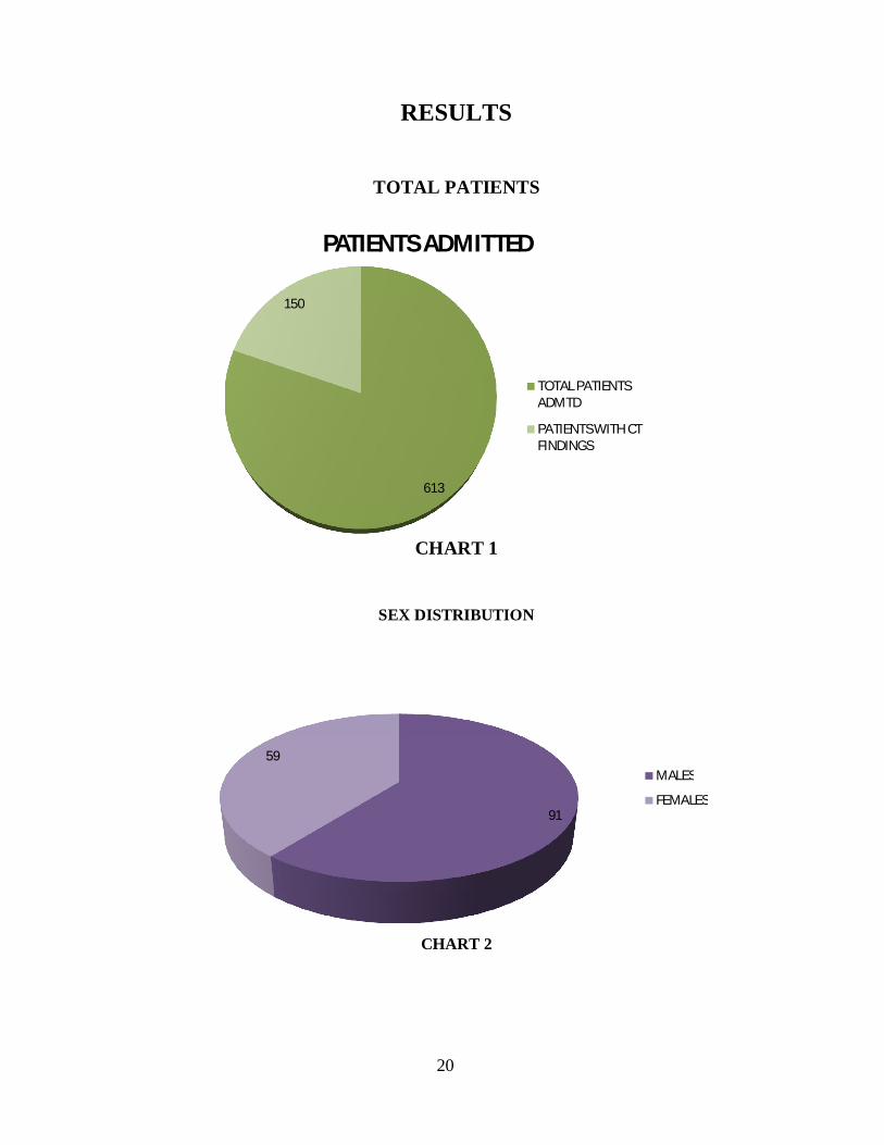
RESULTS
TOTAL PATIENTS
CHART 1
SEX DISTRIBUTION
CHART 2
613
150
PATIENTS ADMITTED
TOTAL PATIENTSADMTD
PATIENTS WITH CTFINDINGS
91
59MALES
FEMALES
20

21
AGE DISTRIBUTION
Group Age Frequency PercentValid
PercentCumulative
Percent I LESS THAN 1
YEAR13 4.0 4.0 4.0
II 1 TO 5 YEAR 67 48.7 48.7 52.7
III MORE THANFIVE YEAR
70 47.3 47.3 100.0
TABLE 1
CHART 3
13
67
70
AGE GROUP
< 1 YEAR
1 - 5 YRS
> 5 YRS

22
MODE OF INJURY
MODE OF INJURIES Frequency Percent
ASSAULT 1 0.7
FALL 79 32.7RTA 67 43.3TA 1 1.3
FALL OF HEAVY OBJECT 2 6.7TOTAL 150 100%
TABLE 2
CHART 4
1
7967
1 2
MODE OF INJURY
ASSAULT
FALL
RTA
TA
FHO

23
Mode of InjuriesASSAULT FALL FHO RTA TA Total
GROUP I 0 9 0 4 0 13
GROUP II 0 33 2 32 0 67
GROUP III 1 37 0 31 1 70
TABLE 3
CHART 5
0
5
10
15
20
25
30
35
40
ASSAULT FALL FHO RTA TA
GROUP I
GROUP II
GROUP III

24
GCS
GCS Frequency Percent
3-8 7 4.6
9-12 15 10.0
13-15 128 84.4
TABLE 4
CHART 6
0
20
40
60
80
100
120
140
GCS 3-8 GCS 9-12 GCS 13-15
GCS
GCS 3-8
GCS 9-12
GCS 13-15

25
GCS
3-8 9-12 13-15
GROUP I 0 0 13
GROUP II 2 7 57
GROUP III 5 8 58
TABLE 5
CHART 7
0 25
0
10
20
30
40
50
60
70
GROUP I GROUP II GROUP II
GCS 3-8
GCS 9-12
GCS 13-15
26
CT FINDINGS
NUMBER IMPROVED DEATH
EDH 14 14 0
SDH 04 04 0
CONTUSION 14 14 0
DAI 2 2 0
FRACTURES 67 67 0
OTHERS 16 16 0
MULTIPLE 34 28 6
TABLE 6
CHART 8
14
4
14
2
67
16
33
0 0 0 0 0 0 50
10
20
30
40
50
60
70
80
CT Findings
IMPROVED
DEATH

27
LOC
LOC
GROUP I 6
GROUP II 35
GROUP III 42TABLE 7
CHART 8
0
20
40
60
80
100
120
GROUP I GROUP II GROUP III
13
67 70
6
3542
LOC
NO OF PTS

28
VOMITING
VOMITING NO OF PTS
GROUP I 4
GROUP II 32
GROUP III 26
TABLE 8
VOMITING
CHART 9
0
10
20
30
40
50
60
70
80
90
100
GROUP I GROUP II GROUP III
13
67 70
4
32 26
VOMITING
NO OF PTS

29
ENT BLEED
ENT BLEED NO OF PTS
GROUP I 1
GROUP II 8
GROUP III 5
TABLE 9
CHART 10
0
10
20
30
40
50
60
70
80
GROUP I GROUP II GROUP III
13
67 70
1
8 5
ENT BLEED
NO OF PTS
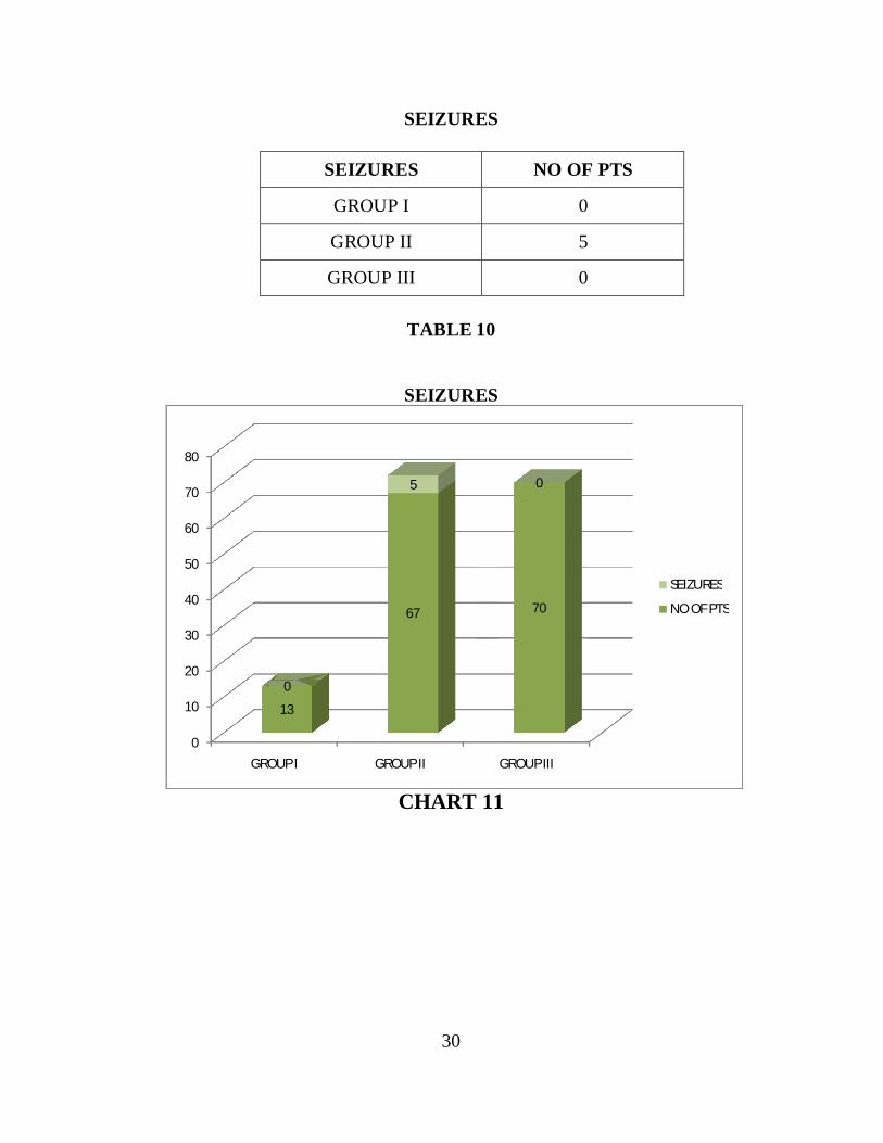
30
SEIZURES
SEIZURES NO OF PTS
GROUP I 0
GROUP II 5
GROUP III 0
TABLE 10
SEIZURES
CHART 11
0
10
20
30
40
50
60
70
80
GROUP I GROUP II GROUP III
13
67 70
0
5 0
SEIZURES
NO OF PTS

31
FRACTURES
FRACTURES NO OF PTSGROUP I 10GROUP II 48GROUP III 39
TABLE 11
CHART 12
DEPRESSED FRACTURES
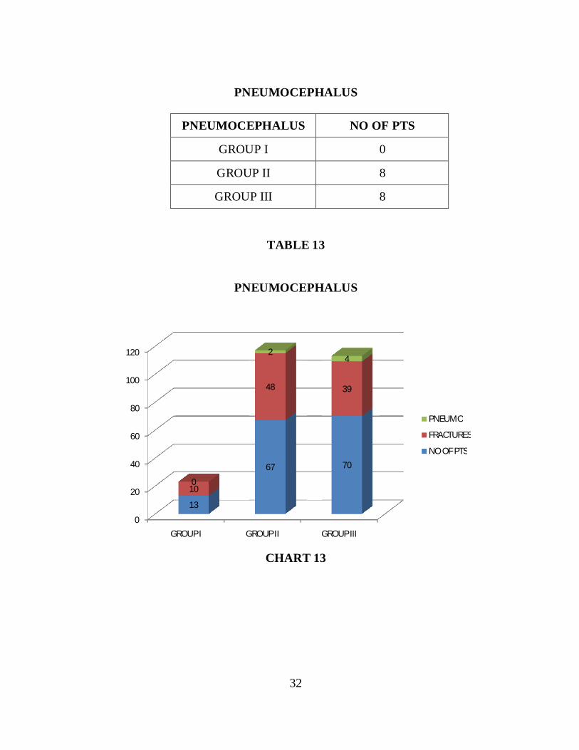
DEPRESSED FRACTURES NO OF PTSGROUP I 0GROUP II 8GROUP III 8
TABLE 12
0
20
40
60
80
100
120
140
GROUP I GROUP II GROUP III
13
67 70
10
48 39
0
88
DEPRESSED
FRACTURES
NO OF PTS
32
PNEUMOCEPHALUS
PNEUMOCEPHALUS NO OF PTS
GROUP I 0
GROUP II 8
GROUP III 8
TABLE 13
PNEUMOCEPHALUS
CHART 13
0
20
40
60
80
100
120
GROUP I GROUP II GROUP III
13
67 70
10
48 39
0
24
PNEUMO
FRACTURES
NO OF PTS

33
EDH
EDH NO OF PTSGROUP I 4GROUP II 10GROUP III 15
TABLE 14
CHART 14
0
10
20
30
40
50
60
70
80
90
GROUP I GROUP II GROUP III
13
67 70
4
1015
EDH
NO OF PTS

34
SDH
SDH NO OF PTS
GROUP I 1
GROUP II 0
GROUP III 3
TABLE 15
CHART 15
0
10
20
30
40
50
60
70
80
GROUP I GROUP II GROUP III
13
67 70
1
03
SDH
NO OF PTS

35
CONTUSION
CONTUSION NO OF PTSGROUP I 2GROUP II 9GROUP III 17
TABLE 16
CHART 16
0
10
20
30
40
50
60
70
80
90
GROUP I GROUP II GROUP III
13
67 70
2
9
17
CONTUSION
NO OF PTS

36
DAI
DAI NO OF PTSGROUP I 0GROUP II 4GROUP III 2
TABLE 17
CHART 17
0
10
20
30
40
50
60
70
80
GROUP I GROUP II GROUP III
13
67 70
0
4 2
DAI
NO OF PTS
37
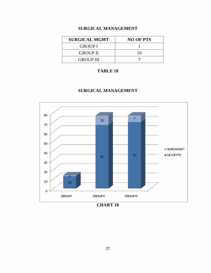
SURGICAL MANAGEMENT
SURGICAL MGMT NO OF PTSGROUP I 1GROUP II 10GROUP III 7
TABLE 18
SURGICAL MANAGEMENT
CHART 18
0
10
20
30
40
50
60
70
80
GROUP I GROUP II GROUP III
13
67 70
1
10 7
SURG MGMT
NO OF PTS

38
CRANIAL NERVE INJURY
SURGICAL MGMT NO OF PTSGROUP I 2GROUP II 8GROUP III 11
TABLE 19
CHART 19
0
10
20
30
40
50
60
70
80
90
GROUP I GROUP II GROUP III
13
67 70
0
01
1
13
1
0
2
0
7
5
VII N
VI N
III N
II N
NO OF PTS

39
GOS
CHART 20
DEATH
DEATH NO OF PTSGROUP I 0GROUP II 1GROUP III 5
TABLE 20
600
4
140
GOS
GOS 1
GOS 2
GOS 3
GOS 4
GOS 5

40
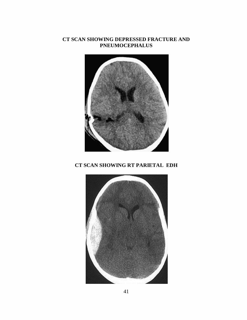
CT SCAN SHOWING DEPRESSED FRACTURE
POST OP PICTURE OF A DEPRESSED FRACTURE
41
CT SCAN SHOWING DEPRESSED FRACTURE ANDPNEUMOCEPHALUS
CT SCAN SHOWING RT PARIETAL EDH

42
CT SCAN SHOWING LT PARIETOOCCIPITAL EDH
CT SCAN SHOWING RT PARIETAL EDH

43
CT SCAN SHOWING INTERHEMISPHERIC BLEED
CT SCAN SHOWING SAH

44
CT SCAN SHOWING RT FTP ACUTE SDH WITH SHIFT
CT SCAN SHOWING TRAUMATIC IVH

45
DISCUSSION
Total number of patients seen from August 2011 to July2012 is 150
patients.
Age and Sex
Total number of male 91Total number of female 59
-----Total 150
-----
Group I (Below 1 year)Male 10Female 3
-----Total 13
-----
Group II (1 - 5 years)Male 35Female 32
-----Total 67
-----
Group III (Above 5 years)Male 46Female 24
-----Total 70

46
AGE VS SEX
Age Male Female Total
Group No % No % No %
Group I 10 76.92 3 23.07 13 8.67
Group II 35 52.24 32 47.76 67 44.67
Group III 46 65.71 24 34.29 70 46.67
It is apparent from this analysis that male children have
outnumbered female in all groups. Overall, Males have outnumbered
female. Male - Female = 1.8: 1. As in the study of RL McLaurin, R.
Towlin and in other Western studies, in this study also, the incidence of
head injury is common with male children, but in group II sex incidence is
similar. It correlates with the Indian equal study of P. Bharthi et al. Male
children tend to be lot more free than female children as they grow older as
reflected by this. Our study shows paediatric head injuries more common
in Group III since the growing children tries to explore things and may
result in frequent falls and RTAs when playing on roadside.
47
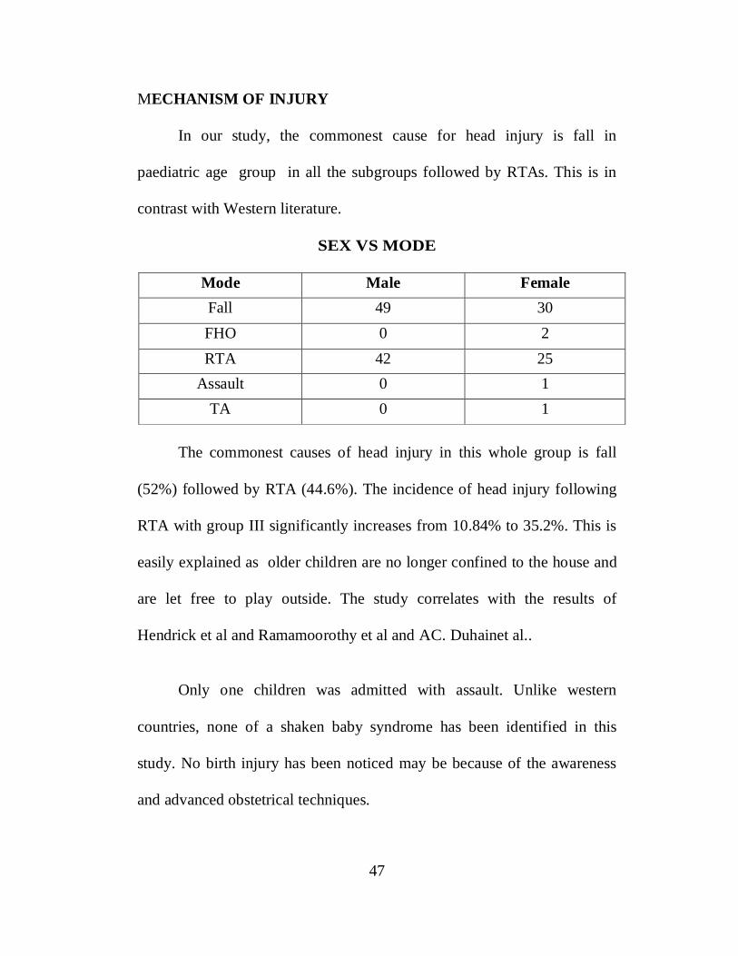
MECHANISM OF INJURY
In our study, the commonest cause for head injury is fall in
paediatric age group in all the subgroups followed by RTAs. This is in
contrast with Western literature.
SEX VS MODE
Mode Male FemaleFall 49 30FHO 0 2RTA 42 25
Assault 0 1TA 0 1
The commonest causes of head injury in this whole group is fall
(52%) followed by RTA (44.6%). The incidence of head injury following
RTA with group III significantly increases from 10.84% to 35.2%. This is
easily explained as older children are no longer confined to the house and
are let free to play outside. The study correlates with the results of
Hendrick et al and Ramamoorothy et al and AC. Duhainet al..
Only one children was admitted with assault. Unlike western
countries, none of a shaken baby syndrome has been identified in this
study. No birth injury has been noticed may be because of the awareness
and advanced obstetrical techniques.
48
Except for the mode of injury "Fall of objects" in all other modes
like, fall, RTA, Domestic accidents, Assault and TA, Male children have
outnumbered female children.
Vomiting
Total number of children presented with vomiting are 62 (41.33%).
Group I - 4 (6.45 %)
Group II - 32 (51.61%)
Group III- 26(41.94%)
Number of operable lesions presented with vomiting was 7. Of this 5
cases are depressed fracture for which wound debridement and evacuation
done and two cases are EDH for which craniotomy and evacuation of EDH
done.
Incidence Post Traumatic vomiting was common in group II
followed by group III children. This study shows though the incidence of
post traumatic vomiting is more common in pediatric head injury, this
symptom correlating with raised ICP resulting in mass effect and surgical
management is very less which is supported by other western literature.

49
ENT Bleed
Total - 14 (9.33%)
Group I - 1 case (7.14%)
Group II - 8 cases (57.14%)
Group III - 5 cases (35.71%)
No one had developed CSF rhinorrhoea in all group. The
commonest findings in these group is frontal bone fracture and naso-
ethmoid complex fracture which is the same in other literature. Three cases
underwent surgery for whom wound debridement and excision of fracture
segment done.
Seizures
Total number of seizures - 5(3.33%)
Group I - 0
Group II - 5
Group III - 0
Seizures are more common in the group II patients.
The types of seizures noted are
Impact seizure - 2cases
Focal seizure - 1 case
GTCS - 2 cases
All the cases are fractures with frontal bone fracture in 4 cases. For
one case surgery was done all cases were started with appropriate anti-
epileptic drugs and patients were seizure free before discharge.

50
AVERAGE HOSPITAL STAY
Group I 4.5 days
Group II 5.7 days
Group III 6.24 days
The duration of stay is lower in Group I and higher in Group III
Cranial nerve deficits
Cranial nerve Injury No of ptsGROUP I 2GROUP II 8GROUP III 11
21 patients out of 150 had cranial nerve involvement (14 %)
2 patients in group I.
8 patients in group II.
11 patients in group III.
No of Pts II N III N VI N VII N
Group I 13 0 1 1 0
Group II 67 0 1 0 7
Group III 70 1 3 2 5

51
The commonest cranial nerves involved in head injury is facial
nerve and the recovery is good in almost all the cases. Secondly the 6th and
3rd nerves are involved only one optic nerve injury is noticed in this group
and there was good outcome. 8th and lower cranial nerves are not involved
in any one of these patients.
CT Findings :
Total no. of patients 613.
CT done for all 613 patients.
Of 613 patients 580 patients admitted with GCS 15 , of these 117 patients
with GCS 15 had some CT findings. Remaining 463 patients with GCS 15
had normal CT finding and were discharged after observation.
Normal CT - 463
Abnormal CT - 150
NUMBER IMPROVED DEATHEDH 14 14 0SDH 04 04 0
CONTUSION 14 14 0DAI 2 2 0
FRACTURES 67 67 0OTHERS 16 16 0
MULTIPLE 34 28 6

52
Study and Mode of Injury
Mode Multiple Fracture EDHSDH OTHERS CONTUSION NormalCT
Fall 13 37 9 4 8 7 6RTA 12 28 4 0 8 7 3TA 1 0 0 0 0 0 0Fall of 0 1 1 0 0 0 0objectsAssaultt 0 1 0 0 0 0 0
Surgery
Out of 150 children, 18 patients had undergone surgical treatment.
The remaining 132 patients were treated conservatively.
18
3
15
SURGERY DONE
TOTAL
EDH
DEPRESSED #

53
Management Group I Group II Group IIIMedical 12 57 63Surgical 1 10 7
OUTCOME:
The outcome in this study are as follows:
Good - 140 93.33%
Moderate disability - 4 2.67%
Severe disability - 0 0%
Vegetative state - 0 0%
Death - 6 4%
Age and Outcome
Age Good MD SD Veg DeadBelow 1 13 0 0 0 0
Below 1-5 66 2 0 0 1Below 6-12 63 2 0 0 5
In this study, the good outcome (90%) in paediatric head injury is
correlating well with the other western studies. The outcome figures are
more or less similar to the study of Michael & Bergers. We have noticed
4% death. This is correlating with the study of Pitts and Dereck D. Bruce
et al. The outcome of moderate disability and severe disability is also

54
correlating with their study. But in their study vegetative states are noticed
2 to 3%. In this study outcome with vegetative state is NIL. Henrick et al.
and Ramamoorthy et al. (1970) have also noticed absence of vegetative
states in their studies.
MODE Vs OUTCOME
Mode Outcome
Good MD SD VS DeathFall 77 1 0 - 1RTA 60 3 0 - 4FHO 2 - - - -
Assault 1 - - - -TA 0 - - - 1
In this study the good outcome from fall of various types is 90.78%
(325/351).
Death due to falls is 1 out of 150= 0.6%
Good outcome from RTA 60 out of 150= 40%
Death due to RTA 12 out of 167 = 2.6%
The ratio of death due to fall and RTA are 1:4
The high percentage of death in RTA may be due to high velocity injuries.
In western studies also, the death rate is more in RTAs comparing with
other modes of injury.
55
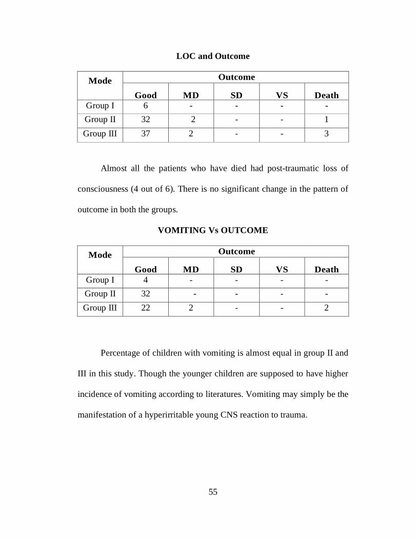
LOC and Outcome
Mode Outcome
Good MD SD VS DeathGroup I 6 - - - -Group II 32 2 - - 1Group III 37 2 - - 3
Almost all the patients who have died had post-traumatic loss of
consciousness (4 out of 6). There is no significant change in the pattern of
outcome in both the groups.
VOMITING Vs OUTCOME
Mode Outcome
Good MD SD VS DeathGroup I 4 - - - -Group II 32 - - - -Group III 22 2 - - 2
Percentage of children with vomiting is almost equal in group II and
III in this study. Though the younger children are supposed to have higher
incidence of vomiting according to literatures. Vomiting may simply be the
manifestation of a hyperirritable young CNS reaction to trauma.

56
SEIZURES Vs OUTCOME
Mode Outcome
Good MD SD VS DeathGroup I 1 - - - -Group II 2 - - - -Group III 2 - - - -
Out of 5 cases of seizures, 5 had good recovery (100%).
HOSPITAL STAY Vs OUTCOME
Group I 4.5 days
Group II 5.7 days
Group III 6.24 days
Good outcome - 10.2 days
Moderate disability - 20 days
Severe disability - -
Vegetative state - -
Dead - 3.65 days

57
GCS / CCS with outcome
Outcome GCS Number of patients
Dead >12 : 0
9-12 : 1
<9 : 5
Vegetative state : 0
Severe : 0
Moderate >12 : 1
9-12 : 2
<9 : 1
Good GCS >12 : 129
9-12 : 11
<9 : 1
No vegetative state has been noticed in this study.
GCS vs OutcomeGCS Dead VS SD MD Good12-15 0 0 0 1 1299-12 1 0 0 2 11
Upto 8 5 0 0 1 1

58
In this study it is observed that higher the GCS or CCS score, better
the prognosis. With GCS, 13 to 15, no patients died out of 130 and there
were 129 good outcome. In GCS 9-12, only 1 patient died out of 14
patients and there were 11 good outcome. In GCS 8 and below 8 out of
7patients5 died and 1 had good outcome 1 patient had moderate disability.
This study proves that higher the GCS or CCS score, better the
prognosis and correlates well with other western literatures.
X-RAY FINDING AND OUTCOME
GCS vs OutcomeCT Dead VS SD MD Good
Contusion - - - - 14EDH - - - - 12DAI 1 - - 1 2
Fractures - - - 1 66
Multiple 5 2 46
Fissure fracture is the commonest pathology and is more common in
and the prognosis was good. The incidence of depressed fracture is almost
the same in both the Groups and the prognosis is good if treated promptly
by surgery.
59
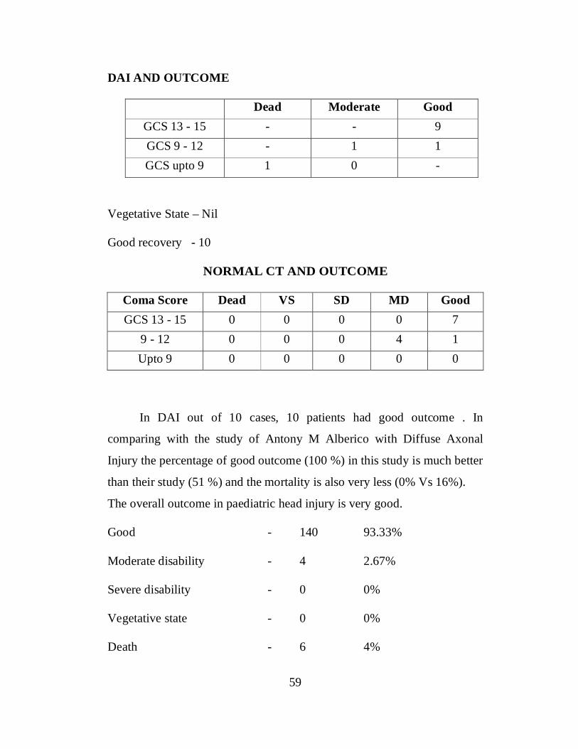
DAI AND OUTCOME
Dead Moderate GoodGCS 13 - 15 - - 9GCS 9 - 12 - 1 1GCS upto 9 1 0 -
Vegetative State – Nil
Good recovery - 10
NORMAL CT AND OUTCOME
Coma Score Dead VS SD MD GoodGCS 13 - 15 0 0 0 0 7
9 - 12 0 0 0 4 1Upto 9 0 0 0 0 0
In DAI out of 10 cases, 10 patients had good outcome . In
comparing with the study of Antony M Alberico with Diffuse Axonal
Injury the percentage of good outcome (100 %) in this study is much better
than their study (51 %) and the mortality is also very less (0% Vs 16%).
The overall outcome in paediatric head injury is very good.
Good - 140 93.33%
Moderate disability - 4 2.67%
Severe disability - 0 0%
Vegetative state - 0 0%
Death - 6 4%

60
93.3 % had good outcome, 2.67% had moderate disability and 0% had
severe disability and there was not a single case of vegetative state. 4 % of
patients died in these series. It is observed that there was poor prognosis in
associated multiple injuries. The mortality rate in the study (4%) is nearly
equal to the study report of Derek A Bruce et a1.(6%)

61
CONCLUSION
Outcome is dependent on GCS score at the time of Therapeutic
Intervention. Higher the score better the prognosis. Parenchymal injuries
like contusions, though not rare, had a good prognosis, seldom requiring
surgery and responding well to conservative treatment itself. Intracranial
haematoma were very rare in children and the prognosis is good. Even in
cases of prolonged unconsciousness no child had vegetative state
indicating the neuronal plasticity in children and the greater ability to
withstand trauma.
Majority of patients with either vomiting or seizures or both had a
normal CT and both had no prognostic value. Multiple injuries carried a
poor prognosis.

62
BIBLIOGRAPHY
1. Paediatric head trauma: influence of age and sex. Berney, J.;
Froidevaux, A. -C.; Favier, J.Child's Nervous System vol. 10 issue 8
November 1994. p. 517 – 523
2. Rivara et al., in low fall like fall from table, bed etc., the common
pathology encountered are concussion brain, fracture skull, ICH.
3. Toft AM, Moller H, Laursen B. The years after an injury: long-term
consequences of injury on self-rated health. J Trauma 2010;69:26–
30.
4. Greenes DS, Schutzman SA. Clinical indicators of intracranial
injury in head-injured infants. Pediatrics.1999;104 :861– 867
5. Durkin et al., in the Journal of Neuro Surgery 1998 Feb, “Article
epidemiology of urban paediatric neurological trauma; evaluation of
and implications for, injury prevention programme”.Durkin et al, in
Journal of Paediatrics June 1999. From “Epidemiology 2 prevention
of traffic injuries to urban children and adolescents”.
6. Myhre MC , Grøgaard JB , Dyb GA , Sandvik L , Nordhov M .
Traumatic head injury in infants and toddlers . Acta Paediatr 2007 ;
96 ( 8 ):1159 – 1163 .
7. Fundavo et al., From clinical nervous system official journal of
international society for paediatric neuro surgery from July 2012
edition.

63
8. Homes et al., from academic emergency medicine: Official journal
of society for academic emergency medicine Sep 2005.
9. Wang MY, Griffith P, Sterling J, et al. A prospective population-
based study of pediatric trauma patients with mild alterations in
consciousness (Glasgow Coma Scale score of 13-14).
10. Scalea TM, Simon HM, Ducan AO, et al. Geriatric blunt multiple
trauma: improved survival with early invasive monitoring. J Trauma
1992;30:129–34
11. Schutzman SA. Clinical indicators of intracranial injury in head-
injured infants. Pediatrics.1999;104 :861– 867
12. Schunk JE, Rodgerson JD, Woodward GA. The utility of head
computed tomographic scanning in pediatric patients with normal
neurologic examination in the emergency department. Pediatr
Emerg Care. 1996;12:160–165
13. Mary E. Aitken, MD; Carla T. Herrerias, MPH; Robert Davis, MD,
MPH; Hanan S. Bell, PhD; John B. Coombs, MD; Lawrence C.
Kleinman, MD, MPH; Charles J. Homer, MD, Minor Head Injury in
Children, Current Management Practices of Pediatricians,
Emergency Physicians, and Family Physician. December 1998, Vol
152, No. 12
14. Rattan, Loudermilk E P, Hartmannsgruber M, Stoltzfus D P,
Langevin P B. A prospective study of the safety of tracheal
extubation using a pediatric airway exchange catheter for patients
64
with a known difficult airway. Chest. 1997;111:1660–1665
15. W R Murshid, Management of minor head injuries: admission
criteria, radiological evaluation and treatment of complications. Acta
Neurochir (Wien). 1998 ;140 (1):56-64 9522909 Cit:17
16. Moran SG, McCarthy MC, Uddin DE, Poelstra RJ. Predictors of
positive CT scans in the trauma patient with minor head injury.
American Surgeon 1994,60(7):533-5
17. Inamasu J, Miyatake S, Suzuki M, et al. Early CT signs in out-of-
hospital cardiac arrest survivors: temporal profile and prognostic
significance. Resuscitation. 2010;81:534– 538.
18. Borzuck et al., from the archives of paediatric adolescent medicine
“Childhood head injuries: accidental or inflected”.
19. Mikhail et al., from school of Health and related research Sheffield
UK “Diagnostic management stagiest for adult and children with
minor head injuries: Systematic review”.
20. Stein et al., Journal Paediatric 2009, “Should head injury child
receive a head injury scar” systemic review of the clinical prediction
rules”.

ABBREVATIONS
TBI - Traumatic Brain InjuryEDH - Extra Dural Haemorrhage
SDH - Sub Dural Haemorrhage
ICH - Intra Cerebral Haemorrhage
IVH - Intra Ventricular Haemorrhage
CNS - Central Nervous System
CTB - Computed Tomography of Brain
RTA - Road Traffic Accidents
MVA - Motor Vehicle Accidents
TTA - Train Traffic Accidents
GCS - Glasgow Coma Scale
GOS - Glasgow Outcome Scale
DAI - Diffuse Axonal Injury

S.No. Name Age Sex Min No Mode of
Injuries
No ofdays inhospital
AdmGCS LOC Vomitin
gENT
bleed Seizures CT. Findings Diagnosis FRACTURE PNEUMOCEPHALUS
DEPFRACTUR
EEDH SDH Contusio
n DAI Management
CranialNerve
Injuries
OtherInjuries GOS
7 Kishore 1 M 21190/11 F 5 15 1 1 0 0 FRA- RT TEMP #Lat.wall of orbit R- Temporal born # 1 0 0 0 0 0 0 C 0 NIL 5
30 Rathinam 1 F 12223 RTA 3 15 1 1 1 1 FRA - RT FRONR-F.B. # withunderlyingHaematoma
1 0 0 1 0 0 0 C 0 Nil 5
76 Kamalesh 1 M 522/12 RTA 1 15 1 0 0 0 N Concussion 0 0 0 0 0 0 0 C 0 Nil 5
60 Ceeyan 1 M 21944 F 3 15 0 0 0 0 FRA - RT FRON R-F.B.# 1 0 0 0 0 0 0 C 0 Nil 5
9 Deepak 2 M 10404 F 5 15 1 1 0 0 N Concussion Brain 0 0 0 0 0 0 0 C 0 Nil 5
18 Satishkumar 2 M 78/11 F 5 15 1 1 0 0 FRA - RT PAR R-Parietal boneFissured # 1 0 0 0 0 0 0 C VI RT Nil 5
28 Rishi Kumar 2 M 6002 RTA 4 15 1 1 0 0 FRA - RT FRON R-F.B.# 1 0 0 0 0 0 0 C 0 Nil 5
70 Asrath 2 M 23703 F 2 15 1 1 0 0 FRA - LT OCCL - Ooccipitalbone # withHaematoma
1 0 0 0 0 0 0 C 0 Nil 5
138 gurupriya 2 F 2334 F 2 15 1 0 0 0 FRA - LT OCC # Occipital bonewith thinn EDH 1 0 0 0 0 0 0 C 0 N 5
141 Kevin 2 M 2556 F 5 15 1 1 0 0 FRA - RT FRON frontal bone # 1 0 0 0 0 0 0 C 0 N 5
147 Dhanalakshmi 1 F 4488 F 2 15 0 0 0 0 CON - RT PAR RT Parietal 0 0 0 0 0 1 0 C 0 N 5
47 Desher 2 F 10771 F 3 15 0 0 0 0 FRA - RT FRON R - F.B. ClosedDep. # 1 0 1 0 0 0 0 C 0 Nil 5
MASTER CHART

54 Nithyasree 2 F 12076 F 3 15 0 0 0 0 MULTIPLE L- P.B. # with thinEDH 1 0 0 1 0 0 0 C 0 Nil 5
20 Dhanalakshmi 2 F 1576/11 F 1 15 0 1 0 0 DAI Fall with DAI withL- L.M.F.Palsy 0 0 0 0 0 0 DAI C 0 NIL 5
14 Absar 2 M 33794/10 RTA 0 15 0 1 0 0 MULTIPLEL-P.Bone # L-T.P.Smallcontusion
1 0 0 0 0 1 0 C 0 Nil 5
25 Ranjini 3 F 4274/11 FHO 2 15 1 0 0 0 FRA - RT FRON R-F.B.Dep # 0 0 1 0 0 0 0 C 0
R - F.Bone #Injury
Roof of R
5
108 Poongavanam 2 F 2333 F 2 15 0 0 0 0 FRA - LT TEMP L - T.B. # - # L -Roof of orbit 1 0 0 0 0 0 0 C 0 Nil 5
114 Jaya 2 F 5336 RTA 13 15 0 0 0 0 FRA - LT TEMP L - T.B. compd.com Dep. # 1 0 0 0 0 0 0
Debridement
excession of #
VII LT Nil 5
57 Deepa 3 F 2270 FHO 15 15 1 0 0 0 EDH- LT FRON L- F.EDH withMass effect 0 0 0 1 0 0 0
BicoronalFlap,
L- Frontalcraniotom
oEvacratio
n EDH
0 Nil 5
11 Deepika 3 F 22701 F 7 15 1 1 0 0 EDH - LTFRONTAL
L - Fraontal EDH #with Mass effect 1 0 0 1 0 0 0
BicoronalFlap,
L- Frontalcraniotom
0 Nil 5
31 Devanish 3 M 8117 F 3 15 1 1 0 0 FRA - OCC Ooccipital bone # 1 0 0 0 0 0 0 C 0 Nil 5
86 Harish 3 M 2206/12 F 8 15 1 0 0 0 FRA - RT PAR R - P. closed DEP# 1 0 0 0 0 0 0
R- P.Brurholeelevation21.02.20
13
VII LT Nil 5
66 Rohith 3 M 6567/11 F 3 15 1 1 1 0 FRA - LT FRON L - F.B. # with #Orbital Bone 1 0 0 0 0 0 0 C 0 Nil 5
119 Priyadarshini 4 F 11023 RTA 11 15 0 1 1 1 FRA - RT PARCCD R - P.CC. Dep. # 1 0 1 0 0 0 0
Debridement
excession of #
0 Nil 5
98 PraveenKumar 3 M 75021 F 3 15 1 0 0 0 FRA - LT FRON L - F.B. # 1 0 0 0 0 0 0 C 0 Nil 5

127 Arthi 3 F 11794 RTA 1 15 1 0 0 0 MULTIPLE BL. IVH. With Diff.Crebral ocedma 0 0 0 0 0 0 0 C 0 N 5
61 Darshini Priya 2 F 9068/11 F 8 10 0 1 0 0 CON - RT TEMPR - T.B.ContusionDAI. Post.Tranmatie seizure
0 0 0 0 0 1 0 C 0 Nil 5
88 Dharshini 3 F 2230 F 6 15 0 1 0 0 Tentoral SAH,IVH Tentoral SAH, IVH 0 0 0 0 0 0 0 C 0 Nil 5
22 Roshini 5 F 2396/11 RTA 8 15 0 0 0 0 FRA - LT PARL-P.Compd.comm.Dep.#
0 0 1 0 0 0 0
WoundDebridem
entelevationof Dep. #Segment
s
0 Nil 5
140 thulasi 3 F 2345 RTA 2 15 1 0 0 0 FRA - RT TEMP Rt temporal bone# 1 0 0 0 0 0 0 C 0 N 5
116 Kavisree 3 M 7651 RTA 13 15 0 1 1 1 FRA - LT TEMPL - T.B.Comp.comm.Elevated #
1 0 0 0 0 0 0 C 0 Nil 5
19 Sevanthi 5 F 1157/11 RTA 8 11 1 0 0 0 FRA - RT PAR R-P.Close.Dep # 0 0 1 0 0 0 0
Debridement
Elevationof Dep. #
0 Nil 4
41 Srikanth 3 M 4931 F 3 15 0 0 0 0 FRA - LT FRON L - F.B. # withPneumocephlus 1 1 0 0 0 0 0 C 0 Nil 5
93 Naveenkumar 4 M 4357 RTA 18 10 1 1 0 0 Tentorial DAI.SAH
Tentorial DAI.SAH 0 0 0 0 0 0 DAI C 0 Nil 5
121 Krithika 4 F 4362 RTA 18 7 1 0 0 0 SAH - LT PAR SAH - LT PAR 0 0 0 0 0 0 0 C 0 Nil 4
52 Dinesh 3 M 11287 RTA 5 15 0 0 0 0 FRA - LT FRON L - F.B. # 1 1 0 0 0 0 0 C 0 Nil 5
110 Dhansingh 4 F 1169 F 3 15 1 1 0 0 EDH- RT FRON R - F.Thin EDH 0 0 0 1 0 0 0 C 0 Nil 5
90 Priyadarshini 3 F 2793 F 8 15 0 0 0 0 FRA - RT FRON
Committed #F.Bone withunderlyingHaematoma
1 0 0 0 0 0 0 C 0 Nil 5
107 Sasikumar 4 M 8077/12 RTA 2 15 1 0 0 0 MULTIPLEResolving R -P.Thin EDH, L -F.B. #
1 0 0 1 0 0 0 C 0 Nil 5
64 Deepa 4 M 15676/11 F 3 15 1 1 0 0 MULTIPLE R - F.B. # with thisEDH 1 0 0 1 0 0 0 C VII RT Nil 5
106 SanjayVignesh 3 M 6199/12 F 5 15 1 0 0 0 MULTIPLE R- T.B. # with L -
F.Contusion 1 0 0 0 0 1 0 C 0 Nil 5
115 Kirankumar 4 M 5506 RTA 7 13 1 1 1 0 FRA - LT TEMPL- T.Bone # with #Body of sphenoid# - R - oribital #
1 0 0 0 0 1 0 C 0 Nil 5
126 Jeevitha 4 F 11009 RTA 1 4 1 0 0 0 MULTIPLER - T.B. Dep. #with B.Stemcontusion
1 0 0 0 0 1 0 C 0 N 1
46 Naresh 5 M 16014 F 19 15 0 1 1 0 FRA - LT PARCCD
L - P.B.Dep.#compd.comm 1 0 1 0 0 0 0
WoundDebridem
entExcessio
0 Nil 5
38 Annakodi 4 F 4679 RTA 11 15 0 1 0 0 MULTIPLE R- F.B. # withEDH 1 0 0 1 0 0 0 C 0 Nil 5
77 Ramesh 4 M 579/12 RTA 2 15 0 1 0 0 FRA - OCC # Occipital bone 1 0 0 0 0 0 0 C 0 Nil 5
118 Naresh 5 M 16014 RTA 19 15 1 0 0 0 FRA - LT FRON L - F.C.C. Dep # 1 0 1 0 0 0 0
Debridement
excession of #
0
Laceration over - LP.Emine
nce
5
148 Harish Kumar 4 M 9876 RTA 1 15 0 1 0 0 FRA - RT FRON Rt Frontal # 1 0 0 0 0 0 0 C 0 N 5
49 Deepak 2 M 10404 F 5 15 0 0 0 0 N Concussion Brain 0 0 0 0 0 0 0 C 0 Nil 5
123 Chithra 4 F 9386 RTA 15 15 1 1 0 0 MULTIPLE L - T.B. # withTempt.contusion 1 0 0 0 0 1 0 C 0 Nil 5
150 ravi 4 M 4556 F 2 15 0 1 0 0 FRA - LT TEMP LT TEMPORAL# 1 0 0 0 0 0 0 C 0 N 5
137 lokesh 4 M 2935 F 2 15 0 0 0 0 FRA - LT TEMP Lt temporal bone# 1 0 0 0 0 0 0 C 0 N 5
139 Thilaggavathy 4 F 4555 F 3 15 0 0 0 0 FRA - LT TEMP Lt temporal bone# 1 0 0 0 0 0 0 C 0 N 5
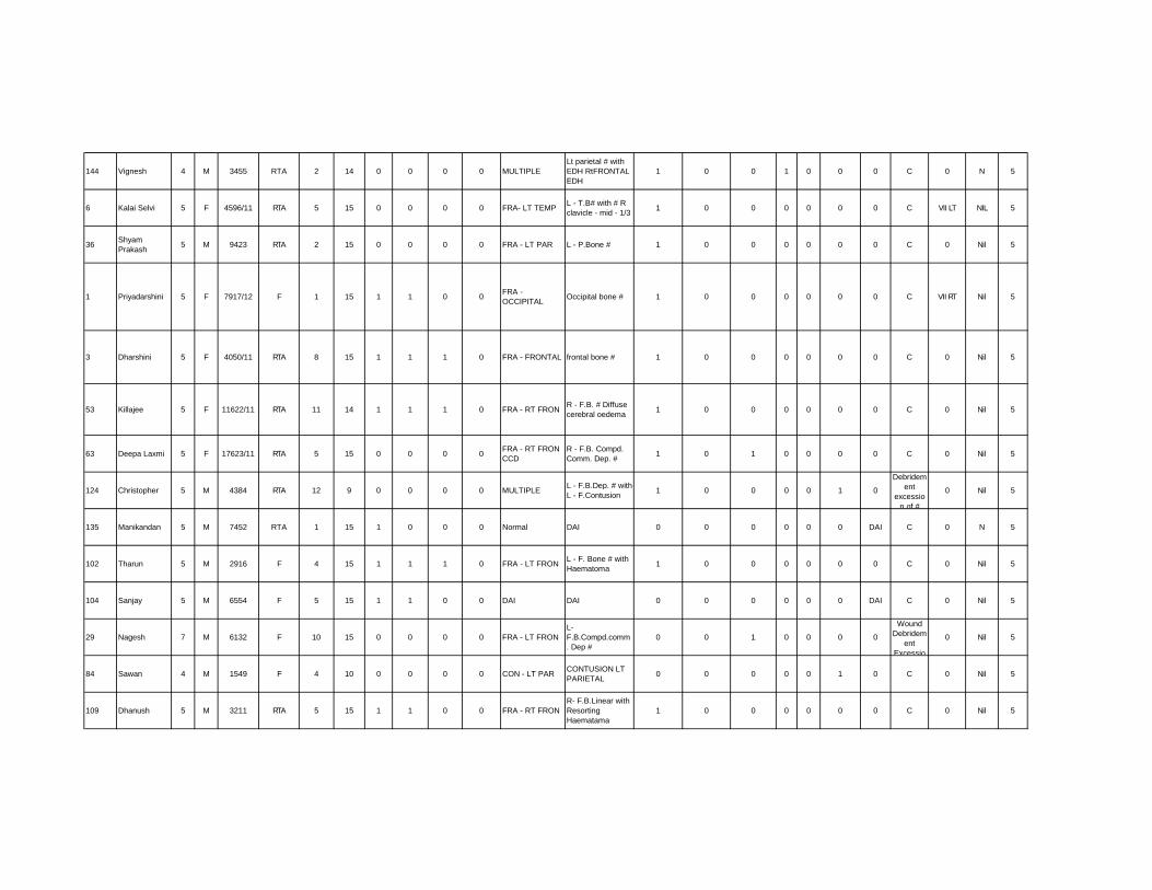
144 Vignesh 4 M 3455 RTA 2 14 0 0 0 0 MULTIPLELt parietal # withEDH RtFRONTALEDH
1 0 0 1 0 0 0 C 0 N 5
6 Kalai Selvi 5 F 4596/11 RTA 5 15 0 0 0 0 FRA- LT TEMP L - T.B# with # Rclavicle - mid - 1/3 1 0 0 0 0 0 0 C VII LT NIL 5
36 ShyamPrakash 5 M 9423 RTA 2 15 0 0 0 0 FRA - LT PAR L - P.Bone # 1 0 0 0 0 0 0 C 0 Nil 5
1 Priyadarshini 5 F 7917/12 F 1 15 1 1 0 0 FRA -OCCIPITAL Occipital bone # 1 0 0 0 0 0 0 C VII RT Nil 5
3 Dharshini 5 F 4050/11 RTA 8 15 1 1 1 0 FRA - FRONTAL frontal bone # 1 0 0 0 0 0 0 C 0 Nil 5
53 Killajee 5 F 11622/11 RTA 11 14 1 1 1 0 FRA - RT FRON R - F.B. # Diffusecerebral oedema 1 0 0 0 0 0 0 C 0 Nil 5
63 Deepa Laxmi 5 F 17623/11 RTA 5 15 0 0 0 0 FRA - RT FRONCCD
R - F.B. Compd.Comm. Dep. # 1 0 1 0 0 0 0 C 0 Nil 5
124 Christopher 5 M 4384 RTA 12 9 0 0 0 0 MULTIPLE L - F.B.Dep. # withL - F.Contusion 1 0 0 0 0 1 0
Debridement
excession of #
0 Nil 5
135 Manikandan 5 M 7452 RTA 1 15 1 0 0 0 Normal DAI 0 0 0 0 0 0 DAI C 0 N 5
102 Tharun 5 M 2916 F 4 15 1 1 1 0 FRA - LT FRON L - F. Bone # withHaematoma 1 0 0 0 0 0 0 C 0 Nil 5
104 Sanjay 5 M 6554 F 5 15 1 1 0 0 DAI DAI 0 0 0 0 0 0 DAI C 0 Nil 5
29 Nagesh 7 M 6132 F 10 15 0 0 0 0 FRA - LT FRONL-F.B.Compd.comm. Dep #
0 0 1 0 0 0 0
WoundDebridem
entExcessio
0 Nil 5
84 Sawan 4 M 1549 F 4 10 0 0 0 0 CON - LT PAR CONTUSION LTPARIETAL 0 0 0 0 0 1 0 C 0 Nil 5
109 Dhanush 5 M 3211 RTA 5 15 1 1 0 0 FRA - RT FRONR- F.B.Linear withResortingHaematama
1 0 0 0 0 0 0 C 0 Nil 5

2 DeepikaPushpalatha 6 F 2272/12 F 9 15 1 0 0 0 N DAI 0 0 0 0 0 0 DAI C 0 Nil 5
145 Saliya Begum 5 F 2O94 RTA 2 15 1 1 0 0 MULTIPLE occipital bone # pfEDH 1 0 0 1 0 0 0 C 0 N 5
81 Shalini 5 F 1544/12 F 1 12 0 1 0 0 FRA - RT TEMP R - Bone # 1 0 0 0 0 0 0 C VII LTR-Claica
# R-Scapila #
5
71 Tharun 1 M 23456 RTA 5 15 0 0 0 0 N DAI 0 0 0 0 0 0 0 C 0 No 5
92 Vadeeswari 5 F 7819 F 6 15 0 0 0 0 FRA - RT FRON R - F.T. Bone # 1 0 0 0 0 0 0 C VII LT Nil 5
149 Yuvasri 5 F 5679 F 3 15 1 0 0 0 CON - LT FRON LT FRONTALCONTUSION 0 0 0 0 0 1 0 C 0 N 5
4 Sanjay 6 M 4108/11 F 5 11 0 0 0 0 EDH - LTFRONTAL
L - F.Bone linear #with small EDH 1 0 0 0 0 0 0 C 0 Nil 5
134 KARTHIKA 5 M 1123 RTA 1 9 0 0 0 0 EDH - LTFRONTAL
EDH - LTFRONTAL 0 0 0 1 0 0 0 C 0 N 5
33 DeepakKumar 7 M 9006 F 21 15 1 0 0 0 SDH - RT FTP R-F.P thin SDH 0 0 0 0 1 0 0 C VII RT Nil 5
100 Md Jakiria 6 M 6474 F 2 15 1 1 0 0 Concussion brain Concussion brain 0 0 0 0 0 0 0 C 0 Nil 5
80 Ramya Sree 6 F 1347/12 RTA 1 15 0 0 0 0 MULTIPLE Bi.P.Bone # withunderlying EDH 1 0 0 1 0 0 0 C 0 Nil 5
142 chandana 6 F 7888 F 3 15 0 0 0 0 EDH - LTFRONTAL Lt frontal EDH 1 0 0 1 0 0 0 C 0 N 5
67 Nandhini 7 F 22073 RTA 6 15 0 0 0 0 FRA - LT PAR L - P.B. Comp.com. Dep. # 1 0 0 0 0 0 0
#Elevation
done0
#CLAVICL
E5

83 DepikaPushpalatha 6 F 2273/11 F 9 11 0 1 0 0 N DAI 0 0 0 0 0 0 0 C 0 Nil 5
10 Nisha 7 F 22271 F 10 15 1 0 0 0 MULTIPLE
L - Fraontal bone# pneumocephalus withCSF Rhinorrhea
1 1 0 0 0 0 0 C 0 Nil 5
78 Raja 7 M 871/12 RTA 1 15 0 0 0 0 MULTIPLE R - Frontal ICH R -F.EDH 0 0 0 1 0 0 0 C 0 Nil 5
39 Raja 8 M 9197/11 RTA 8 15 1 1 1 0 FRA - LT PARCCD
L - P.Compd.Comm.Dep # 0 0 1 0 0 0 0
WoundDebridem
entExcessio
0 Nil 5
68 Nisha 7 F 22271 F 10 9 1 0 0 0 FRA - LT FRON L - F.B. # withPneumocephlus 1 1 0 0 0 0 0 C 0 NIL 5
48 Kalaiselvi 7 F 9107 RTA 4 15 0 1 0 0 FRA - LT TEMP L - T.B. # 1 0 0 0 0 0 0 C 0 Nil 5
120 Diwakar 7 M 3290 RTA 12 13 1 0 0 0 CON - RT FRON R - F.Ich. 0 0 0 0 0 0 0 C 0 Nil 5
50 Ajay 7 M 10868 RTA 3 15 0 0 0 0 CON - LTPERISYLVIAN
L - Peri sylviancontusion 0 0 0 0 0 1 0 C 0 Nil 5
111 PraveenKumar 7 M 5741 F 8 15 1 0 0 0 FRA - LT PAR
CCDL - P.Compd com.Dep. # 1 0 0 0 0 0 0 C 0 Nil 5
130 Balaji 7 M 15321 F 4 15 1 1 0 0 FRA - RT FRON undisplaced R -F.Bone # 1 0 0 0 0 0 0 C 0 N 5
79 Harini 7 F 251/12 RTA 3 13 1 1 0 0 MULTIPLER- orbital bone #with Haemoohagiccontusion
1 0 0 0 0 1 0 C III RT
1. # Shaf -L
Human,2. # L -
5
122 Kannan 7 M 1770 F 8 13 1 0 0 0 FRA - LT PAR # P.O. Bone with# Haematoma 1 0 0 0 0 0 0 C 0 Nil 5
12 Deepakumar 7 M 9006 F 5 13 1 1 0 0 EDH - RTPARIETAL
R-P-Occipital thinEDH - Midline shift 0 0 0 1 0 0 0 C 0 Nil 5
43 DhaxshnaMoorthi 8 M 8425 RTA 11 15 0 0 0 0 FRA - LT PAR L - P.B.Dep.# 1 0 1 0 0 0 0 C VII RT Nil 5

51 Nivedha 9 F 11252 F 5 15 0 1 0 0 SDH - LT TEMP L - T.B. THIN SDH 0 0 0 0 1 0 0 C VI RT Nil 5
35 Anitha 9 F 9124/11 A 5 15 1 1 0 0 FRA - RT OCC R - OCCIPITALDep.# 0 0 1 0 0 0 0 C 0 Nil 5
23 Kesavan 9 M 3521/11 RTA 4 15 1 1 0 0 N Concussion 0 0 0 0 0 0 0 C 0 Nil 5
87 Jothika 8 F 3005/12 F 3 11 1 0 0 0 CON- LTTEMPPAR L - T.P. Contusion 0 0 0 0 0 1 0 C 0 Nil 5
96 Kesavan 8 M 5934 RTA 4 15 1 0 0 0 MULTIPLEOoccipital bone #with cerelellarHaemorrhage
1 0 0 0 0 1 0 C 0 Nil 5
65 Dinesh 8 M 19454 RTA 5 15 0 0 0 0 FRA - LT TEMP L - T.B. # 1 0 0 0 0 0 0 C 0 Nil 5
75 Gokul 8 M 77/12 RTA 1 15 0 0 0 0 FRA - RT FRONCCD
R - F.B. Compd.Comm. Dep. # 1 0 0 0 0 0 0
BicoronalFlap,
wounddebridde
0 Nil 5
62 Keertha 8 F 11040 F 13 15 1 0 0 0 FRA - LT PAR L - P.B. Fissured # 1 0 0 0 0 0 0 C 0 Nil 5
143 chandra 7 F 5678 RTA 2 15 1 0 0 0 EDH - LT TEMP LTemporal polarEDH 0 0 0 1 0 0 0 C 0 N 5
13 Shameer 9 M 3379 RTA 6 15 1 0 0 0 CON-RT FP R-F.T.P.gangccap contusion 0 0 0 0 0 1 0 C 0 Nil 5
105 Sivasakti 6 M 6422 F 3 15 0 0 0 0 SAH - LT PAR L - High ParietalSAH 0 0 0 0 0 0 0 C 0 Nil 5
32 Aravinth 10 M 5677 F 1 15 0 0 0 0 FRA - RT PAR R- P.Bone closedDep.# 0 0 1 0 0 0 0 C 0 Nil 5
91 Ja 8 F 3873 F 4 15 1 0 0 0 FRA - LT TEMP L - T.Bone # 1 0 0 0 0 0 0 C 0 Nil 5
27 Santhia 9 F 4584 F 3 15 1 0 0 0 MULTIPLE R-T.B.# with B1Fronta contusion 1 0 0 0 0 1 0 C 0 0 5
103 Sidharth 7 M 1302 RTA 3 15 0 0 0 0 EDH - LTFRONTAL L - F.thin EDH 0 0 0 1 0 0 0 C 0 Nil 5
95 RaheshKumar 9 M 5827 RTA 6 8 1 0 0 0 CON - RT TEMP
PAR P.T.Concussion 0 0 0 0 0 1 0 C VI LT Nil 5

101 Hari Prasad 9 M 4322 F 12 15 0 0 0 0 MULTIPLE R - F.B. # withEDH 1 0 0 1 0 0 0 C 0 Nil 5
42 Kalaivani 9 F 6341/11 RTA 5 15 1 1 1 0 FRA - RT FRON R - F.B. Fissured# 1 0 0 0 0 0 0 C 0
EthmaxB. #
5
89 Arun 9 M 2884 F 7 15 1 1 0 0 FRA - OCC Ooccipital bone #committd 1 0 0 0 0 0 0 C 0 Nil 5
82 Jesima 9 F 4012 F 1 15 1 0 0 0 EDH - RTPARIETAL R - P.Thint EDH 0 0 0 1 0 0 0 C 0 N 5
99 Keerthana 10 F 7908/12 RTA 5 14 0 0 0 0 CON - RT FRON R - Frontalcontusion 0 0 0 0 0 1 0 C VII LT Nil 5
85 Nithish Kumar 10 M 2205 F 5 15 0 1 0 0 MULTIPLE
L - F.P. ClosedDEP. # withunderlying thisSDH
1 0 0 1 0 1 0 C 0 Nil 5
24 Selvan 10 M 4126 RTA 4 15 1 1 1 1 FRA - RT TEMPR-T.B. # with R -VII CN UMN-PALSY
1 0 0 0 0 0 0 C VII RT 0 5
26 Asriah 10 M 8265 F 7 15 1 1 1 0 FRA - LT FRONL - F.B.Compd.comm.Dep #
0 0 1 0 0 0 0
WoundDebridem
entelevationof Dep. #
BoneSegment
0 0 5
97 Babu 10 M 6189 F 7 15 0 1 1 1 FRA - RT FRON R - F.B. # with thinHamemorrhage 1 0 0 1 0 0 0 C 0 Nil 5
125 Jallaya 10 F 7016 TA 28 4 0 1 0 0 MULTIPLER - T.B. Dep. #with L -F.Contusion
1 0 1 0 0 1 0 C 0 N 1
129 Thiruvikraman 10 M 7557 RTA 4 5 1 0 0 0 DAI Diff. C. Oedemaclaviece # 0 0 0 0 0 0 0 C 0
R-Clavicle #
1
131 Baby 10 F 11086 RTA 1 5 0 1 0 0 MULTIPLEL - F.T.P. MultipleHaemavagiccontusion
1 0 0 0 0 1 0 C 0 Nil 1
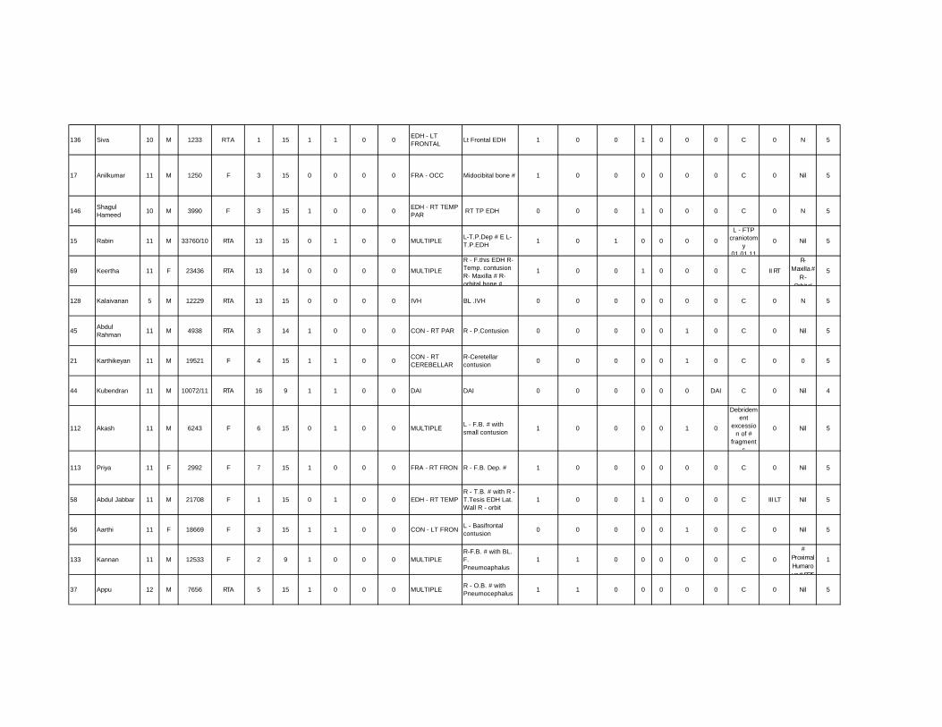
136 Siva 10 M 1233 RTA 1 15 1 1 0 0 EDH - LTFRONTAL Lt Frontal EDH 1 0 0 1 0 0 0 C 0 N 5
17 Anilkumar 11 M 1250 F 3 15 0 0 0 0 FRA - OCC Midocibital bone # 1 0 0 0 0 0 0 C 0 Nil 5
146 ShagulHameed 10 M 3990 F 3 15 1 0 0 0 EDH - RT TEMP
PAR RT TP EDH 0 0 0 1 0 0 0 C 0 N 5
15 Rabin 11 M 33760/10 RTA 13 15 0 1 0 0 MULTIPLE L-T.P.Dep # E L-T.P.EDH 1 0 1 0 0 0 0
L - FTPcraniotom
y01.01.11
0 Nil 5
69 Keertha 11 F 23436 RTA 13 14 0 0 0 0 MULTIPLE
R - F.this EDH R-Temp. contusionR- Maxilla # R-orbital bone #
1 0 0 1 0 0 0 C II RT
R-Maxilla #
R -Orbital
5
128 Kalaivanan 5 M 12229 RTA 13 15 0 0 0 0 IVH BL .IVH 0 0 0 0 0 0 0 C 0 N 5
45 AbdulRahman 11 M 4938 RTA 3 14 1 0 0 0 CON - RT PAR R - P.Contusion 0 0 0 0 0 1 0 C 0 Nil 5
21 Karthikeyan 11 M 19521 F 4 15 1 1 0 0 CON - RTCEREBELLAR
R-Ceretellarcontusion 0 0 0 0 0 1 0 C 0 0 5
44 Kubendran 11 M 10072/11 RTA 16 9 1 1 0 0 DAI DAI 0 0 0 0 0 0 DAI C 0 Nil 4
112 Akash 11 M 6243 F 6 15 0 1 0 0 MULTIPLE L - F.B. # withsmall contusion 1 0 0 0 0 1 0
Debridement
excession of #
fragments
0 Nil 5
113 Priya 11 F 2992 F 7 15 1 0 0 0 FRA - RT FRON R - F.B. Dep. # 1 0 0 0 0 0 0 C 0 Nil 5
58 Abdul Jabbar 11 M 21708 F 1 15 0 1 0 0 EDH - RT TEMPR - T.B. # with R -T.Tesis EDH Lat.Wall R - orbit
1 0 0 1 0 0 0 C III LT Nil 5
56 Aarthi 11 F 18669 F 3 15 1 1 0 0 CON - LT FRON L - Basifrontalcontusion 0 0 0 0 0 1 0 C 0 Nil 5
133 Kannan 11 M 12533 F 2 9 1 0 0 0 MULTIPLER-F.B. # with BL.F.Pneumoaphalus
1 1 0 0 0 0 0 C 0
#ProximalHumarous # SOF
1
37 Appu 12 M 7656 RTA 5 15 1 0 0 0 MULTIPLE R - O.B. # withPneumocephalus 1 1 0 0 0 0 0 C 0 Nil 5

40 SadamHussain 12 M 4943/11 F 8 11 1 0 0 0 MULTIPLE
R - Squmons -T.B. # R-OC.B. # L - P.T.B.#
1 0 0 0 0 0 0 C 0 Nil 5
94 Suresh 12 M 4563 RTA 6 15 0 0 0 0 CON - RTPERISYLVIAN
R - Peri Sylincontusion 0 0 0 0 0 1 0 C 0 Nil 5
73 Nizamudeen 12 M 32/12 F 5 15 1 0 0 0 EDH - RT OCC P.O. area with minEDH 1 0 0 1 0 0 0 C 0 Nil 5
132 Chithan 12 M 13034 RTA 12 7 1 0 0 0 MULTIPLE
R - T.B. # R-Sphoid Bone #Diffuse .C.Oedema
1 0 0 0 0 0 0 C VII LT Nil 1
117 Kavi Raj11
MONTH
M 3687 RTA 5 15 0 0 0 0 FRA - LT TEMP # L - T.B. 1 0 0 0 0 0 0 C 0 Nil 5
59 Arun 12 M 21900 RTA 11 14 1 0 0 0 MULTIPLEL - F.Contusionwith # L - 2ygomacomplex
1 0 0 0 0 1 0 C III LT 0 5
72 Satishkumar3
MONTH
M 21222 F 7 15 1 0 0 0 SDH - LT PAR L - P.O. This SDH 1 0 0 0 1 0 0 C 0 Nil 5
16 Nihhar 5MON F 355/11 F 5 15 1 0 0 0 FRA - RT TEMP R-T.Bone # 1 0 0 0 0 0 0 C 0 Nil 5
55 Asis 10MON M 18526 F 16 15 0 0 0 0 MULTIPLE R- P.B. # with thin
EDH 1 0 0 1 0 0 0 C III RT Nil 5
74 Manikandan5
MONTH
M 31.06.2012 F 1 15 0 1 0 0 FRA - RT FRON
CCDR - F.compd.Comm. DEP # 1 0 0 0 0 0 0
WoundDebridem
entExcessio
n of
VI RT Nil 5
5 Dilipkumar 12 M 4198/11 F 8 12 1 1 0 0 SDH - RT FTP R-FTP acute SDH 0 0 0 0 1 0 0 C 0 Nil 4
34 Nithish 8MON M 10899 F 1 15 1 1 0 0 MULTIPLE R - P.Bone with #
Haematoma 1 0 0 1 0 0 0 C 0 Nil 5
8 Nithish8
MONTH
M 10899/11 F 5 15 0 0 0 0 MULTIPLE
R - Parietal #R-P. #Haematoma R-P.contusion
1 0 0 1 0 1 0 C 0 Nil 5
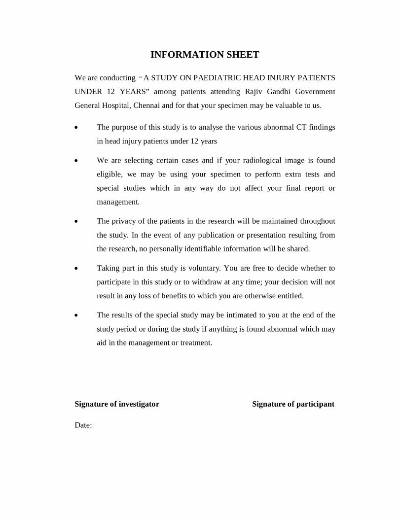
INFORMATION SHEET
We are conducting “ A STUDY ON PAEDIATRIC HEAD INJURY PATIENTS
UNDER 12 YEARS” among patients attending Rajiv Gandhi Government
General Hospital, Chennai and for that your specimen may be valuable to us.
The purpose of this study is to analyse the various abnormal CT findings
in head injury patients under 12 years
We are selecting certain cases and if your radiological image is found
eligible, we may be using your specimen to perform extra tests and
special studies which in any way do not affect your final report or
management.
The privacy of the patients in the research will be maintained throughout
the study. In the event of any publication or presentation resulting from
the research, no personally identifiable information will be shared.
Taking part in this study is voluntary. You are free to decide whether to
participate in this study or to withdraw at any time; your decision will not
result in any loss of benefits to which you are otherwise entitled.
The results of the special study may be intimated to you at the end of the
study period or during the study if anything is found abnormal which may
aid in the management or treatment.
Signature of investigator Signature of participant
Date:


INFORMED CONSENT FORM
Title of the study : “ A STUDY ON PAEDIATRIC HEAD INJURY PATIENTSUNDER 12 YEARS”Name of the Participant: Dr. M.A. BoseName of the Principal (Co-Investigator): Prof. Dr. DeiveeganName of the Institution: Institute of Neurology, MadrasMedicalCollege andRajivGandhiGovernment GeneralHospital, ChennaiName and address of the sponsor / agency (ies) (if any):None.Documentation of the informed consentI _____________________________ have read the information in this form (orit has been read to me). I was free to ask any questions and they have beenanswered. I am over 18 years of age and, exercising my free power of choice,hereby give my consent to be included as a participant in “ A STUDY ONPAEDIATRIC HEAD INJURY PATIENTS UNDER 12 YEARS”1. I have read and understood this consent form and the information
provided to me.2. I have had the consent document explained to me.3. I have been explained about the nature of the study.4. I have been explained about my rights and responsibilities by the
investigator.5. I have been informed the investigator of all the treatments I am taking or
have taken in the past ________ months including any native (alternative)treatment.
6. I have been advised about the risks associated with my participation inthis study.*
7. I agree to cooperate with the investigator and I will inform him/herimmediately if I suffer unusual symptoms. *
8. I have not participated in any research study within the past________month(s). *
9. I have not donated blood within the past _______ months—Add if thestudy involves extensive blood sampling. *
10. I am aware of the fact that I can opt out of the study at any time withouthaving to give any reason and this will not affect my future treatment inthis hospital. *
11. I am also aware that the investigator may terminate my participation inthe study at any time, for any reason, without my consent. *
12. I hereby give permission to the investigators to release the informationobtained from me as result of participation in this study to the sponsors,regulatory authorities, Govt. agencies, and IEC. I understand that they arepublicly presented.
13. I have understand that my identity will be kept confidential if my data arepublicly presented
14. I have had my questions answered to my satisfaction.

15. I have decided to be in the research study.I am aware that if I have any question during this study, I should contact
the investigator. By signing this consent form I attest that the information givenin this document has been clearly explained to me and understood by me, I willbe given a copy of this consent document.
For adult participants:Name and signature / thumb impression of the participant (or legalrepresentativeif participant incompetent)Name _________________________ Signature_________________Date________________Name and Signature of impartial witness (required for illiterate patients):Name _________________________ Signature_________________Date________________Address and contact number of the impartial witness:Name and Signature of the investigator or his representative obtaining consent:Name _________________________ Signature_________________Date________________For Children being enrolled in research:Whether child’s assent was asked: Yes / No (Tick one)[If the answer to be above question is yes, write the following phrase:You agree with the manner in which assent was asked for from your child andgiven by your child. You agree to have your child take part in this study].[If answer to be above question No, give reason (s) :___________________________________.Although your child did not or could not give his or her assent, you agree to yourchild’s participation in this study.Name and Signature of / thumb impression of the participant’s parent(s) (or legalrepresentative)
Name _________________________ Signature_________________
Date________________
Name _________________________ Signature_________________
Date________________
Name and Signature of impartial witness (required for parents of participant
child illiterate):
Name _________________________ Signature_________________
Date________________

