a spider and other arachnids from the devonian of new york
TRANSCRIPT

A S P I D E R A N D O T H E R A R A C H N I D S FROM T H E D E V O N I A N OF NEW Y O R K , A N D
R E I N T E R P R E T A T I O N S OF D E V O N I A N A R A N E A E
by P A U L A . S E L D E N , W I L L I A M A . S H E A R and P A T R I C I A M . B O N A M O
ABSTRACT. The oldest known spider, from the Devonian (Givelian) of Gilboa. New York, is Atlercopus fimbriunguis (Shear. Sclden and Rolfc). parts of which were originally described as a trigonotarbid. possibly of the genus Gelasinotarbus. Previous reports of Devonian spider fossils, from the Lower Emsian of Alken-an-der-Moscl. Germany, and the Pragian of Rhynie. Scotland, are shown to be erroneous identifications. Atlercopus is placed as sister-laxon to all living spiders, on the basis of characters of the spinneret and the arrangement of the patella-tibia joint of the walking legs. A cladogram of the relationships of all pulmonate arachnids is presented. A pulmonate arachnid from Gilboa. related to Arancae and Amblypygi. is described as Ecchosis pulchribolhrium Selden and Shear, gen. ct sp. nov., and additional arachnid material is described.
A D E V O N I A N age for the oldest k n o w n fossil spider was set by Hirst when he described Palaeocieniza crassipes Hirs t , 1923. from the Pragian Rhyn ie Cher t of Aberdeenshire , Scot land. T h e descr ipt ion of a n o t h e r fossil assigned to the Araneae , Archaeometal devonica S tormer , 1976, from the Emsian of Alkcn-an-der -Mose l . G e r m a n y , added more evidence for the ant iqui ty of the order . T h e find of a spider spinneret (Shear. Pa lmer el al. 1989) from the Givetian of G i lboa . N e w York , provided conclusive evidence for the validity of the Devonian as the earliest per iod in which spider fossils a rc known to occur . In this paper , results of a re-examinat ion of the Rhynie and Aiken spider fossils a r e presen ted : the fossils a re not spiders, and arc reinterpreted as a p robab le juvenile t r igonota rb id and an inde te rmina te fossil, respectively. T h e Gi lboa spider is placed in a new genus . Atlercopus. described here. T h e new genus includes only the animal previously called Gelasinolarbus? fimbriunguis (Shear el al. 1987). which we now regard as the only k n o w n Devonian spider, and the oldest k n o w n fossil o f the Araneae . In addi t ion , podomeres originally placed in Arachn ida inceriae sedis by Shear el al. (1987) arc rcdescribed here, with the add i t ion of new material , as Ecchosis pulchribolhrium gen. ct sp. nov. , and placed in Pu lmona ta inceriae sedis (it may be an amblypygid) . and o ther a rachnid remains from Gi lboa are described.
R H Y N I E PALAEOCTEN/ZA
In 1923. Hirst described Palaeocieniza crassipes a s a spider from the Pragian Rhynie Cher t of Scot land . J ames Locke and W.A.S . carried ou t a detailed pho tog raph ic study o f the specimen (British M u s e u m (Na tu ra l His tory) ( B M ( N H ) ) In 24670) in 1987 and 1988. T h e fossil is in a small ch ip of chert m o u n t e d on a microscope slide. Even if the fossil were lo be removed from the slide, n o addi t iona l views could be ob ta ined , owing to the opaci ty of the chert behind the specimen. T h e specimen itself is highly three-dimensional , as are many o f the a r t h ropod remains from Rhynie , and thus difficult to pho tog raph . Add ing to the prob lems are the cloudiness of the matr ix , o p a q u e inclusions, and the very small size of the specimen, a b o u t 0-85 m m long.
In add i t ion to p h o t o g r a p h s of the whole specimen at low magnifications (Text-fig. I), a series of a b o u t thirty-five optical sections was made at higher magnificat ion, using the very shallow depth-of-ficld character is t ic of N o m a r s k i Differential Interference Con t r a s t ( N D I C - see below. Methods). These p h o t o g r a p h s were pr in ted at a large size and each was carefully examined for evidence of
IP.Iacontologv. Vol. 34. P .r t 2, 1991. pp. 241-281 . 7 pls.| O Tt» Palaeontologies! Association

P A L A E O N T O L O G Y . V O L U M E 3 4
TEXT-FIG. 1. Pataeocteniza crassipes Hirst, 1 9 2 3 . A. B, two views, at different planes of focus, of the holotype (and only known) specimen (RM(NH) In 2 4 6 7 0 ) , seen from the left side, anterior to the left, x 130.
spider a u t a p o m o r p h i c s . In add i t ion , each pho tog raph was traced seriatim on a graphics pad and the resul tant digitized images were stacked and reconsti tuted as a ro ta tab le virtual solid using the Jandel c o m p u t e r p rog ram P C 3 D ™ (see below. Methods). We had hoped that James Locke 's efforts to reconst ruct the specimen using this p r o g r a m would a l low us t o examine further detai ls , bu t this was not t o be. T h e level o f resolution a t t a inab le was too low. and there were cons iderable difficulties in digitizing the images, since shallow as the dcpth-of-ficld was. at the necessary magnifications subjective judgement was still required as to wha t was in the p lane of focus and what was not, result ing in further blurr ing of the lines. A careful examina t ion of the specimen itself a n d of the serial p h o t o g r a p h s proved to give the most information.
T h e general condi t ion of the specimen, much crumpled and folded, suggests that it may be a moul t . Hirst (1923) noticed a small , thin, scarcely visible object dorsal to the a b d o m e n , which he supposed to be the detached ca rapace . Since the ca rapace detaches when a rachn ids moul t , if this identification is correct , its presence and posi t ion arc further evidence for the specimen being a cast exoskeleton. T h e p r o s o m a is a lmos t entirely concealed behind the dorsal ly flexed legs and palps. While the pa lps a p p e a r to be comple te , all of the legs on the left side of the specimen (facing the viewer) lack their dis ta l por t ions . T h e a b d o m e n is complexly crushed and folded.
Hirst (1923) provided a detailed drawing, which, however, incorpora tes some er rors . The p r o p o r t i o n s of the right p a l p are not correct in compar i son with the left, to which a segment has been added . In ' r e s t o r i n g ' the loose piece of cuticle to its supposed posi t ion as carapace , the

S E L D E N ET AL.: D E V O N I A N A R A C H N I D S 243
mass of wrinkles and folds above Ihe leg coxae (perhaps the t rue carapace) has been omit ted , and some of the folds in this s t ruc ture a p p e a r to have been confused with par t s of the palps. The second or third left leg has the tibia omi t ted . In the region of the supposed a b d o m e n . Hirst noted that what had been made in the d rawing to resemble spinnerets might be folds of cuticle. Th is is definitely s o : the a p p a r e n t internal s t ruc tures of the a b d o m e n are also cut icular folds on the right side of the specimen, seen th rough the left side.
In a t t emp t ing to de te rmine the affinity of this fossil, a process of el iminat ion was followed. T h e general appea rance and s t ructure of the body (a p r o s o m a with five pairs of leg-like appendages , a n d an a b d o m e n a t tached by a nar rowed por t ion) establishes that it is an a rachnid , and that it may belong to the k n o w n orders Araneae . Amblypygi , Uropygi . Schizomida. or T r igono ta rb ida . T h e presence of leg-like (not raptor ia l ) palps rules ou t Amblypygi . Uropygi . and Schizomida . at least a s they are presently known .
Devon ian t r igono ta rb ids differ from potential ly c o n t e m p o r a n e o u s spiders in a number of ways . While bo th g roups m a y have segmented a b d o m e n s , t r igono ta rb ids have three tergal plates per segment and lack spinnere ts . T h e eyes of any c o n t e m p o r a n e o u s spiders were likely to have been g rouped on a centrally located tubercle, as in the modern mcsothele spiders, while those of Devonian pa laeochar in id t r igono ta rb ids are dispersed in three g r o u p s : a median g r o u p of two. and t w o lateral g roups which may consist of several minor and major lenses each (Shear el al. 1987). All the Devonian t r igonotarb ids we have examined have a simple b icondylar hinge joint between the patella a n d tibia, and spiders have a m o n o c o n d y l a r rocking jo in t in this posi t ion.
Close examina t ion of the a b d o m e n of the specimen failed to reveal any evidence for o r against segmenta t ion (despite the clear segmental lines in his i l lustration, Hirst (1923, p . 460) w r o t e : " . . . i t is impossible to be qui te certain whether this [the a b d o m e n ] is segmented o r not."). T h u s the number o f tergites that might be present for each segment canno t be ascer ta ined. T h e "sp innere t s ' have a l ready been al luded t o ; a s Hirst inferred, this is in fact a fold of the abdomina l cuticle that can be traced cont inuous ly until it merges with o ther folds o f the s t ructure . T h e entire a b d o m e n was also carefully examined for spinnerets , because we suspected that it might have been twisted th rough 180°, and because in living mesothele spiders the spinnerets are located a b o u t in the middle of the ventral surface o f the a b d o m e n , which is supposedly their primitive posi t ion. W e found no indicat ion whatsoever of sp innere ts .
Careful focusing revealed that a m o n g the crushed mass of the p r o s o m a was an object that resembles an eye tubercle and seems to bea r al least two hemispherical lens-like pro t rus ions . Unfor tuna te ly this evidence is inconclusive, because at least two eye lenses would be present on a median tubercle both in t r igonotarb ids a n d spiders. T h e complicated folding and dis tor t ion of the ca rapace and its concealment behind the legs m a d e it impossible for us to find any indication of lateral eye g roups .
T h e patella tibia ar t iculat ion can be seen on just one o f the legs, p robab ly the left third leg. It m a y be possible to m a k e ou t two dorsal ly si tuated ar t icular condyles on the distal end of the patella, but at the level of magnification required to see them, the optical proper t ies of the chert interfere significantly.
In s u m m a r y , the fossil carr ies none of the au t apomorph ie s of spiders that could be seen on a specimen of this size and level o f preservat ion, but its identity as a t r igonotarbid is only suggested (by the possible pat tern of patella tibia ar t icula t ion) . It should be pointed ou t . however , tha t scores of t r igonotarb ids have been seen in the Rhynie chert , and that this specimen is the only one for which a spider identity has been suggested. O u r hypothesis is that Palaeocieniza crassipes Hirst is a moul ted exoskeleton from an early instar t r igonotarb id .
A I . K E N ARCH A F.O MET A
O n e of only four fossil sites with Devonian terrestrial animals . Alken-an-der -Mose l . G e r m a n y , has yielded impression fossils of lower Emsian age. including t r igonotarb ids . scorpions , eurypter ids . and a r th rop leur ids (S lormer 1976; Brauckmann 1987). One fossil from this deposi t . Archaeomeia.'
>

244 P A L A E O N T O L O G Y . V O L U M E 3 4
devonica S to rmcr . 1976. was identified as a spider (Stormer 1976). A policy against type-specimen loans at the Scnckenberg M u s e u m , which houses this specimen, meant that we were unable to examine the original . However , wc were able to s tudy a plaster cast, and the p h o t o g r a p h a n d d rawing publ ished by Stormcr . T h e specimen consists of an e longa te b lob with a few transverse lines at one end a n d a vaguely indicated region at the o ther which may be par t of some plant remains (S tormer 1976. figs 48 and 4 9 ; pi. 5. fig. la,b). S to rmcr indicated that he had before him Pet runkcvi tch ' s d rawing of Archaeomeia nephilina Pocock, 1911. from the U p p e r Carbon i fe rous of Britain. This d rawing (Pctrunkevi tch 1949. fig. 159) shows a featureless ca rapace with seven legs radia t ing from it. and an e longate a b d o m e n with two longitudinal lines and four o r live terminal segments .
There are two similar specimens o f A. nephilina in the British M u s e u m ( N a t u r a l History) which were examined in 1986 by W.A.S . , a n d subsequent ly by P .A.S. Specimen In 15863 is the m o r e c o m p l e t e a n d w a s t h e specimen figured by Pct runkevi tch . It is relatively poor ly preserved a n d little can be a d d e d to the d i a g r a m m a t i c i l lustration and brief descr ipt ion. Specimen In 31259. the ho lo typc . docs not show the t ransverse ' s e g m e n t a l ' lines seen in In 15863. The cuticle is luberculate a n d the a b d o m e n bears longi tudinal folds: neither of these features are found in c o n t e m p o r a n e o u s spider fossils (e.g. Eocteniza silivicola, figured on Pocock ' s pi. II. fig. 4) . but a rc more reminiscent of o ther Carbon i fe rous a rachnid groups . There are o ther details visible on this specimen which would reward a detailed rcstudy. Nevertheless , there arc n o features which would distinguish cither o f these specimens as a spider ra ther than any o ther a rachn id .
In any case, the resemblance of Archaeomeia? devonica to these two specimens is vague and probably coincidental . There seems to be n o reason to consider Archaeomeia'?devonica a s a spider o r a fossil a rachn id o f any sor t .
T H E G I L B O A A R A C H N I D S
Early r epor t s on the Gi lboa fauna (Shear el al. 1984) raised the possibility of spiders being a m o n g the an imals present . T h e tip of an a rachnid walking leg tarsus was illustrated, and diagnosed as being from a spider largely on the basis of serrate ventral setae similar to the s i lk-handling accessory claws found in some living arancoid spiders. However , in later studies, the possibility of spiders being present receded as it became clear that ano the r related g r o u p o f a rachn ids , the Tr igono ta rb ida . domina t ed the fauna. W e were also unable to demons t r a t e conclusively in the fossils any a u t a p o m o r p h i c s of spiders . Shear el al. (1987). in a detailed s tudy of the t r igono ta rb ids . assigned all pu lmona le a rachnid fossils from Gi lboa to this extinct order , which was placed as the plesiomorphic sister g r o u p to the o ther p u l m o n a l e orders . One animal represented only by legs was assigned with some doub t to the t r igonotarb id genus Gelasinotarbus, and given ihe species epithet fimbriunguis. This name referred to ihe characteris t ic claws, set with ventral culicular fimbriae, not found in any o ther t r igonotarb ids . O the r characters in these legs, present but undetected in 1987. we now recognize as conclusive evidence of a spider. A single femur with a patch of acute spinules near its base was called Arachn ida Incertae scdis B : its cuticle is similar to that of fimbriunguis, a n d o the r similar femora have now been found in direct connect ion with pieces o f undoub ted fimbriunguis. A third g r o u p o f spec imens , consis t ing o f p o d o m e r e s a n d cul icular f ragments , w a s referred t o Arachn ida Incertae sedis A. Re-examinat ion of these specimens and of new material with the same dist inctive cuticle has produced evidence that they belong to a p u l m o n a t e arachnid , close to Amblypygi and Araneae . T o complicate mat te r s further, the ta rsus illustrated as a possible spider in Shear el al. (1984. fig. I B) is undoubted ly t r igono ta rb id : it has s m o o t h claws and lacks a tarsal o rgan .
Late in 1988. conclusive evidence for spiders finally turned u p in the Gi lboa mater ia l : a spinneret (Shear . Palmer el al. 1989). This discovery triggered a search for o ther possible spider par t s , and it was s o o n realized that the spinneret belonged with the legs described in 1987 as Gelasinotarbus? fimbriunguis. In addi t ion , some previously unassigned chelicerae and some pieces of ca rapace belong to this an imal .

S E L D E N F.T Al..: D E V O N I A N A R A C H N I D S 245
T h e ' c lasp-kni fe ' form of the chilecera. places it in the P u l m o n a t a ( = Arachn idea sensu van dcr H a m m e n 1977; m a d e u p of the o rders T r igono ta rb ida . Uropygi . Schizomida . Amblypygi . and Araneae ) . I l lustrated here for compar i son are chelicerae of the uropygid Masiigoprocius gigaiueus (PI. 7. fig. 5) . and the amblypygid Heterophrynus elaphus (PI. 7. fig. 6) . and see Shea r el al. (1987. figs 7. 67. 68) for p h o t o g r a p h s of t r igonotarb id chelicerae. A n u m b e r of characters unequivocal ly place the chelicera in A r a n e a e (see discussion under PHYLOGENETIC RELATIONSHIPS). A chcliccral g land , found only in spiders, is present . T h e chcliccral fang o f A. fimbriunguis lacks setae, which are present in all o ther pu lmona tcs . In all o the r o rders of Pu lmona ta , the largest cheliceral teeth are al the end o f the too th row oppos ing the tip of the fang (subchelatc condi t ion) , while in A.
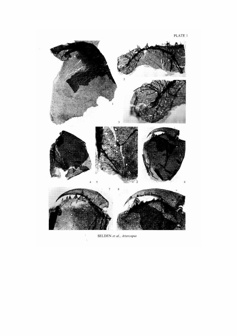
fimbriunguis. a s in the vasl majori ty of spiders , the largest teeth occur par t -way a long the row a n d nearer to the fang ar t iculat ion than to the fang lip (the subchelale condi t ion occurs in a small n u m b e r of spiders , but the described a r rangement is found only in spiders, a m o n g the pu lmona tes ) . On the basis of o u t g r o u p compar i son with, for example , scorpions, the subchelate state is primitive. T h u s there arc three definite spider synapomorph ie s present in the chelicera. A significant a p o m o r p h y of spiders is the presence of cheliceral venom glands. Whilst the evidence is not entirely cer ta in , in at least two specimens of A. fimbriunguis chelicerae there may be a sublerminal venom pore nea r the fang tip (PI. 1. fig. 7). In add i t ion , as discussed in the detailed descript ions, the ar t icu la t ions present m a k e it clear that the A. fimbriunguis chelicera must have been o r t h o g n a t h .
T h e legs of A. fimbriunguis bea r numerous lyriform o r g a n s : only in spiders are lyriform organs found on podomeres o the r than the meta tars i .
T h e pieces of ca rapace are referred to A. fimbriunguis on the basis of their similarity of cul icular pa t t e rn ing .
T h e evidence that the spinneret , chelicera. legs, and ca rapace fragments all come from the same morphospec ics is overwhelming. All the chelicerae are identical, except for some size differences, and all o f t h e p o d o m e r e types ( t rochanter , femur, etc.) a re identical within each type. All specimens, including the spinneret a n d ca r apace fragments, have the same distinctive cut icular o r n a m e n t a t i o n , a pat tern which a p p e a r s in no o ther Gi lboa specimens except those that can be unequivocal ly assigned to the spider on the g rounds given above . Finally, the chelicerae and basal leg p o d o m o r e s occur in o rgan ic connec t ion on a n u m b e r of slides. Therefore these Gi lboa specimens are considered to belong to the s a m e species. Altercopusfimbriunguis.
There are n u m e r o u s fragments of cuticle a m o n g the Gi lboa slides which resemble the cuticle of A. fimbriunguis at first sight, and which we at first thought could belong to the body of the spider. Some of these were figured by Shear el al. (1987) and referred to as Arachn ida Incertae scdis A. This an imal is character ized by: generally large size: scale-like o rnamen t ra ther than re t icula t ion; setal sockets which range from small to very large; striated macrose tae and thick, str iated, bifid spines (PI. 7. figs 4 a n d 8) : g roups o f slit sensilla and lyriform o r g a n s : o rnamen ted t r ichobolhr ia l base on the patel la . Minu te , c. 0 0 0 5 m m . circular o rgans occur on the cuticle surface and appear , at low magnif icat ion, similar to the characteris t ic little slit sensilla of Attercopus. but examina t ion at higher magnif icat ions reveals a circular hole ra ther than a central slit, so they are not the same o rgan . N o n e of these minute pores bears a seta, a n d their function is u n k n o w n : nevertheless, the difference in m o r p h o l o g y from the little slit o rgans of Attercopus gives a useful criterion for dist inguishing the t w o cuticle types. New informat ion on Arachn ida Incertae scdis A has been discovered dur ing the present s tudy, and the an imal is named Ecchosis pulchribothrium gen. et sp. nov. , below. T h e presence o f lyriform o rgans suggests that E. pulchribothrium could be a spider, but the distinctive o rnamen ted t r ichobolhr ia l socket on the patella is puzzling. Virtually identical t r ichobolhr ia l sockets are found on the living amblypygid Helerophrynus elaphus (PI. 7. fig. 2). but this an imal has a qt ite different leg ar t icula t ion pat tern to that in E. pulchribothrium, and a lyriform o rgan only on the meta ta r sus . T h e identity of E. pulchribothrium thus remains unclear, bul we suggest that it is either an abe r r an t amblypygid o r a m e m b e r o f an extinct, undiagnosed arachnid order .
i

246 P A L A E O N T O L O G Y . V O L U M E 3 4
G E O L O G I C A L S E T T I N G
Stratigraphy T h e fossils occur in a grey shale in the upper part of the Panther M o u n t a i n F o r m a t i o n at a locality on Brown M o u n t a i n , Gi lboa . Schohar ie C o . . New York (7J' quad rang le sheet 6168 IV N W 1945. a p p r o x . 271272 m N by 142951 m E ; Banks et al. 1985). Fur the r locality details can be found in Banks et al. (1972). T h e original site has now been destroyed to m a k e way for a pump-s to rage power p lant associated with Schohar ie Reservoir , but much of the fossil-bearing shale was removed to the D e p a r t m e n t of Biology, State Universi ty of New York at B inghamton , for later processing. T h e Pan the r M o u n t a i n Fo rma t ion is par t of the Hami l ton G r o u p , upper Middle Devonian Erian Series, and is equivalent to the middle Givc t ian of E u r o p e .
Palaeoecology Detai led discussion of the t a p h o n o m y and palaeoecology o f the biota is given in Shear (1986). Shear et al. (1987) and Shear and B o n a m o (1988). The Gi lboa li thology is a da rk grey muds tone . T h e fauna occurs in close associat ion with ma t s of inter locking spiny s tems of the lycopod Leclercqia. Cons ide ra t ion of the m a n n e r of preservat ion of the p lan ts suggested to Banks et al. (1985) that they were buried in situ by low-energy flood waters . Shear et al. (1984) suggested that the animals , which were living at the site or may have been carried in by the flow, c a m e to rest by the localized reduct ion of velocity created by the mesh of Leclercqia. the ' n a t u r a l sieve" effect would exclude large pieces of a r t h r o p o d cuticle, while the most minute particles could have passed th rough .
Almos t all the a r t h r o p o d s recovered from the Gi lboa site were undoub ted ly terrestrial . T h e only exception to this is the occurrence o f curypter id fragments . In the Devon ian , these an imals lived in both mar ine and freshwater aqua t ic habi ta t s , and some were amphib ious (Selden 1984. 1985). so their presence in the Gi lboa muds tones is not problemat ica l . In addi t ion to the external evidence o f sedimentology and associated land flora for the habi ta t of the a r t h r o p o d s , palaeophysiology provides further proof of their terrcstriali ty (Selden and Je ram 1989). Tr ichobothr ia are line hairs sensitive to high-frequency vibrat ions, and could only function in air. They occur on the Gi lboa p u l m o n a t e s Gelasinotarhus bonamoae. G. bifidus (Shear el al. 1987, figs 105-120), and Ecchosis pulchribolhrium (see below), a n d the pseudoscorp ion (Shear. Schawaller and B o n a m o 1989). Book-lungs for air b rea th ing occur in the t r igono ta rb ids of Gi lboa (Shear et al. 1987). Whi le we have no evidence of t r i chobothr ia o r book- lungs in the Gi lboa spider Atlercopus. all living spiders are terrestrial apar t from the secondari ly aqua t ic Argyroneta aquatica. found in fresh waters of Europe , and the l i t toral, southern hemisphere Des idae . T h e phylogenetic discussion (below) indicates that if Atlercopus were aqua t i c , it would a lso have been secondari ly so, since all o ther P u l m o n a t a are primari ly terrestrial .
M A T E R I A L A N D M E T H O D S
Preservation T h e animal fossils are preserved as minute , undist inguished, b rown to black flakes, which are unrecognizable as an imals when in the rock and under incident light microscopy, but t ransmit ted light reveals their zoological na ture . T h e cuticle appears brown in t ransmi t ted light, and the depth of co loura t ion is directly correlated with the thickness of the cuticle (o r the n u m b e r of layers of cuticle super imposed in the specimen). T h e chemical composi t ion of the cuticle is not k n o w n ; the b rown co loura t ion suggests it is organic , but the reduct ion of much of the plant mater ial in the same beds to c a r b o n indicates the likelihood that the a r t h r o p o d cuticle has also been altered, p robab ly by rcpolymerizat ion of the organic molecules, dur ing diagenesis. T h e a r t h r o p o d s are s trongly compressed , necessitating the use of special techniques , such as N D I C , to separate over lapping layers of cuticle. F o r the s a m e reason, scanning electron microscopy ( S E M ) is virtually useless for the s tudy of these fossils, revealing only surface features: bo th original s t ructures and diagenctic effects.
i
S E L D E N ET AL.: D E V O N I A N A R A C H N I D S 247
T h e fossils are f ragmentary ; only rarely are p o d o m e r e s and o ther par t s found in organic connec t ion with o thers . However , the occurrence of such specimens is vital for the correct identification o f loose podomeres and reconstruct ion of the an imals . T h e dea r th of pieces of ca rapace and a b d o m e n of the a rachnids can be explained by the fact tha t podomeres have two surfaces, so that when compressed together they remain coherent and are less likely to fragment than the body par t s which consist of a single sheet of cuticle. T h e ca rapace and a b d o m e n cuticle is represented by the m a n y ' s c raps" which occur on the slides. T h e nearly complete t r igonotarb id ca rapaces and a b d o m e n s described by Shear el al. (1987) are rare, a n d mostly consist of both left and right (o r dorsa l a n d ventral) surfaces compressed together.
Fur the r discussion of the preservat ion o f the Gi lboa fauna is given in Shear el al. (1987).
Methods T h e specimens were recovered from the rock matr ix by digestion in concent ra ted hydrofluoric and hydrochlor ic acids (see Shear el al. 1987; Shear and B o n a m o 1988, for detai ls) . After washing in distilled water , the an imal fossils were separated from the a b u n d a n t plant fragments, a s far a s possible, and m o u n t e d in C M C or Clearcol on plain microscope slides. The p repara t ion was done in the l abora to ry of P .M.B. in B inghamton . and the prepared slides were then sent to H a m p d e n -Sydney for s tudy by P .A.S. and W.A.S .
T h e slides were studied using an Olympus Vanox II biological microscope with a N o m a r s k i Differential Interference Cont ras t ( N D I C ) facility. Th is i l lumination is part icularly useful at high magnificat ion a n d for the optical separat ion of closely adpressed layers of cuticle. Use was made of an O l y m p u s S Z H s tereomicroscope for low magnification work , part icularly on compara t ive extant ma te r i a l ; for p h o t o g r a p h y , this was cleared of muscles by soaking overnight in a solut ion of po tass ium hydroxide . C a m e r a lucida a t t a chmen t s to bo th microscopes facilitated accura te d rawing of the specimens, and p h o t o g r a p h s were taken on 35 mm K o d a k Technical Pan film at ASA 50 with O l y m p u s P M 1 0 camera s m o u n t e d on these ins t ruments . On plates and text-figures, unless stated otherwise , all p h o t o g r a p h s were taken in t ransmi t ted light with N D I C on the Vanox.
T h e c o m p u t e r p rog ram Jandel P C 3 D ™ (available from Jandel Scientific. 2526 Bridgeway. Sausal i to . California 94965. U S A ) was used for the three-dimensional reconstruct ion of Palaeocteniza crassipes, and the p rogram MacClade 2.1 (Maddison and Maddi son 1987) was extremely useful in the phylogenet ic analysis.
Abbrev ia t ions and convent ions used in text-figures are as follows: a. an te r ior , a n t e r o - ; ar. a r t i cu la t ion ; ch. chel icera( l) : cl. c law; co cx, costa coxal is ; cu. cut ic le; Cx. c o x a ; d. d o r s a l : d i . d i s t a l ; e. edge ; f, fold: Fe , f emur ; gl, g l and : i. inferior, infero-; m, ar throdia l m e m b r a n e : m a . marg ina l ; me . m e d i a n ; ms . mac rose l a ; M l . m e t a t a r s u s ; p . poster ior , pos te ro- ; pa sp . palpal spinules ; Pa. pa te l la : pd . pa i red : p o . poison duct open ing ; pr . p r o x i m a l ; ps. p r o s o m a : r. r idge : s. superior , s u p e r o - ; sc. sclerile; si, slit sensil la; sr, se r ra ted : st. s t e rnum, su. surface; l b , t r ichobothr ia l ba se : T a . t a r sus ; ta or, tarsal o r g a n ; T i . t ibia; T r . t rochan te r ; tv, t ransverse; v. vent ra l ; X. ar tefact .
Unless stated otherwise in the legend to camera lucida d rawings : dashed lines show linear features showing th rough cuticle from beh ind ; finely do t t ed a reas arc internal surfaces; coarse do t s show ar th rod ia l m e m b r a n e : setal sockets and slit sensilla (where shown) are infilled in black when on surfaces showing th rough from b e h i n d ; p rominen t spores (where shown) are in black.
Repository and authorship T y p e and figured mater ial is deposi ted in the Depar tmen t of Inver tebrates . Amer ican M u s e u m of Na tu ra l His tory. N e w York (numbers prefixed A M N H ) . but are referred to in the text by their slide numbers . Mos t slide n u m b e r s consist of a series number (the first two numbers , e.g. 411.7, or the first only if only two numbers are present , e.g. 329). followed by the number of the slide within the series. T h e last, slide, n u m b e r is prefixed with the letters A R (or Ar) on the slide itself, and quo ted thus in earlier pub l i ca t ions ; these letters are omit ted here for brevity. T h e slide may include m o r e than one specimen, c o m m o n l y of a different a r t h r o p o d , but quo t i ng the slide number makes retrieval of specimens for future s tudy easier, facilitates references to earlier papers on the Gi lboa
l

248 PALAEONTOLOGY. VOLUME 34
Slide No. AMNH No. Illustration Brief description
Atlercopus fimbriunguis 329.1 43162 PI. 3. fig. 4: Text-fig. 6o palpal femur + patella 329.3 43163 PI. 3. fig. 2: Text-fig. 6 B femur 329.3 43163 PI. 4. fig. 1; Text-fig. 7A distal tibia 329.3 43163 PI. 4. fig. 10: Text-fig. 7F metatarsus 329.38 43168 PI. 4. fig. 8 metatarsus 329.39 43098 Text-fig. 12 B patella 329.53 43099 PI. 4. fig. 9 tibia 329.57 43100 Text-fig. 12 F metatarsus 329.58 43101 Shear el al. 1987. fig. 134 HOLOTYPE. metatarsus, tarsus 329.59 43102 PI. 3. fig. 3; Text-fig. 6 c distal femur + patella 329.59 43102 Text-fig, I2c trochanter 329.69 43106 PI. 2. fig. 5; Text-fig. 5E various: femur, patella, tibia 329.69 43106 PI. 6, fig. 5; Text-fig. 9D palpal tarsus 329.70 43107 Text-fig. 12 A PARA TYPE, fcmur + patclla 329.70 43107 Text-fig. I2D, E 2 metatarsi, proximal tarsus 329.16.34 43164 PI. 5. fig. 2 tarsus 329.22.9 43165 PI. 1, fig. 7; Text-fig. 4E chclicera 329.31a.Ml 43166 PI. 3. fig. 7; Text-fig. 6E various; femur + patella 329.31a.M2 43047 PI. 6, fig. 4 legs 334.la.4 43170 PI. 5. figs 1 and 3; Text-figs 8A-C 2 legs, patella to tarsus 334.la.6 43171 PI. 2. fig. 4; Text-fig. 5D femur 334.la.7 43172 PI. 1. figs 6 and 8: Text-fig. 4c chelieera 334.la.8 43173 PI. 4. figs 6; Text-fig. 7E libia 334.la.9 43174 PI. 2, fig. 1; Text-fig. 5A femur 334.1*. 12 43175 PI. 3, fig. 5; Text-fig. 6G distal femur + patclla 334.1*.34 43176 Text-figs 10. and 11 A, B, C spinneret 334.1*.38 43177 PI. 5. fig. 5; Text-fig. 8D tarsus 334.1*.86 43178 PI. 3. fig. 6; Text-fig. 6F femur + patella 41i.02.12M.6 43179 PI. 6. figs 1 and 2: Text-fig. 9A metatarsus + tarsus 411.7.19 43052 PARATYPE, femur 411.7.33 43180 PI. 1. figs 4 and 5: Text-fig. 4D chclicera 411.7.45 43181 PI. 4. fig. 3: Text-fig. 7c distal tibia 411.19.83 43182 PI. 2. fig. 2: Text-fig. 5 B coxa 411.19.98 43183 PI. 4. fig. 7; Text-fig. 7c distal tibia 411.19.102 43184 PI. 2. fig. 7; Text-fig. 5H 3 coxae, 1 trochanter 411.19.243 43185 PI. 3. fig. 8 proximal femur 411.19.248 43186 PI. 4, fig. 5: Text-fig. 7D patella 411.19.250 43187 PI. 2. fig. 8: Text-fig. So coxa 411.19.251 43188 PI. 4, fig. 11 metatarsus 411.20.25 43189 PI. 4. fig. 2: Text-fig. 7 B patella 2002.12.49 43190 PI. 4. fig. 4 tibia 2002.12.79 43191 PI. 3, fig. 1; Text-fig. 6 A femur 2002.12.90 43192 PI. 1. figs 2 and 3; Text-fig. 4 B chcliceral teeth 2002.12.102 43193 PI. 1, fig. 1: Text-fig. 4 A anterior carapace
Ecchosis pulchribolhrium 411.1.33 43194 PI. 7. fig. 1 PARAIYPE. distal femur 411.7.37 43195 PI. 6, fig. 6: Text-fig. 9 B MOI.OTYPE. patella + prox. tibia 411.7.86 43111 Shear el al. 1987. figs 149 and 150 PARATYPE. distal patella 411.19.96 43198 PI. 6, fig. 3; Text-fig. 9 c patella 411.19.137 43169 PI. 7. fig. 4 large, bifid spine 411.19.184 43195 PI. 7. fig. 3 lyriform organ 411.19.188 43196 PI. 7, fig. 8 PARATYPE. probable (ibia 411.19.206 43197 PI. 7. fig. 7 sheet of cuticle 2002.9.13 43097 PI. 2, fig. 3; Text-fig. 5c coxa
Arachnida inceriae sedis 334.la.4 43198 PI. 5. Fig. 3 flagelliform appendage 2002.9.20 43199 PI. 5. Fig. 4 flagelliform appendage
1
TABLE I. List of specimens mentioned in text.

SELDEN ET AL.: DEVONIAN ARACHNIDS 249
fauna in which slide n u m b e r s are used, and locales ihe specimen lo the original rock sample . T h u s it will be possible in the future to collate da ta on the whole Gi lboa b io ta t o a fine degree o f accuracy. T a b l e 1 lists the descr ibed specimens both by their A M N H accession number and the slide number . A comple te list of the microscope slides which bea r fragments of Atlercopus fimbriunguis. Ecchosis pulchribolhrium. a n d Arachn ida inceriae sedis is deposi ted as Supplementa ry Publicat ion N o . S U P 14040. 5 pp . . at the British Library . Boston Spa . Wcthe rby . Yorksh i re LS23 7 B Q . England. Copies o f this can be ob ta ined by writ ing to the British Library a t the above address , enclosing prepaid c o u p o n s avai lable from mos t libraries th roughout the world.
In add i t ion to the fossils, the following material (both males and females, and from the W. A. Shea r Collect ion, unless otherwise s ta ted) of extant a rachnids was studied for compara t ive pu rposes : A r a n e a e : Liphislius sumairanus Thorel l . Sumat ra . Amer ican M u s e u m of Natura l His tory col lec t ion: Amblypyg i : Helerophrynus elaphus Pocock. E c u a d o r ; U r o p y g i : Masligoprocius giganieus (Lucas) . F lo r ida : Sch izomida : species indct. . Mexico .
Fol lowing previous pract ice (Shear el al. 1987). au tho r sh ip o f new taxa is a t t r ibu ted to Selden and Shear . B o n a m o discovered and supervised the prepara t ion of the Gi lboa mater ia l ; Selden and Shear a r e responsible for o t h e r informat ion a n d ideas in this pape r .
R E C O N S T R U C T I O N O F T H E G E N E R A L I Z E D L E G O F ATTERCOPVS
T h e reconstruct ion (Text-fig. 2) reflects a combina t ion of the known morphology of var ious legs, some of which arc suspected to be leg 1 by their close re la t ionship with palpal femora and chclicerae. but for most specimens the leg-to which they belong is not known. T h e reconstruct ion is to be used as a key to in terpre ta t ion of the fossils, and for compara t ive purposes in a general sense. However , it mus t be remembered that no one leg of Atlercopus fimbriunguis looked exactly like this recons t ruc t ion , and in par t icular , the relative p ropor t ions o f the p o d o m e r e s would have varied between legs.
T h e r e arc a n u m b e r of ways in which the or ienta t ion o f p o d o m o r e s can be inferred. Inferior and super ior a r e fairly s t ra igh t forward : compar i son of the ar t iculat ion points with those of living spiders , together with a considerat ion of the way the leg has to work as a functional unit , is normal ly sufficient. Assessing which is an ter ior and which poster ior is less easy. The t rochan te r can be or iented by observing its re la t ionship to the coxa, the or ienta t ion of which is known because of the asymmet ry in the joint and compar i son with extant a rachnids . However , there are n o t rochanters connected to femora which are sufficiently well preserved to enable the following of the or ienta t ion d o w n the leg. Since most jo in t s beyond the coxa are symmetr ical , their morpho logy is of little use in o r ien ta t ion , but there is an asymmetr ical d is t r ibut ion of slit sensilla and lyriform o rgans a round the distal jo in t s o f podomeres . T h e palpal femur bears a patch of spinulcs in an inferior posit ion, to one side of its sagittal p lane . T h e function o f these spinules is not known, but we are assuming tha t , whatever their function (see below), they arc most likely lo occur on the anter ior side of ihe p o d o m e r e . Therefore , the pa lpa l femur can b e or ien ted , a n d since this p o d o m c r c is a t t ached t o a patella, this p o d o m e r e can also, and so on d o w n the leg. A further logical s tep is required in the a s sumpt ion that the apparen t similar dis t r ibut ion of slit sensilla on palpal podomeres a n d on the p o d o m e r e s o f o the r legs reflects a real serial homology. These assumpt ions have only been made in o r d e r to provide an or ien ta t ion for the reconstructed generalized leg. and not for any o ther purpose . Should the or ienta t ion prove to be incorrect , then the references to anter ior and poster ior would simply require reversal.
P H Y L O G E N E T 1 C R E L A T I O N S H I P S O F ATTERCOPUS FIMBR1UNGUS
Cladislic analysis Charac t e r s and charac te r states used in the analysis are listed in T a b l e 2. the da ta matr ix is given in T a b l e 3 . a n d the c l a d o g r a m in Text-figure 3 . T h e tree w a s roo ted by arbi t rar i ly including a n ances to r p lcs iomorphic for all characters .

250 PALAEONTOLOGY. VOLUME 34
TEXT-FIG, 2. A ttercopus fimbriunguis (Shear. Selden and Rolfe. 1987). A. reconstruction of a typical walking leg. posterior aspect. B. diagrammatic representation of walking leg joints, distalmost to the left; each joint is viewed from the distal direction with the anterior to Ihe left, the inner circle represeniing Ihe distal podomere. the outer the proximal podomere; solid circles are articulation points and straight lines are articulation axes, short lines represent slit sensilla. The body-coxa joint is highly diagrammatic; Ihe lower articulation representing Ihe coxoslcmal attachment, the upper triangle representing ihe attachment of the coxa to Ihe prosomal marginal cuticle. The upper coxa-trochantcr articulation is a movable sclerite sel in the arthrodial membrane, which allows rocking. Slit sensilla omitted from coxal distal joint. The trochanter -femur joint is a horizontal pivot. The femur-patella joint is a superior bicondylar hinge, and there is a sclerite embedded in Ihe inferior arthrodial membrane. The patella tibia joint has a superior articulation, but a close connection of the podomeres inferiorly allows the joint to work as a loose vertical pivot. The tibia-metatarsus joint is a superior bicondylar pivot. The metatarsus tarsus joint bears aniero- and poslcrosupcrior articulalions forming a superior bicondylar hinge, bul the joint may be uncoupled on relaxation of Ihe muscles, allowing rocking.
Shear el al. (1987) presented a cladistic analysis based on 23 of the same characters as used here. T h e addi t iona l charac te rs a c c o m m o d a t e the division of the Araneae into Attercopus, Mesolhe lae . M y g a l o m o r p h a c . and A r a n e o m o r p h a e . If a charac te r is not discussed below, the discussion will be found in the 1987 paper . Some of the previously used 23 charac te rs have been re-evaluated; in the following discussion, the charac te r n u m b e r given is from Table 2. and the charac te r number from Shea r el al. (1987) is in bracke ts .
Original characters. Character 8 | 5 | has been recoded. Further investigation of the patella tibia articulation demonstrated lhat the joint in living spiders has an additional specialization, compression zone Y (CZY. see later), not present in Attercopus. Further, while Ihe joint is immobilized (fixed) in Amblypygi. considerable movement is possible at that articulation in legs 2 4 ofUropygi and Schizomida (in leg I the patella and libia are entirely fused without trace of a suture). We do not know if the condition on the more posterior legs of Uropygi and Schizomida represents a reversal or the retention of a primitive condition, bul we decided to code it as a primitive retention on the grounds of parsimony. Character 9 (16| has also been recoded. because an
l

SELDEN ET AL.: DEVONIAN ARACHNIDS 251
TABLE 2. Characters and character states used in the phylogenetic analysis.
Characters Plesiomorphic state Apomorphic state
1. cheliceral segmentation 3-segmented 2-segmented 2. plagula ventralis absent present 3. book-lungs absent present 4. sperm flagcllum 9 + 2 9 + 3 5. segment 7 broad narrowed 6. eggs not protected protected by secretions 7. lateral eyes minor lenses present minor lenses absent 8. Pa-Ti joint bicondylar hinge 1. rocking, no CZY 8. Pa-Ti joint
2. rocking with CZY 3, immovable
9. labium absent present 10. grouped slits/lyriforms absent present 11. tarsal organ absent present 12. cheliceral poison gland absent present 13. silk glands absent present 14. tibial lyriforms absent present 15. cheliceral fang setose naked 16. cheliceral gland absent present 17. male palp unmodified modified 18. abdominal segments visible hidden 19. tartipores absent present 20. sternum broad, unitary reduced, divided 21. palps leg-like raptorial 22. leg 1 leg-like antenniform 23. posterior sucking stomach present absent 24, abdominal flagellum absent present 25. palp coxae free fused 26. postabdomen 2-scgmented 3-scgmcnted 27. abdominal tergites entire divided 28. fimbriae on claws absent present 29. spinules on palpal Fe absent present 30. Ti-Ml organ absent present 31. clavalc trichobothria absent present 32. anterior media spinnerets absent 1, present 32. anterior media spinnerets
2. lost 33. chelicerae orthognath labidognath 34. cleaning brush on palp absent present 35. anal glands absent present 36. male flagellum unmodified modified 37. central nervous system partly in abdomen wholly in prosoma 38. trichobothria present absent
examination of specimens has convinced us that a labium (sternitc of the palpal segment modified as a lower lip) does not in fact occur in Amblypygi, Uropygi. and Schizomida. In amblypygids, a long projection goes forward from the sternitc of the first leg, but could not function as a labium. In uropygids and schizomids. the palpal sternum is an immovable pentagonal sclerite and the ventral wall of the preoral cavity (camerostome) is formed by the fused palpal coxae. Character 5, the narrowing of segment 7, has replaced [18]: presence or absence of a pedicel. We think that the key feature here is the reduction in width of that segment, which occurs to a greater (Araneae, Amblypygi) or lesser (Trionotarbida, Uropygi. Schizomida) degree in all of the taxa involved.

252 PALAEONTOLOGY. VOLUME 34
TABLE 3. Dala matrix used in the phylogenetic analysis. 0 = plesiomorphic stale. 1 = apomorphic state. 2. 3 = alternative apomorphic states. ? = character state uncertain. See text for details.
Characters 12345 i
67890 12345 2
67890 12345 3
67890 12345 (>7S
Trigonotarbida 11171 .'0000 00000 00000 00?00 01000 00000 0?l Atlercopus 111?? ??l ?l urn 1 ??0? 00??0 ??I10 0?00() ll'l Mcsothclae inn 11211 inn 11000 00000 00001 11000 010 Mygalomorphae inn 11211 urn 11110 00000 00000 02000 010 Araneomorphae urn 11211 urn 111 10 00000 00000 01100 010 Amblypygi urn 11301 10000 00001 11000 00000 00010 010 Uropygi urn 11101 10000 00001 urn 10000 00001 000 Schizomida urn 11101 10000 00001 urn 10000 00000 100
New characters. Characters 10 and 14: slit sensilla are unique to chelieerates. Wc have assumed that the primitive arrangement was scattered, single slits on most or all body surfaces, and these still occur in all arachnids. However, the slits, which function as cuticular strain gauges, are found in greater numbers near articulations or points where the cuticle is likely lo be stressed (Barth 1978, 1985). This has led in turn to the formation of loosely organized groups of slits, and thence lo tightly coupled, parallel slits, commonly surrounded by a cuticular border, known as lyriform organs. In true lyriform organs the slit sensilla are ncurally integrated to act as a single organ; this integration is recognized morphologically where the slits arc as close together as their individual widths, and are parallel to each other. They may change in length gradually across Ihe organ, giving the appearance of Ihe arrangement of strings in a lyre or harp. A multiplicity of lyriforms is clearly apomorphic, and in character 14, the presence of lyriforms on the leg tibiae stands in for this increase in their number. In trigonotarbids. we have not detected grouped slits or lyiforms. though large slits occur in greater numbers near the distal ends of podomeres (see Shear ei al. 1987. figs 11. 46. 79-81). Lyriforms occur in amblypygids and uropygids only on the distal ends of the metatarsi of legs 2-4, and are oriented parallel to the long axis of the leg; spiders have this metatarsal lyriform. which is oriented perpendicular to the long axis of ihe leg. as well as many additional lyriforms on other podomeres which are oriented parallel to ihe long axis (Barth 1985; Barth and Stagl 1976; Moro and Bali 1986).
Character 11: typical tarsal organs (Blumenthal 1935; Forster 1980) occur on the walking leg tarsi of all living Pulmonata (Amblypygi and spiders, Forster 1980. and pers. obs.: anlenniform legs of Amblypygi. Foelix el al. 1975 ("pit organ ' ) ; walking legs of Uropygi. pers. obs. and R. Forsler. pers. comm.; walking legs of Schizomida, pers. obs. and R. Forster, pers. comm.). Wc have not detected this organ on the tarsi of trigonotarbids. but il is present in Atlercopus. While similar structures arc found on the tarsi of scorpions and ticks (Foelix and Axlell 1972; Foelix and Schabronaih 1983), ihey appear ullrastruclurally different and their homology has not been established. Thus the presence of Ihe tarsal organ is treated here as a synapomorphy for the orders of Pulmonata excepting Trigonotarbida. though it may later be shown to be more widespread in Arachnida.
Character 15: a naked cheliccral fang is apomorphic by comparison with the setose condition of the palp and walking legs, with which the chclicera is serially homologous. Among the Pulmonata. a naked cheliccral fang is found only in spiders, all other pulmonate orders have a brush of setae on the fang (sec. for example. PI. 7. figs 5 and 6; Shear el al. (1987) figs 7, 67, 68).
Character 16: the cheliccral gland described by Forster and Platnick (1984) has been reported only in spiders; it has been found in all species so far examined from a wide selection of families (R. Forsler. pers. comm.). Raymond Forster (pers. comm.) stated that he has found a series of scattered pores near the midpoinl of the ventral surface of the chclicera in amblypygids, which he considers a cheliccral gland. Using light microscopy (including oil immersion examination of cleared cuticle) wc were not able to confirm these observations, bul a purposeful search for ihe gland may reveal it in orders other than Araneae. In pscudoscorpions. glands also open on the chelicera (Vachon 1966). bul ihey are very distinct in appearance and probably not homologous. We propose the presence of this distinctive gland is yel another aulapomorphy for the order Araneae.
Character 18: in opislhothcle spiders, the segmentation of ihe abdomen is suppressed and is either entirely
l

S E L D E N ET AL.: D E V O N I A N A R A C H N I D S 253
concealed from external view, or revealed only on the maturity of males of a few species of mygalomorphs. and even then only in the anterior pan . This is a synapomorphy for Mygalomorphae and Arancomorphae.
Character 19: tartipores these peculiar structures, like small, collapsed pastries (hence the name), evidently mark the position of spigots on the spinnerets in previous instars (Kovoor 1986; Coddington 1989). They do not occur in Attercopus nor in mesotheles (pcrs. obs. on Liphistius sumatranus and L. inatayanus). The number of spigots on spider spinnerets increases with each inslar: in mesotheles the increase is accomplished by adding more pseudosegments to the spinneret. We consider this mechanism primitive, and the presence of tartipores synapomorphic for mygalomorph and araneomorph spiders.
Character 26: a two-segmented postabdomen is present in trigonotarbids, spiders, and amblypygids. Counting segments shows that uropygids and schi/omids have added a third, basal segment (probably by the narrowing of the segment just in front of the primitive two-segmented postabdomen). which we consider a synapomorphy for that group, correlated with the postanal abdominal flagellum.
Characters 28 and 29: fimbriate claws and palpal femoral spinulcs arc autapomorphies of Attercopus. by outgroup comparison and the criterion of'special structures'.
Characters 30 and 31: a highly specialized organ for detecting deflection of the mctarsus with respect to the tibia is present among spiders only in living mesotheles (Platnick and GolobofT 1985). Likewise, special club-shaped trichobothria (Foclix 1985) are unique to this group (Platnick and GolobofT 1985).
Character 32: by outgroup comparison, the loss of the anterior median spinnerets is autapomorphic for mygalomorph spiders. We might add here that there arc other spinneret and spigot characters that may prove useful for phylogenetic analysis among spiders; some of these have already been described by Coddington (1989) and others arc under study by J. M. Palmer and J. A. Coddington.
Character 3 3 : labidognath chelicerae are found only in araneomorph spiders and arc autapomorphic for that group.
( haractci ' 8 ; the distribution of trichobothria in the -\i .ichnuia ha) been rlJUffllnTftl b) Kacincr 11968), . I I K ! Reissland and Gorner (1985). They arc found in spiders, amblypygids, uropygids, schizomids, palpigradcs. scorpions, pscudoscorpions, and mites, but not in solifuges, ricinuleids, or opilionids. Their occurrence in scorpions and palpigradcs. both considered primitive arachnids, and their general appearance elsewhere argues for considering their absence in any arachnid a loss. We have not found trichobothria in irigonotarhids. nor in Attercopus. Shear el al. (1987) described trichobothria in the supposed trigonotarbid Gelasinotarbus honamoae, bul new studies of this animal have convinced us thai il is not. after all, a trigonotarbid. nor docs it seem lo be a spider. The loss of trichobothria is thus proposed as another autapomorphy for Trigonolarbida.
We are more concerned about the complete lack of trichobothria encountered during our high-magnification studies of well-preserved podomeres of Attercopus. We have found no mention in the literature of spiders without trichobothria, and R. Forstcr and N. Platnick. who have surveyed hundreds of species using SEM. reported that they have found no spiders which lack these sense organs (R. I'orster, pcrs. comm.). Had we noi found tarsal organs and longitudinally oriented lyrilbrmson Attercopus podomorcs. as well as having been able to match their cuticle to that of the isolated spinneret, we would question our assignment of these fossils to Araneae. We must regard the loss of trichobothria in Attercopus as an autapomorphy independent of ihcir loss in trigonotarbids.
Cladogram. Using these 38 characters , we have produced a 36-slep c ladogram (Text-fig. 3) with a consistency index o f 0-97.
In an earlier, prel iminary report on the spinneret of Attercopus fimbriunguis. Shear, Palmer el al. (1989). were able to na r row down the number of possible c l adograms for sp ider sub- and infra-o rders to three, a rgu ing as follows. Recent views of spider evolut ion divide the Orde r Araneae into two suborde r s . Subo rde r Mcsothelae includes a small number of species today restricted to southeas t Asia , Indonesia , and J a p a n : they are uni ted by a n u m b e r of synapomorph ies . including a pecul iar sense o r g a n between the t ib iae a n d meta tars i o f the legs (sec above) . Mesothe les arc bet ter k n o w n to a rachnologis t s for their primitive charac te rs , including an externally segmented abdomen a n d the possession of eight (rarely seven) spinnerets, which are located not at the end of the a b d o m e n , but near the middle of its ventral surface. Suborder Opis tho the lae includes all o ther spiders , in which the n u m b e r of spinnerets has been reduced to six, four, or two and moved to (he pos ter ior end o f the a b d o m e n , which is not externally segmented. With in this g roup . M y g a l o m o r p h a e ( " t a ran tu las ' in the Nor th American sense) have lost all vestiges of ihe anter ior median spinnerets , while A r a n e o m o r p h a e carry a cribellum (repeatedly lost in m a n y lines)
254 PALAEONTOLOGY. VOLUME 34
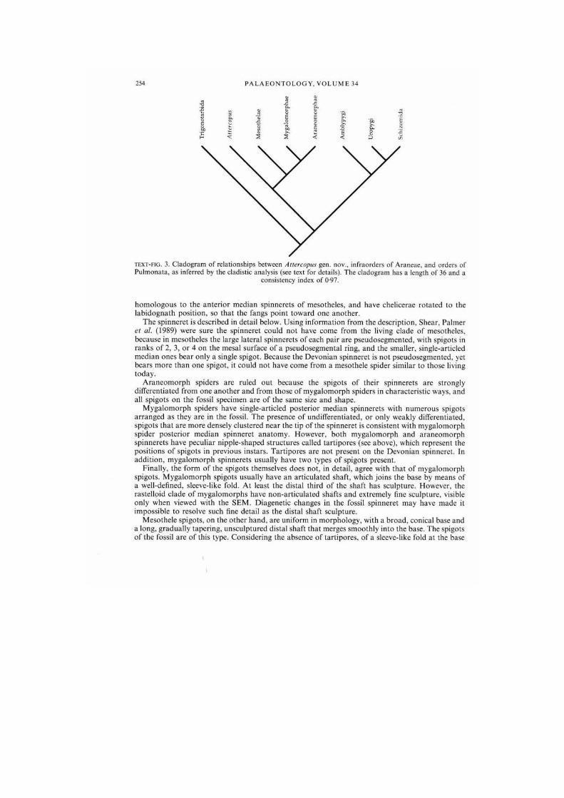
at v 2 1
text - f ig . 3. Cladogram of relationships between Auercopus gen. now, infraordcrs of Araneae. and orders of Pulmonata, as inferred by the cladistic analysis (sec text for details). The cladogram has a length of 36 and a
consistency index of 0-97.
h o m o l o g o u s to the an te r io r median spinnerets of mesotheles. a n d have chelicerae rota ted to the l ab idogna th posi t ion, so that the fangs poin t t oward one ano ther .
T h e spinneret is described in detail below. Using informat ion from the descr ipt ion. Shear. Palmer el al. (1989) were sure the spinneret could not have come from the living c lade of mesotheles. because in mesotheles the large lateral spinnerets of each pair a re pseudosegmcnted . with spigots in r anks o f 2. 3 , o r 4 on the mesal surface of a pseudosegmental ring, and the smaller, single-articled median ones bear only a single spigot . Because the Devonian spinneret is not pseudosegmented. yet bears m o r e than one spigot, it could not have come from a mesothele spider similar to those living today .
A r a n e o m o r p h spiders arc ruled out because the spigots of their spinnerets are strongly differentiated from one ano the r and from those o f myga lomorph spiders in characteris t ic ways, and all spigots on the fossil specimen are of the same size and shape.
M y g a l o m o r p h spiders have single-articled poster ior median spinnerets with n u m e r o u s spigots a r r anged as they are in the fossil. The presence of undifferentiated, o r only weakly differentiated, spigots that are m o r e densely clustered near the tip of the spinneret is consistent with myga lomorph spider poster ior median spinneret ana tomy . However , both m y g a l o m o r p h and a r a n e o m o r p h spinnerets have peculiar nipple-shaped s t ructures called lar l ipores (see above) , which represent the posi t ions of spigots in previous instars . Tar t iporcs arc not present on the Devonian spinneret . In add i t ion , m y g a l o m o r p h spinnerets usually have two types of spigots present.
Finally, the form of the spigots themselves does no t . in detail , agree with that of m y g a l o m o r p h spigots . M y g a l o m o r p h spigots usually have an art iculated shaft, which joins the base by means of a well-defined, slecvc-like fold. At least the distal third of the shaft has sculpture . However , the rastelloid clade of myga lomorphs have non-ar t icula ted shafts and extremely fine sculpture, visible only when viewed with the S E M . Diagenet ic changes in the fossil spinneret may have made it impossible to resolve such fine detail as the distal shaft sculpture.
Mesothe le spigots , on the o ther hand, are uniform in morpho logy , with a b road , conical base and a long, gradual ly taper ing, unsculptured distal shaft tha t merges smooth ly into the base. The spigots of the fossil a re of this type. Consider ing the absence of ta r t ipores . of a slecvc-like fold at the base
•
S E L D E N ET AL.. D E V O N I A N A R A C H N I D S
of the spigot shaft, and the likelihood that distal sculpture is absent , the spigots arc m o r e like mesothcle spigots than m y g a l o m o r p h ones .
Therefore , the c o m b i n a t i o n s of apomorph i e s found in the three living clades would seem to exclude the fossil from all of them. T h e prob lem then becomes placement of the fossil a s a sister g r o u p of one . two or all of these clades. T h e presently accepted 3-taxon s ta tement for the groups of spiders so far discussed is : Mesolhe lae ( M y g a l o m o r p h a c (Araneomorphae ) ) . T h e fossil spinneret is p r o b a b l y no t from a spider be longing t o t h e sister g r o u p o f e i ther A r a n e o m o r p h a e o r M y g a l o m o r p h a c . because to place it in either of those posi t ions would require the ad hoc secondary loss o f t a r t iporcs in the fossil clade. Thus , cither Atlercopus fimbriunguis would prove to be the sister g r o u p of all o ther spiders, o f only mesothcles. o r of opis thotheles . leaving a basal t r ichotomy in the e l a d o g r a m of spider suborders . Shear . Palmer et al. (1989) ended their a rgument at this point , because addi t iona l Atlercopus fragments had not yet been identified, and no charac te rs were avai lable to resolve the t r i cho tomy.
Careful examina t ion of the legs of A. fimbriunguis has provided evidence that the t r ichotomy can be resolved in favour of this Devon ian c lade as the sister g r o u p of all o ther spiders. This evidence c o m e s from the s t ruc ture o f the pate l la- t ib ia joint , which, a s we (Shear el al. 1987) and others ( M a n t o n 1977: van de r H a m m e n 1977. 1985. 1986: Shultz 1989) have shown, is o f great phylogcnet ic significance.
In t r igono ta rb ids . this jo in t is a s imple b icondylar hinge, p robab ly the p les iomorphic form at least for P u l m o n a t a (Shear et al. 1987). In the o ther pu lmona le orders , it becomes a specialized rocking j o i n t , wi th a single do r sa l condyle and held together with s t rong muscles. In spiders , three lyriform o rgans arc found on the pos ter ior surface a n d two on the an te r ior , a n d this rich a r ray of p ropr iocep to r s is associated with the complex movement o f this joint in more than one p lane ( M a n t o n 1977). T h e addi t ional complex mobili ty of the patella tibia jo in t is conferred at least in par t by a pos ter ior emarg ina t ion . occupied by lightly sclcrotized cuticle and extending proximally from the distal edge, which M a n t o n called ' compress ion zone Y ' (CZY) . The presence of C Z Y pushes the middle lyriform of the three poster ior ones a lmost to the proximal edge of the p o d o m e r e . However , in amblypygids , this jo in t , while retaining vestiges of the rocking ar t iculat ion, is nearly immobi le . In uropygids and schizomids the first leg patel lae and t ibiae are entirely fused and n o separa te patella appears . On the walking legs (2-4) the joint is movable , but . a s discussed above , we arc not cer ta in if this mobil i ty is p r imary or secondary .
T h e condi t ion o f this jo in t in A. fimbriunguis is o f great interest : the rocking ar t iculat ion is present but C Z Y is absent . Funct ional ly , this suggests substant ial ly less mobili ty at this jo in t t han in o the r spiders, but m o r e than in t r igonotarb ids .
It is suggested that the c o m m o n ances tor of A r a n e a e and the ' p e d i p a l p ' orders (Uropyg i . Amblypygi . Schizomida) had the type of joint found in A. fimbriunguis. which is still present in Uropygi a n d ' l o c k e d ' in the legs of Amblypyg i : the presence of C Z Y in Mcsothclae a n d Opis tho thc lac is a s y n a p o m o r p h y for them a lone . The mean ing o f this is that A. fimbriunguis represents a clade of spiders forming the sister g r o u p to Mesolhe lae + Opis thothc lac . and could justifiably be m a d e the single member of a new suborder .
There a r e several interesting a u t a p o m o r p h i e s for the Devonian spider. Most obvious are the fimbriate claws, described above. These d o not occur on any o ther spider k n o w n to us and dilTcr s trongly from the s m o o t h claws of t r igonotarb ids . Secondly, the patches of acute spinulcs at the inner base of the palpal femora would appear to be unique a m o n g spiders. Somewhat worr isome, but a potential third a u t a p o m o r p h y . is the absence of t r i chobothr ia . It may be that they are present and we have not found them, bul given o u r close examina t ion o f the mater ia l , this is extremely unlikely.
These addi t iona l observa t ions have a n effect o n the e l a d o g r a m publ ished by Shea r el al. (1987). One result has been to affirm the basal posi t ion in the e ladogram of Tr igono ta rb ida as the p les iomorphic sister g r o u p o f all the o ther included orders of Pu lmona t a . T h e evidence lies in the lack o f tarsal o rgans and lyriforms in t r igonotarb ids . a n d the presence of these features can be considered s y n a p o m o r p h i c for the o ther orders . (However , if the "tarsal o r g a n ' of scorpions and the

256 PALAEONTOLOGY. VOLUME 34
Hal ler ' s O r g a n in ticks are homologous to the tarsal organ of spiders , amblypygids and uropygids . then the loss of it may be an a u t a p o m o r p h y of t r igonotarbids . ) T h e basal , p les iomorphic posi t ion of the t r igonotarb ids . which in general resemble ' sp iders wi thout sp innere t s ' , emphasizes the s trongly derived n a t u r e of Amblypygi , Uropygi . and Schizomida.
Secondly, the earlier conclusion that the Amblypygi arc the sister g r o u p of Uropygi + Schizomida. and not of Araneae , is reinforced. It can be further suggested that the key adap t a t i ons of the ances tor of the ' p e d i p a l p ' c lade were the development of raptorial palps, p robab ly ar t icula t ing in the hor izonta l p lane, an tenni form first legs used as a ranging device for palpal strikes, and finally, as M a n t o n ( 1 9 7 7 ) wrote , par t ia l or total immobil izat ion o f the pate l la- t ib ia jo in t to s t rengthen the knee, which must undergo extreme flexure in connec t ion with the o ther modifications of legs lo al low the an imals to slip sideways into na r row crevices. In uropygids . the jo ints are far m o r e mobi le on legs 2 - 4 than in amblypygids . but the patella tibia joint has been entirely lost in the first legs. Schizomids may be seen as a derived clade of u ropyg ids : the movement of their pa lps in the vertical p lane and the subdivis ion of the ca rapace are secondary changes designed to increase the flexibility of the whole body to al low for movement in the small spaces between soil particles. But the fused patel lot ibia of the first leg remains as a vestige of their c o m m o n ancestry with uropygids .
It should also be recognized that naked cheliceral fangs, cheliceral glands, transversely oriented meta ta rsa l lyriforms. and the presence of lyriforms on podomeres o ther than metatars i , a re p robab le a u t a p o m o r p h i c s of Araneae . jo in ing the bet ter known features of cheliceral poison glands, op i s thosomal silk g lands and spinnerets , and the palpal in t romi t ten t o rgan in ma tu re males.
S Y S T E M A T I C P A L A E O N T O L O G Y
Order ARANEAE Clerck, 1757
Emended diagnosis. P u l m o n a t a with paired abdomina l appendages modified as si lk-spinning o r g a n s : chelicera with cheliceral g l and ; cheliceral fang with poison gland opening, and wi thout se t ae ; adult male palps modified for sperm t ransfer : numerous longitudinally oriented lyriform o rgans present on walking legs in addi t ion to transverse one on distal meta ta rsus .
G e n u s ATTERCOPUS gen. nov.
Derivation of name. English dialect (from Old English) allercop. a spider.
Type anil only known species. Attercopus fimbriunguis (Shear. Selden and Rolfe. 1987).
Diagnosis. Spider with patch of minute cut icular spinulcs on proximal infero-' . 'anterior surface of palpal femur ; minute cuticular fimbriae on inferior surface of all tarsal c laws: wi thout longitudinal emarg ina t ion on pos ter ior side of distal edge o f patella of walking legs.
Attercopus fimbriunguis (Shear. Selden and Rolfe, 1987)
Plate I ; Plate 2. figs I, 2. 4 - 8 ; Plate 3 ; Plate 4 ; Plate 5, figs 1-3, 5; Plate 6. figs I. 2. 4. 5: Text-ligs 4 : 5A, B, D-H; 6; 7; 8; 9A, C; 10; 12.
1987 Gelasinoiarbus?fimbriunguis. Shear, Selden and Rolfe; Shear et al.. pp. 60-65. 71 . figs 128-140. 1987 Araehnida Incertae sedis B, Shear. Selden and Rolfe; Shear et al.. pp. 70. 71. figs 151-157.
Type specimens. Listed in Shear et al. (1987). p. 60.
Additional material. A complete list of the specimens referred to this species is deposited in the British Library. Boston Spa. Yorkshire. England, as Supplementary Publication No. SUP 14040. 5 pp. ; see Repository above for availability of this publication.

SELDEN ET AL.: DEVONIAN ARACHNIDS 257
Diagnosis. As for the genus .
Description Cuticle. The cuticle pattern of Attercopus fimbriunguis is characteristic, and readily identifiable. The surface sculpture was described in Shear el al. (1987. p. 64) as being reticulate, with one side (distal, normally) of each polygonal cell being thicker than the other sides; the sculpture of Incertae sedis B was described (Shear el al. 1987. p. 70) as being similar. This sculpture pattern can be confirmed here, but with added detail: first, the distal side of each polygon of the reticulum actually forms the proximal side of the distally adjacent cells, and second, the sculpture dissolves into smooth cuticle in places, such as over most of the distal parts of the tarsus and the chelicera. Two distinct sizes of sctal socket and the presence of long, fine setae without bifid tips were mentioned by Shear el al. (1987): the cuticle of Incertae sedis B was described as lacking this bimodality of setal sockets. The present study confirms that two sizes of setal sockets may be present, for example, on most of the leg segments there are small sockets with long, fine setae, and larger sockets bearing larger, long setae. This bimodality can, in fact, be seen on the published figures of Incertae sedis B (Shear el al. 1987. figs 151-154). but it is somewhat variable, and is not, alone, diagnostic for the genus. Many of the setae can be seen to be finely serrate, and the macroselae bear serrae on their convex surface.
Most characteristic of Attercopus fimbriunguis is the presence of very small culicular organs scattered across the cuticle surface (PI. 1. fig. I). Their distribution may be quite dense, for example on the spinneret (Text-figs 10 and 11 A. B). At low magnification (up to about x 100). these appear very much like small setal sockets: a circle or oval of dark cuticle, about 0006 mm in diameter. At higher magnification, however, the central pore is revealed as a slit, and thus these organs are true slit sense organs. In addition, larger slit sensilla are found at the joints. They may occur singly, at the distal end of the tarsus for example, in groups, such as those adjacent to the distal articulations of the femur, or in lyriform organs, examples of which can be seen at the distal ends of the patella, the tibia and the metatarsus. The distribution of the larger slits and lyriforms on the generalized leg is shown in the reconstruction (Text-fig. 2).
A major surprise in the present study was to find no evidence of trichobothria on any of the leg segments. The report of one on specimen 411.7.19 (Shear it al. 1987. p. 70) is incorrect; study of many more specimens of femora has shown that these podomeres are susceptible to the occurrence of circular dark patches, the origin of which is unknown, but which may be pre- or post-mortem fungal or parasitic attacks. That the dark patches occur only rarely, and then in different places on the same podomere (e.g. on palpal femora), is evidence that they are not a feature of A. fimbriunguis.
Carapace and abdomen. Three pieces of cuticle may represent parts of the carapace. 2002.12.102 is a sheet of typical reticulate A. fimbriunguis cuticle, with small slit organs scattered over the surface, which lacks setal sockets except at one end where large sockets occur, adjacent to two large, oval holes; nearby are what appear to be the edges of two further holes (PI. I. fig. 1). On one side of the specimen is an edge with a narrow doublure, and that part of the specimen which is folded over also has an edge to it. The holes are interpreted as possible eyes, and the edges as the carapace margin. The margin is not scalloped, as it is in trigonotarbids. A similar edge, with a narrow doublure, occurs on specimen 329.31. It is noteworthy that the carapace of Liphislius is almost devoid of setae except around the margins, and adjacent to the group of eyes (which are situated in the midline at the anterior edge of the carapace) some large setae are present. Specimen 411.11.3 is a chelicera of A. fimbriunguis which is superimposed on a large sheet of A. fimbriunguis cuticle. The cuticle sheet is torn down ihe centre and displaced so that it is overlapping: short lengths of edge can be seen on the sheet, but no eyes are present. Three characteristics suggest that this specimen belongs to the carapace: first, the size of the sheet in comparison to the size of the chelicera. second, the lack of podomere structures, and third, the features of ihe presumed carapace fragmenl 2002.12.102 mentioned above (lack of sctal sockets except near the presumed anterior edge) also occur in this specimen.
Sternum. The sternum, which consisted of a cushion-like surface in life, occurs in the fossil as a rectangular strip of cuticle, about five limes as long as wide (not all of it may be preserved), on specimen 411.19.83 (PI. 2. fig. 2). Articulations arc present at Ihe points where the coxae meet the sternum. There arc three pairs of these visible in the specimen, one side of each pair adjacent to each of the two coxae preserved. The anterior end docs not preserve Ihis feature, and the posterior end is missing. If the well-preserved coxa on this specimen belongs to leg 4 (see below), then ihe sternum is probably produced backward between coxae 4.
Chelicera. The chelicera (PI. I. figs 2-8) is equant in shape. Specimen 334. In. 7 is nearly complete and shows proximal articulations along a joint plane which is nearly at righi-angles to the tooth row. The articulations
l

258 P A L A E O N T O L O G Y . V O L U M E 3 4
TEXT-FIG. 4. Anercopus fimbriunguis (Shear. Selden and Rolfe. 1987). explanatory drawings for specimens illustrated on Plate 1. A. 2002.12.102, anterior part of carapace, small slit sensilla shown on internal surface only. B. 2002.12.90, distal end of chelicera. c, 445. l a . 7. whole chclicera with fang, proximal joint edges shown at left (near side is partly detached), foreign cuticle fragment (X) lying behind specimen, D. 411.7.33, nearly complete chclicera lacking fang, showing tooth row and cheliceral gland (both on far side). E. distal end of chclicera with fang, tooth row (distal end partly obscured by artefact). Scale bar represents 0.5 mm for all
specimens; sec MATERIALS AND METHODS for abbreviations and conventions.
E X P L A N A T I O N O F P L A T E I
Figs 1-8. Atlercopus fimbriunguis (Shear, Selden and Rolfe. 1987). I. anterior pari of carapace showing possible eyes and large setal sockets al anterior, also typical cuticle sculpture and small slit sensilla elsewhere, explanatory drawing in Text-figure 4 A. 2002.12.102. x 70. 2. distal end of chclicera showing tooth row. fang articulations, and position of cheliceral gland, explanatory drawing in Tcxl-figure 4B. 2002.12.90, x 107. J, distal end of tooth row of specimen shown in fig. 2. showing cheliceral gland. 2002.12.90. x 2 ! 5 . 4, chclicera, lacking fang, showing general shape, tooth row, and position of cheliccral gland, explanatory drawing in Text-figure 4i>. 411.7.33, x 95. 5. distal end of tooth row of specimen shown in fig. 3, showing cheliccral gland at end of tooth row, 411.7.33. x 235. 6. whole chclicera. showing general shape, articulation of fang, and poison gland opening, foreign cuticle fragment lying across part of tooth row. explanatory drawing in Text-figure 4 c . 334. l a . 7 x 55. 7. distal part of chclicera showing tooth row. fang articulation, poison duel opening, and serrated ridge on fang, artefact lying across distal end of tooth row. explanatory drawing in Text-figure 4E. 329.22.9, x 132. 8. distal part of specimen shown in figure 6. showing details of fang articulation, poison gland opening, serrate ridge, and tooth row. 334. l a . 7 x 10⁵.
P L A T E I
S E L D E N el al.. Atlercopus

260 P A L A E O N T O L O G Y . V O L U M E 3 4
TEXT-FIG 5. For legend see p. 262.
E X P L A N A T I O N OF PLATE 2
Figs I, 2. 4 8. Attercopus fimbriunguis (Shear. Selden and Rolfc, 1987). I. femur in posterior aspect, distal to right, explanatory drawing in Tcxt-ligure 5 A. 334. la. 9. x 64. 2. left coxa (probably of leg 4). sternum (top to right), fragment of coxa ?3. and piece of marginal cuticle of prosoma. posterior aspect, explanatory drawing in Text-figure 5B . 411.19.83. x 62. 4. femur, posterior aspect, distal to left, explanatory drawing in Text-figure 5D . 334. l a . 6 . x93. 5. complex grouping of podomeres. including chelicera (dark mass on right), palpal femur, leg ?2 femur, patella, tibia, and tarsus (all on left), and plant cuticle and spores, explanatory drawing on Text-figure 5E. 329.69. x80. 6. trochanter, distal aspect, inferior to top. fragment of coxa attached al bottom left, explanatory drawing in Text-figure 5F. 411.19.102. x 93. 7. three coxae (two at top. one at bottom left) and trochanter (bottom right), explanatory drawing in Text-figure 5tl. 334.1«,9. x 66. 8. coxa, posterior aspect, explanatory drawing in Text-figure 5G. 411.19.250. x 117.
Fig. 3. Ecchosis pulchribothrium gen. ct. sp. nov. Ventral part of coxa, posterior aspect, explanatory drawing in TV.vi.fioi.i-/. ir- tnm 9 n x l t n

P L A T E 2
S E L D E N el al.. Atlercopus. Ecchosis

2I<2 PALAEONTOLOGY. VOLUME 34
arc arranged in such a way lhat it is difficult to envisage this chclicera being anything other than orthognalh. The teeth are in a single row of about 8-11 teeth (8 in small. 11 in large specimens). The smallest teeth occur near the fang tip. the larger occur closer to the basal articulation of the fang, and largest of all is third or fourth from the end of the row nearest the fang articulation. There arc no subsidiary teeth, and the teeth are not greatly different in size, the smallest is about half the size of the largest. The fang curves gently lo a point adjacent to end of looth row. A possible orifice for the poison gland may be seen subterminal to ihe fang lip on specimens 334. l a . 7 and 329.22.9 (PI. I. figs 7 and 8); other specimens do not show the fang tip. The inner surface of the fang bears a ridge of fine serrations extending ihe length of the looth row. Most of the cuticle surface bears only a sparse scattering of setal sockets; setae are numerous near the leeth. but do not occur in a comb or brush. The setae are finely serrate. There are no setae on the fang. The cheliceral gland openings can be seen on specimens 2002.12.90. 329 .31a .Ml , and 411.7.33 at the end of the tooth row near the fang tip (PI. I. figs 3 and 5). A few slii sensilla occur adjacent to the fang articulations.
Coxa. Coxae are present on a number of specimens, but commonly these bear numerous other podomeres compressed together (on PI. 2, fig. 7 three coxae and a trochanter occur together), so the coxal morphology is better interpreted from the few isolated examples (e.g. PI. 2. figs 2 and 8). Understanding ihe coxal morphology is aided by sludy of the coxa of Liphistius in conjunction with the fossils. The coxa on specimen 411.19.83 probably belongs to leg 4. since it occurs at the rear of the sternum (sec below) which appears to have attachment points for at least two. and probably three, coxae in front. If this coxa is not leg 4 then it would be leg 3. Adjacent, and anterior to. the main example on this specimen, is a small portion of the medial side of the next coxa anterior, also attached to the sternum, with some membrane between the two. The coxa is of the boat-like form typical of mosl arachnids, although on this specimen the ventral surface is mainly missing. The anterior dorsal edge runs with a thickened line from an attachment point with the sternum towards the distal margin, but about two-thirds of the way along towards the distal margin, it dips ventrally; the next part up to the distal edge is missing. The posterior dorsal edge is also thickened in a line, which runs horizontally for aboul one-third of Ihe way to the distal edge then dips towards the ventral, for a distance of about half the length from the sternum lo the dip. and then runs lo the distal edge at this lower elevation. Specimen 411.19.250 (PI. 2, fig. 8) is most useful for reconstructing the shape of the podomere. The anterior articulation at Ihe distal joint lies at the end of a long ridge of thickened cuticle (Ihe costa coxalis) which extends in a proximodorsal direction towards, and closely approaching, the anterior dorsal edge. The posterior articulation consists of a sclcritc which originates al the posterior edge of the joint in an anterior position, and runs dorsally. separated from the joint edge by membrane (see PI. 2. fig. 8). The morphology of Ihe distal joint is very similar to that of the Recent Liphistius. The strip of cuticle running along the dorsal side of the coxae, the lateral marginal plate, and also seen in Liphistius. can be seen on 411.19.83. On this specimen the posterior sclcritc is folded onto the anterior side of the distal joint.
Trochanter. Trochanter morphology is difficult to interpret because so many of the few specimens are folded together with coxae or femora. The best specimens are 334. l a . 9 (PI. 2. fig. 7). which is attached to coxae, but relatively easy to make out. and 411.19.102 (PI. 2. fig. 6). a separate trochanter. The trochanter is a short podomere. the inferior surface is nearly twice as long as the superior and was bulbous in life. The interior surface bears numerous large setal sockets. Proximal articulations consist of a prominent, thick triangular projection which marks the anterior articulation, slightly inferior in position; the posterior articulation shows
TEXT-FIG. 5. Atlercopus fimbriunguis (Shear. Selden and Rolfe. 1987) (A. I). t ) -H) and Ecchosis pulchribolhrium gen. el sp. nov. (c). explanatory drawings for specimens illustrated on Plate 2 . A. 3 3 4 . l a . 9 . femur of walking leg. posterior aspect, B. 4 1 1 . 1 9 . 8 3 , left coxa (of leg 4 ? ) , sternum, fragment of next anterior coxa, and piece of marginal prosomal cuticle (folded and twisted), ventral surface of coxa ?4 absent, c. 2 0 0 2 . 9 . 1 3 . ventral part of coxa of Ecchosis. ventral surface to lower right, distal joint lo left (superior side absent), spore in black. D. 3 3 4 . la. 6 . femur of walking leg. posterior aspect. E, 3 2 9 . 6 9 . complex group of podomeres (setae omitted), plant cuticle, spores (in black). F, 4 1 1 . 1 9 . 1 0 2 . trochanter, distal aspect, inferior to top. posterior lo left, including fragment of coxa (shaded) with posterior articulation. G. 4 1 1 . 1 9 . 2 5 0 . ventral half of coxa, torn and folded, posterior aspect, inferior to lower right, distal joint to left (superior side absent), setae omitted. H, 3 3 4 . 1 a. 9 . Ihrcc coxae and irochantcr. two coxae at top. one at lower left, trochanter at lower right, setae and interior surfaces of upper two coxae omitted. Scale bar represents 0 .5 mm for all specimens: sec MATERIAL
AND METHODS for abbreviations and conventions.
I

S E L D E N ET AL.: D E V O N I A N A R A C H N I D S 263
only as a darkened edge, the major part of this being on the coxal side of the joint. However. 411.19.102 (PI. 2. fig. 6) shows a portion of the coxa attached al this point, and reveals the detail of the articulation well. The dislal joint bears anterior and posterior articulations: these are not well expressed, being only dark but discrete edges to the podomere. They are connected by a fold of cuticle on the inferior edge which marks the distal termination of the bulbous part of the inferior surface. Superoanteriorly on the distal edge there is a group of slit sensilla; a group of slit sensilla occurs in exactly the same position in Liphislius. and is useful for identifying the orientation of loose podomeres. Specimen 329.59. which was figured by Shear et al. (1987. fig. 140) as a possible median organ of some kind, is now reinterpreted as half of a trochanter. The cuculliform shape described by Shear el al. (1987. p. 64). is incorrect, since there is only a single layer of cuticle present, the inferior surface of the trochanter, and basally. the two proximal articulations can be seen.
Femur. The femur is an easily recognizable podomere. and occurs on many slides. The characteristic palpal femur, with a patch of spinules. is described below. The femur is a long podomere, with a bicondylar horizontal pivot joint proximally (PI. 3. fig. 8) and a greatly inferiorly emarginated distal joint with a dorsal bicondylar hinge. Specimens 334. l a .6 , 334. l a . 9 . 2002.12.79. and 329.3 (PI. 2. figs I and 4 ; PI. 3. figs I and 2) show typical podomeres. Longitudinal rows of sctal sockets occur on the superior surface, and similar rows are found on the inferior surface. The anterior and posterior sides arc devoid of setae. The articulations on the proximal joint occur on pronounced promontories. The distal joint bears curved rows of slit sensilla adjacent to the articulations, which arc situated superoposleriorly and superoanteriorly. Fewer slits occur in the anterior group than in the posterior. Some variation in the femora is noticeable, in greater or lesser amounts of emargination at the inferior side of the distal joint. This can be accounted for by differences between the legs. In Liphislius. the emargination is greatest on legs 2 and 3. whereas on leg 4 and on the palp there is less emargination; the least emargination of all occurs on leg 1. The amount of emargination is correlated with the degree of flexure required during stepping of the legs, and the activities of the palp. Specimen 329.3 la . Ml (PI. 3. fig. 7) shows a femur with little emargination in connection with a chelicera and palpal femora; this presumably belongs to leg I.
The palpal femur is not very large (the largest is specimen 329.63. figured in Shear el al. 1987. fig. 155). and bears a patch of cuticular spinules on its proximal infcro-?anterior surface (PI. 3, fig. 4). The spinules arc not setae, but cuticular projections, and were figured by Shearer al. (1987. figs 156 and 157). By assuming that these were used towards the mouth or towards the antcrior/mcsal. then they would be on the inner, proximal prolateral side. The bases for the supposition that this podomere is palpal arc. first, that modifications to the prosomal limbs in spiders are more likely to affect the palp than any other leg. and second, that when this podomere is found connected together with other organs, it is found adjacent to the chelicera in all cases. Apart from the patch of spinules. the palpal femur is similar to the other femora. There is a bicondylar pivot joint with a horizontal axis al the proximal end of the podomere. and a superior bicondylar hinge distally. with a greatly emarginated inferior surface. Rows of slit sensilla occur adjacent to the distal articulations. Setae on the podomere occur in rows; principally two rows superiorly, two inferiorly. and one retrolaterally. Specimens 329.3 (PI. 3. fig. 2) and 329.63 show right femora, and 329.1 (PI. 3, fig. 4) shows the left femur in connection with the patella. Two palpal femora are present on 329 .31a .Ml . together with the chelicera. and other podomeres.
Patella. The patella is a short podomere, with the curved superior surface more than twice the length of the inferior surface. Specimens are shown on Plate 3. figures 3-7. and Plate 4. figures 2 and 5. The proximal joint bears superoanterior and supcroposterior articulations corresponding to those distally on the femur. The inferior part of this joint, however, is emarginated. more so posteriorly than anteriorly, and two dark, recurved areas are present in infcroposlcrior and inferoanterior positions. By comparison with living spiders, amblypygids. and uropygids. these areas mark the sites of suspension of the arcuate sclerite: a distally procurved sclerite lying in the membrane of the greatly emarginated Fe-Pa joint, and facilitating flexion from the extreme extension possible at this joint. The sclerite itself seems unlikely to be preserved, but nevertheless, one appears to be present on specimen 329 .31a .Ml . on leg ?l (PI. 3. fig. 7). Distally. there is a strong superior articulation; the distal joint is not a bicondylar pivot, as stated by Shear el al. (1987. p. 63). but is monocondylar. Three lyriform organs are situated in an inferoposterior position, and two occur infcroantcriorly. on the dislal joint. Of especial interest here, is the lack of a pronounced emargination (CZY) on the posterior side of the distal joint, seen in Liphislius and all other spiders. In this respect, the A. jimhriunguis patella more closely resembles that of the ambulatory legs of uropygids. The superior surface bears about four large setae in addition to the smaller ones. Smaller setae occur elsewhere, especially superoproximally and inferiorly.
t

264 PALAEONTOLOGY. VOLUME 34
TEXT-FIG. 6. Atlercopus fimbriunguis (Shear. Selden and Rolfe. 1987). explanatory drawings for specimens illustrated on Plate 3. A. 2002.12.79. posterior aspect of right walking leg femur, spores omitted, B. 329.3. anterior aspect of right palpal femur, spore omitted, c . 329.59, distal end of left femur and attached patella, posterior aspect, p, 329.1. posterior aspect of left palpal femur and attached patella, spores omitted. E. 329.31«. M l . detail of joints of left femur and patella, including, sclerite. posterior aspect. F. 334.1A. 86. femur and patella, foreign cuticle omitted. G, 334.16.12. distal femur and patella. Scale bar represents 0.5 mm for
all specimens; see MATERIAL AND METHODS for abbreviations and conventions.
E X P L A N A T I O N OF P L A T E 3
Figs 1-8. Attercopus fimbriunguis (Shear, Selden and Rolfe, 1987). 1. femur, posterior aspect, distal to right, circular spores attached, explanatory drawing in Text-figure 6A. 2002.12.79, x84 . 2. right palpal femur, anterior aspect, distal to right, patch of spinulcs on near (anterior) surface, black spore attached, explanatory drawing in Text-figure 6B. 329.3. x 133. 3. distal end of femur, patella, posterior aspect, explanatory drawing in Text-figure 6c. 329.59. x 73. 4, left palpal femur and patella, patch of spinulcs on far (anterior) surface, dark spores attached, explanatory drawing in Text-figure 6 D , 329.1 . x 74. 5, distal end of femur and patella, explanatory drawing in Text-figure 6G. 334.1 A. 12. x 115. 6. femur and patella, foreign cuticle fragment overlying proximal part of femur, explanatory drawing in Text-figure 6F. 334.16.86, x 71. 7, part of complex grouping of podomeres showing distal femur and patella, posterior aspect, distal to left, details including sclcritc at proximal joint of patella, distal patella with attached fragment of tibia, explanatory drawing in Text-figure 6E. 329 .31a .Ml . x68 . 8, proximal end of femur showing large setal sockets. 411.19.243. x60.

P L A T E 3
S E L D E N el al.. Allercopus

266 P A L A E O N T O L O G Y . V O L U M E 3 4
f jaar -—s
o ° K s l
TEXT-FIG. 7. Allen-opus fimbriunguis (Shear. Selden and Rolfe. 1987). explanatory drawings for specimens illustrated on Plate 4. A. 329.3. distal end of tibia, detritus shown at X. setal sockets not differentiated according to surface and setae omitted. B. 411.7 .45. distal end of tibia, inferior aspect, spore shown in black, c . 411.20.25. patella, inferior aspect, spore shown in black, detritus by X. D. 411.19.248. distal aspect of patella, focused to show details of distal joint, superior to left. E. 3 3 4 . l a . 8 , tibia, setae omitted. F, 411.19.98. distal end of tibia and proximal piece of metatarsus, superior aspect, setae and sockets omitted. G. 329.3. metatarsus, proximal end to left, distal lo right, superolateral aspect, setal sockets omitted. Scale bar represents
0.5 mm for all specimens: sec MATERIAL AND METHODS for abbreviations and conventions.
E X P L A N A T I O N OE P L A T E 4
Figs l - l 1. Atlercopus fimbriunguis (Shear, Selden and Rolfe. 1987). I. distal end of tibia, dark mass of detritus, explanatory drawing in Texl-figurc 7 A. 329.3. x 70. 2. patella, inferior aspect, distal to left, pieces of detritus on right, and spore, explanatory drawing in Text-figure 7B. 411.20.25. x 132. 3. distal end of tibia, showing sensilla and articulations, spore in top left, explanatory drawing in Text-figure 7c . 411.7 .45, x 125. 4. distal end of tibia showing sensilla. 2002.12.49. x 65. 5. patella, details of distal joint, explanatory drawing in Text-figure 7D. 411.19,248. x 190. 6. tibia, distal lo righl. explanatory drawing in Text-figure 7E. 334. l a . 8 . x 66. 7. distal end of tibia attached to proximal part of metatarsus, superior aspect, explanatory drawing in
Text-figure 7G. 411.19.98. x 124. 8. metatarsus, distal to left, attached spore at lop. 329.38. x 58. 9. metatarsus, distal to left. 329.53. x 53. 10. mctarsus. broken into two parts, distal to right, superolateral aspect, cxplanalorv drawing in Text-figure 7F. 329.3. x46. II . metatarsus, superolateral aspect, distal to left. 411.19.251. x92 .


268 P A L A E O N T O L O G Y , V O L U M E 34
TEXT-PIG. 8. Attercopus fimbriunguis (Shear, Selden and Rolfe. 1987) (A-D) and Arachnida incertae sedis (B). explanatory drawings for Plale 5. A-C. 334. l a .4, two walking legs, patella to tarsus: A. detail of distal end of tarsus, setae, sockets, and slit sensilla omitted: B. complete specimen with adjacent Arachnida incertae sedis tlagelliform appendage, setae and sockets omitted for clarity; c . detail of metatarsus-tarsus joint, distal to top. setae and sockets omitted, arthrodial membrane shown in coarse stipple. i>, 334.16.38. tarsus, showing tarsal organ and slit sensilla. setae omitted, spore at proximal end. Scale bar represents 0.25 mm for A and c. 1.5 mm
for B. and 0.5 mm for D ; sec MATERIAL AND METHODS for abbreviations and conventions.
Tibia. This podomere is about three times as long as wide (PI. 4. fig. 6). When flattened in the fossils, it appears rectangular, lacking the distal emargination and the proximal promontories of the femur. It can be distinguished from the metatarsus by the superodistal lyriform organ of the latter, which has the slit sensilla arranged transversely. The proximal joint of the tibia bears a strong superior articulation. The distal joint is a superior bicondylar hinge. Adjacent to one side of the distal articulations is a row of slit sensilla. and (here are lyriforms situated close to the inferior on this side, and on the opposite side of Ihe joint in an anterior/posterior position. Features of the distal joint are shown on Plate 4. figs I. 3, 4, 9. It is not possible to orient the tibia since the only specimens which are in direct connection with the patella and also preserve the distal joint are obscured by other podomeres.
Metatarsus. The metatarsus is the longest podomere on the leg. the longest being nearly four times as long as wide, in the flattened state. The proximal joint is a superior bicondylar hinge (sec tibia, above). The distal joint
E X P L A N A T I O N OF P L A T E 5
Figs 1-3. 5. Attercopus fimbriunguis (Shear. Selden and Rolfe. 1987). I, detail of distal end of tarsus shown in upper part of fig. 3. showing sciation. serrate macroscta inferiorly. tarsal organ superiorly, and arrangemeni of paired and median fimbriate claws, explanatory drawing in Text-figure 8A . 334. l a . 4 . x 165. 2. tarsus, dislal to lop. 329.16.34, x92 . 3, complex grouping of two walking legs of Attercopus with adjacent tlagelliform appendale of Arachnida incertae sedis, explanatory drawing in Text-figure 8B . 334. l a . 4 . x 94. 5. tarsus, dislal to lefl. showing tarsal organ, claws, spore at proximal end. explanatory drawing in Text-figure 8 a. 334.1 b. 38. x 76.
Figs 3 and 4. Arachnida incertae sedis. 3 flagclliform appendage with 12 segments (including dislal?). showing selae and slil sensilla. adjacent lo legs of Attercopus. explanatory drawing in Text-figure 8B . 334. l a . 4 . x 94. 4. 8-scgmenled flagclliform appendage (including dislal?). showing setae and sin sensilla. 2002.9.20. x 80.

P L A T E 5
S E L D E N el al.. Atlercopus. Arachn ida incertae sedis

270 P A L A E O N T O L O G Y . V O L U M E 3 4
TEXT-FIG. 9. Attercopus fimbriunguis (Shear. Selden and Rolfe. 1987) (A. D) and Ecchosis pulchribothrium gen. el. sp. nov. (B, c) . explanatory drawings for Plate 6. A. 411.02.12M .6. mclarsus and overlying larsus. setal sockets nol differentiated according to surface and setae ommited. B. 411.7.37. patella and proximal end of tibia, inferior aspect, dislal to lop. c, 411.19.96. patella, superior aspect, distal to left. D . 329.69. palpal larsus. setal sockets not dilTcrentiatcd according to surface and setae omitted. Scale bar represents 0.5 mm for all
specimens: see MATERIAL AND METHODS for abbreviations and conventions.
is readily recognized by Ihe large lyriform organ situated in a superior position, which characteristically has the slits arranged at right angles to ihe long axis of Ihe leg. The lyriform is situated at the base of a cuticular projection which bears articulations at eilher side (PI. 4. figs 8— I I ; PI. 5. fig. 3 : PI. 6. fig. I). Though resembling a bicondylar hinge, the arrangement here is actually a rocking joint. As in spiders, the two 'condyles' are projections which articulate with the tarsus only loosely, the joint being held by muscles, and the joinl allows rocking in an anlcro-postcrior direction as well as flexure, as necessary (sec Manlon 1977; Clarke 1984. 1986). The metatarsus is well clothed with setae, some of which are long and Ihin. and maerosetae are present
E X P L A N A T I O N OF PLATE 6
Figs I. 2. 4. 5. Attercopus fimbriunguis (Shear. Selden and Rolfe. 1987). I. metatarsus and overlying tarsus. distal to left, explanatory drawing in Text-figure 9 A. 411.02.12M. 6. x 60. 2. detail of dislal end of larsus shown in fig. I, showing claw fimbriae and tarsal organ, explanatory drawing in Text-figure 9A. 411.02.12M .6. x 154. 4. complex grouping of walking leg podomeres. including tibiae, metatarsi, and tarsi. showing selac, claws, and larsal organs. 329.3I« .M2. x98 . 5. palpal tarsus, showing attachment to metatarsus fragment, setae, and single fimbriate claw, explanatory drawing in Texl-ligure 9l>. 329.69. x 80.
Figs 3 and 6. Ecchosis pulchribothrium gen. et sp. nov. 3. patella, superior aspect, distal to left, explanatory drawing in Text-figure 9c . 411.19.96. x 65. 6. patella and proximal end of tibia, inferior aspect, distal IO top. explanatory drawing in Text-figure 9B. 411.7.37. x 90.
i

P L A T E 6
S E L D E N el al., Atlercopus. Ecchosis

272 PALAEONTOLOGY. VOLUME 34
TEXT-FIG. 10. Atlercopus fimbriunguis (Shear. Selden and Rolfe. 1987). explanalory drawing for Texl-figurc 11, poslerior median spinnerel. distal lo right, specimen is folded into three layers at thickest, small slit sensilla shown on near surfaces, setal sockets on far surfaces shown in dotted lines for clarity. Scale bar represents
0.5 mm: see MATERIAL AND METHODS for abbreviations and conventions.
inferiorly and infcrodistally. The macrosetae arc very prominent, sec. for example. Plate 5. figure 3. and Shear el al. (1987, figs 132 and 133). A few slits arc present inferoanteriorly and inferoposicriorly around the joint margin, adjacent lo Ihe macrosetae. No trichobothria have been seen on this podomere.
Tarsus. The tarsus (PI. 5. figs 1-3. 5: PI. 6. figs I. 2. 4. 5) is about five-sixths the length of the metatarsus, and is similarly profusely clothed with two sizes of setae, and macrosetae occur inferiorly. Except proximally in larger specimens, the reticulate pattern characteristic of this genus is absent. The proximal joint bears two articulations which correspond lo the articulations on the metatarsus. The leg tarsi arc parallel-sided, and the distal joint bears three cmarginalions. in inferior, anterior, and poslerior positions. Pairs of slil sensilla occur adjacent to ihe anterior and posterior cmbayments. There are three claws on the leg tarsi: the lateral claws arc long and curved. Ihe median claw is also quite long, and is thicker in mid-section and shorter than the lateral claws. All claws bear rows of fimbriae along their inferior edges (PI. 5. fig. 1; Shear ei al. 1987. figs 137-139). Trichobothria cannot be seen on any of the fossil specimens. A tarsal organ is present in a superodistal position (PI. 5. fig. 1: PI. 6. figs 2 and 4).
The palpal tarsus (PI. 6. fig. 5; Shear el al. 1987, fig. 135) is not parallel-sided, but tapers distally and is also distinguished by Ihe presence of only a single fimbriate claw.
Spinnerel. The single spinneret (specimen 334.16.34; Text-figs 10 and 11 A-c) . believed for reasons already discussed (Shear. Palmer el al. 1989) to be a posterior median spinneret, is about 0-94 mm long and represents a nearly complete single article of typical scmifusiform shape. The specimen appears to have been lorn along the median surface, and subsequently the torn edge (now nearest the observer as the spinnerel is mounted on a microscope slide) was folded under itself. The cuticle is typical of A. fimbriunguis. ornamented wilh distinct polygonal cells, setal sockets and slil sense organs, ihe latter two structures densely but evenly scaltered over the entire surface. Some of the sockets bear large, smooth setae, and a single serrate sela is present (Texl-fig. 10). Spigols arc scattered along the median surface only, and more densely clustered distally. Though folding and consequent superposition of structures makes an exact count difficult, al least 24 distinct individual spigots can be seen. There appears to be no significant variation in spigot size and form. Each spigot consists of a conical
I
t

S E L D E N ET AL.: D E V O N I A N A R A C H N I D S 27.1
TEXT-FIG. II . Attercopus fimbriunguis (Shear. Selden and Rolfe. 1987), A - C . 334.IA.34. A. poslcrior median spinnerel. dislal lo righi. explanatory drawing in Text-figure 10. x 70. B. detail of base of lowermost, distally-directed. terminal spigot shown in A and c. with ?setal socket, distal to lop. x 1200. c. distal end of spinneret
showing detail of cuticle a rd spigots, x 130.
base approximately twice as long as wide, which narrows abruptly to a slender shaft no more than three limes as long as the base. What appears to be a large sctal socket is found on some of the spigot bases; the consistent position of this structure and careful focussing confirms that it is on the spigot base and is not a feature of overlying or underlying spinneret cuticle. The articulation of the base with the shaft is smooth, lacking a collar. No sculpturing is detectable on the distal part of the shaft, but extraordinarily fine sculpture, as is found on the shafts of some rastclloid mygalomorph spiders (J. Palmer, pcrs. comm.). may have been obliteraled during diagenesis.
Subclass PULMONATA (seiisu F i rs tman 1973) incertae sedis G e n u s ECCHOSIS gen. nov.
Derivation of name. Greek, <•<•-. out of. from, and chosis. a heaping-up of earth; referring lo the carlh-dam for the pump-storage power station which now covers the Gilboa locality.
Type and only known species. Ecchosis pulchribothrium sp. nov.
Diagnosis. Pu lmona te with patellar t r i chobothr ium, the basal collar of which is o rnamented with reticulate pat tern of oval a n d lunate reticulate th ickenings; thick, striated spines with bifid lips on some o the r podomeres .

274 PALAEONTOLOGY. VOLUME 34
ms TEXT-FIG. 12. Atlercopus fimbriunguis (Shear. Selden and Rolfe. 1987). explanatory drawings for specimens illustrated in Shear el al. (1987). A. 329.70. left walking leg femur and patella, anterior aspect, sec Shearer al. (1987. fig. 129). B, 329.39, patella, inferior aspect, distal to right, see Shear et al. (1987. fig. 128). c. 329.59. trochanter, inferior aspect, distal to top. superior surface absent, specimen previously described and illustrated in Shear et al. (1987. fig. 140) as •undetermined median structure". D, 329.70. distal end of metatarsus and proximal tarsus, distal to right, setal sockets not differentiated according to surface and setae omitted from tarsus, sec Shear et at. (1987. fig. 132). E. 329.70, distal end of metatarsus showing slit sensilla and serrate macrosetae. distal to left, setal sockets not differentiated according to surface, spores in black, see Shear ei al. (1987. fig. 133). F. 329.57. distal part of metatarsus, distal to left, setae (except macrosetae) omitted, see Shear et al. (1987. fig. 131). Scale bar represents 0-5 mm for all specimens; see MATERIAL AND METHODS for
abbreviations and conventions. I
t

SELDEN ET A I..: DEVONIAN ARACHNIDS 275
Ecchosis pulchribothrium sp. nov.
Plale 2. fig. 3 : Plate 6, figs 3 and 6: Plate 7. figs I. 3. 4. 7, 8; Text-figs 5 c and 9B. C.
1987 Arachnida incertae sedis A. Shear, Selden and Rolfe; Shear el al., pp. 70. 71. figs 146-150.
Derivation of name, Latin, pulcher. beauty, and bothrium. a cup.
Type specimens. Holotypc: patella and base of tibia, on slide 411.7.37. Paratypcs: patella? on slide 411.7 .86: distal end of femur, on slide 411 .1 .33 ; two parts of unknown podomere with large sockets and striated spines, on slide 411.19.188.
Additional material. A complete list of the specimens referred to this species is deposited in the British Library. Boston Spa. Yorkshire. England, as Supplementary Publication No. SUP 14040. 5 pp.; see Repository above for availability of this publication.
Diagnosis. As for the genus .
Description Cuticle. Large sheets of cuticle (PI. 7. figs I and 7) of this animal occur in the Gilboa material, and are characterized by an ornament of small scales, resembling a reticulate ornament, thickened at one side, in which the connections between the thickenings have been lost. The scales are arranged in straight or arcuate parallel rows; the arcuate patterned cuticle is presumed to represent podomeres which have become opened out. The cuticle bears setal sockets whose diameters range in size from small (0015 mm) to extremely large (015 mm), and additionally there arc small (0005 mm), circular pores scattered across the cuticle. The largest sockets only occur on one type of podomere. On what is presumed lo be body cuticle, the setal sockets range up to 0075 mm in diameter, and these larger ones commonly have a raised rim or broad spine on one side of the socket. This pattern is particularly emphasized on what arc presumed to be edges of tergitcs. where a large thorn has a small spine articulated at its base; such an arrangement appears to be common on the cuticle of amblypygids. Large slit sensilla arc also present on these pieces. The macrosctae are conspicuously striated, and the very large, thick spines are not only striated but also have bifid tips, a feature lacking on smaller setae (PI. 7. figs 4 and 8). The cuticle of Ecchosis resembles that or l.iphistius in the following features: scale-like sculpture, minute pores on cuticle surface, raised rim to larger sctal sockets, and striations on macrosctae.
Coxa. The inferior surface and distal joint of the coxa is preserved on slide 2002.9.13 (PI. 2. fig. 3). The costa coxalis can be seen to run as a thickened ridge towards the anterior dorsal edge of the podomere (which is not preserved). Close lo the preserved proximal termination of the costa coxalis. and running at an angle from it towards the dislal edge, is a folded piece of cuticle which is believed to represent the stiffened cuticle by which the coxa arliculatcs dorsodistally with ihe body wall, in comparison wilh ihe coxa of Liphislius. The posterior and superior margins of the distal joint are folded across the anterior surface and the costa coxalis. The ventral surface is covered wilh sclal sockets and richly supplied with pores: the inferoantcrior surface bears fine setae. There is a fragment of the dorsal edge preserved al the proximal end of ihe podomere. No olher specimen of this podomere is known.
Femur, A large femur is present on slide 411.1.33 (PI. 7. fig. I). Only the distal half is preserved, including pans of the dislal joint: one of the articulations, a small group of slit sensilla adjacent and jusl superior lo the articulation, and the emarginated inferior border. A number of small setal sockets are present, and two longitudinal rows of three or four larger sockets run along the inferior side of ihe podomere.
Patella. One definite patella is present, on slide 411.7.37 (PI. 6. fig. 6). attached lo ihe proximal end of a tibia. The patella is easily recognized by its emarginated inferior proximal edge, which bears inferoantcrior and inferoposlerior crescentic articulation points, for attachment of the arcuale sclerite (not preserved). The superior edge of the proximal joint is not preserved. The superior surface of the patella is twice lhe length of the inferior surface; it bears four or five setal sockets, some wilh setae, and a shori distance proximal to the superior articulation of the distal edge lies an ornamented trichobolhrial base. Three small slil sensilla occur between Ihis bolhrium and the articulation point, which is present al the extremity of the distally produced superior side of the dislal joint. The inferior side of the dislal joinl is fairly straight, and is characterized by
t

PALAEONTOLOGY. VOLUME 3 4
two groups of large sill sensilla. ihe slils al an angle distally diverging from the midline, and an inferiorly positioned single large slil which runs at an angle of about 80° from ihe others (this slit may be part of anoiher group, bul dark malerial obscures the podomere at this poinl). The presence of an inferior articulation al ihe distal joint is suspected, but not clearly seen because of the obscuring dark mailer, because there is an articulation on the corresponding inferior side of the piece of tibia which is inserted into the patella (PI. 6. fig. 6).
Two other podomeres bear an ornamented trichobothrial base. The best preserved specimen is 411.7.86 (Shear el al. 1987. figs 149 and 150). The bolhrium consists of a ring of thickened cuticle surrounding a hole. Outside this ring is a collar of patterned cuticle which is more than three times the diameter of ihe hole. The pattern consists of a reticulum of thickened cuticle defining elliptical and lunate shapes. In the other specimens (411.7.37, 411.19.96) ihe morphology appears lo be identical, as far as can be made out in these less well preserved examples. In no case is a hair seen emerging from the hole.
In Ihe original description, the podomere bearing the well preserved example (411.7.86) was described as a possible femur because its distal end appears to have an inferior emarginalion (Shear it al. 1987. lig. 149). Now thai the femur of Ecchosis pulchribolhrium is known, it is certain thai the earlier described podomere is not a femur. Specimen 411.7.86 could, however, be another patella. The bothrium occurs adjacent to the superior dislal articulation: slit sensilla may be present on the emarginated inferior side of the distal joint, bul could not be seen because of ihe folding (Shear et al. 1987. fig. 149). Both ihe inferior side and the proximal joint are not well preserved in 411.7 .86: it is uncertain whether this specimen represents a different podomere with the same kind of trichobothrial base, or another patella.
The third specimen bearing an ornamented trichobothrial base (411.19.96. PI. 6. fig. 3) resembles a trochanter al first sight; closer inspection, however, reveals thai il has been proximodislally compressed lo some degree, and ihe proximal joint is incompletely preserved. It. loo. could be a patella. The preserved inferior surface is short, and bears ihrcc groups of slit sensilla. Two of these are situated close lo the inferior articulation (which is not strongly developed), and they diverge distally at an angle from the midline. The olhcr group diverges at an 80° angle from the first two groups, and is situated on the other side of the midline from them. The trichobothrial organ is obscured by folding: it is situated, like those on the other two podomeres. a short distance behind and a little to one side of the superior dislal arliculation.
Tibia. Only the fragment attached lo the patella in 411.7.37. described above, is known wilh certainty. This piece has superior and inferior proximal articulations. Il is interesting in thai its lateral sides appear to diverge distally: possibly il was a tumid podomere in this leg in life. In addition, a number of examples of a long podomere wilh extremely large setal sockets occurs among the specimens; 411.19.188 (PI. 7. fig. 8) and 329.46 are good examples. The proximal end of the podomere docs not occur on any of these specimens, bul these podomeres are at least three limes as long as wide, and have IW'O rows of large sockets, each row with al least 8 sockets, along their length. In addition to the rows of major sockets, there are about 10 rows of smaller sclal sockets running along the length of the podomere. There is commonly a smaller seta adjacent to each major socket. The large sockets bear thick, spindle-shaped movable spines, each about four times as long as maximally wide in the compressed stale. The spines have straight strialions running along their length, are
E X P L A N A T I O N OE P L A T E 7
Figs I. 3. 4. 7. 8. Ecchosis pulchribolhrium gen. ct sp. nov. I. infcrodistal part of femur, inferior lo top. distal to right, showing cuticle sculpture. 411.1 .33 . x 53. 3 . part of dislal joint of unknown podomere showing slil sensilla grouped into lyriform organ. 411.19.184. x 72. 4. thick, striated, bifid spine on unknown podomere. 411.19.137. x 53. 7. patch of cuticle (part of body not known) showing cuticle sculpture. 411.19.206. x 89. X. superodistal part of unknown podomere showing cuticle sculpture, selae. spine and iheir sockets, and lyriform organ. 411.19.188. x 118.
Figs 2 and 6. Extanl amblypygid lleierophrynus elaphus, specimens cleared in potassium hydroxide. 2. trichobothrial base adjacent to superior articulation at dislal joint of tibia 4. x 135. 6. left chclicera. ectal aspect, dense sctaiion around teeih removed for clarity, transmiltcd light under cthanol on Olympus SZH slcrcomicroscope. x 7-5.
Fig. 5. Exlant uropygid Masiigoprocius giganteus. lefl chclicera. mcsal aspect, specimen cleared in potassium hydroxide, dense selalion around teeth removed for clarity, transmuted light under elhanol on Olympus SZH stercomicroscopc. x 7-5.

P L A T E 7
S E L D E N el al., Ecchosis. Masiigoproclus. llelerophrynus

278 P A L A E O N T O L O G Y . V O L U M E 3 4
broad al ihe base, and have a bifid lip (PI. 7. fig. 4). The normally shaped macrosetae present on the podomeres are also striated, and do not have bifid tips. The smallest setae arc relatively short. The distal end of one podomere is preserved (PI. 7. fig. 8). and shows a longitudinal lyriform organ.
There is no conclusive evidence of the identity of these large podomeres. The short trochanter and patella, and ihe terminal tarsus can all be ruled out. Of the long podomeres. all pulmonale metatarsi have a lyriform or group of slit sensilla at the distal end. in which the slits are aligned transversely. Pulmonate femora bear rows of slil sensilla rather than lyriforms. characteristic articulation poinls. and arc normally distinctly cmarginatcd. It is therefore most likely that the long podomeres represent libiae. Well developed lyriform organs occur on the distal ends of the tibiae of spiders, but not of amblypyaids or uropygids (Barth 1978. fig. 3).
Discussion. Is the o rnamen ted sense o rgan , which is one of the characteris t ics of Ecchosis. a true t r i c h o b o l h r i u m ? A m o n g living arachnids , the t r i chobo lhr ium is fairly widespread, occurr ing in all g roups except Ricinulei. Solifugae. and Opil iones. O r n a m e n t e d t r ichobothr ia l bases are known from living spiders, a l though none has the same type of pat tern see in Ecchosis. It is also rare to find a t r i chobo lhr ium on the patella of an a rachn id : they occur more commonly on the more distal p o d o m e r e s of the legs. A l i terature search for spiders with patel lar t r ichobothr ia revealed none, and R. Fors te r (pers. comm. ) is aware of n o spider with patellar t r ichobothr ia . However , a study of specimens o f o ther P u l m o n a t a revealed that whereas uropygid patel lae bear no t r ichobothr ia . they arc present on the patel lae of legs 2. 3 . and 4 of Amblypygi . Weygoldt (1972) described two t r ichobothr ia on each walking-leg (2. 3, 4) patella of all species of Charinus. and we observed this same pa t te rn on Heterophrynus elaphus. Qu in te ro (1980) described these o rgans on the patella of Acanthophrynus coronalus. and called them "campaniform sensilla*. bul they d o not seem to differ in morpho logy from the tibial t r ichobothr ia . They bear a fine ha i r emerging from the central hole, a s d rawn by Quin te ro (1980. fig. 6) and so are not campani form sensilla. Of especial interest is the o r n a m e n t a t i o n o f the collar (PI. 7. fig. 2) ; it is remarkably similar to that observed in E. pulchrihoihrium, and qui te different from that on the t r ichobothr ia found on uropygids and spiders. T h e patella of amblypygids is different in shape from that of E. pulchribolhrium. being specialized for immobil i ty and twisted to enable the crevice locomot ion of these bizarre an imals ( M a n t o n 1977). so that whilst their patel lar t r ichobothr ia lie adjacent to the super ior distal ar t icula t ion, this ar t iculat ion is si tuated in a t r iangular notch in the distal edge of the podomere .
It is therefore possible thai Ecchosis is an amblypygid. but wi thout addi t iona l evidence, the genus canno t be assigned to that g r o u p . It is p robab le thai in the Devonian there were Pu lmona ta with a mosaic of charac te rs which today are found i.i separa te taxa.
Class A R A C H N I D A Lamarck . 1801 inceriae sedis
Plate 5. figs 3 and 4
Five specimens (329.60. 329.62. 334. lo . 4 .411 .2 .4 .2002.9 .20: PI. 5. figs 3 and 4) of lengths of short segments are present in the Gilboa material. The segments arc about one and a half limes as long as wide and all are virtually identical, apart from the terminal one in some specimens. No more than 12 occur together. Each has a distal collar inio which the nexl succeeding segment is inserted, and this collar bears setal sockets all round. The cuticle is palterned wilh transversely elongate reticulate sculpture, and scattered across the surface arc some small pores which resemble the little slit sensilla of Atlercopus (they differ slightly, however, in that these always appear eliptical or lunate even at low magnification). The setae are very long and thin, and do not have bifid tips (there are many specimens of another type of flagellar appendage in which Ihe segments are about three limes as wide as long, in ihe compressed slate, in which the setae have bifid tips with branches of different lengths). There is no evidence to link these flagellar appendages with any arachnids, except that the little pores, if they arc slil sensilla. wrould confirm an arachnid rather lhan any oiher arthropod group. These organs mighi be the caudal flagellum of a uropygid (and evidence is amounting that Gelasinotarbus bonamoae may prove lo be one ol these animals) or could be the flagelliform first leg of an amblypygid. Similar antenniform appendages wilh slit sensilla have also been found in Slephanian deposits from Kansas (A. J. Jcram. pers. comm.).

S E L D E N ET AL.: D E V O N I A N A R A C H N I D S 279
Acknowledgements. Wc thank Ray Forster and Norman Platnick for sharing their observations on the morphology of a wide range of pulmonale arachnids wilh us. Andy Jeram for information on the many new fossil Pulmonata he is turning up. and Jonathan Coddington and Jacqueline Palmer for discussion on the identity of the spinneret. Wc thank Sam Morris and Norman Platnick for the loan of material in the care of The British Museum (Natural History) and The American Museum of Natural History respectively, and W. Struve (Scnckenberg Museum) for the preparation and loan of a plaster cast of Archaeomeia? devonica. P.A.S. is extremely grateful to the faculty and staff of Hampdcn-Sydney College for their hospitality during an extended study visit in 1989. This work was supported by a grant from the US National Science Foundation (HSR 88-180-27) to W.A.S. and P.M.B.. and by travel funds for P.A.S. from The University or Manchester and The Royal Society of London.
R E F E R E N C E S
BANKS, II. P., BONAMO. P. M and GRIERSON. J. D. 1972. Isclercqia complexa gen. et sp. nov.. a new lycopod from the laic Middle Devonian of eastern New York. Review of Paleobotany- and Palvnologv. 14. 19-40.
1985. The flora of the Calskill clastic wedge. Geological Socielv of America Special Papers. 201. 1-22.
BAKIII. K. <;, 1978. Slit sense organs: 'Strain gauges' in the arachnid exoskeleton. Symposia of the Zoological Society of London. 42. 439-448.
1985. Slit sensilla and the measurement of cuticular strains. 162-188. In BARTII. F. C . (ed.). Neurobiology of arachnids. Springer-Verlag. Berlin. Heidelberg. New York and Tokyo, x + 385 pp.
and STAGE, i. 1976. The slit sense organs of arachnids. A comparative study of their topography on the walking legs (Chelicerata. Arachnida). Zoomorphologie. 86. 1-23.
BLUMFNTHAL. H. 1935. Untcrsuchungcn uber das 'Tarsal-organ' der Spinnen. Zeiischrifl fur Morphologic und Okologie der Tiere. 29, 667 719.
BRAUCKMANN. C. 1987. Ncuc Arachniden-Fundc (Scorpionida. Trigonotarbida) aus dem westdeutschen Unlcr-Dcvon. Geologica et Palaeonlologica. 21. 73-85.
CLARKE, J. 1984. On the relationship between structure and function in the leg joints of Heteropodu venaloria (L.) (Araneae: Eusparassidac). bulletin of the British Araclmological Society. 6. 181-192, — 1986. The comparative functional morphology of Ihe leg joints and muscles of five spiders. Bulletin of the British Araclmological Society. 7. 37-47.
CODDINGTON. L A . 1989. Spinneret silk spigot morphology: evidence for the monophyly of orbweaving spiders. Cyrlophorinae (Araneidae). and the group Thcridiidac plus Ncsticidac. Journal of ArachnoUfgy, 17. 71-95.
FIRSTMAN. B. 1973. The relationship of the cheliceratc arterial system to the evolution of the endostcrniie Journal of Arachnology I . I 54,
FOELIX, R. F. 1985. Mechano- and chemoreceptive sensilla. 118-137. In BARTII. F. G. (ed.). Neurobiology of arachnids. Springer-Verlag. Berlin. Heidelberg. New York and Tokyo, x I 385 pp. — and AXTELL. R. C. 1972. Ullrastructurc of Haller's organ in the tick Amblyomnta americum L . Zeiischrifl fiir Zellforschung und Mikroskopische Anatomic. 124. 275-292.
CHU-WANG. I. w. and BECK. L. 1975. Fine structure of tarsal sensory organs in the whip spider AdntetUl pumilio (Amblypygi. Arachnida). Tissue and Cell. 7. 331 346.
— and SHARBRONATH. I. 1983. The fine structure of scorpion sensory organs. 1. Tarsal sensilla. Bulletin of the British Araclmological Society. 6. 53-67.
FORSTER. R. R. 1980. Evolution of the tarsal organ, the respiratory system and the female genitalia in spiders. 269 284. In GRUBER. J. (ed.). Verhandlungen der 8 buernationaler Arachnologen-Kongress. Wien. H. Engermann. Vienna. 505 pp.
and PLATNICK, N. I. 1984. A review of the archaeid spiders and their relatives, with notes on the limits of the supcrfamilv Palpimanoidca (Arachnida. Araneae). Bulletin of the American Museum of Natural History, 181. 1-230.
HAMMFN. L. VAN DER, 1977. A new classification of Chelicerata. Zoologische Mededclingen. Leiden. 51. 307-319. — 1985. Functional morphology and affinities of extant Chelicerata in evolutionary perspective. Transactions of the Royal Society of Edinburgh (Earth Sciences). 76. 137-146.
1986. Comparative studies in Chelicerata IV. Apatellala. Arachnida. Scorpionida. Xiphosura. Zoologische Verhandclingen. Leiden. 226, 1-52.

280 P A L A E O N T O L O G Y . V O L U M E 3 4
HIRST , s. 1923. On some arachnid remains from the Old Red Sandstone (Rhynie Chen Bed. Aberdeenshire). Annuls and Magazine of Natural History (9). 12. 455-474.
KAESTNER. A . 1968. Invertebrate zoology II. (Translated and adapted from Ihe German by H. w. and L. R. LEVI . ) .
Wiley. New York. London and Sydney, ix + 472 pp. KOVOOR. J. 1986. Comparative structure and histochemistry of silk-producing organs in arachnids. 160 186. In
N E N T W I G . w. (cd.). Ecophysiology of arachnids. Springer-Vcrlag. Berlin. Heidelberg, New York. London. Paris and Tokyo, ix + 448 pp.
MADDISON, w. p. and MADDISON. D . R . 1987. MacClade. version 2.1. (A phylogenetics program for the Apple Macintosh™ computer, distributed by the authors al : Museum of Comparative Zoology. Harvard University. Cambridge. MA 02138. USA.)
M A N T O N , s. M. 1977. 77«' Arthropoda. Habits, functional morphology, and evolution. Clarendon Press. Oxford. xxii + 527 pp.
MORO, s. D . and BALL G . 1986. The topography of slit sense organs in the whip scorpion. Thelyphonus buttons (Arachnida. Uropygida). Verhandlung der Nalurwissenschafllichen Verein im Hamburg (NF), 28. 91-105.
PETRUNKEVITCH. A. 1949. A study of Paleozoic Arachnida. Transactions of the Connecticut Academy of Arts and Sciences. 37. 69-315.
PLATNICK. N . I. and GOLOBOEE . p. A . 1985. On the monophyly of the spider suborder Mesolhelae (Arachnida: Araneae). Journal of the New York Entomological Society. 93. 1265-1270.
POCOCK. R. I. 1911. A monograph of the terrestrial Carboniferous Arachnida of Great Britain. Monograph of the Palaeontographical Society. 64 ( ). 1-84. 3 pis.
QUINTERO. D . 1980. Systcmatics and evolution of Acanthophrynus Kraepclin (Amblypygi. Phrynidac). 341-347. In G R U B E R . i. (cd.). Verhandhmgen der S Internationaler Arachnologen-Kongress. Wien. H. Engermann. Vienna. 505 pp.
REISSLAND, A . and GORNER, P . 1985. Trichobothria. 138 161. In BARTH. E. G . (cd.). Neurobiology of arachnids. Springer-Vcrlag. Berlin. Heidelberg. New York and Tokyo, x + 385 pp.
S H U L T Z , J. w. 1989. Morphology of locomotor appendages in Arachnida: evolutionary trends and phylogenctic implications. Zoological Journal of the Linnean Society 97. 1-56.
SELDEN . p. A . 1984. Autecology of Silurian euryplerids. 39-54. In BASSETT. M. G . and L A W S O N . J. D . (eds). Autccology of Silurian organisms. Special Papers in Palaeontology. 32. 1-295.
1985. Eurypterid respiration. Philosophical Transactions of the Royal Society of London. Series B. 309. 219-226.
and JERAM. A. J. 1989. Palaeophysiology of terrcslrialisation in Ihe Cheliccrata. Transactions of ihe Royal Society of Edinburgh (Earth Sciences). 80. 303-310.
SHEAR , w. A . 1986. A fossil fauna of early terrestrial arthropods from the Givctian (upper Middle Devonian) of Gilboa. New York, USA. Adas X Congreso Inlernacional de Aracnologia. Jacu. Espaiia. I . 387-392.
and BONAMO. P. M. 1988. Devonobiomorpha. a new order of cenlipcds (Chdopoda) from ihe Middle Devonian of Gilboa. New York State. USA. and the phylogeny of centiped orders. American Museum Novitales. 2927. 1-30.
GRIERSON. J. p., ROLFE. W . D . L , SMITH. E. L. and NORTON. R. A . 1984. Early land animals in North America: evidence from Devonian age arthropods. Science. 224. 492-494.
PALMER, J. M.. C O D D I N G T O N . i. A . and BONAMO . p. M. 1989. A Devonian spinneret: early evidence of spiders and silk use. Science. 246. 479-481. — SCHAWALLER , w. and BONAMO. P. M . 1989. Record of Palaeozoic pscudoscorpions. Nature. 341. 527 529. — SELDEN , p. A . , ROLFE. W . D . L . BONAMO. P. M. and GRIERSON . J. D. 1987. New terrestrial arachnids from ihe Devonian of New York (Arachnida. Trigonotarbida). American Museum Novitales. 2901. 1-74.
STORMER. L . 1976. Arthropods from the Lower Devonian (Lower Emsian) of Alkcn-an-der-Mosel. Germany. Pari 5: Myriapoda and additional forms, with general remarks on fauna and problems regarding invasion of land by arthropods. Senckenbergiana Lcthaea. 57. 87-183.
V A C H O N . M. 1966. Lcs conduits evacuateurs des glandcs chcliccricnncs chcz les pscudoscorpions (Arachn.). Senckenbergiana Biologicu. 47. 29-33.
W E Y G O L D T . P. 1972. Charontidac (Amblypygi) aus Brasilien. Beschreibung von zwei neuen Charinus-Arien. mil Anmerkungcn zur Entwicklung, Morphologic mid Ticrgeographie und mit einem Bcslimmungschlusscl fiir die Gallung Charinus. Zoologisches Jahrhucher. Abteilung fiir Svstematik. Geographic und Biologic der Tiere. 99. 107-132.

S E L D E N KT AL.: D E V O N I A N A R A C H N I D S
P A U L A, SELDEN
Department of Exlra-Mural Studies Universily of Manchester
Manchester M13 9PL. UK
Typescript received 2 February 1990 Revised typescript received 31 March 1990
WILLIAM A. SHEAR
Departmcnl of Biology Hampden-Sydney College
Hampden-Sydney, Virginia 23943. USA
PATRICIA M. BON A MO
Center for Evolution and the Paleocnvironmem State Universily of New York
Hinghamton, New York 13901. USA