a revision of anthomyza macra czerny a. pleuralis czerny ... · ann. naturhist. mus. wien 88/89 b...
TRANSCRIPT
Ann. Naturhist. Mus. Wien 88/89 B 593-606 Wien, November 1986
A Revision of Anthomyza macra CZERNY and A. pleuralis CZERNY(Diptera, Anthomyzidae)
By Jindfich ROHÂCEK1)
(With 24 Figures)
Manuskript eingelangt am 28. Oktober 1985
Abstract
Anthomyza macra CZERNY, 1928 and A. pleuralis CZERNY, 1928 are redescribed with figures ofthe male and female genitalia and lectotypes of them are designated. Eggs of A. macra and A. pleuralisare described and illustrated, data about their biology and distribution summarized and supplementedwith new findings, their relationships discussed and a key to them, comprising also A. socculata(ZETTERSTEDT, 1847), is presented.
ZusammenfassungAnthomyza macra CZERNY, 1928 und A. pleuralis CZERNY, 1928 werden wiederbeschrieben und
die männlichen und weiblichen Genitalia abgebildet. Die Lectotypen werden festgelegt. Eier vonAnthomyza macra und A. pleuralis werden beschrieben und dargestellt. Die Daten über die Biologieund Verbreitung werden zusammengefaßt und durch neue Fundortangaben ergänzt. Es wird dieVerwandtschaft diskutiert und ein Bestimmungsschlüssel präsentiert, der auch A. socculata (ZETTER-STEDT, 1847) berücksichtigt.
Anthomyza macra CZERNY, 1928 and A. pleuralis CZERNY, 1928 are very little-known European species of Athomyzidae. Their close relationships have not beenrecognized up to the present because CZERNY (1928) distinguished them accordingto strikingly different colouring of pleurae, and, in consequence of this, he did notcompare A. macra with A. pleuralis but with A. ungulata LOEW, 1873 [= A.socculata (ZETTERSTEDT, 1847)], which is not closely related though a superficiallysimilar species. This fact, along with the previously unknown variability of colour-ing of pleurae in A. macra and poor original descriptions of both the speciesconcerned, caused much confusion as to their interpretation in the subsequentliterature (see COLON, 1944; TROJAN 1962; STACKELBERG, 1970; ROHACEK, 1983).
This study is therefore aimed at rectifying all mistakes involved in publisheddescriptions, keys and distributional data by means of lectotype designations,detailed redescriptions (including first illustrations of the male and female ter-
') Author's address: Dr. Jindfich ROHÄÖEK, Slezské muzeum, CS-746 46 Opava, Czechoslo-vakia.
Ann. Naturhist. Mus. Bd. 88/89 B, 1986 38
©Naturhistorisches Museum Wien, download unter www.biologiezentrum.at
594 J. RoHÀCEK
minalia) and revision of previous records of A. macra and A. pleuralis. Also theirbiology is commented upon the basis of recently acquired material and a new keyto the identification of A. macra, A. pleuralis and the externally similar A.socculata is constructed. Finally, a brief discussion is included dealing with therelationships of the sister-pair studied.
Material examined
The type material and other specimens examined (100) are deposited in the museums andcollections as follows: IPE - Institut für Pflanzenschutzforschung der Akademie der Landwirtschafts-wissenschaften, Eberswalde (GDR); JZP - Collection of J. Zuska, Praha; MBP - Collection of M.Bartâk, Praha; MHK - Krajské muzeum vychodnîch Cech, Hradec Krâlové; NMP - Nârodnî muzeum,Praha (all Czechoslovakia); NMW - Naturhistorisches Museum, Wien (Austria); OMS - Okresnïmuzeum, Sobëslav; SMO - Slezské muzeum, Opava (both Czechoslovakia); TMB - Természet-tudomânyi Müzeum Âllattâra, Budapest (Hungary); UMO - University Museum, Oxford (England);VMZ - Collection of V. Martinek, Praha-Zbraslav (Czechoslovakia); ZIL- Zoological Institute, Lund(Sweden).
Anthomyza macra CZERNY, 1928(Figs. 1-16)
Anthomyza macra CZERNY, 1928: 4; TROJAN, 1962: 39; ANDERSSON, 1984: 52
Type material: Lectotype 6 (hereby designated) labelled: "Austria sup. Kremsm. CZERNY","21/5" and "Anthomyza macra CZERNY S det. L. CZERNY". Paralectotypes: 4(5 19 with the samedata as for lectotype. All types deposited in NMW. Abdomens of one male and one female paralectoty-pes detached, dissected and preserved in plastic microtubes with glycerine, pinned below respectivespecimens. Abdomen of another male paralectotype is missing.
Material examined (besides type specimens): 24c5 349.Czechoslovakia: 22c? 329 (MBP, NMP, OMS, SMO, TMB, VMZ) - Bohemia: Nove Mesto v
KruSnych horâch (MARTINEK leg.), Sobëslav (MÄCA leg.), Jizerské hory-Karlov (MACEK leg.);Moravia: Brno-Kozf hora, Brno-Bystrc, Brno-dam (BARTÄK leg.), Strâznice-Pffvoz (MARTINEK leg.),Bëlâ p. Prad. (ROZKOSNY leg.); Slovakia: Nova Vieska, Cenkov nr. Stûrovo (ROHÀCÉK leg.),Kovâoovské kopce Mts. (ZEMAN leg.), Tatranskâ Kotlina (ROHÄCEK leg.), Bârtfa (= Bardejov)(KERTÉSZ leg.), Snina-Cirocha shores, Stakéîn, Vihorlat-Sninsky kamen Mt., Rusky Potok, Streda n.Bodr. (all ROHÄCEK leg.).
Roumania: 16* 19 (TMB) - Mehâdia (KERTÉSZ leg.).Sweden: \S 19 (ZIL) - Sk: Lünd (HANSSON leg.), Lund-Palsjö (ANDERSSON leg.).
Description
Male. Total body length 2.1-2.7 mm. General colour brown to dark brown,relatively sparsely pollinose, subshining. Head somewhat higher than long (7:6),with hardly projecting frons and non-receding face. Most of head yellow, onlyocciput (darker) and frontal triangle including ocellar triangle (paler) brown.Frontal triangle less pollinose, distinctly shining and reaching middle of frons.Orbits, praefrons (face) and partly also gena whitish dusted. Cephalic chaetotaxy:postvertical setae longer than usual in Anthomyza ssp. (more than half length ofinternal verticals); external and internal vertical bristles long; 3 orbital setae - 2posterior long, the most anterior reduced and hair-like though distinctly longer
©Naturhistorisches Museum Wien, download unter www.biologiezentrum.at
A Revision of Anthomyza macra CZERNY and A. pleuralis CZERNY 595
than in members of the A. gracilis-group; 2-3 pairs of minute hairs arising on fronsbehind frontal lunule; one long vibrissa; peristomal and postocular setulae short.Eye large, oval, its longest diameter about 1.6 times as long as the shortest one.Gena very low; its height only about one-eight to one-sixth of the smallest eye-diameter. Antenna yellowish white to yellow; 3rd segment with white cilia notlonger than those of arista. Arista about twice length of antenna, with brown basalsegments and blackish, shortly ciliate, terminal seta.
Thorax brown, sparsely but distinctly greyish pollinose and subshining onmesonotum, more dusted an duller on pleurae. Colouring of pleurae variable -from pale yellowish ochreous to brown, thus sometimes distinctly paler thanmesonotum, but usually concolorous with it. Thoracic chaetotaxy typical of An-thomyza species. 3 postsutural dorsocentral bristles (the anterior short, the po-sterior very long) ; about 5 dorsocentral setulae in front of the anterior dorsocent-ral. Apical saltellar bristle slightly shorter than posterior dorsocentral bristle butlateral saltellar longer than the foremost dorsocentral bristle. 2 strong sternopleu-ral bristles which are subequal in length and a row of pale hairs on sternopleuron.Legs yellow with brownish darkened distal half of apical segment of tarsi. Forefemur with the usual strong posteroventral spine being slightly longer than width offore tibia. Ventroapical bristle of mid tibia short; hind leg without special arma-ture. Wing long but less narrow than that of A. gracilis, more widened in its middleand with apex somewhat more rounded. Wing membrane pale brownish, veinsyellowish brown. Venation typical of Anthomyza spp., with very long R2+3 andsubparallel R4+5 and Ml+2. 3rd costal sector (between apices of R2+3 andR4+5) slightly to distinctly longer than 4th (between apices of R4+5 and Ml+2).Discal cell not very narrow, with ratio ta-tp:tp being about 2.4-2.6. Wing measure-ments: length 2.3-2.9 mm, width 0.8-1.0 mm. Haltère whitish to orange yellow.
Abdomen dark brown, sparsely greyish pollinose, subshining. Terga large,extended onto pleural part of abdomen; sterna narrow, pale brownish. 6th tergumbipartite, divided dorsomedially, 6th to 8th sternum dark brown, asymmetrical,situated left laterally (6th and 7th sternum) to dorsally (8th sternum).
Genitalia. Periandrium (Figs. 1, 2) very broad, with 2 pairs of longer setae,otherwise relatively shortly setose. Gonostylus (Figs. 3-5) extremely robust, inlateral view almost as long as periandrium; its external side (Fig. 3) convex andcovered by dense micropubescence; internal concave side (Fig. 4) with numeroussetulae, particularly at posterior and anterior margins, but several (the longest) arealso situated in the middle. Generally, the gonostylus much resembles that of A.pleuralis but it is larger, with more dilated apical rounded half; it also possesses anexternal perpendicular impression at anterior margin and posteriorly an internallycurved, thorn-like small projection. Intraperiandrial sclerite (Fig. 1) relativelywide, especially broad ventrally. Cerei weakly sclerotized, pale pigmented and withfine hair-like setae. Internal genitalia (Figs. 6, 7) complex as usual. Hypandrium(Fig. 7, hy) not very robust; pregonites fused with it and bent internally, each with2-3 closely arising anterior setae and 2 (usually shorter) posterior setae situated onrelatively slightly protruding ventral tubercle (Fig. 7,prg). Postgonite (Fig. 7,pg)
• 3 8 *
©Naturhistorisches Museum Wien, download unter www.biologiezentrum.at

596 J. ROHÂCEK
slender, pale pigmented, apically rounded, without setulae. Basal membrane(Fig.7,bm) without spine-like armature, only with various small and pale warts ortubercles; its connecting function is best visible in the erected position of aedeagus(Fig. 7). Aedeagal apodeme (Figs. 6,7,ap) of the usual form. Aedeagus complex,with small, short, frame-like phallophore (Fig. 7,pp). Epiphallus not developed.Distiphallus robust, bifid from its basal portion, forming a sclerotized and darkpigmented slender branch (Fig. 7,f) and large, dilatable membraneous saccus(Fig. 7,s). Slender branch of distiphallus band-like, twisted, with simply taperedand pale apex; saccus contracted in the rest position (Fig. 6) but very dilated whenerected (Fig. 7), covered by pale membraneous tubercles and internally bearingcharacteristic coiled stripe. Ejaculatory apodeme (Fig. 6,ea) well developed,somewhat resembling a human foot.
Female. Head, thorax, legs, wing and preabdomen as in male unless men-tioned otherwise. Total body length 2.2-3.1 mm. Wing measurements: length2.6-3.5 mm, width 0.9-1.2 mm. Posteroventral spine on fore femur much longerand thicker than in male. Preabdomen with sterna more narrow.
Postabdomen (Figs. 12-14) telescopic, in dry specimens retracted into 6thsegment. 6th to 8th tergum dark pigmented. 6th tergum long, far extended ontopleural part. 7th tergum shorter, posteriorly tapered, laterally extended down toventral side and its anterior corners almost meeting ventromedially but not fusedwith 7th sternum. 8th tergum small, forming a transversely oblong plate. 10thtergum (supraanal plate) small, pale pigmented, roundedly triangular, with a pairof long, slightly sinuate setae. 6th sternum normally developed though narrow andpale pigmented. 7th sternum reduced to a very narrow, anteriorly pointed, palepigmented sclerite, with about 4 longer setae at posterior rounded margin. 8thsternum longitudinally divided into 2 short sclerites, each being medially invagi-nated into postabdomen to form an external sclerotization of the gonopore. Insideof 8th segment with internal sclerites (Figs. 13, 14,is) which actually serve assclerotization of lateral walls of uterus (see ANDERSSON, 1976). This structure(Fig. 15) is formed by 2 pale pigmented and somewhat bent sclerites and by amedial elongate „ring". Spermathecae (Fig. 16) of elongate form, composed of twoparts, the basal of which is short, paler and spinulose and the terminal is long, dark,densely, transversely striated and usually with one or more subapical constrictions.The shape of spermatheca is rather variable. 10th sternum (subanal plate) narrow,pale pigmented and tapered posteriorly. Cerei slender, each with 2 longer sinuatehairs and some shorter setae.
Preimaginal stages: Egg (Figs. 9, 10) white to yellowish white, about0.7 mm long and 0.2 mm wide. Micropyle simple, situated on somewhat projectingfront (Fig. 9,mp). Sculpture of chorion different on dorsal and ventral side.Dorsally (Fig. 9) the egg is more flat, with chorion sculptured by very subtle stripesconnected in a characteristic net, the meshes of which are very finely dotted(Fig. 8). Ventral surface of egg (Fig. 10) more convex and with several longitudinalfurrows; anteriorly there are few additional incomplete, more slender and sinuate
©Naturhistorisches Museum Wien, download unter www.biologiezentrum.at

A Revision of Anthomyza macra CZERNY and A. pleuralis CZERNY 597
furrows extending over about the anterior fourth of egg. Sculpture of chorionbetween furrows finely reticulate (Fig. 11).
Discussion
Anthomyza macra CZERNY ist very closely related to A. pleuralis CZERNY
redescribed below. CZERNY (1928) distinguished these two species by differentcolouring of pleurae; however, this feature proved to be unreliable for identifica-tion because in A. macra the pleurae may sometimes be pale as in A. pleuralis. Infact, both species can be separated with certainty only by the form of the malegonostylus; other differences found are relatively small but do allow identificationalthough with much difficulty - this especially holds for females (see the key underA. pleuralis).
Specimens of A. macra with dark pleurae (more common form) can easily bemisidentified as A. socculata (ZETTERSTEDT, 1847), if only external features areused. However, the male and female terminalia of the latter species (see ANDERS-
SON, 1976) are structurally much different from those of A. macra. Previously, A.macra was considered to be probable synonym of A. ungulata LOEW, 1873 (cf.STACKELBERG, 1970) but when ROHÄCEK (1984) recently revised types of thisspecies, A. ungulata was found to be synonymous with A. socculata. Because A.macra has not been collected outside the continental Europe (see below), it isplausible that the records of A. ungulata from Great Britain (CZERNY, 1928;COLLIN, 1944; COGAN, 1976)*) all belong to A. socculata. On the contrary, therecords of A. ungulata from Czechoslovakia (ROHÂCEK, 1983) refer to A. macra.However, the Polish (TROJAN, 1962) and North European (ELBERG, 1968; STACKEL-
BERG, 1970) records may well apply to any one of the species in question(documentary specimens not seen) although A. socculata is more probable.
Biology
Judging from the material examined, A. macra occurs mainly in undergrowthof wet deciduous forests, preferably along brooks and rivers in lowlands andsubmountains. The flight period of adults ranges from May (first occurrence date14. 5.) to the beginning of July (last record from 3. 7.). Life habits of larva areunknown.
Distribution
This poorly known species was previously only recorded from one locality inAustria (Kremsmünster - CZERNY, 1928), one in Roumania (Mehâdia - Soós,1946) and from a few in Czechoslovakia (ROHÂCEK, 1983). Almost all new as well
*) As noted by COLLIN (1944: 269) the British records of A. ungulata are from Scotland, not fromEngland as erroneously stated by CZERNY (1928). All specimens identified by J. E. COLLIN as A.ungulata and revised by me (2c? 69 from Scotland: Aviemore, Nairn, Spey Bridge, Inverness-Grantown; deposited in UMO) proved to be A. socculata (ZETT.).
©Naturhistorisches Museum Wien, download unter www.biologiezentrum.at
598 J. ROHÀÒEK
as verified records (see the material examined) are also from these countries butthere is a new record from southern Sweden. Moreover, a female syntype of A.pleuralis from Berlin (GDR) seems to belong to A. macra.
Anthomyza pleuralis CZERNY, 1928(Figs. 17-24)
Anthomyza pleuralis CZERNY, 1928: 4; COLLIN, 1944: 268, 271; TROJAN, 1962: 38; STACKELBERG,
1970: 329; Soós, 1981: 110; ANDERSSON, 1984: 52.
Type material: Lectotype 6 (hereby designated) labelled: "Porthcawl 22.6.06 Col-Y."(circular label) and "Anthomyzapleuralis CZERNY" (CZERNY'S handwritting on oblong label); abdomendetached, genitalia dissected and all parts preserved in plastic microtube in glycerine, pinned belowspecimen; deposited in UMO. Paralectotypes: 19 with same data as for lectotype; lc? with samedata but collected 9.7.06; 19 labelled: "Woodditton Wd 30.V.09" and with determination label byCZERNY as in lectotype (all in UMO); 19 heavily damaged (only remnants of thorax and abdomenpreserved) and labelled "Berlin, Strausbg., 27.5.01", "Anthomyza pleuralis CZERNY", "coll. Olden-berg" and "Typus", "Holotypus 1972" (two last labels red); deposited in IPE. This female is not theholotype (see CZERNY 1928: 5) though it is mentioned at first place in the original description;moreover, it seems to belong to A. macra (terminalia examined).
Material examined (besides type specimens) 15<?169Great Britain: 16 59 (UMO) - England: Cambs: Chippenham Fen, Woodditton Wood;
Suffolk: Barton Mills (all COLLIN leg.); Wales: Glamorgan: Porthcawl (YERBURY leg.); Scotland: Nairn(? YERBURY leg.).
Sweden: 29 (ZIL) - Sk: Torup (HANSSON leg.), Kullaberg (ANDERSSON leg.).Czechoslovakia: 80 89 (SMO, TMB, JZP, MBP, OMS, VMZ) - Bohemia: Karlov nr.
Mirotice (MÂCA leg.), Nasavrky nr. Tâbor (ZUSKA leg.), Tynec n. Labem (MARTINEK leg.); Moravia:Brno-dam (BARTÄK leg.); Slovakia: Postyén (= PieSïany) (KERTÉSZ leg.), Murân-Hrdzavâ dolina,Snina-Cirocha shores, Vihorlat-Sninsky kamen Mt., Rusky Potok (ali ROHÂÔEK leg.).
Roumania: 19 (TMB) - Körösmezö (KERTÉSZ leg.).
Description
Male: Total body length 1.9-2.6 mm. General colour paler than is usual in A.macra, particularly pleurae always yellow to ochreous. Head higher than long, withvery indistinctly protruding frons and the same colouring and chaetotaxy as in A.macra, including well developed anterior orbital setula and crossed postverticalbristles. Eye large, oval, its longest diameter about 1.5-1.6 times as long as theshortest one. Gena narrow but slightly higher than in A. macra, with its smallestheight about one fifth of the shortest eye diameter. Antenna yellow, with shortlyciliate 3rd segment. Arista brown to dark brown (apical seta), about 1.9 times aslong as antenna and distinctly ciliate (with cilia of the same length as those on 3rdantennal segment).
Thorax with dark brown, subshining, but distinctly sparsely greyish pollinosemesonotum. Humeral callus usually pale brown and pleurae yellowish to ochreouswith darkened dorsal parts of mesopleuron and pteropleuron; metapleuron andpostnotum usually entirely brownish. Thoracic chaetotaxy as described for A.macra. Legs yellow to pale yellowish white, with brownish apical half of last tarsal
©Naturhistorisches Museum Wien, download unter www.biologiezentrum.at
A Revision of Anthomyza macra CZERNY and A. pleuralis CZERNY 599
segment. Armature of fore femur as in A. macra, thus with relatively shortposteroventral spine. Wing without differences against that of A. macra. Wingmeasurements: length 2.3-2.8 mm, width 0.7-0.9 mm. Haltère white to whitishyellow.
Abdomen dark brown, relatively sparsely greyish pollinose and rather shining.Terga dark, large, extended laterally; sterna pale and narrow. 6th tergum drosome-diaily divided into 2 plates; 6th-8th sternum as in A. macra.
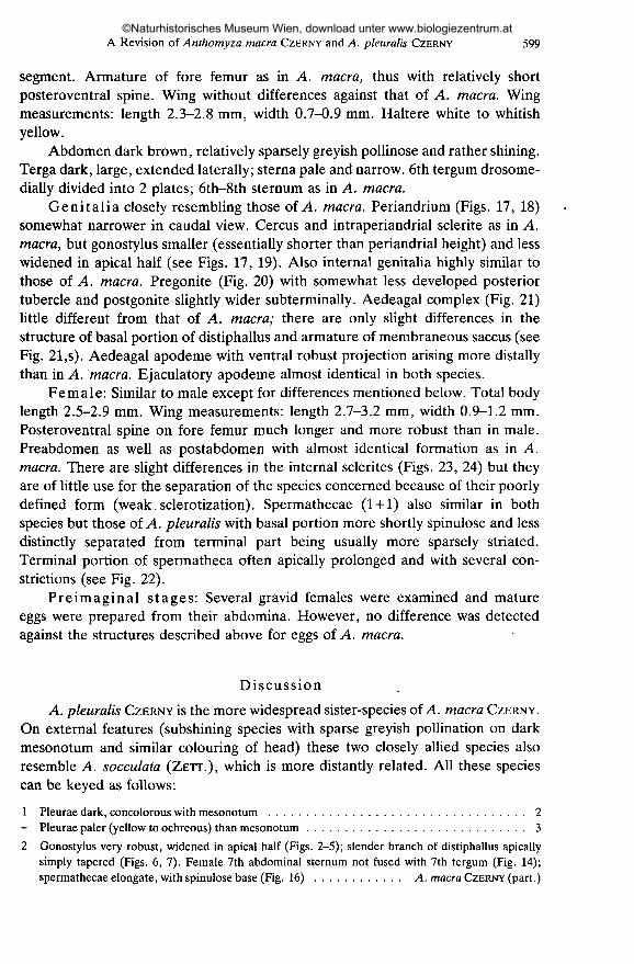
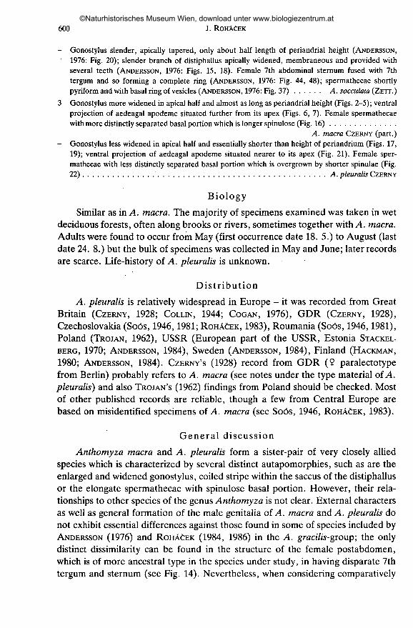
Genitalia closely resembling those of A. macra. Periandrium (Figs. 17, 18)somewhat narrower in caudal view. Cercus and intraperiandrial sclerite as in A.macra, but gonostylus smaller (essentially shorter than periandrial height) and lesswidened in apical half (see Figs. 17, 19). Also internal genitalia highly similar tothose of A. macra. Pregonite (Fig. 20) with somewhat less developed posteriortubercle and postgonite slightly wider subterminally. Aedeagal complex (Fig. 21)little different from that of A. macra; there are only slight differences in thestructure of basal portion of distiphallus and armature of membraneous saccus (seeFig. 21 ,s). Aedeagal apodeme with ventral robust projection arising more distallythan in A. macra. Ejaculatory apodeme almost identical in both species.
Female: Similar to male except for differences mentioned below. Total bodylength 2.5-2.9 mm. Wing measurements: length 2.7-3.2 mm, width 0.9-1.2 mm.Posteroventral spine on fore femur much longer and more robust than in male.Preabdomen as well as postabdomen with almost identical formation as in A.macra. There are slight differences in the internal sclerites (Figs. 23, 24) but theyare of little use for the separation of the species concerned because of their poorlydefined form (weak.sclerotization). Spermathecae (1 + 1) also similar in bothspecies but those of A. pleuralis with basal portion more shortly spinulose and lessdistinctly separated from terminal part being usually more sparsely striated.Terminal portion of spermatheca often apically prolonged and with several con-strictions (see Fig. 22).
Preimaginal stages: Several gravid females were examined and matureeggs were prepared from their abdomina. However, no difference was detectedagainst the structures described above for eggs of A. macra.
Discussion
A. pleuralis CZERNY is the more widespread sister-species of A. macra CZERNY.
On external features (subshining species with sparse greyish pollination on darkmesonotum and similar colouring of head) these two closely allied species alsoresemble A. socculata (ZETT.), which is more distantly related. All these speciescan be keyed as follows:
1 Pleurae dark, concolorous with mesonotum 2- Pleurae paler (yellow to ochreous) than mesonotum 3
2 Gonostylus very robust, widened in apical half (Figs. 2-5); slender branch of distiphallus apicallysimply tapered (Figs. 6,1). Female 7th abdominal sternum not fused with 7th tergum (Fig. 14);spermathecae elongate, with spinulose base (Fig. 16) A. macra CZERNY (part.)
©Naturhistorisches Museum Wien, download unter www.biologiezentrum.at
600 J. ROHÄCEK
- Gonostylus slender, apically tapered, only about half length of periandrial height (ANDERSSON,1976: Fig. 20); slender branch of distiphallus apically widened, membraneous and provided withseveral teeth (ANDERSSON, 1976: Figs. 15, 18). Female 7th abdominal sternum fused with 7thtergum and so forming a complete ring (ANDERSSON, 1976: Fig. 44, 48); spermathecae shortlypyriformand with basal ring of vesicles (ANDERSSON, 1976: Fig. 37) A. socculata (ZETT.)
3 Gonostylus more widened in apical half and almost as long as periandrial height (Figs. 2-5); ventralprojection of aedeagal apodeme situated further from its apex (Figs. 6, 7). Female spermathecaewith more distinctly separated basal portion which is longer spinulose (Fig. 16)
A. macra CZERNY (part.)- Gonostylus less widened in apical half and essentially shorter than height of periandrium (Figs. 17,
19); ventral projection of aedeagal apodeme situated nearer to its apex (Fig. 21). Female sper-mathecae with less distinctly separated basal portion which is overgrown by shorter spinulae (Fig.22) A. pleuralis CZERNY
Biology
Similar as in A. macra. The majority of specimens examined was taken in wetdeciduous forests, often along brooks or rivers, sometimes together with A. macra.Adults were found to occur from May (first occurrence date 18. 5.) to August (lastdate 24. 8.) but the bulk of specimens was collected in May and June; later recordsare scarce. Life-history of A. pleuralis is unknown.
Distribution
A. pleuralis is relatively widespread in Europe - it was recorded from GreatBritain (CZERNY, 1928; COLLIN, 1944; COGAN, 1976), GDR (CZERNY, 1928),Czechoslovakia (Soós, 1946,1981; ROHÂCEK, 1983), Roumania (Soós, 1946,1981),Poland (TROJAN, 1962), USSR (European part of the USSR, Estonia STACKEL-
BERG, 1970; ANDERSSON, 1984), Sweden (ANDERSSON, 1984), Finland (HACKMAN,
1980; ANDERSSON, 1984). CZERNY'S (1928) record from GDR (9 paralectotypefrom Berlin) probably refers to A. macra (see notes under the type material of A.pleuralis) and also TROJAN'S (1962) findings from Poland should be checked. Mostof other published records are reliable, though a few from Central Europe arebased on misidentified specimens of A. macra (see Soós, 1946, ROHACEK, 1983).
General discussion
Anthomyza macra and A. pleuralis form a sister-pair of very closely alliedspecies which is characterized by several distinct autapomorphies, such as are theenlarged and widened gonostylus, coiled stripe within the saccus of the distiphallusor the elongate spermathecae with spinulose basal portion. However, their rela-tionships to other species of the genus Anthomyza is not clear. External charactersas well as general formation of the male genitalia of A. macra and A. pleuralis donot exhibit essential differences against those found in some of species included byANDERSSON (1976) and ROHÄCEK (1984, 1986) in the A. gracilis-group; the onlydistinct dissimilarity can be found in the structure of the female postabdomen,which is of more ancestral type in the species under study, in having disparate 7thtergum and sternum (see Fig. 14). Nevertheless, when considering comparatively
©Naturhistorisches Museum Wien, download unter www.biologiezentrum.at

A Revision of Anthomyza macra CZERNY and A. pleuralis CZERNY 601
great diversity of features of the male and female terminalia within the A. gracilis-group it is possible to include tentatively also A. macra and A. pleuralis into thisgroup as its relatively primitive members (plesiomorphic features: absence ofepiphallus, simple slender branch of distiphallus, saccus without sclerotized spines,unarmed basal membrane, female 7th abdominal tergum and sternum not fusedinto tergosternal complex). In any case, even without the above species, the A.gracilis-group appears to be an artificial unit containing a number of sister-pairswhich are not closely related to each other (see ROHACEK, 1984, 1986). Aredefinition of the A. gracilis-group, based on sound synapomorphies, is muchneeded but it will be possible only when all species of Anthomyza are thorouglyknown (including the postabdominal structures).
Acknowledgements
My most sincere thanks are due to following persons who enabled me to study anthomyzidmaterial in museums or private collections: Dr. H. ANDERSSON and Mr. R. DANIELSSON (ZIL); Dr. M.BARTÂK, CSC. (Praha), Dr. M. C. BIRCH (UMO), Dr. RUTH CONTRERAS-LICHTENBERG(NMW), Dr. J.
JEZEK, CSC. (NMP), Dr. J. MÂCA (Parazitological Institute CSAV, Ceské Budéjovice), Doc. Ing. V.MARTINEK, CSC. (Praha-Zbraslav), Dr. B. MOCEK (MHK), late Prof. Dr. G. MORGE (IPE), Dr. L.PAPP, CSC. (TMB) and Dr. J. ZUSKA, CSC. (Praha). I am also deeply indebted to Mr. J. C. DEEMING(National Museum of Wales, Cardiff) for his critical reading of the manuscript and valuable comments.
References
ANDERSSON, H. (1976): Revision of the Anthomyza species of Northwest Europe (Diptera: Anthomy-zidae) I. The gracilis group. - Ent. scand., 7: 41-52.
— (1984): Family Anthomyzidae. - In: Soós, A. (ed.): Catalogue of Palaearctic Diptera. Vol. 10,402 pp. (pp. 50-53), Akadémiai Kiadó, Budapest.
COGAN, B. H. (1976): 64. Anthomyzidae. - In: KLOET, G. S. & HINCKS, D. H. (eds.): A check list ofBritish Insects. Second edition (completely revised). Part 5: Diptera and Siphonaptera. Handb.ident. br. Ins., Vol. 11, 139 pp. (pp. 82-83), Royal Entomological Society, London.
COLON, J. E. (1944): The British species of Anthomyzidae (Diptera). - Ent. mon. Mag., 80: 265-272.CZERNY, L. (1928): 54b. Anthomyzidae. - In: LINDNER, E. (ed.): Die Fliegen der Palaearktischen
Region. Vol. 6, Pt. 1, 8 pp., E. Schweitzerbart'sche Verlagsbuchhandlung, Stuttgart.ELBERG, K. J. (1968): New data on the fauna of Anthomyzidae (Diptera) from Baltic area. - Ent.
Obozr., 47: 629-632 (in Russ. with Engl. summ.).HACKMAN, W. (1980): A check list of the Finnish Diptera. II. Cyclorrhapa. - Notul. ent., 60:117-162.ROHACEK, J. (1983): Faunistics of the Czechoslovakian species of Anthomyzidae and Stenomicridae
(Diptera). - Cas. siez. Muz. Opava (A), 32: 125-135.— (1984): New species and records of Palaearctic species of the Anthomyza gracilis-group (Diptera,
Anthomyzidae). - Acta ent. bohemoslov., 81: 384-394.— (1986): Two new species of the Anthomyza gracilis-group (Diptera, Anthomyzidae) from Nepal.
- Acta ent. bohemoslov., 83: in press.Soós, Â. (1946): Die acalypteren Museiden des Karpatenbeckens III. - Fragm. faun, hung., 9: 2-10.
— (1981): 58. csalâd: Anthomyzidae - Tüskescombü legyek. Fauna hung. 149, pp. 106-117,Akadémiai Kiadó, Budapest.
STACKELBERG, A. A. (1970): 83. sem. Anthomyzidae. - In: BEJ-BIENKO, G. J. (ed.): Opredelitelnasekomykh evropeiskoi chasti SSSR. Vol. 5, pt. 2, pp. 326-329, Nauka, Leningrad (in Russ.).
TROJAN, P. (1962): Odiniidae, Clusiidae, Anthomyzidae, Opomyzidae, Tethinidae. Klucze do oznac-zania owadów Polski, XXVIII (54-58), 68 pp., PWN, Warszawa.
©Naturhistorisches Museum Wien, download unter www.biologiezentrum.at

602 J. ROHÀÒEK
Figs. 1-5. Anthomyza macra CZERNY: 1 - external genitalia caudally, 2 - dtto laterally, 3 - gonostylussublaterally externally (in the widest extension), 4 - same internally (Figs. 1-4 based on males fromCzechoslovakia), 5 - gonostylus sublaterally externally (paralectotype; micropubescence omitted).
Scales 0.1 mm.
©Naturhistorisches Museum Wien, download unter www.biologiezentrum.at

A Revision of Anthomyza macra CZERNY and A. pleuralis CZERNY 603
op
Figs. 6-11. Anthomyza macra CZERNY: 6 - aedeagal apodeme and aedeagus in rest position laterally(o", Czechoslovakia), 7 - internal genitalia in erected position laterally (<?, paralectotype), 8 - dorsalsculpture of egg chorion, 9 - egg dorsally, 10 - dtto ventrally, 11 - ventral sculpture of egg chorion (Figs.8-11 based on eggs prepared from females, Czechoslovakia). Abbreviations: ap - aedeagal apodeme,bm - basal membrane, dp - distiphallus, ea - ejaculatory apodeme, f- slender branch of distiphallus, hy- hypandrium, mp - micropyle, pg - postgonite, pp - phallophore, prg - pregonite, s - saccus of
distiphallus. Scales: Figs. 6, 7 = 0.1 mm, Figs. 9, 10 = 0.3 mm, Figs. 8, 11 = 0.05 mm.
©Naturhistorisches Museum Wien, download unter www.biologiezentrum.at

604 J. ROHÀCEK
T10
Figs. 12-16: Anthomyza macra CZERNY (9 paralectotype): 12 - postabdomen dorsally, 13 - dttolaterally, 14 - dtto ventrally, 15 - internal sclerites laterally, 16 - spermathecae. Abbreviations: ce -cercus, is-internal sclerites, S-sternum, T - tergum. Scales: Figs. 12-14 = 0.2 mm, others = U.I mm.
©Naturhistorisches Museum Wien, download unter www.biologiezentrum.at
A Revision of Anthomyza macra CZERNY and A. pleuralis CZERNY 60S
18
Figs. 17-19. Anthomyza pleuralis CZERNY (6, lectotype): 17 - genitalia laterally (of the internalgenitalia only aedeagus, postgonite and ejaculatory apodeme dotted), 18 - external genitalia caudally,19 - gonostylus sublaterally (in the widest extension; micropubescence omitted). Abbreviations: ae -aedeagus, ap - aedeagal apodeme, ce - cercus, gs - gonostylus, hy - hypandrium, ip - intraperiandrial
sclerite, p - periandrium. Scales 0.1 mm.
©Naturhistorisches Museum Wien, download unter www.biologiezentrum.at
606 J. ROHAÒEK
aphy 20
Figs. 20-24. Anthomyza pleuralis CZERNY: 20 - hypandrium and associated structures laterally, 21 -aedeagal apodeme and aedeagus laterally (both based on lectotype), 22 - spermathecae, 23 - internalsclerites laterally, 24- dtto ventrally (Figs. 22-24 based on females from G. Britain). Abbreviations: ap- aedeagal apodeme, bm - basal membrane, dp - distiphalîus, ea - ejaculatory apodeme, f- slenderbranch of distiphalîus, hy - hypandrium, pg - postgonite, pp - phallophore, prg - pergonite, s - saccus
of distiphalîus. Scales 0.1 mm.
©Naturhistorisches Museum Wien, download unter www.biologiezentrum.at