a review and empirical study of the composite scales of
TRANSCRIPT

© 2009 McCrea, publisher and licensee Dove Medical Press Ltd. This is an Open Access article which permits unrestricted noncommercial use, provided the original work is properly cited.
Psychology Research and Behavior Management 2009:2 59–79 59
O R I G I N A L R E S E A R C H
A review and empirical study of the composite scales of the Das–Naglieri cognitive assessment system
Simon M McCrea
JP Das Developmental Disabilities Center, Department of Educational Psychology, Faculty of Education, University of Alberta, Edmonton, Alberta, Canada
Correspondence: Simon M McCreaDepartment of Neurology and Neuro-ophthalmology, University of British Columbia, 2550 Willow Street, Vancouver, British Columbia, Canada V5Z 3N9Tel +1 604 875 4111 × 62929Fax +1 604 875 4302Email [email protected]
Abstract: Alexander Luria’s model of the working brain consisting of three functional units
was formulated through the examination of hundreds of focal brain-injury patients. Several
psychometric instruments based on Luria’s syndrome analysis and accompanying qualitative
tasks have been developed since the 1970s. In the mid-1970s, JP Das and colleagues defi ned
a specifi c cognitive processes model based directly on Luria’s two coding units termed
simultaneous and successive by studying diverse cross-cultural, ability, and socioeconomic
strata. The cognitive assessment system is based on the PASS model of cognitive processes
and consists of four composite scales of Planning–Attention–Simultaneous–Successive (PASS)
devised by Naglieri and Das in 1997. Das and colleagues developed the two new scales of
planning and attention to more closely model Luria’s theory of higher cortical functions. In this
paper a theoretical review of Luria’s theory, Das and colleagues elaboration of Luria’s model,
and the neural correlates of PASS composite scales based on extant studies is summarized.
A brief empirical study of the neuropsychological specifi city of the PASS composite scales in a
sample of 33 focal cortical stroke patients using cluster analysis is then discussed. Planning and
simultaneous were sensitive to right hemisphere lesions. These fi ndings were integrated with
recent functional neuroimaging studies of PASS scales. In sum it was found that simultaneous
is strongly dependent on dual bilateral occipitoparietal interhemispheric coordination whereas
successive demonstrated left frontotemporal specifi city with some evidence of interhemispheric
coordination across the prefrontal cortex. Hence, support for the validity of the PASS composite
scales was found as well as for the axiom of the independence of code content from code type
originally specifi ed in 1994 by Das, Naglieri, and Kirby.
Keywords: stroke, focal cortical lesions, Alexander Luria, syndrome analysis, Planning–Attention–
Simultaneous–Successive (PASS), cognitive assessment system, hierarchical agglomerative cluster
analysis, specifi city
IntroductionThe cognitive assessment system (CAS) has now been used in neuropsychological
assessment contexts for both children and adults;1 although it was not initially designed
as a neuropsychological instrument. The CAS is modeled on the Planning–Arousal/
Attention–Simultaneous–Successive (PASS) theory of cognitive processes.2–5 The
PASS model is an elaboration, standardization, and development of core concepts
of Luria’s qualitative theory in the context of Western neuropsychology’s demands
for quantitative methods, sensitivity, and specifi city.6,7 The CAS has a broad range
of complexity of items within subtests, several different types of tasks within each
composite scale, qualitative analysis, sensitivity, excellent test and retest character-
istics as well as reliability and acceptable construct validity parameters.5 A Japanese
version of the CAS has recently been published8 in addition to earlier clinical trials
with a prior Spanish version.

Psychology Research and Behavior Management 2009:260
McCrea
Previous attempts to operationalize Luria’s theory
include the Luria-Nebraska Neuropsychological Battery
(LNNB)9 in which 11 clinical scales were constructed
from 269 pass–fail individual items. The LNNB has met
with mixed reviews since establishing split-half reliability
is not possible with 269 qualitatively different items.10
The LNNB was standardized on a sample of 50 subjects
(mean age = 42) and educational level (12 years) and
unfortunately subjects with nonlocalized, diffuse lesions,
or no lesions at all were included in the reference sample.
Although the LNNB purports to separate brain-damaged
from nonbrain-damaged subjects, the data concerning
this battery’s effi cacy in localizing subtle brain damage
within specifi c regions of each hemisphere is limited.
The scales do not have adequate content validity, there
is an over emphasis on verbal responding, tasks are con-
founded with other cognitive functions, nor are all major
neuropsychological functions examined, and fi nally the
LNNB does not lend itself to qualitative interpretation in
its scoring system.11 Additional studies have shown that
the LNNB does not appear to be able to identify lesion
laterality to a satisfactory degree12 although there have
been subsequent attempts to comprehensively summarize
Luria’s clinical–theoretical approach to assist with the
interpretation of the results.13
The Kaufman Assessment Battery for Children (K-ABC)
is yet another cognitive model that is based on Luria’s6,14
and Das, Kirby, and Jarman’s15 simultaneous and successive
cognitive processes theory.16 However, unlike the DN-CAS
or the LNNB the K-ABC has not been scaled for use in
young adults. Of the 10 mental processing scales of the
K-ABC seven are labeled “simultaneous” and three are
“sequential”. The simultaneous tasks include: (i) object
naming from partial view of a picture through a hole; (ii)
arrangement of photos like picture arrangement of the WAIS;
(iii) gestalt closure; (iv) a variation of Koh’s block design
using triangles; (v) matrices; (vi) spatial memory; and (vii)
face recognition. The sequential tasks include: (i) hand
movements; (ii) digit repetition; and fi nally (iii) silhouette
seriation via pointing.17
The K-ABC is purported to break down into left
(analytical–sequential) and right (gestalt–holistic–
simultaneous) hemisphere functions16 as noted by Spreen
and Strauss.18 However Das, Kirby, and Jarman note that
visuospatial functions can be processed successively and
that auditory information can be processed simultaneously.19
That is, simultaneous is not synonymous with nonverbal
visual processing nor is successive by necessity the same
as verbal processing! This point is illustrated in Das and
colleagues’s15,19 theory by showing that simultaneous aspects
of grammar-based spatial language can occur in addition to
the reverse scenario of successive nonlinguistic seriation-
based tasks such as in Corsi block tapping.20
Moreover, Luria did not emphasize that these two types
of information integration and syntheses were necessarily
lateralizable; although the K-ABC does include a mix of
both verbal and nonverbal simultaneous and successive
tasks.6 Donders noted that, in a sample of 43 children with
traumatic brain injury, the K-ABC did not discriminate
any better than the revised Wechsler Intelligence Scale
for Children (WISC-R).21 Morris and Bigler were more
optimistic in their appraisal and found some degree of
concordance of lateralization of K-ABC dimensions in
79 neurologically impaired children based on patterns of
localization inferred from mainstream neuropsychological
tests.22 However, caution was urged in terms of the gener-
alizability of these fi ndings since the results were based on
the levels of performance on other marker neuropsychologi-
cal tests and were not based on structural neuroimaging
fi ndings per se.
Gutentag, Naglieri, and Yeates fi rst demonstrated that
both PASS scales and select subtests of the CAS reliably
discriminated between adolescents with traumatic brain
injury and controls.23 Moreover, the PASS scales and the CAS
subtests were found to be of diagnostic utility in distinguish-
ing between younger and older Down’s syndrome patients
with and without organic dementia of the Alzheimer’s type
(DAT). This DAT patient data converged with the fi ndings
from the standard Mattis Dementia Rating Scale and Peabody
Picture Vocabulary Test-Revised score patterns.24
Subsequently Wysocki and colleagues successfully
implemented the CAS along with other standard neuropsy-
chological instruments in the assessment of children with
diabetic symptomatology.25 It was found that the CAS subtests
and PASS scales were sensitive enough to be used for neuro-
pharmacological baseline purposes after 9 and 18 months of
medication for severe diabetes. Ryan, Atkinson, and Dunham’s
study of 262 adults found that the CAS’s planning subtests
tapping executive functions were sensitive and diffi cult enough
to discriminate among college student’s cognitive functioning.26
The latter fi nding is important in supporting the conclusion
that the broad range of diffi culty of items within subtests and
composite scales could render this battery useful in adult bed-
side neuropsychological populations. Davis and colleagues
found that a fi ve-days-per-week, four-month-long intensive
exercise program for obese elementary school children likely

Psychology Research and Behavior Management 2009:2 61
Cognitive assessment system PASS composite scales
provided cerebrovasculature benefi ts and hence enhanced
cognitive functioning as measured by the Das–Naglieri (DN)
CAS planning composite scale.27
In response to critics, Haddad noted that the qualitatively-
rated planning tasks of the CAS were not tapping only
speed of processing and defi nitively required the use of
cognitive strategies for optimal completion of these items.28
This study demonstrated that the CAS planning subtests are
robust executive functions tasks and that the Luria-modeled
qualitative descriptions of performance are useful and
meaningful for interpretation of composite scales and perhaps
implementation of remedial programs.5 Perez-Alvarez and
colleagues found that 35 patients treated for idiopathic
epilepsies with the anticonvulsant topiramate for six months
demonstrated signifi cant improvement on the planning scale
composite.29 Subsequently Mack and colleagues found that
surgically restoring portal blood fl ow to the liver in children
with primary extrahepatic thrombosis (and thus at risk
for hepatic central nervous system [CNS] neurotoxicity),
improved performance on the attention composite scale of
the CAS.30
Using Luria’s syndrome analysis method, the concordance
between PASS scale scores and cognitive and linguistic
impairments have also been studied.6 Perez-Alvarez and
Timoneda-Gallart found that planning was specifically
impaired in a large attention-defi cit hyperactivity disorder
(ADHD) Spanish sample.31 In two South African case
studies Jordaan and colleagues determined that a subject
with specifi c language impairment (SLI) presented with
a defi ciency in successive processing and had diffi culty
in acquiring the surface features of both English and
Afrikaans.32 In contrast a subject with a semantic–pragmatic
disorder (SPD) demonstrated a planning and attention
defi ciency with strength in successive processing and com-
petency in both languages. In a Dutch sample, Van Luit,
Kroesbergen, and Naglieri found that subjects with ADHD
demonstrated signifi cantly lower scores on planning and
attention scales and normal scores on simultaneous and
successive scales.33
Finally Naglieri and colleagues found a number of
clinically signifi cant correlations between CAS composite
scales and the Conners’ Continuous Performance Test
(CPT).34 There were in fact more clinically signifi cant and
meaningful correlation indicators with the CAS’s PASS
scales and the CPT than with the newly standardized
WISC-III and the CPT. This is particularly noteworthy
with respect to the study of learning disabilities since
the CPT is one of the most widely-used marker tests of
ADHD diagnosis. Collectively these neuropsychological
and learning disability cross-cultural studies suggest that
across ages, language, cultural, and neurological impair-
ments there are convergent fi ndings which could imply
construct validity of the PASS model.
Purpose of the studyHence, there will be fi ve inter-related purposes to this
theoretical review and accompanying short empirical
study. Firstly, psychometric instantiations spanning the
1970’s to more recent elaborations of Luria’s qualitative
method and validity studies with the CAS have been
compared and contrasted. Secondly, a brief overview of
the sociohistorical and neuropsychological background
surrounding Alexander Luria’s theory of higher cortical
functions will be undertaken in the context of Soviet
nonreductively materialist psychology. Thirdly, Das and
Naglieri’s specifi c instantiation of Luria’s theory in the
PASS model of cognitive functions will be discussed specifi -
cally with reference to the CAS. Fourthly, core differences
between the coding processes of simultaneous and successive
will be contrasted with other dual cognitive process models
in cognitive psychology and the functional neuroanatomical
correlates of these processes will be summarized. Finally,
a brief empirical study examining the construct validity of
the DN-CAS composite PASS scales will be undertaken in a
sample of focal cortical lesion stroke patients and the overall
utility of this unique neuropsychological instrument will be
discussed. Future directions in the further development of
this model that attempts to bridge individual differences,
neuropsychology, and cognitive and behavioral rehabilita-
tion will be highlighted.
Luria’s theory of higher cortical functionsEilam (2003) notes that the philosophical foundations of
Luria’s research program was based on a cultural–historical
psychological theory that was nonreductively materialist in
its core assumptions.35 Along with Vygotsky and Leont’ev,
these three investigators developed a comprehensive
theory of consciousness as a phenomenon in which mental
functions refl ect social relations as manifested by human
action in a world of concrete and theoretical objects.36 For
these theorists, language was the most important cultural
means that affects the contents and structure of cognitive
development. That is, when experts or adults name objects
they also implicitly defi ne the a priori relationships between
these objects such that a nonacculturated person or child will

Psychology Research and Behavior Management 2009:262
McCrea
invariantly create new ways of representing reality. In this
context the appropriation of cultural means requires the use
of (i) not only objects (be these concrete tools or abstract
linguistic tools), but more importantly (ii) the acquisition of
this object’s sociocultural meaning in context.35
In Luria’s theory then appropriation of cultural tools
are essential for the establishment of functional connec-
tions between localized modules and thus in producing
higher cognitive functions through ontogeny. These
higher cortical functions were first to appear on the
interpsychological sociological plane and only then on
the intrapsychological plane as plans that may be used to
direct activity of the organism.37 Therefore in this analysis
a person’s higher mental functions do not originate solely
from within the totality of the CNS but rather is conceived
as a direct consequence of internalization and in essence
refl ection of the sociocultural environment and milieu.
This theoretical proposition suggests that higher mental
functions do not in and of themselves arise solely as a
consequence of the genetic constitution of the individual.
Rather as a consequence of the mediation of sociocultural
products and signs there is a resultant development of
a capacity for the human organism to perform abstract
planful activity. Thus these refl ections of the sociocultural
milieu and historical accumulation of knowledge and skills
of a particular culture have both (i) material and (ii) theo-
retical correlates that develop and form during the history
of human social life.
Since the acquisition of these cultural means differ in
terms of their historical periods and different cultures there
was no fi xed innately determined localization of higher mental
functions in brain structure.35 Meccaci’s review of Luria’s
unitary view of brain and mind provides an excellent example
in the Japanese writing systems.38 The two Japanese writing
systems have entirely different functional organizations than
Western writing systems.39 As Meccaci notes, according to
Luria’s theory “…the development of these new ‘higher’
functional systems implies a reorganization of ‘lower’
cortical functions, a kind of Gestalt-like restructuration where
inferior components acquire a new functional [meaning] at
the moment in which they become part of the new superior
organization….” (p. 818).38
Das and Naglieri’s PASS model of cognitive functionsDas describes a multidimensional view of cognitive processes4
based on Luria’s theory6,7 as consisting of four functions
including: planning, arousal–attention, simultaneous, and
successive syntheses. In this model, planning is required
when for instance an individual makes decisions about how
to solve a problem, carry out a novel activity, or compose
a narrative. Attention–arousal is the process that allows a
person to selectively attend to some stimuli while ignoring
others, resist distractions, and maintain vigilance. Simul-
taneous processing integrates percepts into groups and as a
result stimuli are conceptualized as a whole, with each piece
being related to the others. Finally successive processing
involves integrating stimuli into a specifi c serial order and
is exemplifi ed in processing words in order to determine
their function as in syntactic comprehension.4
The theory links the four processes with particular
regions of the brain. Planning is associated with the frontal
lobes, attention–arousal with the reticular activating system,
and its associated brainstem catecholaminergic projections
throughout the cortex. The two coding units simultaneous and
successive are associated with occipito-temporoparietal junc-
tion and frontotemporal and perisylvian opercular regions,
respectively. This PASS model is directly based on Luria’s6,7
model of higher cortical functions in man. The PASS
model is an elaboration of Das, Kirby, and Jarman’s19 early
psychometric and cross-cultural work demonstrating two
coding units as well as with subsequent studies incorporating
planning and attention in Luria’s complete model of higher
cortical functions.2,40,41
Planning and the prefrontal cortexPerhaps the most important and overlooked and yet still
highly relevant contributions of Luria’s work to neuro-
psychology have been in the area of problem-solving and
frontal lobe functions.42,43 Luria’s qualitative methods and
syndrome analysis have been infl uential in the development
of subsequent models of these processes.44–46 In these later
works the rudiments of Luria’s model encompassing goal-
weighting, anticipatory processes, evaluation, feedback, and
corollary discharge between motor and sensory systems are
present. These components of planning actually antedated
modern notions of a hierarchical distributed control system
or supervisory attentional system (SAS) associated with
the prefrontal cortex. These control processes appear to be
essential in bridging the sociocultural contigencies of envi-
ronmental contexts with self-directed organized, purposeful,
and planful behaviors. A full discussion of Luria’s theory
of planning is beyond the scope of this article. Readers are
directed to Das, Kar, and Parrila’s text41 for an extensive
review of Alexander Luria’s and JP Das and colleagues’s
cognitive models of planning as well as being presented

Psychology Research and Behavior Management 2009:2 63
Cognitive assessment system PASS composite scales
with theoretical integration with more recent cognitive
psychological and cognitive neuroscience theories.
Attention–arousal and the reticulothalamic formationDas’s description of the arousal–attention unit contains the
brainstem reticular activating system (RAS) extending from
the spinal cord up to inhibitory nuclei within the thalamus.4 The
RAS contains both ascending and descending projections from
the forebrain and pyramidal tracts acting together on sensory
relay nuclei. The RAS innervates many regions of the CNS and
thus represents the major source of general regulatory systems
associated with ‘brain excitability’. Moreover, reticular neurons
are not specialized for relaying and analyzing signals that are
exclusively transmitted within a particular modality such as
vision, auditory sensations or touch. The functional importance
of the RAS was fi rst described by Moruzzi and Mangoun in
1949 and it was shown that it induces arousing effects on the
thalamus of the diencephalon and cortex.47 A more contempo-
rary view of the RAS shows that it is not a ‘nonspecifi c’ and
undifferentiated structure as originally proposed by Luria6,7
and that instead different neurotransmitter systems and their
associated nuclei projections exert arousing effects on the
diencephalon and telencephalon in specifi c manners.48
Two cholinergic pathways have been described and are
depicted in green in Figure 1. Cholinergic mesopontine cells
(PN) directly innervate the centrolateral nucleus (CL) of the
thalamus and constitute about 30% of cholinergic synapses.
In contrast, projections originating within the basal nucleus
of Meynert (BN) in the forebrain send projections directly
throughout the cortex via the cingulate bundle and constitute
the remaining 70% of synapses. In addition, pontine-thalamic
cholinergic projections within the CL of the thalamus relay
modulated signals throughout the cortex via glutaminergic
long-range excitatory amino acid synapses.48 The latter
glutaminergic excitatory synaptic projections throughout
the cortex are depicted in light blue in Figure 1. Finally, BN
or substantia innominata located in the anterior perforated
substance sends projections back to the reticular thalamic
nucleus (RE) of the thalamus. These two cholinergic systems
NA
RECL
FN
PN LC
HPBN
Figure 1 The reticular activating system. Yellow, red, and green denote elements of the noradrenergic, serotinergic and cholinergic pathways; respectively. Dark blue denotes modulatory glutaminergic thalamic nuclei.Notes: Notice the close connectivity of the RAS with specifi c nuclei of the thalamus as well as with basal parts of the forebrain. Hence, Luria’s notion of a tight link between the reticular activating system and the reciprocally connected prefrontal cortex is evident.Abbreviations: CL, centrolateral nucleus of the thalamus; RE, reticular thalamic nucleus; HP, hypothalamus; LC, locus coeruleus; FN, raphe nucleus; PN, pedunculopontine nucleus; BN, basal nucleus of meynert; NA, nucleus accumbens.

Psychology Research and Behavior Management 2009:264
McCrea
are effective in inducing and maintaining the enhanced
excitability of neurons in higher cortical structures49 and are
important in modulating signal–noise ratios of thalamocorti-
cal projections. Thus cholinergic innervation of the thalamus
is provided by the brainstem whereas cholinergic innervation
of the cortex is provided by the basal forebrain.
The serotonergic (5HT) projections of the raphe nucleus
(FN) project to a large number of forebrain structures such
as the nucleus accumbens (NA) depicted in red in Figure 1.
Also the 5HT projections innervate the hypothalamus along
the way to projecting throughout the cerebral cortex via the
cingulum. Noradrenergic projections originating in the locus
coeruleus (LC) of the rostrolateral pons course through many
areas including the forebrain, cerebellum, spinal cord, and
cerebral cortex. The noradrenergic fi bers also innervate the
hypothalamus on the way to the cerebral cortex and these
projections are depicted in yellow in Figure 1.50 Both the
serotonergic raphe nucleus and adrenergic locus coeruleus
innervate and modulate the hypothalamus (HP) depicted in
white on their way to the cerebral cortex.
It is important to mention that the brainstem reticular
formation innervates by means of ascending and descending
axons many regions of the CNS; although ascending tracts
are mainly depicted in Figure 1. Differentiation in terms of
functions of the RAS occurs through the actions of four main
types of neurotransmitter codes: (i) acetylcholine (green),
(ii) serotoninergic (red), (iii) adrenergic (yellow), and (iv)
glutaminergic (light blue) projections. Recently it has been
demonstrated that both thalamocortical and corticothalamic
glutaminergic neurons use metabotropic long-term activation
synapses which mediate prolonged excitatory actions similar
to that induced by acetylcholine. Finally from a functional
perspective only cholinergic and glutaminergic projections
are activated during both arousal and rapid eye movement
(REM) sleep whereas noradrenergic and serotonergic recep-
tors are only activated during arousal.48
During the natural states of vigilance commonly encoun-
tered during simple neuropsychological attention or arousal
all of these transmitters are released, however the interaction
of these dynamic systems on the activity of the thalamus and
cortex together are not yet fully understood. It seems then that
Luria’s notion of an arousal–attention unit is highly relevant if
we elaborate his monolithic nonspecifi c view of the RAS into
a distributed and multiple-action site of neurotransmitter func-
tions. A fi nal difference between Luria’s model of an arousal–
attention unit6 and current models48 is a recognition that there
are series of activating and arousing subsystems and cascades
beginning within the brainstem, pontine, rostral mesencephalic
nuclei and fi nally cresting in specifi c neuromodulatory thalamic
nuclei complexes. This revised model of Luria’s is quite con-
cordant with other neuropsychological theorists of brainstem
sensory and thalamic attention and tonic arousal.51
The two coding units and contemporary viewsThe fi rst natural philosophical description of the precursors
of simultaneous and successive cognitive processes were
characterized by the Russian physiologist Ivan Sechenov
in 189152 and then only subsequently elaborated and further
articulated more fully by Luria.53 Luria arrived at the con-
clusion that these two cognitive processes exist in human
information processing through the systematic assessment of
hundreds of brain-injured patients. Contemporarily these pro-
cesses can be best conceived of as functional neural pathways
or neurocomputational systems with some degree of invariant
core structures such that representations can be elaborated
and built upon to increase the complexity and fl exibility of
the organism’s behavior and knowledge-base.
Luria and Artem’eva did not disparage factor analysis,
rather they viewed it as a technique for categorizing and
classifying the mass of secondary symptoms accruing from a
primary localized brain injury.54 Using syndrome analysis and
the comparison of symptomological profi les resulted in the
eduction of dual primary ‘higher-order’ processes common
across sensory modalities, types of tasks, subjects, and variet-
ies of brain lesions. Such higher-order processes by which
information is registered, encoded, elaborated upon, stored
and fi nally integrated with the individual’s extant knowledge
base then corresponds to core cognitive systems associated
with specifi c functional neuroanatomical systems. In contrast,
Das and colleagues primarily used factor analysis and task
decomposition15 to identify statistically simultaneous and
successive processes and then conducted systematic studies
cross-culturally and across demographic variables in order to
verify the existence of these hypothesized processes.
Although Luria acknowledged that perceptual and mnestic
forms of these two processes could be found, it is the latter
memory encoding functions that are most closely associated
with Das, Kirby, and Jarman’s theory of simultaneous and suc-
cessive cognitive processes.15 It is important to note that simul-
taneous and successive are not synonymous with nonverbal
and verbal processing; respectively. As an example, it is pos-
sible that visual information can be processed successively;55
and auditory information can be processed simultaneously as
in sound localization.56 Luria notes that the second functional
unit is important “ …for obtaining, processing and storing

Psychology Research and Behavior Management 2009:2 65
Cognitive assessment system PASS composite scales
information arriving from the outside world…” (p. 43) and
thus is essentially a coding component.14
In their review of the signifi cance of Luria’s work in its
entirety, Stuss and Benson57 note that
…Posterior (parietal-occipital) brain regions are important
in the simultaneous synthesis of incoming information;
damage in these areas leads to particular syndromes,
including deficits in decoding phonetic elements and
in grasping logical, grammatical relations in language.
Anterior cortex, on the other hand, including both frontal
and frontal-temporal zones [anterior temporal opercular
regions in particular], is relevant in the synthesis of suc-
cessive elements into a single continuous sequential series.
Disturbances of “successive synthesis” may be observed
in the reproduction of rhythms, movements, words or
numbers, and series of actions. Anterior brain damage may
cause deterioration in the smooth fl ow from subject to verb.
Luria described this as a failure in syntagmatic organiza-
tion, a defi cit of internal speech, eventually resulting in a
telegraphic style… (p. 31).
The distinction between two orthogonal yet mutually
interacting types of encoding processes is not without prece-
dence in cognitive psychology. Broadbent’s initial descrip-
tion of fi lter theory distinguished between early parallel and
then only later subsequent serial processing.58 Treisman’s
widely accepted feature integration theory is a reformulation
of Broadbent’s original theory in which there is similarly
parallel-then-serial feedforward architecture but proposing a
major role for the inferior parietal lobe in the spatial integra-
tion of features at selected locations.59 Therefore these two
processes are primarily responsible for the encoding, storage,
as well as the transitory representation of information. In the
example of seriation, each item is linked to the next in succes-
sion, or, in the case of simultaneous processing each item in
a related gestalt is surveyable from any vantage point. These
concepts are analogous to analytic and holistic processing,
respectively of Peterson and Rhodes.60 In this sense simultane-
ous and successive processing constitute the two major modes
of information integration in cognitive functioning. Table 1
describes in more detail tasks in different modalities, means of
input and output that have been demonstrated to tap simultane-
ous and successive processing through factor analysis across
ability, age, cultural, and socioeconomic groupings.
Similar dual processing dichotomiesA generalized dual dichotomy of cognitive processes is not
without precedence in cognitive neuropsychology. There
are at least half-a-dozen well articulated cognitive process
dichotomies that bear some theoretical resemblances to the
simultaneous and successive dichotomy.15 Imagery and verbal
processes or synchronic thinking is the fi rst and perhaps most
closely related set of concepts to the simultaneous successive
model.71 Paivio’s model can be seen as a verbal–nonverbal
distinction that is based on the two hemispheres of the brain;
whereas Das and colleague’s model recognizes that there can
be verbal simultaneous tasks as well as nonverbal successive
tasks and may associated with a rostral-posterior designation
of function in addition to laterality distinctions.
Paradigmatic and syntagmatic dual functions have tra-
ditionally been associated with the study of different types
of aphasia in the context of developmental psychological
theory.72 Paradigmatic organization of language is then
associated with simultaneous processing or classifi cation
of words within a certain category; whereas syntagmatic
organization (successive) is the joining together of words
into a coherent expression.73 These two functions of para-
digmatic and syntagmatic functions were associated with the
posterior and anterior zone(s); respectively of the dominant
hemisphere.74
Serial and parallel processes are typical of visual search
tasks and share some characteristics with the successive and
simultaneous processes; respectively. Parallel processes have
been conceived of as “pre-attentive” in that the entire search
array is processed simultaneously. This type of pre-attentive
Table 1 Simultaneous and successive cognitive processes. Adapted and redrawn from Das, Kirby, and Jarman15,19
Simultaneous tasks
1. Syllogisms: Huttenlocher and Higgins61
2. Similarities: Wechsler62
3. Paired associate; Concrete words: Paivio63
4. Paper folding: French, Ekstrom, and Price64
5. Figure copying: Benton65
6. Memory for designs: Graham and Kendall66
7. Matrices: Raven67
8. Visual short-term memory: Sperling68
9. Cross-modal coding (audiovisual synaesthesia): Birch and Belmont69
Successive tasks
1. Digit span: Wechsler62
2. Paired associate abstract (verbal mediation): Paivio63
3. Memory span – Concrete words (written): Paivio and colleagues70
4. Memory span – Abstract words (written): Paivio and colleagues70
5. Auditory serial recall (semantic versus phonological similarity): Paivio and colleagues70
6. Auditory free recall (semantic versus phonological similarity): Paivio and colleagues70
Psychology Research and Behavior Management 2009:266
McCrea
search demonstrates the pop-out effect such that reaction
times are not infl uenced by the number of distracters in the
array.75,76 Moreover, such pre-attentive or parallel processes
are manifestations of maps of individual features that are
characteristically associated with bilateral low-level visual
cortex. In contrast, serial processes are associated with
“attention-requiring” processes such that reaction times are
linearly related with array size. These serial processes are
defi ned by the conglomeration of two or more features or
conjunctions which are detected as integration at the level
of the “master map of locations” and rely on the integrity of
the parietal lobes.75,76
Another popular distinction has been between holistic
and analytic processes corresponding to simultaneous and
successive processes; respectively.60 Holistic processes are
confi gural in the sense that these are spatial-relational pro-
cesses, however these entities do not denote a spatial feature
per se, rather these processes refer to integrated information
regarding components. In contrast, analytic processes are
componential and part-based with a fi ner grain of resolution.
Behrmann and colleagues’s study of an integrative agnosic
might be illustrative of key differences between simultaneous
and successive processes, their possible neural correlates,
and principles of the functional organization of related dual
processing concepts.77
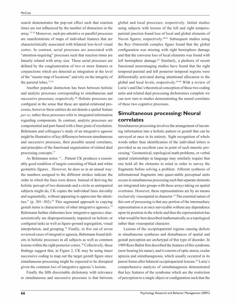
As Behrmann notes: “…Patient CK produces a reason-
ably good rendition of targets consisting of black and white
geometric fi gures…However, he does so in an unusal way:
the numbers assigned to the different strokes indicate the
order in which the lines were drawn. Instead of deriving the
holistic percept of two diamonds and a circle as unimpaired
subjects might do, CK copies the individual lines slavishly
and segmentally, without appearing to appreciate the identi-
ties.” (p. 301–302).78 This segmental approach to copying
gestalt items is characteristic of other integrative agnosics.79
Behrmann further elaborates how integrative agnosics char-
acteristically are disproportionately impaired on holistic or
confi gural tasks as well as fi gure-ground segregation, visual
interpolation, and grouping.78 Finally, in fi ve out of seven
reviewed cases of integrative agnosia, Behrmann found defi -
cits in holistic processes in all subjects as well as common
lesions within the right posterior cortex.78 Collectively, these
fi ndings suggest that, in Figure 2, CK may be using intact
successive coding to map out the target gestalt fi gure since
simultaneous processing might be expected to be disrupted
given the common loci of integrative agnosic’s lesions.
Finally the fi fth discernable dichotomy with relevance
to simultaneous and successive processes is that between
global and local processes; respectively. Initial studies
using subjects with lesions of the left and right temporo-
parietal junction found loss of local and global elements of
Navon fi gures; respectively.80,81 Subsequent studies using
the Rey–Osterreith complex fi gure found that the global
confi guration was missing with right hemisphere damage
and that the converse loss of local elements was found with
left hemisphere damage.82 Similarly, a plethora of recent
functional neuroimaging studies have found that the right
temporal-parietal and left posterior temporal regions were
differentially activated during attentional allocation to the
global and local levels, respectively.83,84 With a review of
Luria’s and Das’s theoretical conception of these two coding
units and related dual processing dichotomies complete we
can now turn to studies demonstrating the neural correlates
of these two cognitive processes.
Simultaneous processing: Neural correlatesSimultaneous processing involves the arrangement of incom-
ing information into a holistic pattern or gestalt that can be
surveyed at once in its entirety. Sight recognition of whole
words rather than identifi cation of the individual letters is
provided as an excellent case in point of such mnestic pro-
cessing.3 Geometrical, topological math problems, or verbal
spatial relationships in language may similarly require that
one hold all the elements in mind in order to survey the
fragments before solving a problem. Afferent synthesis of
informational fragments into quasi-stable perceptual units
occurs in simultaneous processing such that separate elements
are integrated into groups with these arrays taking on spatial
overtones. However, these representations are by no means
exclusively visuospatial in character.19 The essential nature of
this sort of processing is that any portion of the intermediary
representation is at once surveyable without any dependence
upon its position in the whole and thus the representation has
what would be best described mathematically as a topological
rather than visuospatial character.
Lesions of the occipitoparietal regions causing defects
in simultaneous syntheses and disturbances of spatial and
gestalt perception are archetypal of this type of disorder. In
1909 Rezo Balint fi rst described the features of this syndrome
(now bearing his name), and it consists of optic ataxia, ocular
apraxia and simultanagnosia, which usually occurred in its
purest forms after bilateral occipitoparietal lesions.85 Luria’s
comprehensive analysis of simultanagnosia demonstrated
that key features of the syndrome which are the restriction
of perception to a single object or confi guration such that the

Psychology Research and Behavior Management 2009:2 67
Cognitive assessment system PASS composite scales
affected patient is likely to perceive only a restricted element
or aspect of the total stimulus pattern.86 Luria alluded to the
fact that ideational or conceptual aspects of this disorder were
not well-characterized or understood yet at that time. Luria
and colleagues subsequently studied several other patients
with characteristic bilateral occipitoparietal lesions and
simultanagnosia and found that these patients demonstrated
“piecemeal perception” such that integration of the gestalt
story-line of a richly defi ned picture was abnormal.87
In a seminal study Coslett and Saffran studied these sec-
ondary cognitive aspects of simultanagnosia in more detail
and found evidence that neither visual fi eld reductions, nor,
an incapacity to process visual features could account for
the disorder.88 Their simultanagnosic patient could easily
identify briefl y presented single words and objects as rap-
idly and reliably as control subjects suggesting that access
to stored structural description of objects per se was not
impaired. Interestingly, with the simultaneous presentation
of two words or drawings the patient was able to identify
both stimuli signifi cantly more frequently when the stimuli
were semantically related than when they were unrelated.
These results suggest that simultanagnosia is fundamentally
attributable to impairment whereby activated and intact
structural descriptions of objects are linked through faulty
informational coding as to the identity of an object. Hence
the defective binding of semantic information with the
structural description of an object fi gures prominently in the
symptomatology of simultaneous agnosia.
Aysto and Hanninen demonstrated that a simultaneous
verbal factor was highly sensitive to left hemisphere more
than right-hemisphere lesions.89 A difference in performance
in favor of the right posterior lesions as opposed to left poste-
rior lesions was marginally signifi cant for this simultaneous
verbal factor. Finally, a nonverbal simultaneous factor was
also isolated through a factor analysis and patients with right
posterior lesions were more impaired compared to those
with left anterior lesions; a trend which reached marginal
signifi cance. Recent reviews of simultaneous agnosia all
point towards the bilateral occipitoparietal regions as being
critically involved.90
Successive processing: Neural correlatesLuria notes that patients with “lesions of the left temporal
region…experiences considerable diffi culty… when [they]
attempt to carry out systematic, successive operations with
Target model drawing CK’s copy
1 8
92
3
10
45
67
Figure 2 Hypothesized successive compensation of simultaneous processing defi ciency in an integrative agnosic. Copy of the target (left) by CK with the numbers indicating the order of the strokes. Note that gestalt circles and square constructions are patently absent. Adapted and redrawn with permission Nature Publishing Group. From Behrmann M. Neuropsychological approaches to perceptual organization: Evidence from visual agnosia. In: Peterson MA, Rhodes G, editors. Perception of faces, objects and scenes: Analytic and holistic processes. New York, NY: Oxford University Press; 2003. p. 295–334.

Psychology Research and Behavior Management 2009:268
McCrea
relationships requiring the constant participation of speech
associations as mediators…” (p. 121).6 Such processes
include diffi culties: (i) sequencing a series of pictures for
a story by numbers instead of using proprioceptive and
kinaesthetic imbued pointing; (ii) understanding a series of
geometrical operations; (iii) step-wise mental arithmetical
operations; (iv) logical problems through loss of access to
word meanings, values, traces and well-established linguistic
sequences; or (v) impairments in syntagmatic relationships
as opposed paradigmatic relationships.74
Testing of the limits demonstrated that such lesioned
patients could successfully perform successive tasks with the
aid of visual depictions which Luria6,7 attributes to the substi-
tution of degraded verbal memory traces with simultaneous
processes. This is a seemingly reverse simultaneous-successive
encoding compensation scenario as compared with the integra-
tive agnosic patient CK (see Figure 2). A critical and early dis-
tinction that Luria made was between the “communication of
events” and the “communications of relations” corresponding
to essentially successive and simultaneous cognitive processes,
respectively.91a This corresponds to the distinction between
syntagmatic (syntactical constructions of temporal-ordering)
analogous to successive processes versus paradigmatic or
categorization-based relationships pertaining to hierarchies of
concepts analogous to simultaneous processes.72,74
Successive then corresponds to temporal ordering of ele-
ments to be processed one by one and the whole sequence is
not surveyable at any one time.15,19 Aysto and Hanninen note
that in successive processing the information components
are not necessarily related to each other in any systematic
way, but may acquire meaning as a result of understand-
ing the whole sequence and subsequent chunking.89 Thus
a correlation or a direct association between stimuli is not
required at the level of central processing; although once a
linguistic or nonverbal sequence has been chunked in long-
term memory this may well occur. Aysto and Hanninen used
Das and colleagues’s15,19 taxonomy of tasks to factor analyze
an eclectic collection of tests including some from the WAIS,
Wechsler Memory Scales, and Benton Visual Retention Test
using factor loadings as criterion.
According to Das, Kirby, and Jarman, once material is
merged with long-term memory, subjects no longer engage
in successive synthesis but characteristically implement a
strategic difference in actual performance.15 The latter fi nd-
ing is entirely compatible with many functional neuroimag-
ing studies of basal ganglia and cerebellar systems that are
robustly activated when subjects engaged in strategic verbal
or nonverbal processes that require the implementation of
highly routinized sequences.93,94
Kim and colleagues found that verbal sequencing abilities
were impaired by left hemisphere lesions whereas nonverbal
sequencing tests were impaired by right hemisphere lesions.95
In a large adult sample of brain-injured patients Aysto and
Hanninen found that left hemisphere-lesioned patients
were signifi cantly more impaired than the right hemisphere
counterparts on successive processing.89 Similarly, a trend
for regional specifi city was also found such that the levels
of performance of left posterior patients was less than that
of left anterior patients, that these were less than that of right
anterior patients, and fi nally that these were all less than that
of right posterior patients.
These authors also noted that female subjects slightly out-
performed their male counterparts on successive processing; a
fi nding that was previously noted in reviews of simultaneous
and successive cognitive processes.1,2,15 These lesion studies of
the neural correlates of simultaneous and successive processes
are for the most concordant with Luria’s view of the neural
specifi city of these cognitive processes. However, the best test
for the functional system supporting simultaneous and succes-
sive processes would not be a lesion study but rather functional
neuroimaging studies. It is to such recent functional magnetic
resonance imaging (fMRI) and electroencephalography (EEG)
coherence studies of simultaneous and successive processes
that we now examine. These functional neuroimaging studies
Table 2 Cognitive processes similar conceptually to simultaneous and successive
Simultaneous Successive References
Imagery Synchronic thinking Paivio71
Paradigmatic Syntagmatic Jakobson72
Parallel Serial Treisman76
Holistic Analytic Peterson and Rhodes60
Global Local Navon80
Coordinate Categorical Kosslyn130
aInterestingly, as an aside, Luria’s major contributions to aphasiology and problem solving in particular were undoubtedly signifi cantly aided by the infrastructure associated with comprehensively assessing over 800 WWII Soviet soldiers.91 These individuals had been highly selected, comprehensively assessed in-depth neuropsychologically, and had sustained highly focalized ‘clean’ low-velocity high-calibre gunshot wounds. Such wounds are characteristically even more potentially focalized than occurs naturally in cases of ischemic and hemorrhagic stroke or traumatic brain injuries.10 Lezak notes that neuropsychologists who have had the opportunity to study such highly focalized wounds that occur virtually everywhere within the cortex have made, and will likely continue to make, major contributions to clinical and experimental neuropsychology as well as neuropsychological theory10 (eg, see also Luria92).

Psychology Research and Behavior Management 2009:2 69
Cognitive assessment system PASS composite scales
might be expected to be superior to focal cortical lesion
studies of stroke patients (despite the latter’s methodological
strengths), since in the former functional and effective con-
nectivity rather than lesion localization can be established.
Functional neuroimaging of simultaneous and successive processesRecently Okuhata, Okazaki and Maekawa examined EEG
coherence patterns during simultaneous and successive
processing tasks.96,97 The tasks from simultaneous and suc-
cessive were retrofi tted for use in an online computer-based
delivery system (Stim System; Neuroscan Inc., Charlotte,
NC, USA). EEG coherence can be described as the degree of
similarity of the frequencies between two brain regions and it
indexes the degree of functional cooperation and connectiv-
ity. EEG coherence is good means of assessing information
processing mechanisms involving short and long range
connections within the cortex.98 Such correlation within a
frequency band can be interpreted as a functional measure for
information transfer between brain regions and is analogous
with functional neuroanatomical systems approach used in
contemporary neuropsychiatry99 or a Luria-based syndromic
approach used in cognitive neuropsychology.6,7
Okahuta and colleagues compared two CAS tasks that
show the highest factor loadings with their respective simul-
taneous and successive composite factors scores.96 Figure
memory loaded 0.68 on simultaneous and word series 0.72
on the successive factor.1 The results showed that there was
an (i) indistinctive difference for the single simultaneous task
in terms of coherence patterns; and a (ii) signifi cant change
in coherence between the bilateral frontal and left temporal
regions in the beta frequency (12–25 Hz) for the successive
task. Beta coherence has usually been considered to indicate
higher cognitive processes especially of a verbal nature.100 (iii)
Moreover there was no clear left–right asymmetry for verbal
and nonverbal dimensions of the two tasks perhaps negating
the left–right brain hypothesis often confused with the simulta-
neous–successive processing dichotomy.101 However, Okahuta
and colleagues noted that the differential pattern could be due
to a genuine difference in processing type or merely be a task-
specifi c (eg, nonverbal vs. verbal) since only one of each type
of task was incorporated in the initial design.96
Therefore a second study was designed incorporating
comparisons among multiple CAS subtests comprising
each simultaneous and successive composite scale scores.97
Nonverbal matrices, verbal spatial relations, and fi gure
memory were used for the simultaneous factor and word
series, sentence repetition, and sentence questions were used
for the successive factor. The tasks varied in task content
(verbal or nonverbal) and modality (auditory or visual) and
specifi c theta coherence patterns were observed irrespective
of task content or modality. Simultaneous processing was
characterized by increase short-range interhemispheric con-
nections (eg, dual parietal activation) over central and parietal
regions compared to successive processing (see Figure 3).
This fi nding is congruent with theta selective responding to
encoding of new information102 and with a previous study
showing no impact of modality difference in the stimulus.103
Moreover, theta oscillations between 4–6 Hz are specifi cally
related to working memory processes.104 Again, a signifi cant
methodological and interpretation diffi culty with the results
is that the DN-CAS contains no nonverbal successive tasks.
However, previous functional neuroimaging studies using
nonverbal sequencing tasks similar to those included on
the successive processing scale may be able to help resolve
this issue.
Control for sequential nonverbal movements has been
found to require activation in the ventral portion of the lateral
premotor cortex105 and the supplementary motor area.106
Bhimani and colleagues used fMRI107 to examine the anatomic
organization the three Luria motor tasks of hand imitation,
fi st–edge–palm (FEP), and piano key (PK).6 All of these tasks
are nonverbal and hand imitation does not involve sequencing
since subjects only imitate from a target image. In contrast
FEP and PK involved a greater degree of movement pacing
and sequencing. Supplementary motor area was more active in
FEP which is the task with the greatest degree of sequencing.
Also parietal activation was found for all tasks due to the
proprioceptive nature of the tasks performed without vision
and all activation was found to be predominately within
the right hemisphere. Poldrack and Willingham note that
premotor, posterior parietal, and right hemisphere Broca’s
area homolog is most often implicated in such explicit
sequence learning tasks and this type of neural network is
most similar to the spatial working memory network.94
This right frontoparietal network involved in nonverbal
sequencing suggests dual verbal and nonverbal sequenc-
ing or successive pathways in contrast to Luria’s single
frontotemporal pathway usually only referred to in the left
hemisphere.6,7 A possible resolution of this inconsistency is
that different confi gurations of dorsal and ventral pathways
could be dominant within and across the hemispheres.83
Evidence from Balint’s patients with bilateral occipitopari-
etal lesions is helpful in this regard. If spatial relationships

Psychology Research and Behavior Management 2009:270
McCrea
between objects is processed by the dorsal stream while
spatial relationships within objects is processed by the
ventral stream then Balint’s patient’s intact ventral stream
would be suffi cient to represent the intrinsic spatial structure
of an object. However the dorsal stream would be required
to represent the spatial relationship between objects. Under
these circumstances the ventral verbal sequencing stream
would be expected to be dominant in the left hemisphere
and the dorsal nonverbal sequencing stream might be
expected to be dominant in the right hemisphere in almost
all people.83 With a review of the theoretical and empirical
foundations of the neuropsychological properties of the
DN-CAS PASS scales complete we now turn to a discus-
sion of a brief empirical study of this instrument in a small
sample of well-characterized focal cortical lesion stroke
patients.
MethodAfter neurology patients were admitted to the University of
Alberta Hospital in Edmonton, Canada, patients who met
inclusion–exclusion criteria were screened by a neurolo-
gist. The Director of Neurology was the coordinating and
supervising physician and nurses or attending physicians
screened subjects daily for inclusion into the study. At these
physician’s discretion subjects were recruited in a consecu-
tive sample spanning nine months. After referral of such
patients to the experimenter subjects were asked in-person
for their written consent to participate in a study of cognitive
functions following stroke or brain injury.
The median delay between stroke onset, acquisition
of structural neuroimaging, and assessment with the CAS
was approximately one month post-stroke. Case number
5 and case number 9 were outliers since these neurosurgi-
cal patients had surgeries for the excision of tumors with
appended computed tomography (CT) scans that were
approximately one year and six years old, respectively
(Table 3). However, because these two patients’ lesions
were so circumscribed and well defi ned they were included
in the study. In the remaining 31 subjects, the average time
between peak stroke onset, intervening CT or MRI scan-
ning and testing just before discharge with the DN-CAS
was calculated (mean = 27 days [SD 22], range = 88 days,
minimum = 15 days, and maximum = 88 days). Under
these circumstances most subjects were assessed while an
in-patient at the stroke unit often just before their discharge
from the hospital. Subjects completed the twelve subtests
of the DN-CAS in either 1½ hour session with as many rest
breaks between subtests as needed, or, alternatively on two
separate 45 minute sessions on adjacent mornings in order
to minimize fatigue and to ensure subjects were performing
their best.
The inclusion criteria included those patients with:
informed consent for participation and review of neuro-
logical charts; patients with localizable single ischemic or
hemorrhagic stroke lesions; approximately equal distribution
of lesions locations across the left and right frontal lobes
(frontal lobes) and the left and right posterior cortices (tem-
poral, occipital, parietal). The frontal lobe lesioned subjects
included in the study had to have lesions with a center of mass
and volume that was greater than or equal to 75% rostral to
the central sulcus. The posterior lesioned patients included
in the study had to have lesions with a center of mass and
volume that were greater than or equal to 75% caudal to the
central sulcus located primarily within either the parietal,
occipital, and/or temporal lobes. The exclusionary criteria
included those patients with: diffuse lesions, moderate
to severe stroke, post-stroke depression, severe receptive
aphasia, under the care of a guardian, extensive primary
occipital cortex lesions and accompanying severe visual fi eld
defect, neurodegenerative disease or advanced age such that
it would be diffi cult to distinguish whether normal aging or
mild lesion’s effects were the primary cause of the patient’s
cognitive dysfunctions (eg, advanced age ∼ � 70).
Subjects with lesions of either left or right hemisphere but
not both were recruited into the study in approximately equal
proportions. A total of ten subjects with either negligible
lesions (eg, subtle frontal atrophy) as determined by neuro-
radiological MRI or CT scan, or small cerebellar, midbrain
lesions or cyst resections with no intrusion into gray or white
matter were used as the control group subjects. Hence, these
consisted of patients for which there was little evidence of
focal cortical lesions or who had a patent subcortical lesion
or alternatively a neurological patient without any visualiz-
able surgically-induced loss of brain tissue (eg, external optic
nerve cyst resection).
Subjects were administered Annett’s 12-point ques-
tionnaire to evaluate handedness.109 At the same time that
this preliminary assessment was completed demographic
information was also gathered. Documentation regarding
lesion locus, severity, lateralization, clinical neurological,
radiological, and neuroradiological fi ndings was collated
under the supervision of participating neurologists. The
sample control group consisted of 33 brain-lesioned patients
of mean age 46 years (SD = 13); male = 24, female = 9; mean
educational level = 12 years (SD = 3); handedness: left = 8,
right = 25. Ninety-one percent of subjects were Caucasian

Psychology Research and Behavior Management 2009:2 71
Cognitive assessment system PASS composite scales
Table 3 Demographics of the DN-CAS focal cortical lesion study sample
Case Lesion Lat A/P Sex Age Hand Educ Ethnic
1 Posterior left frontal lobe Left Ant. M 4 Right 4 C
2 LT frontal horn and LT basal ganglia Left Ant. F 3 Left 2 C
3 Anterior left frontal lobe Left Ant. F 4 Left 2 C
4 Left frontal lobe Left Ant. M 3 Right 2 C
5 LT inf. frontal and cingulate gyrus Left Ant. M 5 Right 2 C
6 LT temporal lobe lesion Left Pos. M 3 Right 2 C
7 Left temporal lobe Left Pos. M 3 Right 2 C
8 Left occipitotemporal lesion Left Pos. M 4 Right 2 C
9 Left parieto-occipital craniotomy Left Pos. M 5 Right 2 C
10 LT parietal arteriovenous lesion Left Pos. M 2 Right 1 F
11 Left paracentral lobule Left Pos. M 2 Right 2 C
12 RT posterior frontal operculum Right Ant. F 4 Left 2 C
13 Right frontal lobe and basal ganglia Right Ant. F 5 Left 2 C
14 Right frontal lobe lesion Right Ant. M 4 Left 3 C
15 Right posterior frontal lobe Right Ant. F 3 Right 2 B
16 RT frontal lobe and frontal operculum Right Ant. M 3 Right 2 C
17 RT frontal lobe and RT basal ganglia Right Ant. M 4 Right 4 C
18 RT frontal lobe and internal capsule Right Ant. M 4 Right 2 C
19 RT c. semiovale and paracentral sulcus Right Pos. M 4 Right 3 A
20 RT c. semiovale and paracentral sulcus Right Pos. M 2 Right 3 C
21 Right frontoparietal lobe Right Pos. M 5 Right 1 C
22 Right temporoparietal region Right Pos. F 3 Right 2 C
23 Right frontoparietal region Right Pos. M 4 Left 2 C
24 Bilateral frontal lobe lesions C C M 1 Left 2 C
25 Left frontotemporal lobar tumor C C F 2 Right 4 C
26 Bilateral frontal lobe atrophy C C M 3 Right 2 C
27 Bilateral frontal lobe atrophy C C F 2 Right 2 C
28 Bilateral medial frontal lesions C C M 1 Right 3 C
29 Right cerebellar lesion C C F 2 Right 2 C
30 RT posterior cerebellar hemisphere C C M 5 Right 2 C
31 Superior right cerebellar hemisphere C C M 3 Right 2 C
32 Postero-central midbrain-pons lesion C C M 4 Right 3 C
33 Unspecifi ed contusion C C M 1 Right 2 C
Abbreviations: Ant, anterior lesion; Pos, posterior lesion; Lat, Laterality of lesion; c, centrum; C (Lat or A/P), control group; M, male; F, female; LT, left; RT, right; Hand, handedness; Educ, educational level in years of formal education (1: � 8, 2: 9–12, 3: 13–14, 4: � 15 years, respectively); Age (1: � 25, 2: 26–40, 3: 41–50, 4: 51–60, 5: � 61 years, respectively); Ethnic group (C, Caucasian; B, Black; A, Asian; F, First Nation).
(N = 30); 3% of subjects were Black (N = 1); 3% of subjects
were Asian (N = 1); and 3% of subjects were First Nations
(N = 1). Previous analysis demonstrated no signifi cant dif-
ferences in these demographic variables on subtest t-scores
at the aggregate sample level.109
Since no adequate norms yet exist for the DN-CAS for
adults with a mean age of 46 (age range = 20–67) Russell’s
average z-score index was used.110 The average z-score
method consists of four steps: (i) choosing a reference group
of tests; (ii) combining the results from those tests into a
reference scale; (iii) deriving scale scores from the reference
scale using multiple regression; (iv) anchoring to some spe-
cifi c group with a known level of absolute performance on
the task(s) in question. The anchoring population used in this
study was 17 year–8-month-old US students derived from the
DN-CAS standardization sample. Lezak notes that “…tests
of mental ability that provide adult norms extending into the
late teens fi nd that the population of 18 year olds does not
perform much differently than the adult population at large…”
(p. 158).10 Moreover, Naglieri and Das’s interpretive manual

Psychology Research and Behavior Management 2009:272
McCrea
shows that across most of the CAS subtests there is near
asymptotic levels of performance cresting near the age of
18 years.1 Similarly, Ryan and colleagues’s study confi rmed
this assertion by fi nding that within a large sample of college
students the CAS’s subtests great range of diffi culty of items
was more than suffi cient to objectively measure changes in
cognitive functioning across subgroups without any fl oor or
ceiling effects.26
Hence, brain-damaged patient’s scores were normed
using the average z-score index of impairment with the raw
scores of the 18-year-old group in the DN-CAS standardiza-
tion sample used as baseline. All the subject’s index scores
were averaged to form the reference scale. That is, using the
z-scores, the 12 CAS subtests were summed and divided by
12 for each subject. In this way the average index of impair-
ment was created with a mean of 1 and each interval was
equivalent to a standard deviation unit. The scaled scores
for individual CAS subtests and participants were derived
through a series of 12 separate multiple regressions for
each subtest. Average z-score indexes of impairment were
regressed on to subtest raw scores yielding predicted raw
scores with distributions that were equated across subtests.
Therefore, a given level of impairment on one CAS subtest
was equivalent to that on any another CAS subtest.
ResultsCluster analysis is the assignment of observations into groups
such that observations in one cluster are more similar to
each other compared to observations from different clusters.
Cluster analysis is particularly useful in pattern recognition
and hence single-case study design.111 Hierarchical clustering
fi nds clusters by fi rst using more basic level structures within
a data set; while agglomerative algorithms are those that are
bottom-up statistical processes that begin with each element
as a separate cluster. Smaller clusters are then merged into
successively larger clusters. Euclidean distance can be used
to separate clusters and it is a symmetrical metric and is the
most common distance measure used in psychology studies.
Hierarchical agglomerative cluster analysis (as used in this
particular study), traditionally utilizes a visualization format
in which the classifi cation of observations is represented in the
form of a tree-like hierarchy or dendrogram (eg, Figure 4). In
this representation individual elements or cases are depicted
at one end and a single higher-order cluster containing every
element is located at the other end of the dendrogram.
The 33 cases with 12 observations of subtest performance
per subject constitutes approximately 400 singlet observations
which is more than satisfactory for a hierarchical agglomerative
cluster analysis (HACA). During HACA determination of the
number of clusters involves: (i) visual inspection of the den-
drograms; (ii) as well as observation of the largest single jump
in the cluster coeffi cient according to the methods described by
Aldenderfer and Blashfi eld.112 Figure 4 illustrates the point at
which the dendrogram fl attens out most and it shows that there
are three unambiguous primary clusters. At least a dozen itera-
tions using several of the most common (i) clustering methods
as well as (ii) interval measures or metrics all converged on
the same solution depicted in Figure 4. This method was most
reproducible, stable, and meaningful using the common metrics
of between-groups linkage and squared Euclidean distance.
G
F7 F3 FZF4 F8
C4C3 CZ
PZP3
01 02
P4
T3
T5
T4
T6
FP1 FP2
1 23
4 6 5
Figure 3 Hypothetical neural networks underlying simultaneous and successive cognitive processes. Top left: International 10–20 system for the placement of EEG electrodes. F, T, C, P, O, and Z refer to the frontal, temporal, central, parietal, and occipital lobes, respectively. Z refers to reference electrodes placed upon the midline. Top middle and right: Diagram of hypothetical electrode pairing coherence connections for simultaneous and successive cognitive processes. 1, Simultaneous left (O1–C3, P3). 2, Simultaneous right (O2–C4, P4). 3, Simultaneous interhemispheric (O1–C4, P4; O2–C3, P3). 4, Successive left (T3, 5–F3, 7; FP1). 5, Successive right (T4, 6–F4, 8; FP2). 6, Successive interhemi-spheric (T3, T5 : F4, F8, FP2; T4, T6 : F3, F7, FP1). Adapted and redrawn with permission of Elsevier: Okuhata ST, Okazaki S, Maekawa H. Differential topographic pattern of EEG coherence between simultaneous and successive coding tasks. Int J Psychophysiol. 2007; 66:66–80.96

Psychology Research and Behavior Management 2009:2 73
Cognitive assessment system PASS composite scales
This qualitative analysis was followed by confi rmatory
statistical inferencing using cross-tabulations of frequency
distributions across the (1) rostral-caudal and (2) laterality axes
(see Goldstein and colleagues for an identical type of analyses
using the LNNB).12 Only one cluster solution is usually found
for a particular data set of this size and it designates the intrinsic
structure of observation co-variance and logical patterns.112 The
cluster solution closely paralleled the confi guration for lateral-
ity of lesions as opposed to the rostral-causal loci of the lesions.
That is, most of the variance was explained by the laterality of
the lesion that maximized the distance between subjects and
across subtests scores. The left hemisphere, right hemisphere
and control groups were highly signifi cantly concordant with
clusters 1, 2 and 3; respectively (r = 0.85, p � 0.0001), such
that only 5 of 33 or 18% of observations did not fi t exactly into
the original laterality groupings. Subsequent procedures using
predicted subtest scores with laterality as covariate demon-
strated that the anterior-posterior grouping did not contribute
any more meaningful variance and that therefore the fi rst
cluster solution was based exclusively on laterality.
That such unequivocal results were found with such a
moderately sized sample attests to the well characterized
recruitment and selection of only highly focalized cortical
lesion patients through appropriate neurologist-screening
and neuroradiological consultation. However, others,
notably Russell, have shown that recruitment of small
samples with good lesion characteristics (of greater than or
equal to N = 30), are more than adequate to provide reliable
fi rst-approximations or estimates of the neuropsychological
specifi city of a new psychometric instrument.110 This is all
the more evident when the collection of subtests has been
well-normed as in the original DN-CAS standardization.1
Table 4 depicts the three factor cluster solution’s fre-
quency tabulations of sex, handedness, age, education and
ethnicity. One way analysis of variance (ANOVA) across the
parametric variables of age and education did not reveal any
signifi cant differences across clusters (age: F(2,32) = 2.28,
p � 0.15; education: F(2,32) = 0.06, p = 0.95).
A Chi-square analysis of the cluster solution along the
variables of sex, handedness, and ethnicity was performed.
There were no signifi cant group differences across the three
cluster groupings in terms of frequency distributions for sex
(χ2 [2] = 1, p = 0.72); handedness (χ2 [2] = 4, p � 0.15) or
ethnicity (χ2 [2] = 5, p = 0.57). ANOVA statistically cor-
rected for four parametric comparisons demonstrated that
for the laterality of the lesion (eg, left-hemisphere, right-
hemisphere and control subjects) there were signifi cant main
effects for planning and simultaneous. In each case the right
2520
1510
50
1 2 10 11 12 6 8 4 9 7 16 25 18 5 14 13 19 21 24 17 23 15 26 20 27 30 22 29 31 32 28 333
Figure 4 Dendrogram of the hierarchical agglomerative cluster analysis. The y-axis depicts the dendrogram cluster coeffi cients and the x-axis lists the case numbers depicted in Table 3. Thirty-three individual cases with twelve Das–Naglieri cognitive assessment system subtest scores per subject for a grand total of approximately 400 singlet observa-tions. This number of observations provides more than enough variance between and across subjects for statistically reliable hierarchical agglomerative cluster analysis.112

Psychology Research and Behavior Management 2009:274
McCrea
hemisphere lesioned groups performed signifi cantly worse
on the planning and simultaneous composite scales than their
control comparisons groups (Table 6).
DiscussionThe two functional neuroimaging studies of simultaneous and
successive processing reviewed suggested that simultaneous
processing involved dual bilateral occipitoparietal coordina-
tion and that successive processing did not involve as much
interhemispheric coordination.96,97 This view is consonant
with Luria’s initial studies of cases of simultanagnosia86,87
that provided some of the impetus for his formulation and
articulation of the concept of simultaneous processing. The
functional neuroimaging studies are also congruent with
extensive reviews and detailed single case studies of mod-
ern simultanagnosic patients using imaging and cognitive
neuropsychological testing.90 All such studies implicate the
centrality of the integrity of bilateral occipitoparietal regions
for eliciting the classic symptoms of simultanagnosia and
presumably also involved in simultaneous processing.
The focal cortical lesion study included with this review
also indicated that simultaneous processing was a function of
the integrity of the right hemisphere, and left-sided lesions did
not result in appreciable impairment in simultaneous process-
ing. The apparent contradiction between the two functional
neuroimaging and lesion study could be explained by the fact
that the left temporoparietal junction appears to be involved in
fi lling in local detail while the right temporoparietal junction
results in diffi culties in the appreciation of the global aspects
of a fi gure.81,83 On this view impairment of global processing
due to right parietal lesions would be much more disruptive
to the core neural network involved in simultaneous pro-
cessing. Our review of the integrative agnosic patient CK
showed that what might be occurring when global processing
is adversely effected (and therefore presumably disrupting
simultaneous processing), is that successive processing
could compensate by slavishly and segmentally coding the
elements of the complete confi guration.77,78 This hypothesis
would also be congruent with the close relationship between
global and local processing integration and simultanagnosia90
as well as the dominant role of the right intraparietal sulcus
in switching between spatial reference frames (eg, local and
global detail).113
Secondly, the two functional neuroimaging studies
implicated the involvement of bilateral frontal and anterior
left temporal regions in successive processing.96,97 These fi nd-
ings are congruent with the hypothesis that the hemispheres
are capable of sequencing either verbally as in the word
series task, or, nonverbally as in the hand movements task
of the K-ABC, Self-Ordered Pointing, or Corsi Blocks. The
evidence taken as a whole seems to be concordant with this
dual processing hypothesis despite the fact that the DN-CAS
does not have a nonverbal sequencing tasks included within
the standardization battery. Aysto and Hanninen found that
their successive factor was sensitive to left hemisphere
lesions; however all their successive tasks were verbal.89 Kim
and colleagues found that verbal and nonverbal sequencing
were impaired by left and right hemisphere lesions, respec-
tively.95 Collectively these fi ndings suggest that successive or
sequencing is not lateralizable in the frontal regions perhaps
given the extensive genual and anterior commissure con-
nectivity required developmentally in the ontogeny of many
bimanual coordination tasks.114,115
Using structural MRI, Kluger and Heilman found that
Luria’s test of reciprocal coordination (which is a test of
bimanual coordination), could be elicited with frontal mesial
lesions alone encompassing the supplementary motor areas,
but sparing the anterior cingulate or corpus callosum.116
Chunking of action sequences may not necessarily require
content domain-specifi city (eg, verbal vs. nonverbal) for inte-
gration in the prefrontal cortex. This is because the spatial and
temporal sinusoidal topology associated with such action rep-
ertoires would be suffi cient for cross-mapping purposes via
prefrontal–basal ganglia reciprocal connectivity.93 However
the data do suggest that in the posterior cortex (eg, anterior
temporal regions) that verbal and nonverbal sequencing is
Table 4 Cluster solution group, gender, handedness, age, education and ethnicity
Cluster M F Handedness Age Education Ethnicity
LT RT Mean S.D. Mean S.D. C A B F
1 8 3 4 7 48 11 12 3 10 – – 1
2 11 3 4 10 50 12 12 3 12 1 1 –
3 5 3 – 8 38 15 11 2 8 – – –
Total 24 9 8 25 46 13 12 3 30 1 1 1
Abbreviations: Sex: M, male; F, female; Ethnicity: A, Asian; B, Black; C, Caucasian; F, First Nations.

Psychology Research and Behavior Management 2009:2 75
Cognitive assessment system PASS composite scales
highly lateralizable. This could mean that there are two points
of diversion for Successive processing that both converge on
the ventral premotor and prefrontal regions and their basal
ganglia connectivity where verbal and nonverbal content
codes are integrated into unitary amodal codes. In contrast,
simultaneous processing could function more seamlessly
as a consequence of the integrity of the bilateral posterior
parietal regions and their splenial connections vis-à-vis
the corpus callosum.117 This suggests an as yet unspecifi ed
triple fasciculi-based coordinated sets of pathway(s) with a
more anterior-posterior loci of connectivity for successive
processing within the frontotemporal regions and a more
laterally expansive foci for simultaneous processing across
the parietal lobes.
The composite factor of attention was not found to be
localizable to any brain region and this fi ts with its extensive
connectivity using at least four different neurotransmitter
systems emanating from the reticulothalamic formation.
However it is important to note that exclusively subcortical
patients with large lesions were excluded from the study. The
bulk of such connections course through the medial forebrain
bundle and synapse with the basal ganglia, medial temporal
lobe and the nucleus accumbens (Figure 1). As such the atten-
tion composite factor is tightly yoked to the functioning of
the prefrontal cortex as originally articulated by Luria.6,7
Of unique interest is that the ventromedial aspect of the
frontal lobe contains numerous autonomic nervous system
nuclei and connections which regulate and modulate the
reticular formation in functions such as sleep, waking, and
tonic arousal.118 Hence, yoking should be expected to be
especially close between planning and attention if the tasks
could be parametrically varied for diffi culty and if the respec-
tive subtests could be retrofi tted for use in other functional
neuroimaging studies. Finally the planning composite scale
was highly sensitive to right hemisphere lesions congruent
with many studies showing that planning tasks are gener-
ally sensitive to frontal lesions (Grafman for an extensive
review119). Subsequent studies have shown that problems with
an ill-structured nature (such as are present in the DN-CAS
planning composite scale), are highly sensitive to right pre-
frontal cortex lesions,120 which is entirely consistent with the
results of the focal cortical lesion patient study.
ConclusionsSeminal contributions to neuropsychology methods by Luria
include: understanding localization of function, higher corti-
cal functions, functional systems, the symptom-complex and
syndrome analysis, qualitative analysis, use of specifi c errors
patterns, single case study design and rehabilitation theory.35
The cognitive neuropsychological analysis of the single case
is in accordance with Luria’s original qualitative method of
syndrome profi les.43,111 In an infl uential review, Robertson
and colleagues noted that the single-case routinely allows
for the generalization beyond the single-case about what is
and what is not possible to deduce or infer about the general
population’s brain functioning.121 In fact, some advocates of
the single-case would go far beyond this statement and sug-
gest that the single patient is the essence of deducing the core
functions of how the brain is actually organized in vivo from
a functional point of view.6,7 An examination of the enormous
empirical, methodological, and theoretical advances within
the last decade about how the ultra-structural white matter
tracts of brain differ extremely from subject to subject sug-
gests that the stronger form of this position is gaining much
wider credence among the cognitive neuroscience commu-
nity (eg, see Catani and fftyche122).
In this sense then Das, Kirby, and Jarman’s concept
of basic cognitive processes (rather that stimulus-specifi c
content), directly mediating intelligent behavior is perhaps
surprisingly contemporary.15,19 From the perspective of func-
tional neuroimaging and cutting-edge statistical techniques
these major simultaneous-successive throughputs could
be conceived as probabilistic functional maps about how
various types of input are invariably processed. Goebel and
colleagues have provided just such an excellent example
using fine-grained time-resolved fMRI in conjunction
with diffusion tensor imaging fi ber tracking and Granger
causality mapping.123 Granger causality is a technique for
determining whether a time series is useful in forecasting
another time series and therefore it can be used to determine
causality interactions between functional systems in highly
complex sets of variables such as are used in fMRI. These
investigators’ studies demonstrated how occipitoparietal
regions invariantly communicate with both homologous
regions of the contralateral hemisphere in addition to
informational ventral frontotemporal pathways presumably
Table 5 Hierarchical agglomerative cluster analysis by laterality frequency
Cluster Laterality Total
Left hem Right hem Control
1 10 1 – 11
2 1 10 3 14
3 – 1 7 8
Total 11 12 10 33

Psychology Research and Behavior Management 2009:276
McCrea
involved in sequencing. These two throughput pathways
or functional systems of the brain could possibly be medi-
ated by cross-modality global-local attentional modulators
located within the intraparietal sulci (particularly within the
right hemisphere), that bridge the superior temporal sulcus
and the occipitoparietal junction.83
The dual hemispheric posterior inferior parietal system
coordinated by the splenium of the corpus callosum would
then correspond to the simultaneous unit.114 In contrast the
frontotemporal operculum would constitute in essence two
separate sequencing systems (one of which was for verbal and
the other for nonverbal communicative purposes), with the
anterior commissure and genu of the corpus callosum used in
mneumonic integration of these two systems.115,121 In view of
these modern fi ndings and the results of this accompanying
study essentially multisensory and distributed dual cognitive
processing rather than unimodal sensory-specifi c content
processors appears to be the rule rather than the exception.
In the CAS there are no nonverbal successive tasks
although, as mentioned in the introduction, there are many
examples of such tasks in use in common neuropsychologi-
cal practice (eg, Self-Ordered Pointing and Corsi blocks).18
Moreover and perhaps more importantly within the K-ABC,
the hand movements task reliably loads on the sequential fac-
tor rather than a nonverbal factor.16 In the CAS there is just
one example of a verbal simultaneous subtest, that being the
verbal–spatial relations. As an example Aysto and Hanninen
found that their “simultaneous verbal” factor was sensitive
to left-hemisphere lesions; however their designation of
WAIS-R information as a simultaneous-loading variable
was questionable.89 It is known that anomia can result from
lesions from many regions of the brain.125 Anomia is pres-
ent in many left hemisphere-injured patients and it rarely
recovers completely and thus invariably patients will have
diffi culty in retrieving information of low lexical frequency.
Therefore Aysto and Hanninen’s choice of information as
an archetypal simultaneous task is doubtful especially in the
context of Das, Kirby, and Jarman’s extensive cross-cultural
and factor analytic designation of more suitable tasks.15
McCrea found that verbal–spatial relations was not sensi-
tive to left hemisphere lesions but rather was instead sensitive
to anterior lesions in either hemisphere.109 Thus there does
indeed seem to be some good support for the assertion that
verbal–spatial relations is not simply a verbal task nor does
it seem likely that it is simply a “verbal simultaneous” task.
Hence, the pivotal axiom of simultaneous–successive theory
that “code content is independent of code type” as originally
hypothesized by Das, Kirby, and Jarman15,19 is strongly
supported by the data from this study and the results of the
review. Indeed the three functional neuroimaging studies
using Lurian tasks are generally concordant with the view
that code content is independent of code type96,97,107 which
was a key maxim of Das, Naglieri, and Kirby’s introduc-
tory text.2
In view of these subsequent studies and the results of
this review, McCrea’s preliminary statement that code
content is not independent of code type should be qualifi ed
and revised.126 Although this initial stroke cortical lesion
study had suggested that the CAS subtests may be useful
in neuropsychological practice,109,127 the usefulness of the
PASS scales per se would be better tested by functional
neuroimaging studies in conjunction with lesion studies.128,129
Hence, McCrea’s stroke lesion study126 may not have all the
requisite design features necessary by itself to adequately
test the axiom of simultaneous–successive theory that code
content is independent of code type.2,15
The CAS subtests and composite scales have thus been
shown to be useful for understanding learning disabilities
and the PASS model could be particularly useful for neu-
ropsychological rehabilitation purposes. Specifi cally, this
instrument might be useful as either a quick screening
instrument in brain injury samples to be used at the bedside
or in fi elds such as occupational theory, speech language
therapy, physical therapy, and psychiatric nursing where
Table 6 ANOVA of CAS PASS scale composite t-scores across the three factor cluster solution. Bold values indicate level of signifi cance levels with Bonferroni correction for multiple comparisons at p � 0.05
PASS scales
Cluster 1 t-score (SD)
Cluster 2 t-score (SD)
Control t-score (SD)
ANOVA Effect size estimate
Planning 49 (12) 44 (7) 56 (8) F(2,30) = 3.9, p = 0.03, 1.2σ
Attention 48 (9) 46 (11) 54 (9) ns ns
Simultaneous 50 (8) 45 (6) 58 (12) F(2,30) = 5.5, p = 0.009, 1.3σ
Successive 46 (10) 48 (8) 54 (11) ns ns
Abbreviations: ANOVA, analyses of variance; CAS, cognitive assessment system; PASS, Planning–Arousal/Attention–Simultaneous–Successive; SD, standard deviation.

Psychology Research and Behavior Management 2009:2 77
Cognitive assessment system PASS composite scales
Luria’s theories permeate many graduate courses and clinical
practices. Recruitment of small neurosurgical patient popu-
lations with focal cortical excisions as used in this sample
could further establish the neural sensitivity and specifi city
of the composite scales and CAS subtests. Finally cognitive
neuropsychological adaptation of CAS subtests for use in
functional neuroimaging experiments and the correlation of
such studies with rehabilitation outcome studies would also
likely be especially useful.
AcknowledgmentsA Social Sciences and Humanities Research Council of
Canada (SSHRC) doctoral research grant #752-2000-1344
awarded to SMM supported this project. This research was
also supported in part by a National Sciences and Engineer-
ing Research Council of Canada (NSERC) post-doctoral
fellowship grant #241750 awarded to SMM. This project
was approved by Section B of the Health Research Ethics
Board of Capital Health of the University of Alberta Hos-
pital and was conducted in accordance with the Declaration
of Helsinki. The author is grateful to Ashfaq Shuaib, MD,
FRCP, Director of the Division of Neurology; Thomas
Snyder, Ph.D., C. Psych. (AB.) of Neuropsychology; and
Robert Ashforth, MD, FRCP of Neuroradiology of the
University of Alberta Hospital for assisting in patient screen-
ing, referral, and lesion characterizations. Recruitment of
select patients with highly focalized lesions would not have
been possible without these individuals. Mark Gierl, Ph.D.
of the Center for Research in Measurement and Evaluation
(CRAME) assisted with statistical analysis. Finally, special
thanks are duly credited to JP Das, Ph.D. whose critical ideas
and enthusiasm for this study enabled it to begun. This manu-
script was presented as a poster at the XVI Annual Meeting
of the American Neuropsychiatric Association in February
of 2005 in Miami, Florida. The author reports no confl ict of
interest in this work.
References 1. Naglieri JA, Das JP. Das–Naglieri Cognitive Assessment System. Itasca,
Il: Riverside Publishing; 1997. 2. Das JP, Naglieri JA, Kirby JR. Assessment of cognitive processes.
Boston, MA: Allyn and Bacon; 1994. 3. Das JP. A neo-Lurian approach to assessment and remediation.
Neuropsychol Rev. 1999;9:107–116. 4. Das JP. A better look at intelligence. Curr Dir Psychol Sci. 2002;
11:28–33. 5. Naglieri JA. Essentials of CAS assessment. New York, NY: John Wiley
and Sons; 1999. 6. Luria AR. Higher cortical functions in man. New York, NY: Basic
Books; 1966. 7. Luria AR. Higher cortical functions in man. 2nd ed. New York, NY:
Basic Books; 1980.
8. Maekawa H, Nakayama K, Okazaki S. Japanese version of the Das–Naglieri Cognitive Assessment System: Interpretive handbook. Tokyo, Japan: Nihon Bunka Kagakusha; 2007.
9. Golden CJ, Purisch AD, Hammeke TA. The Luria-Nebraska neuro-psychological battery: Forms I and II. Los Angeles, CA: Western Psychological Services; 1985.
10. Lezak MD. Neuropsychological Assessment. 3rd ed. New York, NY: Oxford University Press; 1995.
11. Spiers PA. Have they come to praise Luria or to bury him?: The Luria-Nebraska Battery controversy. J Consult Clin Psychol. 1981;49:331–341.
12. Goldstein G, Shelly C, McCue M, Kane RL. Classifi cation with the Luria-Nebraska neuropsychological battery: An application of cluster and ipsative profi le analysis. Arch Clin Neuropsychol. 1987;2:215–235.
13. Moses JA Jr, Purisch AD. The evolution of the Luria-Nebraska neu-ropsychological battery. In: Goldstein G, Incagnoli TM, editors. Con-temporary approaches to neuropsychological assessment. New York, NY: Plenum; 1997. p. 131–170.
14. Luria AR. The working brain: An introduction to neuropsychology. New York, NY: Basic Books; 1973.
15. Das JP, Kirby JR, Jarman RF. Simultaneous and successive cognitive processes. New York, NY: Academic Press; 1979.
16. Kaufman AS, Kaufman NL. K-ABC: Kaufman assessment battery for children. Circle Pines, MN: American Guidance Services; 1983.
17. Reynolds CR, Kamphaus RW, Rosenthal BL, Heimenz JR. Applications of the Kaufman assessment battery for children (K-ABC) in neuropsy-chological assessment. In: Reynolds CR, Fletcher-Janzen E, editors. Handbook of clinical child neuropsychology. 2nd ed. New York, NY: Plenum; 1997. p. 252–269.
18. Spreen O, Strauss E. A compendium of neuropsychological tests. 2nd ed. New York, NY: Oxford University Press; 1998.
19. Das JP, Kirby JR, Jarman RF. Simultaneous and successive syntheses: An alternative model for cognitive abilities. Psychol Bull. 1975;82:87–103.
20. Milner B. Interhemispheric differences in the localization of psycho-logical processes in man. Br Med Bull. 1971;27:272–277.
21. Donders J. Validity of the Kaufman assessment battery for children when employed with children with traumatic brain injury. J Clin Psychol. 1992;48:225–230.
22. Morris JM, Bigler ED. Hemispheric functioning and the Kaufman assessment battery for children: Results in the neurologically impaired. Dev Neuropsychol. 1987;3:67–79.
23. Gutentag S, Naglieri JA, Yeates K. Performance of children with trau-matic brain injury on the Cognitive Assessment System. Assessment. 1998;5:263–272.
24. Das JP, Davis B, Alexander J, Parrila RK, Naglieri JA. Cognitive decline due to aging among persons with Down syndrome. Res Dev Disabil. 1995;16:461–478.
25, Wysocki T, Harris MA, Mauras N, et al. Absence of adverse effects of severe hypoglycemia on cognitive function in school-aged children with diabetes over 18 months. Diabetes Care. 2003;26:1100–1105.
26. Ryan JP, Atkinson TM, Dunham KT. Sports-related and gender dif-ferences on neuropsychological measures of frontal lobe functioning. Clin J Sport Med. 2004;14:18–24.
27. Davis CL, Tomporowski PD, Boyle CA, et al. Effects of aerobic exer-cise on overweight children’s cognitive functioning: A randomized controlled trial. Res Q Exercise Sport. 2007;78:510–519.
28. Haddad FA. Planning versus speed: An experimental examination of what Planned Codes of the Cognitive Assessment System measures. Arch Clin Neuropsychol. 2004;19:313–317.
29. Perez-Alvarez F, Timoneda-Gallart C, Baus-Rosell J. Topiramate and epilepsy in the light of Das–Naglieri Cognitive Assessment System. Rev Neurologica. 2006;42:3–7.
30. Mack CL, Zelko FA, Lokar J, et al. Surgically restoring portal blood fl ow to the liver in children with primary extrahepatic portal vein thrombosis improves fl uid neurocognitive ability. Pediatrics. 2006;117:405–412.
31. Perez-Alvarez F, Timoneda-Gallart C. PASS neurocognitive dysfunc-tion in attention defi cit. Rev Neurologica 2001;32:30–37.

Psychology Research and Behavior Management 2009:278
McCrea
32. Jordaan H, Shaw-Ridley G, Serfontein J, Orelowitz K, Monaghan N. Cognitive and linguistic profi les of specifi c language impairment and semantic-pragmatic disorder in bilinguals. Folia Phoniatr Logo. 2001;53:153–165.
33. Van Luit JE, Kroesbergen EH, Naglieri JA. Utility of the PASS theory and cognitive assessment system for Dutch children with and without ADHD. J Learn Disabil. 2005;38:434–439.
34. Naglieri JA, Goldstein S, Delauder BY, Schwebach A. Relationships between the WISC-III and the Cognitive Assessment System with Connor’s rating scales and continuous performance tests. Arch Clin Neuropsychol. 2005;20:385–401.
35. Eilam G. The philosophical foundations of Alexandr R. Luria’s neuropsychology. Sci Context. 2003;16:551–577.
36. Wertsch JV, Tulviste P. Lev Semyonovich Vygotsky and contemporary developmental psychology. In: Parke RD, Ornstein PA, Rieser JJ, Zahn-Waxler C, editors. A century of developmental psychology. Washington, DC: American Psychological Association; 1994. p. 333–355.
37. Miller GA, Galanter EH, Pribram KH. (1960). Plans and the structure of behavior. New York, NY: Holt, Rinehart and Wilson; 1960.
38. Meccaci L. Luria: A unitary view of human brain and mind. Cortex. 2005;41:816–822.
39. Tsunoda T. The Japanese brain: Uniqueness and universality. Tokio, Japan: Taishukan; 1985.
40. Ashman F, Das JP. Relation between planning and simultaneous-successive processing. Percept Motor Skill. 1980;51:371–382.
41. Das JP, Kar BC, Parrila RK. Cognitive planning: The psychological basis of intelligent behaviour. Thousand Oaks, CA: Sage Publications; 1996.
42. Luria AR, Tsvetkova LS. The neuropsychological analysis of problems solving. Orlando, Fl: Deutsch Press; 1990.
43. Goldberg E. Contemporary neuropsychology and the legacy of Luria. Hillsdale, NJ: Lawrence Erlbaum and Associates; 1990.
44. Shallice T. From neuropsychology to mental structure. Cambridge, UK: Cambridge University Press; 1987.
45. Stuss DT, Benson DF. The frontal lobes. New York, NY: Raven Press; 1986.
46. Stuss DT, Knight RT. Principles of frontal lobe function. New York, NY: Oxford University Press; 2002.
47. Moruzzi G, Magoun HW. Brain stem reticular formation and the activa-tion of the EEG. Electroen Clin Neuro. 1949;1:455–473.
48. Steriade M. Arousal: Revisiting the reticular activating system. Science. 1996;272:225–226.
49. Kruk ZL, Pycock CJ. Neurotransmitters and drugs. 3rd ed. New York, NY: Chapman and Hall; 1991.
50. Kandel ER. Disorders of mood: Depression, mania, and anxiety dis-orders. In: Kandel ER, Schwartz JH, Jessell TM, editors. Principles of Neural Science. 4th ed. New York, NY: McGraw-Hill; 2000. p. 1209–1226.
51. Heilman KM, Watson RT, Valenstein E. Neglect and related disorders. In: Heilman KM, Valenstein E, editors. Clinical neuropsychology. 4th ed. New York, NY: Oxford University Press; 2003. p. 296–346.
52. Sechenov IM. Physiology of the nervous centers. 2nd ed. Moscow, USSR: Izd. Akad. Med; 1891. (Reprinted in 1952).
53. Luria AR. The origin and cerebral organization of man’s conscious action. Proceedings of the Nineteenth International Congress of Psychology. 1971;19:37–52.
54. Luria AR, Artem’eva EY. Two approaches to an evaluation of the reli-ability of psychological evaluation. Sov Psychol. 1972;8:271–282.
55. Pavese A, Coslett HB, Saffran E, Buxbaum LJ. Limitations of attentional orienting: Effects of abrupt visual onsets and offsets on the naming of two objects in a patient with simultanagnosia. Neuropsychologia. 2002;40:1097–1103.
56. Lie E, Coslett HB. The effect of gaze direction on sound localization in brain-injured and normal adults. Exp Brain Res. 2006;168:322–336.
57. Stuss DT, Benson DF. The frontal lobes and language. In: Goldberg E, Editor. Contemporary neuropsychology and the legacy of Luria. Hillsdale, NJ: Lawrence Erlbaum Associates; 1990. p. 29–49.
58. Broadbent D. Perception and communication. London, UK: Pergamon; 1958.
59. Treisman A. Features and objects: The fourteenth Bartlett memorial lecture. Q J Exp Psychol–A. 1998;40:201–237.
60. Peterson MA, Rhodes G. Perception of faces, objects, and scenes: Analytic and holistic processes. New York, NY: Oxford University Press; 2003.
61. Huttenlocher J, Higgins ET. On reasoning, congruence and other matters. Psychol Rev. 1972;79:420–427.
62. Wechsler D. The measurement an appraisal of adult intelligence. Baltimore, MD: Williams and Wilkins; 1958.
63. Paivio A. Mental imagery in associative learning and memory. Psychol Rev. 1969;76:241–263.
64. French J, Ekstrom R, Price L. Kit of reference tests for cognitive factors. Princeton, NJ: Educational Testing Services; 1963.
65. Benton AL. A visual retention test for clinical use. New York, NY:Psychological Corporation; 1946.
66. Graham FK, Kendall BS. Memory-for-designs test: Revised general manual. Percept Motor Skill. 1960;11:147–188.
67. Raven JC. Guide to the standard progressive matrices. London, UK: H.K. Lewis; 1960.
68. Sperling G. The information available in brief visual presentations. Psychol Monogr. 1960;74:(11,Whole No. 498).
69. Birch HG, Belmont L. Auditory-visual integration in normal and retarded readers. Am J Orthopsychiatry. 1964;36:852–861.
70. Paivio A, Yuille JC, Madigan SA. Concreteness, imagery, and mean-ingfulness values for 925 nouns. J Exp Psychol. 1968;76:1–25.
71. Paivio A. Imagery and memory. In: Gazzaniga MS, editor. The cognitive neurosciences. Cambridge, MA: MIT Press; 1995. p. 977–986.
72. Jakobson R. Studies on child language and aphasia. The Hague: Mouton; 1971.
73. Luria AR. Basic problems in neurolinguistics. The Hague: Mouton; 1976.
74. Bournot-Trites M, Jarman RF, Das JP. Luria’s language theory within a cognitive theory: A Canadian perspective. Aphasiology. 1995;9:123–135.
75. Sagi D, Julesz B. “Where” and “what” in vision. Science. 1985;228:1217–1219.
76. Treisman A. Feature binding, attention and object perception. Philos T Roy Soc B. 1998;353:1295–1306.
77. Behrmann M, Winocur G, Moscovitch M. Dissociations between mental imagery and object recognition in a brain-damaged patient. Nature. 1992;359:636–637.
78. Behrmann M. Neuropsychological approaches to perceptual organiza-tion: Evidence from visual agnosia. In: Peterson MA, Rhodes G, editors. Perception of faces, objects and scenes: Analytic and holistic processes. New York, NY: Oxford University Press; 2003. p. 295–334.
79. Farah MJ. Visual agnosia. 2nd ed. Cambridge, MA: MIT Press; 2004.80. Navon D. Forest before tress: The precedence of global features in
visual perception. Cognitive Psychol. 1977;9:353–383.81. Robertson LC, Lamb MR, Knight RT. Effects of lesions of the temporal-
parietal junction on perceptual and attentional processing in humans. J Neurosci. 1988;8:3757–3769.
82. Robertson LC, Lamb MR. Neuropsychological contributions to theories of part/whole organization. Cognitive Psychol. 1991;23:299–330.
83. Robertson LC. Space, objects, minds and brain. New York, NY: Psychology Press; 2004.
84. Yamaguchi S, Yamagata S, Kobayahi S. Cerebral asymmetry of the “top-down” allocation of attention to global and local features. J Neurosci. 2000;20:1–5.
85. Balint R. Seelenlahhmung des ‘Schauens’, optische Ataxie, raumliche Storung der Aummerksamkeit. Monatsschr Psychiatr Neurol. 1909;25:5–81. (Translated in Cognitive Neuropsych. 1995;12:265–281.)
86. Luria AR. Disorders of “simultaneous perception” in a case of bilateral occipito-parietal brain injury. Brain. 1959;82:437–449.
87. Luria AR, Pravdina EN, Yarbus AL. Disorders of ocular movement in a case of simultanagnosia. Brain. 1963;86:219–228.

Psychology Research and Behavior Management 2009:2 79
Cognitive assessment system PASS composite scales
88. Coslett HB, Saffran E. Simultanagnosia: To see but not to see. Brain. 1991;113:1523–1545.
89. Aysto S, Hanninen R. Simultaneous and successive cognitive pro-cesses in brain damaged adults: Hemispheric and anterior-posterior effects. Arch Clin Neuropsychol. 1988;3:9–32.
90. McCrea SM, Buxbaum LJ, Coslett HB. Illusory conjunctions in simultanagnosia: Coarse coding of visual feature location? Neuro-psychologia. 2006;44:1724–1736.
91. Luria AR. Brain disorders and language analysis. Lang Speech. 1958;1:14–34.
92. Luria AR. Traumatic aphasia. Paris, France: Mouton; 1970. 93. Graybiel AM. The basal ganglia and chunking of action repertoires.
Neurobiol Learn Mem. 1998;70:119–136. 94. Poldrack RA, Willingham DT. Functional neuroimaging of skill
learning. In: Cabeza R, Kingstone A, editors. Handbook of functional neuroimaging. 2nd ed. Cambridge, MA: MIT Press; 2006. p. 113–148.
95. Kim Y, Royer F, Bonstelle C, Boller F. Temporal sequencing of verbal and nonverbal materials: The effect of laterality of lesion. Cortex. 1980;16:135–143.
96. Okuhata ST, Okazaki S, Maekawa H. Differential topographic pattern of EEG coherence between simultaneous and successive coding tasks. Int J Psychophysiol. 2007;66:66–80.
97. Okuhata ST, Okazaki S, Maekawa H. EEG coherence pattern during simultaneous and successive processing tasks. Int J Psychophysiology. 2008 Nov 5. [Epub ahead of print]
98. Singer W. Time as a coding space in the cerebral cortex. In: Kanwisher N, Duncan J, editors. Functional neuroimaging of visual cognition: Attention and performance XX. New York, NY: Oxford University Press; 2004. p. 99–123.
99. Hurley RA, Taber KH, editors. Windows to the brain: Insights from neu-roimaging. Washington, DC: American Psychiatric Publishing; 2008.
100. Weiss S, Muller HM, Schack B, King JW, Kutas M, Rappelsberger P. Increased neural communication accompanying sentence comprehen-sion. Int J Psychophysiol. 2005;57:129–141.
101. Nebes RD. Direct examination of cognitive function in the right and left hemispheres. In: Kinsbourne M, editor. Asymmetrical function of the brain. Cambridge, UK: Cambridge University Press; 1978. p. 99–137.
102. Klimesch W. EEG alpha and theta oscillations refl ect cognitive and memory performance: A review and analysis. Brain Res Rev. 1999;29:169–195.
103. Weiss S, Rappesberger P. Long-range EEG synchronization during word encoding correlates with successful memory performance. Cognitive Brain Res. 2000;9:299–312.
104. Schack B, Klimesch W, Sauseng P. Phase synchronization between theta and upper alpha oscillations in a working memory task. Int J Psychophysiol. 2005;57:105–114.
105. Harrington DL, Rao SM, Haaland KY, et al. (2000). Specialized neural systems underlying representations of sequential movements. J Cognitive Neurosci. 2000;12:56–77.
106. Deiber MP, Honda M, Ibanez V, Sadato N, Hallett M. Mesial motor areas in self-initiated versus externally triggered movements exam-ined with fMRI: Effect of movement type and rate. J Neurophysiol. 1999;81:3065–3077.
107. Bhimani AA, Hlustik P, Small SL, Solodkin A. Complex motor func-tions in humans: Validating and extending the postulates of Alexadr R. Luria. Cog Behav Neurol. 2006;19:11–20.
108. Annett M. The binomial distribution of right, mixed and left-handedness. Q J Expl Psychol. 1967;19:327–333.
109. McCrea SM. A nonparametric study of the performance of cortical lesion patients on the Cognitive Assessment System. Arch Clin Neuropsychol. 2006;21:321–325.
110. Russell EW. A reference method for constructing neuropsychological test batteries. J Clin Exp Neuropsychol. 1987;9:376–392.
111. Yin RK. Case study research: Design and methods. 2nd ed. London, UK: Sage Publications; 1994.
112. Aldenderfer MS, Blashfi eld RK. Cluster analysis. Beverly Hills, CA: Sage Publications; 1984.
113. Yantis S, Serences JT. Cortical mechanisms of space-based and object-based attentional control. Curr Opin Neurobiol. 2003;13:187–193.
114. Berlucchi G, Aglioti S. Interhemispheric disconnection syndromes. In: Denes G, Pizzamiglio L, editors. Handbook of clinical and experi-mental neuropsychology. East Sussex, UK: Psychology Press; 1999. p. 635–670.
115. Doty RW. Unity from duality. Acta Neurobiol Exp. 2003;63:163–170.116. Kluger BM, Heilman KM. Disruption of reciprocal coordination by a
medial frontal stroke sparing the corpus callosum. Cog Behav Neurol. 2007;20:230–231.
117. Heilman KM, Valenstein E, editors. Clinical neuropsychology. 4th ed. New York, NY: Oxford University Press; 2003.
118. Salloway S, Malloy P, Cummings JL, editors. The neuropsychiatry of limbic and subcortical disorders. Washington, DC: American Psychiatric Press; 1997.
119. Grafman J. Experimental assessment of adult frontal lobe function. In: Miller BL, Cummings JL, editors. The human frontal lobes: Functions and disorders. New York, NY: Guilford Press; 1999. p. 321–344.
120. Goel V, Grafman J. Role of the right prefrontal cortex in ill-structured planning. Cognitive Neuropsychol. 2000;17:415–436.
121. Robertson LC, Knight RT, Rafal R, Shimamura AP. Cognitive neu-ropsychology is more than single-case studies. J Exp Psychol Learn. 1993;19:710–717.
122. Catani M, ffytche DH. The rises and falls of disconnection syndromes. Brain. 2005;128:2224–2239.
123. Goebel R, Roebroek A, Kim DS, Formisano E. A framework for the investigation of directed cortical interactions: Theoretical background and application to dynamic sensorimotor mapping. In: Kanwisher N, Duncan J, editors. Functional neuroimaging of visual cognition: Attention and performance XX. New York, NY: Oxford University Press; 2004. p. 439–462.
124. Lewine JD, Doty RW, Astur RS, Provencal SL. Role of the forebrain commissures in bihemispheric mneumonic integration in macaques. J Neurosci. 1994;14:2515–2530.
125. Denes G, Pizzamiglio D, editors. Handbook of clinical and experimen-tal neuropsychology. East Sussex, UK: Psychology Press; 1999.
126. McCrea SM. A multiple case study examining neuropsychological properties of the Cognitive Assessment System. Unpublished doctoral dissertation. Edmonton, Canada: University of Alberta Press; 2001.
127. McCrea SM. A cognitive-neuropsychological examination of the Das-Naglieri Cognitive Assessment System subtests: A report of three stroke cases studied longitudinally during recovery. Int J Neurosci. 2009;119(4):553–599.
128. McCrea SM, Scott M. A case study of strategic infarct dementia investigated with the Cognitive Assessment System. Brain Cognition. 2002;49:207–210.
129. McCrea SM. Measurement of recovery after traumatic brain injury: A cognitive-neuropsychological comparison of the WAIS-R with the Cognitive Assessment System (CAS) in a single case of atypical language localization. Appl Neuropsychol. 2007;14:296–304.
130. Kosslyn SM, Chabris CF, Marsolek CJ, Koenig O. Categorical and coordinate spatial relations. Computational analyses and com-puter simulations. J Exp Psychol Hum Percept Perform. 1992;18:562–577.
