a retrospective study of postoperative complications after ... · med kroppsvikt under 6kg som...
TRANSCRIPT

Faculty of Veterinary Medicine and Animal Science Institution of clinical sciences
A retrospective study of postoperative complications after fracture repair in dogs and cats, with focus on fractures in the radius and
ulna
Yusi Fang
Uppsala
2018
Degree Project 30 credits within the Veterinary Medicine Programme
ISSN 1652-8697 Examensarbete 2018:56


A retrospective study of postoperative complications after fracture repair in dogs and cats, with focus on fractures in the radius and ulna Retrospektiv studie av postoperativa komplikationer efter frakturkirurgi, med focus på frakturer i radius och ulna Yusi Fang Supervisor: Annika Bergström, Institution of clinical sciences
Examiner: Jens Häggström, Institution of clinical sciences Degree Project in Veterinary Medicine Credits: 30 Level: Second cycle, A2E Course code: EX0830 Place of publication: Uppsala Year of publication: 2018 Number of part of series: Examensarbete 2018:56 ISSN: 1652-8697 Online publication: http://stud.epsilon.slu.se Key words: Fracture, surgery, complications, radius/ulna Nyckelord: Frakturkirurgi, komplikationer, radius/ulna Sveriges lantbruksuniversitet Swedish University of Agricultural Sciences Faculty of Veterinary Medicine and Animal Science
Institution of clinical sciences


SUMMARY
Orthopaedic surgeries, including fracture surgeries, are performed on a routine basis at large hospitals in Sweden. Complications after surgeries are inevitable but can probably be reduced by the understanding of potential complications that can arise and risk factors behind them. This study aims to understand the complications after fracture surgeries. The author also sought to provide descriptive data over different types of surgically treated fractures. To the author’s knowledge, there are no such study done in Sweden up to date, and similar studies from other countries are out of date. Fractures in the radius/ulna in small and toy breed dogs are not only common, but also poses great challenges in fracture repair and high complication risks during the postoperative recover period due to various biomechanical factors. The author will therefore focus on fractures in the radius/ulna in small and toy breed dogs in this study in terms of surgery and complications. Data from a four-year period (2012–2015) was collected from medical records at University Animal Hospital in Uppsala, Sweden. A total of 161 surgically treated fracture cases were studied, of which 122 (76%) were dogs and 39 (24%) cats. Non-surgery treatments and fractures in the mandibula and ribs are excluded. One-year postoperative follow-up was done, patients with missing information was contacted through telephone or e-mail interview. Median age for all surgically treated fractures and as well as surgically treated fractures in the radius/ulna was less than one year of age for dogs and < 2 year of age for cats. There are significantly more dogs with body weight less than 6kg with fractures in the radius/ulna compared to other fractures. The five most common surgically treated fracture types in dogs were fracture in the radius/ulna (38%), tibia/fibula (24%), femur (12%), metacarpus/metatarsus and phalanges (9%) and humerus (7%). The five most common surgically treated fracture types in cats were fracture in femur (39%), tibia/fibula (15%), radius/ulna (15%), metacarpus/metatarsus and phalanges (13%) and humerus (8%). The overall complication rate for all fracture for dogs and cats were 63.8 and 50.0% respectively. The overall complications rate for fractures in the radius/ulna was 52.2% in dogs and 16.7% in cats. Implant related, splint/bandage related, gastrointestinal, surgical site infections and delayed union were common complications after surgical repair of all fractures and fractures in the radius/ulna. The most commonly used surgical technique for fracture repair in the radius/ulna was with plate and screws (72.3%). The vast majority of all radius/ulna fractures received perioperative antimicrobials. Deaths within one year after surgery were around 5% for all surgically repaired fractures dogs and cats combined. When attempting to compare overall complications rate, deaths and reoperations/amputations between groups with all fractures excluding radius/ulna and fractures in the radius/ulna, no significant difference were found due to small sample size.

SAMMANFATTNING
Ortopedisk kirurgi, bland annat kirurgisk reparation av frakturer, är idag vanligt och utförs rutinmässigt på de större djursjukhusen i Sverige. Komplikationer efter ortopedisk kirurgi är omöjliga att undvika, men kan rimligtvis minskas med tillräcklig förståelse om de potentiella komplikationer som kan förekomma efter operationen och riskfaktorer som ligger bakom komplikationerna. Ett av målen med denna studie är att bidra till förståelse av komplikationer som uppstår efter frakturkirurgi på Universitetsdjursjukhuset i Uppsala, Sverige. Författaren syftar även till att deskriptivt kartlägga de olika frakturtyper som har opererats. Så vitt författaren vet har sådana studier hittills inte gjorts i Sverige. Liknande kartläggande studier från andra länder är utdaterade. Radius- och ulnafrakturer, speciellt på små- och dvärgrashundar, är inte bara vanliga utan löper även större risk för postoperativa komplikationer på grund av en rad olika biomekaniska faktorer. Författaren kommer därför att fokusera på dessa frakturer i denna studie. Data under en period av fyra år (2012–2015) har samlats in från journaler av patienter på Universitetsdjursjukhuset i Uppsala, Sverige. Totalt har 161 fall av kirurgiskt åtgärdade frakturer studerats, varav 76% var hundar och 24% katter. Frakturer som inte var kirurgiskt åtgärdade samt frakturer i mandibula och revben var exkluderade från denna studie. Patienter följdes upp ett år postoperativt. Patienter som saknade uppgifter i journalen kontaktades via telefon eller e-mail. Medianålder för alla kirurgisk behandlade frakturer och kirurgiskt behandlade frakturer i radius/ulna var under ett år för hundar och under två år för katter. Det var signifikant fler hundar med kroppsvikt under 6kg som opererades för frakturer i radius/ulna jämfört med andra frakturer. De fem vanligast kirurgisk åtgärdade frakturtyperna hos hundar var fraktur i radius/ulna (38%), tibia/fibula (24%), femur (12%), metakarpus/metatarsus och falanger (9%) och humerus (7%). För katter var de fem mest förekommande frakturerna som genomgick kirurgi fraktur i femur (39%), tibia/fibula (15%), radius/ulna (15%), metakarpus/metatarsus och falanger (13%) och humerus (8%). Bland hundar var den totala komplikationsgraden för alla frakturtyper 63.8% och hos katter var den 50.0%. De vanligaste postoperativa komplikationstyper för alla frakturtyper såväl som för frakturer i radius/ulna var implantatrelaterade, skena/bandagerelaterade, gastrointestinala, postoperativa sårinfektioner och försenad frakturläkning. Den vanligaste reparationstekniken för radius- och ulnafrakturer var med platta och skruv (72.3%). De allra flesta som genomgick kirurgi för fraktur i radius- och ulna fick perioperativ antibiotika. Dödligheten inom ett år postoperativt var ca 5% för alla frakturer med hundar och katter kombinerat. Vid jämförelse mellan alla frakturer och gruppen med enbart frakturer i radius/ulna avseende generell komplikationsgrad, dödlighet och reoperation/amputation kunde ingen signifikant skillnad hittas, troligen på grund av för liten urvalsstorlek.

CONTENT/TABLE OF CONTENT
INTRODUCTION ................................................................................................................... 1
Background and aim .......................................................................................................... 1
LITERATURE REVIEW ......................................................................................................... 2
Fracture biology and fracture management ....................................................................... 2
Anatomy of the bone and bone structure ....................................................................... 2 Fracture definition and causes ....................................................................................... 3 Process of bone healing ................................................................................................ 3 Diagnostic tools for fractures – radiography and computer tomography (CT) ................. 4 Classification of fractures ............................................................................................... 4 Fracture assessment score ............................................................................................ 6 Methods of fracture repair .............................................................................................. 6 Perioperative administration of antimicrobials during fracture repair .............................. 8 Coaptation splints and casts .......................................................................................... 8
Complications after fracture repair ..................................................................................... 9
Healing complications .................................................................................................... 9 Implant Failure ............................................................................................................. 10 Surgical Site Infections and Biofilm .............................................................................. 10
Common fractures in dogs and cats ................................................................................ 11
Fracture of the radius and ulna .................................................................................... 11 Humerus fractures ....................................................................................................... 13 Fractures of the pelvis and multitrauma ....................................................................... 13 Femur fractures ........................................................................................................... 14 Fractures of the tibia and fibula .................................................................................... 14
MATERIALS AND METHODS ............................................................................................. 15
Data collection ............................................................................................................. 15 Patient information ....................................................................................................... 15 The injury ..................................................................................................................... 15 Surgery ........................................................................................................................ 15 Antimicrobials .............................................................................................................. 15 Postoperative complications ........................................................................................ 15 Deaths ......................................................................................................................... 16 Statistical analysis ....................................................................................................... 16
RESULTS ............................................................................................................................ 17
Descriptive statistics for all fractures ................................................................................ 17
Descriptive statistics for fractures in radius and ulna ....................................................... 21
Comparative statistics ..................................................................................................... 24

DISCUSSION ...................................................................................................................... 25
CONCLUSION .................................................................................................................... 28
ACKNOWLEDGEMENTS .................................................................................................... 29
REFERENCES .................................................................................................................... 30
ATTACHMENTS ................................................................................................................. 34

1
INTRODUCTION
Background and aim
Orthopaedic surgeries are commonly performed on a routine basis at large animal hospitals in Sweden. It is not know exactly how many orthopaedic surgeries are performed each year in Sweden, however based on the author’s personal experiences fracture surgeries are common, with almost 200 cases during a 4-year period at the University hospital in Uppsala, Sweden. The incidences of motor vehicle injuries in dogs, which have long been known as the major cause of fractures (Phillips, 1979) have increased over recent years (Agria, 2015). The constant developments in veterinary medicine increase diagnostic and treatment possibilities for companion animals, including more advanced surgery. However, complications following surgeries are inevitable despite medical developments, including the ones after orthopaedic surgery. Complications mean suffering for the animal as well as poses both psychological and economic burdens for the owner. Many studies have been conducted in both human and veterinary medicine to investigate potential complications after orthopaedic surgery. Bergström et al found in a recent study that the use of a surgical safety checklist significantly decreases the number of surgical complications after surgeries including fracture repair, arthrodesis, cranial cruciate ligament repair, patella luxation correction and arthroscopy (Bergström et al., 2016). Another way of avoiding surgical complications could be identifying potential risk factors correlated to the specific surgery. To the best of this author’s knowledge there are no studies up to date doing an overall mapping of complications following orthopaedic surgeries. Most of them tend to either focus on one surgical procedure and its following complications or one specific complication after different types of surgeries. Part of this retrospective study aims to provide descriptive data over prevalence of various surgically treated fractures in dogs and cats at a university hospital. Studies reporting incidence of fractures are old and date back 20 to 40 years why a new report is needed presenting the current status. To the authors knowledge there are no such studies done at all in Sweden. This paper also sought to contributes to the basic understanding of complications and find out overall rate of various complications following fracture surgery, focusing on radius/ulna fractures, in dogs and cats. It also serves as part of a greater study about complications after orthopaedic surgery with the aim to identify risk factors correlated with complications.

2
LITERATURE REVIEW
Fracture biology and fracture management Anatomy of the bone and bone structure
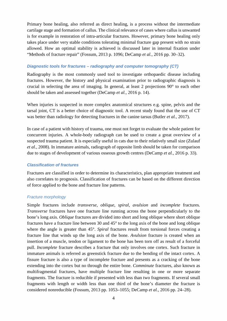
The appendicular skeleton is mostly made up of long bones divided anatomically into different regions: epiphysis, metaphysis and diaphysis (Figure 1). The physis, growth plate, is located within the metaphysis. Articular cartilage covers the joint surfaces, usually at each end of the long bone (Zachary and McGavin, 2012 pp. 925–926). Bone in a mature individual is divided into cortical and cancellous bone depending on its porosity. Cortical bone, also known as hard bone or compact bone, generally has porosity between 5 and 30% and forms the outer shell of almost all bones and forms the medullary canal by the diaphysis. The porosity of cancellous bone, also called spongy or trabecular bone, lies between 30 to 90% and are mostly found in the epiphysis and metaphysis of long bones (Whittick, 1990 pp. 167–170).
Figure 1. Gross anatomy of the long bone. (By Yusi Fang modified from Zachary and McGavin 2012, p. 926, FIG 16-10)

3
Fracture definition and causes
A fracture occurs when the force applied to the bone is greater than the strength of the bone leading to the disruption of the continuity of the bone or cartilage (Tobias and Johnston, 2012 p. 566; DeCamp et al., 2016 p. 24). Different types of forces, for instance axial, torsional and bending force, acting on the bone creates different types of fractures. In general, sudden force with high energy often result in complex fractures with excessive surrounding soft tissue damage whereas a bone under exposure of lower force will result in simple fractures with limited surrounding soft tissue damage (Tobias and Johnston 2012, p.568). Pathological fractures are fractures that occur in a site of bone that are not healthy, with the most common underlying disease being malignant bone tumours. These fractures often develop spontaneously as a result of minimal trauma (Rubin et al., 2015). If a bone is under cyclic stress there is a risk of developing fatigue fractures. An example of this is central tarsal bone fractures in racing greyhounds because of repetitive stress have been long known as a common cause of fatigue fractures. The majority of central tarsal bone fractures in greyhounds affect the right hind leg due to higher compression forces acting on the bone when the dogs run counter clockwise on the bended racing track (Boudrieau et al., 1984; Muir et al., 1999). Process of bone healing
Bones heal by restitutio ad integrum meaning restore to original condition. In other words, a properly healed bone after trauma is “as good as new”. Bone healing can be divided into primary (direct) and secondary (indirect) healing. The healing process is divided into 3 phases: the inflammatory phase, the reparative (proliferative) phase and the remodelling phase (Tobias and Johnston, 2012 p. 568). When motion between fracture fragments is present, indirect healing, which considering being the normal course of bone healing, will take place (Tobias and Johnston 2012 p. 568). The local deformation at the fracture site caused by motion is calculated as strain, the ratio between changes in gap width in correlation to total gap width given in percentage. In the healing process, different tissue will be formed depending on the amount of strain present at the fracture site. During the inflammatory phase shortly after a fracture occurs, haematoma carrying immunocompetent cells is formed adjacent to the fracture site. The haematoma is then replaced by granulation tissue, tolerating high strain in the initially very instable fracture site. With these tissues in place, motion as well as strain will decrease. Another factor decreasing strain is the resorption of bone that widens the fracture gap. With less strain present, fibrous connective tissue and fibrocartilage replace the granulation tissue and provide further stabilization of the fracture. During the reparative phase, mineralization of the fibrous cartilage into a callus can only occur when the fracture is stable enough. The revascularization carries osteogenic cells from the live bone tissue around and callus is formed both externally and internally. During the final remodelling phase that may last for years, mineralized callus is continuously remodelled and increase in strength and stiffness resulting in bone that does not differ in structure or function from bone prior to the injury (Fossum, 2013 pp.1095–1097).
4
Primary bone healing, also referred as direct healing, is a process without the intermediate cartilage stage and formation of callus. The clinical relevance of cases where callus is unwanted is for example in restoration of intra-articular fractures. However, primary bone healing only takes place under very stable conditions tolerating minimal fracture gap present with no strain allowed. How an optimal stability is achieved is discussed later in internal fixation under “Methods of fracture repair” (Fossum, 2013 p. 1096; DeCamp et al., 2016 pp. 30–32). Diagnostic tools for fractures – radiography and computer tomography (CT)
Radiography is the most commonly used tool to investigate orthopaedic disease including fractures. However, the history and physical examination prior to radiographic diagnosis is crucial in selecting the area of imaging. In general, at least 2 projections 90° to each other should be taken and assessed together (DeCamp et al., 2016 p. 14). When injuries is suspected in more complex anatomical structures e.g. spine, pelvis and the tarsal joint, CT is a better choice of diagnostic tool. A recent study found that the use of CT was better than radiology for detecting fractures in the canine tarsus (Butler et al., 2017). In case of a patient with history of trauma, one must not forget to evaluate the whole patient for concurrent injuries. A whole-body radiograph can be used to create a great overview of a suspected trauma patient. It is especially useful in cats due to their relatively small size (Zulauf et al., 2008). In immature animals, radiograph of opposite limb should be taken for comparison due to stages of development of various osseous growth centres (DeCamp et al., 2016 p. 33). Classification of fractures
Fractures are classified in order to determine its characteristics, plan appropriate treatment and also correlates to prognosis. Classification of fractures can be based on the different direction of force applied to the bone and fracture line patterns. Fracture morphology
Simple fractures include transverse, oblique, spiral, avulsion and incomplete fractures. Transverse fractures have one fracture line running across the bone perpendicularly to the bone’s long axis. Oblique fractures are divided into short and long oblique where short oblique fractures have a fracture line between 30 and 45° to the long axis of the bone and long oblique where the angle is greater than 45°. Spiral fractures result from torsional forces creating a fracture line that winds up the long axis of the bone. Avulsion fracture is created when an insertion of a muscle, tendon or ligament to the bone has been torn off as result of a forceful pull. Incomplete fracture describes a fracture that only involves one cortex. Such fracture in immature animals is referred as greenstick fracture due to the bending of the intact cortex. A fissure fracture is also a type of incomplete fracture and presents as a cracking of the bone extending into the cortex but no through the entire bone. Comminute fractures, also known as multifragmental fractures, have multiple fracture line resulting in one or more separate fragments. The fracture is reducible if presented with less than two fragments. If several small fragments with length or width less than one third of the bone’s diameter the fracture is considered nonreducible (Fossum, 2013 pp. 1053–1055; DeCamp et al., 2016 pp. 24–28).

5
Figure 2. Classification of fractures based on fracture morphology. (By Yusi Fang, modified from Fossum, 2013 p. 1054 FIG 32-18) Open fractures
An open fracture means that the fracture end has communication with the outer environment. It can generally be classified into 3 grades depending on the extent of soft tissue damage at the fracture site. A Grade I open fracture has small skin wounds created by the fracture end penetrating from the inside. The bone is then often drawn back to below the skin and is not necessarily visible in the wound. In a Grade II fracture, the wound is greater than in Grade I and usually caused by external forces. Grade III open fractures have extensive soft tissue damage sometimes combined with skin loss and the fracture is often comminute (Tobias and Johnston, 2012 pp. 572–573; Fossum, 2013 p.1054). Salter-Harris Fractures
The Salter Harris classification scheme describes the different fracture patterns that involve the physis, growth plate, in immature animals. Salter-Harris Type I has a fracture line running through the physis. Type II has fracture line running through the physis and part of the metaphysis. Type III is considered as an intra-articular fracture with fracture line through the physis and epiphysis. Type IV is also intra-articular with fracture line extending from the epiphysis to the metaphysis by crossing the physis. The final Type V results from crushing of the physis (Fossum, 2013 pp. 1054–1055).
Figure 3. Salter-Harris classification. By Yusi Fang, Modified from (Fossum, 2013 p.1054 FIG 32-18)

6
Fracture assessment score
Fossum (2013) suggested a fracture-assessment score system based on some of the fracture classifications mentioned above. Apart from considering the fracture itself, it also weighs in patient and client/owner information and functions as a guide to choose the most optimal approach of fracture management. The fracture-assessment score includes three different factors: mechanical, biological and clinical. Mechanical factors includes reducibility of the fracture, patient size and weight, and if injury or disease is present on other limbs. Evaluation of mechanical factors helps to evaluate how strong the fracture fixation should be for the individual patient. Biological factors take into account the age and general health of the patient, extent of soft tissue damage around the fracture both due to high-velocity injury and the surgeon’s skills during fracture repair. The biological factors play a role in estimating the healing time of the fracture. Clinical factors help assess the fracture healing during the postoperative period. It includes patient and owner compliance, the activity level of the patient and the comfort during fracture healing. The 3 factors should all be taken into account when assessing the prognosis of a fracture and fracture surgery. Fractures with high scores generally heal with lower complications risk whereas fractures assigned lower scores one can expect greater risk for complications (Fossum, 2013 pp.1055–1058). Methods of fracture repair
The aim of fracture repair is to restore function of the injured bone and for the patient to be able to use the limb (DeCamp et al., 2016 p. 33). There are many ways of managing a fracture, below are some of the surgical techniques of fracture repair. Intramedullary pins and Kirschner wires
Intramedullary (IM) pins are mostly used for diaphyseal fractures in femur, humerus, tibia, ulna, metacarpal and metatarsal fractures. The IM pins should not be used as stabilization of radial fractures due to interference of the elbow joint proximally and the carpal joint distally. The IM pins are highly resistant to bending force in all directions, but not to rotational force and are not either suitable in case of unstable fractures. Instead, the IM pins should be combined with other implants to achieve optimal stabilization. Kirschner wires (K-wire) can be applied as cross pin for fixation of physeal fractures of immature animals (Fossum, 2013 p. 1079–1082). Rush pinning is another technique useful for repair of physeal fractures, for instance one located at the distal femur. Similar to cross pinning, the rush pins are inserted distally to the physis into the medullary canal where it deflects off the opposite cortex from the inside at the proximal part of the bone (Tobias and Johnston, 2012 p. 899). Dowel pinning is a special method of fixation with IM pins, usually K-wires, that is a widely used technique in metacarpal and metatarsal fractures. The K-wire is inserted to the longer
7
fracture fragment leaving a part of the K-wire protruding. The shorter fracture fragment is then placed on the protruding K-wire until the fracture gap is closed (Zahn et al., 2007). Pins in combination with a tension band can be used as fixation method of avulsion fractures. The combination of the implants acts as counterforce for the muscle and hold the torn off fracture piece in place (Fossum, 2013 p. 1086). Screws
Different types and sizes of bone screws are available. Bone screws can be divided into cortical and cancellous (Tobias and Johnston, 2012 pp. 590–600). Bone screws can either be used as a lag screw or as a position screw. The aim of lag screw technique is to achieve interfragmental compression when the screw is tightened. Lag screws can be used when anatomical reconstruction is desired, for example reduction of intra-articular fractures where callus is unwanted. Under absolute stability of the lag screw, the fracture will heal by primary bone healing (AO Foundation, 2017). On the contrary to lag screw, the purpose of a position screw is to hold fragments in place without interfragmentary compression (Tobias and Johnston 2012, p.590). Bone plates
Bone plates and screws is widely used as internal fixation of fractures. Advantages with such fixations are immediate weigh bearing after surgery and little postoperative maintenance needed. Bone plate and screws can be used for stabilization of most fractures, but are more suitable for fractures with low fracture assessment scores (Fossum, 2013 p.1086). Many different bone plates are available depending on their intended application (Tobias and Johnston, 2012 p. 591). Stainless steel plates are used more often because it is cheaper than titanium plates. Depending on the desired function of the stabilization, a bone plate can be used as compression, neutralization or buttress plate. Different compression plates differ by both plate and plate hole configuration. A dynamic compression plate (DCP) has oval hole shape with an inclined plane. When the screw is tightened on each side of the fracture line, the screw heads will glide down the incline compressing the fracture line from each side. A limited-contact DCP (LC-DCP) has undercuttings between screw holes limiting the contact between plate and bone and thereby minimize the interruption of underlying blood flow (Fossum, 2013 p. 1088). A locking compression plate (LCP) has threaded plate holes enabling screws with threaded head to be lock in the plate and acting as one unit. In this way, the plate is not pressed to the bone and precise anatomical contouring of the plate is no longer necessary (Frigg, 2001; Miller and Goswami, 2007). A neutralization plate holds a bone section in anatomical position. It can for instance be used in case of a comminute fracture with several fragments reduced with primary fixations such as lag screws or cerclage wires. A neutralization plate then acts to prevent distraction force on the primary fixation implants (Colton, 2013).
8
Buttress or bridging plates aim to maintain the length of the bone as well as preventing rotation. In case of an unstable comminute fracture in the diaphysis where reconstruction is not possible, a buttress plate can be used (Fossum, 2013 p. 1089). External skeletal fixators (ESF)
External skeletal fixators can be used as fixation of long bone fractures. An ESF consists of pins placed on each side of the fracture line, joint together with a connecting bar outside the skin. The number of pins and connecting bars placed depend on the strength and stiffness one would like to achieve with the ESF. External skeletal fixators are also suitable for stabilization of unstable, comminute or open fractures. Usually, fractures located at the proximal long bones such as the humerus and femur cannot be adequately stabilized with ESFs only. To increase the fixation rigidity of such fractures, an IM pin can be combined with ESF. External skeletal fixators require postoperative care including management of pinholes to avoid the development of surgical site infections (SSI) (Fossum 2013, p.1067–1072). Cerclage wire
Cerclage wire can be used alone or in addition to the implants mentioned above in case of oblique or spiral fractures by compressing the fragment and holding them in place. However, cerclage wire as stabilizing device should be avoided in multifragmental fracture due to high risk of movement of the segment postoperatively causing the repaired fracture to collapse (Fossum, 2013 p. 1084) Perioperative administration of antimicrobials during fracture repair
The use of antibiotics adjacent to surgery depends on the type of injury and the risk of developing postoperative SSI. Surgical wounds are divided into a) clean, b) clean-contaminated, c) contaminated and d) dirty wounds. The general rate of SSI of all surgical wounds is about 5%. Perioperative prophylactic administration of antibiotics aims to prevent postoperative SSI whereas the use of antibiotics in therapeutic purpose is to eliminate an already established infection. However, some cases fall into the grey zone. For instance, a fairly fresh open fracture given perioperative antibiotics is considered to be early therapeutic rather than prophylactic. According to the Swedish Veterinary Association, the use of surgical implants itself in fracture surgery is not a sufficient indication for the use of prophylactic antibiotics (Sveriges Veterinärförbund, 2009 pp. 9–10). On the contrary, international literatures warrant the use of prophylactic antibiotics in connection with orthopaedic procedures such as open and extensive fracture repair (Fossum, 2013 p 92).
Coaptation splints and casts
Various different splints and cast can be used solemnly as indirect fixation method or to provide greater stability to surgically treated fractures. Generally, the cast or splint should cover at least one joint proximally and one joint distally to the injury site. Therefore, it is not suitable for injuries above the elbow or stifle joint.
9
Casts are moulded circularly around a limb. There are 2 types of casts that are often used: the thermomouldable or thermoplastic material and fiberglass/resin material. Both can be custom-made for optimal fit for the individual animal. Circular casts can be cut into half longitudinally (bivalved) to facilitate further cast changes. There are also premade splints that serve its supporting function by being placed on one side of the limb. All cast and splints should have adequate padding underneath to prevent abrasions and pressure wounds. Protruding structures on the limb should be carefully protected. The cast or splint should be checked at least once in a week, and sooner if the animal expresses discomfort towards the device (DeCamp et al., 2016 pp. 49–66). Complications after fracture repair Healing complications
Delayed union and nonunion
Delayed union and nonunion are complications that can occur during fracture healing. In a delayed union, the fracture is expected to eventually heal but the healing time is prolonged, whereas in case of nonunion the fracture is not able to heal regardless of healing time. The expected time of healing depends on many factors including mechanical, biological and clinical factors used in fracture assessment score (Tobias and Johnston, 2012 pp. 647–656; DeCamp et al., 2016 pp. 163–166). The most common cause of delayed union is excessive fracture gap, fracture instability causing movement at fracture site and inadequate blood supply (Jackson and Pacchiana, 2004). If the original reduction and fixations is satisfactory, restriction of the animal’s activity is usually enough as treatment of delayed unions to give the fracture a chance of healing. However, if the original fixation is considered to be inadequate, further surgical intervention is needed (DeCamp et al., 2016 pp. 163–166). Nonunion is failure for the fracture ends to unite and is divided into viable and nonviable. In case of nonunion the fracture cannot heal without surgical intervention (Jackson and Pacchiana, 2004). Most fractures have an expected healing time of at least 4 week. It is therefore unlikely for one to expect delayed or nonunion before 4 weeks after surgery (DeCamp et al., 2016 p. 163). Malunion
Malunion of a fracture is when the fracture has healed improperly or has healed in a anatomically noncorrect position. The causes can be due to failures in fracture repair, such as inadequately fixation or inappropriately treated reduction, but can also be caused by premature weight bearing shortly after surgery. Short-term consequences of malunions are reduced or impaired limb function whereas in the long term malunions can cause degenerative joint disease in adjacent joints. In serious cases of malunion, correction surgery is needed to restore limb function and prevent joint diseases (Jackson and Pacchiana, 2004; Tobias and Johnston, 2012 pp. 653–654).

10
Implant Failure
Implant failure associated with orthopaedic surgery is well documented in literature. In small animal medicine, fatigue failure is considered to be the most important type of implant failure. An example from large animal practice is the stabilization of a long bone fracture in a horse. When the horse attempts to stand up after surgery, enough force acting on the implant can cause acute implant breakage. However in small animal medicine there are rarely such extensive forces acting on an implant causing acute failure. Instead, the cyclic stress below the implants breaking point applied creates microscopic cracks in the implant and permanent changes in the structure of the implant whilst the implant takes the main load during fracture healing. It usually present clinically as the implant “suddenly” breaks several weeks after surgery. In practice, it is important for the surgeon to choose an appropriate size of implant for the animal that does not reach the fatigue failure point before bone healing. Other unusual causes that can contribute to implant failure are electrochemical and oxidation-reduction erosion, and implant manufacture defects (Coughlan et al., 1998 pp. 311–315). Surgical Site Infections and Biofilm
A surgical site infection can occur anywhere in the surgical area postoperatively (Tobias and Johnston, 2012 p. 135). Superficial incisional only involve the skin and subcutaneous tissues. Due to the use of implants, deep incisional and organ/space SSI are also relevant complications after fracture repair surgery (Mangram et al., 1999). Osteomyelitis, an inflammation in the bone or bone marrow, is classified as organ/space SSI. The infection occurs either as a result of trauma, the presence of foreign body like implants or inoculation of microbes capable of producing biofilms (Mangram et al., 1999; Tobias and Johnston, 2012 p. 669). According to IUPACs definition, biofilm is an aggregate of microorganisms, usually bacteria, in which cells that are frequently embedded within a self-produced matrix of extracellular polymeric substance adhere to each other and/or to a surface (Vert et al., 2012; Turk, 2013). Biofilms situated on orthopaedic implants is believed to cause chronic infections that develop over months and years (Stoodley et al., 2011). Bacteria within a biofilm are less susceptible to antimicrobial agents as a result of several different resistance mechanisms. First of all, the biofilm matrix itself acts as a physical protection barrier. The environment in the biofilm leads to decreased growth and metabolism of the bacteria and therefor limiting the effect of antimicrobials that affect bacterial growth, e.g. β-lactams. Resistance genes, e.g. efflux pumps and beta-lactamase contribute to antimicrobial resistance in biofilms (Percival et al., 2011 pp. 223–252).

11
Common fractures in dogs and cats
According to a survey study by Phillips, fractures of the radius and ulna, pelvis, femur and tibia were most common in dogs whereas fractures of femur, pelvis and mandible were more common in cats. The same author also found that 80% of the fractues occurred in animals under 3 years of age and 50% of the animals were under one year of age. Road accidents and fall and crush injuries were the main causes of fractures. Far from all fractures are surgically treated. Some animals are treated conservatively with or without coaptation and others are euthanized (Phillips, 1979). Fracture of the radius and ulna
The canine and feline antebrachium is composed of the radius and ulna as 2 paired long bones with the radius acting as the main weight bearing bone. The elbow joint is located proximally and the carpal joint distally. Fractures in the radius and ulna account for about 18% of all fractures in dogs (De Arburn Parent et al., 2017) and less than 10% of all fractures in cats (Phillips, 1979). Diaphyseal fractures, especially in the distal third of the bone, are the most common fracture site (Milovancev and Ralphs, 2004) and in most cases, the fracture involves both of the bones (Phillips, 1979). The choice of treatment for fractures in the radius and ulna depends partly on the nature of the fracture. Conservative treatment with cast or splint is only appropriate if the fracture is closed, incomplete, simple (transverse, oblique or spiral) with minimal displacement or only involving one of the 2 bones. The most commonly used fracture repair technique is bone plate and screws with the plate generally placed on the dorsal aspect of the radius. External skeletal fixators also had successful treatment results for radial and ulna fractures and is especially suitable for comminute and open fractures with tissue loss. In general, IM pinning is not recommended due to the curved anatomy of the radius and the risk of hurting the adjacent joints (Coughlan et al., 1998, pp. 197–201). Fracture of the radius and ulna in toy breed dogs
Small and toy breed dogs < 6kg body weight have greater prevalence of these fractures, often the result of minimal trauma from jumping or falling (Fossum, 2013 p. 1140; Gilbert et al., 2017). A study by Brianza et al. suggested that the antebrachii of small and toy breed dogs are more susceptible to fractures compared to large dogs due to morphological differences in cross-section properties (Brianza et al. 2006). Radial and ulnar fractures are also more prone to complications after surgical repair and there are several other factors suggested to be contributing risk factors for complications in these fractures. Biomechanical factors include high frequency of short oblique fractures with limited distal fracture fragment making it difficult to achieve anatomical reduction. Poor vascularization, limited soft tissue coverage and limited bone surface contact at fracture site due to small bone diameter are contributing biological factors. All the factors combined poses great challenges in fracture treatment (Ramírez and Macías, 2016; Gilbert et al., 2017).

12
The distal radius and ulna is the most common site to develop delayed union (Jackson and Pacchiana, 2004). Other documented complications associated with fracture repair of radius and ulna fractures in small dogs are malunion and refracture after implant removal (Larsen et al., 1999; Bierens et al., 2017). Treatment options for fractures in the radius and ulna
Several different treatment methods for fractures in the radius and ulna have been described, especially for toy breed dogs. The treatment options are conservative management, internal fixations or external fixations. Many studies has shown that open reduction with bone plate and screws in small dogs with distal radius and ulna fracture yield excellent functional outcome and low complication rate (Larsen et al., 1999; Kang et al., 2016; Ramírez and Macías, 2016; De Arburn Parent et al., 2017; Gilbert et al., 2017). In these studies, a bone plate is placed on the cranial aspect of the radius. Advantages of this method are the possibility to achieve optimal reduction and stability of the fracture as well as immediate weight bearing after surgery. However, due to open reduction technique, it may result in some degree of blood supply disruption at the fracture site (Milovancev and Ralphs, 2004). Hudson et al. stated that by using minimal invasive plate ostesosynthesis (MIPO) technique of the radius, vascular supply and soft tissue in the fracture site could be better preserved compared to open reduction. With MIPO, a plate is placed to the fracture site by making small incisions remote from the injury. This technique requires good anatomical knowledge because the fracture site cannot be directly visualized. The authors suggests that the best candidate for MIPO is an acute closed diaphyseal fracture that is mildly comminute, minimally displaced and has little adjacent soft tissue damage. In reality however, there are few cases that fits perfectly with these criteria (Hudson et al., 2012). The complications associated with bone plate and screw fixation techniques include implant failure, angulation, infection, and disuse osteopenia in addition to delayed and nonunion described earlier (Larsen et al., 1999). Another author group suggests that a bone plate placed to the medial aspect of the radius is an alternative method of fixating fractures in the radius and ulna in dogs of different sizes. The authors claim that medial approach eases implant application and can avoid the extensor tendons in the distal part of the radius. Also, the radius has a greater mediolateral thickness allowing longer screws to be placed. According to the authors, this method should be avoided if the fracture is in the proximal third of the forearm due to anatomical difficulties in applying the plate and in case of complex fractures (Sardinas and Montavon, 1997). ESF is preferred in case of open fractures. When appropriately placed, ESF disturbs vascular supply to a less extent and the implants are removed after fracture healing (Milovancev and Ralphs, 2004). The ESF method requires more postoperative management and documented complications associated with ESF as repair method include pin loosing, pin tract drainage, delayed, nonunion and malunion (Larsen et al., 1999). One author group found that miniature circular ESF can be used to manage fractures in the radius and ulna in young miniature dogs with successful outcome (Bierens et al., 2017).

13
ESF can also be combined with dowel pinning. In a study by Yu et al, toy breed dogs with a mean body weight of 3.13kg treated for fracture in the radius and ulna had an excellent alignment in fracture fragment when combining ESF with dowel pinning. However, this method resulted in some delayed time to bone healing compared to the group with closed reduction and ESF. One out of 51 dogs in that study included in the “dowel-pinning group” suffered a severe complication in the form of re-fractured radius and ulna several month after ESF removal (Yu et al., 2010). Therefore, the ESF should only be removed when radiological and clinical signs of bone healing is confirmed (Piermattei et al. 2006, p.94). Radius and Ulna Fractures in Cats
Although fracture in the radius and ulna is not as common in cats compared to dogs, repair of these fractures are challenging and the complication rate is high. Similar to the human forelimb, the antebrachium of cats have more ability for rotation (supination and pronation), approximately twice as much compared to the movement of dogs. Therefore, fixation of one of the bones only is often insufficient. The dual bone fixation method with an IM pin in the ulna in addition to a cranial bone plate in the dorsal aspect of the radius has shown to have lower complications rate compared to fixation of the radius only, most likely due to more rigid fixation (Wallace et al., 2009; Preston et al., 2016).
Humerus fractures
Humerus fractures account for approximately 10% of all fractures in dogs and cats (Phillips, 1979; Tobias and Johnston 2012 pp. 709–710) and most of these involve the middle and distal one third of the bone (Harari et al., 1986). Motor vehicle trauma seems to account for the majority of humeral fractures in dogs and cats (Bardet et al., 1983). Therefore, it is crucial to carefully evaluate for concurrent trauma to other parts of the body, especially the thorax and the skull (Tobias and Johnston, 2012 p. 709). The humerus has a unique shape and complex surrounding anatomy with important nerves and vessels making fracture repair a challenge. Depending on the nature of injury, both internal and external fixations are options for fracture repair in the humerus (Simpson, 2004). Fractures of the pelvis and multitrauma
Pelvic fractures account for 16–30% of all fractures in dogs and cats (Kipfer and Montavon, 2011; Phillips, 1979) with fracture in the ilium being the most common fracture in the pelvis accounting for 46% of all pelvic fractures (Harasen, 2007). Due to the box-like structure of the pelvis, bone fragment only displaces when fractures occur at 3 different sites. The weight-bearing axis accounting for the load transmission from the hind leg to the spine includes the sacroiliac joint, ilium, acetabulum and femoral head/neck. Therefore, patient with fracture in these areas are candidates for surgical treatment (Fossum, 2013 pp. 1169–1173). In case of acetabula fractures, surgical stabilization is especially important to prevent degenerative joint disease due to the involvement of the joint surface (Tobias and Johnston, 2012 p. 806). The most common cause of pelvic fractures is motor vehicle accidents, but pelvic fractures can also result from fall from heights. Patients with pelvic fractures often suffer from injuries

14
involving other body parts, with thoracic injuries, abdominal injuries, spinal injuries, soft tissue injuries and ruptured urine bladder being the common ones. Due to the great risk of anaesthetic complications in a multitrauma patient, soft tissue injuries should be identified and manage before surgical repair of the pelvic fracture, however within 7 to 10 days from trauma (Harasen, 2007; Tobias and Johnston 2012 p. 801). Femur fractures
Femoral fractures account for about 45% of all long bone fractures and approximately 15 to 30% of all fractures in dogs and cats (Phillips, 1979; Piermattei et al. 2006, p. 512). The majority of these fractures are results of motor vehicle trauma, and young animals less than 5 years of age are significantly overrepresented. As with pelvic fractures, concurrent injuries to other organ systems should be carefully examined. Because of abundant amount of thigh muscles around the femur that acts as protection, only 17% of femur fractures are open fractures (Tobias and Johnston 2012, p. 865; Fossum 2013, p.1181–1182). Fractures of the tibia and fibula
Fracture of tibia and fibula is also a common type of fracture making up about 10% of all fractures and 20% of all long bone fractures with motor vehicle trauma being the main cause. A significant majority of the fractures happen to dogs and cats less than one year of age and affect the diaphysis of the bone. Due to the sparse soft tissue coverage of the tibia and fibula, open fractures are fairly common, about 12–37% (Tobias and Johnston 2012, 1000–1001). Avulsion of the tibial tuberosity is a special type of tibial fracture and the most common type of fracture of the proximal tibia in immature dogs (Deahl et al., 2017), usually affecting animals between 4 and 8 months of age. The tibial tuberosity is a separate growth-centre that later fuses with the proximal tibia when the animal reaches maturity. The quadriceps muscle inserts at the tibial tuberosity through the straight patellar ligament (Piermattei et al. 2006, p. 639–640). If the quadriceps muscle is contracted simultaneously as the stifle joint is flexed and the foot standing on the group, the tibial tuberosity will be torn off resulting in an avulsion fracture (DeCamp et al., 2016 p. 682).

15
MATERIALS AND METHODS Data collection
Data were collected from patient’s medical records at University Animal Hospital, Uppsala, Sweden. Searches were made on all fractures during the time period from year 2012 to 2015 June in order to do the one year after surgery follow up study with start in year 2016. One case had surgery date November 2011. A total of 161 surgically treated fracture cases were studied. Fractures in the mandibula, fracture in ribs and nonsurgical treated fractures were excluded. Two fractures in the same patient occurring with a time span over a year were managed as two different cases, which is why there is a case with date of surgery in year 2016. Patient information
Basic patient information include gender, date of birth, and weight at date of surgery. All breeds were listed according to Federation Cynologique Nationale (FCI), the World Canine Organization. Breeds not recognized by FCI were categorized as mixed-breed. Cats without breed specified were listed as domestic cats regardless of coat length. The injury
Orthopaedic diagnosis 1 states the type of fracture and if the fracture was in addition an open fracture, intra-articular or Salter Harris fracture it was noted in orthopaedic diagnosis 2 prioritized in the order stated. Multitrauma (trauma that includes other organ systems as well as extensive soft tissue injury near fracture site) and comminute fracture (identified radiological or during surgery) were also noted. Surgery
Aspects of the surgery studied were date of surgery, type of surgery and whether implant was used or not. Intraoperative death was noted if it happened during anaesthesia. Intraoperative complications and anaesthetic complications were recorded but not included in this study. Antimicrobials
The types of antimicrobials used pre-operatively (the day or days before surgery), peri-operatively (induction of anaesthesia up to 24 hours after surgery) and post-operatively (antimicrobials given directly 24 hours after surgery) were noted. Postoperative complications
Short term (within 30 days) and long term (30–365 days) complications were evaluated. Many patients were referral cases from all over the country. Patients with missing information up to 1 year after surgery were followed up through telephone or e-mail interviews with owner. In some cases, the referral or after care hospital was contacted to obtain medical record and the complications studied. For complications occurring within 30 days, the most severe complication was documented as Complication 1. If more than one complication was developed, the second most severe complication was stated as Complication 2. Surgical site infection was one of the complications amongst all that was of interest regarding complications that occurred within 30 days after first surgery. Only superficial SSI were included in this study
16
and defined according to the Centres for Disease Control and Prevention guidelines (Mangram et al., 1999) with or without bacterial culture confirmation. Surgical site infection secondary to reoperation or pin removal were not included in this study. Implants predicted before surgery that needed to be removed were not classified as a complication, likewise incidents that could not be associated with the surgery and complications to other injuries in case of a multitrauma. Deaths
If death occurred, it was noted time (during surgery, within 30 days after surgery or 30–365 days after surgery) as well as cause of death whether it was directly related to the surgery or surgical disease. Statistical analysis
Continuous data were presented as median (Q1, Q3) due to non-normalized data. χ2 test was used for comparison between groups and Fisher’s exact when values were zero. A p < 0.5 was considered significant for all analysis. The statistics software program used for data analysis is Minitab (State College, Pennsylvania, USA). In case of data missing due to lost to follow up or data not applicable because of deaths, these cases were excluded in the current analysis.

17
RESULTS
Descriptive statistics for all fractures
Of all fracture cases studied (n = 161), 122 (76%) were dogs and 39 (24%) were cats. A total of 12 cases were lost to one-month or one-year follow-up. Weight and age of the dogs and cats that underwent fracture surgery are presented in Table 1 and Table 2. Table 1. Weight for dogs and cats in kg, for all surgically treated fractures
Weight
Median Q1 Q3 Min Max
Dogs (n=122)
7.45 2.88 19.63 1.00 44.00
Cats (n=39)
3.90 3.00 4.70 1.70 6.00
Table 2. Age for dogs and cats surgically treated for all fractures. Age presented as both years and months for easier overview
Age
Median Q1 Q3 Min Max
Years Months Years Month Years Months Years Months Years Months
Dogs (n=121*)
0.84 10.0 0.50 5.7 2.79 33.4 0.19 2.2 9.14 109.7
Cats (n=39)
1.53 18.3 0.62 7.4 3.34 40.1 0.31 3.7 15.0 179.6
*One dog excluded in this calculation due to unknown birth date The most common fracture type in dogs was fracture in the radius/ulna, while the most common fracture type in cats was femur fractures. The 5 most common types of fractures for dogs and cats respectively that underwent surgery are shown in Figure 4 and 5.

18
Figure 4. Five most common surgically treated fracture types in dogs. *Others include: fractures in the tarsus, pelvis, scapula, carpus, patella and tail.
Figure 5. Five most common surgically treated fracture types in cats. *Others include: fractures in pelvis and scapula.
38%
24%
12%
9%
7%
10%
Dogs (n total=122)
Radius/ulna
Tibia/fibula
Femur
Metacarpus/metatarsus andphalanges
Humerus
Others*
39%
15%
15%
13%
10%
8%
Cats (n total=39)
Femur
Tibia/fibula
Radius/ulna
Metacarpus/metatarsus andphalanges
Humerus
Others*

19
For dogs, the overall complication rate is 63.8% and for cats 50.0% (Table 3). The overall number of complications and complication rate is presented in Table 4. Additionally, the complication type and rate within 30 days after surgery and between 30 days and 1 year after surgery are presented in Table 4 and 5. Deaths and cause of deaths are shown in Table 6. Table 3. Overall postoperative complication rate for all fractures, dogs and cats shown together and separately
No complications Complications Complication rate, % X* Dogs and cats (n total=161) 60 93 60.8 8
Dogs (n total=122) 42 74 63.8 6 Cats (n total=39) 19 19 50.0 1
*Number of cases excluded in current analysis due to lost to follow up or deaths Table 4. Complication types and rate within 30 days after surgery (two most severe complications documented) for all fractures, an overview of dogs and cats together and separate. Some animals were documented with more than one complication explaining the fact that there are more complications than the number of cases with complications
Dogs and cats (n total=161)
Dogs (n total=122)
Cats (n total=39)
Complication type No. % No. % No. % Implant related 30 26.8 28 40.6 2 13.3
Surgical site infection 14 12.5 10 14.5 4 26.7
Splint/bandage related 22 19.6 17 24.6 5 33.3
Gastrointestinal 11 9.8 11 15.9 0 0 Circulatory 1 0.9 0 0 1 6.7
New fracture on same bone 1 0.9 0 0 0 0
New fracture at same site 2 1.8 0 0 2 13.3
Local ischemia in surgical area or distal
of surgical area 2 1.8 2 2.9 0 0
Others 2 1.8 1 1.4 1 11.6
Total of 112 complications in 67
cases X*=3
Total of 69 complications in 54
cases X*=2
Total of 15 complications in 13
cases X*=1
*Number of cases excluded in current analysis due to lost to follow up or deaths.

20
Table 5. Complication types between 30 days and 1 year after surgery (the most severe complications documented) for all fractures presented for dogs and cats together and separately. Only one complication was documented for this time period, therefore the number of complications is equal to the number of cases with complications
Dogs and cats (n total=161)
Dogs (n total=122)
Cats (n total=39)
Complication type No. % No. % No. % Implant related 23 47.9 18 45 5 62.5 Splint/bandage
related 4 8.3 4 10 0 0
Delayed union 6 14.6 6 15 1 12.5 Nonunion 4 8.3 4 10 0 0
Gastrointestinal 1 2.1 1 2.5 0 0 Urogenital 1 2.1 0 0 1 12.5
New fracture on same bone 2 4.2 2 5 0 0
New fracture at same site 1 2.1 1 2.5 0 0
Local ischemia in surgical area or distal
of surgical area 1 2.1 1 2.5 0 0
Others 4 8.3 3 7.5 1 12.5
Total of 48 complications in 48
cases X*=18
Total of 40 complications in 40
cases X*=16
Total of 8 complications in 8
cases X*=2
*Number of cases excluded in current analysis due to lost to follow up or deaths Table 6. Deaths within one year after all fracture repairs and cause of deaths, dogs and cats presented together and separately
Deaths Cause of death No. % X* Directly
related to surgery/surgical disease
Other cause not related to
surgery/surgical disease
Death related to surgery/surgical disease
but associated with other disease/cause
Dogs and cats (n total=161)
8 5.4 13 4 3 1
Dogs (n total=122)
6 5.4 10 4 1 1
Cats (n total=39)
2 5.6 3 0 2 0
*Number of cases excluded in current analysis due to lost to follow up or deaths

21
Descriptive statistics for fractures in radius and ulna
Fracture in the radius and/or ulna accounted for 32.9% (53/161) of all surgically treated fractures in dogs and cats. Of these, 88.7% (n = 47) were dogs and 11.3% (n = 6) were cats. A larger percentage of dogs underwent surgery for fractures in the radius/ulna of radial and ulnar fractures compared to cats, 38.5% (47/122) and 15.4% (6/39) respectively, see Figure 4 and 5. The 5 most common dog breeds that underwent surgery for fractures in the radius/ulna were mixed-breed (40.4%, 19/47), Poodle (14.9%, 7/47), Chihuahua (6.4%, 3/47), German spitz (6.4%, 3/47) and Continental toy spaniel (4.3%, 2/47). Weight and age for dogs and cats that underwent surgery after fracture in the radius/ulna are shown in Table 7 and 8. Table 7. Weight for dogs and cats surgically treated for fractures in the radius/ulna
Weight in kg
Median Q1 Q3 Min Max
Dogs (n=47)
3.00 2.00 8.70 1.00 33.00
Cats (n=6)
3.90 2.75 4.93 1.70 5.30
Table 8. Age for dogs and cats surgically treated for fractures in the radius/ulna, age presented as both years and months for easier overview
Age Median Q1 Q3 Min Max Years Months Years Month Years Months Years Months Years Months
Dogs (n=46*)
0.72 8.6 0.56 6.7 2.26 27.1 0.26 3.2 8.20 98.4
Cats (n=6)
1.76 21.1 1.01 12.12 3.02 36.2 0.31 3.7 4.37 52.4
*One dog excluded in this calculation due to unknown birth date Out of all 47 dogs with radius and ulna fractures, 10.4% (n = 4) had open fractures, 6.4% (n = 3) of the fractures involved a joint and 4.3% (n = 2) were Salter Harris fracture without joint involvement. One dog (2.1%) had multitrauma, 5 dogs (10.6%) comminute fractures and 2 dogs (4.3%) a combination of multitrauma and comminute fractures. Two of the 6 cats (33.3%) had open fractures and 1 cat (16.7%) had a fracture involving a joint. Only one of the cats (16.7%) suffered from both multitrauma and comminute fracture. All fracture repair surgeries used one or more implants. Plates and screws (72.3%) was the most commonly used surgical technique for repair of radius/ulna fractures in dogs (Figure 7.). Other surgical techniques are also presented for both dogs and cats in Figure 7. The vast majority of the cases of radius/ulna fracture repair did not receive antimicrobials before or after surgery. Instead, almost all animals received cephalosporin’s perioperatively (Figure 8).

22
Figure 7. Surgery techniques for repair of fractures in the radius and ulna in dogs and cats.
Figure 8. Antimoicrobial administration in conjunction with fracture repair of the radius and ulna fractures in dogs (n total=47). The overall postoperative complication rate for fractures in the radius/ulna, as well as complication type within 30 days after surgery and between 30 days and 1 year after surgery are presented in the same fashion in Table 9, 10 and 11 as for all fractures.
1 2
34
3 3 310
2 2 1 0 1 00
5
10
15
20
25
30
35
40
IM pin ESF Plate andscrews
Plate,screws and
IM pin
Pin andtensionband
Crosspinning
Rushpinning
Num
ber o
f cas
es
Type of surgery
Dogs (n total=47)
Cats (n total=6)
43
2
39
3
45
7
1 0 10
5
10
15
20
25
30
35
40
45
50
Preoperative Perioperative Postoperative
Num
ber o
f cas
es
Time correlated to surgery
No antimicrobials
Cephalosporins
Clindamycin

23
Table 9. Overall postoperative complication rate for fractures in the radius/ulna, dogs and cats shown together and separately
No complications Complications Complication rate, % X* Dogs and cats (n total=53) 26 25 49.0 2
Dogs (n otal=47) 21 24 53.3 2 Cats (n total=6) 5 1 16.7 0
*Number of cases excluded in current analysis due to lost to follow up or deaths Table 10. Complication types and rate within 30 days after surgery (two most severe complications documented) for fractures in the radius/ulna. Some animals were documented with more than one complication explaining the fact that there are more complications than the number of cases with complications
Dogs (n total = 47)
Cats (n total = 6)
Complication type No. % No. % Implant related 9 50.0 0 0
Surgical site infection 2 11.1 0 0
Splint/bandage related 1 5.6 1 100.0
Gastrointestinal 5 27.8 0 0 New fracture on
same bone 1 5.6 0 0
Total of 18 complications in 14
cases X* = 2
Total of 1 complications in 1
cases X* = 0
*Number of cases excluded in current analysis due to lost to follow up or deaths Table 11. Complication types between 30 days and 1 year after surgery (the most severe complications documented) for fractures in the radius/ulna. Only one complication was documented for this time period, therefore the number of complications is equal to the number of cases with complications
Dogs (n total = 47)
Cats (n total = 6)
Complication type No. % No. % Implant related 5 35.7 0 0 Delayed union 5 35.7 0 0
Nonunion 2 14.3 0 0 New fracture on
same bone 1 7.1 0 0
New fracture at same site 1 7.1 0 0
Total of 14 complications in 14
cases X* = 8
Total of 0 complications in 0
cases X* = 1
*Number of cases excluded in current analysis due to lost to follow up or deaths

24
The number of cases that went for reoperation or amputation are presented in Table 12. Details about some of the cases are discussed further below. Deaths and cause of death for dogs and cats undergoing fracture repair in the radius/ulna is shown in Table 13. Table 12. For all radius and ulna fractures, only dogs (n total = 47) suffered from reoperations and amputation. The second surgery closest in time to original fracture repair surgery is documented. If a patient first underwent reoperation and later amputation, only the reoperation is noted
No. % X* All reoperations 6 14.0 3
Unplanned reoperation due to failure of previous surgery
1 2.3 4
Amputation 2 4.5 3 *Number of cases excluded in current analysis due to lost to follow up or deaths Table 13. Deaths within one year after all fracture repair of fractures in the radius and ulna with cause of deaths, dogs and cats presented together and separately
Deaths Cause of death No. % X* Directly
related to surgery/surgical disease
Other cause not related to
surgery/surgical disease
Death related to surgery/surgical disease
but associated with other disease/cause
Dogs and cats (n total=53)
4 8.2 4 2 1 1
Dogs (n total=47)
3 7.0 4 2 0 1
Cats (n total=6)
1 16.7 0 0 1 0
*Number of cases excluded in current analysis due to lost to follow up or deaths Comparative statistics
Small and toy breed dogs weighing < 6kg accounted for 61.7% (29/47) of all fracture in the radius/ulna in dogs and there were significantly more dogs with body weight < 6kg compared to dogs > 6kg (p < 0.001). There were no significant difference in overall complication rate between dogs and cats for all surgically treated fractures (p > 0.05). Furthermore, a comparison was made between groups with fracture in the radius/ulna and other fractures excluding fractures in the radius/ulna in terms of overall complications, deaths and reoperations/amputations (Attachment 1). There were no significant differences between any of the compared groups (p > 0.05). There is a trend showing fewer overall postoperative complications in the group with radius/ulna fractures compared other factures, but the difference did not come out significant (p = 0.09). Odds ratio (OR) was also calculated. However, the OR cannot be interpreted due to confidential interval spans over one implying that there is no association between the 2 groups compared.

25
DISCUSSION
This retrospective study has provided a recent update of all surgically treated fractures at a university hospital in Sweden. Because fractures in the radius and ulna are common in dogs, especially small and toy breed dogs, the author focuses on these fractures in attempt to compare this group with other fractures regarding postoperative complications. There is a large spread of dog weight; both between minimum and maximum weigh, but also between Q1 and Q3. A possible explanation is the great size variety of dog breeds that suffered from fractures. There were significantly more dogs with body weight less than 6kg with surgically treated fractures in the radius/ulna compared to other fractures (p < 0.001). In the radius/ulna group, the median weight as well as Q1 and Q3 were lower supporting the theory that these fracture more often affect smaller dogs (Larsen et al., 1999; Brianza et al., 2006; Yu et al., 2010; De Arburn Parent et al., 2017). The median and spread of cat weight did not differ much between the group with all fractures and fractures in the radius/ulna. The median age for both dogs and cats with all fractures and fractures in the radius/ulna were low, < 1 year of age for dogs and < 2 years for cats. These numbers were consistent with earlier studies that have shown that 50% of all fracture in dogs and cats were in animals < 1 year of age (Phillips, 1979). The cause of injury was not documented in this current study, but the author’s hypothesis is that younger animals are more active and therefore exposed to greater risk of traumatic injuries, that has been shown to be the main cause of fractures (Phillips, 1979; Johnson et al., 1994). Due to differences in study design, the distribution of fracture types in the current study is hard to compare to previous studies. This study maps the distribution of various surgically treated fractures whereas other studies take into account all fractures, including the ones that are not surgically treated. A study reported almost 50% of all fractures were treated surgically, but it was not possible to decipher what proportion of each fracture type that were surgically repaired (Phillips, 1979). With safer anaesthesia and implants of better quality specialized for animals (Fossum 2013, p. 1086), one could assume that the proportion of surgically treated fractures today is much larger. In case of pelvic fractures, they were according to earlier studies the most common fracture type in dogs with 22.4% (Johnson et al., 1994) and 15.8% (Phillips, 1979) of all fractures in dogs and 24.8% in cats (Phillips, 1979). In the current study however, < 10% of all fractures in dogs and cats underwent surgery were pelvic fractures. One explanation to the differing data is that far from all pelvic fractures need to be treated surgically. An early report showed that 75% of dogs with pelvic fractures recovered completely with conservative treatment (Denny, 1978). Another explanation is that animals with pelvic fractures are more often euthanized due to concurrent injuries on other body parts. In the current study, surgically treated fractures in the radius and ulna make up 38.5% of all 161 fractures that underwent surgery, also being the most commonly surgically treated fracture type in dogs. Because non-surgically treated fractures are not included in this study, it is not possible to know how big portion of the fractures in radius/ulna were surgically repaired. However, earlier studies revealed that more than 80% of the conservatively treated fractures in the radius/ulna with external coaptation, especially in small and toy breed dogs, developed
26
complications that were serious, such as nonunions and malunion (Lappin et al., 1983 and Waters et al., 1993 see Milovancev and Ralphs, 2004). Therefore, the surgically treated fractures in the radius/ulna in the current study presumably represent a majority of all fractures in the radius/ulna. Even if the numbers are not comparable with earlier studies of fracture incidences from the 20th century, results from this current study with a percentage of almost 40 % surgically treated fractures in the radius/ulna is still high. Because the majority of radius/ulna fractures in dogs affect small and toy breed dogs, one reason behind the higher prevalence of these fractures is the increased popularity of small and toy breed dogs. Another possible reason is the differences in geographical distribution of small and toy breed dogs, with a higher occurrence in larger cities such as Uppsala where this study was carried out. The overall complication rate after fracture repair is 63.8% in dogs and 50.0% in cats. For both dogs and cats, the most common complication types are implant related complications, surgical site infections (superficial), splint/bandage related complications and delayed union. Each complication type are well described separately, but as mentioned earlier, there are no reports on overall complication rate and complication types for all fracture surgery to the author’s best knowledge. An overall complications rate of more than 60% in dogs might seem high, however one must not forget that it includes both major as well as minor complications such as gastrointestinal problems due to NSAID treatment. The number shows how many percent of all cases that have had at least one complication during a one-year period postoperatively. The proportion of patients that recover completely after surgery is far > 40% that did not have any complications postoperatively whatsoever. Almost all animals undergoing surgery for fractures in the radius/ulna received perioperative cephalosporins only. Previous studies have shown that perioperative use of either cefazolin (first generation cephalosporin) or potassium penicillin G as prophylaxis significantly decreases postoperative infection rate after elective orthopedic procedures (Whittem et al., 1999). Another study by Pratesi et al. found an overall SSI rate after clean orthopedic surgery with implants to be 12.9% (Pratesi et al., 2015). The number is well consistent with results in this study where the SSI rate for all fractures is 12.5% and for fractures in the radius/ulna being 11.1%. The same author group also found that postoperative antimicrobial administration has a protective effect against SSI postoperatively (Pratesi et al., 2015). It would be interesting to investigate whether perioperative administration of antimicrobials decreases the postoperative SSI rate. However, since there are only 2 cases in this study that did not receive perioperative antimicrobials, this comparison could not be done due to insufficient sample size. The overall complication rate for dogs undergoing surgical repair for fractures in the radius/ulna was 53.4%. Because the majority of dogs that went to surgery for fracture in the radius/ulna were small and toy breed dogs under 6kg (61.7%) and the main surgical technique used is with plate and screws (72.3%) the results in this study is considered comparable with other studies examining complication after fracture repair of the radius/ulna with plates and screws in toy breed dogs. Larsen et al. revealed an overall complication rate of 54% in their study (Larsen et al., 1999), well consistent with the findings in this study. Another study found an overall complication rate of 25% (De Arburn Parent et al., 2017). However, there are still some differences in the 3 studies, especially in the definition of complication. Both De Arburn et al

27
and Larsen et al considered mild angulation, lameness and thermal conduction as complications, which is not the case in the current study. On the contrary, gastrointestinal complications following NSAID treatment are included as complications in this study. The rate of gastrointestinal complication was fairly high, making up almost 30% of all complications within 30 days postoperatively. The most common types of postoperative complications within one year after surgical repair of fractures in the radius/ulna are implant related, delayed union, gastrointestinal, nonunion and surgical site infections (summarized data of Table 9 and 10). These are well-documented complications after fracture repair of radius/ulna in small and toy breed dogs. Implant related complications range from minor implant movements seen on radiographs to plate breakage. The risk for toy breed dogs to developing delayed union and nonunion after surgical repair of fractures in the radius/ulna is high due to various factors mentioned earlier. Gastrointestinal ulceration is a long known common side effect of treatments with non-steroidal anti-inflammatory drugs (NSAID) (Lascelles et al., 2007). Some of the complications lead to reoperations. Only one out of 6 reoperations were due to failure of previous surgery (Table 11). It was a 1.5 year old mixed-breed weighing 3kg that reoperated three weeks after the initial fracture repair with plate and screws due to angulation of the forelimb and movement of screw and fracture fragment. In 2 cases, amputation was used as salvation procedure due to complications in fracture healing. The first case was a 6.5 year old poodle weighing 4.7kg at the time of first surgery where the fracture was repaired with plate and screws. The fracture was healing without complications until 10 weeks postoperatively when the dog got into a fight with the family’s other dogs and fractured the same leg at one of the screw holes. The prognosis for amputation was considered better than that for reoperation. The dog recovered well after amputation and did not have any further complications. The other case was a 2-year old Chihuahua weighing 2.2kg at time of first surgery. Due to a distal fracture fragment < 5mm to the carpal joint, the fracture was fixated with cross pins and cerclage and stabilized with splint postoperatively. After no signs of fracture healing radiographically 2 months postoperatively, the affected leg was amputated with excellent outcome. Overall complications rate, deaths and reoperations/amputations were compared between groups with all fractures excluding radius/ulna and fractures in the radius and ulna with different inclusion criteria (Attachment 1). The comparison between all fractures excluding radius/ulna dogs and cats together, and fracture in the radius/ulna dogs and cats together showed a trend that there were more overall postoperative complications in other fracture compared to fracture in radius/ulna. This finding does not support earlier finding with higher complication rate after fracture repair in the radius/ulna, especially in small and toy breed dogs. However, there are many limitations in this study behind that finding. First of all, the p-value for the difference was 0.09 meaning it was not significant, and is therefore only a trend observed. Second of all, the 2 groups were not entirely comparable in terms of many other factors such as fracture type, animal size, complexity of the fractures, multitrauma etc. When attempting to compare all long bone fractures excluding radius/ulna fractures in dogs < 6kg body weight with

28
radius/ulna fractures in dogs < 6kg body weight there were no significant difference or trend regarding overall complications, deaths and reoperation/amputations. One main reason could be the result of a limited sample size in attempt to make the 2 groups comparable. This study has limitations due to its retrospective nature. Some data, especially follow-up records, can be non-reliable due to larger time span between the surgeries and follow-ups, which in a large portion of the cases done through contact with the owner. However, data concerning basic patient information and data in connection to the surgery are considered reliable. Most of the comparative analyses in this study were not significant. Due to the rare outcome of some of the parameters analyzes, a larger sample size is needed in hope of obtaining significant data.
CONCLUSION
Out of a total of 161 cases studied, fracture in the radius/ulna is the most common fracture type in dogs, making up 38% of all surgically treated fractures. The median age for both dogs and cats were young, < 2 years of age. There were significantly more dogs with body weight less than 6kg suffering from fractures in the radius/ulna compared to dogs weighing more than 6kg. The overall complication rate for all fractures dogs and cats combined was 60.8% and for radius/ulna 49.0%. A larger sample size is needed in order to determine a significant difference between groups with radius/ulna fractures and all fractures excluding radius/ulna regarding overall complication rate, deaths and reoperation/amputation. As mentioned earlier, the current study is part of a greater study about postoperative complications after orthopaedic surgery in hope of providing further interesting data in this field.

29
ACKNOWLEDGEMENTS
I would like to thank my supervisor Annika Bergström, Swedish University of Agricultural Sciences in Uppsala, for being helpful and a huge inspiration during the journey. Ulf Emanuelsson, Swedish University of Agricultural Sciences in Uppsala, for being kind and educative with the statistical analysis. Jens Häggström, Swedish University of Agricultural Sciences in Uppsala, for being a knowledgeable and humorous examiner-in-chief.

30
REFERENCES Agria (2015) Ökade antal hundar i olyckor. Available:
https://www.agria.se/pressrum/pressmeddelanden-2015/okat-antal-hundar-i-olyckor/ [2017-11-11]
AO Foundation. Lag screw technique. Available: https://www2.aofoundation.org/wps/portal/!ut/p/a0/04_Sj9CPykssy0xPLMnMz0vMAfGjzOKN_A0M3D2DDbz9_UMMDRyDXQ3dw9wMDAx8jfULsh0VAdAsNSU!/?soloState=true&contentUrl=/srg/popup/further_reading/41/41_X10-Lag-screw-technique.jsp - JumpLabelNr1 [2017-11-11]
AO Foundation. Surgical fixation with absolute stability. Available: https://www2.aofoundation.org/wps/portal/!ut/p/a0/04_Sj9CPykssy0xPLMnMz0vMAfGjzOKN_A0M3D2DDbz9_UMMDRyDXQ3dw9wMDAx8jfULsh0VAdAsNSU!/?bone=Humerus&segment=Shaft&soloState=lyteframe&contentUrl=srg/popup/further_reading/PFxM2/12_44_Fx_w_abs_stability.jsp [2017-11-11]
Bardet, J.F., Hohn, R.B., Rudy, R.L., Olmstead, M.L. (1983). Fractures of the humerus in dogs and cats a retrospective study of 130 cases. Veterinary Surgery, vol. 12(2), 73–77.
Bergström, A., Dimopoulou, M., Eldh, M. (2016). Reduction of surgical complications in dogs and cats by the use of a surgical safety checklist: surgical checklist to reduce postoperative complications. Veterinary Surgery, vol. 45(5), 571-576.
Bierens, D., Unis, M.D., Cabrera, S.Y., Kass, P.H., Owen, T.J., Mueller, M.G. (2017). Radius and ulna fracture repair with the IMEX miniature circular external skeletal fixation system in 37 small and toy breed dogs: A retrospective study. Veterinary Surgery, vol. 46(4), 587–595.
Boudrieau, R.J., Dee, J.F., Dee, L.G. (1984). Central tarsal bone fractures in the racing Greyhound: a review of 114 cases. Journal of the American Veterinary Medical Association, vol. 184(12), 1486–1491.
Brianza, S.Z.M., Delise, M., Maddalena Ferraris, M., D’Amelio, P., Botti, P. (2006). Cross-sectional geometrical properties of distal radius and ulna in large, medium and toy breed dogs. Journal of Biomechanics. vol. 39(2), 302–311.
Butler, D., Nemanic, S., Warnock, J.J. (2017). Comparison of radiography and computed tomography to evaluate fractures of the canine tarsus. Veterinary Radiology & Ultrasound. https://doi.org/10.1111/vru.12558
Colton C, Orsen J. (2013). Plates - form and function. AO trauma ORP.
Coughlan, A.R., Miller, A. (1998). BSAVA manual of small animal fracture repair and management. British Small Animal Veterinary Association, Shurdington, Cheltenham, U.K.
Deahl, L., Ben-Amotz, R., Caceres, A.V., Agnello, K.A. (2017). Proximal tibial metaphyseal fractures in immature dogs: Veterinary and Comparative Orthopaedics and Traumatology. DOI: 10.3415/VCOT-16-11-0154.
De Arburn Parent, R., Benamou, J., Gatineau, M., Clerfond, P., Planté, J. (2017). Open reduction and cranial bone plate fixation of fractures involving the distal aspect of the radius and ulna in miniature- and toy-breed dogs: 102 cases (2008–2015). Journal of the American Veterinary Medical Association, vol. 250(12), 1419–1426.
DeCamp, C.E., Johnston, S.A., Déjardin, L.M., Schaefer, S.L. (2016). Brinker, Piermattei, and Flo’s handbook of small animal orthopedics and fracture repair, Fifth edition. ed. Elsevier, St. Louis, Missouri.

31
Denny, H.R. (1978). Pelvic fractures in the dog: a review of 123 cases. Journal of Small Animal Practice. vol. 19(1-12), 151–166.
Fossum, T.W. (2013). Small animal surgery, 4th ed. ed. Elsevier Mosby, St. Louis, Mo.
Frigg, R., (2001). Locking Compression Plate (LCP). An osteosynthesis plate based on the dynamic compression plate and the point contact fixator (PC-Fix). Injury, vol. 32, 63–66.
Gilbert, E.D., Lewis, D.D., Townsend, S., Kim, S.E. (2017). Comparison of two external fixator systems for fracture reduction during minimally invasive plate osteosynthesis in simulated antebrachial fractures. Veterinary Surgery. https://doi.org/10.1111/vsu.12687
Harari, J., Roe, S.C., Johnson, A.L., Smith, C.W. (1986). Medial plating for the repair of middle and distal diaphyseal fractures of the humerus in dogs. Veterinary Surgery, vol. 15, 45–48.
Harasen, G. (2007). Pelvic Fractures. The Canadian Veterinary Journal, vol. 48(4), 427–428.
Hudson, C.C., Lewis, D.D., Pozzi, A. (2012). Minimally invasive plate osteosynthesis in small animals. Veterinary Clinics of North America: Small Animal Practice, vol. 42(5), 983–996.
Jackson, L.C., Pacchiana, P.D. (2004). Common complications of fracture repair. Clinical Techniques in Small Animal Practice, vol. 19(3), 168–179.
Johnson, J.A., Austin, C., Breur, G.J. (1994). Incidence of canine appendicular musculoskeletal disorders in 16 veterinary teaching hospitals from 1980 through 1989. Veterinary and Comparative Orthopaedics and Traumatology, vol. 7, 5–18.
Kang, B.-J., Ryu, H.-H., Park, S., Kim, Y., Kweon, O.-K., Hayashi, K. (2016). Clinical evaluation of a mini locking plate system for fracture repair of the radius and ulna in miniature breed dogs. Veterinary and Comparative Orthopaedics and Traumatology, vol. 29(6), 522–527.
Kipfer, N.M., Montavon, P.M. (2011). Fixation of pelvic floor fractures in cats: Veterinary and Comparative Orthopaedics and Traumatology, vol. 24(2), 137–141.
Lappin, M., Aron, D., Herron, H. (1983). Fractures of the radius and ulna in the dog. Journal of the American Animal Hospital Association, vol. 19, 643–50.
Larsen, L., Roush, J., McLaughlin, R. (1999). Bone plate fixation of distal radius and ulna fractures in small- and miniature-breed dogs. Journal of the American Animal Hospital Association, vol. 35(3), 243–250.
Lascelles, B.D.X., Court, M.H., Hardie, E.M., Robertson, S.A. (2007). Nonsteroidal anti-inflammatory drugs in cats: a review. Veterinary Anaesthesia and Analgesia, vol. 34(4), 228–250.
Mangram, A.J., Horan, T.C., Pearson, M.L., Silver, L.C., Jarvis, W.R. (1999). Guideline for prevention of surgical site infection, 1999. American Journal of Infection Control, vol. 27(2), 97–134.
Miller, D.L., Goswami, T. (2007). A review of locking compression plate biomechanics and their advantages as internal fixators in fracture healing. Clinical Biomechanics, vol. 22(10), 1049–1062.
Milovancev, M., Ralphs, S.C. (2004). Radius/Ulna fracture repair. Clinical Techniques in Small Animal Practice. vol. 19(3), 128–133.
Muir, P., Johnson, K.., Ruaux-Mason, C. (1999). In vivo matrix microdamage in a naturally occurring canine fatigue fracture. BONE, vol. 25, 571–576.
Percival, S., Knottenbelt, D., Cochrane, C. (2011). Biofilms and veterinary medicine, springer series on biofilms. Springer Berlin Heidelberg, Berlin, Heidelberg.

32
Phillips, I.R. (1979). A survey of bone fractures in the dog and cat. Journal of Small Animal Practice, vol. 20(11), 661–674.
Piermattei, D.L., Flo, G.L., DeCamp, C.E. (2006). Brinker, Piermattei, and Flo’s handbook of small animal orthopedics and fracture repair, 4th ed. Saunders/Elsevier, St. Louis, Mo.
Pratesi, A., Moores, A.P., Downes, C., Grierson, J., Maddox, T.W. (2015). Efficacy of postoperative antimicrobial use for clean orthopedic implant surgery in dogs: a prospective randomized study in 100 consecutive cases: postoperative antimicrobials in orthopedic implant surgery in dogs. Veterinary Surgery. vol. 44(5), 653–660.
Preston, T.J., Glyde, M., Hosgood, G., Day, R.E. (2016). Dual bone fixation: a biomechanical comparison of 3 implant constructs in a mid-diaphyseal fracture model of the feline radius and ulna: dual bone fixation in cats. Veterinary Surgery, vol. 45(3), 289–294.
Ramírez, J., Macías, C. (2016). Conventional bone plate fixation of distal radius and ulna fractures in toy breed dogs. Australian Veterinary Journal, vol. 94, 76–80.
Rubin, J.A., Suran, J.N., Brown, D.C., Agnello, K.A. (2015). Factors associated with pathological fractures in dogs with appendicular primary bone neoplasia: 84 cases (2007–2013). Journal of the American Veterinary Medical Association, vol. 247(8), 917–923.
Sardinas, J.C., Montavon, P.M. (1997). Use of a medial bone plate for repair of radius and ulna fractures in dogs and cats: a report of 22 cases. Veterinary Surgery, vol. 26(2), 108–113.
Simpson, A.M. (2004). Fractures of the humerus. Clinical Techniques in Small Animal Practice. vo. 19(3), 120–127.
Stoodley, P., Ehrlich, G.D., Sedghizadeh, P.P., Hall-Stoodley, L., Baratz, M.E., Altman, D.T., Sotereanos, N.G., William Costerton, J., DeMeo, P. (2011). Orthopaedic biofilm infections. Current Orthopaedic Practice. vol. 22(6), 558–563.
Sveriges Veterinärförbund (2009), Antibiotikapolicy för hund och katt-sjukvård
Tobias, K.M., Johnston, S.A. (2012). Veterinary Surgery: Small Animal. Elsevier, St. Louis, Mo.
Turk, R. (2013). Prospective Evaluation of the Epidemiology and Microbiology of Surgical Site Infections. Diss. University of Guelph, Ontario, Canada
Vert, M., Doi, Y., Hellwich, K.-H., Hess, M., Hodge, P., Kubisa, P., Rinaudo, M., Schué, F. (2012). Terminology for biorelated polymers and applications (IUPAC Recommendations 2012). Pure and Applied Chemistry, 84. https://doi.org/10.1351/PAC-REC-10-12-04
Wallace, A., De La Puerta, B., Trayhorn, D., Moores, A., Langley-Hobbs, S. (2009). Feline combined diaphyseal radial and ulnar fractures: A retrospective study of 28 cases. Veterinary and Comparative Orthopaedics and Traumatology. vol. 22(1): 38-46.
Waters, D., Breur, G., Toombs, J. (1993). Treatment of common forelimb fractures in miniature and toy breed dogs. The Journal of the American Animal Hospital Association, vol. 29, 442–448.
Whittem, T.L., Johnson, A.L., Smith, C.W., Schaeffer, D.J., Coolman, B.R., Averill, S.M., Cooper, T.K., Merkin, G.R. (1999). Effect of perioperative prophylactic antimicrobial treatment in dogs undergoing elective orthopedic surgery. Journal of the American Veterinary Medical Association, vol. 215(2), 212–216.
Whittick, W.G. (1990). Canine Orthopedics, 2nd ed. Lea & Febiger, Philadelphia.

33
Yu, J., DeCamp, C.E., Rooks, R. (2010). Improving surgical reduction in radial fractures using a “dowel” pinning technique in miniature and toy breed dogs: Veterinary and Comparative Orthopaedics and Traumatology, vol. 24(1), 45–49.
Zachary, J.F., McGavin, M.D. (2012). Pathologic Basis of Veterinary Disease, 5th ed. ed. Elsevier, St.Louis, Mo.
Zahn, K., Kornmayer, M., Matis, U. (2007). “Dowel” pinning for feline metacarpal and metatarsal fractures: Veterinary and Comparative Orthopaedics and Traumatology. vol. 20(4): 256-63.
Zulauf, D., Kaser-Hotz, B., Hässig, M., Voss, K., Montavon, P. (2008). Radiographic examination and outcome in consecutive feline trauma patients: Veterinary and Comparative Orthopaedics and Traumatology. vol. 21(1):36-40.

34
ATTACHMENTS
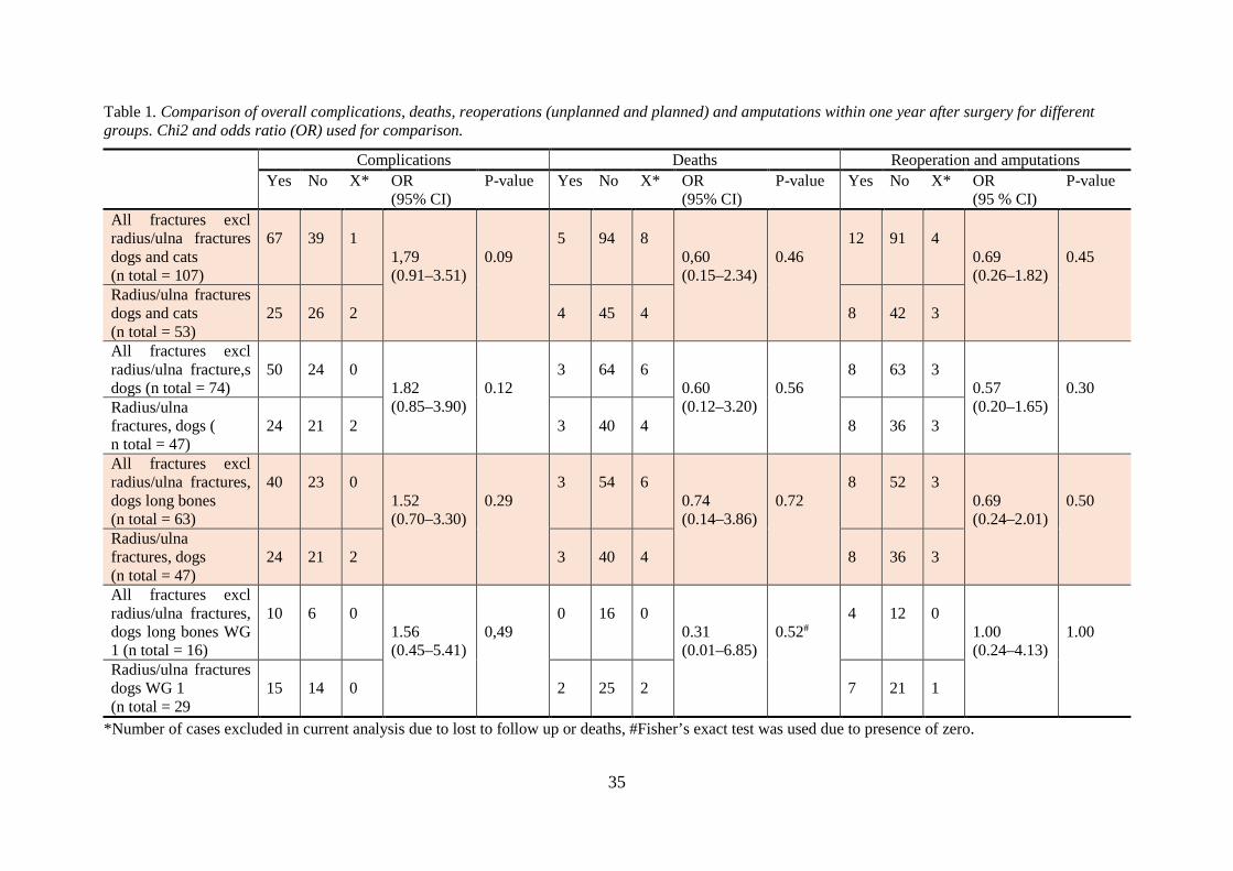
35
Table 1. Comparison of overall complications, deaths, reoperations (unplanned and planned) and amputations within one year after surgery for different groups. Chi2 and odds ratio (OR) used for comparison.
Complications Deaths Reoperation and amputations Yes No X*
OR (95% CI)
P-value Yes No X*
OR (95% CI)
P-value Yes No X*
OR (95 % CI)
P-value
All fractures excl radius/ulna fractures dogs and cats (n total = 107)
67
39
1
1,79 (0.91–3.51)
0.09
5
94
8
0,60 (0.15–2.34)
0.46
12
91
4
0.69 (0.26–1.82)
0.45
Radius/ulna fractures dogs and cats (n total = 53)
25
26
2
4
45
4
8
42
3
All fractures excl radius/ulna fracture,s dogs (n total = 74)
50
24
0
1.82 (0.85–3.90)
0.12
3
64
6
0.60 (0.12–3.20)
0.56
8
63
3
0.57 (0.20–1.65)
0.30
Radius/ulna fractures, dogs ( n total = 47)
24
21
2
3
40
4
8
36
3
All fractures excl radius/ulna fractures, dogs long bones (n total = 63)
40
23
0
1.52 (0.70–3.30)
0.29
3
54
6
0.74 (0.14–3.86)
0.72
8
52
3
0.69 (0.24–2.01)
0.50
Radius/ulna fractures, dogs (n total = 47)
24
21
2
3
40
4
8
36
3
All fractures excl radius/ulna fractures, dogs long bones WG 1 (n total = 16)
10
6
0
1.56 (0.45–5.41)
0,49
0
16
0
0.31 (0.01–6.85)
0.52#
4
12
0
1.00 (0.24–4.13)
1.00
Radius/ulna fractures dogs WG 1 (n total = 29
15
14
0
2
25
2
7
21
1
*Number of cases excluded in current analysis due to lost to follow up or deaths, #Fisher’s exact test was used due to presence of zero.