a novel approach to rapidly and sensitively detect and ......sarcina aurantiaca was used as a model...
TRANSCRIPT

A Novel Approach to Rapidly and Sensitively Detect and Purify Water Contaminated with
Shigella, E. coli, Salmonella, and Cholera
Authors: Ryan Thorpe and Rachel Chang, Manhasset High School, New York
Entry into the Stockholm Junior Water Prize (2017)
`

2
Abstract
Waterborne diseases cause 3.4 million deaths annually, concentrated in countries lacking
sanitary water. Conventional methods, such as PCR, can be costly, take several days, and have
detection limits of up to 1,000 CFUs. Thus, the purpose of this study was to engineer a system
that could efficiently detect and purify bacterial presence in a more rapid time frame and a lower
detection threshold. Graphene was utilized to create biosensors through the immobilization of
enzymes that target analytes released during respiration of Salmonella, Shigella, Cholera, and E.
coli. When these bacteria respire, they produce analytes that, when in contact with the enzymes,
produce a varying electric current that can be read by an ammeter to determine the presence of
bacteria. The sensors were able to detect the presence of at least 1 CFU of bacteria in 1 L of
water instantaneously. A mechanized approach was then taken to purify contaminated water
samples through the use of arduino microcontrollers. This system was successful in detecting
minute levels of bacteria in a rapid time frame and purifying the water of pathogens.

3
Table of Contents
Chapter Title Page Number
Abstract 2
Preliminary Matters 4
Introduction 5-7
Materials and Methods 7-12
Discussion and Results 12-19
Conclusion 20
Bibliography 20-21

4
III. Key Words
Waterborne diseases, graphene, biosensor, π-π interactions, enzyme immobilization,
Arduino, hydroxyl radicals, bacterial contamination
IV. Abbreviations and Acronyms
1-pyrenebutyric acid (PBA), Colony Forming Unit (CFU), Escherichia coli (E. coli),
CVD (chemical vapor deposition), PCR (polymerase chain reaction), DMSO (dimethyl
sulfoxide), hydroxyl radicals (OH⠂),
V. Acknowledgements
We would like to thank our amazing research mentor, Ms. Alison Huenger, for her
continued support and guidance. Without her dedication and expertise, we could not be where we
are today.
VI. Biography
Rachel Chang is a rising senior at Manhasset High School. After Rachel was fortuitously
put into her school’s science research program by accident, she came to truly love research and
hopes to pursue it as a career. Rachel has also been playing violin since she was 4 years old,
picking up new instruments along the way. She is the concertmaster of the Metropolitan Youth
Orchestra and the school’s symphony orchestra, and was fortunate enough to participate in the
All-State Symphony Orchestra this past year. Rachel hopes to continue working in the field of
Environmental Engineering in order to create novel solutions to environmental problems faced
around the world.
Ryan Thorpe will be entering his senior year at Manhasset High School. Ryan first
became interested in research in the 8th grade after he visited the science research lab at his
school. Since then, Ryan has developed an intense passion for science, and he has researched
sleep deprivation, bacterial contamination, and breast cancer development in both a high school
and professional lab setting. Outside of science, Ryan is an avid cross country and track runner,
musician, and is a founding member of the Manhasset Historical Society. In the future, Ryan
wishes to continue his education in the field of biomedical or environmental engineering and
hopes to develop sensors as a means of improving medical science and the environment.

5
Introduction
Waterborne diseases cause 3.4 million deaths annually. Worldwide, there exists 783
million individuals that do not have access to clean, potable water. The consumption and usage
of contaminated water is the leading cause of the development of debilitating waterborne
diseases such as Cholera, Shigellosis, E. coli, and Salmonella. 319 million of these people do not
have access to purification methods in order to eliminate biological contaminants in their water.
[4] Yet, despite this severity, the traditional methods used to detect the presence of these bacteria
can take 1-2 days, and have a detection limits of up to 1,000 CFUs [3]. As these pathogens have
no known cure, a more sensitive, rapid detection device and purification method must be created
in order to identify and eliminate microorganism presence in water sources to prevent
contamination and the outbreak of pathogenic disease.
A biosensor is a type of analytical device that is capable of detecting a biological
component and converting it into an electric signal through an electrode and a transducer. These
sensors can detect analytes rapidly, specifically, and sensitively utilizing receptors, nucleic acids,
antibodies, or enzymes [7, 8]. In this study, biosensors were constructed using graphene and
enzymes in order to detect analytes produced by waterborne pathogens. Graphene is a two-
dimensional allotrope of carbon that has a one atom thick hexagonal molecular structure with
benzene rings. Due to the unique nature of its structure, electrons are able to flow rapidly
throughout the thin material [5]. With this high conductivity and electron mobility, it serves as an
ideal electrode for the creation of biosensors. Putzbach’s study [14] determined that enzymatic
biosensors can be created through the incorporation of nanomaterials. Electrochemical
biosensors detect materials through an electric signal that is created by an enzymatic reaction.
Putzbach[14] determined that enzymes act as the transducer of the biosensor. Enzymes provide
selectivity to the biosensor, which make it the optimal transducer for detection devices. The
enzyme layer is then able to catalyze the production of a current that generates a signal.
Enzymes can be used to trigger an electrochemical response from a graphene biosensor to
detect the presence of specific analytes released by bacteria during respiration. Utilizing enzymes
in order to create electrochemical biosensors has been done in previous studies. For example,

6
Labroo [12] constructed a nanosensor in order to detect lactate in human sweat. This was done
by transferring a graphene electrode onto a flexible substrate and immobilizing lactate
dehydrogenase for lactate onto the electrode. When the lactate came into contact with this
enzyme, it was converted into an electrical stimulus that displayed the presence of this
biomolecule. [12] Similarly, Ang et. al [2] created an amperometric biosensor in order to
measure the glucose content of fruit. Ang’s study immobilized glucose oxidase onto a chitosan
membrane and attached it onto platinum electrode in order to measure electrical output. This
allows the presence of glucose to be monitored on the electrodes.
These methods were utilized in the formation of the biosensors for bacterial analyte
detection, as the utilization of oxidase enzymes and graphene electrodes were essential in
converting bacterial respiratory products into a detectable current. In this experiment, the
enzymes Lactate Oxidase, Glucose Oxidase, Glycerol Oxidase, and Galactose Oxidase were
immobilized by utilizing a linker molecule, 1-pyrenebutyric acid (PBA). This acid is a linker
molecule that acts as a bridge between the enzyme and the graphene through π-stacking. PBA
has an aromatic molecular structure, containing benzene rings that non-covalently stabilize atop
the graphene hexagonal rings. These enzymes are immobilized onto the graphene through π-π
interactions. π-interactions are a type of non-covalent bond that can exist between π-systems. π-
interactions are extremely stable and control a broad spectrum of biological interactions, such as
interactions between aromatic molecules and protein structures. [7] π-interactions were analyzed
in their applications in enzyme immobilization. π-π interactions occur between 2 aromatic
molecules, or 2 molecules that contain hexagonal benzene rings in their molecular structure. The
two benzene rings, or the π rings, are able to stack up one another and align themselves to stably
bind the molecules together. Due to this property, enzymes can be utilized to engineer enzymatic
biosensors to detect various biomolecules. The linker molecule then bonds with the enzyme
through an amide bond that occurs between the amine group of the enzyme and the pyrene group
of the PBA, a process known as amidation. [10,12] This safely and securely immobilizes the
enzymes onto the graphene without damaging either structure. These π-interactions are necessary
to allow for the permanent immobilization of enzymes, which act as the transducer of the
biosensor for the detection of biomolecules.

7
Equally as important as the detection of bacteria is the elimination of bacteria from water
sources. Hydrogen peroxide and sodium hydroxide can be utilized in order to eliminate organic
material in water. When hydrogen peroxide and sodium hydroxide are added to water, the water
will experience a raise in pH that contributes to a process known as hydrogen peroxide
bleaching, a process by which hydroxyl radicals are formed. These radicals have the potential to
degrade lipids and proteins. As bacterial pathogens are made of lipids and proteins, these radicals
can eliminate bacterial presence. [11] This would effectively purify water and make it safe for
consumption, as the only byproducts of this reaction are carbon dioxide and water.
Engineering Goal
Despite the widespread nature and severity of bacterial contamination in water, there is a
lack of sensitive and rapid technology to detect these microorganisms. The purpose and
engineering goal of this study was to address the limitations of both the detection and
purification of pathogens. This experiment aimed to create various graphene-based biosensors in
order to recognize minute levels of specific bacteria in a rapid time frame and fabricate a
mechanized purification system to eliminate this bacterial presence.
Materials and Methodology
Phase 1:
A graphene biosensor was created in order to rapidly and sensitively detect bacterial
contamination in water by S. typhi, V. cholerae, E. coli, and Shigella through the isolation of
graphene and immobilization of specific enzymes that target the analytes produced by each
bacterium.
In this study, E. coli K12 was used as a model organism for E. coli O157:H7,
Enterobacter aerogenes modelled Shigella dysenteriae, Sarcina aurantiaca was used as a model
for Salmonella typhi, and Vibrio fischeri modelled Vibrio cholerae, because of the metabolic and
respiratory parallels between the organisms. Firstly, both E. coli K12 and E. coli O157:H7 are
gram-negative, rod-shaped, and both perform mixed acid anaerobic and aerobic respiration. It is
through respiration that both strains of E. coli can generate D-lactate (Forester, 2014). Second,
Sarcina aurantiaca was used as a model organism for S. typhimurium as both organisms have
CrsA-monitored respiration pathways by which glucose-6-P is produced [1]. Third, Vibrio

8
fischeri was used as a model organism for Vibrio cholerae because both organisms belong to the
Vibrio genus, and both organisms produce ɑ-D-Galactose-1,P as a byproduct of glycolysis.
Finally, Enterobacter aerogenes was used as a model organism for Shigella dysenteriae because
both organisms produce glycerol during respiration [13].
Graphene Isolation
Graphene was isolated in two manners in order to analyze the efficiency of more cost
effective methods.
First, CVD Graphene was purchased on a Ni/SiO2/Si wafer from Graphene Supermarket.
In order to isolate the graphene layers, the nickel and silicon/silicon
dioxide layers were removed. To accomplish this, thermal release tape
was adhered onto the graphene with applied pressure in order to remove
air bubbles and increase the continuity of the graphene layer. It was then
exposed to 30 mL of deionized water and ultrasonicated for 180 seconds.
The ultrasonicator produces 20 kHz of sound waves that intervene
between the layers of the wafer, breaking the intermolecular interactions
and removing the silicon/silicon dioxide layers from the graphene wafer.
Then, in order to remove the nickel layer on the wafer, nickel etching
was performed using a 0.1 M solution of ferric chloride. The ferric
chloride was warmed to 55°C on a hot plate underneath a fume hood. The graphene with tape
was then placed in the solution and stirred for approximately 10 minutes, removing the nickel
layer. The graphene on the tape was then removed from the solution and rinsed with deionized
water [12]. These procedures were repeated for the isolation of graphene for the 3 remaining
graphene wafers. At this point, only graphene remained on the thermal release tape. In order to
transfer it onto a flexible substrate, the thermal release tape was adhered onto a PET film and
placed on a hot plate set to 130°C, the release point of the tape, for 10 seconds. [40] When at
130°C, the tape released the graphene molecules onto the plastic substrates. The graphene layer
following isolation procedures is seen in Figure 1.
Graphite-Flake Dispersion
Despite CVD graphene’s ability to form a highly functioning electrode, a graphite-flake
Fig. 1 Isolated graphene layer
on PET film.
Photo by Author

9
dispersion between oil and water can also be utilized in order to reduce overall costs of the
biosensor. Graphite from a standard graphite pencil was rubbed against a square of thermal
release tape, which was then adhered to another piece of thermal release tape. 15 mL of water
was added to a beaker, and 15 mL of oil was pipetted on top of the water. The two substances
remained separated due to the differences of polarity of water and oil. These two substances
formed an interface caused by the surface tension that arose from the hydrogen bonding of water.
The graphite thermal release tape was then placed in the oil and water interface and
ultrasonicated for 3 minutes, which would create an emulsification. The thermal release tape was
then removed and a single-layer of graphite, or graphene, existed on the plastic tape films. [16]
Graphene Functionalization
The graphene on the plastic films, both from CVD and graphite
origin, were submerged into a 5 mM solution of 1-pyrenebutyric acid
dissolved in DMSO and left at room temperature. After 2 hours, the
substrates were removed and rinsed with distilled water. This bonded the
PBA onto the graphene through π-π interactions. [10] Glucose Oxidase,
Glycerol 3-Phosphate Oxidase, Galactose Oxidase, and Lactate Oxidase,
purchased from Sigma-Aldrich in anhydrous form, were immobilized onto
the graphene. In order to immobilize the enzymes onto the graphene, 24
µL of distilled water was added to the anhydrous molecules of the enzyme
and the solution was ultrasonicated for 180 seconds. This was repeated for the remaining
enzymes. One aliquot of each enzyme was then micro-pipetted onto one of the graphene
substrates and left at 7°C overnight to immobilize the enzymes onto the graphene. Conductive
paint terminals were then applied onto opposite, parallel sides of the graphene biosensor. The
completed biosensor is seen on Figure 2.
Testing
Fig. 2 Immobilized glucose
oxidase on graphene with
conductive paint.
Photo by Author
10
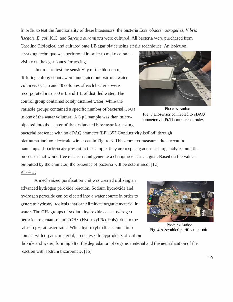
In order to test the functionality of these biosensors, the bacteria Enterobacter aerogenes, Vibrio
fischeri, E. coli K12, and Sarcina aurantiaca were cultured. All bacteria were purchased from
Carolina Biological and cultured onto LB agar plates using sterile techniques. An isolation
streaking technique was performed in order to make colonies
visible on the agar plates for testing.
In order to test the sensitivity of the biosensor,
differing colony counts were inoculated into various water
volumes. 0, 1, 5 and 10 colonies of each bacteria were
incorporated into 100 mL and 1 L of distilled water. The
control group contained solely distilled water, while the
variable groups contained a specific number of bacterial CFUs
in one of the water volumes. A 5 µL sample was then micro-
pipetted into the center of the designated biosensor for testing
bacterial presence with an eDAQ ammeter (EPU357 Conductivity isoPod) through
platinum/titanium electrode wires seen in Figure 3. This ammeter measures the current in
nanoamps. If bacteria are present in the sample, they are respiring and releasing analytes onto the
biosensor that would free electrons and generate a changing electric signal. Based on the values
outputted by the ammeter, the presence of bacteria will be determined. [12]
Phase 2:
A mechanized purification unit was created utilizing an
advanced hydrogen peroxide reaction. Sodium hydroxide and
hydrogen peroxide can be ejected into a water source in order to
generate hydroxyl radicals that can eliminate organic material in
water. The OH- groups of sodium hydroxide cause hydrogen
peroxide to denature into 2OH⠂(Hydroxyl Radicals), due to the
raise in pH, at faster rates. When hydroxyl radicals come into
contact with organic material, it creates safe byproducts of carbon
dioxide and water, forming after the degradation of organic material and the neutralization of the
reaction with sodium bicarbonate. [15]
Fig. 4 Assembled purification unit
Photo by Author
Fig. 3 Biosensor connected to eDAQ
ammeter via Pt/Ti counterelectrodes
Photo by Author

11
A mechanized unit was created using an Arduino microprocessor to regulate the
implementation of chemicals into water sources. The Arduino Zero was then connected to a
Darrington Board using jumper cables. The pins of the Arduino (D8-D11) were connected to the
corresponding number of the Darrington Board (INT1-INT4). This allowed for the Arduino to
power the board as well as upload the rotation code onto it. The Arduino and the Darrington
Board were then mounted onto a RobotGeek workbench using metal wiring. A stepper motor
was attached to the Darrington Board using 5 lead adjoined wires.
A stepper motor code provided with the Arduino module was manipulated to fit the
parameters of the purification unit. Once this code was uploaded, a spring was attached to a
micropipette via adhesive glue. The micropipette was then elevated off the RobotGeek
Workbench using a stack of 4, 3.5x2.7x0.3 cm plastic prisms and adhered using glue. This
system would allow for the safe uptake and ejection of purification chemicals into water.
This process was repeated in order to mount two micropipettes onto the workbench. The
micropipettes were filled with sodium hydroxide (0.5M, 10µL) and hydrogen peroxide (3%
10µL). The activation of the Arduino units led to the ejection of these chemicals into a water
source. After ejection, sodium bicarbonate was added to water in order to disrupt the functioning
of the hydroxyl radicals and was filtered out of water with a coffee filter. [11] In order to test the
functionality of the device, bacteria were inoculated into a water source, and a sample of the
water was placed on the biosensors in order to confirm the presence of bacteria in water. Next,
the purification unit was activated, leading to the ejection of chemicals loaded in the
micropipettes. Samples of water were then removed from the decontaminated water source in
order to confirm that the purification unit had success in eliminating bacterial presence in water.
This was accomplished by placing volumes of water onto the biosensor and monitoring the
current with the EDAQ ammeter. A flat, unchanging current graph designated that the machine
had success in eliminating bacterial presence in water.

12
Results
Phase 1:
Graph 1. A) The amperage output of a control group of distilled water. B) The output of 1 CFU of S.
aurantiaca in 100 mL of water. The values of the amperage outputted by the ammeter are used to
calculate the range of the electric current.
During testing, each of the biosensors were exposed to a sample of contaminated water for 15
seconds. Samples contained varying volumes of water (100mL and 1000mL) and varying CFU
counts (0, 1, 5, and 10 CFU). Graph 1A shows the output of a sample of solely distilled water.
As there are no bacteria present in a sample, no analytes are freeing electrons, which creates this
unchanging current output. However, when bacteria are present as seen in Graph 1B, the bacteria
releases analytes that undergoes a series of chemical reactions to generate a changing current. A
changing current output represents that bacteria are present in the sample. The range of the
current values that were outputted by the eDAQ ammeter was calculated for each of these trials.
Current Created by Bacterial Presence
13
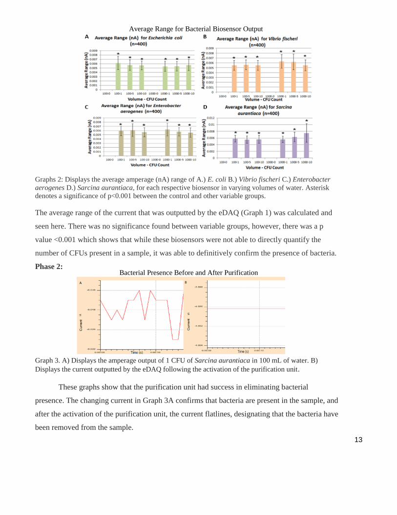
Graphs 2: Displays the average amperage (nA) range of A.) E. coli B.) Vibrio fischeri C.) Enterobacter
aerogenes D.) Sarcina aurantiaca, for each respective biosensor in varying volumes of water. Asterisk
denotes a significance of p<0.001 between the control and other variable groups.
The average range of the current that was outputted by the eDAQ (Graph 1) was calculated and
seen here. There was no significance found between variable groups, however, there was a p
value <0.001 which shows that while these biosensors were not able to directly quantify the
number of CFUs present in a sample, it was able to definitively confirm the presence of bacteria.
Phase 2:
Graph 3. A) Displays the amperage output of 1 CFU of Sarcina aurantiaca in 100 mL of water. B)
Displays the current outputted by the eDAQ following the activation of the purification unit.
These graphs show that the purification unit had success in eliminating bacterial
presence. The changing current in Graph 3A confirms that bacteria are present in the sample, and
after the activation of the purification unit, the current flatlines, designating that the bacteria have
been removed from the sample.
Average Range for Bacterial Biosensor Output
Bacterial Presence Before and After Purification

14
Discussion
The purpose of the first phase of this study was to create graphene biosensors that were
capable of detecting the presence of prevalent waterborne pathogens such as S. typhi, V.
cholerae, S. dysenteriae, and E. coli, which are the bacteria that cause Salmonella, Cholera, and
Shigellosis, and E. coli, respectively. These biosensors successfully detected the presence of
these microorganisms at distinctly low levels in varying volumes of water and a rapid time
frame. These two devices improve greatly on current methods of bacterial detection and
purification. PCR, a traditional method of measuring bacterial contamination in water, requires a
thermal cycler which can cost thousands of dollars, constant hands on work for 1-2 days, and
prior training before use. PCR and traditional plating methods have detection limits from 100 to
1000 CFUs [3]; meanwhile, the World Health Organization deemed that even 1 CFU of bacteria
in 100 mL of water was unsafe. These biosensors are drastically more rapid, sensitive,
inexpensive, and user-friendly.
When a sample is pipetted onto the graphene biosensors in contact with the ammeter, the
presence of a changing electric current can be used to signal the presence of bacteria. When
analytes that are released during bacterial respiration come into contact with its designated
enzyme, it’s catalyzed into hydrogen peroxide and pyruvate. The hydrogen peroxide is then
oxidized through the applied voltage given by the ammeter, which frees electrons that the
graphene harnesses in order to generate an electric current. As seen in Graph 1 above, the EDAQ
ammeter graphed the change in nanoamps outputted by the biosensor over 15 seconds. Graph 1a
shows that there was no change in current outputted by the biosensor when uncontaminated
distilled water was placed on it, as no analytes were present from bacterial respiration to be
converted into electrical stimuli. However, graph 1b shows the results of a sample containing 1
CFU of S. aurantiaca. There is a fluctuating output, as the analytes come into contact with the
enzyme and free electrons. The analytes do not all couple with the enzyme at the same time,
therefore, electrons are not all freed at one time. Additionally, bacteria respire at different rates
which causes the level of analytes present in a water sample to vary over a time interval.
50 trials were run for each variable group, with 15 seconds per trial. The range of these
trials were calculated and compared on Graph 2. It was found that in groups where no bacteria

15
contaminated the water, the biosensor outputted a constant value, and therefore the range was
equal to zero. However, in water in which there was microorganism contamination, there existed
a range of the values that was not equal to zero. This varying current and range of values signals
the presence of these specific bacteria.
The data received in this experiment proved that the biosensors were able to detect and
signal the presence of bacteria through the immobilization of enzymes onto the graphene
substrates. Enzymes can be immobilized onto graphene through π-π interactions, which is a type
of noncovalent bond that occurs between two aromatic molecules. Because graphene and 1-
pyrenebutyric acid are both aromatic, meaning that they contain hexagonal benzene rings in their
molecular structure, the parallel, aromatic rings align themselves with each other [8] when 1-
pyrenebutyric acid solution is incubated onto the layer of graphene. This binds the PBA onto the
graphene. This acid then acts as a linker molecule to allow for enzyme immobilization, as the
enzyme then latches onto the acid through an amide bond that occurs between the amide group
of the enzymes and the pyrene group of the pyrene butyric acid. This secures and immobilizes
the enzymes onto the graphene molecules [9].
When the analytes that are released during bacterial respiration come into contact with
the designated enzyme that is immobilized on the graphene, a series of chemical reactions occurs
that signals the presence of bacteria through a changing electric current, as seen in Graph 1b. The
analyte is catalyzed into hydrogen peroxide and pyruvate, and the hydrogen peroxide is oxidized.
The oxidation of hydrogen peroxide frees electrons that the graphene is able to harness due to its
conductive properties to generate an electric signal [9]. For example, V. fischeri produces the
metabolite ɑ-D-Galactose-1 as a byproduct of respiration. When the metabolite comes into
contact with galactose oxidase, the enzyme catalyzes the analyte into hydrogen peroxide and
pyruvate. The hydrogen peroxide is then oxidized through the applied voltage of the ammeter,
which frees electrons that graphene harnesses in order to generate a current measurable by the
ammeter [14]. Because bacteria are respiring at different rates, the concentration of analytes in
the sample vary, which causes the current produced by the biosensor to vary as presented by
Graph 1b. Therefore, when water was contaminated with the specific bacteria designated to the
enzyme, the EDAQ ammeter measured a varying current value that signaled the presence of

16
bacteria in water. When bacteria are not present in water, no metabolites are being produced by
bacterial respiration. Therefore, no analytes are freeing electrons, which cause the current to
flatline as seen in Graph 1a.
After running a One-Way ANOVA followed by a post-hoc scheffe test, there was no
statistical significance between variable groups containing various CFU counts. However, a
p value of <0.001 was discovered between the control group with solely distilled water and all
other variable groups containing bacteria. This shows that while these biosensors did not appear
to have the ability to quantify the number of bacterial CFU’s present in a sample based on the
threshold it was tested at, these biosensors were capable of definitively signaling the presence of
bacteria through a changing electrical current. This was found in Labroo’s study [12], in which
there existed a threshold that the lactate concentration had to surpass in order to create a
significantly different electrical output. This suggests that if these biosensors were to be tested
with higher levels of bacteria, and thus a higher concentration of analytes, the electrical output
would correlate with the number of CFU’s of bacteria present in a sample.
The purpose of the second phase of this study was to develop a purification unit that
could purify water contaminated with bacteria, creating potable and usable water. Using the
EDAQ ammeter and biosensors, it was confirmed that the Arduino-programmed micropipette
purification unit had the ability to decontaminate water that contained bacterial pathogens, thus
fulfilling the purpose of this study. Current methods for purification are costly, take a long period
of time, and poses risk for further contamination as water must be shipped out to external facility
to be treated. This purification system allows bacteria to be eliminated from water directly within
the water source in a cheap, safe, manner.
When the biosensors confirm the presence of bacteria in water via a varying current, the
purification unit can be activated to eliminate the bacteria. The rotation code adapted in this
study can be activated on the two Arduino boards. This would allow for stepper motors to rotate,
and push forward rails on rail mounts that push forward the plungers of micropipettes, releasing
sodium hydroxide and hydrogen peroxide into the water. These two chemicals can lead to the
generation of hydroxyl radicals that mark a very effective, but safe method by which water can
be purified. Hydroxyl radicals uncouple bonds that exist in organic material, combining with

17
hydrogen and carbon atoms to form water and carbon dioxide. The radicals will interact with the
organic material into all the bacteria have been degraded, where upon sodium bicarbonate can be
added into the water in order stop the functioning of the radicals, and allow for complete
elimination of bacteria in water. The biosensors can be used in order to confirm the success of
the purification unit in eliminating bacteria, as it can monitor bacterial presence regardless of any
other substance that may exist in water as the enzymes immobilized on the biosensors only free
electrons when the specific correlate analyte binds with the enzyme. When the bacteria are
eliminated, there are no analytes being produced in the water, and therefore the current read by
the ammeter remains at a constant value, which shows that the purification unit had success in
eliminating bacterial presence. [15]
The purification unit in this study consisted of several features that greatly improve upon
current methods of purification. For one, the purification unit had the dimensions of 44cm x
21cm x 0.5cm. This size is much smaller than current municipal water treatment facilities, and
therefore, upon implementation in a real-world situation, there exists no real size constraints due
to the compact nature of the device. This would allow for the system to be utilized onsite in
water sources in order to apply an immediate purification response to water if the biosensors
were to confirm bacterial presence in water. Additionally, the purification system construction
was much cheaper than that of a water treatment plant. While water treatment plants have the
capacity to eliminate other forms of water contamination like nitrates, the unit constructed in this
study costs a couple of hundred dollars, whereas most water treatment facilities cost multiple
hundreds of millions of dollars. Moreover, the purification unit in this study that was created
allowed for cost-effective, on-site bacteria elimination that marked a critical improvement on
past methods of water purification. [17]
Using these two systems, bacterial detection and water purification can occur
immediately, on-site, and at a lower cost to ensure the sanitation and affordability of clean
drinking water.
One area of this study that could be improved upon was the use of water-based
conductive paint. Working in a high school laboratory, silver-based paint could not be utilized
due to its low flash point. Therefore, water based paint was used as an alternative; however, this

18
allows only a small volume of a sample to be pipetted onto the biosensor rather than immersing
the entire substrate in a water source. If this study were to be replicated in a professional
laboratory in which hydrophobic silver-based paint could be utilized, the procedure to manually
transfer the sample onto the biosensor could be eliminated. The biosensors could then be simply
placed in a water source, such as a well, and able to continually monitor the presence of bacteria.
In spite of this limitation, however, it is important to recognize that this system had the
capabilities of detecting 1 CFU of the four major bacterial contaminants of water in 1 second,
and purifying water contaminated with bacterial in a rapid manner.
Two different electrode configurations were used in this study in order to try to generate
biosensors that are more cost effective for those living in poorer regions in the world. The
purchased CVD graphene and the graphene harvested from pencil graphite exhibited no
observable differences in output. This reflects that utilizing graphite in order to harvest graphene
would be a significantly more cost-effective alternative, while retaining the biosensors’
sensitivity and rapidity. Thus, countries in the developing world, with a more readily available
supply of graphite over graphene, could utilize graphite as an effective electrode alternative for
the developing world.
Overall, this system has important implications for the developing world in terms of
water contamination monitoring and purification. The detection devices and purification unit has
the potential to be implemented into a drinking source in the developing world in order to
consistently monitor the presence of E. coli, Shigella, Salmonella, and Cholera in the water. If
the detection device identifies that there exists bacteria in the water, the purification unit can be
implemented in order to add the chemical agents necessary to generate hydroxyl radicals in order
to purify water contaminated with the bacteria. These results are fundamental to the advancement
of water sanitation throughout the world, in the developing and developed world alike. In the
developing world, women and children spend upwards of 6 hours each day collecting water [18].
This system can alleviate this issue, allowing women and children to receive an education, by
providing a user-friendly, labor-saving sanitation system for drinking water. Similarly, access to
safe water in the developing world has the potential to provide almost $32 billion dollars in
economic benefits. [4]

19
These systems can be applied to industrial settings as well. Biosensors can be formatted
into a cylindrical structure in order to be placed in a pipe of flowing water. The biosensor can
then be covered in electrically conductive carbon black epoxy, which will prevent high volumes
of water at high speeds from causing the device to flow away. This device implementation would
allow for countries in the developed world to constantly monitor bacterial presence and reduce
the threat that waterborne pathogens have on the world.
Conclusion
This detection/purification system was successful in various areas of interest:
1. These sensors were capable of detecting bacterial presence in less than 1 second, which is
far more rapid than the current methods of bacterial detection.
2. These biosensors were also able to discern lower levels of bacteria (1 CFU) than these
conventional methods, such as PCR and colony counting, are capable of.
3. In addition to these improvements upon the functionality of detection devices, they are
also user friendly and can be implemented easily in the developing world.
4. The mechanized purification unit was successful in eliminating bacterial presence in
water in a more rapid, cost effective parameter.
In conclusion, this study yielded the creation of sensors that had the ability to detect model
organisms for E. coli, Salmonella, Shigella, and Cholera. Working in conjunction with these
biosensors, a mechanized purification system could monitor and purify microorganism presence
in order to sanitize water sources contaminated with bacterial pathogens. This system could
potentially eliminate the threat of waterborne diseases and greatly expand sanitary water
resources throughout the world.

20
7. References
[1] Ali, Mohamed M (2014) "Fructose-Asparagine Is a Primary Nutrient during Growth of
Salmonella in the Inflamed Intestine." PLOS. PLOS Pathogens 25 Mar. 2017
[2] Ang, Lee Fung (2015) "Development of an Amperometric-Based Glucose Biosensor to
Measure the Glucose Content of Fruit." Plos One 10.3: n. pag. Web.
[3] Deshmukh, Rehan A., Kopal Joshi, Sunil Bhand, and Utpal Roy. "Recent Developments in
Detection and Enumeration of Waterborne Bacteria: A Retrospective Minireview."
MicrobiologyOpen 5.6 (2016): 901-22. Web.
[4] “Facts and Statistics about Water and Its Effects.” The Water Project. N.p., 31 Aug. 2016.
Web. 22 June 2017.
[5] Fuente, Jesus De La. "Graphene - What Is It?" Graphenea. N.p., n.d. Web. 25 Mar. 2017.
[6] "Graphene Transfer Guide." Graphene Supermarket :: Graphene Transfer Guide. N.p., n.d.
Web. 22 June 2017.
[7] Hunter, Christopher A., and Jeremy K. M. Sanders (1990) "The Nature of .pi.-.pi.
Interactions." J. Am. Chem. Soc. Journal of the American Chemical Society 112.14: 5525-
534. Web.
[8] Hinnemo, Malkolm. "On the Road to Graphene Biosensors." Digital Comprehensive
Summaries of Uppsala Dissertations from the Faculty of Science and Technology (2017):
n. pag. Web. 22 June 2017.
[9] Karachevtsev, Victor A., and Stepan G. Stepanian (2011) "Noncovalent Interaction of
Single-Walled Carbon Nanotubes with 1-Pyrenebutanoic Acid Succinimide Ester and
Glucoseoxidase." The Journal of Physical Chemistry C 115.43: 21072-1082. Web.
[10] Kathyayini Nagaraju (2015) A Review on Protein Functionalized Carbon Nanotubes: n.
pag. Journal of Applied Biomaterials & Functional Materials. Web.
[11] Kommineni, Suril (2016) "Advanced Oxidation Processes." SpringerBriefs in Molecular
Science Novel Catalysts in Advanced Oxidation of Organic Pollutants : 23-34. Web.
[12] Labroo, Pratima, and Yue Cui (2013) "Flexible Graphene Bio-nanosensor for
Lactate." Biosensors and Bioelectronics 41 : 852-56. Web.
[13] Müller, Ulrike Maria, Liang Wu, Lourina Madeleine Raamsdonk, Aaron Adriaan Winkler,

21
and Dsm Ip Assets B.V (2008) "Acetyl-coa Producing Enzymes in Yeast." Google Books.
BibTex, Web. 21 Nov. 2016.
[14] Putzbach, William, and Niina Ronkainen (2013) "Immobilization Techniques in the
Fabrication of Nanomaterial-Based Electrochemical Biosensors: A Review." Sensors 13.4:
4811-840. Web.
[15] Swaminathan, Meenakshiundaram, Manickavachagam Muruganandham, and Mika
Sillanpaa. "Advanced Oxidation Processes for Wastewater Treatment." International
Journal of Photoenergy (2013): 1-216. Web.
[16] Tang, Zhihong (2010) "Exfoliation of Graphene from Graphite and Their Self-Assembly at
the Oil-Water Interface." Research Gate. Tsinghua University, Web. 12 Apr. 2017.
[17] "Treatment Plant - Water Treatment Program - Water and Waste - City of
Winnipeg."Winnipeg. N.p., 27 Jan. 2017. Web. 22 June 2017.
[18] UN Water. (2013). UN-Water factsheet on water and gender, World Water Day 2013.
8. Bibliography
[19] Arco, L.g. De (2009) "Synthesis, Transfer, and Devices of Single- and Few-Layer Graphene
by Chemical Vapor Deposition." IEEE Transactions on Nanotechnology 8.2: 135-38. Web.
[20] Bourzac, Katherine (2014) "Clean Water for the Developing World." MIT Technology
Review. MIT Technology Review. Web. 25 Mar. 2017.
[21] Lazcka, Olivier, and F. Javier Del Campo (2007) "Pathogen Detection: A Perspective of
Traditional Methods and Biosensors." Biosensors and Bioelectronics 22.7: 1205-217. Web.
[22] Leonard, Paul, and Stephen Hearty (2003) "Advances in Biosensors for Detection of
Pathogens in Food and Water." Enzyme and Microbial Technology 32.1: 3-13. Web.
[23] Leprince-Wang, Yamin (2015) "ZnO-Nanowire-Based Nanogenerators: Principle,
Characterization and Device Fabrication." Piezoelectric ZnO Nanostructure for Energy
Harvesting: 65-103. Web.
[24] Rassaei, Liza, and Wouter Olthuis (2013) "Lactate Biosensors: Current Status and
Outlook." Analytical and Bioanalytical Chemistry 406.1: 123-37. Web.
[25] Ray, M. B., J. Paul Chen, and Lawrence K. Wang (2006) "Advanced Oxidation
Processes." Advanced Physicochemical Treatment Processes: 463-81. Web.