a new southern laramidian ankylosaurid, akainacephalus ... wiersma & irmis 2018... · a new...
TRANSCRIPT

A new southern Laramidian ankylosaurid,Akainacephalus johnsoni gen. et sp. nov.,from the upper Campanian KaiparowitsFormation of southern Utah, USA
Jelle P. Wiersma1,2,3 and Randall B. Irmis2,3
1 Department of Geosciences, James Cook University, Townsville, QLD, Australia2 Natural History Museum of Utah, Salt Lake City, UT, USA3 Department of Geology & Geophysics, University of Utah, Salt Lake City, UT, USA
ABSTRACTA partial ankylosaurid skeleton from the upper Campanian Kaiparowits
Formation of southern Utah is recognized as a new taxon, Akainacephalus johnsoni,
gen. et sp. nov. The new taxon documents the first record of an associated
ankylosaurid skull and postcranial skeleton from the Kaiparowits Formation.
Preserved material includes a complete skull, much of the vertebral column,
including a complete tail club, a nearly complete synsacrum, several fore- and hind
limb elements, and a suite of postcranial osteoderms, making Akainacephalus
johnsoni the most complete ankylosaurid from the Late Cretaceous of southern
Laramidia. Arrangement and morphology of cranial ornamentation in
Akainacephalus johnsoni is strikingly similar to Nodocephalosaurus kirtlandensis
and some Asian ankylosaurids (e.g., Saichania chulsanensis, Pinacosaurus grangeri,
and Minotaurasaurus ramachandrani); the cranium is densely ornamented with
symmetrically arranged and distinctly raised ossified caputegulae which are
predominantly distributed across the dorsal and dorsolateral regions of the nasals,
frontals, and orbitals. Cranial caputegulae display smooth surface textures with
minor pitting and possess a distinct conical to pyramidal morphology which
terminates in a sharp apex. Character analysis suggests a close phylogenetic
relationship with N. kirtlandensis, M. ramachandrani, Tarchia teresae, and
S. chulsanensis, rather than with Late Cretaceous northern Laramidian
ankylosaurids (e.g., Euoplocephalus tutus, Anodontosaurus lambei, and
Ankylosaurus magniventris). These new data are consistent with evidence for
distinct northern and southern biogeographic provinces in Laramidia during the
late Campanian. The addition of this new ankylosaurid taxon from southern Utah
enhances our understanding of ankylosaurid diversity and evolutionary
relationships. Potential implications for the geographical distribution of Late
Cretaceous ankylosaurid dinosaurs throughout the Western Interior suggest
multiple time-transgressive biogeographic dispersal events from Asia into
Laramidia.
Subjects Biodiversity, Biogeography, Evolutionary Studies, Paleontology, Taxonomy
Keywords Paleontology, Taxonomy, Biodiversity
How to cite this article Wiersma and Irmis (2018), A new southern Laramidian ankylosaurid, Akainacephalus johnsoni gen. et sp. nov.,
from the upper Campanian Kaiparowits Formation of southern Utah, USA. PeerJ 6:e5016; DOI 10.7717/peerj.5016
Submitted 10 September 2016Accepted 28 May 2018Published 19 July 2018
Corresponding authorJelle P. Wiersma,[email protected]
Academic editorFabien Knoll
Additional Information andDeclarations can be found onpage 68
DOI 10.7717/peerj.5016
Copyright2018 Wiersma and Irmis
Distributed underCreative Commons CC-BY 4.0

INTRODUCTIONThe Ankylosauridae is a monophyletic clade of herbivorous, armored ornithischian
dinosaurs that are predominantly recorded from the Late Cretaceous (Turonian—late
Maastrichtian) of Asia and latest Cretaceous (early Campanian—late Maastrichtian) of
western North America (Laramidia) (Lambe, 1902; Brown, 1908; Parks, 1924; Nopcsa,
1928; Sternberg, 1929; Gilmore, 1933; Marya!nska, 1977; Sullivan, 1999; Carpenter, 2004;
Miles & Miles, 2009; Arbour, Burns & Sissons, 2009; Arbour & Currie, 2013a, 2016; Arbour,
Currie & Badamgarav, 2014; Arbour et al., 2014; Penkalski, 2014; Arbour, Zanno & Gates,
2016; Penkalski & Tumanova, 2017; Arbour & Evans, 2017). The majority of Laramidian
ankylosaurid specimens are known from northern localities (Fig. 1) and include:
Euoplocephalus tutus, Dyoplosaurus acutosquameus, and Scolosaurus cutleri from the
Dinosaur Park and Scollard formations of Alberta, Canada; Anodontosaurus lambei
from the Horseshoe Canyon Formation of Alberta, Canada; Zuul crurivastator from the
Figure 1 Distribution of Late Cretaceous Laramidian ankylosaurid dinosaurs. Overview of stratigraphic, temporal, and biogeographic dis-tribution of Late Cretaceous (upper Campanian–upper Maastrichtian) ankylosaurid dinosaurs, including Akainacephalus johnsoni, across northernLaramidian (Alberta, Montana, Wyoming), and southern Laramidian (Utah, New Mexico) basins. Noticeable is the higher number of taxa andmore widespread distribution of ankylosaurids in northern Laramidia. Colored bars within stratigraphic formations represent ankylosaurid taxaand their respective temporal range. Paleogeographic map modified after Sampson et al. (2010). Stratigraphic intervals modified after Arbour &Currie (2013a); Roberts et al. (2013); Johnson, Nichols & Hartman (2002). Full-size DOI: 10.7717/peerj.5016/fig-1
Wiersma and Irmis (2018), PeerJ, DOI 10.7717/peerj.5016 2/76

Judith River Formation of Montana; Oohkotokia horneri from the Two Medicine
Formation of Montana, USA; and Ankylosaurus magniventris from the upper
Maastrichtian Lance, Hell Creek, and Scollard formations of Wyoming, USA and Alberta,
Canada, respectively (Lambe, 1902; Brown, 1908; Parks, 1924; Nopcsa, 1928; Sternberg,
1929; Carpenter, 2004; Arbour, Burns & Sissons, 2009; Arbour & Currie, 2013a; Penkalski,
2014; Arbour & Evans, 2017). Ankylosaurid taxa from southern Laramidia were unknown
until the discovery of Nodocephalosaurus kirtlandensis from the upper Campanian–
Maastrichtian Kirtland Formation of New Mexico (Sullivan, 1999). However, the number
of Late Cretaceous ankylosaurid taxa recorded from the Kirtland and Fruitland
formations of NewMexico has tripled within the last 15 years, and additional taxa include
Ziapelta sanjuanensis (Arbour et al., 2014) and Ahshislepelta minor (Burns & Sullivan,
2011a), leading to a rapidly increasing taxonomic diversity within southern Laramidian
basins during the Late Cretaceous of western North America. In addition, several
ankylosaurid specimens (UMNH VP 19472, UMNH VP 19473, UMNH VP 21000,
UMNH VP 20202) have been recorded from the upper Campanian Kaiparowits
Formation of southern Utah (Loewen et al., 2013a; Wiersma & Irmis, 2013) but UMNH
VP 20202 is the first newly-described ankylosaurid taxon from the Late Cretaceous of
Utah. Despite these recent discoveries from New Mexico and Utah, Late Cretaceous
southern Laramidian ankylosaurid specimens remain rare from upper Campanian
terrestrial deposits of the Kaiparowits, Kirtland, and Fruitland formations, and the
majority of taxa are represented by a single specimen.
Here, we describe and compare a new genus and species of ankylosaurid dinosaur,
Akainacephalus johnsoni (UMNH VP 20202), from the upper Campanian Kaiparowits
Formation of southern Utah, to other known Late Cretaceous taxa from Asia and
western North America. UMNH VP 20202 represents the most complete ankylosaurid
specimen from the Kaiparowits Formation and southern Laramidia to date. The specimen
consists of a complete skull and mandibles, a nearly complete synsacrum, various cervical,
dorsal, and caudal vertebrae, including the tail club handle and knob, a large number
of fore- and hindlimb elements, two cervical half rings, and a suite of postcranial
osteoderms. The distinct combination and arrangement of conical and pyramid-shaped
caputegulae, the massive and backswept postorbital horns, and the ventrally descending,
triangular squamosal horns in Akainacephalus johnsonimake this specimen taxonomically
unique among other Late Cretaceous ankylosaurid dinosaurs from Asia and western
North America. The overall morphology of A. johnsoni suggests a close phylogenetic
relationship with Nodocephalosaurus kirtlandensis and Late Cretaceous Asian
ankylosaurids.
Geologic settingAkainacephalus johnsoni was discovered in the Kaiparowits Formation; an ∼860-m-thick
terrigenous siliciclastic stratigraphic succession (Fig. 2A) that crops out in the Kaiparowits
Plateau within the Grand Staircase-Escalante National Monument (GSENM) in
southern Utah, USA (Fig. 2B) and contributes to a substantial part of the 2 km thick
Late Cretaceous stratigraphic sequence within the Kaiparowits Basin (Roberts, 2007).
Wiersma and Irmis (2018), PeerJ, DOI 10.7717/peerj.5016 3/76

LEGEND
Ms Ss Cg
Akainacephalus johnsoni
Kai
paro
wit
s F
m
700-
600-
500-
400-
300-
200-
100-
0-
UP
PE
R U
NIT
MID
DL
E U
NIT
LO
WE
R U
NIT
- mudstone
- channel sandstone
- sandstone
- conglomerate
- bentonite
WW
Fm
CP
Fm(m)
900-
800-74.69 +/- 0.18 Ma; Ar/Ar
75.51 +/- 0.15 Ma; Ar/Ar
75.97 +/- 0.18 Ma; Ar/Ar76.26 +/- 0.10 Ma; U-Pb
76.46 +/- 0.14 Ma; Ar/Ar
75.51 +/- 0.15 Ma; Ar/Ar
38 0N
112 0W
BoulderTable CliffsPlateau
Escalante
Tropic
UTAHGSENM
UTAHARIZONA
KaiparowitsPlateau
HW
Y 1
2
HWY 12
DU
Paun
saug
unt f
ault
D U
Sevi
er fa
ult
25 kmD U
N
Claron, Pine Hollow, Cn PeakFormation, undifferentiated
Kaiparowits Formation
Wahweap Formation
Straight Cliffs Formation
Monument boundary
Normal fault
Grand Staircase - Escalante NationalMonument
LEGEND
Lake Powell
B
A
Figure 2 Location and stratigraphy of the Kaiparowits Formation. Map of Grand Staircase-Escalante National Monument (GSENM), southernUtah (A), and generalized stratigraphic section of the upper Campanian Kaiprowits Formation (B). The approximate stratigraphic position ofAkainacephalus johnsoni is located near the base of the middle unit within the Kaiparowits Formation. The map highlights the GSENM boundary(dashed line), showing the geological distribution and outcrops of the Cretaceous Kaiparowits, Wahweap, and Straight Cliffs formations. The redstar indicates the Horse Mountain area, from which Akainacephalus johnsoni was recorded. Map and stratigraphic column modified from Roberts(2005). Radioisotopic dates used from Roberts, Deino & Chan (2005) and Roberts et al. (2013), respectively.
Full-size DOI: 10.7717/peerj.5016/fig-2
Wiersma and Irmis (2018), PeerJ, DOI 10.7717/peerj.5016 4/76

From a paleontological perspective, the Kaiparowits Formation preserves a unique record
of Late Cretaceous terrestrial vertebrate ecosystems in the Western Interior of North
America. This thick succession of strata was deposited at an unusually rapid rate within a
time frame of ∼2 million years, making it one of the most rapidly deposited terrestrial
formations in the world (Roberts, 2005). Recently recalibrated radioisotopic dates for the
Kaiparowits Formation indicate a late Campanian age range of 76.46 ± 0.14 Ma for
the upper portion of the lower unit to 74.69 ± 0.18 Ma for the uppermost portion of the
upper unit of the Kaiparowits Formation (Fig. 2A) (Roberts, 2005; Roberts, Deino & Chan,
2005; Roberts et al., 2013), making it contemporaneous with dinosaur-bearing strata from
the Dinosaur Park Formation, Alberta, Judith River and Two Medicine formations,
Montana, Fruitland and Kirtland formations, New Mexico, and the Aguja Formation,
Texas (Fig. 1).
Topographically, the strata within the Kaiparowits Formation are characterized by their
badland-forming bluish-gray siltstone and mudstone and grayish sandstone outcrops,
overlying the more cliff-forming predominantly yellow-brownish fluvial channel deposits
that form the lower-middle Campanian Wahweap and upper Turonian–Santonian
Straight Cliffs formations (Eaton et al., 2001; Roberts, 2005, 2007; Roberts, Deino & Chan,
2005; Jinnah et al., 2009; Jinnah, 2013; Roberts et al., 2013). Akainacephalus johnsoni
(UMNH VP 20202) was discovered at the Horse Mountain Gryposaur (HMG) Quarry,
UMNH VP Locality 1109, a multitaxic and multidominant bonebed, deposited in a fine-
to medium-grained sandstone crevasse splay, intercalated within a silty mudstone. The
quarry, and therefore UMNH VP 20202, is located within the lower section of the
informal middle unit of the Kaiparowits Formation, approximately 190 m above the basal
contact of the formation in the Horse Mountain region (Roberts et al., 2013). This
bonebed has also produced a nearly complete skull and postcranial skeleton of the
hadrosaurid ornithischian dinosaur Gryposaurus (UMNH VP 20181 in Gates et al., 2013),
the type specimen of the baenid turtle Arvinachelys goldeni (Lively, 2015), a nearly
complete articulated skull and postcranial skeleton of a new taxon of small alligatoroid
(Irmis et al., 2013), and a poorly preserved partial skull of a small theropod dinosaur
(Zanno et al., 2013).
MATERIALS AND METHODSUMNH VP 20202 was prepared using small airscribes, dental tools, brushes, and a
microscope. Cleaning was performed exclusively by using water and paper towels.
Polyvinal acetate (VinacTM) dissolved in acetone was applied as a consolidant, and
cyanoacrylate and two-part epoxy were used as adhesives. Two-part epoxy putty was
applied to selected areas to complement stabilization or fill in large fractures but was not
applied for reconstructive purposes. After preparation, the skull was subjected to
computed tomography (CT) scanning in order to reveal the internal anatomy, particularly
in the endocranial cavity and nasal regions. CT scanning was performed at the University
of Texas High Resolution X-ray Computed Tomography Facility in Austin, TX, USA.
UMNH VP 20202 was scanned in January 2014, using a NSI scanner equipped with a
Titan GE source set at 450 kV and 3.0 mA and voxel size of 0.1692 mm. Anatomical
Wiersma and Irmis (2018), PeerJ, DOI 10.7717/peerj.5016 5/76
comparisons were made with closely related taxa; comparisons to other taxa in the
description are referenced, following standard modern paleontological descriptive
practice, citing specimen numbers when material was observed personally, and citing
published references when comparisons are made with the literature. A complete list of all
examined taxa and specimen numbers are summarized in Table S1 (Supplemental
Material S1). Measurements performed on UMNH VP 20202 are summarized in
Supplemental Material S4.
UMNH VP 20202 is permanently reposited in the collections of the Natural History
Museum of Utah, Salt Lake City, UT, USA. Detailed locality information is on file and
available to qualified researchers as per museum policy. All specimens were collected
under permits obtained from the United States Department of the Interior Bureau
of Land Management (BLM) in the BLM-administered GSENM.
Nomenclatural actsThe electronic version of this article in portable document format will represent a
published work according to the International Commission on Zoological Nomenclature
(ICZN), and hence the new names contained in the electronic version are effectively
published under that Code from the electronic edition alone. This published work and
the nomenclatural acts it contains have been registered in ZooBank, the online
registration system for the ICZN. The ZooBank Life Science Identifiers (LSIDs) can be
resolved and the associated information viewed through any standard web browser by
appending the LSID to the prefix http://zoobank.org/. The LSID for this publication is:
[urn:lsid:zoobank.org:pub:42CB4F68-9E0D-42BB-943D-8A66E3D9DA12]. The online
version of this work is archived and available from the following digital repositories: PeerJ,
PubMed Central, and CLOCKSS.
RESULTSSystematic paleontology
Dinosauria Owen, 1842 sensu Padian and May, 1993
Ornithischia Seeley, 1887 sensu Padian and May, 1993
Thyreophora Nopcsa, 1915 sensu Sereno, 1986
Ankylosauria Osborn, 1923 sensu Carpenter, 1997
Ankylosauridae Brown, 1908 sensu Sereno, 1998
Ankylosaurinae Brown, 1908 sensu Sereno, 1986
Ankylosaurini Arbour and Currie, 2016
Akainacephalus, gen. nov.
Akainacephalus johnsoni, sp. nov.
HolotypeUMNH VP 20202, a partial skeleton comprising a complete skull, both mandibles,
predentary, four dorsal, four dorsosacral, three sacral, one caudosacral, and eight caudal
Wiersma and Irmis (2018), PeerJ, DOI 10.7717/peerj.5016 6/76

vertebrae, dorsal ribs, a complete tail club, both scapulae, left coracoid, right humerus,
right ulna, partial left ilium, left femur, left tibia, left fibula, phalanx, two partial cervical
osteoderm half rings, and 17 dorsal and lateral osteoderms of various sizes and
morphologies.
Type localityUMNH VP Locality 1109 (“HMG Quarry”), Horse Mountain area, GSENM, Kane
County, southern Utah, USA.
Type stratigraphic horizon and ageUMNH VP Locality 1109 is a multitaxic bonebed deposited in a crevasse splay sandstone
within the lower portion of the middle unit of the upper Campanian Kaiparowits
Formation (Fig. 2A). The stratigraphic position of this site is approximately 190 m from
the base of the formation (Roberts et al., 2013: fig. 6.3) and within approximately one
meter stratigraphic proximity of the recently dated bentonite ash bed KP-07, which has
produced a U-Pb zircon age of 76.26 ± 0.10 Ma (Roberts et al., 2013), providing a precise
age constraint for Akainacephalus johnsoni.
EtymologyThe genus name is derived from the Greek akaina, meaning “thorn” or “spine,”
referring to the thorn-like cranial caputegulae of the holotype; and “cephalus,” the Greek
meaning for head. The specific epithet honors Randy Johnson, volunteer preparator at the
Natural History Museum of Utah, who skillfully prepared the skull and lower jaws of
UMNH VP 20202.
DiagnosisAkainacephalus johnsoni possesses the following autapomorphies: massive supraorbital
bosses in lateral view, forming a tall backswept flange extending laterally over the orbits,
and enveloping the anterodorsal and posterior margins of the orbit; nearly vertical
projecting triangular quadratojugal horns; frontal possesses a large, flat, and centrally
positioned hexagonal-shaped caputegulum; a combination of tightly spaced,
symmetrically positioned pyramidal and conical-shaped caputegulae across the
frontonasal region; a distinct midline row of conical-shaped caputegulae across the nasal
region, symmetrically separating caputegulae situated dorsolaterally; basioccipital
foramen anterior and dorsally to the occipital condyle. A. johnsoni also possesses a unique
combination of character states: shares with Nodocephalosaurus kirtlandensis the presence
of a large, laterally oriented supranarial osteoderm forming the postmaxillary/lacrimal
ridge dorsal to the external nares; differs from Tsagantegia longicranialis, Talarurus
plicatospineus, Pinacosaurus grangeri, all northern Laramidian taxa and Ziapelta
sanjuanensis but shares with Nodocephalosaurus kirtlandensis, Minotaurasaurus
ramachandrani, Saichania chulsanensis, and Tarchia kielanae the presence of well-
pronounced cranial ornamentation located along the nasal and frontal regions of the skull
that are characterized by a dense array of well-defined caputegulae with a distinct conical
(N. kirtlandensis) and pyramidal (M. ramachandrani, S. chulsanensis, T. kilanae)
Wiersma and Irmis (2018), PeerJ, DOI 10.7717/peerj.5016 7/76

morphology; shares with Euoplocephalus and Zuul crurivastator a globular surface texture
on the tail club knob, which differs from the smoother texture in Ankylosaurus
magniventris; differs from ZPAL MgD I/113, cf. Pinacosaurus, Saichania chulsanensis, and
Dyoplosaurus acutosquameus, but similar to Anodontosaurus lambei, Euoplocephalus tutus,
Zuul crurivastator, and Ankylosaurus magniventris in having a wider than long tail club
knob ratio; and shares with ZPAL MgD I/113, cf. Pinacosaurus, D. acutosquameus, and
Zuul crurivastator triangular osteoderms along the lateral surfaces on the proximal
portion of the tail.
OSTEOLOGICAL AND COMPARATIVE DESCRIPTIONPreservationUMNH VP 20202 is well-preserved overall and comprises ∼45% of the skeleton,
including the armor. Among the postcrania, only a few elements are poorly preserved. The
ulna and fibula are all heavily postdepositionally fractured, missing large portions of their
proximal and/or distal ends. The skull is complete, including both mandibles and the
predentary. Upon discovery, the skull was positioned vertically in the sediment, the
snout facing downwards. Fractures along the posterior margins of the postorbital bosses
and the anteroventral margin of the orbits suggests that the skull underwent hinge-like
anteroposterior deformation. This resulted in anteroventral rotation of the posterior part
of the skull, including the basicranial elements (pterygoids, quadrates, basisphenoid,
basioccipital, supraoccipital, exoccipitals), which kept interelemental breakage to a
minimum. Areas most prominently affected by the deformation include the premaxilla-
maxillary and orbital regions, and the choanae. No teeth are preserved in the maxillae. The
orbits are dorsoventrally oblong as a result of significant anteroposterior compression.
A broken, partial right squamosal horn is preserved, and the left squamosal horn is
completely broken away, making assessment of their original morphology impossible.
Both mandibles are preserved but are incomplete due to postdepositional breakage and
predepositional weathering along most of their surfaces. The anterior and dorsal surfaces
of the dentaries are broken and preserve no teeth. Medially, the dentary, splenial,
surangular, and prearticular are broken and show signs of predepositional weathering.
The articular is broken away from the right mandible. The predentary is nearly complete
but is missing the left lateral portion that articulates with the left dentary.
CraniumPerhaps the most striking feature of Akainacephalus johnsoni is the exterior surface of the
skull (Fig. 3), which contains a unique suite of cranial ornamentation comprising several
symmetrical rows of small pyramidal and conical caputegulae along the dorsolateral
surface of the skull, including a distinct midline row of tall, well-pronounced caputegulae
(Figs. 3, 4A and 4C). Ornamentation across the midline of the frontal region is limited to a
single, transversely long, hexagonal-shaped caputegulum of low relief and contains
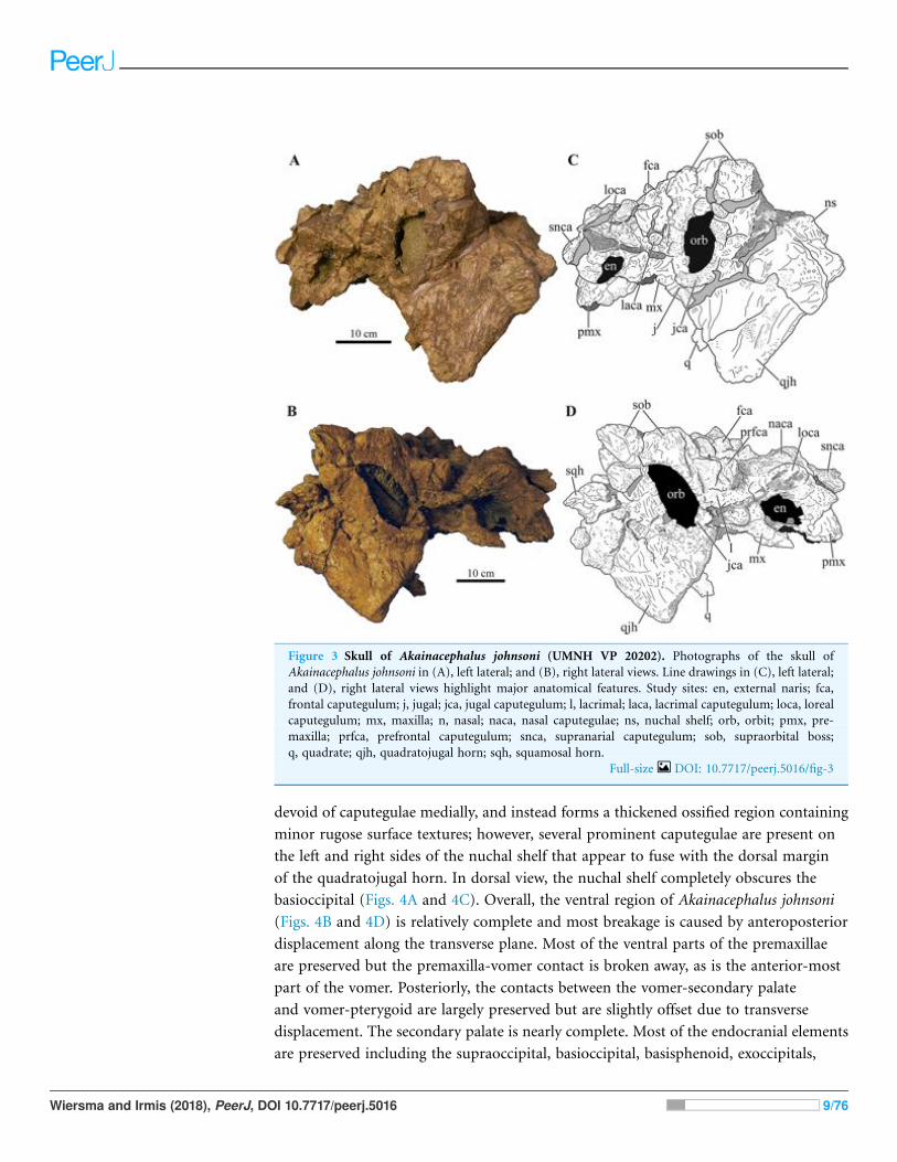
minor rugose surface texture (Fig. 4A). The nuchal shelf is an anteroposteriorly short
structure that is positioned transversely across the dorsal surface of the skull. It contacts
the posteriormost margin of the frontals. The anterior portion of the nuchal area is
Wiersma and Irmis (2018), PeerJ, DOI 10.7717/peerj.5016 8/76
devoid of caputegulae medially, and instead forms a thickened ossified region containing
minor rugose surface textures; however, several prominent caputegulae are present on
the left and right sides of the nuchal shelf that appear to fuse with the dorsal margin
of the quadratojugal horn. In dorsal view, the nuchal shelf completely obscures the
basioccipital (Figs. 4A and 4C). Overall, the ventral region of Akainacephalus johnsoni
(Figs. 4B and 4D) is relatively complete and most breakage is caused by anteroposterior
displacement along the transverse plane. Most of the ventral parts of the premaxillae
are preserved but the premaxilla-vomer contact is broken away, as is the anterior-most
part of the vomer. Posteriorly, the contacts between the vomer-secondary palate
and vomer-pterygoid are largely preserved but are slightly offset due to transverse
displacement. The secondary palate is nearly complete. Most of the endocranial elements
are preserved including the supraoccipital, basioccipital, basisphenoid, exoccipitals,
Figure 3 Skull of Akainacephalus johnsoni (UMNH VP 20202). Photographs of the skull ofAkainacephalus johnsoni in (A), left lateral; and (B), right lateral views. Line drawings in (C), left lateral;and (D), right lateral views highlight major anatomical features. Study sites: en, external naris; fca,frontal caputegulum; j, jugal; jca, jugal caputegulum; l, lacrimal; laca, lacrimal caputegulum; loca, lorealcaputegulum; mx, maxilla; n, nasal; naca, nasal caputegulae; ns, nuchal shelf; orb, orbit; pmx, pre-maxilla; prfca, prefrontal caputegulum; snca, supranarial caputegulum; sob, supraorbital boss;q, quadrate; qjh, quadratojugal horn; sqh, squamosal horn.
Full-size DOI: 10.7717/peerj.5016/fig-3
Wiersma and Irmis (2018), PeerJ, DOI 10.7717/peerj.5016 9/76

and paroccipital processes, but experienced significant predepositional weathering,
removing most of the surface texture details and external margins of each element. In
overall proportions, the mandibles are dorsoventrally tall compared to most other
ankylosaurid mandibles (e.g., Euoplocephalus tutus [UALVP 31, AMNH FARB 5403,
AMNH FARB 5405], Ankylosaurus magniventris [AMNH FARB 5214, CMN 8880],
M. ramachandrani [INBR 21004], and P. grangeri [MPC 100/1014]), but are similar to
Saichania chulsanensis (MPC 100/151). No teeth are preserved with the mandibles, but the
alveolar cavities in the left dentary suggest the presence of 16–18 teeth during life. Both
mandibles are heavily weathered and eroded, predominantly along the posterior and
anterior margins, removing most of the articular surfaces that contact the quadrates
as well as the mandibular symphyseal surfaces that articulate with the predentary.
Figure 4 Skull of Akainacephalus johnsoni (UMNH VP 20202). Photographs of the skull of Akai-nacephalus johnsoni in (A), dorsal; and (B), ventral views. Line drawings in (C), dorsal; and (D), ventralviews highlight major anatomical features. Study sites: bpt, basipterygoid; bs, basisphenoid; ch, choana;exo, exoccipital; fm, foramen magnum; fca, frontal caputegulum; ins, internarial septum; laca, lacrimalcaputegulum; loca, loreal caputegulum; mx, maxilla; mxtr, maxillary tooth row; naca, nasal capute-gulum; ns, nuchal shelf; oc, occipital condyle; pal, palatine; prfca, prefrontal caputegulum; pmx, pre-maxilla; pmxs, interpremaxillry suture with oblong depression; pop, paroccipital process; ptv, pterygoidvacuity; q, quadrate; qj, quadratojugal; qjh, quadratojuga horn; so, supra occipital; snca, supranarialcaputegulum; sob, supraorbital boss; sqh, squamosal horn.
Full-size DOI: 10.7717/peerj.5016/fig-4
Wiersma and Irmis (2018), PeerJ, DOI 10.7717/peerj.5016 10/76
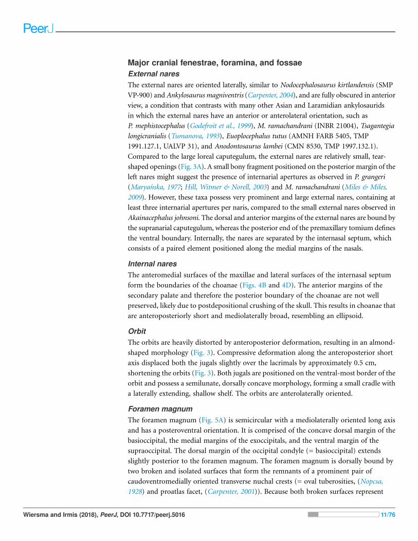
Major cranial fenestrae, foramina, and fossaeExternal naresThe external nares are oriented laterally, similar to Nodocephalosaurus kirtlandensis (SMP
VP-900) andAnkylosaurus magniventris (Carpenter, 2004), and are fully obscured in anterior
view, a condition that contrasts with many other Asian and Laramidian ankylosaurids
in which the external nares have an anterior or anterolateral orientation, such as
P. mephistocephalus (Godefroit et al., 1999), M. ramachandrani (INBR 21004), Tsagantegia
longicranialis (Tumanova, 1993), Euoplocephalus tutus (AMNH FARB 5405, TMP
1991.127.1, UALVP 31), and Anodontosaurus lambei (CMN 8530, TMP 1997.132.1).
Compared to the large loreal caputegulum, the external nares are relatively small, tear-
shaped openings (Fig. 3A). A small bony fragment positioned on the posterior margin of the
left nares might suggest the presence of internarial apertures as observed in P. grangeri
(Marya!nska, 1977; Hill, Witmer & Norell, 2003) and M. ramachandrani (Miles & Miles,
2009). However, these taxa possess very prominent and large external nares, containing at
least three internarial apertures per naris, compared to the small external nares observed in
Akainacephalus johnsoni. The dorsal and anterior margins of the external nares are bound by
the supranarial caputegulum, whereas the posterior end of the premaxillary tomium defines
the ventral boundary. Internally, the nares are separated by the internasal septum, which
consists of a paired element positioned along the medial margins of the nasals.
Internal naresThe anteromedial surfaces of the maxillae and lateral surfaces of the internasal septum
form the boundaries of the choanae (Figs. 4B and 4D). The anterior margins of the
secondary palate and therefore the posterior boundary of the choanae are not well
preserved, likely due to postdepositional crushing of the skull. This results in choanae that
are anteroposteriorly short and mediolaterally broad, resembling an ellipsoid.
OrbitThe orbits are heavily distorted by anteroposterior deformation, resulting in an almond-
shaped morphology (Fig. 3). Compressive deformation along the anteroposterior short
axis displaced both the jugals slightly over the lacrimals by approximately 0.5 cm,
shortening the orbits (Fig. 3). Both jugals are positioned on the ventral-most border of the
orbit and possess a semilunate, dorsally concave morphology, forming a small cradle with
a laterally extending, shallow shelf. The orbits are anterolaterally oriented.
Foramen magnumThe foramen magnum (Fig. 5A) is semicircular with a mediolaterally oriented long axis
and has a posteroventral orientation. It is comprised of the concave dorsal margin of the
basioccipital, the medial margins of the exoccipitals, and the ventral margin of the
supraoccipital. The dorsal margin of the occipital condyle (= basioccipital) extends
slightly posterior to the foramen magnum. The foramen magnum is dorsally bound by
two broken and isolated surfaces that form the remnants of a prominent pair of
caudoventromedially oriented transverse nuchal crests (= oval tuberosities, (Nopcsa,
1928) and proatlas facet, (Carpenter, 2001)). Because both broken surfaces represent
Wiersma and Irmis (2018), PeerJ, DOI 10.7717/peerj.5016 11/76

individual elements, it appears that an incisive notch (sensu Vickaryous, 2001) was present
during life.
Cranial foraminaThe endocranial cavity, including a total of five cranial fenestrae have been identified with
the aid of CT scanning (Figs. 6A–6E). These foramina form the accommodation space for
various cranial nerves (CN). The anterior-most cranial foramen forms the housing for
trigeminal nerve (CN V) and is located on the ventral margin between the laterosphenoid
and prootic bones (Vickaryous, Marya!nska & Weishampel, 2004). In Akainacephalus
johnsoni, the opening for the trigeminal nerve is circular in dorsal view (Fig. 6C). Situated
posterior to CN V, and positioned between the prootic and opisthotic is the fenestra
ovalis, which is a laterally oblong opening in dorsal view that exits directly anterior to
the exoccipitals (Fig. 6C). Posterior to the fenestra ovalis are three foramina for the
glossopharyngeal (CN IX), vagus (CN X), and accessory nerves (CN XI), respectively
(Fig. 6C). The morphology of the openings are very narrow and needle-like. The foramen
Figure 5 Skull of Akainacephalus johnsoni (UMNH VP 20202). Photographs of the skull ofAkainacephalus johnsoni in (A), posterior; and (B), anterior views. Line drawings in (C), posterior; and(D), anterior views highlight major anatomical features. Study sites: fm, foramen magnum; fca, frontalcaputegulae; loca, loreal caputegulum; naca; nasal caputegulae; ns, nuchal shelf; oc, occipital condyle;pmx, premaxilla; pop, paroccipital process; prfca, prefrontal caputegulum; q, quadrate; qjh, quad-ratojugal horn; snca, supranarial caputegulum; so, supraoccipital; sob, supraorbital boss; sqh, squamosalhorn. Full-size DOI: 10.7717/peerj.5016/fig-5
Wiersma and Irmis (2018), PeerJ, DOI 10.7717/peerj.5016 12/76
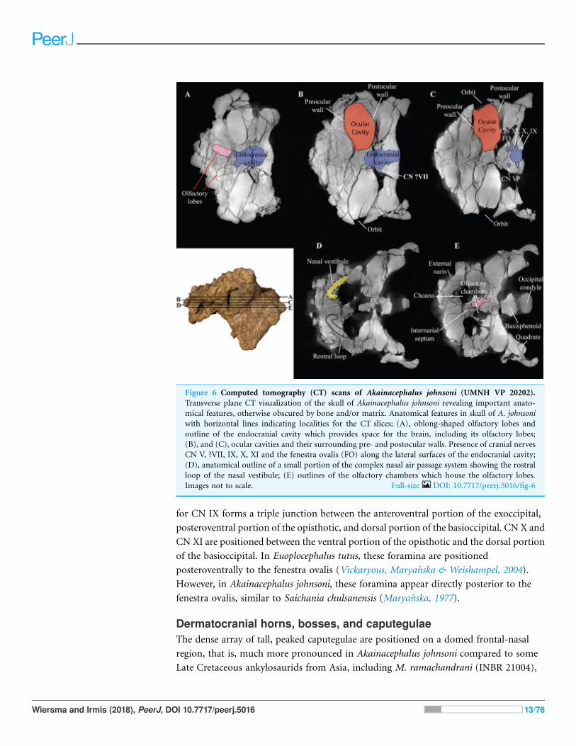
for CN IX forms a triple junction between the anteroventral portion of the exoccipital,
posteroventral portion of the opisthotic, and dorsal portion of the basioccipital. CN X and
CN XI are positioned between the ventral portion of the opisthotic and the dorsal portion
of the basioccipital. In Euoplocephalus tutus, these foramina are positioned
posteroventrally to the fenestra ovalis (Vickaryous, Marya!nska & Weishampel, 2004).
However, in Akainacephalus johnsoni, these foramina appear directly posterior to the
fenestra ovalis, similar to Saichania chulsanensis (Marya!nska, 1977).
Dermatocranial horns, bosses, and caputegulaeThe dense array of tall, peaked caputegulae are positioned on a domed frontal-nasal
region, that is, much more pronounced in Akainacephalus johnsoni compared to some
Late Cretaceous ankylosaurids from Asia, including M. ramachandrani (INBR 21004),
Figure 6 Computed tomography (CT) scans of Akainacephalus johnsoni (UMNH VP 20202).Transverse plane CT visualization of the skull of Akainacephalus johnsoni revealing important anato-mical features, otherwise obscured by bone and/or matrix. Anatomical features in skull of A. johnsoniwith horizontal lines indicating localities for the CT slices; (A), oblong-shaped olfactory lobes andoutline of the endocranial cavity which provides space for the brain, including its olfactory lobes;(B), and (C), ocular cavities and their surrounding pre- and postocular walls. Presence of cranial nervesCN V, ?VII, IX, X, XI and the fenestra ovalis (FO) along the lateral surfaces of the endocranial cavity;(D), anatomical outline of a small portion of the complex nasal air passage system showing the rostralloop of the nasal vestibule; (E) outlines of the olfactory chambers which house the olfactory lobes.Images not to scale. Full-size DOI: 10.7717/peerj.5016/fig-6
Wiersma and Irmis (2018), PeerJ, DOI 10.7717/peerj.5016 13/76

Saichania chulsanensis (Marya!nska, 1977), and Tarchia teresae (Penkalski & Tumanova,
2017) (Fig. 7). Instead, the dome is morphologically similar to Late Cretaceous
Laramidian ankylosaurids such as Ankylosaurus magniventris (Brown, 1908; Carpenter,
2004; Arbour & Mallon, 2017), Anodontosaurus lambei (CMN 8530; TMP 1997.132.1),
Euoplocephalus tutus (e.g., ROM 1930; TMP 1991.127.1; UALVP 31), and Ziapelta
sanjuanensis (Arbour et al., 2014). The dome covers most of the nasal region,
extendingtowards the posteriormost part of the frontals, where it contacts the nuchal
shelf. Akainacephalus johnsoni possesses two supranarial caputegulae that are
morphologically similar to Nodocephalosaurus kirtlandensis (SMP VP-900); in both taxa,
the supranarial caputegulae are anteriorly protruding, mediolaterally broad caputegulae,
situated dorsally on the premaxilla (Figs. 3A–3D, 4A, 4C, 7A–7D and 8). Two flange-like
and anteroposteriorly elongated loreal caputegulae cover the nares dorsally, each
succeeded by a large, tetrahedral-shaped, prefrontal caputegulae. A small lacrimal
psob
sqh
sqh
qjh
qjhasob
loca
snca
acc po sqh
qjh
pos
lacapmxqptptmx
pmxpmx
mxsnca
pt qqjh
pnca
sqh
loca
psobasobprfca
naca
nucafrca
orb orb orborb
orb
n nn
UMNH VP 20202Akainacephalus johnsoni
SMP VP-900Nodocephalosaurus kirtlandensis
PIN 3142/250Tarchia teresae
INBR 21004Minotaurasaurus ramachandrani
A C E G
B DF H
prfca
mso
laca
prfcaprfca
prfcalaca
Figure 7 Variation in cranial ornamentation in selected Laramidian and Asian taxa, including Akainacephalus johnsoni. Comparative linedrawings highlighting major areas of cranial ornamentation in Akainacephalus johnsoni and closely related Laramidian and Asian taxa. Akaina-cephalus johnsoni (UMNHVP 20202) in (A), dorsal; (B), left lateral view compared toNodocephalosaurus kirtlandensis (SMP VP-900) in (C), dorsal;and (D) left lateral view; Tarchia teresae (PIN 3142/250) in (E) dorsal; (F), left lateral view and Minotaurasaurus ramachandrani (INBR 21004) in(G), dorsal; and (H), left lateral view. Study sites: acc po, accessory postorbital ossification; asob, anterior supraorbital boss; frca, frontal capu-tegulum; laca, lacrimal caputegulum; loca, loreal caputegulum; mso, medial supraorbital; mx, maxilla; n, external naris; naca, nasal caputegulae;nuca, nuchal caputegulae; orb, orbital; pmx, premxilla; pnca, postnarial caputegulum; pos postocular ossicles; prfca, prefrontal caputegulum; psob,posterior supraorbital boss; pt, pterygoid; q, quadrate; qjh, quadratojugal horn; snca, supranarial caputegulum; sqh, squamosal horn. Color schemeafter Arbour & Currie (2013a). Dorsal view of N. kirtlandensis modified after Arbour et al. (2014). T. teresea (=Saichania chulsanensis in Arbour,Currie & Badamgarav, 2014) and M. ramachandrani modified after Arbour, Currie & Badamgarav (2014).
Full-size DOI: 10.7717/peerj.5016/fig-7
Wiersma and Irmis (2018), PeerJ, DOI 10.7717/peerj.5016 14/76

caputegulum is positioned ventral to the prefrontal caputegulum. The postorbital horns
are dorsoventrally tall, backswept, and project laterally in dorsal view. Only a partial
squamosal horn is preserved but it is badly damaged and largely anatomically
uninformative. The quadratojugal horns display a triangular morphology with a ventrally
projecting apex and cover the majority of the ventral portion of the postocular region
of the skull. Individual caputegulae are described below.
Supranarial caputegulaeThe external nares in Akainacephalus johnsoni are oriented laterally and their position
is placed posteriorly relative to the front of the snout, similar to Nodocephalosaurus
kirtlandensis (Fig. 8). In most other ankylosaurids, including M. ramachandrani
(INBR 21004), Euoplocephalus tutus (UALVP 31, AMNH FARB 5205), Anodontosaurus
lambei (CMN 8350), Tarchia teresae (PIN 3142/250), and Saichania chulsanensis
(MPC 100/151), the nares are oriented entirely anteriorly or anterolaterally and are
dorsally and transversely ornamented with a thickened rim of ossification—the
Figure 8 Skull of Akainacephalus johnsoni compared with Nodocephalosaurus kirtlandensis.Comparison of cranial features between closely related southern Laramidian taxa; (A), Akainacepha-lus johnsoni (UMNH VP 20202) from the Late Cretaceous Kaiparowits Formation of Utah; and (B),Nodocephalosaurus kirtlandensis (SMP VP-900) from the Late Cretaceous Kirtland Formation of NewMexico, in left lateral views. Various synapomorphies are shared with N. kirtlandensis (highlighted inblack and white arrows) and includes “flaring nostrils”; enlarged, laterally projecting, loreal osteodermsthat are situated directly dorsal to the external nares. Other synapomorphies include pyramid-shapednasal and frontal osteoderms positioned on the dorsal regions of the skull. A number of significantdifferences have been observed between both specimens; in A. johnsoni, the anterior, and posteriorsupraorbital bosses form an enlarged element that is somewhat backswept, whereas in N. kirtlandensis,the posterior and anterior supraorbital bosses are clearly defined as individual osteoderms, and are muchsmaller in size. Additionally, the squamosal horn in Akainacephalus is very small but is prominent andtetrahedrally shaped in Nodocephalosaurus. The quadratojugal horn in Akainacephalus is massive, has asubtriangular morphology in lateral view and projects almost entirely ventral, whereas in Nodocepha-losaurus, the quadratojugal horn is smaller and has a typical fin-shaped morphology. Study sites: asob,anterior supraorbital boss; ext naris, external naris; laca, lacrimal caputegulum; loca, loreal caputegu-lum; naca, nasal caputegulae; orb, orbit; psob, posterior supraorbital boss; qjh, quadratojugal horn;sqh, squamosal horn. Full-size DOI: 10.7717/peerj.5016/fig-8
Wiersma and Irmis (2018), PeerJ, DOI 10.7717/peerj.5016 15/76
supranarial caputegulae (Arbour & Currie, 2013a). In Akainacephalus johnsoni, each naris
is anteriorly ornamented with an anteroposteriorly broad, supranasal caputegulum that is
positioned transversely and dorsolaterally along the premaxillary beak (Fig. 3). A similar
caputegulum was described for Nodocephalsaurus kirtlandensis by Sullivan (1999). Given
that the caputegulum is situated directly above the external nares, it seems appropriate
to use the term supranarial caputegulum, which is consistent with the terminology
used by Arbour & Currie (2013a, 2016). The surface texture of this large caputegulum
is smooth, with some rugosity along the dorsal surface. The caputegulum is
anteroposteriorly elongated (Figs. 4A and 4C) and contains a lateral margin that forms a
longitudinal keel along the entire length of the caputegulum (Fig. 3). Posteriorly, the
caputegulum contacts the anterior margin of the lacrimal portion of the circumorbital
complex. Posterodorsally it is accompanied by a prominent, single, and tetrahedral-
shaped lacrimal caputegulum with minimal rugose surface texture, which is present in
Nodocephalosaurus kirtlandensis (SMP VP-900) and is referred to as the prefrontal
caputegulum by Sullivan (1999). However, in N. kirtlandensis (SMP VP-900), the
caputegulum is morphologically more conical rather than tetrahedral and is heavily pitted
(Fig. 8). Anteriorly, each supranarial caputegulum contacts a large anterolaterally
positioned caputegulum that forms the anterior-most ornamentation and is positioned
transversely along the premaxilla, nearly identical to N. kirtlandensis (SMP VP-900).
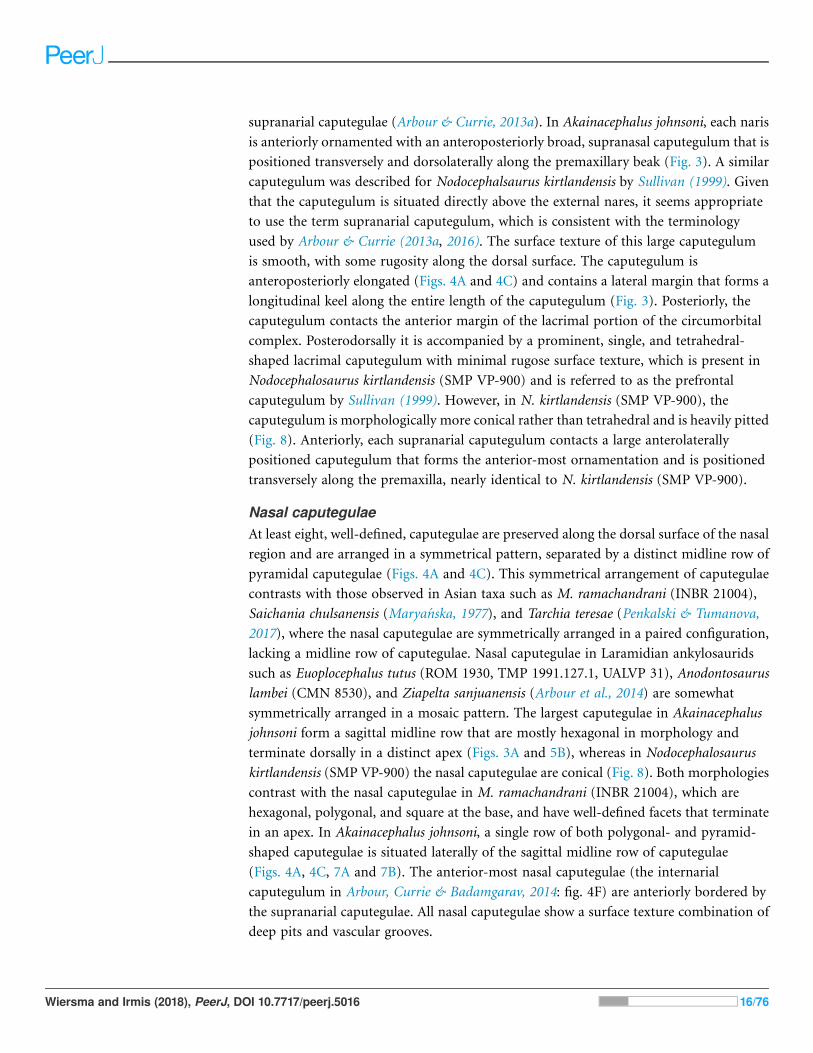
Nasal caputegulaeAt least eight, well-defined, caputegulae are preserved along the dorsal surface of the nasal
region and are arranged in a symmetrical pattern, separated by a distinct midline row of
pyramidal caputegulae (Figs. 4A and 4C). This symmetrical arrangement of caputegulae
contrasts with those observed in Asian taxa such as M. ramachandrani (INBR 21004),
Saichania chulsanensis (Marya!nska, 1977), and Tarchia teresae (Penkalski & Tumanova,
2017), where the nasal caputegulae are symmetrically arranged in a paired configuration,
lacking a midline row of caputegulae. Nasal caputegulae in Laramidian ankylosaurids
such as Euoplocephalus tutus (ROM 1930, TMP 1991.127.1, UALVP 31), Anodontosaurus
lambei (CMN 8530), and Ziapelta sanjuanensis (Arbour et al., 2014) are somewhat
symmetrically arranged in a mosaic pattern. The largest caputegulae in Akainacephalus
johnsoni form a sagittal midline row that are mostly hexagonal in morphology and
terminate dorsally in a distinct apex (Figs. 3A and 5B), whereas in Nodocephalosaurus
kirtlandensis (SMP VP-900) the nasal caputegulae are conical (Fig. 8). Both morphologies
contrast with the nasal caputegulae in M. ramachandrani (INBR 21004), which are
hexagonal, polygonal, and square at the base, and have well-defined facets that terminate
in an apex. In Akainacephalus johnsoni, a single row of both polygonal- and pyramid-
shaped caputegulae is situated laterally of the sagittal midline row of caputegulae
(Figs. 4A, 4C, 7A and 7B). The anterior-most nasal caputegulae (the internarial
caputegulum in Arbour, Currie & Badamgarav, 2014: fig. 4F) are anteriorly bordered by
the supranarial caputegulae. All nasal caputegulae show a surface texture combination of
deep pits and vascular grooves.
Wiersma and Irmis (2018), PeerJ, DOI 10.7717/peerj.5016 16/76
Loreal caputegulaeA very characteristic, large, and laterally oriented loreal caputegulum (= postmaxillary/
lacrimal ridge in Sullivan, 1999) is positioned dorsally above the external nares where it
ascends anteriorly and “flares” out laterally (Fig. 3). The loreal caputegulum extends
posteriorly and contacts the anterior margin of the circumorbital complex, enveloping the
external nares along the anterodorsal and posterior margins. Sullivan (1999) referred to
this particular loreal caputegulum in Nodocephalosaurus kirtlandensis (SMP VP-900) as
the postmaxillary/lacrimal ridge, but this term is somewhat misleading because the
caputegulum in Akainacephalus johnsoni is positioned directly dorsal to the maxilla and
obscures the lacrimal. The lateral projection of the nasal caputegulum forms a thickly
rimmed, overhanging shelf, producing a very characteristic flaring nostril in lateral view.
This condition is currently only observed in Akainacephalus johnsoni and
Nodocephalosaurus kirtlandensis (Sullivan, 1999).
Prefrontal caputegulaeA distinct, tetrahedral/subtriangular caputegulum with a laterally projecting, keeled
apex is positioned on the lateral side of each prefrontal (Figs. 4A and 4C). The
caputegulum is directly anterior to the anterior-most supraorbital boss and dorsal to the
flanged nasal caputegulum, and shows a morphology and position similar to the
prefrontal caputegulae in M. ramachandrani (INBR 21004) and Saichania chulsanensis
(Marya!nska, 1977: fig. 5; Arbour, Currie & Badamgarav, 2014: figs. 4E-F and 5D). In
UMNH VP 20202 it does not form a continuous surface with the supraorbital bosses, in
contrast with M. ramachandrani. It is situated directly anterior to the circumorbital
complex and dorsal to the supranarial caputegulum and lacrimal. The surface texture is
similar to that of the supranarial caputegulum and is relatively smooth with some
vascularity but no pitted textures.
Frontal caputegulaeMany caputegulae are eroded away but seven distinct caputegulae are preserved across the
frontal region of the skull and are arranged in a mosaic pattern (Figs. 4A and 4C). The
posterior-most caputegulum is the largest and positioned medially. It has a mediolaterally
oblong, somewhat hexagonal base and a bluntly bulbous morphology with relatively
smooth surface texture. This caputegulum is laterally and anteriorly surrounded by six
smaller caputegulae. The morphology of the smaller surrounding caputegulae is very
different from the large, posterior caputegulum, but similar to the nasal caputegulae;
they preserve irregularly polygonal-shaped base, are sharply conical rather than bulbous,
and possess a rugose and pitted surface texture. The arrangement of the frontal
caputegulae in Akainacephalus appears similar to Anodontosaurus lambei (TMP 1997.59.1
(Arbour & Currie, 2013a: fig. 4.)) and Ankylosaurus magniventris (Brown, 1908; Carpenter,
2004; Arbour & Mallon, 2017); however, the morphology of these smaller caputegulae is
more similar to those observed in Nodocephalosaurus (SMP VP-900) (Fig. 7C), and to
some extentM. ramachandrani (INBR 21004) (Fig. 7G) and Zaraapelta nomadis (Arbour,
Currie & Badamgarav, 2014), although the latter two taxa display a more pyramidal
Wiersma and Irmis (2018), PeerJ, DOI 10.7717/peerj.5016 17/76

than conical morphology. Frontal caputegulae in other Asian taxa such as Tarchia
(Tumanova, 1977; Penkalski & Tumanova, 2017) and Saichania (Marya!nska, 1977) are
morphologically very different from Akainacephalus johnsoni and display a more rectangular
base and a nearly flat or slightly bulbous dorsal surface. In Laramidian ankylosaurid
dinosaurs, including Euoplocephalus tutus (Arbour &Currie, 2013a) and Ziapelta sanjuanensis
(Arbour et al., 2014), very few or no frontal caputegulae are known, either because they did
not preserve or perhaps these taxa lack extensive cranial ornamentation in this region.
Nuchal caputegulaeThe nuchal shelf forms a dorsoventrally raised, tabular shelf, similar to Minotaurasaurus
ramachandrani (INBR 21004), Tarchia kielanae (Arbour, Currie & Badamgarav, 2014;
Penkalski & Tumanova, 2017), Tarchia teresae (Penkalski & Tumanova, 2017), Saichania
chulsanensis (Marya!nska, 1977), and some specimens of Euoplocephalus tutus (ROM
1930), Oohkotokia horneri (MOR 433), and Anodontosaurus lambei (AMNH FARB 5238).
In Akainacephalus johnsoni, the nuchal shelf shows little rugosity, but is damaged in
several places across the dorsal surface and a large portion is broken away from the left
side, leaving a transversely oblong cavity (Figs. 4A and 4C). Nuchal caputegulae are
present on the posterior-most portion of the nuchal shelf. A total of three, poorly
preserved caputegulae are visible (Fig. 4A), and their morphology varies between
subrounded with a small apex to elongate polygonal with a small transverse dorsal ridge.
This condition is dissimilar from other ankylosaurids such as Ziapelta sanjuanensis
(Arbour et al., 2014), Zaraapelta nomadis (MPC D100/1338 (Arbour, Currie &
Badamgarav, 2014)), Pinacosaurus mephistocephalus (IMM 96BM3/1 (Godefroit et al.,
1999)), and P. grangeri (MPC 100/1014 (Tumanova, 1987)), in which the nuchal shelf and
the nuchal caputegulae are flat. The surface texture of the nuchal caputegulae in
Akainacephalus johnsoni is smooth with shallow pitting. A distinct furrow separates
the nuchal caputegulae from the posterior supraorbital bosses.
Circumorbital complexThe large circumorbital complex consists of a series of co-ossified ornamental elements
that surround the orbital cavity and are best preserved on the left lateral side of the skull
(Fig. 3A). The complex comprises a large supraorbital horn, a small lacrimal
caputegulum, a semicircular jugal osteoderm, and the thickened rim along the posterior
margin of the orbital. The anterior- and posterior supraorbital caputegulae appear
fused together, forming a large, and tall, postorbital horn with a prominent apex that
projects posterolaterally, covering the entire dorsal surface of the supraorbital (Figs. 3 and 4);
a condition unique to Akainacephalus johnsoni. In anterior and posterior view (Fig. 5),
the postorbital horn projects dorsolaterally and laterally in dorsal view (Figs. 4A and 4C),
forming the transversely widest part of the skull as preserved. Usually, cranial elements
such as the squamosal horns and quadratojugal horns exceed the width of the postorbital
horns as in M. ramachandrani (INBR 21004), Tarchia teresae (Penkalski & Tumanova,
2017), Pinacosaurus mephistocephalus (Godefroit et al., 1999). This cannot be evaluated
in Akainacephalus johnsoni because the squamosal horns are badly damaged, and the
Wiersma and Irmis (2018), PeerJ, DOI 10.7717/peerj.5016 18/76

quadratojugal horns are more vertically oriented compared to other ankylosaurid taxa
(e.g.,M. ramachandrani [INBR 21004], Euoplocephalus tutus [AMNH FARB 5405, UALVP
31], Ankylosaurus magniventris [AMNH FARB 5214], and Anodontosaurus lambei
[CMN 8530]). In dorsal view, the postorbital horns are large and triangular with a
posterolaterally projecting apex (Fig. 4A). The surface texture is coarsely rugose and
bulbous. In lateral view, the apex projects posterodorsally and is less rugose compared
to its dorsal surface. The lacrimal caputegulum is positioned ventral to the supraorbital
boss and comprises a small tabular caputegulum with a much smoother surface texture.
The jugal caputegulum forms the ventral-most border of the circumorbital complex and
has a semilunate, dorsally concave morphology that cradles the orbit and possesses a
relatively smooth surface texture. The caputegulum extends laterally and results in a small
shelf. Posteriorly, several small, tabular caputegulae complete the circumorbital complex.
They are more rugose than the lacrimal and jugal caputegulae but less so than the
supraorbital boss.
Quadratojugal hornsThe quadratojugal horns cover the majority of the ventral postocular region of the skull
(Fig. 3). Their morphology and orientation are unusual in that they represent an
asymmetrical triangle with a vertically positioned apex, which is somewhat similar to
Zaraapelta nomadis (Arbour, Currie & Badamgarav, 2014) and Shamosaurus scutatus
(Tumanova, 1983), except that the apices in Z. nomadis project lateroventrally, and
Akainacephalus johnsoni lacks the presence of interstitial postocular ossicles, separating
the quadratojugal from the squamosal horn and orbit. In anterior and posterior view, the
quadratojugal horns project nearly vertically, which appears to be close to the original
anatomical position of the horns, given that little deformation and breakage of other
(basicranial) elements is expressed in this area of the skull. In lateral view, the anterior
part of the quadratojugal cradles the ventral margin of the jugal portion of the
circumorbital complex, whereas the posterior end extends posterodorsally and contacts
the posterior margin of the circumorbital complex. The extent of the posterodorsal
boundaries of the quadratojugal horns are poorly defined and blend together with the
squamosal, making it difficult to assess their exact position. The quadratojugal horns are
nearly straight or slightly convex laterally and have an overall smooth surface texture,
accompanied by dorsoventral-oriented furrows or vascular grooves (Fig. 3). The
quadrates are visible in lateral view, anterior to the quadratojugal horns, a condition
similar to Nodocephalosaurus kirtlandensis (SMP VP-900), Gobisaurus domoculus
(Vickaryous et al., 2001), Shamosaurus scutatus (Tumanova, 1983), and Tarchia teresae
(Penkalski & Tumanova, 2017). In all other ankylosaurids (e.g., Saichania chulsanensis,
P. grangeri, Euoplocephalus tutus, Zuul crurivastator, and Anodontosaurus lambei), the
quadrates are obscured by the quadratojugal horns.
Squamosal hornOnly a partial right squamosal horn is preserved (Fig. 3B), but is largely broken, limiting
the amount of observable anatomical detail. The right squamosal horn forms the
Wiersma and Irmis (2018), PeerJ, DOI 10.7717/peerj.5016 19/76

posterior-most caputegulum on the skull (Fig. 3) and is positioned directly posterior to
the supraorbital boss. Various Asian (e.g., Tarchia kielanae (Marya!nska, 1977), Saichania
chulsanensis (Marya!nska, 1977), Zaraapelta nomadis (Arbour, Currie & Badamgarav,
2014), Minotaurasaurus ramachandrani [INBR 21004]) and most Laramidian taxa (e.g.,
Anodontosaurus lambei [CMN 8530], Euoplocephalus tutus [e.g., ROM 1930], O. horneri
[MOR 433], Ankylosaurus magniventris (Carpenter, 2004), Ziapelta sanjuanensis (Arbour
et al., 2014)) display a clear separation between the quadratojugal and squamosal horns, a
space that is sometimes occupied by postocular ossicles and visible in several specimens of
Anodontosaurus lambei (CMN 8530, NHMUK R4947, TMP 1997.132.1) and Zaraapelta
nomadis (Arbour, Currie & Badamgarav, 2014), but this area is poorly defined in
Akainacephalus johnsoni (Figs. 3 and 7). The left squamosal horn is not preserved and the
area is damaged.
Mandibular caputegulumAn anteroposteriorly elongated and dorsoventrally deep mandibular caputegulum forms
the ventral border of the mandible (Fig. 9). Morphologically, the caputegulum is
subtriangular with a blunt keel and a near-ventral projection; a condition unique to
Akainacephalus johnsoni. The caputegulum is positioned along the ventrolateral margin of
the jaw, covering the ventral, and lower half of the lateral portion of the angular. The total
length of the mandibular caputegulum covers >50% of the entire anteroposterior length
of the lower jaw. The mandibular caputegulum is short compared to other ankylosaurid
taxa, including specimens of Euoplocephalus tutus (AMNH FARB 5403 and 5405),
M. ramachandrani (INBR 21004), and to some extent, Tarchia teresae (=Saichania
chulsanensis (Arbour, Currie & Badamgarav, 2014)), in which the caputegulae are
dorsoventrally narrow and encompass nearly the entire length of the mandible. Although
the orientation of the mandibular caputegulum is unique to Akainacephalus johnsoni,
morphologically it shares some similarities to the mandibular caputegulum seen in the
holotype of Saichania chulsanensis (MPC 100/151 (Arbour, Currie & Badamgarav, 2014)).
In anterior and posterior view, the mandibular caputegulum is oriented ventrolaterally
and extends well below the dentary and angular, forming the ventral-most portion of
the jaw. Posteriorly, the caputegulum is convex and curves upward in a lobe-shaped
morphology. It terminates against, and contacts with, the ventral border of the surangular.
A distinct longitudinal furrow delineates the medial border of the mandibular
caputegulum from the angular. The ventral surface is mediolaterally broad and
horizontally flat. The external surface texture is smooth and shows no signs of furrowing
and pitted textures. Measurements for individual elements are summarized in
Supplemental Material S4.
Bones of the dermatocraniumPremaxillaeThe premaxillae form a broad U-shaped beak in ventral view (Figs. 4B and 4D), similar to
Euoplocephalus tutus (ROM 1930; TMP 1997.132.1 (Arbour & Currie, 2013a: fig. 6A–B)),
Anodontosaurus lambei (CMN 8530), and Ziapelta sanjuanensis (Arbour et al., 2014).
Wiersma and Irmis (2018), PeerJ, DOI 10.7717/peerj.5016 20/76

In Ankylosaurus magniventris (AMNH FARB 5214), Pinacosaurus mephistocephalus
(Godefroit et al., 1999), P. grangeri (Hill, Witmer & Norell, 2003), Tarchia teresae (Penkalski
& Tumanova, 2017), Saichania chulsanensis (Marya!nska, 1977), and M. ramachandrani
(INBR 21004), the premaxillae slightly taper anteriorly, resulting in a narrower beak. In
anterior view, the rostral portion of the premaxilla is transversely wide and tall, dorsally
Figure 9 Mandibles and predentary. Photographs of the mandibles and predentary of Akainacephalusjohnsoni (UMNH VP 20202). Left mandible in (A), lateral; (B), medial; (C), ventral; and (D), dorsalviews. Right mandible in (E), lateral; (F), medial; (G), ventral; and (H), dorsal view. Predentary in (I),anterior; (J), posterior; (K), dorsal; and (L), ventral views. Study sites: cp, coronoid process; cs, pre-dentary cutting surface; mos, mandibular osteoderm; ms, mandibular symphysis; mtr, mandibular toothrow; pp, predentary protuberance. Full-size DOI: 10.7717/peerj.5016/fig-9
Wiersma and Irmis (2018), PeerJ, DOI 10.7717/peerj.5016 21/76

bound by a distinct transverse and ventrally concave-oriented nasal ridge which is bisected
by two supranarial caputegulae (Fig. 5B). The ventral margin that forms the premaxillary
tomium is eroded, but preserved areas reveal a ventrally convex morphology for each
premaxilla. A significant portion of the right premaxillary tomium is broken. Anteriorly,
the midline possesses the typical interpremaxillary suture (sensu Vickaryous, 2001)
and is characterized by a small midline notch (incisura premaxillaris; Vickaryous, 2001)
(= interpremaxillary notch; Sereno, 1999) bisecting the left and right premaxillae.
In ventral view, the buccal margin of the premaxilla is U-shaped and shallow. The
premaxillary palate is wider than it is long. The lateral halves of the premaxillary palate are
slightly concave ventrally but become more convex medially, a morphology similar to
Euoplocephalus tutus (ROM 1930) and Ankylosaurus magniventris (Carpenter, 2004:
fig. 4C). The majority of the vomer is broken away along the rostral part of the skull
and only a dorsal remnant that articulates with the roof of the oral cavity remains. The
premaxilla-maxilla contact is the widest part of the premaxillae in ventral view. In lateral
view, it borders the ventral margin of the external nares. The ventral cutting surface is
convex and forms a distinct premaxillary notch along the midline, separating both
premaxillae. This notch is clearly visible in ventral view and continues posteriorly to
form a small tear drop-shaped foramen (Figs. 4B and 4D). The ventral portion of the
premaxillary beak is anteroposteriorly shorter than in other ankylosaurids (e.g.,
M. ramachandrani [INBR 21004]) and contacts the anteriormost alveolar cavities of
the maxillae. The premaxillary tomial crest is short and terminates well before the
anterior-most maxillary alveolar cavity.
MaxillaeThe maxillae are poorly preserved, andmost of the surface textures and margins have been
eroded to some extent. However, both maxillae in Akainacephalus johnsoni are unusually
well exposed in lateral view, revealing important anatomical features (Fig. 3). In lateral
view, the anterior margin is concave and borders the posterior and partial ventral portion
of the external nares until it contacts the premaxilla. The ventral portion of the anterior
half of the maxilla is convex but becomes concave along the posterior half, where it
contacts the anteroventral margin of the circumorbital complex. A large but shallow
sulcus forms the majority of the posterior half of the maxilla. In most ankylosaurids (e.g.,
Pinacosaurus grangeri [MPC 100/1014 (Hill, Witmer & Norell, 2003: fig. 4)], Saichania
chulsanensis [MPC 100/151], M. ramachandrani [INBR21004], Euoplocephalus tutus
[e.g., TMP 1991.127.1], Anodontosaurus lambei [CMN 8530], and Ankylosaurus
magniventris [AMNH FARB 5214 (Carpenter, 2004: fig. 4C)]) only the ventral-most
portion of the maxilla is visible in lateral view. Lateral and ventrally extending nasal
and lacrimal caputegulae obscure the maxillae in the aforementioned taxa, but
Akainacephalus johnsoni possess no such lateral covering, instead, the only ornamentation
that is present along the maxilla is contributed dorsally by the supranasal caputegulum,
and posteriorly by the anteroventral margin of the circumorbital complex (Fig. 3), similar
to Nodocephalosaurus kirtlandensis (Sullivan, 1999). Approximately 75% of the entire
tomial crest is located on the maxillae and terminates just dorsal of the ectopterygoid.
Wiersma and Irmis (2018), PeerJ, DOI 10.7717/peerj.5016 22/76

Breakage and erosion have damaged both tooth rows, and the four to six anterior-most
alveolar cavities on the right maxilla are eroded away. All the alveolar cavities on the
posterior half of the left maxilla are eroded away, preserving only the anterior alveoli. A
transverse fracture displaces the posterior half of the preserved alveoli, offsetting them
medially. Preserved alveolar cavities suggest that each maxilla would have held at least 16
teeth during life. Both maxillae are laterally concave and together form an hourglass
configuration, a condition typical for ankylosaurid and nodosaurid dinosaurs. The
anterior half of the maxilla forms the widest part of the element and becomes the ventral
border of the external nares. Posteriorly, the width of the maxilla decreases, and it contacts
with the dorsal surface of the ectopterygoid. In lateral view, a deep sulcus is positioned
between the supranarial caputegulum and the posterior half of the maxilla. Ventrally, the
anterodorsal secondary palate forms the lingual part of the maxilla and is dorsally depressed
compared to its anteriorly bordering premaxillary shelf (Figs. 4B and 4D).
NasalsThe nasals are completely obscured by the co-ossification of caputegulae along their
entire dorsal and lateral surfaces. Ventrally, they are obscured by the maxillary secondary
palate (Figs. 4B and 4D). Observations of the nasals have been made with the aid of CT
scans where features are obscured. The internasal septum is morphologically similar to
that described in Pinacosaurus grangeri (Marya!nska, 1977; Hill, Witmer & Norell, 2003)
and forms a paired element that resembles elongate wings that project ventrally and
are formed by the medial margins of the nasal bones. Together, they comprise the
insertion space for the vomer. Posteriorly, the internasal septum forms a steeply
descending ventral flange that articulates with the midline of the pterygoid complex.
Anteriorly, the septum expands transversely as well as anteroposteriorly, resulting in an
elliptical, bulging process that articulates along the midline on the posterior margin of the
premaxillae. The choanae are deep and have bony fragments encased in sandstone matrix
in the dorsal-most regions, which might belong to the roof of the nasals.
LacrimalsIn ankylosaurs, the lacrimals form the anterior border of the orbit and circumorbital
complex, and contact the maxillae anteroventrally, the supraorbitals posterodorsally,
and the jugals posteroventrally (Vickaryous, 2001). Only a few ankylosaurian dinosaurs,
such as juvenile Pinacosaurus grangeri (Vickaryous, 2001; Burns et al., 2011) and
Kunbarrasaurus ieversi (Leahey et al., 2015), display clearly identifiable lacrimals, which
form the anterior-most circumorbital bones. In Akainacephalus johnsoni, the lacrimals
cannot be identified as distinct elements because of the presence of remnants of unprepared
matrix along the medial and ventral portions of this region (Fig. 3). In addition, co-
ossification of the rugose secondary surface on the lateral side fully obscures the elements. In
his description of Nodocephalosaurus kirtlandensis, Sullivan (1999) mentioned the presence
of a prominent lacrimal, comparable to P. grangeri (Vickaryous, 2001). In Akainacephalus
johnsoni the lacrimals are poorly visible laterally, which is possibly due to deformation.
Dorsolaterally, the lacrimals are ornamented with a tetrahedral-shaped lacrimal
Wiersma and Irmis (2018), PeerJ, DOI 10.7717/peerj.5016 23/76

caputegulum (Fig. 3), which has a conical morphology in N. kirtlandensis (Sullivan, 1999).
In contrast, in Nodocephalosaurus kirtlandensis the lacrimal is well defined in lateral view
and has a rugose surface texture (Sullivan, 1999: fig. 2). In Akainacephalus johnsoni, the long
axis of the lacrimals is oriented anteromedially in lateral view.
PostorbitalThe postorbital forms the posterior-most contribution to the circumorbital complex and
contacts anteriorly with the supraorbital and ventrally with the jugal (Fig. 3). Its exact
morphology cannot be determined because it is co-ossified with caputegulae along its
exterior surface and obscured by matrix medially. The ornamented lateral surface
that forms the posterior border of the orbit is continuous with the posterodorsal
ornamentation of the jugal. It forms a thin and narrow, dorsally projecting, and anteriorly
concave wedge that expands dorsally and longitudinally where it contacts the ventral
border of the supraorbital boss. The pre- and postocular walls border the orbital cavities
but are only visible with the aid of CT data (Figs. 6B and 6C) because the orbits are infilled
with matrix. They form a transverse wall of bone, occluding the anterior and posterior
borders of the orbit.
JugalsThe right jugal is damaged by breakage and erosion (Fig. 3B). The left jugal is better
preserved and comprises a small and transversely broad element that contacts the
anterior-most part of the quadratojugal and the posterior margin of the maxilla (Fig. 3A).
The long axis of the element is anteromedially oriented. The jugal curves anteromedially
and ventrally towards the pterygoid, suggesting a contact between both elements;
however, this area is damaged and a suture confirming this contact could not be observed.
In lateral view, the jugal forms a dorsally concave semilunate shelf that is laterally
ornamented with a small ridge of rugose co-ossified bone that forms the ventral border for
both the orbit and the circumorbital complex.
SquamosalsCo-ossification along the lateral (Fig. 3) and dorsal surfaces (Figs. 4A and 4C) of the
squamosal, and the paroccipital processes posteriorly, fully obscure the squamosal.
QuadratojugalsThe quadratojugal forms the anterior extension of the quadratojugal horn. The left
quadratojugal is best preserved. It is a small, anteriorly projecting wall of bone that
appears as a small point in lateral view. It curves medially along its posterior half and
contacts the lateral distal shaft and condyle of the quadrate. The anterior half contacts
the jugal along its dorsal surface (Fig. 3).
PalatinesOnly fragmentary remains of the posteroventral portion of the palatines are preserved,
making it difficult to provide detailed anatomical descriptions of this element. These
remnants are positioned medial to the maxillae, lateral to the internasal septum and jugal,
and anteromedial to the pterygoids (Figs. 4B and 4D). The anterior margin is concave
Wiersma and Irmis (2018), PeerJ, DOI 10.7717/peerj.5016 24/76
and shallow, forming the posterior border of the choanae. Posteriorly, the palatines
reside entirely against the pterygoids to form a solid surface. Some taxa, such as
Pinacosaurus grangeri (Hill, Witmer & Norell, 2003), Anodontosaurus lambei (CMN 8530),
and Euoplocephalus tutus (e.g., Arbour & Currie, 2013a, TMP 1997.132.1, and ROM 1930)
have a posteriorly located postpalatal fenestra or lateral palatal aperture which are absent
in UMNH VP 20202.
EctopterygoidThe ectopterygoid forms a poorly preserved, small, wedge-shaped bone that is
morphologically consistent with other ankylosaurid dinosaurs (e.g., Ankylosaurus) and is
positioned ventromedially to the left pterygoid flange (= pterygoid wing in Arbour &
Currie, 2013a), and lateral to the maxilla (Figs. 4B and 4D). Similar to the description of
the ectopterygoid by Vickaryous (2001), it contributes to the rostral border of the
suborbital fenestra and the caudoventral secondary palate. Significant breakage is present
along the ectopterygoid-pterygoid complex, resulting in the loss of the right ectopterygoid
and the lateral-most portion of the right pterygoid-ectopterygoid articular surface.
PterygoidsThe pterygoid complex is preserved along its posterior region and a small portion of the
left pterygoid flange is visible, but it is completely missing on the right side (Figs. 4B
and 4D). The pterygoid shields are ventrally concave and medially curve downward to
form a blunt keel along the posterior margin. Together with the basipterygoids, they
form a large, anteriorly tapering, teardrop-shaped pterygoid vacuity, similar to
Pinacosaurus grangeri (Hill, Witmer & Norell, 2003), M. ramachandrani (INBR 21004),
Euoplocephalus tutus (e.g., TMP 1997.132.1; ROM 1930), and Zuul crurivastator (ROM
75860). In Saichania chulsanensis (Marya!nska, 1977), this vacuity is much smaller. The
medial margins along the anterior portion of the pterygoid shields do not contact each
other but extend anteriorly where they contact the internasal septum. Laterally, the
pterygoid shields contact the medial margin of the quadrate shaft. The anterolateral half
becomes the pterygoid flange, is ventrally convex, and forms the contact for the
ectopterygoid. The pterygoid flanges extend anterior to the quadratojugal horn and are
visible in lateral view.
Bones of the chondrocraniumLaterosphenoidThe laterosphenoids are not visible in external view because they are entirely obscured by
matrix. CT scans reveals little detail, but do allow visualization of the position of cranial
fenestrae that accommodated CN IX and X (Fig. 6C), confirming the position of the
laterosphenoid. Additionally, the large fenestra ovalis (fenestra vestibuli) is clearly visible
and positioned dorsally and anterior to the aforementioned fenestrae (Fig. 6C),
demarcating the boundary between the posterior border of the prootic and anterior
border of the opisthotic bones. The laterosphenoid forms the dorsal accommodation
space for cranial fenestrae IX and X and contacts the anterior surface of the supraoccipital.
Transverse plane CT imaging of the laterosphenoid show it broadening posterolaterally
Wiersma and Irmis (2018), PeerJ, DOI 10.7717/peerj.5016 25/76
and transitioning into the exoccipitals, but the boundary between these elements is
not clear. The laterosphenoid forms the lateral walls of the posterior portion of the
endocranial cavity.
ProoticSimilar to the laterosphenoid, the prootic is not distinguishable in external view due to
heavily eroded external surfaces and partial obscural by matrix, making anatomical
descriptions and comparison with other taxa difficult. Internally, CTscan data reveal little
anatomical detail as well, because this region is severely fractured (Figs. 6A–6E).
However, these CT data do provide information regarding the position of the prootic with
respect to the endocranial cavity and, together with the laterosphenoid and opisthotic,
indicate it forms the lateral portion of the endocranial cavity. Anatomically, the prootic is
positioned anterior to the opisthotic and posterior to the laterosphenoid (Vickaryous,
Marya!nska & Weishampel, 2004), but sutures between these elements are fully obliterated.
Nonetheless, the presence of the fenestra ovalis (Fig. 6C) confirms the position of the
prootic in UMNH VP 20202.
OpisthoticOnly the anterior and ventral portions of the left opisthotic are exposed. In ventral view it
displays a wedge-like morphology, widest medially and tapering laterally until it forms a
continuous surface with the shaft of the exoccipital process. A small depression on the
ventral side of the opisthotic, just anterior to the exoccipital, suggests the presence of a
foramen. The exoccipitals and paroccipital processes are transversely straight and
dorsoventrally short, forming a mediolaterally long structure along the majority of the
occiput which extends from the lateral borders of the foramen magnum onto the
quadrates. Dorsally and ventrally they are slightly concave and only the dorsolateral
margin contacts the parietals. Ventrally, the lateral surfaces contact the posterior surfaces
of the quadrates. Dorsolateral to the foramen magnum two broken and isolated buttresses
form the remnants of a prominent pair of caudoventromedially oriented transverse
nuchal crests (= oval tuberosities of Nopcsa, 1928; and proatlas facet of Carpenter, 2001).
The inclination of the nuchal crest corresponds to the orientation of the foramen
magnum. Dorsal to each of the nuchal crests are two shallow, mediolaterally transverse
furrows. The long axis of the exoccipitals is mediolaterally oriented in posterior view and
laterally oriented in dorsal view, a condition common in other ankylosaurid dinosaurs
from both Asia and western North America, including Saichania chulsanensis (Marya!nska,
1977), Tarchia teresae (Penkalski & Tumanova, 2017), M. ramachandrani (INBR 21004),
Euoplocephalus tutus (Arbour & Currie, 2013a, TMP 1997.132.1, ROM 1930),
Anodontosaurus lambei (CMN 8530), Ankylosaurus magniventris (Carpenter, 2004), and
Oohkotokia horneri (MOR 433). In posterior view, the distal ends of the paroccipital
processes are rounded, bend ventrally and are dorsoventrally expanded (Fig. 5A).
SupraoccipitalThe supraoccipital is a single subrectangular element that laterally contacts the exoccipital
processes and dorsally contacts the parietals (Fig. 5A). It is dorsoventrally broad, similar
Wiersma and Irmis (2018), PeerJ, DOI 10.7717/peerj.5016 26/76

to M. ramachandrani (INBR 21004), Saichania chulsanensis (Carpenter et al., 2011),
Zaraapelta nomadis (Arbour, Currie & Badamgarav, 2014), and Euoplocephalus tutus
(ROM 1930). Its ventral border preserves a small posteriorly oriented ridge that forms the
roof of the foramen magnum. The dorsal contact with the parietals is poorly exposed.
Much of the original posterior surface texture is eroded away and distinct boundaries
and sutures are no longer visible. Only small remnants of the proatlas facets are preserved
and would extend posteriorly to the same extent as the occipital condyle to form the
articular surface for the atlas. There appears to be no indication of posterior thickening
along the ventral margin of the supraoccipital, a condition that is unique to
M. ramachandrani.
BasioccipitalThe basioccipital is an unpaired, transversely broad midline element that contacts the
basisphenoid anteriorly and opisthotic laterally (Figs. 4B and 4D). The ventral surface of
the basioccipital is transversely convex and anteroposteriorly concave and lacks a distinct
foramen located on the ventral midline, similar to Zaraapelta nomadis (Arbour, Currie &
Badamgarav, 2014). A basioccipital foramen is clearly present in some other
ankylosaurids, including Saichania chulsanensis (Marya!nska, 1977; Arbour, Currie &
Badamgarav, 2014) and Nodocephalosaurus kirtlandensis (Sullivan, 1999). Posteriorly, the
basioccipital is widest and extends laterally beyond the occipital condyle (Fig. 4B),
whereas in N. kirtlandensis the basioccipital is laterally concave, not extending beyond the
occipital condyle (Sullivan, 1999: fig. 5B). The basioccipital-basisphenoid complex in
Akainacephalus johnsoni tapers anteriorly along their lateral margins, preserving a
triangular morphology. However, both elements are heavily eroded along their lateral
margins, so this shape is not entirely original (Figs. 4B and 4D). In lateral view, the
basioccipital in Akainacephalus johnsoni forms a shallow ventrally concave depression,
whereas in Nodocephalosaurus kirtlandensis there is a distinct, deeply excavated, and
saddle-shaped concavity (see Fig. 5A in Sullivan, 1999). The dorsal portion of the
basioccipital contacts the opisthotic and exoccipital bones and also forms the ventral
accommodation space for three cranial foramina that would house CN IX, X, and XI
(Fig. 6C). The occipital condyle lacks a neck that elevates it from the rest of the
basioccipital and therefore contributes to the ventral-most border of the foramen
magnum. Most of the ventral surface of the occipital condyle is eroded away but the
overall morphology is reniform and it is oriented posteroventrally.
BasisphenoidAnterior to the basioccipital is the basisphenoid; an anteroposteriorly short and unpaired
midline element that contacts the pterygoid complex anteriorly with the basipterygoid
processes, the basioccipital posteriorly, and the laterosphenoid and prootic dorsally
(Figs. 4B and 4D). Two thin but long basipterygoid processes are positioned along the
anterolateral margin of the basisphenoid and have an anterolateral orientation. The
basipterygoids articulate with the posterior surface of the pterygoids. In ankylosaurs,
contact between the basioccipital and basisphenoid is often characterized by presence of a
Wiersma and Irmis (2018), PeerJ, DOI 10.7717/peerj.5016 27/76

mediolateral transversely running basal tuberum, but superficial erosion along the ventral
surface has removed any evidence of this structure in UMNH VP 20202. Instead, the
ventral surface of the basisphenoid forms a continuous surface with the basioccipital and
is longitudinally strongly concave and transversely convex, similar to Nodocephalosaurus
kirtlandensis (SMP VP-900), M. ramachandrani (INBR 21004), and Euoplocephalus
tutus (e.g., TMP 1997.132.1, UALVP 31). The anterior-most portion of the basisphenoid
curves sharply ventral to form a vertical projection. Erosion has removed most of the
lateral margins of the basisphenoid.
Bones of the splanchnocraniumQuadratesThe quadrates are relatively robust and tall elements that have a strong anterior
inclination of >60! in lateral view. The dorsal half contacts with the exoccipitals
posteriorly, but the exoccipitals do not appear to be fused with the quadrates. Laterally
and medially the quadrate shaft expands to contact the medial face of the quadratojugals
and the lateral face of the pterygoids, respectively. Ventrally, the medially expanded
portion of the shaft possesses a sharp keel (Figs. 4B and 4D). Both the quadrates are
clearly visible in lateral view and the quadrate condyles are positioned anterior to the
quadratojugal horns (Fig. 3). In most Laramidian (e.g., Euoplocephalus tutus [e.g.,
ROM 1930, TMP 1991.127.1, UALVP 31), Ankylosaurus magniventris (Carpenter, 2004),
Anodontosaurus lambei [CMN 8530]) and Asian ankylosaurids (e.g., Tarchia teresae
(Penkalski & Tumanova, 2017), Saichania chulsanensis (Marya!nska, 1977)) the quadrates
are entirely obscured by the quadratojugal horn. In contrast, Nodocephalosaurus
kirtlandensis (SMP VP-900) and a single specimen of Euoplocephalus tutus (AMNH FARB
5337) possess a condition in which the quadrates extend ventrally below the quadratojugal
horns, possibly caused by breakage or deformation of the quadrate shaft. Despite being
rotated anteriorly due to crushing, the quadrates, the immediately surrounding
basicranial elements (pterygoids, exoccipitals) and the quadratojugal horns appear
to be anatomically intact. Sutural contacts between the quadrate-pterygoid, pterygoid-
basipterygoid, quadrate-exoccipital, and contacts between the quadrate-quadratojugal
horns show no signs of breakage or offset. In Akainacephalus johnsoni the quadrate
condyles are largely eroded away and are therefore anatomically uninformative.
PredentaryMost of the predentary is preserved, but the left lateral-most side is broken away as is
the posterior-most portion along the right side that articulates with the anterior region
of the dentary (Figs. 9I–9L). It is an unpaired, dorsoventrally depressed, and
anteroposteriorly broad element that forms the mandibular counterpart to the
premaxillary rostrum (Vickaryous, Marya!nska & Weishampel, 2004) and articulates
with the mandibular symphysis.
Very few ankylosaurid specimens preserve the predentary, and it has been primarily
described in Pinacosaurus grangeri (Hill, Witmer & Norell, 2003), M. ramachandrani
(INBR 21004), Tarchia teresae (Penkalski & Tumanova, 2017), and Saichania chulsanensis
Wiersma and Irmis (2018), PeerJ, DOI 10.7717/peerj.5016 28/76

(Marya!nska, 1977). In Akainacephalus johnsoni, the predentary is transversely almost
straight, a condition observed in other ankylosaurid dinosaurs (e.g.,M. ramachandrani [INBR
21004], and P. grangeri [MPC 100/1014]). The posterior-most portion of the right lateral side
of the predentary is missing, but still preserves the reminiscence of a crescent-shaped
morphology of the predentary in dorsal view (Figs. 9K–9L). Most of the left lateral margin is
broken away. The external surface is irregular, highly rugose, and heavily ornamented with
shallow and short vertical fovea and deeply pitted nutrient foramina. Its ventral midline is
characterized by a distinct, ventrally oriented, sagittal protuberance (tuberculum predentale).
Posteriorly, the predentary has a smooth surface texture.
DentariesThe dentary constitutes the largest portion of the mandible and is morphologically
similar to the same element in Saichania chulsanensis (no dentary is known for
Nodocephalosaurus). In Euoplocephalus tutus (UALVP 31, Arbour & Currie, 2013a), and
Ankylosaurus magniventris (Carpenter, 2004), the dentaries are dorsoventrally shallower.
The dentary contacts the angular ventrally and the surangular posteriorly in lateral
view. The dorsal surface of the right dentary is nearly entirely eroded away, preserving no
alveoli and overall providing little anatomical information for this element (Figs. 9E
and 9H). The left dentary is well-preserved and posseses a tall, anterodorsally convex
alveolar border which is medially inset from the lateral margin of the jaw. This differs from
other ankylosaurids such as Zuul crurivastator (Arbour & Evans, 2017), Euoplocephalus
tutus (Vickaryous & Russell, 2003), Ankylosaurus magniventris (Carpenter, 2004),
M. ramachandrani (INBR 21004) and Tarchia teresae (Penkalski & Tumanova, 2017),
in which the dentaries are dorsoventrally shallower. The anterior margin of the
alveolar border curves steeply ventrally, forming a deep concave opening between
the alveolar border and mandibular symphysis. The alveolar border forms a sinuous tooth
row in dorsal view (Figs. 9C and 9G). At least sixteen alveolar cavities are present but the
anterior- and posterior-most alveoli have been eroded away, thus the total number of
alveoli remains unknown. Most of the mandibular symphyses are missing, but a small
remnant of the predentary sulcus (= predentary groove in Carpenter, 2001) is present on
the lateral side of the left dentary and is positioned directly anterior to the alveolar cavities
(Fig. 9A). A small keel runs longitudinally along the lateral surface of the mandibular
symphysis and turns lateroventrally towards its anterior margin, slightly rotating the axis
of this portion of the dentary. In medial view, the anterior-most portion of the alveolar
border and mandibular symphysis gently curve anterolaterally. Medially, the dentary
contacts the splenial and the coronoid, and a small portion of the Meckelian groove is
visible. In lateral view, the anterior half of the dentary is dorsoventrally robust and
laterally somewhat concave (Figs. 9A and 9E).
SplenialsThe splenial is an anteroposterior, long, and thin bone that is positioned along the
ventromedial surface of the mandible. It is ventrally convex and bordered by the
mandibular caputegulum (Figs. 9A and 9E). In ventral view, the splenial has an
Wiersma and Irmis (2018), PeerJ, DOI 10.7717/peerj.5016 29/76

undulating shape, medially convex along the anterior half and becoming medially concave
along its posterior terminus where it contacts the prearticular. This morphology differs
greatly from M. ramachandrani (INBR 21004) and Pinacosaurus grangeri (Hill, Witmer &
Norell, 2003), which have splenials that are strictly concave medially, similar to
Ankylosaurus magniventris (Carpenter, 2004) and Euoplocephalus tutus (Vickaryous &
Russell, 2003). In their general description of ankylosaurid mandibles, Vickaryous,
Marya!nska & Weishampel (2004) mention that the splenial has a lengthy contact with the
angular, but this contact is not visible in Akainacephalus johnsoni even though the region is
clearly visible (i.e., not obscured by the mandibular caputegulum). A subtle ridge is
present along the posterodorsal region of the splenial, cradling the prearticular, and
appears to be the splenial–prearticular contact. This contact is anterodorsally inclined and
curves sharply upward along its anterior margin, where it dorsally terminates at the
ventral border of the coronoid, forming a triple contact between the prearticular,
coronoid, and splenial. A large intermandibular foramen (foramen intermandibulare
caudale) pierces the splenial. Vickaryous, Marya!nska & Weishampel (2004) report that
the intermandibular foramen is positioned ventral to the posterior-most tooth in
Euoplocephalus tutus. Although the posterior margin of the alveoli are broken in
Akainacephalus johnsoni, the location of the intermandibular foramen appears to be
consistent with this description. This contrasts with some ankylosaurids such as
M. ramachandrani (INBR 21004), where this foramen is located posterior to the posterior-
most alveolus. The anterior margin of the splenial—dentary contact is not visible and
the presence of a Meckelian groove can only be inferred. This area is damaged in both
mandibulae, but a longitudinal furrow is present that might be part of theMeckelian groove.
SurangularsTogether with the posterior margin of the dentary, the surangular contributes to the tallest
portion of the mandible, forming the coronoid process that terminates in a broadly
triangular apex (Figs. 9A, 9B, 9E and 9F). Compared to other ankylosaurid taxa (i.e.,
Tarchia teresae (Penkalski & Tumanova, 2017), Saichania chulsanensis (Marya!nska, 1977),
M. ramachandrani [INBR 21004], Euoplocephalus tutus (Vickaryous & Russell, 2003),
and Ankylosaurus magniventris (Carpenter, 2004)) in which the coronoid process only
projects slightly more dorsally than the rest of the dentary, they extend much further
dorsally in Akainacephalus johnsoni. Its posterior margin is convex and descends down
where it curves medially and contacts with the prearticular and articular. Medially,
the surangular surrounds a large and deep mandibular fossa, situated between the
prearticular, coronoid, and angular. The anterior suture with the dentary and the medial
sutures with the coronoid and splenial are not visible, but this condition is not observed in
all ankylosaurids. In M. ramachandrani (INBR 21004), for example, the surangular—
dentary contact is characterized by a distinct saw-tooth suture pattern.
CoronoidsThe coronoid is difficult to identify and rarely observed in ankylosaurian dinosaurs
(Vickaryous, Marya!nska &Weishampel, 2004). In Akainacephalus johnsoni, the coronoid is
Wiersma and Irmis (2018), PeerJ, DOI 10.7717/peerj.5016 30/76
present on both mandibles but is best preserved on the left mandible (Fig. 9B).
It forms a dorsally concave sheet of bone that contacts the medioposterior surface of the
alveolar border, the anteromedial margin of the surangular, the anterodorsal border of
the prearticular, and the posterodorsal border of the splenial.
PrearticularsThe prearticular is a subrectangular element that contacts the surangular medially, and the
anteroventral margin contacts the splenial (Figs. 9A, 9B, 9E and 9F). Together with the
surangular, the prearticular contributes to the retroarticular process. In dorsal view, the
posterior half of the prearticular is the widest and gradually tapers to form a medially
concave surface that joins with the splenial and the coronoid. Its dorsal surface is nearly
horizontal but the posterior half inclines posteroventrally to form a shallow, dorsally
concave surface.
AXIAL SKELETONCervical vertebraeA total of two partial cervical vertebrae are preserved with the skeleton; an isolated neural
arch, and a nearly complete cervical vertebra which exhibits postdepositional lateral
compression in addition to breakage that removed the posterior right half of the articular
surface of the centrum (Figs. 10B and 10D). The cervical vertebra is morphologically
similar to those observed in other ankylosaurids (e.g, Euoplocephalus tutus (Vickaryous,
Marya!nska & Weishampel (2004); Arbour & Currie, 2013a), Ankylosaurus magniventris
(Brown, 1908; Carpenter, 2004), and Scolosaurus cutleri (Penkalski & Blows, 2013)). The
vertebra is relatively compact and contains a subrounded, anteroposteriorly longer than
dorsoventrally tall and mediolaterally wide, spool-shaped centrum, suggesting it is a
posterior cervical vertebra, similar to the sixth cervical of Ankylosaurus magniventris
(Brown, 1908; Carpenter, 2004; Vickaryous, Marya!nska & Weishampel, 2004). Articular
facets of the centrum are amphicoelous, and the posterior face is dorsally elevated
compared to the anterior face. A distinct, longitudinal keel is present on the ventral side of
the centrum. The parapophyses are positioned on the anterodorsal area of the centrum
and the diapophyses are located anterolaterally along the neural arch, similar to
Euoplocephalus tutus (Arbour & Currie, 2013a) and Ankylosaurus magniventris (Carpenter,
2004). The neural canal is large and round and is roofed by a short and broad neural spine
from which the dorsal-most portion is missing. The prezygopophyses are positioned at
the base of the neural canal, forming stout and anterodorsally projection elements which
are broken terminally, preserving no articular facets. The dorsoposterior surface of
the neural spine accommodates the postzygopophyses. The articular facets of the
postzygopophyses are nearly horizontal with a ventral orientation.
Dorsal vertebraeA total of four dorsal vertebrae are preserved. Two anterior dorsal vertebrae display heavy
postdepositional deformation, lateral compression, and lack the fused ribs (Fig. 11). The
centra are spool-shaped, laterally constricted, and anteroposteriorly longer than
Wiersma and Irmis (2018), PeerJ, DOI 10.7717/peerj.5016 31/76

Figure 10 Cervical vertebra. Photographs of the cervical vertebra of Akainacephalus johnsoni (UMNHVP 20202) in (A), anterior; (B), posterior; (C) right lateral; (D), left lateral; (E), ventral; and (F), dorsalviews. Study sites: c, centrum; na, neural arch; nc, neural canal; ns, neural spine; poz, postzygopophyses;prz, prezygopophyses. Full-size DOI: 10.7717/peerj.5016/fig-10
Wiersma and Irmis (2018), PeerJ, DOI 10.7717/peerj.5016 32/76

dorsoventrally tall. Because the vertebrae are laterally compressed, the centra appear
dorsoventrally oblong in anterior and posterior views. A single dorsal vertebra preserves
the notochordal protuberance on the posterior facet of the centrum. A small, recurving,
Figure 11 Free anterior dorsal vertebrae. Photographs of the two anterior dorsal vertebrae of Akai-nacephalus johnsoni (UMNHVP 20202) in (A) and (F), anterior; (B) and (G), posterior; (C) and (H), rightlateral; (D) and (I), left lateral; (E) and (J), dorsal views. Ribs are broken away in both vertebrae butarticulation with the diapophyses and parapophyses are clearly visible. Study sites: c, centrum; diap,diapophysis; na, neural arch; nc, neural canal; ns, neural spine; parp, parapophysis; poz, postzygopophyses;prz, prezygopophyses; tp, transverse process. Full-size DOI: 10.7717/peerj.5016/fig-11
Wiersma and Irmis (2018), PeerJ, DOI 10.7717/peerj.5016 33/76

hook-like projection is present ventrally on the posterior surface of the centrum
(Figs. 11H–11I) contrary to other ankylosaurids (e.g., Talarurus plicatospineus (Maleev,
1956); Ankylosaurus magniventris (Carpenter, 2004); AMNH FARB 5337 (Arbour & Currie,
2013a); cf. Pinacosaurus (MPC 100/1305 [=Saichania chulsanensis, Carpenter et al., 2011;
Arbour & Currie, 2013b)). This projection is anteriorly followed by the presence of a
shallow and longitudinal keel (= ventral medial ridge in Carpenter, 2004) that extends
along the ventral length of the centrum. The articular surface on the anterior facet of the
centra is slightly elevated with respect to the posterior facet, which is consistent in
ankylosaurian dinosaurs. The neural canals are oblong, with the long axis running
dorsoventrally in both vertebrae; this is a condition common in the anterior dorsal
vertebrae of ankylosaurids (Vickaryous, Marya!nska & Weishampel, 2004). No ribs are
preserved with the dorsal vertebrae, but the parapophyses are present at the junction of
the neurocentral suture and the diapophyses are located on the distal ends of the
transverse processes. The prezygopophyses are positioned directly above the neural canal,
merge medially, and form a rigid, single structure with a distinct V-shape in which
the articular surfaces for the postzygopophyses face mediodorsally. Some of the
prezygopophyses do not extend beyond the anterior portion of the centrum. The
postzygopophyses form a laterally thickened structure, at the ventral continuation of the
neural spine, and the articular surfaces face ventrolaterally. Two steeply dorsolaterally
oriented transverse processes and a anteroposteriorly broad neural spine form the roof of
the neural canal.
The other two dorsal vertebrae preserve large portions of the fused ribs (Fig. 12).
The morphology of the centra, neural canals, and prezygopophyses, suggests that they
belong to the mid-dorsal series. Laterally, the vertebrae show minor deformation. In
anterior and posterior view, both centra are severely broken and weathered but possess
a substantially rounder morphology compared to the anterior dorsal vertebrae in
Akainacephalus johnsoni. They are somewhat ovoid compared to the round dorsal
vertebrae observed in Ankylosaurus magniventris (Carpenter, 2004). Although the neural
canal becomes significantly rounder in the posterior dorsal vertebrae in ankylosaurids
(e.g., Ankylosaurus magniventris (Carpenter, 2004), Euoplocephalus tutus (Vickaryous,
Marya!nska & Weishampel, 2004; Arbour & Currie, 2013a), and Pinacosaurus grangeri
(Hill, Witmer & Norell, 2003)), it retains an ovoid morphology with a vertical long axis
in Akainacephalus johnsoni, likely caused through postdepositional mediolateral
deformation. Similar to the anterior dorsal vertebrae, the prezygopophyses are
obliquely oriented dorsally and merge medially to form a V, with the articulation surfaces
oriented dorsomedially. The postzygopophyses are situated directly below the posterior
extension of the neural spine and articulation surfaces are oriented ventrolaterally. The
fused ribs are T-shaped in cross section proximal to their articulation with the transverse
processes of the vertebrae, but this morphology becomes distally more ovoid and the
distal portions are broken away. The ribs extend from the vertebrae in a curved manner at
approximately 45! ventrolaterally. The diapophyses are located on the ventral surface
of the transverse processes and form the surface upon which the capitulum fuses, whereas
the tubercle fuses on the parapophyses, which are located on the lateral side of the
Wiersma and Irmis (2018), PeerJ, DOI 10.7717/peerj.5016 34/76

Figure 12 Free mid-dorsal vertebrae. Photographs of two mid-dorsal vertebrae with fused ribs ofAkainacephalus johnsoni (UMNH VP 20202) in (A) and (D), anterior; (B) and (E), posterior; (C) and(F), dorsal views. Study sites: c, centrum; nc, neural canal; ns, neural spine; poz, postzygopophyses; prz,prezygopophyses. Full-size DOI: 10.7717/peerj.5016/fig-12
Wiersma and Irmis (2018), PeerJ, DOI 10.7717/peerj.5016 35/76

vertebra, between the centrum and neural arch. A dorsoventrally tall and
anteroposteriorly broad neural spine is present on all the dorsal vertebrae, but is shorter in
comparison to the neural spines on the anterior dorsal vertebrae. The anterior end of the
neural spine forms a steeply anteroventral descending margin, whereas the posterior
margin is vertical. Measurements for the dorsal vertebrae are summarized in
Supplemental Material S4.
Sacral vertebraeThe well-preserved, nearly complete synsacrum contains eight co-ossified vertebrae,
including dorsosacral, sacral, and caudosacral vertebrae (Fig. 13). The synsacrum
experienced slight postdepositional dextral deformation (Figs. 13E and 13F). A total of
four dorsosacral, three sacral vertebrae (a first primordial, inserted, and second
primordial sacral (cf. Nesbitt, 2011: fig. 29)), and a single caudosacral vertebra make up
the synsacrum. All vertebrae are fused along the centra and the neural spines, forming an
immobile element. Sutural contacts are nearly obliterated between individual vertebrae of
Akainacephalus johnsoni, unlike the synsacrum of some ankylosaurid specimens such as
Euoplocephalus tutus (AMHH FARB 5409 (Coombs, 1971)), in which the sutures are
clearly visible, which could be an ontogentic feature. Individual vertebrae are longer than
wide, spool-shaped, and display laterally compressed, concave surfaces (Figs. 13A and 13B).
The neural arches are tall and narrow on the dorsosacral vertebrae but are short and wider
on the caudosacral vertebra. Neural canals on the sacral vertebrae are difficult to observe,
because they are obscured by matrix and slightly dorsoventrally compressed. The neural
spines are broken on the dorsosacral vertebra but overall are short and fused into a large
sheet that runs across the dorsal surface of the sacral vertebrae. The three sacral and single
caudosacral vertebrae are morphologically almost identical; however, the caudosacral
vertebra possesses a well-defined articular pleurofossal facet for the first free caudal vertebra.
In addition, a prominent notochordal protuberance is present in the middle of the posterior
face of the centrum, indicating this is the terminal vertebra in the synsacrum.
The ribs and transverse processes on dorsosacral vertebrae 1–3 are broken away from
their respective centra (Figs. 13A–13D). The remaining vertebrae contain short,
horizontally oriented ribs that are fused to the centrum and transverse processes and
typically decrease in length with each successive posterior vertebra. Distally, the ribs
expand anteroposteriorly and dorsoventrally to form broad, ovoid contact surfaces that
articulate with the medial surface of the ilium (Figs. 13A and 13C).
Complete ankylosaurid synsacra are only known for a few taxa and display both
ontogenetic and taxonomic variation seen in juvenile and adult specimens of Pinacosaurus
(Maleev, 1954), cf. Pinacosaurus (MPC 100/1305 (=Saichania chulsanensis, Carpenter
et al., 2011; Arbour & Currie, 2013b)), Talarurus plicatospineus (Maleev, 1952, 1956),
Euoplocephalus tutus (Coombs, 1979; Arbour & Currie, 2013a), but depend heavily on the
preserved condition of the specimen. Distinguishing between dorsosacral and sacral
vertebrae can be a challenging undertaking. In their taxonomic revision of Euoplocephalus
tutus, Arbour & Currie (2013a) distinguish between the dorsosacral and sacral ribs based
on their cross-sectional morphology; they argue that the dorsosacral ribs express the
Wiersma and Irmis (2018), PeerJ, DOI 10.7717/peerj.5016 36/76

typical T-shaped morphology in cross section, whereas the sacral ribs do not express
any dorsal expansion and are ellipsoid in cross section, and that the sacral ribs exclusively
bracket the acetabulum. Carpenter et al. (2011) provided a more detailed anatomical
description of the morphology and position of the dorsosacral and sacral ribs but ignored
Figure 13 Sacrum, presacral, and caudosacral vertebrae. Photographs of the sacrum, presacral rod,and caudosacral vertebrae of Akainacephalus johnsoni (UMNH VP 20202) in (A), ventral; (B), dorsal;(C), right lateral; (D), left lateral; (E), posterior; and (F), anterior views. Study sites: c, centrum; cs,caudosacral vertebra; csr, caudosacral ribs; ds, dorsosacral vertebrae; dsr, dorsosacral ribs; ns, neuralspine; sr, sacral ribs. Full-size DOI: 10.7717/peerj.5016/fig-13
Wiersma and Irmis (2018), PeerJ, DOI 10.7717/peerj.5016 37/76

the specific location of the sacral ribs with respect to the acetabulum. Nesbitt (2011)
discusses how the sacral ribs articulate with the ilium, which subsequently allows for
the identification of the primordial and inserted sacral vertebrae.
In this section, the criteria provided by Carpenter et al. (2011), Nesbitt (2011), and
Arbour & Currie (2013a) are combined in order to differentiate between the dorsosacral
and sacral verterbrae of Akainacephalus johnsoni. The sacral ribs are directed nearly
horizontal and expand distally to form the broad surface that articulates with the medial
face of the ilium. These features are present on all synsacral vertebrae with the exception of
dorsosacral vertebrae 1–3 (Fig. 13). The ribs of dorsosacral 4 are long and possess a narrow
shaft. Ventrally, the surface of the proximal half is narrow, whereas the dorsal surface
expands horizontally, forming a well-defined T-shaped morphology. Distally, the ribs
flatten dorsoventrally, losing the T-shape morphology, and the distal ends articulate with
the medial face of the ilium but do not form a broad contact. Medially, the dorsosacral ribs
articulate with the dorsolateral surface of the centrum and the ventral surface of the
tranverse processes. In contrast, the sacral and caudosacral ribs fuse to the ventral side of
both the centra and transverse processes, forming dorsoventrally tall and robust ribs
(Fig. 13). The articular surface on the sacral rib of sacral vertebra one (= primordial sacral
one of Nesbitt, 2011) is dorsoventrally ovoid, anteroposteriorly narrow, and articulates
with the preacetabular process of the ilium. The articular surface of the sacral rib on sacral
vertebra three (= primordial sacral two ofNesbitt, 2011) is larger than in sacral rib one and
is teardrop shaped; it articulates with the postacetabular process of the ilium. Situated
between the primordial sacral vertebrae is the inserted sacral vertebra (sensu Nesbitt,
2011). Its sacral rib possesses a dorsoventrally tall and broad articular surface that contacts
the mediodorsal portion of the acetabulum. The dorsal surfaces of the sacral ribs expand
horizontally but are less developed in this respect compared to the ribs on the dorsosacral
vertebrae, resulting in a much less pronounced T-shaped cross-sectional morphology.
This differs from the sacral ribs of cf. Pinacosaurus (MPC 100/1305 (=Saichania
chulsanensis, Carpenter et al., 2011; Arbour & Currie, 2013b)) which represent an I-beam in
cross section (Carpenter et al., 2011). The sacral and caudosacral ribs are much shorter
and decrease in length with each posteriorly successive vertebra.
Ventrally, the synsacral vertebrae lack the presence of a longitudinal groove, a condition
commonly observed in other ankylosaurids (Carpenter et al., 2011) (e.g., Euoplocephalus
tutus [AMNH 5409], MPC 100/1305, and Anodontosaurus lambei (Arbour & Currie,
2013a)). Longitudinal grooves along the ventral surface of the sacral vertebrae have been
observed in most nonankylosaurid thyreophoran dinosaurs, e.g., Gastonia burgei,
Edmontonia longiceps, Nodosaurus textilis, and Cedarpelta bilbeyhallorum (Kirkland, 1998;
Carpenter et al., 2008; Loewen & Kirkland, 2013), but they have been reported to be absent
in the nodosaurid Silvisaurus condrayi (Carpenter & Kirkland, 1998). The sacral vertebrae
are bowed to form a shallow, ventrally concave arch.
Caudal vertebraeA total of eight anterior caudal vertebrae are preserved and form the mobile portion of the
tail (Fig. 14). All caudal vertebrae experienced crushing, breakage, and deformation to
Wiersma and Irmis (2018), PeerJ, DOI 10.7717/peerj.5016 38/76

Wiersma and Irmis (2018), PeerJ, DOI 10.7717/peerj.5016 39/76
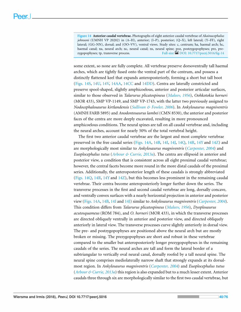
some extent, so none are fully complete. All vertebrae preserve dorsoventrally tall haemal
arches, which are tightly fused onto the ventral part of the centrum, and possess a
distinctly flattened keel that expands anteroposteriorly, forming a short but tall boot
(Figs. 14S, 14U, 14V, 14AA, 14CC and 14DD). Centra are laterally constricted and
preserve spool-shaped, slightly amphicoelous, anterior and posterior articular surfaces,
similar to those observed in Talarurus plicatospineus (Maleev, 1956), Oohkotokia horneri
(MOR 433), SMP VP-1149, and SMP VP-1743, with the latter two previously assigned to
Nodocephalosaurus kirtlandensis (Sullivan & Fowler, 2006). In Ankylosaurus magniventris
(AMNH FARB 5895) and Anodontosaurus lambei (CMN 8530), the anterior and posterior
faces of the centra are more deeply excavated, resulting in more pronounced
amphicoelous conditions. The neural spines are tall on all caudal vertebrae and, including
the neural arches, account for nearly 50% of the total vertebral height.
The first two anterior caudal vertebrae are the largest and most complete vertebrae
preserved in the free caudal series (Figs. 14A, 14B, 14I, 14J, 14Q, 14R, 14Y and 14Z) and
are morphologically most similar to Ankylosaurus magniventris (Carpenter, 2004) and
Euoplocephalus tutus (Arbour & Currie, 2013a). The centra are ellipsoid in anterior and
posterior view, a condition that is consistent across all eight proximal caudal vertebrae;
however, the central facets become more round in the more distal caudals of the proximal
series. Additionally, the anteroposterior length of these caudals is strongly abbreviated
(Figs. 14Q, 14R, 14Y and 14Z), but this becomes less prominent in the remaining caudal
vertebrae. Their centra become anteroposteriorly longer further down the series. The
transverse processes in the first and second caudal vertebrae are long, dorsally concave,
and ventrally convex surfaces with a nearly horizontal projection in anterior and posterior
view (Figs. 14A, 14B, 14I and 14J) similar to Ankylosaurus magniventris (Carpenter, 2004).
This condition differs from Talarurus plicatospineus (Maleev, 1956), Dyoplosaurus
acutosquameus (ROM 784), and O. horneri (MOR 433), in which the transverse processes
are directed obliquely ventrally in anterior and posterior view, and directed obliquely
anteriorly in lateral view. The transverse processes curve slightly anteriorly in dorsal view.
The pre- and postzygopophyses are positioned above the neural arch but are mostly
broken or missing. The prezygopophyses are short and robust in these vertebrae
compared to the smaller but anteroposteriorly longer prezygopophyses in the remaining
caudals of the series. The neural arches are tall and form the lateral border of a
subtriangular to vertically oval neural canal, dorsally roofed by a tall neural spine. The
neural spine comprises mediolaterally narrow shaft that strongly expands at its dorsal-
most region. In Ankylosaurus magniventris (Carpenter, 2004) and Euoplocephalus tutus
(Arbour & Currie, 2013a) this region is also expanded but to a much lesser extent. Anterior
caudals three through six are morphologically similar to the first two caudal vertebrae, but
Figure 14 Anterior caudal vertebrae. Photographs of eight anterior caudal vertebrae of Akainacephalusjohnsoni (UMNH VP 20202) in (A–H), anterior; (I–P), posterior; (Q–X), left lateral; (Y–FF), rightlateral; (GG–NN), dorsal; and (OO–VV), ventral views. Study sites: c, centrum; ha, haemal arch; hc,haemal canal; na, neural arch; nc, neural canal; ns, neural spine; poz, postzygopophyses; prz, pre-zygopophyses; tp, transverse process. Full-size DOI: 10.7717/peerj.5016/fig-14
Wiersma and Irmis (2018), PeerJ, DOI 10.7717/peerj.5016 40/76

the centra are anteroposteriorly less abbreviated. Most of the neural spines and the
majority of the transverse processes are postdepositionally broken away. The articular
facets of the centra in the third through sixth caudal vertebrae are round in anterior and
posterior view and the anterior facet is dorsally elevated compared to the posterior facet.
Anterior caudals seven and eight are the smallest of the preserved caudal series and the
interecentral articular surfaces are nearly round (Figs. 14G, 14H, 14O and 14P). Although
they represent the smallest of the free caudal series, the anteroposterior length of their
centra is relatively larger compared to the first six caudal vertebrae (Figs. 14Q–14FF).
Similar to the other six anterior caudal and the two dorsal vertebrae, the anterior facets of
the centrum are dorsally elevated relative to the posterior facets. Similar to caudals
three through six, the transverse processes form reduced, horizontally directed projections
with their distal-most ends eroded away. The transverse processes are positioned on
the dorsal half of the centrum. Caudal vertebrae seven and eight in Akainacephalus
johnsoni differ strongly from SMP VP-1149 and SMP VP-1743, which preserve more
robust, and ventrolaterally projecting transverse processes. The transverse processes on the
distal vertebrae of the free caudal series in Andodontosaurus lambei appear to have been
fully lost (Arbour & Currie, 2013a: fig.9K–I). Only the proximal-most portions of the
haemal arches are preserved and are firmly fused onto the ventral margin of the centrum.
The neural canal is vertically oblong-subtriangular. This condition differs from SMP
VP-1149 and SMP VP-1743, which possess round neural canals in the distal vertebrae of
the proximal caudal series. The neural spines are dorsoventrally shorter and directed more
obliquely posterior compared to the other free caudal vertebrae. Contrary to the
mediolaterally expanded distal ends of the neural spines in the first six anterior caudals,
the distal ends of the neural spines in caudals seven and eight are mediolaterally flat,
similar to a distal anterior caudal vertebra preserved in Anodontosaurus lambei (Arbour &
Currie, 2013a).
Fused posterior caudal vertebrae (handle) and knobHandleThe well-preserved handle comprises eleven fused vertebrae (Figs. 15A and 15B) and
terminates in a large tail club knob of two lateral osteoderms and what appears to be a
single distal osteoderm (Figs. 15C, 15D, 15E, 15F and 15G). Although the overall
morphology of the handle and tail club of Akainacephalus johnsoni differs from
Dyoplosaurus acutosquameus (ROM 784), the number of fused caudal vertebrae is
identical; this character varies among different ankylosaurid taxa and can be as many as
seventeen, as has been observed in the handle of ZPAL MgD I/113 (previously referred to
Tarchia cf. gigantea (Arbour et al., 2013)). The centra are spool-shaped with laterally-
compressed concave surfaces. Each individual vertebra is nearly twice as long as wide and
vertebrae are approximately equal in size, ranging between 98 and 105 mm in
anteroposterior length, but the width and height of the centra decrease posteriorly across
successive vertebrae. The handle is dorsally reinforced by posteriorly elongated, wedge-
shaped prezygopophyses that extend across nearly 50% of the length of the adjacent
vertebra and interlock with the shorter postzygopophyses (Fig. 15G). Chevrons are fused
Wiersma and Irmis (2018), PeerJ, DOI 10.7717/peerj.5016 41/76

Figure 15 Fused caudal vertebrae (handle) and tail club knob. Photographs of distal caudal vertebrae(handle) and tail club of Akainacephalus johnsoni (UMNH VP 20202). Handle in (A), left lateral; and(B), right lateral view. Tail club in (C), dorsal; (D), ventral; (E), left lateral; and (F), right lateral views.Reconstructed handle and tail club in (G), dorsal view. Study sites: lo, lateral osteoderms; to, terminalosteoderms. Full-size DOI: 10.7717/peerj.5016/fig-15
Wiersma and Irmis (2018), PeerJ, DOI 10.7717/peerj.5016 42/76

onto the ventral surface of the centra and dorsoventrally compressed so that they look like
elongated rods in lateral view. In ventral view, the chevrons are posterior-oriented
V-shaped structures that interlock with one another and reinforce the handle ventrally.
Some ankylosaurids (e.g., D. acutosquameus [ROM 784], cf. Pinacosaurus [MPC
100/1305], ZPAL MgD I/113, and UMNH VP 19472) preserve a dense network of ossified
tendons along the lateral sides or the handle, but these are not preserved in Akainacephalus
johnsoni.
KnobA large, wider than long, robust tail club knob is fused onto the two posterior-most
caudal vertebrae and comprise three distinct osteoderms; two anteroposteriorly elongated
lateral major osteoderms and what appears to represent a single, subtriangular terminal
minor osteoderm (Figs. 15C and 15D), revealing no evidence of multiple minor
osteoderms, but its poor preservation makes this uncertain. In dorsal and ventral view, the
knob itself displays an overall subtriangular morphology with a broad and blunt apex, a
condition uncommon in other Laramidian ankylosaurids in which the knobs are rounder
(e.g., Ankylosaurus magniventris (Carpenter, 2004), CMN 349, TMP 1983.36.120, TMP
2001.42.9, and SMP VP-2074 (Burns & Sullivan, 2011b)) and more elongate (e.g., Zuul
crurivastator (Arbour & Evans, 2017)). Because some of the lateral and dorsal areas are
eroded, the original morphology was possibly rounder along the lateral margins. The
tail club knob morphology of Akainacephalus johnsoni also differs fromUMNHVP 19472,
the North American D. acutosquameus (ROM 784) and some Asian taxa (e.g., cf.
Pinacosaurus [MPC 100/1305], and ZPAL MgD I/113). In these taxa, the knobs are
much smaller and the major osteoderms are transversely narrower. The tail club in
Akainacephalus johnsoni has a posterodorsal inclination of 10–15! in lateral view (Figs.
15E and 15F). The medial bases of the major osteoderms that make up the tail club knob
are deeply excavated and envelope the distal-most caudal vertebrae.
Both major osteoderms are anteroposteriorly longer than they are mediolaterally wide
in dorsal and ventral views, similar to Euoplocephalus tutus (CMN 2251), Ankylosaurus
magniventris (AMNH FARB 5214), and other specimens referred to Ankylosauridae
such as USNM 10753, CMN 349, TMP 1983.36.120, UALVP 16247 (Arbour & Currie,
2013a), and SMP VP-2074. In dorsal view, the osteoderms are badly damaged but preserve
and overall rounded morphology along the medial and lateral margins. The lateral
margins are clearly rounded, convex, and differ from Anodontosaurus lambei (Arbour &
Currie, 2013a), in which the lateral margins terminate in a distinct apex and curve
dorsolaterally upward. In ventral view, the osteoderms have a semicircular morphology
and the posterior halves extend further medially and contact each other, enclosing the
ventral surface on the posterior end of the tail club and nearly obscuring all of the ventral
portion of the minor osteoderm, forming a small semicircular wedge.
The posterior portion of the knob is far smaller compared to the anteriorly positioned
lateral osteoderms, and forms the terminal part of the tail club. Dorsally, this area is
transversely wider than it is long and has a diamond-shaped morphology, most similar to
certain tail club knob specimens assigned to Anodontosaurus lambei (AMNH FARB 5216)
Wiersma and Irmis (2018), PeerJ, DOI 10.7717/peerj.5016 43/76
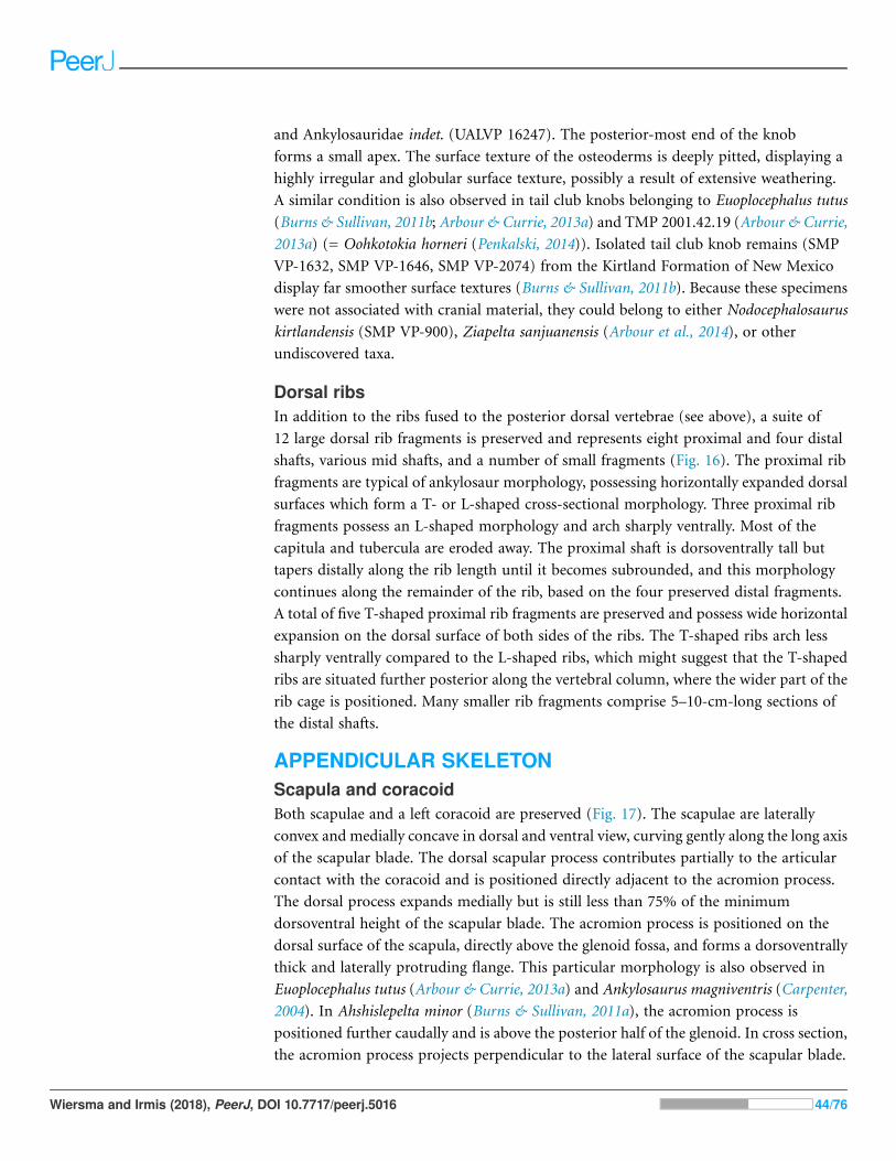
and Ankylosauridae indet. (UALVP 16247). The posterior-most end of the knob
forms a small apex. The surface texture of the osteoderms is deeply pitted, displaying a
highly irregular and globular surface texture, possibly a result of extensive weathering.
A similar condition is also observed in tail club knobs belonging to Euoplocephalus tutus
(Burns & Sullivan, 2011b; Arbour & Currie, 2013a) and TMP 2001.42.19 (Arbour & Currie,
2013a) (= Oohkotokia horneri (Penkalski, 2014)). Isolated tail club knob remains (SMP
VP-1632, SMP VP-1646, SMP VP-2074) from the Kirtland Formation of New Mexico
display far smoother surface textures (Burns & Sullivan, 2011b). Because these specimens
were not associated with cranial material, they could belong to either Nodocephalosaurus
kirtlandensis (SMP VP-900), Ziapelta sanjuanensis (Arbour et al., 2014), or other
undiscovered taxa.
Dorsal ribsIn addition to the ribs fused to the posterior dorsal vertebrae (see above), a suite of
12 large dorsal rib fragments is preserved and represents eight proximal and four distal
shafts, various mid shafts, and a number of small fragments (Fig. 16). The proximal rib
fragments are typical of ankylosaur morphology, possessing horizontally expanded dorsal
surfaces which form a T- or L-shaped cross-sectional morphology. Three proximal rib
fragments possess an L-shaped morphology and arch sharply ventrally. Most of the
capitula and tubercula are eroded away. The proximal shaft is dorsoventrally tall but
tapers distally along the rib length until it becomes subrounded, and this morphology
continues along the remainder of the rib, based on the four preserved distal fragments.
A total of five T-shaped proximal rib fragments are preserved and possess wide horizontal
expansion on the dorsal surface of both sides of the ribs. The T-shaped ribs arch less
sharply ventrally compared to the L-shaped ribs, which might suggest that the T-shaped
ribs are situated further posterior along the vertebral column, where the wider part of the
rib cage is positioned. Many smaller rib fragments comprise 5–10-cm-long sections of
the distal shafts.
APPENDICULAR SKELETONScapula and coracoidBoth scapulae and a left coracoid are preserved (Fig. 17). The scapulae are laterally
convex and medially concave in dorsal and ventral view, curving gently along the long axis
of the scapular blade. The dorsal scapular process contributes partially to the articular
contact with the coracoid and is positioned directly adjacent to the acromion process.
The dorsal process expands medially but is still less than 75% of the minimum
dorsoventral height of the scapular blade. The acromion process is positioned on the
dorsal surface of the scapula, directly above the glenoid fossa, and forms a dorsoventrally
thick and laterally protruding flange. This particular morphology is also observed in
Euoplocephalus tutus (Arbour & Currie, 2013a) and Ankylosaurus magniventris (Carpenter,
2004). In Ahshislepelta minor (Burns & Sullivan, 2011a), the acromion process is
positioned further caudally and is above the posterior half of the glenoid. In cross section,
the acromion process projects perpendicular to the lateral surface of the scapular blade.
Wiersma and Irmis (2018), PeerJ, DOI 10.7717/peerj.5016 44/76
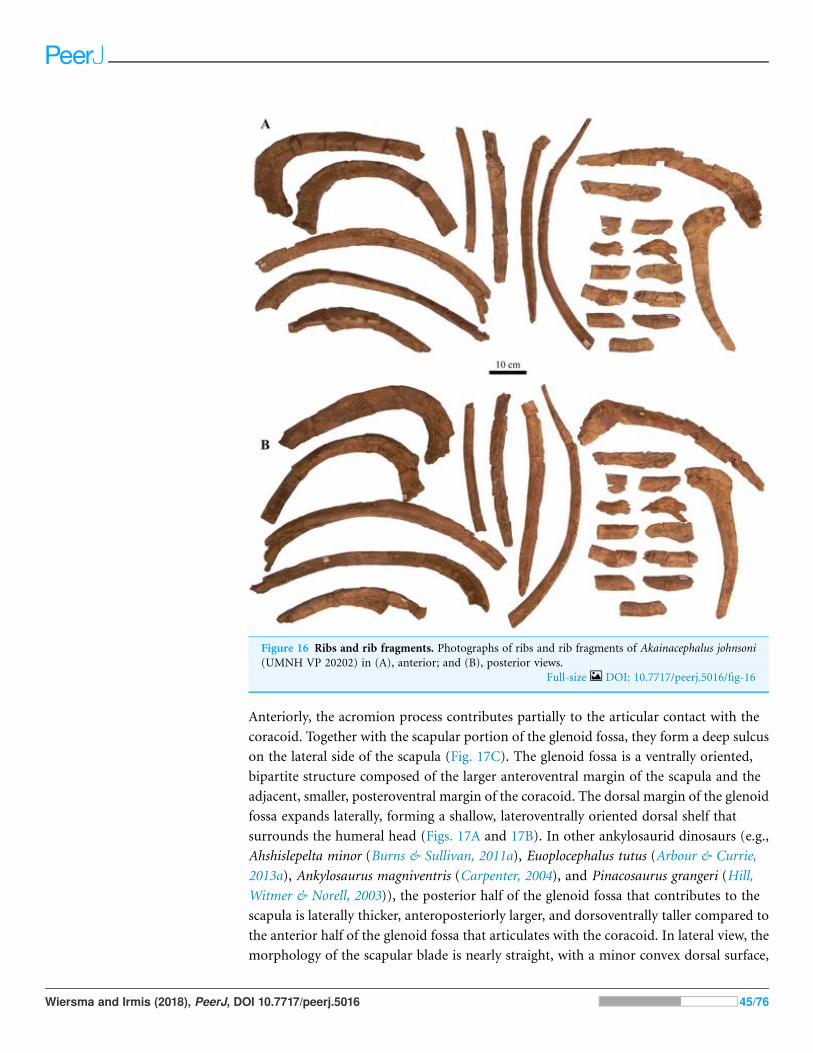
Anteriorly, the acromion process contributes partially to the articular contact with the
coracoid. Together with the scapular portion of the glenoid fossa, they form a deep sulcus
on the lateral side of the scapula (Fig. 17C). The glenoid fossa is a ventrally oriented,
bipartite structure composed of the larger anteroventral margin of the scapula and the
adjacent, smaller, posteroventral margin of the coracoid. The dorsal margin of the glenoid
fossa expands laterally, forming a shallow, lateroventrally oriented dorsal shelf that
surrounds the humeral head (Figs. 17A and 17B). In other ankylosaurid dinosaurs (e.g.,
Ahshislepelta minor (Burns & Sullivan, 2011a), Euoplocephalus tutus (Arbour & Currie,
2013a), Ankylosaurus magniventris (Carpenter, 2004), and Pinacosaurus grangeri (Hill,
Witmer & Norell, 2003)), the posterior half of the glenoid fossa that contributes to the
scapula is laterally thicker, anteroposteriorly larger, and dorsoventrally taller compared to
the anterior half of the glenoid fossa that articulates with the coracoid. In lateral view, the
morphology of the scapular blade is nearly straight, with a minor convex dorsal surface,
Figure 16 Ribs and rib fragments. Photographs of ribs and rib fragments of Akainacephalus johnsoni(UMNH VP 20202) in (A), anterior; and (B), posterior views.
Full-size DOI: 10.7717/peerj.5016/fig-16
Wiersma and Irmis (2018), PeerJ, DOI 10.7717/peerj.5016 45/76

similar to Euoplocephalus tutus (Arbour & Currie, 2013a). In contrast, in Ankylosaurus
magniventris (Carpenter, 2004) this dorsal surface is strongly convex dorsally, and in
Saichania chulsanensis (Marya!nska, 1977) it is slightly concave. A distinct surface for the
insertion of theM. triceps longus caudalis is situated on the ventral surface of the scapular
blade, directly posterior to the area where the blade is at its minimum dorsoventral height.
In lateral view, the scapular blade is paddle-shaped with a convex distal margin, similar
to Euoplocephalus tutus (Arbour & Currie, 2013a). This morphology is the result of
dorsoventral expansion of the distal margin, primarily along the ventral margin, and the
total expansion attributes more than 150% of the minimum dorsoventral height of
the scapular blade.
The somewhat square coracoid is anteroposteriorly longer than it is dorsoventrally
tall, and is fused to the right scapula. The coracoid is oriented anteromedially compared
to the scapular blade. The posteromedial surface that articulates with the scapula is
medially expanded to form a uniform structure with the dorsal scapular process (Figs. 17A
and 17B). A large oval coracoid foramen is situated directly dorsal to the glenoid fossa.
The dorsal and anterior surfaces are largely broken but appear to be concave. In cross
section, the dorsal border of the coracoid is convex, whereas the ventral border is concave.
A short anteroventral process is present and is anteriorly separated from the glenoid
by a distinct ventrally concave notch.
HumerusA complete right humerus and a partial left humeral head are preserved. The right
humerus is robust like other ankylosaurids; an enlarged and tall deltopectoral crest spans
>50% of the total humeral length and is followed by a short and thick humeral shaft that
Figure 17 Left and right scapulae and right coracoid. Photographs of the left and right scapulae andright coracoid of Akainacephalus johnsoni (UMNH VP 20202). Right scapula and coracoid in (A),medial; and (B), lateral views. Left scapula in (C), lateral; and (D), medial views. Study sites: acr,acromium; cor, coracoid; gl, glenoid; glf, glenoid fossa; mtlc, enthesis of M. triceps longus caudalis; scb,scapular blade; stp, sternal process. Full-size DOI: 10.7717/peerj.5016/fig-17
Wiersma and Irmis (2018), PeerJ, DOI 10.7717/peerj.5016 46/76

comprises the ventral half of the humerus. Most of the external surface experienced
predepositional weathering, removing most of the original surface texture and
subsequently eroding away the medial and dorsal margins of the humeral head. The
remaining portion of the humeral head is separated from the greater tubercle by a small
notch which is commonly seen in most other ankylosaurids, except Ankylosaurus
magniventris (Carpenter, 2004) and Crichtonpelta benxiensis (=Crichtonsaurus, Junchang
et al., 2007), which lack a notch and form a continuous surface between these elements.
Posterior and directly ventral to the humeral head is a large, well-expressed,
posteroventrally projecting process that terminates in a ventrally projecting hook
(Figs. 18C and 18D). In some ankylosaurids this process is absent (i.e., AMNH FARB
5337) but expressed to some extent in others (e.g., AMNH FARB 5404), however, it is
prominent Akainacephalus johnsoni. Directly ventral to the process, the humeral shaft
expands posteriorly, resulting in distinctly separated humeral heads of the triceps
(Figs. 18A and 18C). Anteriorly, the area between the humeral head and the greater
tubercle (supracoracoideus) form a large but shallow sulcus. The lateral external
margin of the greater tubercle curves downward and forms a continuous surface that
transitionss into the mediotransversely expanded and strongly anterolaterally oriented
deltopectoral crest. A large and round protuberance is situated on the lateral side of the
deltopectoral crest and forms the articulation surface for the M. latissimus dorsi. The
internal tuberosity is damaged but large and well expressed (Figs. 18A, 18B and 18D).
Distally, the humeral shaft is followed by the medial and lateral epicondyles. Both
epicondyles suffered postdepositional breakage and deformation, resulting in the removal
of the articular surface on the medial epicondyle (Fig. 18D), whereas the semicircular
articular surface on the lateral epicondyle is preserved and bears a prominent
protuberance on the anterior margin (Fig. 18C). The medial epicondyle extends slightly
more distal compared to the lateral epicondyle (Figs. 18A and 18B). In anterior and
posterior view, the epicondyles are separated by a shallow concave sulcus (Figs. 18A and
18B); however, the ventral-most portion is deeply excavated due to breakage and erosion
(Fig. 18F). Measurements of the humerus are summarized in Supplemental Material S4.
UlnaOnly the left ulna is present, but it is broken along the mid-shaft and largely incomplete,
preserving only the olecranon process and a partial shaft (Fig. 19). Most of the olecranon
process is broken away, leaving only the base preserved. The coronoid process is
transversely distorted and is poorly preserved, missing most of the ventral margin.
The radial notch has a typical ankylosaur morphology and is mostly intact, forming a
large, laterally projecting flange with a ventrally tapering shaft, from which small
fragments are broken away. Little is left of the distal end of the ulna, but the posteriorly
oriented flange of the ulna head is still visible.
IliumA nearly complete left ilium is preserved and shows some breakage along the acetabulum,
ventral margin of the ilium, and the distal margins on the pre- and postacetabular
Wiersma and Irmis (2018), PeerJ, DOI 10.7717/peerj.5016 47/76

Figure 18 Right humerus. Photographs of the right humerus of Akainacephalus johnsoni (UMNH VP20202) in (A), anterior; (B), posterior; (C), lateral; (D), medial; (E), dorsal; and (F), ventral views. Studysites: dpc, deltopectoral crest; hh, humeral head; it, internal tuberosity; mc, medial condyle; lc, lateralcondyle. Full-size DOI: 10.7717/peerj.5016/fig-18
Wiersma and Irmis (2018), PeerJ, DOI 10.7717/peerj.5016 48/76

processes (Figs. 20A–20D). In dorsal view, the ilium is broadly concave along the lateral
margin and nearly straight medially, but dorsally convex in medial and lateral views
(Figs. 20A, 20C and 20D). The dorsal portion of the acetabular region curves laterally
and ventrally, forming a broad supra-acetabular shelf that extends laterally and obscures
the acetabulum in lateral view (Fig. 20C). Articular surfaces for the attachement of sacral
ribs are not preserved on the ilium and are difficult to identify on the along the medial
margin of the acetabulum, pre- and postacetabular processes. The large preacetabular
process forms >50% of the entire iliac length and projects anterolaterally at an angle of ∼23!.Most of the medial and anterior margins are broken and would have provided the contact
surfaces for the dorsosacral ribs. Posteriorly, the postacetabular process forms a short
buttress, but it is longer than the acetabulum and curves strongly posteroventrally at ∼45!.A large portion of the posterior margin is postdepositionally broken. A deep, concave sulcus
is located on themedial side of the ilium and forms the ventrally oriented, closed acetabulum.
The acetabulum itself is only partially preserved, and it shows breakage particularly around
the articular surfaces that contact the proximal margins of the pubis and ischium.
IschiumA partial, right ischium is preserved (Figs. 20E and 20F) and has a typical ankylosaurid
morphology; it is Y-shaped in lateral and medial view, and comprises a laterally
compressed shaft, containing virtually no anterior curvature, consistent with ankylosaurid
ischia (Vickaryous, Marya!nska & Weishampel, 2004). The lateral surface of the ischium is
relatively straight in anterior and posterior view but the medial surface is slightly concave.
Most of the proximal part of the ischium that forms the ischial and pubic peduncles are
broken away, making it difficult to distinguish between a left or right ischium. A small
remnant of the lateral sulcus is preserved on the dorsolateral part of the ischium and
contributes to the closed acetabulum. The distal-most end of the ischium is also broken
so an exact length of the element cannot be determined.
Figure 19 Left ulna. Photographs of a partial left ulna of Akainacephalus johnsoni (UMNH VP 20202)in (A), anterior; (B), posterior; (C), lateral; (D), medial; (E), distal; and (F), proximal views. Study sites:op, olecranon process. Full-size DOI: 10.7717/peerj.5016/fig-19
Wiersma and Irmis (2018), PeerJ, DOI 10.7717/peerj.5016 49/76

Figure 20 Left ilium and right ischium. Photographs of the left ilium and right ischium of Akaina-cephalus johnsoni (UMNH VP 20202). Left ilium in (A), dorsal; (B), ventral; (C), lateral; and (D), medialviews. Right ischium in (E), medial; and (F), lateral views. Study sites: ac, acetabulum; ip, iliac peduncle;poa, postacetabular process; pp, pubic peduncle; prea, preacetabular process.
Full-size DOI: 10.7717/peerj.5016/fig-20
Wiersma and Irmis (2018), PeerJ, DOI 10.7717/peerj.5016 50/76
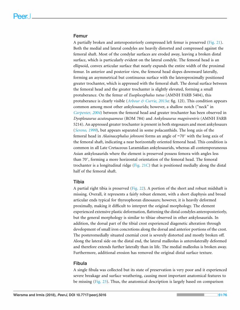
FemurA partially broken and anteroposteriorly compressed left femur is preserved (Fig. 21).
Both the medial and lateral condyles are heavily distorted and compressed against the
femoral shaft. Most of the condylar surfaces are eroded away, leaving a broken distal
surface, which is particularly evident on the lateral condyle. The femoral head is an
ellipsoid, convex articular surface that nearly expands the entire width of the proximal
femur. In anterior and posterior view, the femoral head slopes downward laterally,
forming an asymmetrical but continuous surface with the lateroproximally positioned
greater trochanter, which is appressed with the femoral shaft. The dorsal surface between
the femoral head and the greater trochanter is slightly elevated, forming a small
protuberance. On the femur of Euoplocephalus tutus (AMNH FARB 5404), this
protuberance is clearly visible (Arbour & Currie, 2013a: fig. 12I). This condition appears
common among most other ankylosaurids; however, a shallow notch (“neck” in
Carpenter, 2004) between the femoral head and greater trochanter has been observed in
Dyoplosaurus acutosquameus (ROM 784) and Ankylosaurus magniventris (AMNH FARB
5214). An appressed greater trochanter is present in both stegosaurs and most ankylosaurs
(Sereno, 1999), but appears separated in some polacanthids. The long axis of the
femoral head in Akainacephalus johnsoni forms an angle of ∼70! with the long axis of
the femoral shaft, indicating a near horizontally oriented femoral head. This condition is
common in all Late Cretaceous Laramidian ankylosaurids, whereas all contemporaneous
Asian ankylosaurids where the element is preserved possess femora with angles less
than 70!, forming a more horizontal orientation of the femoral head. The femoral
trochanter is a longitudinal ridge (Fig. 21C) that is positioned medially along the distal
half of the femoral shaft.
TibiaA partial right tibia is preserved (Fig. 22). A portion of the short and robust midshaft is
missing. Overall, it represents a fairly robust element, with a short diaphysis and broad
articular ends typical for thyreophoran dinosaurs; however, it is heavily deformed
proximally, making it difficult to interpret the original morphology. The element
experienced extensive plastic deformation, flattening the distal condyles anteroposteriorly,
but the general morphology is similar to tibiae observed in other ankylosaurids. In
addition, the dorsal part of the tibial crest experienced diagenetic alteration through
development of small iron concretions along the dorsal and anterior portions of the crest.
The posteromedially situated cnemial crest is severely distorted and mostly broken off.
Along the lateral side on the distal end, the lateral malleolus is anterolaterally deformed
and therefore extends further laterally than in life. The medial malleolus is broken away.
Furthermore, additional erosion has removed the original distal surface texture.
FibulaA single fibula was collected but its state of preservation is very poor and it experienced
severe breakage and surface weathering, causing most important anatomical features to
be missing (Fig. 23). Thus, the anatomical description is largely based on comparison
Wiersma and Irmis (2018), PeerJ, DOI 10.7717/peerj.5016 51/76
Figure 21 Left femur. Photographs of the left femur of Akainacephalus johnsoni (UMNH VP 20202) in(A), anterior; (B), posterior; (C), lateral; (D), medial; (E), dorsal; and (F), ventral views. Study sites:fh, femoral head; lc, lateral condyle; mc, medial condyle. Full-size DOI: 10.7717/peerj.5016/fig-21
Wiersma and Irmis (2018), PeerJ, DOI 10.7717/peerj.5016 52/76

with other ankylosaurid specimens of Ankylosaurus magniventris (AMNH FARB 5214)
and Euoplocephalus tutus (AMNH FARB 5266).
The fibula is long and narrow. Proximally, the shaft expands to form an asymmetrical
anteroposteriorly wide surface. A small depression appears to be present on this
surface and could potentially represent the medial face based on the morphology of
Ankylosaurus magniventris (AMNH FARB 5214) (Brown, 1908; Carpenter, 2004). Minor
anteroposterior expansion due to mediolateral compression occurs along the distal
portion of the fibula, which contains the malleolus of the fibula, but the articular surfaces
are highly damaged. The fibula is relatively long (397 mm) but ∼14% shorter than the
Figure 22 Right tibia. Photographs of the right tibia of Akainacephalus johnsoni (UMNH VP 20202) in(A), anterior; and (B), posterior views. Study sites: cn, cnemial crest; im, inner malleolus; om, outermalleolus. Full-size DOI: 10.7717/peerj.5016/fig-22
Wiersma and Irmis (2018), PeerJ, DOI 10.7717/peerj.5016 53/76
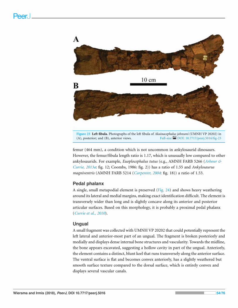
femur (464 mm), a condition which is not uncommon in ankylosaurid dinosaurs.
However, the femur/fibula length ratio is 1.17, which is unusually low compared to other
ankylosaurids. For example, Euoplocephalus tutus (e.g., AMNH FARB 5266 (Arbour &
Currie, 2013a: fig. 12; Coombs, 1986: fig. 2)) has a ratio of 1.55 and Ankylosaurus
magniventris (AMNH FARB 5214 (Carpenter, 2004: fig. 18)) a ratio of 1.53.
Pedal phalanxA single, small metapodial element is preserved (Fig. 24) and shows heavy weathering
around its lateral and medial margins, making exact identification difficult. The element is
transversely wider than long and is slightly concave along its anterior and posterior
articular surfaces. Based on this morphology, it is probably a proximal pedal phalanx
(Currie et al., 2010).
UngualA small fragment was collected with UMNHVP 20202 that could potentially represent the
left lateral and anterior-most part of an ungual. The fragment is broken posteriorly and
medially and displays dense internal bone structures and vascularity. Towards the midline,
the bone appears excavated, suggesting a hollow cavity in part of the ungual. Anteriorly,
the element contains a distinct, blunt keel that runs transversely along the anterior surface.
The ventral surface is flat and becomes convex anteriorly, has a slightly weathered but
smooth surface texture compared to the dorsal surface, which is entirely convex and
displays several vascular canals.
Figure 23 Left fibula. Photographs of the left fibula of Akainacephalus johnsoni (UMNH VP 20202) in(A), posterior; and (B), anterior views. Full-size DOI: 10.7717/peerj.5016/fig-23
Wiersma and Irmis (2018), PeerJ, DOI 10.7717/peerj.5016 54/76

POSTCRANIAL ARMORCervical half ringsA partial anterior cervical half ring and a nearly complete posteriorcervical half ring is
preserved (Figs. 25A–25F). Both cervical half rings are broken along the anterior and
posterior margins, so the exact shape of these surfaces is unclear. The incomplete element
(Figs. 25A–25D) is possibly the anterior-most (first) half ring, and the near-complete
element (Figs. 25E and 25F) is likely the posterior (second) half ring. The presence of
dorsoventrally raised and thickened surfaces along the dorsal and lateral regions of each
half ring suggests they are comprised of six individual secondary osteoderms, similar to
Euoplocephalus tutus (CMN 210, UALVP 31), Anodontosaurus lambei (CMN 8530), and
Scolosaurus cutleri (Penkalski & Blows, 2013). The secondary osteoderms are heavily fused
with the dorsal surfaces of the half rings and possibly experienced resorption to some
extent, however, the dorsal surface of each half ring is severely weathered, making it
challenging to assess the morphology of individual osteoderms. Several ankylosaurid
dinosaurs such as Euoplocephalus tutus (e.g., AMNH FARB 5337, UALVP 31, UALVP
45931, USNM 7943, TMP 2007.12.52) display distinct and clearly visible saw-tooth
sutures across the junctures of the half ring segments, fusing together the individual bony
segments that make up the cervical band (see Figs. 13H–13K and 13T–13V in Arbour &
Currie, 2013a). These sutures were not observed in Akainacephalus johnsoni.
The overall morphology of the half rings is ventrally concave and dorsally convex;
however, when observed more closely in anterior and posterior view, the loci that
accommodate the secondary osteoderms are thickened and elevated compared to the empty
interstitial areas, resulting in a profile that is rather sinusoidal (Figs. 25A, 25E and 25F).
Figure 24 Pedal phalanx. Photographs of a single pedal phalanx of Akainacephalus johnsoni (UMNHVP 20202) in (A), anterior; (B), posterior; (C), dorsal; (D), ventral; (E), lateral; and (F), medial views.
Full-size DOI: 10.7717/peerj.5016/fig-24
Wiersma and Irmis (2018), PeerJ, DOI 10.7717/peerj.5016 55/76

Figure 25 Cervical half rings and postcranial osteoderms. Photographs of postcranial osteoderms ofAkainacephalus johnsoni (UMNHVP 20202). Nearly complete cervical half ring in (A), anterior; and (B),posterior views. Right lateral portion of the partial half ring in (C), dorsal; (D), ventral; (E), posterior;and (F), anterior views. Various sharply keeled, type 1 (pup-tent), osteoderms associated with UMNHVP 20202. The osteoderms are displayed in (G), dorsal; (H), ventral; (I), medial; and (J), lateral views.No consensus currently exists regarding the exact position of type 1 osteoderms, but they appear to bepositioned either laterally on the body along the sacral and proximal portion of the tail or dorsolaterallyalong the thoracic and pectoral regions as seen in Euoplocephalus and Scolosaurus (Marya!nska, 1977;Ford, 2000). Full-size DOI: 10.7717/peerj.5016/fig-25
Wiersma and Irmis (2018), PeerJ, DOI 10.7717/peerj.5016 56/76

These dorsally elevated loci are bulbous and oval, with an anteroposteriorly oriented long
axis, suggesting that the overlying secondary osteoderms would have had excavated bases
and possibly a similar oval circumference. A total of two large foramina are present and
situated dorsal to the ventral-most articular surfaces for the secondary osteoderms.
Other noncervical osteodermsA total of 14 noncervical partial osteoderms are preserved and provide some insight
regarding the morphology and possible locations of armor across the body of UMNH VP
20202. The majority of the osteoderms are anteroposteriorly elongate and mediolaterally
compressed. All the osteoderms are tall, with strongly keeled dorsal margins (Figs.
25G–25J), and contain deeply excavated bases, the so-called “pup-tent” morphology
(Ford, 2000), and they contain slightly vascular and heavily pitted surface textures. Their
sizes range between a few centimeters and >10 cm in length. Little is known about the
exact locations and positions of postcranial osteoderms in ankylosaurid dinosaurs because
they commonly dissociate and disarticulate from the body after death. Only a few known
specimens, including Scolosaurus cutleri Nopcsa, 1928 (NHMUK R5161, holotype),
Dyoplosaurus acutosquameus, Parks, 1924 (ROM 784, holotype), Zuul crurivastator,
Arbour and Evans, 2017, Pinacosaurus grangeri Gilmore, 1933 (IVPP V16853, PIN 614
(“Syrmosaurus”: Maleev, 1954)), cf. Pinacosaurus, Arbour and Currie, 2013b (MPC 100/
1305 (“Saichania chulsanensis”: Carpenter et al., 2011)), and ZPAL. MgDI/113 (previously
referred to as Tarchia cf. gigantea Maleev, 1956), preserve osteoderms articulated with the
rest of the skeleton, providing at least partially detailed anatomical information regarding
the position and orientation of postcranial ornamentation among ankylosaurid
dinosaurs. Combined with other studies on ankylosaurian osteoderms by other workers
(Marya!nska, 1969; Carpenter, 1982; Burns, 2008; Arbour, Burns & Sissons, 2009;
Penkalski, 2014, Arbour et al., 2013; Burns & Currie, 2014; Brown et al., 2017; Brown, 2017)
these data provide additional insights on the positions of postcranial osteoderms
preserved within Akainacephalus johnsoni. Thus, based on the morphology and base width
of the preserved osteoderms in Akainacephalus johnsoni, it appears that their locations are
predominantly positioned along the dorsal (wider base) and lateral (narrower base)
portions of the body, and possibly the forelimbs (Lambe, 1902; Parks, 1924; Nopcsa, 1928;
Sternberg, 1929; Carpenter, 1982; Ford, 2000; Carpenter, 2004). Preserved osteoderms are
subdivided into three distinct morphologies based on morphotypes described in detail by
Marya!nska (1969) and include: type 1, “pup-tent” shaped (Ford, 2000) with S-twisted
keels and a backswept apex; type 2, dorsoventrally compressed triangular; and type 5,
flat-based with an off-centered, longitudinal keel.
Five type 1 osteoderms, referred to as “pup-tent” shaped (Figs. 25G–25J) by Ford
(2000), are taller than wide with narrow, deeply excavated bases, and thin-walled. Most
are incomplete, with portions of the base broken away. The osteoderms are asymmetrical
in lateral view, with tall, sharply pronounced keels that are anteriorly convex and nearly
twice as long as the short concave anterior margin. This morphology results in
dorsoposteriorly backswept and rounded apexes (Figs. 25G and 25H). In dorsal view,
the keels are sinusoidal (= S-twist in Marya!nska, 1969) compared to the straight keels
Wiersma and Irmis (2018), PeerJ, DOI 10.7717/peerj.5016 57/76

observed in the type 2 and type 5 osteoderms described below. Surface texture ranges from
minimally rugose with some dorsoventral furrowing in some osteoderms to rugose and
densely distributed, shallow pitting in others (Figs. 25G and 25H). Type 1 osteoderms
appear to be positioned on the thoracic region of the body as seen in Euoplocephalus tutus
(Ford, 2000) or the pectoral region in Scolosaurus cutleri (Penkalski & Blows, 2013).
Eight partial type 2 osteoderms of various sizes (55–112 mm long and 65–91 mm tall)
are dorsoventrally compressed with narrow but deeply excavated concave bases and
display a triangular dorsoventral morphology that terminates laterodistally in a distinct
apex. Their surface texture is slightly rugose with minor transverse running, shallow
furrows, and minimal pitting. These morphologies are typical for osteoderms positioned
along the lateral margins of the thorax and tail, as is apparent in specimens of Scolosaurus
cutleri (Penkalski & Blows, 2013),Dyoplosaurus acutosquameus (ROM 784), Tarchia teresae
(Penkalski & Tumanova, 2017), and Saichania chulsanensis (Marya!nska, 1977), but the
type 2 osteoderms in Akainacephalus johnsoni lack the two distinct dorsal grooves that
were observed by Arbour et al. (2013) in Tarchia. The entire external margins are keeled
along the posterior and anterior sides, including the apex. One side is convex and the
other side is concave, but it is difficult to determine the posterior and anterior face because
the orientation of these osteoderms varies among different ankylosaurid taxa. For
example, the lateral osteoderms on the proximal and distal tail of ZPAL MgD I/113
(Tarchia cf. gigantea (Arbour et al., 2013: fig. 4)) and proximal tail of D. acutosquameus
(Parks, 1924: plate 1; Arbour, Burns & Sissons, 2009: fig. 1) show convex anterior and
concave posterior faces with a posterolaterally projected apex. This condition contrasts
with the orientation of lateral caudal osteoderms observed in Saichania chulsanensis
(Arbour et al., 2013: fig. 4), in which the anterior face is concave and the posterior convex,
followed by an anterolaterally projecting apex.
A poorly preserved, single, type 5 dorsal osteoderm is heavily crushed and distorted.
The remaining fragments suggest a round, flat base with a distinctly off-centered, straight
keel that runs longitudinally along its dorsal surface, a condition and morphology that
somewhat resemble the dorsally positioned osteoderms in Scolosaurus (Penkalski & Blows,
2013). The visible surface texture is smooth and accompanied by small, shallow pits.
PHYLOGENETIC ANALYSISIn order to reconstruct the phylogenetic relationships of Akainacephalus johnsoni among
other ankylosaurid dinosaurs, we conducted a phylogenetic analysis using cladistic
parsimony. Characters for Akainacephalus johnsoni were scored and coded using the
character descriptions and character matrix constructed by Loewen & Kirkland (2013),
providing the most comprehensive taxon and character sampling for Ankylosauridae to
date, with 35 taxa and 293 characters (Supplemental Material S2). However, character
scorings for the majority of the in-group ankylosaurid taxa were verified or modified
through personal observations of original specimens or casts and specimens in published
literature, and are reflected in the matrix used for the phylogenetic analysis and
interpretation of its results.
Wiersma and Irmis (2018), PeerJ, DOI 10.7717/peerj.5016 58/76

A total of 11 nonankylosaurid taxa were also included to make sure character-states
were optimized correctly at the base of Ankylosauridae. The Jurassic ornithischian
Lesothosaurus diagnosticus was constrained as the outgroup taxon because it is just basal to
the Thyreophora–Neornithischia split and known from nearly complete remains
(Crompton, 1968; Thulborn, 1971; Galton, 1978; Sereno, 1991). The 10 nonankylosaurid
thyreophoran taxa include the early thyreophorans Scutellosaurus (Colbert, 1981),
Emausaurus (Haubold, 1990), and Scelidosaurus (Owen, 1859), the stegosaurs
Huayangosaurus (Dong, Tang & Zhou, 1982) and Tuojiangosaurus (Dong et al., 1977), the
basal ankylosaurian Minmi (Molnar, 1980), the nodosaurids Europelta (Kirkland et al.,
2013) and Cedarpelta (Carpenter et al., 2001), and the polacanthids Mymoorapelta
(Kirkland & Carpenter, 1994) and Gargoyleosaurus (Carpenter, Miles & Cloward, 1998).
Lesothosaurus diagnosticusScutellosaurus lawleri
Emausaurus ernesti
Huayangosaurus taibaiiTuojiangosaurus multispinus
Scelidosaurus harrisoniMinmi paravertebra
Mymoorapelta maysiGargoyleosaurus parkpinorum
Europelta carbonensisCedarpelta bilbeyhallorum
Gobisaurus domoculus
Shamosaurus scutatusZhongyuansaurus lauyangensis
Tsagantegia longicranialisCrichtonsaurus benxiensis
Euoplocephalus tutusOohkotokia horneriScolosaurus cutleri
Ankylosaurus magniventrisDyoplosaurus acutosquameus
Ziapelta sanjuanensis
Zaraapelta nomadis
Shanxia tianzhenensis
Minotaurasaurus ramachandraniTarchia kielanae
Tarchia giganteaSaichania chulsanensis
Tianzhenosaurus youngi
Ahshislepelta minor
Pinacosaurus grangeri
Pinacosaurus mephistocephalus
Anodontosaurus lambei
Nodocephalosaurus kirtlandensisAkainacephalus johnsoni
Thyreophora
Stegosauria
Ankylosauria
Ankylosauridae
Nodosauridae
Polacanthidae
(
(
(
(
(
Thyreophora
Stegosauria
Ankylosauria
Ankylosauridae
Nodosauridae
Polacanthidae
(
(
(
(
(
Consistency index 0.507Retention index 0.739
A B
Consistency index 0.504Retention index 0.737
Figure 26 Results of phylogenetic analyses for Akainacephalus johnsoni. Phylogenetic position ofAkainacephalus johnsoni in (A), a strict consensus of 21 equally most parsimonious trees, including thewildcard taxon Ahshislepelta minor, placing A. johnsoni within a large polytomy, consisting of crowngroup taxa that include Asian and all Laramidian ankylosaurids; and (B), the resulting strict consensus ofsix equally most phylogenetic trees, from which the wildcard taxon Ahshislepelta minor has been pruned.The crown group taxa are slightly better resolved in the pruned analysis, in which Akainacephalusjohnsoni forms a clade with its sister taxon Nodocephalosaurus kirtlandensis, nested within the clade thatalso includes the Asian taxaMinotaurasaurus ramachandrani, Tarchia kilanae, and Shanxia tianzhenensis,suggesting a close taxonomic relationship with Nodocephalosaurus kirtlandensis and Asian taxa.
Full-size DOI: 10.7717/peerj.5016/fig-26
Wiersma and Irmis (2018), PeerJ, DOI 10.7717/peerj.5016 59/76

The analysis contains 24 putative ankylosaurid taxa and include recently described taxa
such as Ziapelta sanjuanensis (Arbour et al., 2014) and Zaraapelta nomadis (Arbour, Currie
& Badamgarav, 2014), Akainacephalus johnsoni (described herein), as well as the
recently re-evaluated Dyoplosaurus acutosquameus (Arbour, Burns & Sissons, 2009),
Scolosaurus cutleri (Penkalski & Blows, 2013), and Oohkotokia horneri (Penkalski,
2014). Pinacosaurus grangeri, Pinacosaurus mephistocephalus, Tarchia kielanae, and
Tarchia teresae, were all coded as separate operational taxonomic units. We treat
M. ramachandrani as a valid taxon and separate operational taxonomic unit because we
agree with Penkalski & Tumanova (2017) that it is not a synonym of Tarchia kielanae
(Arbour, Currie & Badamgarav, 2014; Arbour & Currie, 2016). Tianzhenosaurus youngi
(Pang & Cheng, 1998) and Shanxia tianzhenensis (Barrett et al., 1998) are considered
synonyms of Saichania chulsanensis in Arbour, Currie & Badamgarav (2014) and Arbour &
Currie (2016), however, the characters used to unite these taxa are poorly defined, and
given rather general descriptions. Tianzhenosaurus, Shanxia, and Saichania are considered
valid taxa and have been treated as individual operational units. Similarly, we consider
O. horneri a separate taxon, not a juvenile synonymwith Scolosaurus cutleri (contra Arbour
& Currie, 2013a, 2016). The main focus of the phylogenetic analysis is to provide a
phylogenetic hypothesis for the position of Akainacephalus johnsoni, not to discuss the in-
depth the phylogenetic relationships of Ankylosauridae, which is far beyond the scope of
this descriptive paper and is better suited for a more in-depth phylogenetic contribution
which is currently in preparation by Loewen & Kirkland (2013).
The matrix (Supplemental Material S3) was constructed in Microsoft Excel and
formatted in Mesquite v.3.01 build 658 (Maddison & Maddison, 2011). The analysis was
conducted using TNT v.1.1 (Goloboff, Farris & Nixon, 2008), and carried out under the
“traditional search” option, with 10,000 replications of Wagner trees under the tree
bisection reconnection (TBR) algorithm, saving 10 trees per replication, followed by an
additional round of TBR on those saved trees. All characters were unweighted, and
characters 1, 2, 3, 7, 10, 13, 16, 18, 23, 25, 30, 36, 38, 48, 49, 54, 64, 87, 98, 101, 103, 104, 105,
140, 141, 143, 145, 148, 149, 156, 162, 165, 174, 177, 194, 201, 205, 209, 217, 229, 231, 232,
236, 237, 238, 260, 268, and 279 were ordered. Bremer values were also recorded, together
with a bootstrap analysis (utilizing sampling with replacement and 10,000 replications).
The analysis recovered 21 equally most parsimonious trees (length = 613), recording
consistency index and retention index values of 0.504 and 0.737, respectively. Examining
the strict consensus of these trees (Fig. 26A and Supplemental Material S1; Fig. S1), the
base of the resulting phylogeny does not differ greatly from previous analyses (e.g.,
Carpenter, 2001; Hill, Witmer & Norell, 2003; Thompson et al., 2012; Arbour & Currie,
2016) of thyreophoran relationships. The ankylosaurids P. mephistecephalus and Ziapelta
sanjuanensis form a small polytomy with the branch leading to Tianzhenosaurus youngi
plus all other ankylosaurids (Fig. 26A). All other ankylosaurid taxa sister to Saichania
chulsanensis form a large polytomy, including Akainacephalus johnsoni (Fig. 26A). In a
majority of the most parsimonious trees, A. johnsoni is nested within a clade of Asian taxa.
The high proportion of missing data for Ahshislepelta minor caused it to act as a wildcard
taxon, collapsing the interrelationships of this large clade of ankylosaurid dinosaurs.
Wiersma and Irmis (2018), PeerJ, DOI 10.7717/peerj.5016 60/76

After removal of Ahshislepelta minor, the pruned dataset resulted in six most
parsimonious trees (length = 610) (Supplemental Material S1; Figs. S2–S7) with a
consistency index value of 0.507 and retention index value of 0.739. A strict consensus of
these trees did not resolve the deeply nested polytomy of P. mephistocephalus, Z. nomadis,
and the branch leading to Tianzhenosaurus plus all other ankylosaurids (Fig. 26B and
Supplemental Materials S1; Fig. S8) but substantially more structure among the
ankylosaurid taxa above S. chulsanensis that formed a large polytomy in the original
analysis, placing Tarchia teresae as more closely related to a clade including all North
American taxa than to S. chulsanensis (Fig. 26B). Akainacephalus johnsoni forms a clade
with the southern Laramidian taxon Nodocephalosaurus kirtlandenesis (Fig. 26B), and is a
sister clade to a polytomy of the Asian taxa Tarchia kielanae, M. ramachandrani, and
Shanxi tianzhenensis (Fig. 26B). This southern Laramidian/Asian clade is sister to a clade
including all other Laramidian taxa: Ziapelta sanjuanensis, Ankylosaurus magniventris,
Dyoplosaurus acutosquameus, Euoplocephalus tutus, Oohkotokia horneri, Anodontosaurus
lambei, and Scolosaurus cutleri, though their interrelationships are unresolved.
The clade of A. johnsoni + N. kirtlandensis is supported by five synapomorphies
(Supplemental Material S1; Fig. S8): the width across the paroccipital processes is less than
195% of the height from the quadrates to the top of the paroccipital process (character 4
[0]); lateral orientation of the external nares (character 10[0]); absence of a suborbital lip
that forms a thin, sharp flange on the lateral edge along the ventral surface of the orbit
(character 41[0]); posterolateral orientation of the supraorbital boss in anterior view
(character 46[1]); and quadrate condyles visible in lateral view, and not obscured by the
quadratojugal horn (character 57[0]). The clade (Supplemental Material S1; Fig. S8) that
includes T. kielanae,M. ramachandrani, S. tianzhenensis, and A. johnsoni +N. kirtlandensis
is supported by three synapomorphies: no dorsoventral expansion of the distal
paroccipital process, compared to the neck of the paroccipital process (character 64[2]);
anteroposterior length of the centrum of dorsal vertebrae is greater than 110% of centrum
height (character 132[0]); and presence of a longitudinal keel along ventral surface of
dorsal vertebrae centra (character 133[1]).
The analysis after the removal of Ahshislepelta has a consistency index value of 0.507,
and retention index of 0.739. Bootstrap values are low along the branches of
Ankylosauridae (Supplemental Material S1; Fig. S9), with most nodes well below 50%; the
highest bootstrap value within Ankylosauridae was for the clade including Crichtonpelta
benxiensis (83%). Bremer support values were generally "3 across the base of the
phylogeny (Supplemental Material S1; Fig. S9), with exception of the branch leading to
the clade including Emausaurus ernesti (1). Bremer support values within Ankylosauridae
are much lower, almost universally 1 or 2 (Supplemental Material S1; Fig. S9), with the
exception of the branches leading to the taxa Gobisaurus domoculus (8), Tsagantegia
longicranialis (4), and Crichtonpelta benxiensis (5). The low support values reflect the high
level of homoplasy, and the small number of taxa with relatively complete specimens
available for study.
We also tested the phylogenetic position of Akainacephalus johnsoni, that is, in the
character-taxon matrix of Arbour & Evans (2017), a modified version of the matrix from
Wiersma and Irmis (2018), PeerJ, DOI 10.7717/peerj.5016 61/76

Arbour & Currie (2016). We analyzed these data in TNTusing the same parameters as our
original analysis. The resulting phylogeny recovered 1990 most parsimonious trees (length
= 469), and the strict consensus tree nested Akainacephalus johnsoni in a large polytomy
consisting of most ankylosaurid and nodosaurid taxa (Fig. 27). Calculating a maximum
agreement subtree (as Arbour & Currie, 2016 did) recovers Akainacephalus johnsoni within
the Ankylosauridae, which itself consists of three smaller polytomies (Supplemental
Material S1; Fig. S10). Surprisingly, none of the phylogenetic trees recovered from our
analysis of the Arbour & Evans (2017) character matrix resolved Akainacephalus johnsoni
as a sister taxon to Nodocephalosaurus. Instead, Nodocephalosaurus kirtlandensis was
consistently resolved as a sister taxon to Talarurus plicatospineus. Both Akainacephalus
johnsoni and the clade N. kirtlandensis + T. plicatospineus are deeply nested with other
Asian ankylosaurids (Supplemental Material S1; Fig. S10), rather than being resolved
within the Ankylosaurini, which constsists almost exclusively of Late Cretaceous
Laramidian ankylosaurids (Arbour & Currie, 2016; Arbour & Evans, 2017).
We refrain from making further interpretations of this analysis because the dataset
appears to be extremely sensitive to taxon and character sampling. Though the strict
consensus tree published by Arbour & Evans (2017) shows better resolution than the
previous results, this is a highly-pruned dataset compared with the taxonomic sampling of
the original Arbour & Currie (2016) analysis. When all taxa from the original dataset are
included, the strict consensus tree from their analysis remains largely unresolved, with
most taxa falling into three large polytomies (Fig. 27). These authors were only able to
make further interpretations of ankylosaurian phylogeny by calculating a 50% majority
rule consensus tree and a maximum agreement subtree. The former method, although
useful for statistical exploration of the relative frequency of branching relationships in a
set of trees, should not be used to make phylogenetic or evolutionary interpretations,
because it is a biased summary of parsimonious trees, and thus can lead to potentially false
precision, as elegantly discussed by Sumrall, Brochu & Merck, (2001). Maximum
agreement subtrees do report phylogenetic structure common to all most parsimonious
trees, but do so at the expense of many of the taxa in the original analysis. Furtheremore,
when the strict consensus of an analysis including the full set of taxa is poorly resolved
(as is the case with the Arbour and Currie matrix), the addition or exclusion of just one
taxa can have a large effect on which taxa remain in the resulting maximum agreement
subtree. In contrast, our primary analysis recovered considerably more phylogenetic
structure in the strict consensus before removing any taxa (using either a priori or a
posteriori methods), and thus did not require a technique such as a maximum agreement
subtree to examine the overall structure of the tree. In any case, the low tree support for
both our analysis and those of Arbour & Currie (2016) and Arbour & Evans (2017) indicate
that much remains to be done in elucidating ankylosaurid phylogeny.
DISCUSSIONThe known taxonomic diversity of Late Cretaceous southern Laramidian akylosaurids
continues to expand with the addition of Akainacephalus johnsoni (Fig. 28), a new taxon of
ankylosaurid dinosaur from the upper Campanian Kaiparowits Formation of southern
Wiersma and Irmis (2018), PeerJ, DOI 10.7717/peerj.5016 62/76

Lesothosaurus diagnosticusScelidosaurus harrisonii
Huayangosaurus taibaii
Mymoorapelta maysiKunbarrasaurus ieversi
Gargoyleosaurus parkpinorum
Tatankacephalus cooneyorum
Sauropelta edwardsorum
Stegopelta landerensis
Panoplosaurus mirusPawpawsaurus campbelli
Glyptodontopelta mimus
“Antarctopelta” oliveroiArgentine ankylosaur
Pinacosaurus grangeri
Akainacephalus johnsoniNodocephalosaurus kirtlandensis
Dyoplosaurus acutosquameusDongyangopelta yangyanensis
Liaoningosaurus paradoxus
“Zhejiangosaurus luoyensis”
Pinacosaurus mephistocephalus
Sauroplites scutiger
Saichania chulsanensis
Taohelong jinchengensisTarchia kielanae
Zaraapelta nomadis
Crichtonpelta benxiensis
Tsagantegia longicranialis
Ziapelta sanjuanensis
Euoplocephalus tutus
Talarurus plicatospineusScolosaurus cutleri
Thyreophora
Ankylosauria
Chuanqilong chaoyanensisCedarpelta bilbeyhallorum
Aletopelta coombsiShamosaurus scutatus
Anodontosaurus lambei
Gobisaurus domoculus
Ankylosaurus magniventris
Gastonia burgeiAhshislepelta minor
Zuul crurivastator
Figure 27 Phylogenetic results using character state codings from Arbour & Evans (2017). Phylo-genetic analysis resulting in a strict consensus of 1990 equally most parsimonious trees and the phy-logenetic position of Akainacephalus johnsoni, implemented into the character matrix from Arbour &Evans (2017). The resulting consensus tree renders A. johnsoni poorly resolved, consistently placing itwithin a large polytomy. Full-size DOI: 10.7717/peerj.5016/fig-27
Wiersma and Irmis (2018), PeerJ, DOI 10.7717/peerj.5016 63/76
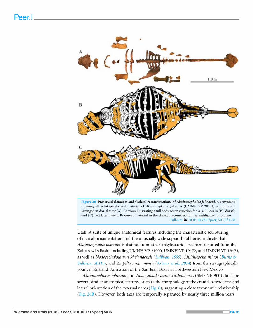
Utah. A suite of unique anatomical features including the characteristic sculpturing
of cranial ornamentation and the unusually wide supraorbital horns, indicate that
Akainacephalus johnsoni is distinct from other ankylosaurid specimen reported from the
Kaiparowits Basin, including UMNH VP 21000, UMNH VP 19472, and UMNH VP 19473,
as well as Nodocephalosaurus kirtlandensis (Sullivan, 1999), Ahshislepelta minor (Burns &
Sullivan, 2011a), and Ziapelta sanjuanensis (Arbour et al., 2014) from the stratigraphically
younger Kirtland Formation of the San Juan Basin in northwestern New Mexico.
Akainacephalus johnsoni and Nodocephalosaurus kirtlandensis (SMP VP-900) do share
several similar anatomical features, such as the morphology of the cranial osteoderms and
lateral orientation of the external nares (Fig. 8), suggesting a close taxonomic relationship
(Fig. 26B). However, both taxa are temporally separated by nearly three million years;
Figure 28 Preserved elements and skeletal reconstructions of Akainacephalus johnsoni. A compositeshowing all holotype skeletal material of Akainacephalus johnsoni (UMNH VP 20202) anatomicallyarranged in dorsal view (A). Cartoon illustrating a full body reconstruction for A. johnsoni in (B), dorsal;and (C), left lateral view. Preserved material in the skeletal reconstructions is highlighted in orange.
Full-size DOI: 10.7717/peerj.5016/fig-28
Wiersma and Irmis (2018), PeerJ, DOI 10.7717/peerj.5016 64/76

Akainacephalus johnsoni is recorded from the lower portion of the middle unit of the
Kaiparowits Formation, which is late Campanian in age (76.26 ± 0.10 Ma (Roberts et al.,
2013)), whereas Nodocephalosaurus kirtlandensis is from the upper-most Campanian-
lower-most Maastrichtian (73.04 ± 0.25 Ma (Fassett & Steiner, 1997); 73.49 ± 0.25 Ma
(Fowler, 2017)) De-na-zin Member of the Kirtland Formation. Such a large
geochronologic gap alone renders it unlikely that A. johnsoni and N. kirtlandensis belong
to the same species. Even igorning this age difference, a number of character states differ
between the skulls of Akainacephalus johnsoni and Nodocephalosaurus kirtlandensis (SMP
VP-900), including those in the basioccipital-basisphenoid region and amongst the
cranial ornamentation, suggesting two separate taxa. The morphology of the
basioccipital-basisphenoid complex in Akainacephalus johnsoni forms an anterior
tapering triangle, whereas in Nodocephalosaurus kirtlandensis (Sullivan, 1999) it is
anteroposteriorly oblong. Furthermore, Nodocephalosaurus kirtlandensis possesses a
deeply concave (saddle-shaped in Sullivan, 1999) basioccipital in lateral view, and
possesses a deeply piercing basioccipital foramen. Akainacephalus johnsoni possesses a
ventrally concave basioccipital but it is shallower and lacks the basioccipital foramen
(Fig. 4C). The quadratojugal horn in N. kirtlandensis is triangular and somewhat fin-
shaped, whereas in Akainacephalus johnsoni, this element is more robust and is a ventrally
projecting triangular protuberance. Although intraspecific variation in cranial
ornamentation is present in ankylosaurid taxa, for example, Euoplocephalus tutus,
Anodontosaurus lambei, Pinacosaurus grangeri (Penkalski, 2001; Arbour & Currie, 2013a),
the size proportions and morphology of the quadratojugal horns between Akainacephalus
johnsoni and Nodocephalosaurus kirtlandensis (SMP VP-900) (Figs. 8 and 9A–9D) differ
significantly and are herein regarded as taxonomically distinct. The near vertical
projection of the quadratojugal horns in Akainacephalus johnsoni are similar in
orientation with the left quadratojugal horn preserved in Nodocephalosaurus kirtlandensis
(SMP VP-900) but contrasts with the more laterally oriented quadratojugal horns
observed in other ankylosaurids such as Tarchia teresae (Penkalski & Tumanova, 2017)
(Figs. 7E and 7F), M. ramachandrani (INBR 21004) (Figs. 7G and 7H), Saichania
chulsanensis (MPC 100/151), Oohkotokia horneri (MOR 433), Euoplocephalus tutus
(AMNH FARB 5405, ROM 1930), Ankylosaurus magniventris (AMNH FARB 5214), and
Anodontosaurus lambei (CMN 8530). Finally, the condylar end on the ventral surface of
the quadrate is thickest medially in Nodocephalosaurus kirtlandensis (SMP VP-900),
Ankylosaurus magniventris (AMNH FARB 5214), Euoplocephalus tutus (AMNH FARB
5337, AMH FARB 5405, ROM 1930), and Anodontosaurus lambei (TMP. 1997.132.1),
whereas in Akainacephalus johnsoni the condylar contact appears to be situated along the
middle of the ventral surface. Akainacephalus johnsoni and Nodocephalosaurus
kirtlandensis are thus separate taxa, but they share morphological similarities that clearly
distinguish them from other Late Cretaceous Laramidian ankylosaurids.
Because the southern Laramidian clade of Akainacephalus johnsoni + Nodocephalosaurus
kirtlandensis is nested within Asian taxa, it suggests that this clade was a separate
biogeographic dispersal event from Asia independent from the main radiation of
Laramidian ankylosaurids (Ankylosaurini (Arbour & Currie, 2016)). Multiple
Wiersma and Irmis (2018), PeerJ, DOI 10.7717/peerj.5016 65/76
intercontinental ankylosaurid dispersal patterns contribute to an already complex
biogeographic pattern than was previously recognized for Late Cretaceous Laramidian
terrestrial and freshwater vertebrate faunas (Gates et al., 2010; Lehman, 1997; Lehman,
McDowell & Connelly, 2006; Loewen et al., 2013c; Sampson et al., 2004, 2010).
Geographic provincialism is present throughout western North America during the
Campanian, as discussed by Kauffman (1984), Lehman (1997, 2001), and Gates et al.
(2010). This resulted in many endemic terrestrial and freshwater vertebrate taxa (Sampson
et al., 2010; Loewen et al., 2013c), and ankylosaurid dinosaurs (Arbour et al., 2014),
including Akainacephalus johnsoni. The exact driving force behind provincialism within
theWestern Interior remains poorly understood to date, and evidence to support presence
of physical geographical barriers appears absent. Latitudinal climatic gradients have been
proposed by Gates et al. (2010). Such gradients produce different biomes, regional annual
temperatures and quantities of meteoric water, causing physiological constraints on
inhabiting organisms, preventing pandemic distributions of taxa. Climate gradient
signatures are particularly well demonstrated within the plant fossil record of Campanian
aged terrestrial environments throughout the Western Interior Basin (Miller et al., 2013).
The dispersal of ankylosaurids into Laramidia appears to be approximately coeval (i.e.,
Campanian in age) with respect to the dispersal of other dinosaur clades, including
tyrannosaurids (Thomson, Irmis & Loewen, 2013; Loewen et al., 2013c), ceratopsians
(Sampson et al., 2010), and ornithopod dinosaurs (Ryan & Evans, 2005; Gates et al., 2013).
These climate gradients, combined with fluctuations in sea level, may have helped
reinforced Campanian provincialism (Loewen et al., 2013c).
The identification of several new ankylosaur taxa from southern Laramidian basins has
resulted in a rapid taxonomic increase in diversity during the last 20 years. Currently, at
least two diagnostic ankylosaurid taxa and potentially a third taxon have been identified
from the Kirtland Formation of the San Juan Basin in northwestern New Mexico:
Nodocephalosaurus kirtlandensis (Sullivan, 1999), Ziapelta sanjuanensis (Arbour et al.,
2014), and Ahshislepelta minor (Burns & Sullivan, 2011a). Loewen et al. (2013a) assessed
the different ankylosaur remains that were recognized and collected from the Dakota,
Wahweap, and Kaiparowits formations, and other laterally contemporaneous formations
within the GSENM area in southern Utah. Within this area, most ankylosaur material is
known from the Kaiparowits Formation, which is largely due to the extensive expeditions
that have been part of the Kaiparowits Basin Project that began in 2000, resulting in
at least two distinct ankylosaurid taxa.
Although the taxonomic diversity of ankylosaurid material from southern Laramidian
basins continues to expand, individual taxa remain largely underrepresented in terms
of the number of specimens available for study, a phenomenon that appears to be
characteristic for ankylosaurian dinosaurs as a whole, regardless of age or formation in
which they are found. When relative abundance and species richness of ankylosaurian
dinosaurs are quantitatively compared to the other coeval ornithischian dinosaur
assemblages, (e.g., ornithopods and ceratopsians) (Brinkman, 1990; White, Fastovsky &
Sheehan, 1998; Ryan & Evans, 2005;Horner, Goodwin &Myhrvold, 2011; Gates et al., 2013,
Wiersma and Irmis (2018), PeerJ, DOI 10.7717/peerj.5016 66/76

Loewen et al., 2013b) from temporally equivalent stratigraphic horizons/formations,
ankylosaurs are always rare and species-poor. For example, ankylosaur specimens
recorded from the Kaiparowits Formation encompass <5% of associated dinosaur sites
throughout the formation, whereas hadrosaurid and ceratopsian sites are at least an order
of magnitude more abundant (Loewen et al., 2013a). When ankylosaurid material from
northern Laramidian basins are compared to coeval dinosaur faunas from the Dinosaur
Park and Hell Creek formations, ankylosaurid material is rarer by several orders of
magnitude, even with the well-known taxon Euoplocephalus tutus and specimens referred
to Euoplocephalus (White, Fastovsky & Sheehan, 1998; Ryan & Evans, 2005; Horner,
Goodwin & Myhrvold, 2011; Gates et al., 2013; Loewen et al., 2013b). Thus, considerable
future effort is required in Laramidian strata to discover additional taxa and specimens
that will improve our understanding of ankylosaurid evolution and paleobiology. These
recent finds from southern Laramidia, such as Akainacephalus johnsoni, demonstrate the
strong potential for new discoveries, despite the overall rarity of ankylosaur fossils.
CONCLUSIONAkainacephalus johnsoni (UMNH VP 20202) is a new taxon of ankylosaurid dinosaur
from the upper Campanian Kaiparowits Formation of southern Utah, USA. It consists of a
complete cranium and significant amount of diagnostic postcranial material that can
be distinguished from all other known Late Cretaceous Laramidian ankylosaurids.
Akainacephalus johnsoni is closely related to its stratigraphically younger sister taxon,
Nodocephalosaurus kirtlandensis from the Kirtland Formation of New Mexico. Both taxa
are more closely related to Asian ankylosaurids than they are to other Laramidian
ankylosaurids. This suggests multiple ankylosaurid dispersal events from Asia to
Laramidia during the Late Cretaceous. Lastly, together with Dyoplosaurus acutossquameus
and Scolosaurus cutleri (∼77 Ma) from northern Laramidia, Akainacephalus johnsoni
represents one of the older known ankylosaurid dinosaurs (∼76.3 Ma) from the Late
Cretaceous of western North America.
ACKNOWLEDGEMENTSThe authors wish to thank Allan A. Ekdale, Colleen G. Farmer, James Kirkland, Mark A.
Loewen, Paul Penkalski, and Alan Titus for their insightful and fruitful discussions,
constructive comments, and reviews of this manuscript. James Kirkland, Atila Osi, and
Victoria Arbour provided thoughtful reviews that significantly improved the manuscript.
The BLM and GSENM provided research permits and funding for fieldwork. Many thanks
to all NHMU staff, students, and volunteers who helped excavate and prepare UMNH VP
20202. We would like to particularly thank NHMU volunteer Randy Johnson for
preparing the majority of UMNH VP 20202. M. Colbert and J Maisano at the University
of Texas High Resolution CT Lab, Austin, Texas, kindly CTscanned UMNHVP 20202. We
thank the following people for granting me access to research specimens: Carolyn Levitt-
Bussian and Mike Getty: Natural History Museum of Utah; D. Henderson, B. Striliski,
T. Courtenay, and H. Lecuyer: Royal Tyrrell Museum of Palaeontology, Drumheller,
Alberta; C. Coy, and M. E. Burns: University of Alberta Laboratory of Vertebrate
Wiersma and Irmis (2018), PeerJ, DOI 10.7717/peerj.5016 67/76
Palaeontology, Edmonton, Alberta; V. M. Arbour, D. C. Evans, and K. Seymour: Royal
Ontario Museum, Toronto, Ontario. This manuscript was part of the senior author’s M.S.
thesis at the University of Utah.
ADDITIONAL INFORMATION AND DECLARATIONS
FundingResearch funding was provided by the Geological Society of America Graduate Student
Research Grant (grant number 10631-14); the Bureau of Land Management National
Landscape Conservation System Grant (grant numbers JSA071004 & L12AC20378); and
the University of Utah, Department of Geology & Geophysics Graduate Student Travel
Grant. The funders had no role in study design, data collection and analysis, decision
to publish, or preparation of the manuscript.
Grant DisclosuresThe following grant information was disclosed by the authors:
Geological Society of America Graduate Student Research Grant: 10631-14.
Bureau of Land Management National Landscape Conservation System Grant: JSA071004
& L12AC20378.
University of Utah, Department of Geology & Geophysics Graduate Student Travel Grant.
Competing InterestsThe authors declare that they have no competing interests.
Author Contributions# Jelle P. Wiersma conceived and designed the experiments, performed the experiments,
analyzed the data, contributed reagents/materials/analysis tools, prepared figures and/or
tables, authored or reviewed drafts of the paper.
# Randall B. Irmis authored or reviewed drafts of the paper.
Data AvailabilityThe following information was supplied regarding data availability:
The raw data has been supplied as a Supplementary File.
New Species RegistrationThe following information was supplied regarding the registration of a newly described
species:
Publication LSID: urn:lsid:zoobank.org:pub:42CB4F68-9E0D-42BB-943D-
8A66E3D9DA12.
Akainacephalus johnsoni urn:lsid:zoobank.org:act:11A47631-A9BE-4D58-9F6A-
49E0897181AA.
Supplemental InformationSupplemental information for this article can be found online at http://dx.doi.org/
10.7717/peerj.5016#supplemental-information.
Wiersma and Irmis (2018), PeerJ, DOI 10.7717/peerj.5016 68/76

REFERENCESArbour VM, Burns ME, Sissons RL. 2009. A redescription of Dyoplosaurus acutosquameus Parks,
1924 (Ornithischia: Ankylosauria) and a revision of the genus. Journal of Vertebrate Paleontology
29:1117–1135.
Arbour VM, Burns ME, Sullivan RM, Lucas SG, Cantrell AK, Fry J, Suazo TL. 2014. A new
ankylosaurid dinosaur from the Upper Cretaceous (Kirtlandian) of New Mexico with
implications for ankylosaurid diversity in the Upper Cretaceous of western North America.
PLOS ONE 9(9):e108804 DOI 10.1371/journal.pone.0108804.
Arbour VM, Currie PJ. 2013a. Euoplocephalus tutus and the diversity of ankylosaurid dinosaurs
in the Late Cretaceous of Alberta, Canada, and Montana, USA. PLOS ONE 8(5):e62421
DOI 10.1371/journal.pone.0062421.
Arbour VM, Currie PJ. 2013b. The taxonomic identity of a nearly complete ankylosaurid dinosaur
skeleton from the Gobi Desert of Mongolia. Cretaceous Research 46:24–30
DOI 10.1016/j.cretres.2013.08.008.
Arbour VM, Currie PJ. 2016. Systematics, phylogeny, and palaeobiogeography of the ankylosaurid
dinosaurs. Journal of Systematic Palaeontology 14(5):385–444
DOI 10.1080/14772019.2015.1059985.
Arbour VM, Currie PJ, Badamgarav D. 2014. The ankylosaurid dinosaurs of the Upper
Cretaceous Barungoyot and Nemegt formations of Mongolia. Zoological Journal of the Linnean
Society 172(3):631–652 DOI 10.1111/zoj.12185.
Arbour VM, Evans DC. 2017. A new ankylosaurine dinosaur from the Judith River Formation of
Montana, USA, based on an exceptional skeleton with soft tissue preservation. Royal Society
Open Science 4(5):161086 DOI 10.1098/rsos.161086.
Arbour VM, Lech-Hernes NL, Guldberg TE, Hurum JH, Currie PJ. 2013. An ankylosaurid
dinosaur from Mongolia with in situ armour and keratinous scale impressions. Acta
Palaeontologica Polonica 58(1):55–64 DOI 10.4202/app.2011.0081.
Arbour VM, Mallon JC. 2017. Unusual cranial and postcranial anatomy in the archetypal
ankylosaur Ankylosaurus magniventris. Facets 2(2):764–794 DOI 10.1139/facets-2017-0063.
Arbour VM, Zanno LE, Gates T. 2016. Ankylosaurian dinosaur palaeoenvironmental associations
were influenced by extirpation, sea-level fluctuation, and geodispersal. Palaeogeography
Palaeoclimatology Palaeoecology 449:289–299 DOI 10.1016/j.palaeo.2016.02.033.
Barrett PM, Hailu Y, Upchurch P, Burton AC. 1998. A new ankylosaurian dinosaur (Ornithischia:
Ankylosauria) from the Upper Cretaceous of Shanxi Province, People’s Republic of China.
Journal of Vertebrate Paleontology 18(2):376–384 DOI 10.1080/02724634.1998.10011065.
Brinkman DB. 1990. Palaeoecology of the Judith River Formation (Campanian) of Dinosaur
Provincial Park, Alberta, Canada; Evidence from vertebrate microfossil localities. Palaeogeography
Palaeoclimatology Palaeoecology 78(1–2):37–54 DOI 10.1016/0031-0182(90)90203-j.
Brown B. 1908. The Ankylosauridae, a new family of armored dinosaurs from the Upper
Cretaceous. Bulletin of the American Museum of Natural History 24(1):187–201
DOI 10.1007/bf01975763.
Brown CM. 2017. An exceptionally preserved armored dinosaur reveals the morphology and
allometry of osteoderms and their horny epidermal coverings. PeerJ 5:e4066
DOI 10.7717/peerj.4066.
Brown CM, Henderson DM, Vinther J, Fletcher I, Sistiaga A, Herrera J, Summons RE. 2017. An
exceptionally preserved three-dimensional armored dinosaur reveals insights into coloration
and Cretaceous predator-prey dynamics. Current Biology 27(16):2514–2521
DOI 10.1016/j.cub.2017.06.071.
Wiersma and Irmis (2018), PeerJ, DOI 10.7717/peerj.5016 69/76

Burns ME. 2008. Taxonomic utility of ankylosaur (Dinosauria: Ornithischia) osteoderms:
Glyptodontopelta mimus Ford, 2000: a test case. Journal of Vertebrate Paleontology
28(4):1102–1109 DOI 10.1671/0272-4634-28.4.1102.
Burns ME, Currie PJ. 2014. External and internal structure of ankylosaur (Dinosauria,
Ornithischia) osteoderms and their systematic relevance. Journal of Vertebrate Paleontology
34(4):835–851 DOI 10.1080/02724634.2014.840309.
Burns ME, Currie PJ, Sissons RL, Arbour VM. 2011. Juvenile specimens of Pinacosaurus grangeri
Gilmore, 1933 (Ornithischia: Ankylosauria) from the Late Cretaceous of China, with comments
on the specific taxonomy of Pinacosaurus. Cretaceous Research 32(2):174–186
DOI 10.1016/j.cretres.2010.11.007.
Burns ME, Sullivan RM. 2011a. A new ankylosaurid from the Upper Cretaceous Kirtland
Formation, San Juan basin, with comments on the diversity of ankylosaurids in New
Mexico. Fossil Record 3. New Mexico Museum of Natural History and Science Bulletin
53:169–178.
Burns ME, Sullivan RM. 2011b. The tail club of Nodocephalosaurus kirtlandensis (Dinosauria:
Ankylosauridae), with a review of ankylosaurid tail club morphology and homology. Fossil
Record 3. New Mexico Museum of Natural History and Science Bulletin 53:179–186.
Carpenter K. 1982. Skeletal and dermal armor reconstruction of Euoplocephalus tutus
(Ornithischia: Ankylosauridae) from the Late Cretaceous Oldman Formation of Alberta.
Canadian Journal of Earth Sciences 19(4):689–697 DOI 10.1139/e82-058.
Carpenter K. 1997. Ankylosaurs. In: Farlow JO, Brett-Surman MK, eds. The Complete Dinosaur.
Indianapolis: Indiana University Press, 307–316.
Carpenter K. 2001. Phylogenetic analysis of the Ankylosauria. In: Carpenter K, ed. The Armored
Dinosaurs. Bloomington: Indiana University Press, 455–483.
Carpenter K. 2004. Redescription of Ankylosaurus magniventris Brown, 1908 (Ankylosauridae)
from the Upper Cretaceous of the Western Interior of North America. Canadian Journal
of Earth Sciences 41(8):961–986 DOI 10.1139/e04-043.
Carpenter K, Bartlett J, Bird J, Barrick R. 2008. Ankylosaurs from the Price River Quarries, Cedar
Mountain Formation (Lower Cretaceous), east-central Utah. Journal of Vertebrate Paleontology
28(4):1089–1101 DOI 10.1671/0272-4634-28.4.1089.
Carpenter K, Hayashi S, Kobayashi Y, Marya!nska T, Barsbold R, Sato K, Obata I. 2011.
Saichania chulsanensis (Ornithischia, Ankylsauridae) from the Upper Cretaceous of Mongolia.
Palaeontographica Abteilung A 294(1–3):1–61 DOI 10.1127/pala/294/2011/1.
Carpenter K, Kirkland JI. 1998. Review of the Lower and Middle Cretaceous ankylosaurs
from North America. In: Lucas SG, Kirkland JI, Estep JW, eds. Lower and Middle Cretaceous
Terrestrial Ecosystems. New Mexico Museum of Natural History and Science Bulletin. Vol. 14.
Albuquerque: New Mexico Museum of Natural History & Science, 249–270.
Carpenter K, Kirkland JI, Burge DL, Bird J. 2001. Disarticulated skull of a new primitive
ankylosaurid from the Lower Cretaceous of eastern Utah. In: Carpenter K, ed. The Armored
Dinosaurs. Bloomington: Indiana University Press, 211–238.
Carpenter K, Miles C, Cloward K. 1998. Skull of a Jurassic ankylosaur (Dinosauria). Nature
393(6687):782–783 DOI 10.1038/31684.
Colbert KH. 1981. A primitive ornithischian dinosaur from the Kayenta Formation of Northern
Arizona. Museum of Northern Arizona Press Bulletin Series 53:61.
Coombs WP Jr. 1971. The ankylosauria. Ph.D. dissertation. New York: Columbia University, 487.
Coombs WP Jr. 1979. Osteology and myology of the hindlimb in the Ankylosauria (Reptillia,
Ornithischia). Journal of Paleontology 53:666–684.
Wiersma and Irmis (2018), PeerJ, DOI 10.7717/peerj.5016 70/76
Crompton AW. 1968. In search of the “insignificant”. Discovery 3:23–32.
Currie PJ, Badamgarav D, Koppelhus EB, Sissons EB, Vickaryous ME. 2010. Hands, feet, and
behavior in Pinacosaurus grangeri (Dinosauria: Ankylosauridae). Acta Palaeontologica Polonica
56:489–504.
Dong Z, Li X, Zhou S, Zhang Y. 1977. On the stegosaurian remains from Zigong (Tzekung),
Szechuan province. Vertebrata PalAsiatica 15:307–312.
Dong Z, Tang Z, Zhou S. 1982. Note on the new Mid-Jurassic stegosaur from Sichuan Basin,
China. Vertebrata PalAsiatica 20:83–87.
Eaton JG, Laurin J, Kirkland JI, Tibert NE, Leckie RM, Sageman BB, Goldstrand PM,
Moore DW, Straub AW, Cobban WA, Dalebout JD. 2001. Crtecaeous and Early Tertiary
geology of Cedar and Parowan canyons, western Markagunt Plateau, Utah: Utah Geological
Association Field Trip Road Log. Utah Geological Association Publication 30:337–363.
Fassett JE, Steiner MB. 1997. Precise age of C33N-C32R magnetic polarity reversal, San Juan
Basin, New Mexico and Colorado. New Mexico Geological Society Guidebook 48:239–247.
Ford TL. 2000. A review of ankylosaur osteoderms from New Mexico and a preliminary review of
ankylosaur armor. New Mexico Museum of Natural History and Science Bulletin 17:157–176.
Fowler DW. 2017. Revised geochronology, correlation, and dinosaur stratigraphic ranges of the
Santonian-Maastrichtian (Late Cretaceous) formations of the Western Interior of North
America. PLOS ONE 12(11):e0188426 DOI 10.1371/journal.pone.0188426.
Galton PM. 1978. Fabrosauridae, the basal family of ornithischian dinosaurs (Reptilia:
Ornithopoda). Palaontologische Zeitschrift 52(1–2):138–159 DOI 10.1007/bf03006735.
Gates TA, Lund EK, Boyd CA, DeBlieux DD, Titus AL, Evans DC, Getty MA, Kirkland JI,
Eaton JG. 2013. Ornithopod dinosaurs from the Grand Staircase-Escalante National
Monument region, Utah, and their role in paleobiogeographic and macroevolutionary studies.
In: Titus AL, Loewen MA, eds. At the Top of the Grand Staircase: The Late Cretaceous of Southern
Utah. Bloomington: Indiana University Press, 463–481.
Gates TA, Sampson SD, Zanno LE, Roberts EM, Eaton JG, Nydam RL, Hutchison JH, Smith JA,
Loewen MA, Getty MA. 2010. Biogeography of terrestrial and freshwater vertebrates from
the Late Cretaceous (Campanian) western interior of North America. Palaeogeography
Palaeoclimatology Palaeoecology 291(3–4):371–387 DOI 10.1016/j.palaeo.2010.03.008.
Gilmore CW. 1933. Two new dinosaurian reptiles fromMongolia with notes on some fragmentary
specimens. American Museum Novitates 679:1–20.
Godefroit P, Pereda-Superbiola XP, Li H, Zhi-Ming D. 1999. A new species of the ankylosaurid
dinosaur Pinacosaurus from the Late Cretaceous of Inner Mongolia (P.R. China). Bulletin
de l’Institut Royal des Sciences Naturelles de Belgique, Sciences de la Terre 69:17–36.
Goloboff PA, Farris JS, Nixon KC. 2008. TNT, a free program for phylogenetic analysis. Cladistics
24(5):774–786 DOI 10.1111/j.1096-0031.2008.00217.x.
Haubold H. 1990. Ein neuer Dinosaurier (Ornithischia, Thyreophora) aus dem Unteren Jura des
nordlichen Mitteleuropa. Revue de Paleobiologie 9:149–177.
Hill RV, Witmer LM, Norell MA. 2003. A new specimen of Pinacosaurus grangeri (Dinosauria:
Ornithischia) from the Late Cretaceous of Mongolia: ontogeny and phylogeny of ankylosaurs.
American Museum Novitates 3395:1–29 DOI 10.1206/0003-0082(2003)3952.0.co;2.
Horner JR, Goodwin MB, Myhrvold N. 2011. Dinosaur census reveals abundant Tyrannosaurus
and rare ontogenetic stages in the Upper Cretaceous Hell Creek Formation (Maastrichtian),
Montana, USA. PLOS ONE 6(2):e16574 DOI 10.1371/jornal.pone.0016574.
Wiersma and Irmis (2018), PeerJ, DOI 10.7717/peerj.5016 71/76

Irmis RB, Hutchison JH, Sertich JJW, Titus AL. 2013. Crocodyliforms from the Late Cretaceous
of Grand Staircase-Escalante National Monument and vicinity, southern Utah, U.S.A. In:
Titus AL, Loewen MA, eds. At the Top of the Grand Staircase: The Late Cretaceous of Southern
Utah. Bloomington: Indiana University Press, 424–444.
Jinnah ZA. 2013. Tectonic and sedimentary controls, age, and correlatipon of the Upper
Cretaceous Wahweap Formation, southern Utah. In: Titus AL, Loewen MA, eds. At the Top of
the Grand Staircase: The Late Cretaceous of Southern Utah. Bloomington: Indiana University
Press, 57–73.
Jinnah ZA, Roberts EM, Deino AL, Larsen JS, Link PK, Fanning CM. 2009. New 40Ar/39Ar and
detrital zircon U-Pb ages for the Upper Cretaceous Wahweap and Kaiparowits formations in
the Kaiparowits Plateau, Utah: implications for regional correlation, provenance, and
biostratigraphy. Cretaceous Research 30(2):287–299 DOI 10.1016/j.cretres.2008.07.012.
Johnson KR, Nichols DJ, Hartman JH. 2002. Hell Creek Formation: a 2001 synthesis. Geological
Society of America Special Paper 361:503–510.
Kauffman EG. 1984. Paleobiogeography and evolutionary response dynamics in the Cretaceous
Western Interior seaway of North America. In: Westerman GEG, ed. Jurassic-Cretaceous
Biochronology and Paleogegraphy of North America: Geological Association of Canada Special
Paper. Vol. 27. Toronto: Geological Association of Canada, 274–306.
Kirkland JI. 1998. A polacanthine ankylosaur (Ornithischia: Dinosauria) from the Early
Cretaceous (Barremian) of Eastern Utah. New Mexico Museum of Natural History and Science
Bulletin 14:271–281.
Kirkland JI, Alcala L, Loewen MA, Espılez E, Mampel L, Wiersma JP. 2013. The basal nodosaurid
ankylosaur Europelta carbonensis n. gen., n. sp. from the Lower Cretaceous (lower Albian)
Escucha Formation of northeastern Spain. PLOS ONE 8(12):e80405
DOI 10.1371/journal.pone.0080405.
Kirkland JI, Carpenter K. 1994. North America’s first pre-Cretaceous ankylosaur (Dinosauria)
from the Upper Jurassic Morrison Formation of western Colorado. Brigham Young University
Geology Studies 40:25–42.
Lambe LM. 1902. New genera and species from the Belly River Series (mid-Cretaceous).
Contributions to Canadian Palaeontology. Geological Survey of Canada 3:25–81.
Leahey LG, Molnar RE, Carpenter K, Witmer LM, Salisbury SW. 2015. Cranial osteology of the
ankylosaurian dinosaur formerly known as Minmi sp. (Ornithischia: Thyreophora) from the
Lower Cretaceous Allaru Mudstone of Richmond, Queensland, Australia. PeerJ 3:e1475
DOI 10.7717/peerj.1475.
Lehman TM. 1997. Late Campanian dinosaur biogeography in the western interior of North
America. In: Wolberg DL, Stump E, Rosenberg GD, eds. Dinofest International. Philadelphia:
Philadelphia Academy of Natural Sciences, 223–240.
Lehman TM. 2001. Late Cretaceous dinosaur provinciality. In: Tanke DH, Carpenter K, eds.
Mesozoic Vertebrate Life. Bloomington: Indiana University Press, 310–328.
Lehman TM, McDowell FW, Connelly JN. 2006. First isotopic (U-Pb) age for the Late
Cretaceous Alamosaurus vertebrate fauna of west Texas, and its significance as a link
between two faunal provinces. Journal of Vertebrate Paleontology 26(4):922–928
DOI 10.1671/0272-4634(2006)26[922:fiuaft]2.0.co;2.
Lively JR. 2015. A new species of baenid turtle from the Kaiparowits Formation (Upper
Cretaceous, Campanian) of southern Utah. Journal of Vertebrate Paleontology 35(6):e1009084
DOI 10.1080/02724634.2015.1009084.
Wiersma and Irmis (2018), PeerJ, DOI 10.7717/peerj.5016 72/76

Loewen MA, Burns ME, Getty MA, Kirkland JI, Vickaryous MK. 2013a. Review of Late
Cretaceous ankylosaurian dinosaurs from the Grand Staircase region, southern Utah. In:
Titus AL, Loewen MA, eds. At the Top of the Grand Staircase: The Late Cretaceous of Southern
Utah. Bloomington: Indiana University Press, 445–462.
Loewen MA, Farke AA, Sampson SD, Getty MA, Lund EK, O’Connor PM. 2013b. Ceratopsid
dinosaurs from the Grand Staircase of southern Utah. In: Titus AL, Loewen MA, eds. At the Top
of the Grand Staircase: The Late Cretaceous of Southern Utah. Bloomington: Indiana University
Press, 488–503.
Loewen MA, Irmis RB, Sertich JJW, Currie PJ, Sampson SD. 2013c. Tyrant dinosaur evolution
tracks the rise and fall of Late Cretaceous oceans. PLOS ONE 8(11):e79420
DOI 10.1371/jornal.pone.0079420.
Loewen MA, Kirkland JI. 2013. The evolution and biogeographic distribution of Ankylosaurisa:
New insights from a comprehensive phylogenetic analysis. Journal of Vertebrate Paleontology,
Program and Abstracts 2013:25.
Junchang L, Qiang J, Yubo G, Zhixin L. 2007. A new species of the ankylosaurid dinosaur
Crichtonsaurus (Ankylosauridae: Ankylosauria) from the Cretaceous of Liaoning Province,
China. Acta Geologica Sinica–English Edition 81(6):883–897
DOI 10.1111/j.1755-6724.2007.tb01010.x.
Maddison WP, Maddison DR. 2011. Mesquite: A Modular System for Evolutionary Analysis.
Version 3.01. Available at http://mesquiteproject.org.
Maleev EA. 1952. A new ankylosaur from the Upper Cretaceous of Mongolia. Doklady Akademiia
Nauk, SSSR 87:273–276 (In Russian; translation by T an F Jeletsky, 1956).
Maleev EA. 1954. The armored dinosaurs from the Cretaceous Period in Mongolia (Family
Syrmosauridae). Doklady Akademiia Nauk, SSSR 48:142–170 (In Russian, translation by
R. Welch).
Maleev EA. 1956. Armored dinosaurs of the Upper Cretaceous of Mongolia, Family
Ankylosauridae. Trudy Palaeontologicheskoi Instytuta, Akademiia Nauk, SSSR 62:51–91
(In Russian, translated by R. Welch).
Marya!nska T. 1969. Remains of armoured dinosaurs from the uppermost Cretaceous in
Nemegt Basin, Gobi Desert. Palaeontologia Polonica 21:23–32.
Marya!nska T. 1977. Ankylosauridae (Dinosauria) from Mongolia. Palaeontologia Polonica
37:85–151.
Miles CA, Miles CJ. 2009. Skull ofMinotaurasaurus ramachandrani, a new Cretaceous ankylosaur
from the Gobi Desert. Current Science 96:65–70.
Miller IM, Johnson KR, Kline DE, Nichols DJ, Barclay RS. 2013. A late Campanian flora from the
Kaiparowits Formation, southern Utah, and a brief overview of the widely sampled but little-
known Campanian vegetation of the western interior of North America. In: Titus AL,
Loewen MA, eds. At the Top of the Grand Staircase: The Late Cretaceous of Southern Utah.
Bloomington: Indiana University Press, 107–131.
Molnar RE. 1980. An ankylosaur (Ornithischia: Reptilia) from the Lower Cretaceous of southern
Queensland. Memoirs of the Queensland Museum 20:65–75.
Nesbitt SJ. 2011. The early evolution of archosaurs: relationships and the origin of major clades.
Bulletin of the American Museum of Natural History 352:1–292 DOI 10.1206/352.1.
Nopcsa F. 1915. Die dinosaurier der Siebenburgischen landesteile Ungarns [Dinosaurs of the
Siebenberger regions of Hungary]. Mitteilungen aus dem Jahrbuche der Kgl. Ungarischen
Geologischen Reichanstalt 23:1–24.
Wiersma and Irmis (2018), PeerJ, DOI 10.7717/peerj.5016 73/76

Nopcsa F. 1928. Palaeontological notes on reptiles. Geologica Hungarica Series Palaeontologica
1:1–84.
Osborn HF. 1923. Two Lower Cretaceous dinosaurs of Mongolia. American Museum Novitates
95:1–10.
Owen R. 1842. Report on British fossil reptiles, part II. Report of the British Association for the
Advancement of Science 11:60–204.
Owen R. 1859. Palaeontology. In: Encyclopaedia Britannica. Eighth Edition. Vol. 17. Edinburgh:
Adam and Charles Black, 91–176.
Pang Q, Cheng Z. 1998. A new ankylosaur of Late Cretaceous from Tianzhen, Shanxi. Progress in
Natural Science 8(3):326–334.
Padian K, May CL. 1993. The earliest dinosaurs. In: Lucas SG, Morales M, eds. The Nonmarine
Triassic. New Mexico Museum of Natural History & Science Bulletin. Vol. 3. Albuquerque:
New Mexico Museum of Natural History & Science, 379–381.
Parks WA. 1924. Dyoplosaurus acutosquameus, a new genus and species of armored dinosaur;
and notes on a skeleton of Prosaurolophus maximus. University of Toronto Studies Geological
Series 18:1–35.
Penkalski P. 2001. Variation in specimens referred to Euoplocephalus tutus. In: Carpenter K, ed.
The Armored Dinosaurs. Bloomington: Indiana University Press, 363–385.
Penkalski P. 2014. A new ankylosaurid from the Late Cretaceous Two Medicine Formation of
Montana, USA. Acta Palaeontologica Polonica 59:617–634 DOI 10.4202/app.2012.0125.
Penkalski P, Blows WT. 2013. Scolosaurus cutleri (Ornithischia: Ankylosauria) from the Upper
Cretaceous Dinosaur Park Formation of Alberta, Canada. Canadian Journal of Earth Sciences
50(2):171–182 DOI 10.1139/cjes-2012-0098.
Penkalski P, Tumanova T. 2017. The cranial morphology and taxonomic status of Tarchia
(Dinosauria: Ankylosauridae) from the Upper Cretaceous of Mongolia. Cretaceous Research
70:117–127 DOI 10.1016/j.cretres.2016.10.004.
Roberts EM. 2005. Stratigraphic, taphonomic, and paleoenvironmental analysis for the Upper
Cretaceous Kaiparowits Formation, Grand Staircase-Escalante National Monument, Utah.
Ph.D. dissertation Salt Lake City, Utah: University of Utah.
Roberts EM. 2007. Facies architecture and depositional environments of the Upper Cretaceous
Kaiparowits Formation, southern Utah. Sedimentary Geology 197(3–4):207–233
DOI 10.1016/j.sedgeo.2006.10.001.
Roberts EM, Deino AL, Chan MA. 2005. 40Ar/39Ar age of the Kaiparowits Formation, southern
Utah, and correlation of contemporaneous Campanian strata and vertebrate faunas along
the margin of the Western Interior Basin. Cretaceous Research 26(2):307–318
DOI 10.1016/j.cretres.2005.01.002.
Roberts EM, Sampson SD, Deino AL, Bowring SA, Buchwaldt R. 2013. The Kaiparowits
Formation: a remarkable record of Late Cretaceous terrestrial environments, ecosystems, and
evolution in western North America. In: Titus AL, Loewen MA, eds. At the Top of the Grand
Staircase: The Late Cretaceous of Southern Utah. Bloomington: Indiana University Press, 85–106.
Ryan MJ, Evans DC. 2005. Ornithischian dinosaurs. In: Currie PJ, Koppelhus EB, eds. Dinosaur
Provincial Park A Spectacular Ancient Ecosystem Revealed. Bloomington: Indiana University
Press, 312–348.
Sampson SD, Loewen MA, Farke AA, Roberts EM, Forster CA, Smith JA, Titus AL. 2010. New
horned dinosaurs from Utah provide evidence for intracontinental dinosaur endemism.
PLOS ONE 5(9):e122292 DOI 10.1371/journal.pone.0012292.
Wiersma and Irmis (2018), PeerJ, DOI 10.7717/peerj.5016 74/76
Sampson S, Loewen M, Roberts E, Smith J, Zanno L, Gates T. 2004. Provincialism in Late
Cretaceous terrestrial faunas: new evidence from the Campanian Kaiparowits Formation of
Utah. Journal of Vertebrate Paleontology Program and Abstracts 2004:108A.
Seeley HG. 1887. On the classification of the fossil animals commonly named Dinosauria.
Proceedings of the Royal Society of London 43(258–265):165–171 DOI 10.1098/rspl.1887.0117.
Sereno PC. 1986. Phylogeny of the bird-hipped dinosaurs (Order Ornithischia). National
Geographic Research 2:234–256.
Sereno PC. 1991. Lesothosaurus, “Fabrosaurids,” and the early evolution of Ornithischia. Journal
of Vertebrate Paleontology 11(2):168–197 DOI 10.1080/02724634.1991.10011386.
Sereno PC. 1998. A rationale for phylogenetic definitions, with applications to the higher-level
taxonomy of Dinosauria. Neues Jahrbuch fur Geologie und Palaontologie–Abhandlungen
210(1):41–83 DOI 10.1127/njgpa/210/1998/41.
Sereno PC. 1999. The evolution of dinosaurs. Science 284(5423):2137–2147
DOI 10.1126/science.284.5423.2137.
Sternberg CM. 1929. A toothless armored dinosaur from the Upper Cretaceous of Alberta. Bulletin
of the National Museum of Canada 54:28–33.
Sullivan RM. 1999. Nodocephalosaurus kirtlandensis gen. et sp. nov., a new ankylosaurid dinosaur
(Ornithischia: Ankylosauria) from the Upper Cretaceous (upper Campanian) Kirtland
Formation of New Mexico. Journal of Vertebrate Paleontology 19:126–139.
Sullivan RM, Fowler DW. 2006. New specimens of the rare ankylosaurid dinosaur
Nodocephalosaurus kirtlandensis (Ornithischia: Ankylosauridae) from the Upper Cretaceous
Kirtland Formation (De-Na-Zin member), San Juan Basin, New Mexico. In: Lucas SG,
Sullivan RM, eds. Late Cretaceous vertebrates from the Western Interior. Vol. 35.
New Mexico Museum of Natural History and Science Bulletin, 259–262.
Sumrall CD, Brochu CA, Merck JW Jr. 2001. Global lability, regional resolution, and majority-
rule consensus bias. Paleobiology 27(2):254–261 DOI 10.1666/0094-8373(2001)0272.0.co;2.
Thompson RS, Parish JC, Maidment SCR, Barrett PM. 2012. Phylogeny of the ankylosaurian
dinosaurs (Ornithischia: Thyreophora). Journal of Systematic Palaeontology 10(2):301–312
DOI 10.1080/14772019.2011.569091.
Thomson TJ, Irmis RB, Loewen MA. 2013. First occurrence of a tyrannosaurid dinosaur from the
Mesaverde Group (Neslen Formation) of Utah: implications for upper Campanian Laramidian
biogeography. Cretaceous Research 43:70–79 DOI 10.1016/j.cretres.2013.02.006.
Thulborn RA. 1971. Origins and evolution of ornithischian dinsosaurs. Nature 234(5324):75–78
DOI 10.1038/234075a0.
Tumanova TA. 1977. New data on the ankylosaur Tarchia gigantea. Paleontological Zhurnal
4:92–100.
Tumanova TA. 1983. The first ankylosaur from the Lower Cretaceous of Mongolia. Sovmestnaya
Sovetsko-Mongol’skaya Paleontogichekaya Ekpeditsiya. Trudy 24:110–120 (In Russian with
English summary).
Tumanova TA. 1987. The armored dinosaurs of Mongolia. Sovmestnaya Sovetsko-Mongol’skaya
Paleontogichekaya Ekpeditsiya. Trudy 32:1–80 (In Russian).
Tumanova TA. 1993. A new armored dinosaur from southeastern Gobi. Paleontologicheskii
Zhurnal 27:92–98 (In Russian).
Vickaryous MK. 2001. Skull morphology of the Ankylosauria. M.S. thesis Calgary, Alberta:
The University of Calgary, 269.
Wiersma and Irmis (2018), PeerJ, DOI 10.7717/peerj.5016 75/76

Vickaryous MK, Marya!nska T, Weishampel DB. 2004. Ankylosauria. In: Weishampel BD,
Dodson P, Osmolska H, eds. The Dinosauria. Second Edition. Berkeley: University of
California Press, 363–392.
Vickaryous MK, Russell AP. 2003. A redescription of the skull of Euoplocephalus tutus
(Archosauria: Ornithischia): a foundationfor comparative and systematic studies of
ankylosaurian dinosaurs. Zoological Journal of the Linnean Society 137(1):157–186
DOI 10.1046/j.1096-3642.2003.00045.x.
Vickaryous MK, Russell AP, Currie PJ, Zhao XJ. 2001. A new ankylosaurid (Dinosauria:
Ankylosauria) from the Lower Cretaceous of China, with comments on ankylosaurian
relationships. Canadian Journal of Earth Sciences 38(12):1767–1780
DOI 10.1138/cjes-38-12-1767.
Wiersma J, Irmis R. 2013. A new ankylosaurid dinosaur (Ornithischia: Thyreophora) from the
upper Campanian Kaiparowits Formation of Grand Staircase-Escalante National Monument,
southern Utah. Journal of Vertebrate Paleontology Program and Abstracts 2013:238.
White PD, Fastovsky DE, Sheehan PM. 1998. Taphonomy and suggested structure of the
dinosaurian assemblage of the Hell Creek Formation (Maastrichtian), eastern Montana and
western North Dakota. Palaios 13(1):41–51 DOI 10.2307/3515280.
Zanno LE, Loewen MA, Farke AA, Kim G-S, Claessens LPAM, McGarrity CT. 2013. Late
Cretaceous theropod dinosaurs of southern Utah. In: Titus AL, Loewen MA, eds. At the Top of
the Grand Staircase: The Late Cretaceous of Southern Utah. Bloomington: Indiana University
Press, 504–525.
Wiersma and Irmis (2018), PeerJ, DOI 10.7717/peerj.5016 76/76