a molecular and culture-based assessment of the microbial diversity of diabetic chronic foot
TRANSCRIPT

Molecular and Culture-Based Assessment of the Microbial Diversity ofDiabetic Chronic Foot Wounds and Contralateral Skin Sites
Angela Oates,a Frank L. Bowling,b Andrew J. M. Boulton,b and Andrew J. McBaina
School of Pharmacy and Pharmaceutical Sciences, The University of Manchester,a and University Department of Medicine and Diabetes, Manchester Royal Infirmary,b
Manchester, United Kingdom
Wound debridement samples and contralateral (healthy) skin swabs acquired from 26 patients attending a specialist foot clinicwere analyzed by differential isolation and eubacterium-specific PCR-denaturing gradient gel electrophoresis (DGGE) in con-junction with DNA sequencing. Thirteen of 26 wounds harbored pathogens according to culture analyses, with Staphylococcusaureus being the most common (13/13). Candida (1/13), pseudomonas (1/13), and streptococcus (7/13) were less prevalent. Con-tralateral skin was associated with comparatively low densities of bacteria, and overt pathogens were not detected. According toDGGE analyses, all wounds contained significantly greater eubacterial diversity than contralateral skin (P < 0.05), although nosignificant difference in total eubacterial diversity was detected between wounds from which known pathogens had been iso-lated and those that were putatively uninfected. DGGE amplicons with homology to Staphylococcus sp. (8/13) and S. au-reus (2/13) were detected in putatively infected wound samples, while Staphylococcus sp. amplicons were detected in 11/13noninfected wounds; S. aureus was not detected in these samples. While a majority of skin-derived DGGE consortial fin-gerprints could be differentiated from wound profiles through principal component analysis (PCA), a large minority couldnot. Furthermore, wounds from which pathogens had been isolated could not be distinguished from putatively uninfectedwounds on this basis. In conclusion, while chronic wounds generally harbored greater eubacterial diversity than healthyskin, the isolation of known pathogens was not associated with qualitatively distinct consortial profiles or otherwise al-tered diversity. The data generated support the utility of both culture and DGGE for the microbial characterization ofchronic wounds.
Chronic wounds occur in approximately 2% of the populationin developed countries (20), where they cause considerable
morbidity and mortality (23, 24, 34). In 2008, over 200,000 pa-tients in the United Kingdom were affected, associated with aneconomic burden of c. £3 billion (34).
Common forms of chronic wounds include pressure sores anddiabetic/venous ulcers. Risk factors for the development of dia-betic ulcers include conditions associated with neuropathy and/orvenous insufficiency, which restrict oxygen supply and impair thetransport and integration of leukocytes and macrophages into tis-sues, leading to ischemic necrosis and subsequent ulceration (2,12, 43). This creates a portal of entry for bacteria and thus, in-creases susceptibility to infections. Infection can then lead to fur-ther tissue damage and impaired healing by exacerbating the in-flammatory state (15, 26, 37).
Clinical laboratory investigations of chronic wound infectionscommonly rely upon bacterial isolation by culture, which mostefficiently detects numerically dominant organisms amenable togrowth on laboratory media. While this is a useful and well-estab-lished approach for the detection of many common pathogenicbacteria associated with wound infections, it may underestimatemicrobial diversity (45). Thus, various culture-independentmethods have been assessed in a limited number of studies aspotential adjuncts or replacements of culture for the microbialcharacterization of chronic wounds (11, 18, 23). Culture-inde-pendent investigations of the bacterial diversity utilizing pyrose-quencing (11), PCR-denaturing gradient gel electrophoresis(DGGE) (8), other DNA fingerprinting techniques (39), andquantitative PCR (qPCR) (31) have generally identified a greaterrange of bacteria than traditional culture techniques, and taxa notpreviously detected in wounds have been reported. For example,
Hill et al. (23) used 16S rRNA gene clone sequence analysis andculture to assess the microbial composition of a chronic venousleg ulcer. Acinetobacter sp. was detected by culture in both swabsand tissue samples; swab samples yielded Proteus sp. and Candidatropicalis, whereas Staphylococcus epidermidis was only isolatedfrom tissue samples. Importantly, however, molecular analysis ofthe same samples identified clones that were closely related to thecultured organisms, together with species that had not been iso-lated from the samples (Morganella morganii, Bacteroides ureolyti-cus, Enterococcus faecalis, and Peptostreptococcus octavius). A studyconducted by Davies et al. (8), which assessed the microbiota ofhealing and nonhealing chronic venous leg ulcers, also reportedgreater eubacterial diversity according to PCR-DGGE than cul-ture. Of the sequences obtained in the Davies study, 40% wereorganisms which had not been isolated from the same samplesusing culture (8). These are among a limited number of reports inregard to the ability of molecular techniques to identify a distinctrange of organisms in wounds and also illustrate the importanceof sampling techniques and sample sites for the outcome of anal-yses (23).
While it is clear that culture-independent methods may pro-
Received 23 January 2012 Returned for modification 4 March 2012Accepted 26 April 2012
Published ahead of print 2 May 2012
Address correspondence to Andrew J. McBain,[email protected].
Copyright © 2012, American Society for Microbiology. All Rights Reserved.
doi:10.1128/JCM.06599-11
July 2012 Volume 50 Number 7 Journal of Clinical Microbiology p. 2263–2271 jcm.asm.org 2263
Dow
nloa
ded
from
http
s://j
ourn
als.
asm
.org
/jour
nal/j
cm o
n 13
Feb
ruar
y 20
22 b
y 12
1.11
1.30
.149
.

vide deeper characterization of microbial diversity, the role thattaxa thus identified play in infection remains poorly understood.This contrasts with isolation methods, where the pathogenicity ofprominent culturable organisms, such as Staphylococcus aureusand Pseudomonas aeruginosa, has been well documented (3, 10,19, 29).
The current study used clinical diagnostic isolation techniquesin combination with eubacterium-specific PCR-DGGE to com-pare bacterial consortia associated with chronic wound debride-ment samples with those from healthy skin (17, 21), to assess theutility of PCR-DGGE in addition to culture, and to determinewhether samples from which pathogens could be isolated wereotherwise compositionally distinct.
MATERIALS AND METHODSChemicals and media. Unless otherwise stated, the chemicals used wereobtained from Sigma (Poole, Dorset, United Kingdom). Dehydrated bac-teriological medium was obtained from Oxoid (Basingstoke, Hampshire,United Kingdom) and prepared according to instructions supplied by themanufacturer.
Ethical approval. This study was reviewed by the North ManchesterResearch Ethics Committee and Central Manchester University HospitalResearch and Development department (reference number 09/H1006/41,protocol number 1.0).
Collection of chronic wound tissue and contralateral skin swabs. Atotal of 26 wound tissue debridement samples from chronic diabetic footwounds (defined as distal to the medial and lateral malleoli, with a knownduration greater than 4 weeks) and 26 contralateral skin swabs were ob-
tained from patients with diabetic chronic foot wounds attending a spe-cialist foot clinic in Manchester, United Kingdom, between 2 February2010 and 2 February 2011 as follows. Wound tissue samples were takenfrom the wound bed and surrounding tissue by the attending clinicianusing a sterile scalpel and placed in sterile 0.85% (wt/vol; 5 ml) saline fortransportation. Skin swabs of an area of intact contralateral skin measur-ing 40 cm2 were also collected using Dual Amies transport swabs (DuoTranswab; MWE, Wiltshire, United Kingdom). Swabs were moistenedwith sterile saline and then used to thoroughly scrub skin sites within thecontralateral area. All samples were transported to the laboratory andprocessed within 3 h of collection, as detailed in the following section.
Semiquantitative culture and differential bacteriological identifica-tion. Tissue samples were dissected with a sterile scalpel in the laboratory,weighed, and homogenized using a sterile tissue pulper (VWR, Leicester-shire, United Kingdom) in 3 ml of sterile saline. Dual Amies swabs wereaseptically separated with sterile scissors, with one swab archived at�80°C for bacterial DNA extraction in 1.5-ml microcentrifuge tubes. Theremaining skin swabs and samples of homogenized tissue were streakedfor isolation onto four quadrants of Health Protection Agency (HPA)-recommended agars and grown under appropriate atmospheres as shownin Table 1 to isolate clinically relevant organisms according to standard-ized methods (25). Residual samples of homogenized tissue were archivedat �80°C for bacterial DNA extraction. Bacterial species isolated from thefour quadrants were reported as scant (�10 colonies), light (first quad-rant), moderate (second quadrant), or heavy (third-fourth quadrant)growth according to methods outlined and validated by Angel et al. (1)and Healy and Freedman (22). Bacterial identification was based uponcolony morphology, Gram staining, catalase reaction, latex coagulase re-action tests, Lancefield group reaction to identify �-hemolytic strepto-
TABLE 2 Microbial characterization of chronic wounds by semiquantitative culture
Bacterial groupa
Characteristic in isolateb from patient:
A B C D E F G H I J K L M N O P Q R S T U V W X Y Z
S. aureus 3�c 2�d 2� 2� 2�c 3�c 2�c 1�c 1�d 1� 2�d 1�c 2�c
CNS 3�c 3�c 3�d 2� 2�c 2�d 3�d 3�c 2�d 3�d 3� 3� 1�d 1�d 3�c 1�d 2�d 3�d 1� 3�d
E. coli 1� 3� 3�Coliform 2�d 3�c 2� 1� 3� 3�c 3�d
GDS 1�d 2�c 2�c 1�GBS 1� 2�GGS 2�c
Corynebacteriumspp.
2� 2� 2�
Pseudomonas spp. 1�Micrococcus spp. 2�Anaerobes
(GPC)1�d
Candida spp. 2�a CNS, coagulase-negative staphylococci; GBS, �-hemolytic streptococci (Lancefield group B); GDS, Enterococcus faecalis (Lancefield group D); GGS, �-hemolytic streptococci(Lancefield group G); GPC, Gram-positive cocci.b Shading indicates isolation of wound pathogens from tissue according to established culture-based criteria. 1�, light growth; 2�, moderate growth; 3�; heavy growth (seeMaterials and Methods for definitions). Blank cells, none detected.c Genus identified on DGGE sequence analysis from bands derived from the sample or aligned sequenced bands.d Species identified on DGGE sequence analysis from bands derived from the sample or aligned sequenced bands.
TABLE 1 Bacteriological agars used in the study
Medium Incubation conditions Target bacterial groupa
Cysteine lactose electrolyte-deficient agar 5% CO2, 37°C Enterobacteriacea and Pseudomonads5% Horse blood agar with vancomycin plus
5-�g metronidazole discAnaerobic, 37°C Gram-negative anaerobes
5% Horse blood agar with neomycin plus5-�g metronidazole disc
Anaerobic, 37°C Gram-positive anaerobes
5% Horse blood agar 5% CO2, 37°C Streptococci (Lancefield groups A, C, and G), Pasteurella spp.,S. aureus, Vibrio spp., and Aeromonas spp.
a Commonly isolated wound-associated pathogens which may be indicative of infection (25).
Oates et al.
2264 jcm.asm.org Journal of Clinical Microbiology
Dow
nloa
ded
from
http
s://j
ourn
als.
asm
.org
/jour
nal/j
cm o
n 13
Feb
ruar
y 20
22 b
y 12
1.11
1.30
.149
.
cocci (Prolex Streptococcal grouping latex kits; Pro-Lab Diagnostics,Cheshire, United Kingdom), and subculture onto brilliance UTI medium(25, 40).
Chronic wound tissue and skin swab bacterial community profilingusing PCR-DGGE. DNA was extracted from archived macerated tissuesamples and swab samples using a DNeasy blood and tissue kit (QiagenLtd., West Sussex, United Kingdom) in accordance with the manufactur-er’s instructions. The V2-V3 region of the 16S rRNA gene was amplifiedusing the eubacterium-specific primers HDA1 (with additional GCclamp) (5=-CGC CCG GGG CGC GCC CCG GGC GGG GCG GGG GCACGG GGG GAC TCC TAC GGG AGG CAG CAG T-3=) and HDA2 (5=-GTA TTA CCG CGG CTG CTG GCA C-3=) (44). The reaction mixturewas as follows: Red Taq DNA polymerase ready mix (25 �l), HDA primers(2 �l of each [5 �M]), NANOpure water (16 �l), and extracted DNAtemplate. The reactions were performed in 0.2-ml DNA-free PCRtubes with a T-Gradient DNA thermal cycler (Biometra, Germany).The thermal amplification program was as follows: 94°C (4 min), fol-lowed by 30 thermal cycles of 94°C (30 s), 56°C (30 s), and 68°C (60 s).The final cycle incorporated a 7-min chain elongation step (68°C).Positive and negative controls (5 �l of microbial DNA extracted fromsaliva and NANOpure water, respectively) were run concurrently witheach reaction run.
Polyacrylamide electrophoresis was done using 30% and 60% dena-turing concentrations using the DCode Universal Mutation DetectionSystem (Bio-Rad, Hemel Hempstead, United Kingdom) according to themanufacturer’s recommendations for perpendicular DGGE. Parallel de-naturing gradient gels of 10% acrylamide– bisacrylamide (37:1:5) (Sigma,Poole, Dorset, United Kingdom) were cast with a linear gradient of ureaand formamide ranging from 60% at the base to 30% at the top (100% ofthe denaturants corresponds to 7 M urea and 40% formamide). The gelswere left to equilibrate at room temperature overnight in the tank con-taining 7 liters of 1� Tris-acetate buffer solution. Gel electrophoresis wascarried out at 140 V at a constant 60°C for 5.5 h (16, 30). All gels werestained using SYBR Gold stain (Molecular Probes, Leiden, The Nether-lands) for 30 min with occasional agitation, after which they were trans-ferred to a UV transilluminator (UVP, Upland, CA), visualized under UVlight at 312 nm, and photographed using a Canon D60 digital single lensreflex (DSLR) camera (Canon, Surrey, United Kingdom).
Construction of dendrograms. Gel images were processed andaligned (with the aid of the positive controls of saliva as internal controlsused on each gel) using Adobe Photoshop Elements version 7 and analyzedusing the BioNumerics Fingerprint package (Applied Maths, Belgium).Bands were detected automatically and then checked manually. Dendro-grams were constructed by cluster comparison using the unweighted pairgroup method with arithmetic mean (UPGMA) algorithm (28).
Diversity indices were determined for each individual sample and alsofor each specimen group (i.e., the presence or absence of cultured woundpathogens) and using the Shannon-Weaver index of diversity (H=) withthe following equation, where s is the number of species and Pi is theproportion of species in the sample i (13, 16):
H � � � �i�1
s
Pi In �Pi�
The resultant indices were compared between wound and skin sam-ples and between the presence or absence of cultured wound pathogencohorts by the Mann-Whitney U test performed with SPSS version 16(SPSS, Chicago, IL).
DGGE band excision and reamplification for sequence identifica-tion. Replicated DGGE bands (visualized on a UV transilluminator), i.e.,those which were present across several samples, and unique bands wereexcised using a sterile scalpel and placed in nuclease-free tubes with 20 �lNANOpure water. The bands were then stored at 4°C for 24 h before beingarchived at �80°C. Before sequence analysis, the tubes were vortexed for30 s and then centrifuged (MSE Microcentaur; Sanyo, Loughborough,United Kingdom) for 10 min at 14,462 � g. Extracts (5 �l) were then usedas templates for PCR using the protocol outlined above for bacterial com-munity profiling. PCR products derived from excised DGGE bands werepurified using a QIAquick PCR purification kit (Qiagen Ltd., West Sussex,United Kingdom) in accordance with the manufacturer’s instructions.PCR products were sequenced using the non-GC clamp (reverse) HDAprimer. The sequencing reaction was as follows: 94°C (4 min) followed by25 cycles of 96°C (30 s), 50°C (15 s), and 60°C (4 min). Once chaintermination was complete, sequencing was carried out in a Perkin-ElmerABI 377 sequencer. DNA sequences were compiled using CHROMAS-LITE (Technelysium Pty. Ltd., Australia). Sequence matching was under-taken using the Basic Local Alignment Search Tool bioinformatics pro-gram (http://www.ncbi.nlm.nih.gov/blast) to mine the prokaryoticdatabase for matching sequences.
PCA. To produce graphical representations in which clusters can bedifferentiated, principal component analysis (PCA) was used. Similaritymatrix data derived from UPGMA algorithms of DNA fingerprints ofchronic wound tissue and intact-skin swabs were utilized to determineprincipal component data. Briefly, similarity matrix data of correlatedvariables were reduced using factor analysis (SPSS; SPSS, Inc., Chicago,IL) in which variances between groups, i.e., the different band position foreach sample when compared to another, are maximized to produce threeoverall uncorrelated variables (principal components). The first principalcomponent accounts for the greatest degree of variance in the overallgroup, with each succeeding component representing the remaining vari-ances. The resulting principal component data were plotted on a 3-axisscatter plot.
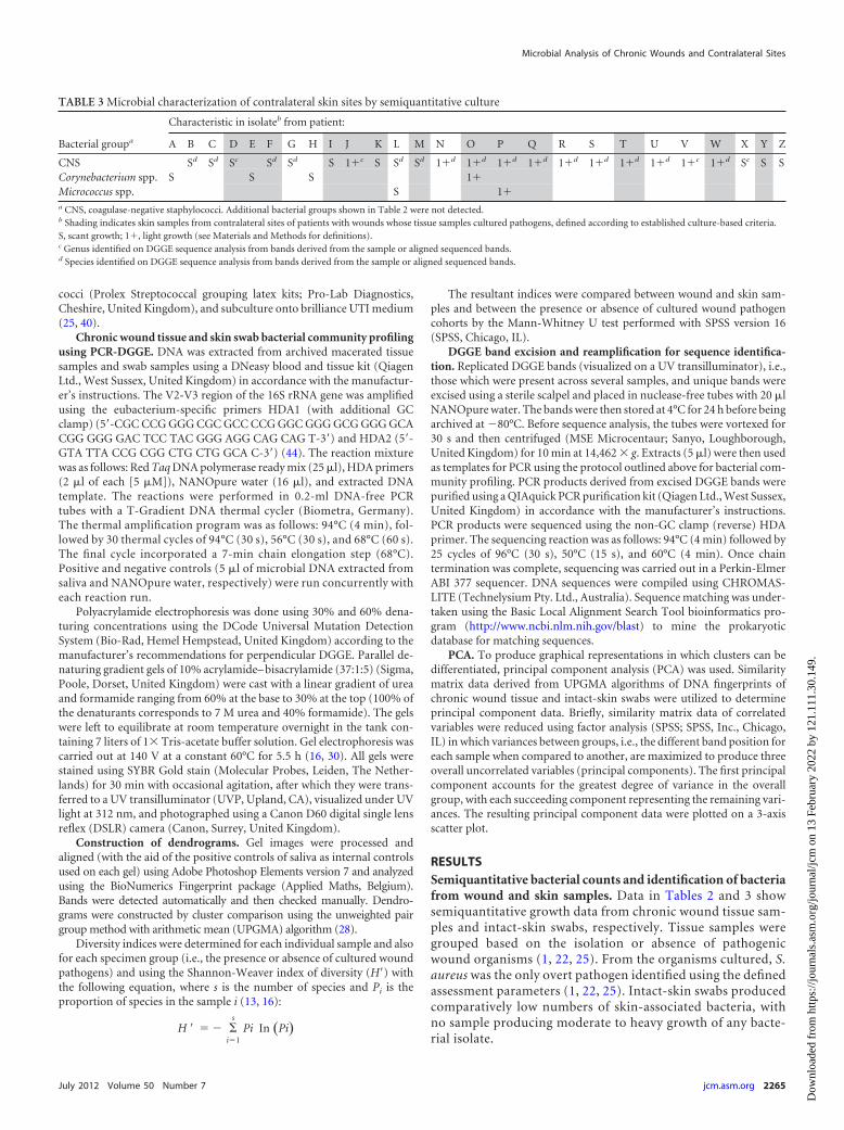
RESULTSSemiquantitative bacterial counts and identification of bacteriafrom wound and skin samples. Data in Tables 2 and 3 showsemiquantitative growth data from chronic wound tissue sam-ples and intact-skin swabs, respectively. Tissue samples weregrouped based on the isolation or absence of pathogenicwound organisms (1, 22, 25). From the organisms cultured, S.aureus was the only overt pathogen identified using the definedassessment parameters (1, 22, 25). Intact-skin swabs producedcomparatively low numbers of skin-associated bacteria, withno sample producing moderate to heavy growth of any bacte-rial isolate.
TABLE 3 Microbial characterization of contralateral skin sites by semiquantitative culture
Bacterial groupa
Characteristic in isolateb from patient:
A B C D E F G H I J K L M N O P Q R S T U V W X Y Z
CNS Sd Sd Sc Sd Sd S 1�c S Sd Sd 1�d 1�d 1�d 1�d 1�d 1�d 1�d 1�d 1�c 1�d Sc S SCorynebacterium spp. S S S 1�Micrococcus spp. S 1�a CNS, coagulase-negative staphylococci. Additional bacterial groups shown in Table 2 were not detected.b Shading indicates skin samples from contralateral sites of patients with wounds whose tissue samples cultured pathogens, defined according to established culture-based criteria.S, scant growth; 1�, light growth (see Materials and Methods for definitions).c Genus identified on DGGE sequence analysis from bands derived from the sample or aligned sequenced bands.d Species identified on DGGE sequence analysis from bands derived from the sample or aligned sequenced bands.
Microbial Analysis of Chronic Wounds and Contralateral Sites
July 2012 Volume 50 Number 7 jcm.asm.org 2265
Dow
nloa
ded
from
http
s://j
ourn
als.
asm
.org
/jour
nal/j
cm o
n 13
Feb
ruar
y 20
22 b
y 12
1.11
1.30
.149
.

DGGE analysis of the microbial diversity of chronic woundtissue samples and contralateral skin swabs. A UPGMA dendro-gram was constructed to compare the overall eubacterial DNAfingerprints of wound and skin communities derived fromchronic wound tissue samples and contralateral intact-skin swabs.The similarity scores ranged from 10 to 60%, with the averagesimilarity score below 50%, indicating that, generally, skin surfaceand wounds were colonized with divergent consortial profiles.However, clustering can be seen for a minority of DGGE profilesderived from wound and skin consortia in the results in Fig. 1.This is explored further via principal component analysis of sim-ilarity matrix scores (Fig. 2) in which two major clusters are ap-parent. While one of these represents a combination of skin andwound profiles, the other is composed primarily of skin-derivedconsortial profiles. Comparisons between diversity indices de-rived from individual wound and skin samples (Table 4) re-
vealed marked differences in eubacterial diversity between allthe wound and skin samples and those where no pathogenswere cultured (P � 0.05), although significant differences werenot detected between wound and skin cohorts where pathogenswere isolated.
Comparisons of 16S DNA sequence data between chronicwound tissue samples and contralateral intact-skin swabs foreach patient. Comparisons were made between bands withmatching positions or taxonomic affiliation and sequences acrossthe wound and the contralateral control skin swab for eachpatient. Examples are given from patients A and G (no patho-gens isolated), presented in Fig. 3 and 4, and patients D and I(wound pathogens isolated), presented in Fig. 5 and 6. In gen-eral, greater proportions of skin-associated bacteria were de-tected in contralateral wound sites (two or more correlatedbands in 8 samples) where no wound pathogens were cultured
FIG 1 A UPGMA dendrogram for patients A to Z, showing percentage matching of wound DGGE fingerprints. Closed triangles, wound debridement samplesfrom which pathogens were isolated; open triangles, wound debridement samples from which pathogens were not isolated; closed circles, contralateral skinsamples from individuals with wounds from which pathogens were isolated; open circles, contralateral skin samples from individuals with wounds from whichpathogens were not isolated.
Oates et al.
2266 jcm.asm.org Journal of Clinical Microbiology
Dow
nloa
ded
from
http
s://j
ourn
als.
asm
.org
/jour
nal/j
cm o
n 13
Feb
ruar
y 20
22 b
y 12
1.11
1.30
.149
.

than in wounds where pathogens were cultured (no correlatedbands in 11 samples).
Corroboration of isolation data by DGGE sequence analyses.DGGE-derived sequence identities were compared to isolationdata for each patient and between sample cohorts. Sequence anal-yses of DGGE amplicons suggested the presence of Staphylococcussp. in 8/13 and S. aureus in 2/13 wound samples from which S.aureus was cultured. PCR amplicons with homology to coagulase-negative staphylococci were detected in 8/13 and Staphylococcussp. in 3/13 of the wound samples where no pathogens were iso-lated (data not shown). For both tissue and skin samples, the mostprevalent genera were Staphylococcus and Bacillus; the latter werenot detected by culture. Additionally, a greater number of obligateanaerobic organisms were identified in both the skin and tissueisolates by DGGE than by culture. According to PCR-DGGE anal-yses, of the 22 genera identified in the wound tissue samples and21 genera in skin swabs, four were unique to the wounds (Kleb-siella sp., Abiotrophia sp., Escherichia coli, and Peptoniphilus sp.)and three were unique to the intact-skin swabs (Kocuria rhizo-phila, Morexellaceae sp., and Rhodocyclaceae sp.).
DISCUSSION
The etiology of chronic wounds commonly relates to underlyingpathologies; initiation is often associated with primary tissuedamage, which creates a portal of entry for microorganisms inwhich complex microbial communities can develop and infectionmay occur (15, 26, 37). The progression and chronicity of woundscan be correlated with infection which, from a microbiologicalperspective, is dependent upon the types of bacteria present andtheir relationship with the host immune responses. Wound infec-tion is commonly defined according to the presence of pathogens
and colonization densities exceeding �106 organisms per gram oftissue and the development of significant tissue damage and clin-ical signs of infection (32, 36, 37).
There has been considerable speculation regarding the poten-tial etiological role of the taxonomically diverse microbial popu-lations which commonly develop within chronic wounds (14, 27,37, 38) and the role that culture-independent techniques couldplay in research and diagnosis (42). The current study investigatedthe relationship between the isolation of overt pathogens fromwounds, as defined by established culture methods, and eubacte-rial diversity, assessed using PCR-DGGE. These techniques werealso used to compare the bacterial composition of wounds andcontralateral healthy skin sites. Semiquantitative culture, oftenused clinically as an indication of infection severity (22, 25, 35,41), was adopted, providing a means by which culturable patho-genic organisms could be detected and a relevant comparator forPCR-DGGE.
Of the 26 chronic wound tissue samples investigated, commonwound pathogens could be isolated from 13; S. aureus was de-tected in all of these, in addition to enteric species and variousrepresentatives of the regional skin microbiota. Additionally, 2 of13 samples (E and M) were also associated with Candida sp., Pseu-domonas sp., and hemolytic group G streptococci. Coliforms wereisolated from seven samples, of which six also harbored skinand/or enteric flora, indicating putative colonization or contam-ination. A moderate growth of coliforms was noted for patientsample Z, which was not considered to be associated with infec-tion based on clinical details. A scant culture of E. coli was alsoisolated from sample A. While E. coli and other coliforms may beconsidered pathogenic and thus significant in specific wound cul-tures, such as gastrointestinal surgical wound sites, in the context
FIG 2 Principal component analysis of DGGE fingerprints of chronic wound samples and intact-skin swabs (patients A to Z). See legend to Fig. 1 for key tosymbols.
Microbial Analysis of Chronic Wounds and Contralateral Sites
July 2012 Volume 50 Number 7 jcm.asm.org 2267
Dow
nloa
ded
from
http
s://j
ourn
als.
asm
.org
/jour
nal/j
cm o
n 13
Feb
ruar
y 20
22 b
y 12
1.11
1.30
.149
.

of the current study, sample A was classified as not harboringwound pathogens due to the low numbers of E. coli isolated andthe wound type from which it was isolated.
Comparisons of the bacteriological composition of woundsfrom which pathogens had or had not been isolated using eubac-terium-specific PCR-DGGE detected S. aureus in 2/13 and Staph-ylococcus sp. in 8/13 of wound samples associated with pathogens.Several sequences obtained using DGGE analysis could not beidentified to species level, an observation which is commonly as-sociated with this method. Isolation methods detected a morelimited range of taxa than DGGE from both wound tissue and skinswabs. Additionally, a greater number of obligate anaerobic or-ganisms were identified in both the skin and tissue isolates usingthe PCR-based technique than the culture, despite the fact thatvalidated anaerobic isolation methods were used. Interestingly, ahigher proportion of those taxa present on the contralateral (con-trol) skin sites occurred in wounds which did not harbor overtpathogens than in those from which pathogens had been isolated.Within the 13 tissue samples where pathogens had been isolated,11 produced no bands (i.e., PCR amplicons) which matched to thecontralateral skin swab profile, whereas all tissue samples whereno pathogens had been isolated were associated with least one ormore matching skin swab bands. Previous diversity profiling stud-ies of the human skin microbiota by Gao et al. (17) and Grice et al.(21) suggest that while there is comparatively little interindividualcompositional similarity in healthy skin microbiotas, high levelsof conservation between the contralateral skin sites in individualscan be demonstrated (17, 21). On this basis, contralateral intact-skin samples may provide an insight into the normal microbiotaof the site and thus, the microbial composition of skin prior towounding.
Primary colonizers of wounds are reportedly often members ofthe autochthonous skin microbiota due to their proximity to thetissue injury (4, 5). However, delayed healing may enable adven-titious bacteria to proliferate and thus compete against autoch-thonous species. Additionally, since the microbiota of healthy skin
TABLE 4 Eubacterial diversity indices and proportions of skinamplicons also detected in woundsa
Patienta
Shannon-Weaverdiversity indexb
Shared ampliconscWound Skin
A 0.583 0.901 4/11B 0.583 0.410 0/5C 1.166 0.328 3/4D 0.510 0.328 0/4E 0.364 0.164 0/2F 0.219 0.819 0/10G 0.656 0.492 2/7H 0.874 0.246 2/3I 0.656 0.410 0/5J 0.801 0.492 1/6K 0.510 0.410 0/5L 0.583 0.655 0/8M 0.729 0.328 1/4N 0.364 0.573 0/6O 0.364 0.328 2/4P 0.801 0.082 0/1Q 0.219 0.246 0/3R 0.437 0.492 2/6S 0.510 0.819 3/10T 0.146 0.328 1/4U 0.801 0.410 1/5V 0.510 0.328 2/3W 1.239 0.410 0/5X 0.364 0.328 0/4Y 0.583 0.492 0/6Z 0.729 0.492 1/6a Shaded rows indicate wounds which harbored pathogens based on established culturecriteria.b Diversity indices were compared between wound and skin and between infected andnoninfected cohorts by the Mann-Whitney U test. Significant differences were foundbetween diversity indices of wound and skin samples and between wound and skinsamples when grouped into noninfected cohorts (P � 0.05). No significant differencewas found between wound and skin when grouped into infected cohorts.c Proportion of bands present in intact-skin DGGE analysis found in chronic woundDGGE analysis.
FIG 3 Characterization of major taxa in wound and skin samples based on dominant PCR amplicons and matched bands derived from DGGE gels (patient A;no pathogens isolated).
Oates et al.
2268 jcm.asm.org Journal of Clinical Microbiology
Dow
nloa
ded
from
http
s://j
ourn
als.
asm
.org
/jour
nal/j
cm o
n 13
Feb
ruar
y 20
22 b
y 12
1.11
1.30
.149
.

is likely to be water and nutrient limited (6), hydration and nutri-ent availability may have a marked influence on the microbialcomposition of wounds. The restricted nutrient and moisturecontent of healthy skin may limit the proliferation of fastidiousorganisms and thus select for Gram-positive bacteria, such as co-agulase-negative staphylococci, corynebacteria, and propionibac-teria (6, 7). In contrast, the comparatively nutrient-rich environ-ment of a wound may facilitate the growth of a wider variety oforganisms, including S. aureus, P. aeruginosa, streptococci, entero-
bacteriaceae, and other facultative anaerobic species (6, 9). Thetransition from healthy skin to colonized/infected wound maytherefore be associated with the clonal expansion of bacteria notnormally associated with health. In most cases, wounds were col-onized by more diverse microbial communities than healthy skin,but the overall eubacterial diversity of wounds which harboredpathogens and those from which no pathogens were isolated didnot differ significantly. It could be agued that this observationhighlights the utility of culture as a straightforward means of de-
FIG 4 Characterization of major taxa in wound and skin samples based on dominant PCR amplicons and matched bands derived from DGGE gels (patient G;no pathogens isolated).
FIG 5 Characterization of major taxa in wound and skin samples based on dominant PCR amplicons and matched bands derived from DGGE gels (patient D;pathogens isolated).
Microbial Analysis of Chronic Wounds and Contralateral Sites
July 2012 Volume 50 Number 7 jcm.asm.org 2269
Dow
nloa
ded
from
http
s://j
ourn
als.
asm
.org
/jour
nal/j
cm o
n 13
Feb
ruar
y 20
22 b
y 12
1.11
1.30
.149
.

tecting wound pathogens. However, it also indicates the need forfurther investigations of potential associations between microbialprofiles and clinical outcome, because the contribution of alteredmicrobial colonization to causality remains poorly understood, incontrast to the involvement of overtly pathogenic species, whichare more readily detectable by culture (25). Previous analysis ofchronic wound diversity by Dowd et al. (11) using pyrosequenc-ing, DGGE, and rRNA gene shotgun sequencing identified severalgenera and species not isolated upon culture, a method which ingeneral failed to identify the primary bacterial populations of thewounds tested (11). Importantly, sequence analyses of DGGE am-plicons within the current study indicated that wounds which didnot harbor pathogens were associated with a greater proportion ofnormal skin bacteria than were infected wounds.
This study provides a cross-sectional assessment of the bacte-rial diversity of wounds which were initially assessed according tothe presence or absence of culture-defined pathogenic species. Italso provides an opportunity to compare isolation methods to aculture-independent DNA profiling technique. In some cases,pathogens detected by isolation were not detected by PCR-DGGE,and conversely, bacterial diversity indicated on DGGE gels wasnot readily detectable by culture. Both DGGE and culture havedistinct characteristics: culture is relatively simple to implement,cost effective, and can detect numerically dominant culturablepathogens. It is, however, limited by the culturability of targetbacteria. Conversely, DGGE can be used to analyze complex com-munities while facilitating the identification of unculturable or-ganisms which may only represent as little as 1% of the total bac-terial population (33). Due to the length of the DNA sequencesthat DGGE produces, however, the taxonomic associations mademay not be categorical.
Analysis of the microbiotas of wound and contralateral skinsites indicated that, in general, healthy skin-associated organismswere underrepresented in wounds from which pathogens werecultured but that significant alterations in total eubacterial diver-sity were not detected. Therefore, while DGGE is a useful tool forthe reproducible culture-independent profiling of bacterial con-
sortia, data presented in the current investigation also highlightthe utility of culture. On this basis, the two analytical approachesare complementary.
ACKNOWLEDGMENTS
This work was partially supported by grants from the BBSRC and Con-vaTec.
REFERENCES1. Angel DE, Lloyd P, Carville K, Santamaria N. 2011. The clinical efficacy
of two semi-quantitative wound-swabbing techniques in identifying thecausative organism(s) in infected cutaneous wounds. Int. Wound J.8:176 –185.
2. Bass MJ, Phillips LG. 2007. Pressure sores. Curr. Probl. Surg. 44:101–143.3. Bourke WJ, O’Connor CM, FitzGerald MX, McDonnell TJ. 1994.
Pseudomonas aeruginosa exotoxin A induces pulmonary endothelialcytotoxicity: protection by dibutyryl-cAMP. Eur. Respir. J. 7:1754 –1758.
4. Bowler PG, Duerden BI, Armstrong DG. 2001. Wound microbiologyand associated approaches to wound management. Clin. Microbiol. Rev.14:244 –269.
5. Brook I, Frazier EH. 1990. Aerobic and anaerobic bacteriology of woundsand cutaneous abscesses. Arch. Surg. 125:1445–1451.
6. Chiller K, Selkin B, Murakawa G. 2001. Skin microflora and bacterialinfections of the skin. J. Invest. Dermatol. Symp. Proc. 6:170 –174.
7. Cogen AL, Nizet V, Gallo RL. 2008. Skin microbiota: a source of diseaseor defence? Br. J. Dermatol. 158:442– 455.
8. Davies CE, et al. 2004. Use of 16S ribosomal DNA PCR and denaturinggradient gel electrophoresis for analysis of the microfloras of healing andnonhealing chronic venous leg ulcers. J. Clin. Microbiol. 42:3549 –3557.
9. Davies CE, et al. 2001. Use of molecular techniques to study microbialdiversity in the skin: chronic wounds reevaluated. Wound Repair Regen.9:332–340.
10. Dinges MM, Orwin PM, Schlievert PM. 2000. Exotoxins of Staphylococ-cus aureus. Clin. Microbiol. Rev. 13:16 –34.
11. Dowd S, et al. 2008. Survey of bacterial diversity in chronic wounds usingpyrosequencing, DGGE, and full ribosome shotgun sequencing. BMCMicrobiol. 8:43. doi:10.1186/1471-2180-8-43.
12. Eaglstein WH, Falanga V. 1997. Chronic wounds. Surg. Clin. North Am.77:689 –700.
13. Edwards ML, Lilley AK, Timms-Wilson TH, Thompson IP, Cooper I.2001. Characterisation of the culturable heterotrophic bacterial commu-nity in a small eutrophic lake (Priest Pot). FEMS Microbiol. Ecol. 35:295–304.
FIG 6 Characterization of major taxa in wound and skin samples based on dominant PCR amplicons and matched bands derived from DGGE gels (patient I;pathogens isolated).
Oates et al.
2270 jcm.asm.org Journal of Clinical Microbiology
Dow
nloa
ded
from
http
s://j
ourn
als.
asm
.org
/jour
nal/j
cm o
n 13
Feb
ruar
y 20
22 b
y 12
1.11
1.30
.149
.

14. Edwards RH, Keith G. 2004. Bacteria and wound healing. Skin Soft TissueInfect. 17:91–96.
15. Fazli M, et al. 2009. Nonrandom distribution of Pseudomonas aeruginosaand Staphylococcus aureus in chronic wounds. J. Clin. Microbiol. 47:4084 –4089.
16. Gafan GP, et al. 2005. Statistical analyses of complex denaturing gradientgel electrophoresis profiles. J. Clin. Microbiol. 43:3971–3978.
17. Gao Z, Tseng C-h, Pei Z, Blaser MJ. 2007. Molecular analysis of humanforearm superficial skin bacterial biota. Proc. Natl. Acad. Sci. U. S. A.104:2927–2932.
18. Gontcharova V, Youn E, Sun Y, Wolcott RD, Dowd SE. 2010. Acomparison of bacterial composition in diabetic ulcers and contralateralintact skin. Open Microbiol. J. 4:8 –19.
19. Gorbet MB, Sefton MV. 2005. Endotoxin: the uninvited guest. Biomate-rials 26:6811– 6817.
20. Gottrup F. 2004. A specialized wound-healing center concept: impor-tance of a multidisciplinary department structure and surgical treatmentfacilities in the treatment of chronic wounds. Am. J. Surg. 187:38S– 43S.
21. Grice EA, et al. 2008. A diversity profile of the human skin microbiota.Genome Res. 18:1043–1050.
22. Healy B, Freedman A. 2006. Infections. BMJ 332(7545):838 – 841. doi:10.1136/bmj.332.7545.838.
23. Hill KE, et al. 2003. Molecular analysis of the microflora in chronicvenous leg ulceration. J. Med. Microbiol. 52:365–369.
24. Howell-Jones RS, et al. 2005. A review of the microbiology, antibioticusage and resistance in chronic skin wounds. J. Antimicrob. Chemother.55:143–149.
25. HPA. 2009. Investigation of skin and superficial and non-surgical woundswabs. UK standards for microbiology investigations, vol 11. V11 Issue 5.Health Protection Agency, London, United Kingdom.
26. Jones SG, Edwards R, Thomas DW. 2004. Inflammation and woundhealing: the role of bacteria in the immuno-regulation of wound healing.Int. J. Low. Extrem. Wounds 3:201–208.
27. Kirketerp-Moller K, et al. 2008. The distribution, organization and ecol-ogy of bacteria in chronic wounds. J. Clin. Microbiol. 46:2717–2722.
28. Ledder RG, et al. 2006. Individual microflora beget unique oral micro-cosms. J. Appl. Microbiol. 100:1123–1131.
29. Lyczak JB, Cannon CL, Pier GB. 2000. Establishment of Pseudomonasaeruginosa infection: lessons from a versatile opportunist. Microbes In-fect. 2:1051–1060.
30. McBain AJ, et al. 2003. Growth and molecular characterization of dentalplaque microcosms. J. Appl. Microbiol. 94:655– 664.
31. Melendez JH, et al. 2010. Real-time PCR assays compared to culture-based approaches for identification of aerobic bacteria in chronic wounds.Clin Microbiol. Infect. 16:1762–1769.
32. Murphy RC, Robson MC, Heggers JP, Kadowaki M. 1986. The effect ofmicrobial contamination on musculocutaneous and random flaps. J.Surg. Res. 41:75– 80.
33. Muyzer G, de Waal EC, Uitterlinden AG. 1993. Profiling of complexmicrobial populations by denaturing gradient gel electrophoresis analysisof polymerase chain reaction-amplified genes coding for 16S rRNA. Appl.Environ. Microbiol. 59:695–700.
34. Posnett J, Franks PJ. 2008. The burden of chronic wounds in the UK.Nurs. Times 104:44 – 45.
35. RNAO. 2005. Assessment and management of foot ulcers for people withdiabetes. Registered Nurses’ Association of Ontario, Toronto, Ontario,Canada.
36. Robson MC. 1979. Infection in the surgical patient: an imbalance in thenormal equilibrium. Clin. Plast. Surg. 6:493–503.
37. Robson MC. 1997. Wound infection: a failure of wound healing caused byan imbalance of bacteria. Surg. Clin. North Am. 77:637– 650.
38. Robson MC, Stenberg BD, Heggers JP. 1990. Wound healing alterationscaused by infection. Clin. Plast. Surg. 17:485– 492.
39. Singh SK, et al. 2009. Detecting aerobic bacterial diversity in patients withdiabetic foot wounds using ERIC-PCR: a preliminary communication.Int. J. Low. Extrem. Wounds 8:203–208.
40. Steel KJ, Barrow GI, Feltham RKA, Cowan ST. 1993. Cowan and Steel’smanual for the identification of medical bacteria. Cambridge UniversityPress, Cambridge, United Kingdom.
41. Stone ND, et al. 2008. Importance of bacterial burden among methicillin-resistant Staphylococcus aureus carriers in a long-term care facility. Infect.Control Hosp. Epidemiol. 29:143–148.
42. Thomsen TR, et al. 2008. The bacteriology of chronic venous leg ulcerexamined by culture-independent molecular methods. Wound Rep.Regen. 18:38 – 49.
43. Vasconez LO, Schneider WJ, Jurkiewicz MJ. 1977. Pressure sores. Curr.Probl. Surg. 14:1– 62.
44. Walter J, et al. 2000. Detection and identification of gastrointestinalLactobacillus species by using denaturing gradient gel electrophoresis andspecies-specific PCR primers. Appl. Environ. Microbiol. 66:297–303.
45. Wilson MJ, Weightman AJ, Wade WG. 1997. Applications of molecularecology in the characterization of uncultured microorganisms associatedwith human disease. Rev. Med. Microbiol. 8:91–102.
Microbial Analysis of Chronic Wounds and Contralateral Sites
July 2012 Volume 50 Number 7 jcm.asm.org 2271
Dow
nloa
ded
from
http
s://j
ourn
als.
asm
.org
/jour
nal/j
cm o
n 13
Feb
ruar
y 20
22 b
y 12
1.11
1.30
.149
.