diabetic foot wound
TRANSCRIPT

6/22/2021
1
Diabetic Foot Wound
Said Atway, DPM, FACFASAssociate Professor-Clinical Department of Orthopaedics
The Ohio State University Wexner Medical Center
“Diabetic foot” variety of pathological conditions that
might affect the feet in patients with diabetes (Boulton 2002)
Team Approach
• Expedited referral and care
• Madagin program proven to decrease amputations 82%
• Driver et al. Diabetes Care 2005
• Swedish study decreased amputations 71% over 11 years
• Wennberg et al Diabetes research 2019
• UK study 40% reduction and 62% reduction in major amputations

6/22/2021
2
•Prevalence‒29.1 Million people 9.3% of the US 2012
• CDC‒2.8% Worldwide 2000 (171 million)
• WHO
Amputations ‒73,000 non‐traumatic amputations in diabetics 2010
• CDC
‒Cost• $4,595 per ulcer and $28,000 >2years• $5billion per year annually
• Clin Ther 1998• $30‐50k amputation according to president
Foot Infections• Any infra‐malleolar infection in a person with diabetes
• Common and costly problem‒DM related amputation cost 3B per year
• Diabetes Care 2003
• Most common reason for a diabetic to be admitted‒National Hospital Discharge Data
• Most common non‐traumatic cause of amputation‒60% of LEA‒Most common cause of nontraumaticlower extremity amputation
• Lancet 2005

6/22/2021
3
Importance of Diabetic Wound care
•Diabetic foot ulcers present >4 weeks have a 5 fold higher risk of infection
• Infection in a foot ulcer increases the risk for hospitalization 55.7 times and risk for amputation 155 times
•5 year mortality after limb amputation is 68%•NIH publication 1995
The FDA defines a healed wound as reepithelialized skin without drainage or dressing requirements confirmed at 2 consecutive visits 2 weeks apart.
Clinical Practice Guidelines• Management of etiologic factors‒Adequate perfusion
• PAD (Twice as common in DM)• Gregg et al 2004Rarely lead to ulcer directlyContributes to 50% of ulcersDiabetes Metab 2008
‒Debridement• Sharp debridement of infection• Urgent for gas/necrotizing infection
‒Infection Control• IDSA guidelines
‒Pressure Mitigation• Offloading• Total contact cast

6/22/2021
4
• ADA recommendations:‒ ABI >50y DM‒ <50y with risk factors
• Smoking• HTN• Hyperlipidemia• >10years DM
‒ Anyone with PAD symptoms
• Dependent rubor
• Pallor on elevation
• Absence of hair growth
• Dystrophic nails
• Cool/Dry/Fissured skin• Diabetes Care 2003
Vascular work up
• Pathophysiologic mechanism complex‒Neuropathy‒Repetitive trauma
‒Focal tissue ischemia
‒Tissue Destruction
• Foot deformities ‒Charcot
• Neuroarthropathy
• Limited joint mobility‒Glycosylation of soft tissue
Deformity• Size
‒% reduction early predictor of outcome• Location
‒WB surface‒ Digits‒ Heel ‒ Legs
• Shape‒Margolins
• Depth ‒ Deep tissue involvement
• Base ‒ Necrotic/Fibrotic/Granular
• Border‒ Abnormal
• Probe ‒ 89% Probe to bone
• Xrays‒ Free air/foreign body
• Infection‒ Advanced imaging work up
Wound Evaluation

6/22/2021
5
Orthopaedic Wound CareManagement of osteomyelitis and bone infection
Anne Sullivan, MDAssistant Professor - ClinicalDepartment of Orthopaedics
The Ohio State University Wexner Medical Center
Why Orthopaedic Wound care?• Things may not be as they appear…..• Bone Expertise provides a margin for safety• Bone Debridement without Expertise
• May Compromise Structural integrity • May Fail to Eradicate infection
• Bone Debridement without Coverage • Exposes previously Protected area to Environment• May Promote Deeper Infection
• Biopsy techniques Must Avoid Contaminating deeper bone –Metaphyseal or Medullary bone.
• Red flag: Persistent Draining Wound despite Meticulous care • What Lies Beneath……
• As many as 64% Diabetic Foot infections involve Bone• Foreign material and Necrotic bone harbor Biofilm• Granulation tissue Obscures, does Not Protect underlying Structures from Environment
• Proper use of Imaging/X‐rays is Essential
Osteomyelitis= Infection involving bone• Timing: acute or chronic
• Organisms: Staphylococcus aureus• Most common, can be any organism
• Causes/ examples: (may be acute or chronic)• Direct contamination – bone contacts environment—
• Open Fracture • Penetrating Trauma• Stage 4 ulcer• Animal or Human bite
• Contiguous spread –from Local wound or abscess,• Diabetic Foot wound, Pressure ulcer, Paronychia, Injection Related abscess
• Infected Fixation Hardware or Prosthetic Joint Infection (PJI)• Hematogenous seeding from Remote source
• Endocarditis, Pyelonephritis,• Infections related to Injected Drug use
OsteomyelitisCierny‐Mader Classification/ Factors (1980’s) • Location of infection in Bone:
• 1. Medullary • 2. Superficial • 3. Localized • 4. Diffuse
• Type of Host:• “A”: Healthy Host
normal healing potential• “B”: Compromised Host
Systemic or Local Factors +/‐ Correctable ??
• “C”: Debilitated host: Treatment is worse than Disease
• Systemic (s):• Organ failure (renal, hepatic)• Malnutrition, • Diabetes Mellitus• Chronic Hypoxia • Extremes of Age• Immune disease • Immune compromise• Malignancy
• Local (l):• Vascular disease, stasis, lymphedema• Scarring, radiation fibrosis• Neuropathy• Small vessel disease• Tobacco abuse
Mader JT, Shirtliff M, Calhoun JH. Staging and Staging Application in
Osteomyelitis. Clinical Infectious Diseases. 1997;25:1303–1309.
* 2021*Substance Abuse,
Noncompliance
Cierny G, Mader JT, Pennick H. A clinical staging system of adult osteomyelitis. Contemp Orthop1985; 10:17 – 37

6/22/2021
6
Treatment Strategies:Wound Closure efforts may require any or all to succeed
• Recognize Source and remove Bioburden
• Assessment, Imaging, Irrigation, Debridement, Bone Resection
• Protect bone
• Avoid Further and Repeat contamination
• Maintain or Restore Structural integrity of bone, joint, tendon, ligament
• May Require Advanced Orthopaedic Reconstruction• Antibiotics: IV, Oral, Local delivery (beads), Antimicrobial dressings
• Avoid spread: Coverage/ closure, Blood supply, Dressings, Control pathogens
• Optimize healing potential‐‐Improve host
• Oxygen, Nutritional resources for Healing,
• Decrease Size of wound–, Negative pressure, Bone resection, Flap/Graft
• Address Soft tissue Issues
• Control Medical Factors (optimization)
• No Tobacco
Late effects of open tibia plateau fracture: plain X‐ray and CT showing foreign body, sclerotic dead bone, and defect
Plain x‐ray: 2 stage reconstruction
MRI proximal tibia: showing metal artifact
= wrong study!**IMAGING USE IN
CLINICAL SCENARIO **
General Surgery and Wound Care
David Renton, MD, MPH, FACSAssociate Professor
Associate Chief Quality Officer for Perioperative ServicesDepartment of Surgery
The Ohio State University Wexner Medical Center
General Surgery and Wound Care•Typically, General Surgery deals with the abdomen, so this is what we deal with in Wound Care as well
•Most of what I am seeing in wound care is chronic abdominal wounds
•This often has to do with infected prosthetics such as hernia mesh that are involved in patient care in the past
•Also help with colostomy creation for help in healing sacral wounds

6/22/2021
7
Laparoscopic Loop Colostomy
•Diverts stool to the abdominal wall through a stoma
•Helps clean up perineum, allows for complex wound closure of sacral wounds
•Can be temporary, or permanent, depending on patient preference
Wound Care
•With a team approach, we can offer our patients a full breadth of options for wound care
•Having Plastic Surgery, Orthopedics, and General Surgery gives patients the best chance of healing their wounds
•Our nursing staff is very experienced, and helps get patients ready for surgery if it is indicated.
Case Presentations on Various Wound Etiologies
Nancy Hale, APRN, CNPComprehensive Wound Center
The Ohio State University Wexner Medical Center

6/22/2021
8
DermatologyRheumatologyVascularNutrition
EndocrinologyPT
EndocrinologySurgical services
Wound Center Provider
ID PMR
Pyoderma
Stage 4 Pressure Injury Arterial Wound

6/22/2021
9
Malignant Melanoma
Advanced Wound Careat the New OSU Wound Center
Rajiv Chandawarkar, MDDirector of OSU WOUND CARE
Director Plastic Surgery, The Ohio State University Wexner Medical Center East Hospital
Professor of Plastic Surgery, Department of Plastic Surgery The Ohio State University Wexner Medical Center
Disclosures
I have no disclosures
New OSU Wound CenterWhat is Advanced Wound Care
Wound care professionals should select the appropriate wound management system based on: 1) Published clinical evidence ;2) Contribution to providing the best outcomes at the total lowest
cost of care, and 3) Comprehensive multidisciplinary care model with highly
specialized care.
We offer the most advanced treatment for nonhealing and hard‐to‐heal wounds, including hyperbaric oxygen therapy, surgical and microsurgical reconstruction, advanced use of biologics and new emerging technology

6/22/2021
10
Referral to Wound Center Wound Care Documentation
Color Coding WoundsPrinciples of Advanced Wound Care
• Differentiate between acute wounds and the development of chronic wounds.
• Identify risk factors for chronic wound development.
• Identify the four most common type of chronic wounds: pressure injury and venous, arterial, and diabetic ulcers.
• Discuss the development of biofilm and the role it plays in wound chronicity.
• Apply the concepts of DIME+S– Debridement, Inflammation Control, Moisture management, Edge effect AND Surgery (when indicated)
New OSU wound care center

6/22/2021
11
Wound Preparation Model
Chronic Wound
RxCause
LocalWound Care
PatientCentered Concerns
Debridement Moisture BalanceInfection
InflammationEdge Effect
SURGERY WHEN INDICATED
New OSU wound care center DEBRIDEMENTWhy is it necessary?
DEBRIDEMENTWhy is it necessary?
Inflammation and Infection ControlWhat Does it Do?

6/22/2021
12
Infection – shades of grey Management of MoistureTips and Tricks
Edge Effect: The Benefits of a Contact Layer
Edge Effect:Choosing the Right Dressings

6/22/2021
13
Overall Cost‐Awareness
Surgical ClosureSurgical Closure
Pre-op EvaluationPre-op Evaluation• History:
–Cardiac, pulmonary, endocrine, oncologic• Location :
–Depth, volume– Involvement of adjacent structures
• Bone, Joint, sinus• Quality:
–Vascularity–Presence of scarring–Extent of infection
• Cellulitis, Heavy bacterial count, C/S
Pre-op EvaluationPre-op Evaluation• Nutritional status
–Cachexia, anemia• Musculoskeletal status
– Spasms, fixed contractures • Distant infection
–Urinary, pulmonary, other• General status
–Neurologic/ psychiatric• Coma, disorientation• Schizophrenia, depression• Social support system

6/22/2021
14
Methods of Surgical ClosureMethods of Surgical Closure• Primary closure
• Skin graft
• Pedicle flap: muscle, musculocutaneous,fasciocutaneous
• Free tissue transfer
Sacral Ulcer ClosureSacral Ulcer Closure• primary closure
• superior gluteus muscle
• gluteal fasciocutaneous
• gluteal turnover
• reverse dermal graft
• V‐Y advancement
• paraspinous based perforator flap
Gluteus maximus
Sacral UlcerSacral Ulcer

6/22/2021
15
Sacral UlcerSacral Ulcer Sacral UlcerSacral Ulcer
Ischial Ulcer ClosureIschial Ulcer Closure• primary closure
• posterior thigh
• inf. gluteal muscle
• gracilis
• biceps femoris & skin
• hamstring
• tensor fascia lata & vastuslateralis
• lateral thigh
• rectus abdominus
Ischial UlcerIschial Ulcer

6/22/2021
16
Ischial UlcerIschial Ulcer Ischial UlcerIschial Ulcer

6/22/2021
17
Trochanteric Ulcer ClosureTrochanteric Ulcer Closure• random fasciocutaneous
• TFL
• TFL V‐Y
• sensate TFL
• TFL w/ skin island
• gluteus medius & TFL
• vastus lateralis
• gluteus maximus, distally based
• gluteal thigh flap
• expansive gluteus max.
Tensor Fascia Lata (TFL)Tensor Fascia Lata (TFL) Tensor Fascia Lata (TFL)Tensor Fascia Lata (TFL)

6/22/2021
18
Trochanteric UlcerTrochanteric UlcerPosterior thigh flapPosterior thigh flap
Fillet (Leg Amputation) FlapFillet (Leg Amputation) Flap
• Hip disarticulation & fillet flap
• High complication rate ‐ OR blood loss, infection, sinus tract, dehiscence, femoral stump rotation
• Hemicorporectomy: For extensive infection, very morbid
Fillet (Leg Amputation) FlapFillet (Leg Amputation) Flap
6/22/2021
19
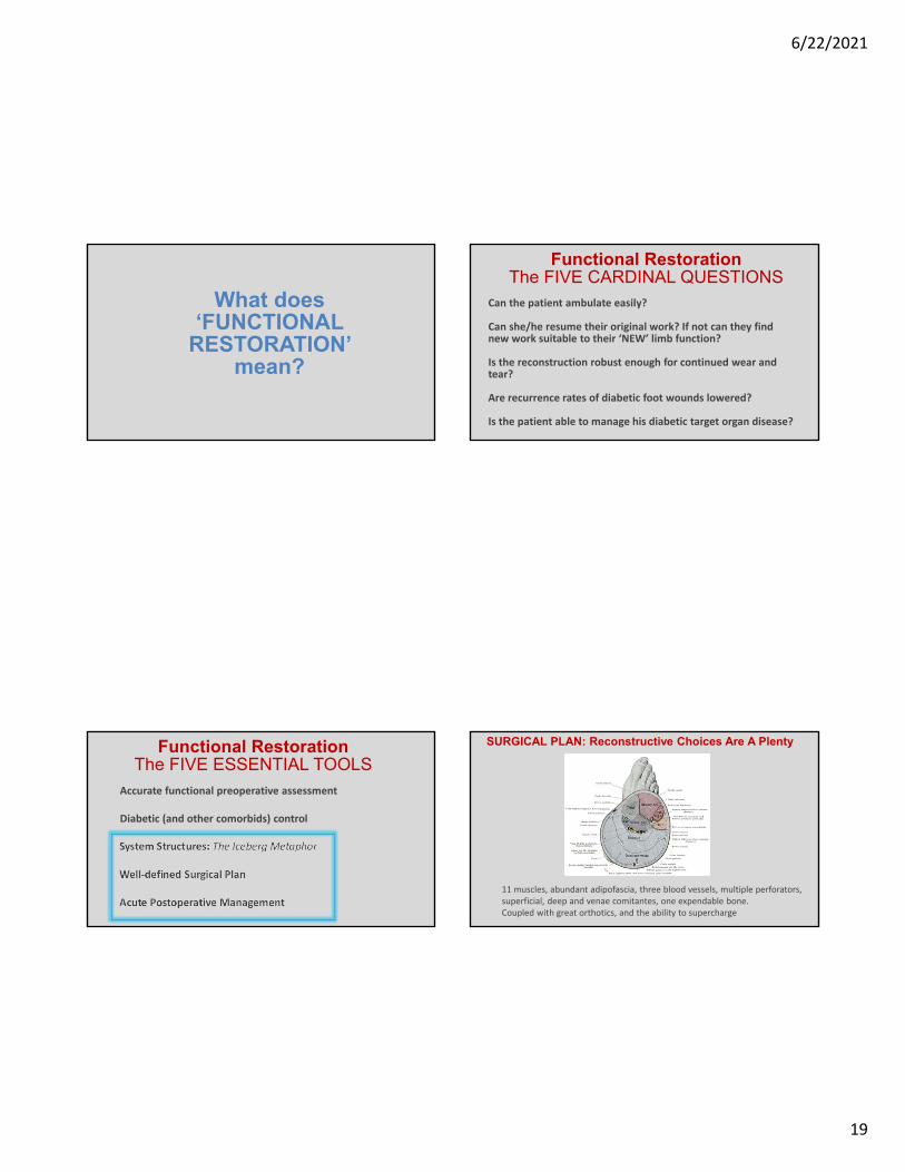
What does ‘FUNCTIONAL
RESTORATION’ mean?
Can the patient ambulate easily?
Can she/he resume their original work? If not can they find new work suitable to their ‘NEW’ limb function?
Is the reconstruction robust enough for continued wear and tear?
Are recurrence rates of diabetic foot wounds lowered?
Is the patient able to manage his diabetic target organ disease?
Functional RestorationThe FIVE CARDINAL QUESTIONS
Accurate functional preoperative assessment
Diabetic (and other comorbids) control
System Structures: The Iceberg Metaphor
Well‐defined Surgical Plan
Acute Postoperative Management
Functional RestorationThe FIVE ESSENTIAL TOOLS
11 muscles, abundant adipofascia, three blood vessels, multiple perforators, superficial, deep and venae comitantes, one expendable bone. Coupled with great orthotics, and the ability to supercharge
SURGICAL PLAN: Reconstructive Choices Are A Plenty

6/22/2021
20
Functionalized Reverse Sural Fasciocutaneous Flap
Reverse SuralFascioCutaneous Flap
Sural And Saphenous Vessels
Vasanervosum and Vasovasorum may play a role(controversial)
Deep fascia is included
Great for Achilles Repair
Funtionalized Reverse SuralFasciocutaneous Flap
Funtionalized Reverse SuralFasciocutaneous Flap

6/22/2021
21
Funtionalized Reverse SuralFasciocutaneous Flap ‐ Transfer
Funtionalized Reverse SuralFasciocutaneous Flap ‐ Inset
Funtionalized Reverse SuralFasciocutaneous Flap
Funtionalized Reverse SuralFasciocutaneous Flap
Currently, this patient walks without support, drives, and has resumed her job as a school teacher

6/22/2021
22
Calcaneal reconstruction with microvascular double/single barrel
fibula osteocutaneus flap
Background
Calcaneal destruction commonly occurs in diabetics ‐usually necessitates a below‐knee amputation since the central weight‐bearing mechanism is lost and reconstructive choices are limited.
Here we present two cases of calcaneal reconstruction, using double/single‐barreled fibular osteocutaneousfree flaps.
88

6/22/2021
23
Currently, this patient walks without support and has resumed his job as a supervisor in a tree‐cutting agency

6/22/2021
24
Currently, this patient walks without support and has resumed his job as a short‐distance truck driver.He even takes his Harley out, once in a while!
95
Reverse Peroneus Brevis Flap Reverse Peroneus Brevis Flap
96

6/22/2021
25
New OSU wound care center
• Strong commitment by the members of the multidisciplinary team
• Physical space and financial support from the sponsoring institution
• Performance metrics, Quality measures• Data Management and Support• Technology• Partnership with an OutPatient Community• Actionable Knowledge• CoMarket Expansion• Defined Drivers of Care and Patient Volumes