a- encoding problem: p rs overview of the visual cortex...
TRANSCRIPT
A- 'Unaware'
Fixed !!
Decoder
Adaptation!
State
Population !
Response
Encoder!!! r
B- 'Aware'
Adaptive !!
Decoder
Adaptation!
State
Population !
Response
Encoder!!! r
s s
s s
Encoding problem: A- 'Unaware'
Fixed !!
Decoder
Adaptation!
State
Population !
Response
Encoder!!! r
B- 'Aware'
Adaptive !!
Decoder
Adaptation!
State
Population !
Response
Encoder!!! r
s s
s s
−180 −90 0 90 1800
25
50
θtest
Resp
on
ses
A − Tuning Curves
−180 −90 0 90 180
0
25
50
θtest
B − Population Response
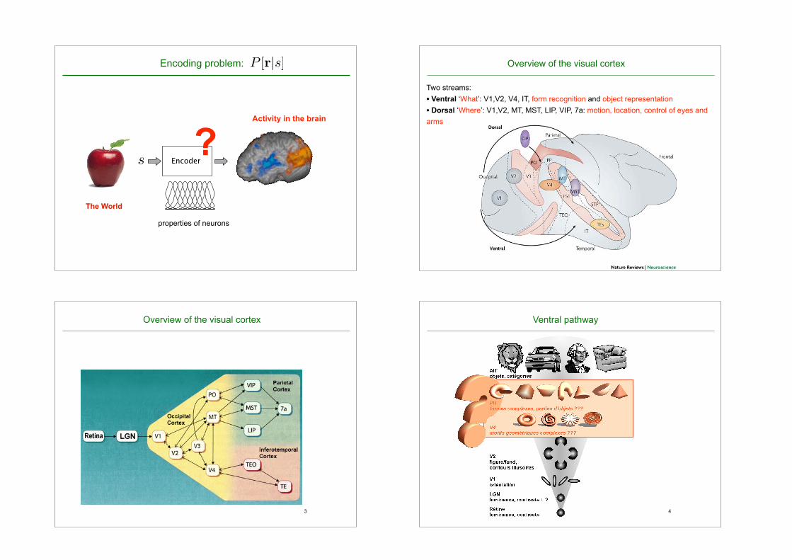
Perception: what we think the world is likeThe World
Activity in the brain
properties of neurons
P [r|s]
?
Overview of the visual cortex
Two streams: • Ventral ‘What’: V1,V2, V4, IT, form recognition and object representation • Dorsal ‘Where’: V1,V2, MT, MST, LIP, VIP, 7a: motion, location, control of eyes and arms
3
Overview of the visual cortex
4
Ventral pathway
5
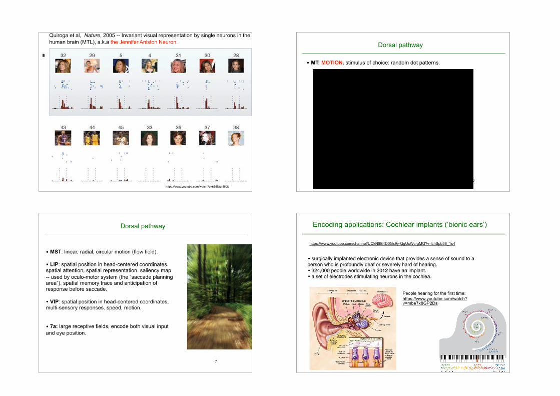
Figure 1a shows the responses of a single unit in the left posteriorhippocampus to a selection of 30 out of the 87 pictures presented tothe patient. None of the other pictures elicited a statistically signifi-cant response. This unit fired to all pictures of the actress JenniferAniston alone, but not (or only very weakly) to other famous andnon-famous faces, landmarks, animals or objects. Interestingly, theunit did not respond to pictures of Jennifer Aniston together with theactor Brad Pitt (but see Supplementary Fig. 2). Pictures of JenniferAniston elicited an average of 4.85 spikes (s.d. ¼ 3.59) between 300and 600ms after stimulus onset. Notably, this unit was nearly silent
during baseline (average of 0.02 spikes in a 700-ms pre-stimulus timewindow) and during the presentation of most other pictures(Fig. 1b). Figure 1b plots the median number of spikes (across trials)in the 300–1,000-ms post-stimulus interval for all 87 pictures shownto the patient. The histogram shows amarked differential response topictures of Jennifer Aniston (red bars).Next, we quantified the degree of invariance using a receiver
operating characteristic (ROC) framework15. We considered as thehit rate (y axis) the relative number of responses to pictures of aspecific individual, object, animal or landmark building, and as
Figure 1 | A single unit in the left posterior hippocampus activatedexclusively by different views of the actress Jennifer Aniston.a, Responses to 30 of the 87 images are shown. There were no statisticallysignificant responses to the other 57 pictures. For each picture, thecorresponding raster plots (the order of trial number is from top to bottom)and post-stimulus time histograms are given. Vertical dashed lines indicateimage onset and offset (1 s apart). Note that owing to insurmountablecopyright problems, all original images were replaced in this and allsubsequent figures by very similar ones (same subject, animal or building,similar pose, similar colour, line drawing, and so on). b, The median
responses to all pictures. The image numbers correspond to those in a. Thetwo horizontal lines show the mean baseline activity (0.02 spikes) and themean plus 5 s.d. (0.82 spikes). Pictures of Jennifer Aniston are denoted byred bars. c, The associated ROC curve (red trace) testing the hypothesis thatthe cell responded in an invariant manner to all seven photographs ofJennifer Aniston (hits) but not to other images (including photographs ofJennifer Aniston and Brad Pitt together; false positives). The grey linescorrespond to the same ROC analysis for 99 surrogate sets of 7 randomlychosen pictures (P , 0.01). The area under the red curve is 1.00.
NATURE|Vol 435|23 June 2005 LETTERS
1103© 2005 Nature Publishing Group
Quiroga et al, Nature, 2005 -- Invariant visual representation by single neurons in the human brain (MTL), a.k.a the Jennifer Aniston Neuron.
https://www.youtube.com/watch?v=635Ntur8K2s
6
Dorsal pathway
• MT: MOTION. stimulus of choice: random dot patterns.
7
Dorsal pathway
• MST: linear, radial, circular motion (flow field).
• LIP: spatial position in head-centered coordinates. spatial attention, spatial representation. saliency map -- used by oculo-motor system (the “saccade planning area”). spatial memory trace and anticipation of response before saccade.
• VIP: spatial position in head-centered coordinates, multi-sensory responses. speed, motion.
• 7a: large receptive fields, encode both visual input and eye position.
Encoding applications: Cochlear implants (‘bionic ears’)
• surgically implanted electronic device that provides a sense of sound to a person who is profoundly deaf or severely hard of hearing.• 324,000 people worldwide in 2012 have an implant.• a set of electrodes stimulating neurons in the cochlea.
17SENSATION AND PERCEPTION | BRAIN FACTSSOCIETY FOR NEUROSCIENCE
Although the process is not yet completely understood, recent findings suggest that visual signals are fed into at least three separate processing systems. One system appears to process information mainly about shape; a second, mainly about color; and a third, movement, location, and spatial organization. These findings of separate processing systems come from anatomical and physiological studies in monkeys. They are supported by human psychological studies showing that the perception of movement, depth, perspective, the relative size of objects, the relative movement of objects, shading, and gradations in texture all depend primarily on contrasts in light intensity rather than on color.
Why movement and depth perception should be emphasized by one processing system may be explained by a school of thought called Gestalt psychology. Perception requires various elements to be orga-nized so that related ones are grouped together. This stems from the brain’s ability to group the parts of an image together and also to sepa-rate images from one another and from their individual backgrounds.
How do all these systems combine to produce the vivid images of solid objects that we perceive? This involves extracting biologically relevant information at each stage and associating firing patterns of neuronal populations with past experience.
Vision studies also have led to better treatment for visual disor-ders. Information from research in cats and monkeys has improved the therapy for strabismus, or squint, a term for cross-eye or walleye. Children with strabismus initially have good vision in each eye. But because they cannot fuse the images in the two eyes, they tend to favor one eye and often lose useful vision in the other. Vision can be restored in such cases, but only during infancy or early childhood. Beyond the age of 6 or so, the blindness in one eye becomes perma-nent. Until a few decades ago, ophthalmologists waited until children reached the age of 4 before operating to align the eyes or prescribing exercises or an eye patch. Now strabismus is corrected very early in life — before age 4, when normal vision can still be restored.
HearingOften considered the most important sense for humans,
hearing allows us to communicate with each other by receiving sounds and interpreting speech. It also gives us information vital to survival; for instance, by alerting us to an approaching car.
Like the visual system, our hearing system distinguishes several qualities in the signals it detects. Our hearing system, however,
HEARING. From the chirping of crickets
to the roar of a rocket engine, sound waves
are collected by the external ear — the pinna
and the external auditory canal — and fun-
neled to the tympanic membrane (eardrum)
to make it vibrate. Attached to the tympanic
membrane, the malleus (hammer) transmits
the vibration to the incus (anvil), which passes
vibration on to the stapes (stirrup). The stapes
pushes on the oval window, which separates
the air-filled middle ear from the fluid-filled
inner ear, to produce pressure waves in the
snail-shaped cochlea of the inner ear. Hair
cells in the cochlea, riding on the vibrating
basilar membrane, have “hair bundles” of
microscopic stereocilia that are deflected by
the overlying tectorial membrane. Hair cells
convert the mechanical vibration to an elec-
trical signal; they, in turn, release chemicals
to excite the 30,000 fibers of the auditory
nerve that carry the signals to the brainstem.
Auditory information is analyzed by multiple
brain centers as it flows to the temporal gyrus
or auditory cortex, the part of the brain
involved in perceiving sound.
People hearing for the first time: https://www.youtube.com/watch?v=mbe7x8GP2Ds
https://www.youtube.com/channel/UCkN8E4D0Gs9y-QgUcWc-gMQ?v=LhSpb36_1s4

Encoding applications: retinal implants (‘bionic eyes’)
• in development • meant to partially restore vision to people with degenerative eye conditions such as macular degeneration• stimulating the retina with array of electrodes -currently 60-100 channels.
Sheila Nirenberg: A prosthetic eye to treat blindnesshttp://www.ted.com/talks/sheila_nirenberg_a_prosthetic_eye_to_treat_blindness.html
https://www.youtube.com/watch?v=CiyGOUHD2nI
10
−180 −90 0 90 1800
25
50
θtest
Resp
on
ses
A − Tuning Curves
−180 −90 0 90 180
0
25
50
θtest
B − Population Response
Decoding populations of neuronsA- 'Unaware'
Fixed !!
Decoder
Adaptation!
State
Population !
Response
Encoder!!! r
B- 'Aware'
Adaptive !!
Decoder
Adaptation!
State
Population !
Response
Encoder!!! r
s s
s s
A- 'Unaware'
Fixed !!
Decoder
Adaptation!
State
Population !
Response
Encoder!!! r
B- 'Aware'
Adaptive !!
Decoder
Adaptation!
State
Population !
Response
Encoder!!! r
s s
s s
In response to a stimulus with unknown orientation s, we observe a pattern of activity r (e.g. in V1). What can we say about s given r?
spik
es/s
preferred orientation
r
?
Decoding populations of neurons
An estimation problem (detecting signal in noise). ➡ Tools : estimation theory, bayesian inference, machine learning
When does the problem occur?:
1 - Point of view of the experimentalist or Neuro-Engineering. Seeking the most effective method (e.g. prosthetics) to read out the code.
✤ Statistical optimality ✤ considering the constraints (e.g. real time?)
2 - Model of the brain’s decoding strategy e.g. mapping from sensory signals to motor response and understanding
the relationship between physiology and psychophysics
✤ statistical optimality ? ✤ optimality within a class ? ✤ or simplicity/ arbitrary choice? (what are the biological constraints ?)

Decoding: to understand the link between Physiology and Psychophysics
• Understanding the relationship between neural responses and performances of the animal:
• Detection Task: e.g. can you see the target ? Measure Detection threshold. • Estimation Task: e.g. What is the angle of the bar ? The contrast of the grating? Measure Estimation errors (bias -- illusions).
• Discrimination Task: e.g. What is the minimal difference you can see?
A- 'Unaware'
Fixed !!
Decoder
Adaptation!
State
Population !
Response
Encoder!!! r
B- 'Aware'
Adaptive !!
Decoder
Adaptation!
State
Population !
Response
Encoder!!! r
s s
s s
−180 −90 0 90 1800
25
50
θtest
Resp
on
ses
A − Tuning Curves
−180 −90 0 90 180
0
25
50
θtest
B − Population Response Perceptionsensory stimulus
Optimal Decoder
✤ optimality criterion? MSE(s) =< (s� s)2 >
1. Optimal Decoding
spik
es/s
s = argmaxsP (r|s)
−180 −90 0 90 1800
25
50
θtest
Resp
on
ses
A − Tuning Curves
−180 −90 0 90 180
0
25
50
θtest
B − Population Response
preferred orientation
✤ Maximum Likelihood: if we know P[r|s] (the encoding model), choose the stimulus s that has maximal probability of having generated the observed response, r.
1. Optimal Decoding
spik
es/s
−180 −90 0 90 1800
25
50
θtest
Resp
on
ses
A − Tuning Curves
−180 −90 0 90 180
0
25
50
θtest
B − Population Response
preferred orientation
?
s = argmaxsP (r|s)
✤ Maximum Likelihood: if we know P[r|s] (the encoding model), choose the stimulus s that has maximal probability of having generated the observed response, r.
1. Optimal Decoding

spik
es/s
−180 −90 0 90 1800
25
50
θtest
Resp
on
ses
A − Tuning Curves
−180 −90 0 90 180
0
25
50
θtest
B − Population Response
preferred orientation
s = argmaxsP (r|s)
✤ Maximum Likelihood: if we know P[r|s] (the encoding model), choose the stimulus s that has maximal probability of having generated the observed response, r.
1. Optimal Decoding
spik
es/s
−180 −90 0 90 1800
25
50
θtest
Resp
on
ses
A − Tuning Curves
−180 −90 0 90 180
0
25
50
θtest
B − Population Response
preferred orientation
✤ Maximum a Posteriori: if we know P[r|s] and have a prior on s, P[s], choose the stimulus s that is most likely, given r.
s = argmaxsP (s|r) = argmaxsP [r|s]P [s]
1. Optimal Decoding
A- 'Unaware'
Fixed !!
Decoder
Adaptation!
State
Population !
Response
Encoder!!! r
B- 'Aware'
Adaptive !!
Decoder
Adaptation!
State
Population !
Response
Encoder!!! r
s s
s s
−180 −90 0 90 1800
25
50
θtest
Resp
on
ses
A − Tuning Curves
−180 −90 0 90 180
0
25
50
θtest
B − Population Response Perceptionsensory stimulus
Simple Decoders
Is the brain able to do ML or MAP estimation ?
- Unknown - It is argued that realistic architectures could perform ML [Deneve, Latham, Pouget al 2001, Ma, Pouget et al 2006, Jazayeri and Movshon 2006]
−180 −90 0 90 1800
25
50
θtest
Resp
on
ses
A − Tuning Curves
−180 −90 0 90 180
0
25
50
θtest
B − Population Response
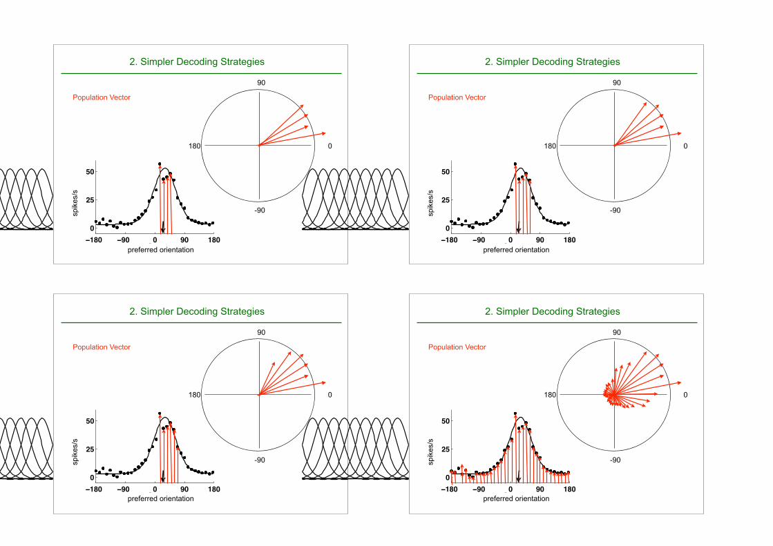
2. Simpler Decoding Strategies
Winner Take All :
If we know the preferred orientation of all neurons, choose the preferred orientation of the neuron that responds most.
preferred orientation
spik
es/s
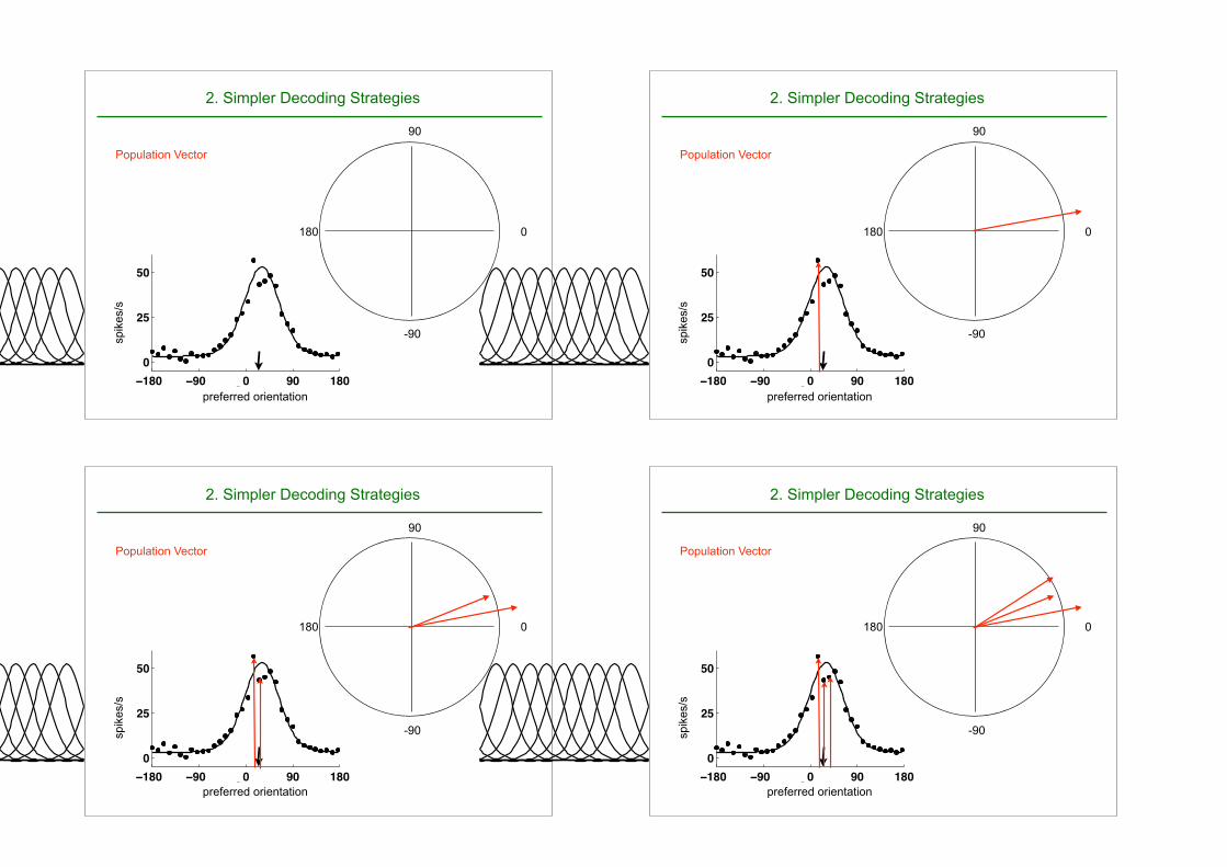
Population Vector
−180 −90 0 90 1800
25
50
θtest
Resp
on
ses
A − Tuning Curves
−180 −90 0 90 180
0
25
50
θtest
B − Population Response
preferred orientation
spik
es/s
0
90
180
-90
2. Simpler Decoding Strategies
−180 −90 0 90 1800
25
50
θtest
Resp
on
ses
A − Tuning Curves
−180 −90 0 90 180
0
25
50
θtest
B − Population Response
preferred orientation
spik
es/s
0
90
180
-90
Population Vector
2. Simpler Decoding Strategies
−180 −90 0 90 1800
25
50
θtest
Resp
on
ses
A − Tuning Curves
−180 −90 0 90 180
0
25
50
θtest
B − Population Response
preferred orientation
spik
es/s
0
90
180
-90
Population Vector
2. Simpler Decoding Strategies
−180 −90 0 90 1800
25
50
θtest
Resp
on
ses
A − Tuning Curves
−180 −90 0 90 180
0
25
50
θtest
B − Population Response
preferred orientation
spik
es/s
0
90
180
-90
Population Vector
2. Simpler Decoding Strategies
−180 −90 0 90 1800
25
50
θtest
Resp
on
ses
A − Tuning Curves
−180 −90 0 90 180
0
25
50
θtest
B − Population Response
preferred orientation
spik
es/s
0
90
180
-90
Population Vector
2. Simpler Decoding Strategies
−180 −90 0 90 1800
25
50
θtest
Resp
on
ses
A − Tuning Curves
−180 −90 0 90 180
0
25
50
θtest
B − Population Response
preferred orientation
spik
es/s
0
90
180
-90
Population Vector
2. Simpler Decoding Strategies
−180 −90 0 90 1800
25
50
θtest
Resp
on
ses
A − Tuning Curves
−180 −90 0 90 180
0
25
50
θtest
B − Population Response
preferred orientation
spik
es/s
0
90
180
-90
Population Vector
2. Simpler Decoding Strategies
−180 −90 0 90 1800
25
50
θtest
Resp
on
ses
A − Tuning Curves
−180 −90 0 90 180
0
25
50
θtest
B − Population Response
preferred orientation
spik
es/s
0
90
180
-90
Population Vector
2. Simpler Decoding Strategies

−180 −90 0 90 1800
25
50
θtest
Resp
on
ses
A − Tuning Curves
−180 −90 0 90 180
0
25
50
θtest
B − Population Response
preferred orientation
spik
es/s
0
90
180
-90
⇥P Population Vector
s
2. Simpler Decoding Strategies2. Simpler decoding strategies:
Optimal Decoders within a class
Optimal decoders often requires much too much data (full model P[r|s]), seem too complex:
The question then is the cost of using non-optimal decoders. - Linear Decoders, eg. OLE, [Salinas and Abbott 1994] - Decoders that ignore the correlations (decode with the “wrong model” which assumes independence) [Nirenberg & Latham 2000, Wu et al 2001, Seriès et al 2004]
s =�
i
wiri
Use of simple decoding methods for prosthetics
Brain-machine interface usually use very simple decoding techniques ... and they show promising results (as well as surprising learning effects).
See eg. lab of M. Nicolelis @ Duke, and A. Schwartz @ Pittsburg
http://www.youtube.com/watch?v=7kctOHnrvuM&feature=related
32
© 2006 Nature Publishing Group
Neuronal ensemble control of prostheticdevices by a human with tetraplegiaLeigh R. Hochberg1,2,4, Mijail D. Serruya2,3, Gerhard M. Friehs5,6, Jon A. Mukand7,8, Maryam Saleh9†,Abraham H. Caplan9, Almut Branner10, David Chen11, Richard D. Penn12 & John P. Donoghue2,9
Neuromotor prostheses (NMPs) aim to replace or restore lost motor functions in paralysed humans by routeingmovement-related signals from the brain, around damaged parts of the nervous system, to external effectors. Totranslate preclinical results from intact animals to a clinically useful NMP, movement signals must persist in cortex afterspinal cord injury and be engaged by movement intent when sensory inputs and limb movement are long absent.Furthermore, NMPs would require that intention-driven neuronal activity be converted into a control signal that enablesuseful tasks. Here we show initial results for a tetraplegic human (MN) using a pilot NMP. Neuronal ensemble activityrecorded through a 96-microelectrode array implanted in primary motor cortex demonstrated that intended hand motionmodulates cortical spiking patterns three years after spinal cord injury. Decoders were created, providing a ‘neuralcursor’ with which MN opened simulated e-mail and operated devices such as a television, even while conversing.Furthermore, MN used neural control to open and close a prosthetic hand, and perform rudimentary actions with a multi-jointed robotic arm. These early results suggest that NMPs based upon intracortical neuronal ensemble spiking activitycould provide a valuable new neurotechnology to restore independence for humans with paralysis.
Hundreds of thousands of people suffer from forms of motorimpairment in which intact movement-related areas of the braincannot generate movements because of damage to the spinal cord,nerves, or muscles1. Paralysing disorders profoundly limit indepen-dence, mobility and communication. Current assistive technologiesrely on devices for which an extant function provides a signal thatsubstitutes for missing actions. For example, cameras can monitoreye movements that can be used to point a computer cursor2.Although these surrogate devices have been available for sometime, they are typically limited in utility, cumbersome to maintain,and disruptive of natural actions. For instance, gaze towards objectsof interest disrupts eye-based control. By contrast, an NMP is atype of brain–computer interface (BCI) that can guide movementby harnessing the existing neural substrate for that action—that is,neuronal activity patterns in motor areas. An ideal NMP wouldprovide a safe, unobtrusive and reliable signal from the discon-nected motor area that could restore lost function. Neurons in theprimary motor cortex (MI) arm area of monkeys, for example,provide information about intended arm reaching trajectories3–5,but this command signal would work for an NMP only if neuralsignals persist and could be engaged by intention in paralysedhumans.In concept, NMPs require a sensor to detect the activity of multiple
neurons, a decoder to translate ensemble firing patterns into motorcommands, and, typically, a computer gateway to engage effectors.BrainGate (Cyberkinetics, Inc.) is an NMP system under development
and in pilot trials in people with tetraparesis from spinal cord injury,brainstem stroke, muscular dystrophy, or amyotrophic lateral sclero-sis. Currently, this system consists of a chronically implanted sensorand external signal processors developed from preclinical animalstudies (see Methods)6–8. The participant described in this report, thefirst in the BrainGate trial, is a 25-yr-old male (MN) who sustained aknife wound in 2001 that transected the spinal cord between cervicalvertebrae C3–C4, resulting in complete tetraplegia (C4 ASIA A)9. Thearray was implanted in June 2004 into the MI arm area ‘knob’10, asidentified on pre-operative magnetic resonance imaging (MRI)(Fig. 1c). Post-operative recovery was uneventful. The data presentedhere are derived from 57 consecutive recording sessions from14 July 2004 to 12 April 2005 (9months).
Signal quality and varietyAction potentials were readily observable on multiple electrodes,indicating that MI neural spiking persists three years after SCI, assuggested indirectly by functional MRI data11–14. Recorded signalsranged from qualitatively well-isolated single neurons to mixtures ofa few different waveforms (Fig. 2a). Different waveform shapes wereidentified visually, using standard time-amplitude windows, butthere was no further attempt to distinguish between well isolatedand intermixed waveforms, both of which we refer to in this report as‘units’. An average of 26.9 ^ 14.2 units were observed each day(range 3–57), with mean peak-to-peak spike amplitudes of76.4 ^ 25.0 mV (mean ^ s.d., n ¼ 56 sessions) (see Supplementary
ARTICLES
1Department of Neurology, Massachusetts General Hospital, Brigham and Women’s Hospital, and Spaulding Rehabilitation Hospital, Harvard Medical School, 55 Fruit Street,Boston, Massachusetts 02114, USA. 2Department of Neuroscience and Brain Science Program, and 3Department of Engineering, Brown University, PO Box 1953, Providence,Rhode Island 02912, USA. 4Center for Restorative and Regenerative Medicine, Rehabilitation Research and Development Service, Department of Veterans Affairs, VeteransHealth Administration, 830 Chalkstone Avenue, Providence, Rhode Island 02908, USA. 5Department of Clinical Neurosciences (Neurosurgery), Brown University, and6Department of Neurosurgery, Rhode Island Hospital, 120 Dudley Street, Suite 103, Providence, Rhode Island 02905, USA. 7Department of Rehabilitation Medicine, BrownUniversity, 593 Eddy Street, Providence, Rhode Island 02903, USA. 8Sargent Rehabilitation Center, 800 Quaker Lane, Warwick, Rhode Island 02818, USA. 9CyberkineticsNeurotechnology Systems, Inc., 100 Foxborough Boulevard–Suite 240, Foxborough, Massachusetts 02035, USA. 10Cyberkinetics Neurotechnology Systems, Inc., 391 ChipetaWay, Suite G, Salt Lake City, Utah 84108, USA. 11Department of Physical Medicine and Rehabilitation, Rehabilitation Institute of Chicago, 345 E. Superior Street, 1146, Chicago,Illinois 60611, USA. 12Department of Neurosurgery, University of Chicago Hospitals, 5841 S. Maryland Avenue, MC3026, Chicago, Illinois 60637, USA. †Present address:Graduate Program in Computational Neuroscience, University of Chicago, Chicago, Illinois 60637, USA.
Vol 442|13 July 2006|doi:10.1038/nature04970
164
http://www.braingate2.org/60mins.html

33
https://www.youtube.com/watch?v=Z3a5u6djGnE
34
http://www.youtube.com/watch?v=ZuATvhlcUU4
Neural Prosthetics: Krishna Shenoy at TEDxStanford
http://www.youtube.com/watch?v=CR_LBcZg_84
Miguel Nicolelis: A monkey that controls a robot with its thoughts.
35
Decoding in humans
http://www.youtube.com/watch?v=6FsH7RK1S2E
Jack Gallant -- decoding the movie you’re viewing from your fMRI scan
https://www.youtube.com/watch?v=1_yaQTR3KHI
36
fMRI
http://videolectures.net/fmri06_mitchell_odmsp/
classification techniques : a machine learning problem

37
lie detection: fMRI now better than polygraphs?
−180 −90 0 90 1800
25
50
θtest
Resp
on
ses
A − Tuning Curves
−180 −90 0 90 180
0
25
50
θtest
B − Population Response
A- 'Unaware'
Fixed !!
Decoder
Adaptation!
State
Population !
Response
Encoder!!! r
B- 'Aware'
Adaptive !!
Decoder
Adaptation!
State
Population !
Response
Encoder!!! r
s s
s s
A- 'Unaware'
Fixed !!
Decoder
Adaptation!
State
Population !
Response
Encoder!!! r
B- 'Aware'
Adaptive !!
Decoder
Adaptation!
State
Population !
Response
Encoder!!! r
s s
s s
spik
e
preferred orientation
r
?
Decoding: Summary of previous slides
✤ Decoding: for neuro-prostheses and/or for understanding the relationship between the brain’s activity and perception or action
✤ Different strategies are possible: optimal decoders (e.g. ML, MAP) vs simple decoders (e.g. winner take all, population vector), depending on what we know about the encoding model, and constraints.