a dna-mediated crosslinking strategy to enhance cellular
TRANSCRIPT

ChemicalScience
EDGE ARTICLE
Ope
n A
cces
s A
rtic
le. P
ublis
hed
on 0
8 D
ecem
ber
2020
. Dow
nloa
ded
on 1
2/24
/202
1 4:
43:0
4 PM
. T
his
artic
le is
lice
nsed
und
er a
Cre
ativ
e C
omm
ons
Attr
ibut
ion-
Non
Com
mer
cial
3.0
Unp
orte
d L
icen
ce.
View Article OnlineView Journal | View Issue
A DNA-mediated
aInstitute of Chemical Biology and Nanom
410082, P. R. China. E-mail: jianhuijiang@bState Key Laboratory of Chemo/Biosensing
and Chemical Engineering, Hunan Universi
† Electronic supplementary informa10.1039/d0sc04977h
Cite this: Chem. Sci., 2021, 12, 1803
All publication charges for this articlehave been paid for by the Royal Societyof Chemistry
Received 8th September 2020Accepted 3rd December 2020
DOI: 10.1039/d0sc04977h
rsc.li/chemical-science
© 2021 The Author(s). Published by
crosslinking strategy to enhancecellular delivery and sensor performance of proteinspherical nucleic acids†
Jing Yan,ab Ya-Ling Tan,b Min-jie Lin,ab Hang Xing *ab and Jian-Hui Jiang *ab
Intracellular delivery of enzymes is essential for protein-based diagnostic and therapeutic applications.
Protein-spherical nucleic acids (ProSNAs) defined by protein core and dense shell of oligonucleotides
have been demonstrated as a promising vehicle-free enzyme delivery platform. In this work, we reported
a crosslinking strategy to vastly improve both delivery efficiency and intracellular sensor performance of
ProSNA. By assembling individual ProSNA with lactate oxidase (LOX) core into a nanoscale particle,
termed as crosslinked SNA (X-SNA), the enzyme delivery efficiency increased up to 5–6 times higher.
The LOX X-SNA was later demonstrated as a ratiometric probe for quantitative detection of lactate in
living cells. More importantly, X-SNA probe showed significantly improved sensor performance with
signal-to-noise ratio 4 times as high as ProSNA when detecting intracellular lactate.
Introduction
Delivery of functional proteins to replace malfunctional ones inlive cells or as intracellular probes to monitor metabolic path-ways plays a key role in protein-based therapies and diag-nosis.1–8 However, efficient delivery of native proteins which arenormally membrane impermeable remains a major challengedue to their inherent biological sensitivity, surface charge, andlarge sizes.9 Tomeet the challenge, a variety of methods that cantune protein-membrane interfaces have been developed tofacilitate protein internalization, including the use of super-charged proteins,10,11 cell-penetrating peptides,12 liposomes,13–15
virus-like particles,16 transfection agents,17 and nano-particles.18–21 While these methods have been proven effective indifferent application scenarios, the use of exogenous deliveryvehicles may lead to potential ill-responses of cells includingcytotoxicity and immunogenicity, especially those based onvirus capsids. Methods involving genetic fusion also suffer fromreduced protein activity and stability.22
Recently, protein with highly oriented DNA strands onsurface, known as protein spherical nucleic acid (ProSNA), hasbeen reported as a transfection agent-free delivery system thatprotects protein activity, facilitates its cellular uptake, andserves as intracellular probe for live-cell analysis.23–27 SNAstructure engages in cell-surface receptor-mediated endocytosis
edicine, Hunan University, Changsha,
hnu.edu.cn; [email protected]
and Chemometrics, College of Chemistry
ty, Changsha, 410082, P. R. China
tion (ESI) available. See DOI:
the Royal Society of Chemistry
to promote protein transfection.28,29 Although ProSNA hasfeatured advantages over its native structure, but still leavesmuch to be desired. One way to further improve cellular uptakeis to increase the DNA density on protein surface, but that aloneshields protein core and thus results in inhibition of enzymaticactivity.30 The use of metal–organic framework to load proteinsin SNA structure also can improve the loading and uptake, butthe introduction of heavy metal ions may affect cellularprocesses.31–33 Indeed, a vehicle-free protein delivery system thatpossesses higher delivery efficiency while retaining its nativestructure and functionality is still sought aer.
Herein, we report a straightforward strategy to signicantlyimprove the intracellular delivery efficiency of proteins throughcontrolled crosslinking of SNA structures (Fig. 1). We termed
Fig. 1 Scheme showing X-SNA enhances intracellular enzymedelivery efficiency and improves sensing performance compared toun-crosslinked protein SNA.
Chem. Sci., 2021, 12, 1803–1809 | 1803

Fig. 2 (a) Schematic illustration of the functionalization of LOX withDNA. (b) Protein-staining image (left) and fluorescence (right) image ofa native PAGE gel showing the LOX SNA. (c) AFM height images of as-synthesized LOX SNA (left) and X-SNA constructs (right). (d) Corre-sponding cross-sectional analysis of SNA (orange) and X-SNA (pink). (e)DLS characterization of native LOX (gray), SNA (orange), and X-SNA(pink). (f) Signal transduction process of the lactate detection usingLOX. The generated H2O2 turned on the fluorescence of indicator. (g)Histogram of fluorescence intensity showing the catalytic activity ofnative LOX, SNA, and X-SNA based on the reaction depicted in (f). (h)Fluorescence spectra of X-SNA probe responding to lactate concen-trations from 0–40mM. (i) Calibration profile of X-SNA probe showingthe increase of lactate concentration resulted in the increase offluorescence intensity. Error bars indicate standard deviations of threeindependent measurements.
Chemical Science Edge Article
Ope
n A
cces
s A
rtic
le. P
ublis
hed
on 0
8 D
ecem
ber
2020
. Dow
nloa
ded
on 1
2/24
/202
1 4:
43:0
4 PM
. T
his
artic
le is
lice
nsed
und
er a
Cre
ativ
e C
omm
ons
Attr
ibut
ion-
Non
Com
mer
cial
3.0
Unp
orte
d L
icen
ce.
View Article Online
these supermolecular structures as crosslinked SNAs (X-SNAs).The central idea is that crosslinking individual proteins intonanoassemblies of well-dened sizes using long double-stranded DNAs (dsDNAs) can increase local protein concentra-tion to facilitate uptake, while still space neighboring proteinsa few nanometers apart to protect bio-functionalities. Theenhanced protein uptake in turn can improve the signal-to-noise ratio of the X-SNA nanoprobes, allowing the develop-ment of better sensors for intracellular diagnosis.
Results and discussion
The key hypothesis of our strategy is that the X-SNA structurecan promote protein delivery while maintaining its catalyticactivity. To demonstrate that this is the case, tetrameric enzymeL-lactate oxidase (LOX) which is a member of alpha hydroxyacid-oxidation enzymes family was chosen as the model system.LOX is chosen for the following reasons. First, monomeric LOXhas a molecular weight of ca. 80 kDa with its tetramer size of ca.5 � 10 � 10 nm.34 Due to its relatively large size, LOX does notefficiently travel across cellular membranes and can serve as anideal model for evaluating the intracellular delivery of func-tional enzymes. Second, lactate can serve as a potentialbiomarker owing to its abnormal accumulation in cancercells.35,36 However, current detection of cellular lactate limits toextracellular environment or cell lysis,37,38 and intracellulardetection is rarely reported.39 Thus, the efficient delivery of itsoxidase, which does not exist in native mammalian cell, willallow the development of intracellular lactate probes bydetecting generated H2O2. Native LOX was functionalized withnucleic acids to prepare ProSNA structure as previously re-ported.40,41 Briey, surface amine groups on LOX were conju-gated with 50-end thiolated DNA strands using N-(3-maleimidocaproyloxy)succinimide (EMCS) as a bifunctionallinker (Fig. 2a). To demonstrate the successful immobilizationof nucleic acids on LOX, formed LOX SNAs were characterizedusing denaturing polyacrylamide gel electrophoresis (SDS-PAGE). Coomassie brilliant blue and SYBR green II were usedto stain protein and oligonucleotide respectively, followed byphotographic and uorescence imaging. As shown in Fig. 2b,LOX SNA exhibited clear multiple bands around 200 kDa inboth LOX and DNA-staining images, while unreacted proteinand free DNA showed distinct lower bands. The decreasedelectrophoretic mobility of LOX SNA can be attributed to theintroduction of negatively charged DNA strands and increasedmolecular weight, suggesting the covalent attachment ofmultiple oligonucleotides onto LOX surface (Fig. 2b). Theformation of LOX SNA was further conrmed by high perfor-mance liquid chromatography (HPLC) and UV-vis spectroscopy(Fig. S1†). The level of DNA modication was estimated usingabsorbance at 260 nm and 280 nm, with ca. 12 strands of DNAper tetrameric LOX and ca. 4 pmol cm�2 surface DNA density(Fig. S2†).42
To form the X-SNA construct, a 36-bp dsDNA crosslinker wasapplied to assemble LOX SNAs (Table S1†). The dsDNA cross-linker was designed with oligo-T10 spacer and 30-base-long 30
sticky end which is complementary to DNAs on LOX. The
1804 | Chem. Sci., 2021, 12, 1803–1809
relatively rigid and extended dsDNA crosslinker connectsproteins with enough interparticle distance, avoiding denselypacking of proteins that may inhibit its catalytic activity. LOXSNAs and dsDNA crosslinkers were mixed in 1 : 1 stoichiometryratio, salt-aged, and then puried by low speed centrifugation.Atomic force microscopy (AFM) and dynamic light scattering(DLS) were employed to study the size and morphology ofassembled X-SNA construct. AFM images showed the obtainedX-SNAs to be roughly spherical with ca. 80 nm diameter and12.5 nm height. The un-crosslinked SNAs appeared smallerthan the X-SNAs with ca. 30 nm diameter and 5 nm height(Fig. 2c and d). The reduction in particle height observed in zaxis can be possibly attributed to tip tapping on particles duringAFM measurements. The increased size of X-SNA aer cross-linking was further conrmed by dynamic light scattering (DLS)(Fig. S3†). Native LOX, SNA, and X-SNA showed an averagehydrodynamic size of ca. 12 nm, 44 nm, and 78 nm, respectively(Fig. 2e).
© 2021 The Author(s). Published by the Royal Society of Chemistry
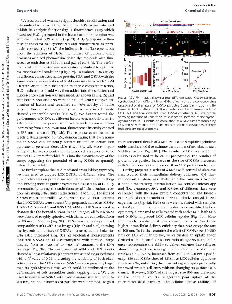
Fig. 3 (a) AFM images showing four different sized X-SNA samplessynthesized from different linker/SNA ratio. Inserts are correspondingcross-sectional analysis of X-SNA particles. Scale bar ¼ 500 nm. (b)Dynamic light scattering (DLS) and zeta potential measurements ofLOX SNA and four different sized X-SNA constructs. (c) Size profileshowing increase of linker/SNA ratio leads to increase of the hydro-dynamic size. (d) Quantitative correlation of X-SNA sizes measured byDLS and AFM images. Error bars indicate standard deviations of threeindependent measurements.
Edge Article Chemical Science
Ope
n A
cces
s A
rtic
le. P
ublis
hed
on 0
8 D
ecem
ber
2020
. Dow
nloa
ded
on 1
2/24
/202
1 4:
43:0
4 PM
. T
his
artic
le is
lice
nsed
und
er a
Cre
ativ
e C
omm
ons
Attr
ibut
ion-
Non
Com
mer
cial
3.0
Unp
orte
d L
icen
ce.
View Article Online
We next studied whether oligonucleotides modication andintermolecular crosslinking block the LOX active site andinhibit its catalytic functionality. A uorescence assay whichmeasured H2O2 generated in the lactate oxidation reaction wasemployed to test LOX activity (Fig. 2f). A H2O2-responsive uo-rescent indicator was synthesized and characterized as previ-ously reported (Fig. S4†).43 The indicator is not uorescent, butupon the addition of H2O2, the release of boronate esterproduces oxidized phenoxazine-based dye molecule with uo-rescence emission at 585 nm and pKa of ca. 8.75. The perfor-mance of the indicator was systematically studied to optimizethe experimental conditions (Fig. S5†). To evaluate LOX activityin different constructs, native protein, SNA, and X-SNA with thesame protein concentration of 5 nM were incubated with 1 mML-lactate. Aer 30 min incubation to enable complete reaction,H2O2 indicator of 1 mM was then added into the solution anduorescence emission was measured. As shown in Fig. 2g andS6,† both X-SNA and SNA were able to efficiently catalyze oxi-dization of lactate and remained ca. 70% activity of nativeenzyme. Further studies of enzymatic activity in cell lysateshowed comparable results (Fig. S7†). We further tested theperformance of X-SNA at different lactate concentrations in 1�PBS buffer. In the presence of lactate with a concentrationincreasing from 0 mM to 40 mM, uorescence intensity centredat 585 nm increased (Fig. 2h). The response curve started toreach plateau around 40 mM, demonstrating that even nano-molar X-SNA can efficiently convert millimolar lactate intopyruvate to generate detectable H2O2 (Fig. 2i). More impor-tantly, the lactate concentration in tumor cells is reported to bearound 10–20 mM,44,45 which falls into the dynamic range of theassay, suggesting the potential of using X-SNA to quantifyintracellular lactate.
To further explore the DNA-mediated crosslinking approach,we then tried to prepare LOX X-SNAs of different sizes. Theoligonucleotides on protein surface offer a preeminent orthog-onal binding motif to guide programmable assembly of LOX. Bysystematically tuning the stoichiometry of hybridization reac-tion via varying SNA : linker ratio from 1 : 1 to 1 : 50, the sizes ofX-SNAs can be controlled. As shown in Fig. 3a, four differentsized LOX X-SNAs were successfully prepared, named as X-SNA-1, X-SNA-5, X-SNA-10, and X-SNA-50. AFM and DLS were used tocharacterize the formed X-SNAs. In AFM images, all four X-SNAswere observed roughly spherical with diameters controlled fromca. 80 nm to 600 nm (Fig. S8†). DLS measurements producedcomparable results with AFM images (Fig. 3b and S9†), showingthe hydrodynamic sizes of X-SNAs increased as the linker-to-SNA ratio increased (Fig. 3c). Zeta-potential measurementsindicated X-SNAs are all electronegative with surface chargeranging from ca. �20 mV to �60 mV, supporting the DNAcoverage (Fig. 3b). The correlation of AFM and DLS resultsshowed a linear relationship between two sets of measured sizeswith a R2 value of 0.98, indicating the reliability of both char-acterizations. The AFM-observed X-SNA size was generally largerthan its hydrodynamic size, which could be attributed to thedeformation of so assemblies under tapping mode. We alsotried to synthesize X-SNA with hydrodynamic size around 300–400 nm, but no uniform-sized particles were obtained. To gain
© 2021 The Author(s). Published by the Royal Society of Chemistry
more structural details of X-SNA, we used a simplied primitivecubic packing model to estimate the number of proteins in eachX-SNA structure (Fig. S10†). The number of LOX in a ca. 80 nmX-SNA is calculated to be ca. 10 per particle. The number ofproteins per particle increases as the size of X-SNA increases,with 500 nm one containing more than 1000 protein molecules.
Having prepared a series of X-SNAs with controlled sizes, wenext studied their intracellular delivery efficiency. Cy5 uo-rophore on a T-base was labeled into the constructs to offera handle for tracking internalization via confocal microscopyand ow cytometry. SNA, and X-SNAs of different sizes werecalibrated with the same protein concentration and uores-cence emission per protein to allow quantitative analysis in cellexperiments (Fig. 4a). HeLa cells were incubated with samplesof 5 nM protein for 4 h and their uptake was quantied by owcytometry. Compared to cells treated with native LOX, both SNAand X-SNAs improved LOX cellular uptake (Fig. 4b). Moreimportantly, X-SNA constructs showed up to ca. 5–6 timeshigher intracellular delivery efficiency than SNA except the oneof 500 nm. To further examine the effect of X-SNA size (80–500nm) on LOX cellular uptake, we calculated an uptake indexdened as the mean uorescence ratio using SNA as the refer-ence, representing the ability to deliver enzymes into cells. Asshown in Fig. 4c, there was a general trend of increased cellularuptake as X-SNA size increased from ca. 80 to 250 nm. Speci-cally, 250 nm X-SNA showed 6.5 times LOX cellular uptake asmuch as SNA, indicating the crosslinking strategy signicantlyimproved protein cell entry without changing its surface DNAdensity. However, X-SNA of the largest size 500 nm presenteduptake index of ca. 0.2, suggesting poor uptake of themicrometer-sized particles. The cellular uptake abilities of
Chem. Sci., 2021, 12, 1803–1809 | 1805

Fig. 4 (a) Schematic illustration of cell entry properties of constructsincluding SNA and four different sized X-SNAs. (b) Cell internalizationproperties of native LOX, SNA, and four different sized X-SNAsdetermined by flow cytometry. (c) Correlation curve showing thecellular uptake index of different sized constructs. (d) Flow cytometryanalysis of HeLa cells incubated with 80 nm X-SNA under differentinhibitor treatments. (e) Confocal images (left) and fluorescenceintensity profiles (right) showing colocalization of X-SNA probes withearly endosome and lysosome in HeLa cells. Error bars indicatestandard deviations of three independent measurements.
Chemical Science Edge Article
Ope
n A
cces
s A
rtic
le. P
ublis
hed
on 0
8 D
ecem
ber
2020
. Dow
nloa
ded
on 1
2/24
/202
1 4:
43:0
4 PM
. T
his
artic
le is
lice
nsed
und
er a
Cre
ativ
e C
omm
ons
Attr
ibut
ion-
Non
Com
mer
cial
3.0
Unp
orte
d L
icen
ce.
View Article Online
different sized X-SNAs were further tested in both upright andinverted cell culture congurations using confocal microscopy(Fig. S11a†). Cells in the upright culture generally showedslightly higher uorescence, possibly due to the gradual particlesedimentation. Importantly, the size-dependent uptake curvesobserved in both setups exhibited the same trend, which wereconsistent with ow cytometry results, conrming the effec-tiveness of crosslinking strategy (Fig. S11b–d†).
To understand the size-dependent cellular uptake of X-SNA,we investigated its internalization mechanism using 80 nmconstructs as the model system. The incubation of HeLa cellswith X-SNA at 4 �C or in the presence of NaN3 led to negligibleuorescence in cells, demonstrating X-SNA relied on energy-dependent endocytosis process (Fig. 4d and S10†). To examinethe endocytic pathway of X-SNA, we employed different types ofbiological inhibitors to inhibit established pathways by block-ing surface receptors on plasma membrane.46 Confocal imagesof cells treated with methyl-b-cyclodextrin (MbCD), an inhibitorof caveolin-mediated endocytosis, showed signicantly reduceduorescence, indicating the suppressed X-SNA uptake(Fig. S12†). Quantitative analysis was further performed byusing ow cytometry. As shown in Fig. 4d, treatment withMbCDresulted in a 60–70% decrease in the cellular uptake of X-SNA.Other inhibitors, such as chlorpromazine (CPZ, an inhibitorof clathrin-mediated endocytosis), amiloride(macropinocytosis-mediated endocytosis inhibitor), andnystatin (lipid-ra-mediated endocytosis inhibitor), showedminimized effects on X-SNA internalization. These results areconsistent with previous observations that nanoscale objectsare internalized by cells via a receptor-mediated endocytic
1806 | Chem. Sci., 2021, 12, 1803–1809
pathway.47,48 In this vein, for nanoscale X-SNAs with sizesmatching endocytic vesicles budded from plasma membrane,larger particles presented higher local protein concentration,resulting in signicantly enhanced intracellular delivery ofproteins.
Having elucidated the cell entry mechanism of X-SNA, wethen studied the subcellular location of X-SNAs aer endocy-tosis. Early endosome and lysosome were stained respectively toinvestigate the colocalization with X-SNAs in HeLa cell. Asshown in Fig. 3e, early endosome uorescence largely over-lapped with X-SNA uorescence, generating yellow colour. ThePearson's correlation coefficient was calculated to be ca. 0.79,suggesting a good colocalization and further conrming thereceptor-mediated endocytic pathway. Lysosome uorescenceshowed localized green dots while red X-SNA uorescencespread in a larger distribution area around nucleus. Confocal z-stack images also showed clear divided green and red uores-cence at different scan depths (Fig. S13†). The calculated Pear-son's correlation coefficient of lysosome and X-SNA decreasedfrom ca. 0.28 to ca. 0.21 with the increase of the incubation timefrom 2 to 4 hours (Fig. S14†). These observations may possiblyindicate that a part of delivered X-SNA particles released fromlysosome through a slow escape process. Importantly, theability to efficiently deliver exogenous oxidase such as LOX intocytoplasm potentially provides a general route to estimatecellular metabolite concentration by simply converting theminto measurable reactive oxygen species using correspondingoxidase.
To ensure that the delivered enzymes are active in cellularmilieu, we designed a ratiometric uorescence assay usingsynthesized H2O2 indicator. Specically, HeLa cells were incu-bated with Cy5-modied LOX X-SNA in OptiMEM for 4 h. Aerwashing cells with PBS buffer, fresh medium containing 20 mMH2O2 indicator was added, and the cells were incubated at 37 �Cfor 30 min. Upon oxidation of lactate catalyzed by LOX, thereleased H2O2 oxidized the indicator, generating uorescenceemission at 585 nm. Meanwhile, Cy5 on X-SNA served as aninternal standard, producing ratiometric signals for lactate thatcan be visualized and quantied by confocal microscopy andow cytometry (Fig. 5a). Confocal z-stack images showed that X-SNA-based uorescence assay produced green uorescence andwell-colocalized Cy5 uorescence throughout the entire cellspace, proving that the delivered enzymes remained functionaland responded to lactate ratiometrically (Fig. S15†). We nexttreated HeLa cells with lactate medium or inhibitor oxamate totest the response of X-SNA assay to different lactate concentra-tions intracellularly. As shown in Fig. 5b, 10 mM lactatemedium treatment increased green uorescence in HeLa cells,while inhibitor quenched most of the emission. IntracellularCy5 emission overlapped with green uorescence and remainedunchanged in treated cells, indicating the consistency of theinternal standard. The green-to-red (G/R) uorescence ratio ofHeLa cells increased from 0.58 to 0.71 with external lactateshowing more redder colour, while decreased to 0.22 withinhibitor showing more bluer colour (Fig. 5b and S16†). Flowcytometry histograms plotted in mean G/R uorescence ratiowere consistent with above-mentioned confocal images
© 2021 The Author(s). Published by the Royal Society of Chemistry
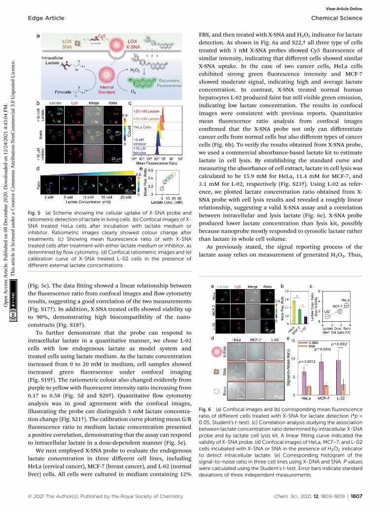
Fig. 5 (a) Scheme showing the cellular uptake of X-SNA probe andratiometric detection of lactate in living cells. (b) Confocal images of X-SNA treated HeLa cells after incubation with lactate medium orinhibitor. Ratiometric images clearly showed colour change aftertreatments. (c) Showing mean fluorescence ratio of with X-SNAtreated cells after treatment with either lactate medium or inhibitor, asdetermined by flow cytometry. (d) Confocal ratiometric images and (e)calibration curve of X-SNA treated L-02 cells in the presence ofdifferent external lactate concentrations.
Fig. 6 (a) Confocal images and (b) corresponding mean fluorescenceratio of different cells treated with X-SNA for lactate detection (*p <0.05, Student's t-test). (c) Correlation analysis studying the associationbetween lactate concentration ratio determined by intracellular X-SNAprobe and by lactate cell lysis kit. A linear fitting curve indicated thevalidity of X-SNA probe. (d) Confocal images of HeLa, MCF-7, and L-02cells incubated with X-SNA or SNA in the presence of H2O2 indicatorto detect intracellular lactate. (e) Corresponding histogram of thesignal-to-noise ratio in three cell lines using X-DNA and SNA. P valueswere calculated using the Student's t-test. Error bars indicate standarddeviations of three independent measurements.
Edge Article Chemical Science
Ope
n A
cces
s A
rtic
le. P
ublis
hed
on 0
8 D
ecem
ber
2020
. Dow
nloa
ded
on 1
2/24
/202
1 4:
43:0
4 PM
. T
his
artic
le is
lice
nsed
und
er a
Cre
ativ
e C
omm
ons
Attr
ibut
ion-
Non
Com
mer
cial
3.0
Unp
orte
d L
icen
ce.
View Article Online
(Fig. 5c). The data tting showed a linear relationship betweenthe uorescence ratio from confocal images and ow cytometryresults, suggesting a good correlation of the two measurements(Fig. S17†). In addition, X-SNA treated cells showed viability upto 90%, demonstrating high biocompatibility of the nano-constructs (Fig. S18†).
To further demonstrate that the probe can respond tointracellular lactate in a quantitative manner, we chose L-02cells with low endogenous lactate as model system andtreated cells using lactate medium. As the lactate concentrationincreased from 0 to 20 mM in medium, cell samples showedincreased green uorescence under confocal imaging(Fig. S19†). The ratiometric colour also changed evidently frompurple to yellow with uorescent intensity ratio increasing from0.17 to 0.58 (Fig. 5d and S20†). Quantitative ow cytometryanalysis was in good agreement with the confocal images,illustrating the probe can distinguish 5 mM lactate concentra-tion change (Fig. S21†). The calibration curve plotting mean G/Ruorescence ratio to medium lactate concentration presenteda positive correlation, demonstrating that the assay can respondto intracellular lactate in a dose-dependent manner (Fig. 5e).
We next employed X-SNA probe to evaluate the endogenouslactate concentration in three different cell lines, includingHeLa (cervical cancer), MCF-7 (breast cancer), and L-02 (normalliver) cells. All cells were cultured in medium containing 12%
© 2021 The Author(s). Published by the Royal Society of Chemistry
FBS, and then treated with X-SNA and H2O2 indicator for lactatedetection. As shown in Fig. 6a and S22,† all three type of cellstreated with 5 nM X-SNA probes showed Cy5 uorescence ofsimilar intensity, indicating that different cells showed similarX-SNA uptake. In the case of two cancer cells, HeLa cellsexhibited strong green uorescence intensity and MCF-7showed moderate signal, indicating high and average lactateconcentration. In contrast, X-SNA treated normal humanhepatocytes L-02 produced faint but still visible green emission,indicating low lactate concentration. The results in confocalimages were consistent with previous reports. Quantitativemean uorescence ratio analysis from confocal imagesconrmed that the X-SNA probe not only can differentiatecancer cells from normal cells but also different types of cancercells (Fig. 6b). To verify the results obtained from X-SNA probe,we used a commercial absorbance-based lactate kit to estimatelactate in cell lysis. By establishing the standard curve andmeasuring the absorbance of cell extract, lactate in cell lysis wascalculated to be 15.9 mM for HeLa, 11.4 mM for MCF-7, and3.1 mM for L-02, respectively (Fig. S23†). Using L-02 as refer-ence, we plotted lactate concentration ratio obtained from X-SNA probe with cell lysis results and revealed a roughly linearrelationship, suggesting a valid X-SNA assay and a correlationbetween intracellular and lysis lactate (Fig. 6c). X-SNA probeproduced lower lactate concentration than lysis kit, possiblybecause nanoprobe mostly responded to cytosolic lactate ratherthan lactate in whole cell volume.
As previously stated, the signal reporting process of thelactate assay relies on measurement of generated H2O2. Thus,
Chem. Sci., 2021, 12, 1803–1809 | 1807

Chemical Science Edge Article
Ope
n A
cces
s A
rtic
le. P
ublis
hed
on 0
8 D
ecem
ber
2020
. Dow
nloa
ded
on 1
2/24
/202
1 4:
43:0
4 PM
. T
his
artic
le is
lice
nsed
und
er a
Cre
ativ
e C
omm
ons
Attr
ibut
ion-
Non
Com
mer
cial
3.0
Unp
orte
d L
icen
ce.
View Article Online
endogenous H2O2 in cell especially in cancer cell may lead toincreased background noise and affect sensing performance.49
To overcome this issue, we proposed to use X-SNA probe basedon the hypothesis that the enhanced protein delivery ability ofX-SNA would in turn enable an improved detection performancein living cells. To test the hypothesis, we compared the perfor-mance of X-SNA with un-crosslinked SNA in previous three celllines to detect lactate. Cells incubated only with indicator, butno LOX were used as control groups. As shown in Fig. 6d andS24–S26,† in all three cell lines, cells incubated with X-SNAexhibited higher green uorescence than that treated by SNA,indicating improved lactate detection ability. Visible greencolour was observed in HeLa and MCF-7 control groups butnegligible in L-02 control, which was attributed to the back-ground H2O2 in cancer cells. To make a quantitative compar-ison, we calculated the signal-to-noise ratio of both constructsusing control cells as background reference. As shown in Fig. 6eand S27,† in three tested cell lines, X-SNA showed up to ca. 9.6signal-to-noise ratio, while un-crosslinked SNA generatedsignal-to-noise ratio of 2–3, suggesting the use of X-SNA cansignicantly improve the assay sensitivity, especially in biolog-ical environment with high background.
Conclusions
In conclusion, we have developed a straightforward strategy bycrosslinking individual DNA-modied enzymes into X-SNAconstruct that has achieved up to 6 times higher delivery effi-ciency than un-crosslinked ProSNA without the use of anytransfection agent. The presented results demonstrate X-SNAenters cells in a size-dependent manner and preserves mostcatalytic functions. The LOX-based X-SNA has been applied asintracellular lactate probe with signicantly improved perfor-mance, showing up to 3–4 times higher signal-to-noise ratiothan regular ProSNA. The strategy is facile and easy to createbiologically active protein materials that outperform previouslyreported un-crosslinked method. Because of the programma-bility and modularity enabled by DNA crosslinker, it is possibleto install a large number of functionalities into the structuresuch as targeting groups, imaging agents, and functionalnucleic acids, leading to potential biological applications suchas cellular imaging, gene regulation, and immunomodulation.Additionally, since lactate is measured through the quantica-tion of the redox product H2O2, the idea of delivering exogenousoxidases as intracellular signal transducer provides a generalstrategy to develop nanoprobes which can monitor a variety ofmetabolites that are difficult to detect in live cells.
Conflicts of interest
The authors declare no conict of interest.
Acknowledgements
The authors gratefully acknowledge National Natural ScienceFoundation of China (No. 21877032, No. 21527810), HunanProvince Talented Young Scientists Program (2019RS2021,
1808 | Chem. Sci., 2021, 12, 1803–1809
2019RS2023), National Key Research Program (No.2019YFA0905800), and the Fundamental Research Funds forthe Central Universities for nancial support.
Notes and references
1 R. M. Hoffman, Lancet Oncol., 2002, 3, 546–556.2 R. J. Desnick and E. H. Schuchman, Nat. Rev. Genet., 2002, 3,954–966.
3 B. Leader, Q. J. Baca and D. E. Golan, Nat. Rev. DrugDiscovery, 2008, 7, 21–39.
4 D. S. D'Astolfo, R. J. Pagliero, A. Pras, W. R. Karthaus,H. Clevers, V. Prasad, R. J. Lebbink, H. Rehmann andN. Geijsen, Cell, 2015, 161, 674–690.
5 W. J. Sun, W. Y. Ji, J. M. Hall, Q. Y. Hu, C. Wang, C. L. Beiseland Z. Gu, Angew. Chem., Int. Ed., 2015, 54, 12029–12033.
6 M. Q. Tolentino, A. K. Hartmann, D. T. Loe and J. L. Rouge, J.Mater. Chem. B, 2020, 8, 5627–5635.
7 D. Zhao, Y. H. Kong, S. S. Zhao and H. Xing, Top. Curr. Chem.,2020, 378, 83–124.
8 Z. M. Huang, M. Y. Lin, C. H. Zhang, Z. K. Wu, R. Q. Yu andJ. H. Jiang, Anal. Chem., 2019, 91, 9361–9365.
9 Z. Gu, A. Biswas, M. X. Zhao and Y. Tang, Chem. Soc. Rev.,2011, 40, 3638–3655.
10 M. S. Lawrence, K. J. Phillips and D. R. Liu, J. Am. Chem. Soc.,2007, 129, 10110–10112.
11 J. J. Cronican, D. B. Thompson, K. T. Beier,B. R. McNaughton, C. L. Cepko and D. R. Liu, ACS Chem.Biol., 2010, 5, 747–752.
12 P. Saalik, A. Elmquist, M. Hansen, K. Padari, K. Saar, K. Viht,V. Langel and M. Pooga, Bioconjugate Chem., 2004, 15, 1246–1253.
13 M. Wang, S. Sun, C. I. Neufeld, B. Perez-Ramirez andQ. B. Xu, Angew. Chem., Int. Ed., 2014, 53, 13444–13448.
14 J. A. Zuris, D. B. Thompson, Y. Shu, J. P. Guilinger,J. L. Bessen, J. H. Hu, M. L. Maeder, J. K. Joung, Z. Y. Chenand D. R. Liu, Nat. Biotechnol., 2015, 33, 73–80.
15 L. L. Sun, Y. J. Gao, Y. G. Wang, Q. Wei, J. Y. Shi, N. Chen,D. Li and C. H. Fan, Chem. Sci., 2018, 9, 5967–5975.
16 S. J. Kaczmarczyk, K. Sitaraman, H. A. Young, S. H. Hughesand D. K. Chatterjee, Proc. Natl. Acad. Sci. U. S. A., 2011,108, 16998–17003.
17 A. Erazo-Oliveras, K. Najjar, L. La Dayani, T. Y. Wang,G. A. Johnson and J. P. Pellois, Nat. Methods, 2014, 11,861–867.
18 R. A. Petros and J. M. DeSimone, Nat. Rev. Drug Discovery,2010, 9, 615–627.
19 G. S. Song, Y. Y. Chen, C. Liang, X. Yi, J. J. Liu, X. Q. Sun,S. D. Shen, K. Yang and Z. Liu, Adv. Mater., 2016, 28, 7143–7148.
20 L. Cheng, F. R. Zhang, S. H. Wang, X. T. Pan, S. C. Han,S. Liu, J. J. Ma, H. Y. Wang, H. Y. Shen, H. Y. Liu andQ. P. Yuan, Angew. Chem., Int. Ed., 2019, 58, 7728–7732.
21 X. Dong, H. J. Liu, H. Y. Feng, S. C. Yang, X. L. Liu, X. Lai,Q. Lu, J. F. Lovell, H. Z. Chen and C. Fang, Nano Lett.,2019, 19, 997–1008.
© 2021 The Author(s). Published by the Royal Society of Chemistry
Edge Article Chemical Science
Ope
n A
cces
s A
rtic
le. P
ublis
hed
on 0
8 D
ecem
ber
2020
. Dow
nloa
ded
on 1
2/24
/202
1 4:
43:0
4 PM
. T
his
artic
le is
lice
nsed
und
er a
Cre
ativ
e C
omm
ons
Attr
ibut
ion-
Non
Com
mer
cial
3.0
Unp
orte
d L
icen
ce.
View Article Online
22 A. L. Fu, R. Tang, J. Hardie, M. E. Farkas and V. M. Rotello,Bioconjugate Chem., 2014, 25, 1602–1608.
23 J. D. Brodin, A. J. Sprangers, J. R. McMillan and C. A. Mirkin,J. Am. Chem. Soc., 2015, 137, 14838–14841.
24 J. D. Brodin, E. Auyeung and C. A. Mirkin, Proc. Natl. Acad.Sci. U. S. A., 2015, 112, 4564–4569.
25 D. Samanta, S. B. Ebrahimi and C. A. Mirkin, Adv. Mater.,2020, 32, 1901743.
26 S. B. Ebrahimi, D. Samanta and C. A. Mirkin, J. Am. Chem.Soc., 2020, 142, 11343–11356.
27 D. Samanta, S. B. Ebrahimi, C. D. Kusmierz, H. F. Cheng andC. A. Mirkin, J. Am. Chem. Soc., 2020, 142, 13350–13355.
28 P. C. Patel, D. A. Giljohann, W. L. Daniel, D. Zheng,A. E. Prigodich and C. A. Mirkin, Bioconjugate Chem., 2010,21, 2250–2256.
29 C. H. J. Choi, L. L. Hao, S. P. Narayan, E. Auyeung andC. A. Mirkin, Proc. Natl. Acad. Sci. U. S. A., 2013, 110, 7625–7630.
30 J. R. McMillan, O. G. Hayes, P. H. Winegar and C. A. Mirkin,Acc. Chem. Res., 2019, 52, 1939–1948.
31 T. T. Chen, J. T. Yi, Y. Y. Zhao and X. Chu, J. Am. Chem. Soc.,2018, 140, 9912–9920.
32 Y. J. Chen, P. Li, J. A. Modica, R. J. Drout and O. K. Farha, J.Am. Chem. Soc., 2018, 140, 5678–5681.
33 S. Z. Wang, Y. J. Chen, S. Y. Wang, P. Li, C. A. Mirkin andO. K. Farha, J. Am. Chem. Soc., 2019, 141, 2215–2219.
34 Y. Umena, K. Yorita, T. Matsuoka, A. Kita, K. Fukui andY. Morimoto, Biochem. Biophys. Res. Commun., 2006, 350,249–256.
35 M. G. V. Heiden, L. C. Cantley and C. B. Thompson, Science,2009, 324, 1029–1033.
36 O. N. Okorie and P. Dellinger, Crit. Care Clin., 2011, 27, 299–326.
© 2021 The Author(s). Published by the Royal Society of Chemistry
37 A. Mongersun, I. Smeenk, G. Pratx, P. Asuri and P. Abbyad,Anal. Chem., 2016, 88, 3257–3263.
38 M. Braendlein, A. M. Pappa, M. Ferro, A. Lopresti,C. Acquaviva, E. Mamessier, G. G. Malliaras andR. M. Owens, Adv. Mater., 2017, 29, 1605744.
39 A. San Martin, S. Ceballo, I. Ruminot, R. Lerchundi,W. B. Frommer and L. F. Barros, PLoS One, 2013, 8, e57712.
40 H. Xing, C. L. Zhang, G. Ruan, J. J. Zhang, K. Hwang andY. Liu, Anal. Chem., 2016, 88, 1506–1510.
41 J. B. Trads, T. Torring and K. V. Gothelf, Acc. Chem. Res.,2017, 50, 1367–1374; H. Li, B. H. Zhang, X. G. Lu,X. Y. Tan, F. Jia, Y. Xiao, Z. H. Cheng, Y. Li, D. O. Silva,H. S. Schrekker, K. Zhang and C. A. Mirkin, Proc. Natl.Acad. Sci. U. S. A., 2018, 115, 4340–4344.
42 H. Li, B. H. Zhang, X. G. Lu, X. Y. Tan, F. Jia, Y. Xiao,Z. H. Cheng, Y. Li, D. O. Silva, H. S. Schrekker, K. Zhangand C. A. Mirkin, Proc. Natl. Acad. Sci. U. S. A., 2018, 115,4340–4344.
43 L. Yi, L. Wei, R. Y. Wang, C. Y. Zhang, J. Zhang, T. W. Tanand Z. Xi, Chem.–Eur. J., 2015, 21, 15167–15172.
44 X. T. Zheng, H. B. Yang and C. M. Li, Anal. Chem., 2010, 82,5082–5087.
45 F. Hirschhaeuser, U. G. A. Sattler and W. Mueller-Klieser,Cancer Res., 2011, 71, 6921–6925.
46 H.M. Ding, J. Li, N. Chen, X. J. Hu, X. F. Yang, L. J. Guo, Q. Li,X. L. Zuo, L. H. Wang, Y. Q. Ma and C. H. Fan, ACS Cent. Sci.,2018, 4, 1344–1351.
47 S. D. Perrault, C. Walkey, T. Jennings, H. C. Fischer andW. C. W. Chan, Nano Lett., 2009, 9, 1909–1915.
48 X. Y. Tan, X. G. Lu, F. Jia, X. F. Liu, Y. H. Sun, J. K. Logan andK. Zhang, J. Am. Chem. Soc., 2016, 138, 10834–10837.
49 M. Giorgio, M. Trinei, E. Migliaccio and P. G. Pelicci, Nat.Rev. Mol. Cell Biol., 2007, 8, 722–728.
Chem. Sci., 2021, 12, 1803–1809 | 1809