cellular internalization of green fluorescence protein ... · cellular internalization of green...
TRANSCRIPT
Cellular Internalization of Green Fluorescence Protein Fused with Herpes
Simplex Virus Protein VP22 via a Lipid-raft-mediated Endocytic Pathway
Independent of Caveolae and Rho Family GTPases but Dependent on Dynamin
and Arf6*
Kenji Nishi1, and Kaoru Saigo
2
Department of Biophysics and Biochemistry, Graduate School of Science,
University of Tokyo, 7-3-1 Hongo, Bunkyo-ku, Tokyo 113-0033, Japan
Running title: VP22 Internalization via Lipid-raft-mediated Endocytosis 1 To whom correspondence should be addressed: Tel.: 81-3-5841-3044; Fax: 81-3-5841-3044; E-mail:
[email protected] 2 To whom correspondence should be addressed: Tel.: 81-3-5841-3044; Fax: 81-3-5841-3044; E-mail:
VP22 is a structural protein of the herpes
simplex virus and has been reported to possess
unusual trafficking properties. Here we examined
the mechanism of cellular uptake of VP22 using a
fusion protein between VP22 C-terminal half and
GFP (VP22-GFP). Adsorption of VP22-GFP onto a
cell surface required heparan sulfate proteoglycans
and basic amino acids, in particular, arginine at
position 164 of VP22. Inhibitor treatment, RNA
interference, expression of dominant-negative
mutant genes and confocal microscopy all indicated
VP22-GFP to enter cells through an endocytic
pathway independent of clathrin and caveolae but
dependent on dynamin and Arf6 activity. As with
CD59, a lipid rafts marker, VP22-GFP cell surface
signals were resistant to Triton X-100 treatment but
cell surface signals of VP22-GFP only partially
overlapped those of CD59. Furthermore, unlike
other lipid-raft-mediated endocytosis, no Rho family
GTPase was required for VP22-GFP internalization.
Internalized VP22 initially entered early endosomes
and then moved to lysosomes and possibly recycling
endosomes.
Short peptides and protein domains capable of
transducing cargo across the plasma membrane are
called protein transduction domains, PTDs3 (reviewed
in ref.1,2). A short basic peptide from TAT protein, a
transcription activator of the human immunodeficiency
virus 1 (3-5), and the third helix of the Drosophila
Antennapedia homeodomain (6) are widely used as
short-peptide-type PTDs. Recent experiments showed
that these short-peptide-type PTDs and proteins
associated with them are most likely to be incorporated
into cells via some endocytic mechanism (7,8).
Endocytosis is a complex mechanism involving
numerous protein-protein and protein-lipid interactions
(9) and may occur as clathrin-dependent endocytosis,
macropinocytosis, caveolae-mediated endocytosis and
lipid raft-dependent/caveolae-independent endocytosis
(9,10). TAT and TAT-PTD fusion proteins are capable
of interacting negatively charged plasma membrane
(11,12), but their endocytosis mechanism differs
considerably depending on cell type or protein moieties
associated with TAT-PTD (12-18). In some cases, they
are internalized via macropinocytosis (13,14), while, in
other cases, they appear to be incorporated into cells via
caveolae-dependent endocytosis (15,16) or
clathrin-dependent endocytosis (12,18). Endosomal
escape of TAT-PTD-fusion proteins may require
endosome acidification and cytosolic Hsp90 activity
(18-20).
VP22 is a major component of the
herpes-simplex-virus type-1 tegment and is situated
between the capsid and envelope (21). This component
has been shown to be secreted from cells expressing it
through a Golgi-independent mechanism (22). The
C-terminal half of VP22, residues 159-301 (termed
VP22.C1) is highly homologous in sequence to
counterparts of other -herpes virus VP22 and is
considered associated with intercellular trafficking
ability (23,24). Although others reported 34 amino acids
(AAs)-long C-terminal region of VP22.C1 is necessary
and sufficient for cellular internalization (25), our results
indicated rather the entire 142 AAs-long region to be
involved in cellular uptake (K.N. & K.S., unpublished
data).
VP22.C1 has been noted to form complexes with
fluorescein-labeled oligonucleotides so as to generate
particles of 0.3-1 μm in diameter (24). These particles
are efficiently taken up into cultured cells or mice and
release active antisense oligonucleotide reagents into the
cytoplasm in response to light activation (24,26,27). The
uptake of VP22.C1-fused green fluorescent protein
http://www.jbc.org/cgi/doi/10.1074/jbc.M703810200The latest version is at JBC Papers in Press. Published on July 20, 2007 as Manuscript M703810200
Copyright 2007 by The American Society for Biochemistry and Molecular Biology, Inc.
by guest on February 16, 2020http://w
ww
.jbc.org/D
ownloaded from
(GFP), hereafter referred to as VP22-GFP, into an entire
tumor has been observed following peritumoral
injection into human pancreatic tumors in SCID mice
(28). VP22.C1-Rab9 has been reported to be
internalized into cells and to exhibit biological activity
(29). Thus possibly, as with short-peptide-type
TAT-PTD, 142-AAs-long VP22.C1 may serve as a
vector for delivering cargo into cells. As in the case of
TAT-PTD and its fusion proteins, VP22.C1-fusion
proteins may also enter a cell via endocytosis since they
exhibited cytoplasmic vesicular distribution (8). To date,
the cellular internalization mechanism of VP22-fusion
proteins is but little understood.
Here we study mechanism of VP22 cellular
internalization using VP22-GFP as a model system.
VP22-GFP was internalized through interactions with
cell surface heparan sulfate proteoglycans and
subsequent lipid-raft-mediated endocytosis independent
of clathrin, caveolae and Rho family GTPases but
dependent on dynamin and Arf6. Also, as with
transferrin internalized via clathrin-dependent
endocytosis, nearly all internalized VP22-GFP signals
were incorporated into the early endosomes and
co-localized with transferrin.
EXPERIMENTAL PROCEDURES
Drugs and antibodies - Transferrin-Alexa Fluor
647, rhodamine phalloidin and Hoechst 33342 were
purchased from Molecular Probes. Heparin (porcine
intestinal mucosa), chlorpromazine hydrochloride,
methyl- -cyclodextrin (M CD), genistein and
Clostridium difficile toxin B were obtained from Sigma.
Cytochalasin D, okadaic acid (OA), PP2 and sodium
orthovanadate were from Calbiochem while
Chloroquine diphosphate salt was from Wako.
Chondroitin sulfate A (whale cartilage), chondroitin
sulfate B (pig skin), chondroitin sulfate C (shark
cartilage), keratan sulfate (bovine cornea), heparitinase
and chondroitinase ABC were from Seikagaku
Corporation. Antibodies used were: anti-heparan sulfate
monoclonal antibody (clone NAH46; Seikagaku
Corporation), anti-HA tag rabbit polyclonal IgG
(Upstate), anti-c-myc monoclonal mouse IgG
(Oncogene), myc-tag polyclonal antibody (Cell
Signaling Technology), mouse anti-human clathrin
heavy chain monoclonal antibody (Affinity
BioReagents), anti-CD71 antibody (transferrin receptor;
Santa Cruz Biotechnology), anti-EEA1 rabbit
polyclonal IgG (Upstate), mouse anti-EEA1
monoclonal antibody (BD Pharmingen), anti-human
CD59 monoclonal antibody (Cedalane Laboratories),
rabbit anti-caveolin1 polyclonal antibody (BD
Transduction Laboratories), mouse anti-GM130
monoclonal antibody (BD Transduction Laboratories),
anti-human CD107a (LAMP1) antibody (BD
Pharmingen), goat anti-mouse IgG conjugated with Cy3
or Cy5 (Amersham Pharmacia Biotech), goat
anti-mouse IgM rhodamine (Upstate) and
FITC-conjugated goat anti-mouse, Cy5-conjugated goat
anti-rabbit and Cy3-conjugated goat anti-rabbit IgG
(Jackson Immunoresearch Laboratories).
Plasmids and VP22 mutagenesis - pVP22-GFP,
an expression plasmid for VP22-GFP production in
Escherichia coli cells, was constructed as follows: A
0.6-kb long EcoR /Not fragment of pEGFP-N3
(Clontech) was inserted into the EcoR /Not site of the
pVP22-myc-his plasmid (Invitrogen). A fragment
encoding the C-terminal half of VP22 (amino acid
residues 159-301) and full-length EGFP was amplified
from the plasmid through polymerase chain reaction,
digested with NheI and Not and inserted into the
NheI/Not site of pET28a (Novagen), which is capable
of producing recombinant protein with an N-terminal
polyhistidine tag. The following primers were used:
5’-AAATTTGCTAGCACGGCGCCAACCCGATC
CAA-3’ and
5’-CGGGTTAGATCTCAATGGTGATGGTGATG
ATGAC-3’.
PCR-based systematic mutagenesis was carried
out and a series of pVP22-GFP derivatives encoding
mutant types of VP22-GFP were generated. Sequences
of primers used for mutagenesis are available upon
request.
pGFP, a GFP expression plasmid, was
constructed by inserting the 0.6-kb-long EcoR /Not
fragment of pEGFP-N2 (Clontech) into the EcoR /Not
site of pET28a.
PcDNA3-HA-Dynamin2a WT and K44A
constructs (30) were obtained from Kazuhisa
Nakayama (Kyoto University). PcDNA3-myc-Rab5
WT and Q79L constructs (31) were kindly provided by
Harald Hirling (Faculté des Science de la Vie).
pRK5-myc-Rac1-T17N was obtained from Gary
Bokoch (The Scripps Research Institute).
pXS-HA-Arf6-Q67L plasmid (32) was kindly provided
by Julie G. Donaldson (National Institutes of Health).
Protein Purification - VP22-GFP and GFP were
produced using BL21 Star (DE3) E. coli cells
(Invitrogen) as host. Cells were first grown in L Broth
with kanamycin at 37°C overnight. The overnight
culture was diluted with the same medium 10 times and
shaking was continued at room temperature. Isopropyl
by guest on February 16, 2020http://w
ww
.jbc.org/D
ownloaded from

-D-thiogalactoside was added to final concentration of
1 mM at the time point of 0.8-1.0 A600. After an
additional 12 h shaking at room temperature, the cell
cultures were cooled on ice and the cells were collected
by centrifugation at 4°C. The pellets were frozen and
stored at –20°C until use. Cells were thawed on ice,
resuspended in lysis buffer (50 mM sodium phosphate
[pH8.0] containing 10 mM imidazole and 300 mM
NaCl), and disrupted by sonication. The lysate was
centrifuged at 12,000g for 1 h at 4°C. The supernatant
was loaded onto a Ni-NTA column (Qiagen)
equilibrated with lysis buffer. The column was washed
with lysis buffer and then wash buffer (50 mM sodium
phosphate [pH8.0] with 20 mM imidazole and 300 mM
NaCl) and finally eluted with the elution buffer (50 mM
sodium phosphate [pH8.0]) containing 250 mM
imidazole and 300 mM NaCl. Purified protein was
immediately used or frozen in 10% glycerol and stored
at –80°C until use.
Cell Culture and Transfection - Wild type
CHO-K1 and HeLa cells were cultured in Dulbecco’s
Modified Eagle Medium (DMEM) supplemented with
10% fetal calf serum (FCS) at 37°C in 5% CO2.
CHO-K1 glycosaminoglycan-deficient (pgs A-745) and
heparan sulfate-deficient (pgs D-677) mutant cells
which were obtained from the American Type Culture
Collection were cultured in F12-K medium
supplemented with 10% FCS at 37°C in 5% CO2.
Transfection was carried out using Lipofectamine 2000
(Invitrogen) according to instructions of the
manufacturer. Usually, cells were analyzed within the
period, 16-48 h following transfection.
Visualization of Internalized Proteins by Confocal
Microscopy - Cells were cultured in 12-well culture
plates (SUMITOMO BAKELITE) on a glass coverslip.
When cells were treated with drug or protein
(VP22-GFP, GFP or transferrin), the culture medium
was replaced with fresh serum-free medium at the
protein and/or drug concentration indicated. The cells
were incubated for 1 h and then washed twice with
ice-cold phosphate-buffered saline (PBS). After being
fixed with 2-4% paraformaldehyde (PA) in PBS for 15
min at room temperature unless otherwise indicated, the
cells were washed twice with ice-cold PBS and
mounted in Vectashield mounting medium (Vector
Laboratories). This was followed by cell visualization
with Olympus Fluoview FV1000 confocal microscopy
system. Using a 60 objective lens, successive 0.25 μm
optical sections were taken. In some cases, after cells
had been incubated on ice, the medium was replaced
with fresh medium containing protein(s) at the indicated
concentration. The cells were then placed on ice for 1 h,
washed twice with serum-free medium, placed in
pre-warmed serum-free medium, incubated for a
specified period of time and then visualized as above.
Transferrin internalization was examined by exposing
cells to the culture medium containing 0.25 μM
Transferrin-Alexa Fluor 647 and observing stained cells
under a confocal microscope.
Quantification of internalized VP22-GFP - Cells
were seeded on each well of 12-well culture plates in
culture medium containing 10% FCS. Culturing was
continued until pre-confluency at 37°C. The medium
was replaced by fresh serum-free medium containing 1
μM VP22-GFP. Cells were incubated for indicated time
or 1 h, washed once with ice-cold PBS, and trypsinized
for 15 min at 37°C to eliminate non-internalized cell
surface protein. This was followed by centrifugation in
1.5ml tubes, the cells were washed three times in
ice-cold PBS, lysed with PBS containing 0.5% Triton
X-100 and disrupted by sonication. The cell lysate was
centrifuged. Protein content of the supernatant was
measured using a Bio-Rad protein assay kit with bovine
serum albumin (BSA) as the standard. Fluorescence
intensity was measured with Hitachi spectrofluorometer
FL-2500 at excitation wavelength of 488 nm and
emission wavelength of 511 nm. Replacement of 0.5%
Triton X-100 with 0.1% SDS gave no appreciable
difference in measurement, indicating that almost all
internalized VP22-GFP is solubilized by 0.5% Triton
X-100 treatment.
Low Temperature Treatment and ATP Depletion -
For low temperature treatment, after being incubated at
4°C for 1 h, the cells were further incubated in the
presence of VP22-GFP for 1 h at 4°C and subjected to
trypsin digestion or fixation. For removal of cellular
ATP, the cells were incubated with ATP depletion
medium (glucose-free DMEM with 10 mM sodium
azide and 6 mM 2-deoxy-D-glucose) containing 10%
FCS for 1.5 h, followed by incubation in the presence of
an indicated concentration of VP22-GFP for 1 h in
serum-free ATP depletion medium. Cellular protein
uptake was visualized or measured as indicated above.
Quantification of cell surface binding VP22-GFP
and anti-heparan sulfate signals - Cells were cultured in
12-well culture plates on a glass coverslip. Cells were
washed twice with serum-free DMEM and incubated
on ice for 1 h. After addition of the indicated
concentration of VP22-GFP or 2 μg/ml
anti-heparan-sulfate antibody in serum-free DMEM,
cells were further incubated on ice for 1 h, washed twice
with ice-cold PBS, fixed with 4% PA at room
by guest on February 16, 2020http://w
ww
.jbc.org/D
ownloaded from
temperature for 15 min and visualized by confocal
microscopy. For anti-heparan sulfate staining, the
second antibody treatment was also carried out before
PA fixation. A projection image was constructed using
successive optical sections 0.25 μm each. A region of
interest was drawn around each cell and average
intensity was calculated using a software equipped with
the confocal microscope.
Immunofluorescence - Cells were cultured in
12-well culture plates on a glass coverslip, incubated
with protein at specified concentration in serum-free
DMEM for the indicated time, washed twice with ice
cold PBS and fixed with 4% PA at room temperature
for 15 min. After being washed twice with ice cold PBS,
cells were permeabilized with PBS containing 0.2%
Triton X-100 and washed twice with ice cold PBS.
Non-specific binding sites were blocked with PBS
containing 5-10% goat whole serum (IBL) at room
temperature for 30 min. Following incubation with the
1st antibody (1:100-400 dilution) in PBS containing
1-5% goat whole serum at room temperature for 1 h or
at 4 overnight, the cells were washed twice with PBS
and treated with the 2nd antibody (1:100-400 dilution)
in PBS containing 1-5% goat whole serum at room
temperature for 1h or at 4 overnight. This was
followed by two washings in PBS and visualization by
confocal microscopy as above.
RNA interference - Two sets of human clathrin
heavy chain siRNA (chc-siRNAs) were selected using
siDirect (33); (GCUUCAGUACCCUGACUAUGG
(chc1 passenger strand), and
AUAGUCAGGGUACUGAAGCCA (chc1 guide
strand), and CCUGGUACGUCGAAAGGAUCC
(chc2 passenger strand) and
AUCCUUUCGACGUACCAGGUA (chc2 guide
strand). Firefly luciferase siRNA (fl-siRNA) used as
control is CGUACGCGGAAUACUUCGAAA
(passenger strand) and
UCGAAGUAUUCCGCGUACGUG (guide strand).
RNA oligonucleotides were synthesized by Proligo and
double-stranded siRNA was prepared as described
previously (34). HeLa cells in 12 well plates were
transfected 3 times at 24-36 h intervals with 50 nM
siRNA. fl-siRNA treated and chc-siRNA treated cells
were trypsinized, mixed of a ratio in 1:1 and seeded in
12 well plates 20 h prior to clathrin heavy chain
depletion-effect observation in the same field. DNA
transfection was conducted with third siRNA
transfection.
DNA-mediated RNAi was also carried out for
knocking-down genes encoding RhoA, Cdc42 and Arf6.
A firefly luciferase short hairpin RNA (shRNA)
expression construct was used as control. Details of
vector construction will be described elsewhere. HeLa
cells in 12 well plates were transfected with a suitable
construct and tranfectants were selected with puromycin
(2 μg/ml) 24 h after transfection. Cells were analyzed 96
h after tranfection.
Drug Treatment - Cells were washed twice with
serum-free DMEM and incubated in serum-free
DMEM containing at indicated concentration for 30
min except for Clostridium difficile toxin B. Following
protein addition to the medium, cells were incubated for
1 h or 4 h. Protein uptake and cell surface binding were
visualized and measured as described above.
Triton X-100 Treatment - After being put on ice
with 0.25 μM VP22-GFP, 2 μg/ml anti-CD59 antibody
or 1 μg/ml anti-transferrin receptor antibody on ice for 1
h, cells were washed twice with serum-free DMEM,
stained with 2nd antibody for 1 h in serum free DMEM,
washed twice with ice-cold PBS, treated with 1% Triton
X-100 in PBS for 7 min and washed twice again with
ice-cold PBS on ice. They were then fixed with 4% PA
and visualized.
Cell Treatment with GAG Lyases - Enzymatic
treatment with GAG lyases was carried out as
previously reported (35). Briefly, pre-confluent cells
were washed once with PBS, followed by incubation
with GAG lyases in PBS containing 0.1% BSA, 0.2%
gelatin and 0.1% glucose for 2 h at 37 °C. Four
washings with PBS or serum free DMEM were carried
out. Following an indicated concentration of VP22-GFP
addition to the medium, cells were incubated for 1 h.
VP22-GFP uptake and binding were visualized and
measured as described.
Real-Time PCR - The total RNA from transfected
cells was isolated using an RNeasy Mini kit (Qiagen).
The RNA was treated with RQ1 DNase (Promega),
purified by an RNeasy Mini kit, and reverse-transcribed
using SuperScript III (Invitrogen). Before the PCR, a
mixture of the synthesized cDNA and SYBR green
PCR Master Mix (Applied Biosystems) was incubated
at 95°C for 10 min. The PCR reaction was then carried
out at 95°C for 15 sec and 60°C for 1 min for 40 cycles.
All reactions were carried out in triplicate. The levels of
PCR products were monitored with an ABI PRISM
7000 sequence detection system and analyzed with ABI
PRISM 7000SDS software (Applied Biosystems). For
each sample, the amount of target mRNA was
normalized using endogenous -actin mRNA.
Following primers were used. Arf6 forward:
5’-ATCAATGACCGGGAGATGAG-3’; Arf6
by guest on February 16, 2020http://w
ww
.jbc.org/D
ownloaded from
reverse: 5’-AGGGCTGCACATACCAGTTC-3’;
cdc42 forward: 5’-CGATGGTGCTGTTGGTAAAA
-3’; cdc42 reverse:
5’-TCCTCTTGCCCTGCAGTATC-3’; RhoA
forward: 5’-CGGTCTGGTCTTCAGCTACC-3’;
RhoA reverse:
5’-CCATCACCAACAATCACCAG-3’; -actin
forward: 5’-CACACTGTGCCCATCTACGA-3’;
-actin reverse:
5’-GCCATCTCTTGCTCGAAGTC-3’.
RESULTS
Cellular uptake of VP22-GFP occurs through
endocytosis - To clarify the mechanism for the cellular
uptake of VP22, VP22-GFP, a fusion protein between
GFP and VP22.C1 (Fig.1A), was prepared from E.coli
cells, and added to the culture medium with HeLa cells.
GFP signal distribution was examined at 1 h incubation
at 37°C using a confocal microscope (Fig.1B).
Significant VP22-GFP signals were observed not only
on the surface (see t.s. (top section) in Fig.1B) but also
inside of the cells (see i.s. (intermediate section)). Fifteen
minutes trypsin treatment at 37°C eliminated almost
completely cell surface signals without affecting
intracellular signal intensity (compare the middle and
lowest panels in Fig.1B). VP22-GFP cellular uptake
appeared to depend on the VP22 moiety, since no
intracellular GFP signal was detected with elimination
of the entire VP22.C1 moiety from the fusion protein
(data not shown). Change in intracellular GFP signal
intensity was examined spectrofluorometrically using
trypsin-treated HeLa cells (Fig.1D). Intracellular
VP22-GFP signal increased gradually for more than 6 h.
Fig.1B shows cytoplasmic distribution of
VP22-GFP in HeLa cells to be vesicular, supporting the
notion that VP22-GFP is internalized through
endocytosis (8). Endocytosis-dependent cellular
internalization would not occur either at 4°C or in the
absence of ATP (7). As shown in Fig.1C,E, no or hardly
any intracellular accumulation of VP22-GFP could be
detected in HeLa cells exposed to VP22-GFP at 4°C. In
contrast to cellular uptake, VP22-GFP adsorption onto
the cell surface appeared quite normally to occur even at
4°C. The cellular ATP pool becomes empty with the
pre-incubation of cells with sodium azide and
deoxyglucose (36). Only slight cellular uptake of
VP22-GFP without significant reduction in surface
signals was apparent in both ATP-depleted HeLa and
CHO-K1 cells (Fig.1E). VP22-GFP may thus be
concluded to be internalized through endocytosis in
HeLa and CHO-K1 cells.
Requirement of heparan-sulfate for cell surface
binding and cellular uptake of VP22-GFP - TAT-PTD
cellular internalization requires proteoglycans (11,12). In
the case of mammalian cell surface glycosaminoglycans
(GAGs), heparan sulfate and chondroitin sulfates A, B
and C are main components. Study was thus made to
determine whether GAGs are required for cell surface
adsorption and cellular uptake of VP22-GFP. After
treating HeLa cells with heparitinase or chondroitinase
ABC, assessment was made of cell surface adsorption
and cellular uptake of VP22-GFP signals. Heparitinase
digests heparan sulfate, while chondroitinase ABC,
chondroitin sulfates A, B and C but not heparan sulfate.
Heparitinase treatment eliminated not only cell surface
VP22-GFP signals but cellularly internalized
VP22-GFP signals as well (Fig.2A). In contrast,
virtually no reduction in VP22-GFP signals on either the
surface of or inside of cells could be detected by
chondroitinase ABC treatment (Fig.2A). GAGs were
added to the culture medium and only heparin
(analogue of heparan sulfate) addition was found to
induce a significant reduction in intracellularly
accumulated VP22-GFP signals (Fig.2B). Thus, in
HeLa cells, heparan sulfate but not chondroitin sulfates
is required for the surface adsorption and intracellular
accumulation of VP22-GFP.
Pgs A-745 and pgs D-677 cells are GAG mutant
strains of CHO-K1 cells (37,38). Pgs A-745 is deficient
in xylosyltransferase, the key enzyme required for GAG
attachment to core protein. Only 1% of GAGs
expressed in wild type cells has been reported to be
expressed in pgs A-745 cells (37). In this study, very
few if any heparan sulfate signals could be detected by
anti-heparan sulfate antibody staining in both pgs A-745
and pgs D-677 cells (Supplemental Fig.1). Pgs D-667 is
deficient in N-acetylglucosaminyltransferase and
glucronyltransferase, both essential for heparan sulfate
polymerization. Pgs D-667 cells have been shown not to
produce heparan sulfate but rather 3-4 times as many
chondroitin-sulfate molecules as wild type cells (38).
Fig.2C shows cell surface and internalized VP22-GFP
signals to undergo significant reduction in pgs A-745
and pgs D-667 cells so that, as with HeLa cells, heparan
sulfate but not chondroitin sulfates is required for
VP22-GFP binding to the cell surface and cellular
internalization in CHO-K1 cells.
To further confirm the close relation between
VP22-GFP cell surface adsorption and heparan sulfate,
examination was made to see whether the distribution of
VP22-GFP signals on the cell surface is in some way
related to that of heparan sulfate signals (Fig.2D).
by guest on February 16, 2020http://w
ww
.jbc.org/D
ownloaded from
Anti-heparan sulfate antibody staining revealed heparan
sulfate on the surface of HeLa cells to be punctate.
Nearly all punctate signals of heparan sulfate were
found to co-localize with VP22-GFP signals that had
distributed in a punctate fashion and vice versa (Fig. 2D).
However, it should be noted that heparan sulfate signal
intensity is not always proportional to VP22-GFP signal
intensity, possibly suggesting involvement of other
factors in VP22-GFP cell surface adsorption.
Involvement of basic amino acids in
VP22.C1 N-terminal region in VP22-GFP cell surface
adsorption and intracellular uptake - As with
TAT-PTD, VP22.C1 is rich in basic amino acids
(Fig.2E; 23). Mutational analysis indicated that 8 basic
amino acids in TAT-PTD are equally requisite for
cellular uptake activity (39). To determine which basic
amino acids of VP22.C1 are required for interactions
between heparan sulfate and VP22-GFP, basic amino
acids of VP22.C1 were systematically replaced with
alanine (neutral amino acid) or glutamic acid (acidic
amino acid) and spectrofluorometrical examination was
made of whether VP22-GFP mutant proteins were
effectively incorporated into cells (Fig.2F). R164A and
R164E mutant proteins were found to lose 90%
intracellular incorporation activity of wild type,
indicating the importance of arginine at position 164 for
VP22-GFP internalization. At positions 174, 192 and
199, only glutamic acid substitutes exhibited significant
reduction in internalization activity, suggesting that the
absence of negative charges from these positions is
required for effective intracellular uptake of VP22-GFP.
Fig.2F shows that most, if not all, other basic amino
acids of VP22.C1, which are associated with moderate
mutational effects, may also be involved in VP22-GFP
internalization to some extent.
Reduction in VP22-GFP internalization activity
appeared virtually proportional to that in cell surface
binding activity. Indeed, cell surface VP22-GFP signals
were almost completely abolished or significantly
reduced in R164E, R164A and R192E, three strong
internalization-defective mutants, while cell surface
VP22-GFP signals in R192A and R207A, associated
with moderate internalization defects, were moderately
reduced (compare Fig.2F and G).
Requirement of dynamin for cellular
internalization of VP22-GFP - Dynamin is a GTPase
essential for various endocytosis pathways (10). Cellular
expression of the GTPase-deficient, dominant-negative
mutant of dynamin (Dynamin K44A) has no effect on
macropinocytosis or certain lipid-raft-mediated
endocytic pathways including that induced by
Arf6-Q67L (32) but almost entirely blocks
clathrin-mediated endocytosis and other
lipid-raft-mediated endocytic pathways such as
caveolae-mediated endocytosis (10,40-43). Using
transferrin as a dynamin-dependent endocytosis marker,
VP22-GFP internalization was examined in HeLa cells
transfected with wild type dynamin2 (dynamin2 WT) or
dynamin2 K44A. Dynamin-overexpressing cells were
identified by anti-HA antibody. Overexpression of
Dynamin2 WT undergoes virtually no change in
VP22-GFP and transferrin internalization (data not
shown). In contrast the internalization of VP22-GFP
and transferrin was noted to be significantly reduced in
nearly 60 and 80% of cells overexpressing Dynamin2
K44A, respectively (Fig.3A,C). No apparent
Dynamin2-K44A-dependent change in cell surface
heparan sulfate signals was observed (Fig.3B).
Dynamin2 is thus essential for the normal cellular
internalization of VP22-GFP and too, VP22-GFP may
not be internalized through macropinocytosis and
lipid-raft-mediated endocytosis induced by Arf6-Q67L
but by clathrin-mediated endocytosis or
dynamin-dependent lipid-raft-mediated endocytosis,
such as that mediated by caveolae. Note that not only
heparan sulfate (Fig.3B) but also VP22-GFP cell surface
signals were not affected by the absence of Dynamin
activity (Fig.3D), indicating that Dynamin is involved
not in VP22-GFP adsorption but in cellular uptake.
VP22-GFP endocytosis is independent of clathrin
- By far, clathrin-mediated endocytosis has been best
characterized and has been shown to be inhibited
specifically by chlorpromazine (44). Cell treatment with
chlorpromazine induces clathrin-coated-pit
misassembly at the plasma membrane so that
clathrin-mediated endocytosis such as transferrin uptake
is prevented to a significant degree in
chlorpromazine-treated cells. To confirm any possible
involvement of clathrin-mediated endocytosis in the
cellular internalization of VP22, examination was made
of the effects of chlorpromazine treatment on
VP22-GFP internalization in HeLa cells (Fig.4A,B).
Chlorpromazine treatment (10 μg/ml, 30 min)
brought about significantly reduction in transferrin
internalization in 60% of the cells (see arrows in Fig.4A)
while VP22-GFP internalization was actually rather
enhanced in virtually all cells exhibiting a significant
reduction in transferrin uptake (Fig.4A).
Clathrin-mediated endocytosis thus apparently is not
involved in VP22-GFP internalization in HeLa cells.
For further clarification of the above, the clathrin
heavy chain (chc) gene was knocked down using RNA
by guest on February 16, 2020http://w
ww
.jbc.org/D
ownloaded from
interference (RNAi), since clathrin-mediated
endocytosis was previously reported to be severely
inhibited in clathrin-depleted cells (45,46). For
observation of clathrin-depleted and non-depleted cells
in the same field, a 1:1 mixture of chc1-siRNA treated
cells and fl-siRNA (control siRNA unrelated in
sequence to the chc gene) treated cells were plated. In
clathrin-negative cells, transferrin uptake was almost
completely inhibited while VP22-GFP uptake was
essentially the same as that in clathrin-positive normal
cells (Fig.4C,D). But unlike the case of chlorpromazine
treatment, no apparent enhancement in VP22-GFP
uptake could be detected (compare Fig.4A and C).
Essentially the same was noted with RNAi using a
different chc-siRNA (chc2; data not shown), indicating
siRNA-induced change not to be due to the off-target
effects (47) but rather to the knock-down of clathrin
gene activity. The cellular uptake of VP22-GFP may
thus be concluded independent of clathrin-mediated
endocytosis.
Lipid-raft dependency of cellular uptake of
VP22-GFP - Dynamin2 is required for certain
lipid-raft-mediated endocytic pathways, such as
caveolae-mediated endocytosis. Examination was thus
made as to whether VP22-GFP uptake may be
dependent on lipid-raft-mediated endocytosis. Lipid
rafts are detergent (Triton X-100)-resistant domains rich
in cholesterol and sphingolipids (48) and CD59 serves
as a marker for a subgroup of lipid rafts (32). Cell
surface signals of not only CD59 but also VP22-GFP
were found resistant to Triton X-100 treatment (Fig.5A).
But virtually all transferrin receptor signals, internalized
in a lipid-raft-independent manner, were completely
eliminated subsequent to Triton X-100 treatment
(Fig.5A). Double staining showed VP22-GFP cell
surface signals to overlap partially if any with those of
CD59 in Triton X-100 treated HeLa cells (Fig.5B),
suggesting that VP22 may be internalized by
endocytosis different in type from that for CD59
internalization. Consistent with this, most VP22-GFP
intracellular signals were found not to be co-localized
with internalized CD59 signals at early stages (Fig.5C,
Supplemental Fig.2A), although, at late stages, in
particular, in most cells expressing Arf6-Q67L,
VP22-GFP signals were co-localized with large
CD59-positive Arf6-Q67L induced vesicles (Fig.5D;
32). Cell surface heparan sulfate binding proteins such
as lipoprotein lipase were previously shown to be
resistant to Triton X-100 treatment (49), indicating the
presence of heparan sulfate on the surface of Triton
X-100-treated cells.
M CD disrupts lipid rafts (50). Genistein is a
tyrosine kinase inhibitor, which prevents lipid
raft-mediated endocytosis such as caveolae-dependent
endocytosis (50-52). Consistent with the notion that
VP22-GFP is internalized through lipid-raft-mediated
endocytosis, VP22-GFP internalization signals in HeLa
cells were significantly reduced subsequent to M CD
(Fig.5E) or genistein (Fig.5F) treatment. M CD
treatment did not change the intensity of cell surface
heparan sulfate signals, while a considerable heparan
sulfate signals were abolished by genistein treatment
(Fig.5H), suggesting that genistein may prevent
VP22-GFP internalization via reduction in cell surface
heparan sulfate.
The actin cytoskelton is required for
lipid-raft-mediated endocytosis such as that mediated by
caveolae (52). On treating HeLa cells with cytochalasin
D, an actin depolymerizing reagent, actin filament
disruption occurred with consequent large membrane
bleb (Fig.5G) or protrusion (53) formation on the cell
surface. Virtually all cell surface VP22-GFP signals
appeared co-localized with membrane blebs induced by
cytochalasin D (Fig.5G). A similar phenomenon has
been reported to occur in the case of Helicobacter pylori
vaculating cytotoxin VacA (53). In cytochalasin
D-treated cells manifesting these membrane blebs,
cellular uptake of VP22-GFP was significantly reduced
(Fig.5F,G) without reduction in cell-surface heparan
sulfate signals (Fig.5H). It may thus follow that
VP22-GFP is internalized via lipid-raft-mediated
endocytosis.
Caveolae is not involved in VP22-GFP
internalization - To determine whether caveolae may be
involved in VP22-GFP internalization, examination was
made to confirm possible requirement of caveolae for
cellular internalization of VP22-GFP. In cells other than
those derived from muscle, caveolin1 is a major
structural component of caveolae and consequently,
ligands of caveolae-mediated endocytosis enter
caveolin1-positive subcellular compartments (54,55).
To determine the subcellular distribution of VP22-GFP
at early stages of cellular uptake, HeLa cells were
incubated in the presence of VP22-GFP at 0 , washed
and re-incubated at 37 for 15 min. Hardly any
internalization signals of VP22-GFP were localized
with caveolin1 signals (Fig.5I, Supplemental Fig.2B),
indicating that VP22-GFP is internalized independently
of caveolae.
Caveolae-mediated endocytosis is enhanced by
OA, an inhibitor of several protein serine/threonine
phosphatases, and vanadate, an inhibitor specific for
by guest on February 16, 2020http://w
ww
.jbc.org/D
ownloaded from

tyrosine phosphatases (56). In HeLa cells, OA treatment
enhanced VP22-GFP cellular uptake, but there was no
apparent OA-dependent stimulation of VP22-GFP
internalization in CHO-K1 cells (data not shown).
Vanadate brought about no enhancement of VP22-GFP
internalization but rather significantly reduce the uptake
of VP22-GFP in either cell type (Fig. 5F). PP2 is a Src
kinase specific inhibitor and prevents caveolae-mediated
endocytosis (57). Fig.5F shows that VP22-GFP
internalization is completely resistant to PP2 treatment.
No appreciable change in cell surface heparan sulfate
signals were found upon treatment with OA, vanadate
or PP2 (Fig.5H). It follows conclusively from these
findings that VP22-GFP internalization takes place
through a caveolae-independent class of
lipid-raft-mediated endocytosis.
Rho family GTPases are not required for cellular
internalization of VP22-GFP - Small GTPases are
comprised of five families, Ras, Rho, Rab, Arf and Ran
(58) and some of which have recently been shown
differentially involved in lipid raft-mediated endocytosis
(10).
Rho family GTPases include Cdc42, RhoA and
Rac1. RhoA and Rac1 are required for lipid
raft-mediated endocytosis of interleukin 2 (IL2)
receptors (42), while Cdc42 is involved in that of
glycosylphosphatidylinositol-anchored proteins
(GPI-APs; 59). Clostridium difficile toxin B specifically
inactivates Rho family GTPases through
monoglucosilation (60). To examine possible
involvement of Rho family GTPases in VP22-GFP
internalization, HeLa cells were treated with 0.2-1
μg/ml toxin B, which resulted in typical morphological
change such as cell rounding and actin filament
disruption. As shown in Fig.6A,B, toxin B treatment
brought about significant change in cell morphology
and actin cytoskeleton, but there was no appreciable
reduction in VP22-GFP internalization signals in toxin
B-treated cells, suggesting that Rho family GTPases are
not involved in VP22-GFP internalization.
To further confirm this point, HeLa cells were
subjected to DNA-mediated RNAi for knocking down
RhoA (Fig.6C,D), Cdc42 (Fig.6E,F). Rac1 activity was
depleted by overexpressing a dominant negative form of
Rac1 (Rac1-T17N; Fig.6G-I). RhoA and Cdc42
mRNA was significantly eliminated by RNAi
(Fig.6C,E), but no appreciable reduction in VP22-GFP
internalization was detected (Fig.6D,F). Rac1-T17N
expression levels varied depending on cells. About 70%
cells expressed Rac1-T17N moderately and exhibited a
slight increment in VP22-GFP internalization signals
without any substantial loss in surface signals (Fig.6G),
supporting the notion that Rac1 is not essential for VP22
cellular internalization. In the remaining, not only
VP22-GFP cell surface signals but cell surface heparan
sulfate signals as well were frequently observed to be
considerably reduced and instead, cellular
internalization signals of both VP22-GFP and heparan
sulfate were increased (Fig.6H,I). We interpreted these
findings as suggesting that high levels of Rac1-T17N
expression might result in rapid cellular uptake of cell
surface complexes of heparan sulfate and VP22-GFP.
Rho family members of small GTPases are thus
concluded not to be essential for normal VP22-GFP
internalization.
ADP ribosylation factor 6 (Arf6) is a member of
Arf family and is essential for certain lipid-raft- or
clathrin-mediated endocytosis (10,61). To show Arf6
involvement in VP22-GFP internalization, shRNA
specific for arf6 was used to knock down arf6 activity.
In Fig.6K,L, internalization signals of VP22-GFP as
well as those of transferrin have been significantly
reduced with elimination of arf6 activity by RNAi
(Fig.6J). Arf6 is thus assumed to be involved in
VP22-GFP cellular internalization. Since cell surface
heparan sulfate signals were occasionally observed to be
reduced moderately in Arf6-knock-down cells (Fig.6K),
Arf6 may be partly involved in recruitment of heparan
sulfate to the cell surface. Transferrin internalization was
inhibited by arf6 depletion in HeLa cells used here; this
is inconsistent with previous findings for HEK 293 and
HeLa cells (61,62) and could suggest differences in cell
line.
Co-localization of VP22-GFP and transferrin in
early endosomes - To determine the subcellular
localization of internalized VP22-GFP signals,
clarification was sought as to whether VP22-GFP
signals are co-localized with cell compartment markers.
HeLa cells were exposed to VP22-GFP at 0°C and
VP22-GFP which could not be bound to cells was
removed by washing at 0°C. Then, VP22-GFP
internalization was examined by shifting cells to 37°C.
Most, if not all, cytoplasmic VP22-GFP signals, at
15min internalization, were found to have co-localized
with EEA1, an early endosomal marker (Fig.7A,
Supplemental Fig.3A), but not with LAMP1, a
lysosomal marker (Fig.7B, Supplemental Fig.3B), or
GM 130, a Golgi marker (Fig.7C, Supplemental
Fig.3C). Internalized VP22-GFP signals are thus
initially incorporated into early endosomes marked with
EEA1.
At 60 min internalization, a considerable number
by guest on February 16, 2020http://w
ww
.jbc.org/D
ownloaded from
of VP22-GFP molecules appeared to have moved from
early endosomes to lysosomes for degradation.
Overlapping between VP22-GFP and EEA1 signals at
60 min became less prominent than that at 15min
(Fig.7E,I, Supplemental Fig.3A); rather, some
VP22-GFP signals appeared to overlap LAMP1 signals
(Fig.7F,I, Supplemental Fig.3B), suggesting that
VP22-GFP is partially transferred to the lysosome at
later stages. There was no overlap between VP22-GFP
and GM 130 signals (Fig.7G,I, Supplemental Fig.3C).
Transferrin, internalized through clathrin-mediated
endocytosis, accumulates in early endosomes initially
and recycling endosomes at later stages (63).
Comparison was thus made between the subcellular
location of VP22-GFP signals and those of transferrin at
15 min and 60 min (Fig.7D,H,I, Supplemental Figs 2C
and 3D). Nearly all VP22-GFP signals were found to be
co-localized with transferrin signals at both 15 min and
60 min internalization. Thus, the above findings may
indicate that VP22-GFP and transferrin, internalized
with different mechanisms, are accumulated in the same
early endosomes immediately following internalization
and recycling endosomes at later stages.
To further confirm the close relationship between
internalized VP22-GFP and transferrin in early
endosomes, constitutive active Rab5 mutant
(Rab5-Q79L), known to stimulate early endosomal
fusion to form ring-shaped large endosomes (64) was
used. In HeLa cells overexpressing rab5-WT, a gene
encoding wild type Rab5 tagged with Myc, the cellular
distribution of VP22-GFP and transferrin was virtually
the same as in non-transfected HeLa cells (data not
shown). But in HeLa cells overexpressing Myc-tagged
Rab5-Q79L, VP22-GFP and transferrin were
co-localized with ring-shaped endosomes, as visualized
with anti-Myc antibody (Fig. 7J, Supplemental
Fig.2D). To rule out the possibility that VP22-GFP
enters Rab5-Q79L positive structures via
clathrin-mediated endocytosis, clathrin heavy chain was
abolished by RNAi (Fig.7K). In clathrin-depleted cells,
VP22-GFP entered ring-shaped endosomes and this
internalization was inhibited by genistein, an inhibitor of
lipid-raft-mediated endocytosis (data not shown). Rab5
and VP22-GFP rings and VP22-GFP and clathrin rings
can be seen to differ slightly in position in Fig.7K, this
possibly reflecting pathway-dependent substructures of
early endosomes.
Stabilization of internalized VP22-GFP signals in
chloroquine-treated cells - Biological activity of
TAT-Cre has been shown to be significantly increased
by chloroquine treatment (13), possibly suggesting that
chloroquine enhances endosomal release of these
proteins. Examination was thus made of whether
chloroquine treatment induces non-vesicular
cytoplasmic VP22-GFP signals in HeLa and CHO-K1
cells. Cell were treated with 50 μM chloroquine and
possible change in intracellular VP22-GFP signals was
examined spectrofluorophotometrically and
microscopically (Fig.7L,M). On the contrary to our
expectation, no apparent increase in non-vesicular
cytoplasmic VP22-GFP signal was detected even after
long chloroquine treatment. Chloroquine treatment
rather increased intracellular vesicular VP22-GFP
signals in both cell lines significantly. We interpret these
results as suggesting that predicted
endosome-cytoplasm transfer of VP22-GFP may occur
in a mechanism different from that of TAT-Cre.
DISSCUSSION
In this paper, we showed that cell surface binding
and cellular internalization of VP22 require interactions
between cell surface heparan sulfate and basic amino
acids in the amino-terminal region of VP22.C1
(C-terminal half of VP22), which includes Arginine at
position 164, and that VP22 is internalized through a
lipid-raft-mediated endocytic pathway independent of
clathrin, caveolae, and Rho family GTPases but
dependent on dynamin and Arf6. We also showed that
internalized VP22 initially enters early endosomes and
then moves to lysosomes and possibly recycling
endosomes. VP22 cellular uptake appeared unrelated to
lipid raft-mediated endocytosis induced in Ar6-Q67L
expressing cells (32).
Differences in cell-surface adsorption between
VP22-GFP and TAT-PTD - Fig.2 shows VP22-GFP to
bind to the cell surface mainly via heparan sulfate. The
contribution of chondroitin sulfate to this binding is
apparently quite small, if at all, since virtually no
adsorption of VP22-GFP signals could be detected in
pgs D-677, a CHO-K1 cell line capable of producing
3-4 times as many chondroitin sulfate molecules as
wild-type cells without production of heparan sulfate.
The intensity of internalized mutant VP22 signals was
basically in proportion to that of signals adsorbed on the
cell surface (see Fig.2F,G), thus possibly indicating that
cell surface adsorption is the first rate-limiting step of
cellular internalization of VP22-GFP.
The role of GAG in cell surface adsorption and
internalization of TAT peptide may differ from that of
VP22-GFP. TAT peptide enters pgs D-677 cells 40% as
much as wild-type cells and TAT-Cre internalization is
significantly inhibited by chondroitin sulfates B and C
by guest on February 16, 2020http://w
ww
.jbc.org/D
ownloaded from
(12,13). But, Fig.2B shows inhibition of VP22-GFP
internalization to hardly be inhibited at all by
chondroitin sulfate addition to the culture medium.
In TAT-PTD, all 8 basic amino acids are equally
required for cellular uptake activity (39). Our mutational
analysis of VP22 (see Fig.2F) indicated the roles of
basic amino acids (23 of 142 AAs) of VP22.C1 in
VP22-GFP cellular internalization to differ significantly,
depending on amino acid position. Arginine at position
164 in VP22 is almost indispensable for VP22-GFP
internalization activity, while others may be only
moderately or slightly necessary. Thus, a long protein
transduction domain such VP22.C1 may differ in GAG
interaction mode from short-peptide-type PTDs such as
TAT-PTD. In the former, amino acid sequence itself
appears much more important than the presence or
absence of any positive charge. No or little similarity
was found between the VP22 AA sequence around
position 164 and the consensus AA sequences for
heparin/heparan sulfate binding domains so far
identified (65).
Not only specific receptors but also heparan sulfate
co-receptors have been shown to be required for cell
surface binding of morphogens such as Hedgehog,
BMP, Wnt and Fibroblast growth factors (66). Our
results in Fig.2D may also support the notion that
VP22-GFP cell surface binding requires not only
heparan sulfate but VP22-specific receptors as well.
VP22-GFP may be internalized through a unique
endocytosis pathway - Our results show that, in HeLa
cells, VP22 is internalized through
clathrin/caveolae/Rho-family-GTPase-independent but
dynamin/Arf6-dependent lipid-raft-mediated
endocytosis.
Various clathrin- and caveolae-independent, lipid
raft-dependent endocytic pathways have recently been
reported (10) and may be classed into two groups based
on dynamin dependence (Table 1). In several cell types,
cellular internalization of GPI-APs, simian virus 40 and
cholera toxin subunit B along with
Arf6-Q67L-dependent internalization (32) is
independent of dynamin (group I, 59,67,68). But
dynamin is required for cellular internalization of IL2
receptor and albumin (group II, 42,57). The former may
be regulated by Cdc 42 or Arf6, and the latter, by RhoA
and Rac1 (32,42,57,59,69-71). Our results show
VP22-GFP internalization to occur via
lipid-raft-mediated endocytosis dependent on dynamin
and arf6 but not Rho family GTPase members such as
RhoA, Rac1 and Cdc42. Thus, as summarized in Table
1, the VP22-GFP endocytosis mechanism may differ
significantly from those previously identified. We
presume that the endocytic pathway for VP22-GFP
internalization may represent the third
lipid-raft-mediated endocytic pathway (group III in
Table 1).
VP22.C1 and short-peptide-type PTDs may not
have the same internalization mechanism. Indeed,
short-peptide-type PTDs such as TAT, N-terminal
peptide of prion protein, PDX-1 and octaarginine
peptide, in addition to their fusion proteins, are
internalized via macropinocytosis (13,14,72-74). TAT
peptide itself is internalized via clathrin-dependent
endocytosis (12) and TAT-GFP is internalized via
caveolae-mediated endocytosis in HeLa cells (15,16).
VP22-GFP internalized is also shown to be
initially incorporated into EEA1-positive early
endosomes (see Fig.7A) with transferrin incorporated
via clathrin-mediated endocytosis (see Fig.7D,H).
Internalized TAT-GFP is not incorporated into
EEA1-positive early endosomes but caveolin1-positive
structures (15,16). This difference in the endocytic
mechanism of TAT and VP22 might arise from that in
their requirement for GAGs.
During the revision process of this paper, Payne et
al. reported cell surface proteoglycans and proteoglycan
binding ligands such as cationic polymers, lipids and
polypeptides to be internalized via clathrin- and
caveolin-independent, flotillin- and dynamin-dependent
endocytosis (75). This system was resistant to the
treatment of cholesterol-binding drugs such as filipin
and nystatin and accordingly appears independent of
lipid rafts (75). In contrast, the
clathlin/caveolae-independent but dynamin-dependent
endocytic pathway found in VP22 internalization in the
present work is mediated by lipid rafts (see Fig.5),
suggesting that these two systems are different from
each other.
In conclusion, we have shown that the
mechanism of cellular uptake for VP22 is significantly
different from that for short-peptide-driven protein
transduction domains and that VP22 internalization is
carried out via a type of lipid-raft-mediated endocytosis
independent of clathrin, caveolae, Rho family GTPases
but dependent on dynamin and Arf6.
REFERENCES
by guest on February 16, 2020http://w
ww
.jbc.org/D
ownloaded from
1. Wadia, J. S., and Dowdy, S. F. (2002) Curr. Opin. Biotech. 13, 52-56
2. Lindsay, M. A. (2002) Curr. Opin. Pharm. 2, 587-594
3. Green, M., and Loewenstein, P. (1988) Cell 55, 1179-1188
4. Frankel, A., and Pado, C. (1988) Cell 55, 1189-1193
5. Vivès, E., Brodin, P., and Lebleu, B. (1997) J. Biol. Chem. 272, 16010-16017
6. Derossi, D., Joliot, A. H., Chassaing, G., and Prochiantz, A. (1994) J. Biol. Chem. 269, 10444-10450
7. Richard, J. P., Melikov, K., Vives, E., Ramos, C., Verbeure, B., Gait, M. J., Chernomordik, L. V., and Lebleu, B.
(2003) J. Biol. Chem. 278, 585-590
8. Lundberg, M., Wikstrom, S., and Johansson, M. (2003) Mol. Ther. 8, 143-150
9. Conner, S. D., and Schmid, S. L. (2003) Nature 422, 37-44
10. Kirkham, M., and Parton, R. G. (2005) Biochim. Biophys. Acta. 1745, 273-286
11. Tyagi, M., Rusnati, M., Presta, M., and Giacca, M. (2001) J. Biol. Chem. 276, 3254-3261
12. Richard, J. P., Melikov, K., Brooks, H., Prevot, P., Lebleu, B., and Chernomordik, L. V. (2005) J. Biol. Chem.
280, 15300-15306
13. Wadia, J. S., Stan, R. V., and Dowdy, S. F. (2004) Nat. Med. 10, 310-315
14. Kaplan, I. M., Wadia, J. S., and Dowdy, S. F. (2005) J. Control. Release 102, 247-253
15. Fittipaldi, A., Ferrari, A., Zoppe, M., Arcangeli, C., Pellegrini, V., Beltram, F., and Giacca, M. (2003) J. Biol.
Chem. 278, 34141-34149
16. Ferrari, A., Pellegrini, V., Arcangeli, C., Fittipaldi, A., Giacca, M., and Beltram, F. (2003) Mol. Ther. 8,
284-294
17. Jones, S. W., Christison, R., Bundell, K., Voyce, C. J., Brockbank, S. M. V., Newham, P., Lindsay, M. A.
(2005) Br. J. Pharmacol. 145, 1093-1102
18. Vendeville, A., Rayne, F., Bonhoure, A., Bettache, N., Montcourrier, P., and Beaumelle, B. (2004) Mol. Biol.
Cell 15, 2347-2360
19. Potocky, T. B., Menon, A. K., and Gellman, S. H. (2003) J. Biol. Chem. 278, 50188-50194
20. Fischer, R., Kohler, K., Fotin-Mleczek, M., and Brock, R. (2004) J. Biol. Chem. 279, 12625-12635
21. Elliott, G. D., and Meredith, D. M. (1992) J.Gen.Virol. 73, 723-726
22. Elliott, G., and O’Hare, P. (1997) Cell 88, 223-233
23. Kueltzo, L. A., Normand, N., O’Hare, P., and Middaugh, C. R. (2000) J. Biol. Chem. 275, 33213-33221
24. Normand, N., van Leeuwen, H., and O’Hare, P. (2001) J. Biol. Chem. 276, 15042-15050
25. Stroh, C., Held, J., Samraj, A. K., and Schulze-Osthoff, K. (2003) Oncogene 22, 5367-5373
26. Brewis, N. D., Phelan, A., Normand, N., Choolun, E., and O’Hare, P. (2003) Mol. Ther. 7, 262-270
27. Zavaglia, D., Normand, N., Brewis, N., O'Hare, P., Favrot, M. C., and Coll, J. (2003) Mol. Ther. 8, 840-845
28. Boenicke, L., Chu, K., Pauls, R., Tams, C., Kruse, M. L., Kurdow, R., Schniewind, B., Bohle, A., Kremer, B.,
and Kalthoff, H. (2003) J. Mol. Med. 81, 205-213
29. Narita, K., Choudhury, A., Dobrenis, K., Sharma, D. K., Holicky, E. L., Marks, D. L., Walkley, S. U., and
Pagano, R. E. (2005) FASEB J. 19, 1558-1560
30. Kasai, K., Shin, H. W., Shinotsuka, C., Murakami, K., and Nakayama, K. (1999) J. Biochem. (Tokyo) 125,
780-789
31. Steiner, P., Sarria, J. C. F., Glauser, L., Magnin, S., Catsicas, S., and Hirling, H. (2002) J. Cell Biol. 157,
1197-1209
32. Naslavsky, N., Weigert, R., and Donaldson, J. G. (2004) Mol. Biol. Cell 15, 3542-3552
33. Naito, Y., Yamada, T., Ui-Tei, K., Morishita, S., and Saigo, K. (2004) Nucleic Acids Res. 32, W124-W129
34. Ui-Tei, K., Zenno, S., Miyata, Y., and Saigo, K. (2000) FEBS Lett. 479, 79–82
35. Summerford, C., and Samulski, R. J. (1998) J. Virol. 72, 1438-1445
36. Tang, Y., and DeFranco, D. B. (1996) Mol. Cell. Biol. 16, 1989-2001
37. Esko, J. D., Stewart, T. E., and Taylor, W. H. (1985) Proc. Natl. Acad. Sci. U. S. A. 82, 3197-3201
38. Lidholt, K., Weinke, J. L., Kiser, C. S., Lugemwa, F. N., Bame, K. J., Cheifetz, S., Massague, J., Lindahl, U.,
and Esko, J. D. (1992) Proc. Natl. Acad. Sci. U. S. A. 89, 2267-2271
39. Wender, P. A., Mitchell, D. J., Pattabiraman, K., Pelkey, E. T., Steinman, L., and Rothbard, J.B. (2000) Proc.
Natl. Acad. Sci. U. S. A. 97, 13003-13008
by guest on February 16, 2020http://w
ww
.jbc.org/D
ownloaded from
40. van der Bliek, A. M., Redelmeier, T. E., Damke, H., Tisdale, E. J., Meyerowitz, E. M., and Schmid, S. L.
(1993) J. Cell Biol. 122, 553-563
41. Oh, P., McIntosh, D. P., and Jan E. Schnitzer, J. E. (1998) J. Cell Biol. 141, 101-114
42. Lamaze, C., Dujeancourt, A., Baba, T., Lo, C. G., Benmerah, A., and Dautry-Varsat, A. (2001) Mol. Cell 7,
661-671
43. Sauvonnet, N., Dujeancourt, A., and Dautry-Varsat, A. (2005) J. Cell Biol. 168, 155-163
44. Wang, L. H., Rothberg, K. G., and Anderson, R. G. (1993) J. Cell Biol. 123, 1107-1117
45. Motley, A., Bright, N. A., Seaman, M. N. J., Margaret S. and Robinson, M. S. (2003) J. Cell Biol. 162,
909-918
46. Hinrichsen, L., Harborth, J., Andrees, L., Weber, K., and Ungewickell, E. J. (2003) J. Biol. Chem. 278,
45160-45170
47. Jackson, A. L., and Linsley, P. S. (2004) Trends Genet. 20, 521-524
48. Simons, K., and Ikonen, E. (1997) Nature 387, 569-572
49. Martinho, R. G., Castel, S., Urena, J., Fernandez-Borja, M., Makiya, R., Olivecrona, G., Reina, M., Alonso, A.,
and Vilaro, S. (1996) Mol. Biol. Cell 7, 1771-1788
50. Nabi, I. R., and Le, P. U. (2003) J. Cell Biol. 161, 673-677
51. Pelkmans, L., and Helenius, A. (2002) Traffic 3, 311-320
52. Robert, G. P., and Richards, A. A. (2003) Traffic 4, 724-738
53. Gauthier, N. C., Ricci, V., Gounon, P., Doye, A., Tauc, M., Poujeol, P., and Boquet, P. (2004) J. Biol. Chem.
279, 9481-9489
54. Pelkmans, L., Kartenbeck, J., and Helenius, A. (2001) Nat. Cell Biol. 3, 473-483
55. Nichols, B. J. (2002) Nat. Cell Biol. 4, 374-378
56. Pelkmans, L., Puntener, D., and Helenius, A. (2002) Science 296, 535-539
57. Cheng, Z. J., Singh, R. D., Sharma, D. K., Holicky, E. L., Hanada, K., Marks, D. L., and Pagano, R. E. (2006)
Mol. Biol. Cell 17, 3197-3210
58. Wennerberg, K., Rossman, K. L., and Der, C. J. (2005) J. Cell Sci. 118, 843-846
59. Sabharanjak, S., Sharma, P., Parton, R. G., and Mayor, S. (2002) Dev. Cell 2, 411-423
60. Aktories, K., and Just, I. (1995) Trends Cell Biol. 5, 441-443
61. Houndolo, T., Boulay, P. L., and Claing, A. (2005) J. Biol. Chem. 280, 5598-5604
62. Hashimoto, S., Hashimoto, A., Yamada, A., Kojima, C., Yamamoto, H., Tsutsumi, T., Higashi. M., Mizoguchi,
A., Yagi, R., and Sabe, H. (2004) J. Biol. Chem. 279, 37677-37684
63. Ghosh, R. N., and Maxfield, F. R. (1995) J. Cell Biol. 128, 549-561
64. Stenmark, H., Parton, R. G., Steele-Mortimer, O., Lutcke, A., Gruenberg, J., and Zerial, M. (1994) EMBO J.
13, 1287–1296
65. Capila, I., and Linhardt, R. J. (2002) Angew. Chem. Int. Ed. 41, 390-412
66. Filmus, J., and Selleck, S. B. (2001) J. Clin. Invest. 108, 497-501
67. Damm, E. M., Pelkmans, L., Kartenbeck, J., Mezzacasa, A., Kurzchalia, T., and Helenius, A. (2005) J. Cell
Biol. 168, 477-488
68. Kirkham, M., Fujita, A., Chadda, R., Nixon, S. J., Kurzchalia, T. V., Sharma, D. K., Pagano, R. E., Hancock, J.
F., Mayor, S., and Parton R. G. (2005) J. Cell Biol. 168, 465-476
69. Gauthier, N. C., Monzo, P., Kaddai, V., Doye, A., Ricci, V., and Boquet, P. (2005) Mol. Biol. Cell 16,
4852-4866
70. Delaney, K. A., Murph, M. M., Brown, L. M., and Radhakrishna, H. (2002) J. Biol Chem. 277, 33439-33446
71. Arnaoutova, I., Jackson, C. L., Al-Awar, O. S., Donaldson, J. G., and Loh, Y. P. (2003) Mol. Biol. Cell 14,
4448-4457
72. Magzoub, M., Sandgren, S., Lundberg, P., Oglecka, K., Lilja, J., Wittrup, A., Goran Eriksson, L. E., Langel, U.,
Belting, M., and Graslund, A. (2006) Biochem. Biophys. Res. Commun. 348, 379-385
73. Noguchi, H., Matsumoto, S., Okitsu, T., Iwanaga, Y., Yonekawa, Y., Nagata, H., Matsushita, M., Wei, F. Y.,
Matsui, H., Minami, K., Seino, S., Masui, Y., Futaki, S., and Tanaka, K. (2005) Cell Transplant. 14, 637-645
74. Nakase, I., Niwa, M., Takeuchi, T., Sonomura, K., Kawabata, N., Koike, Y., Takehashi, M., Tanaka, S., Ueda,
K., Simpson, J. C., Jones, A. T., Sugiura, Y., and Futaki, S. (2004) Mol. Ther. 10, 1011-1022
by guest on February 16, 2020http://w
ww
.jbc.org/D
ownloaded from
75. Payne, C. K., Jones, S. A., Chen, C., and Zhuang, X. (2007) Traffic 8, 389-401
FOOTNOTES
* We thank K. Nakayama, J. Pessin, G. Bokoch, and J. G. Donaldson for giving us some plasmids and K. Ui-Tei
for helpful discussion. This work was supported in part by a Special Coordination Fund for promoting Science and
Technology and grants from the Ministry of Education, Culture, Sports, Science and Technology of Japan to K.S.
3The abbreviations used are: AA, amino acid; Arf, ADP ribosylation factor; BSA, bovine serum albumin; chc,
clathrin heavy chain; DMEM, Dulbecco’s Modified Eagle Medium; FCS, fetal calf serum; fl, firefly luciferase;
GAG, glycosaminoglycan; GFP, green fluorescent protein; GPI-AP, glycosylphosphatidylinositol-anchored
proteins; IL2, interleukin 2; M CD, methyl- -cyclodextrin; OA, okadaic acid; PA, paraformaldehyde; PBS,
phosphate-buffered saline; PTD, protein transduction domain; RNAi, RNA interference; shRNA, short haipin
RNA.
FIGURE LEGENDS
FIGURE 1. VP22 cellular uptake via endocytosis. A, Structure of VP22-GFP. Six AAs-long Histidine-tag,
VP22.C1 and GFP are colored in yellow, red and green, respectively. B, VP22-GFP signals in HeLa cells with (the
lowest panel) or without (two upper panels) trypsin treatment. HeLa cells were exposed to 0.5 μM VP22-GFP at
37°C for 1 h, fixed with PA, and observed under a confocal microscope. Cells were trypsinized before PA fixation.
VP22-GFP signals (green) at top and intermediate optical sections (see the lower margin) and a differential
interference contrast image were merged. Arrows indicate internalized VP22-GFP signals. C, HeLa cells were
exposed to VP22-GFP at 4°C. Note the absence of internalized VP22-GFP signals. D, Time course of intracellular
accumulation of VP22-GFP in HeLa cells at 37°C. Cells were exposed to 1 μM VP22-GFP. The amount of
intracellular VP22-GFP signals was measured spectrofluorometrically after trypsin treatment. Two measurements
were averaged. E, Effects of low temperature and ATP depletion on cellular uptake of VP22-GFP in HeLa and
CHO-K1 cells. HeLa and CHO-K1 cells were exposed to 1 μM VP22-GFP at 4°C or under an ATP depletion
condition. Three independent spectrofluorometrical measurements obtained after trypsin treatment were averaged.
Error Bar, Standard deviation. Control, cellular uptake of VP22-GFP found in cells cultured under a normal
condition at 37°C.
FIGURE 2. Requirement of cell-surface heparan sulfate and VP22.C1 basic amino acids for cellular
internalization of VP22-GFP. Error Bar, Standard deviation. A, Effects of heparitinase or chondroitinase
treatment on VP22-GFP cell surface binding and intracellular accumulation. HeLa cells were treated with 25
mU/ml heparitinase or chondroitinase ABC and exposed to 0.25 μM (cell-surface binding) or 1 μM (intracellular
accumulation) VP22-GFP for 1 h. Filled boxes, VP22-GFP cell surface signals measured using a confocal
microscope (n 23; see Experimental Procedures). Open boxes, intracellular GFP signals measured
spectrofluorometically after trypsin treatment. Three independent measurements were averaged. B, Inhibition of
VP22-GFP intracellular accumulation by GAGs. Cells were exposed to 1 μM VP22-GFP in the medium
containing 1-25 μg/ml of heparin (analogue of haparan sulfate), 25 μg/ml of chondroitin sulfate A (CS-A), B
(CS-B), C (CS-C), or keratan sulfate (KS) for 1 h and intracellular GFP signals measured spectrofluorometically
after trypsin treatment. Three independent measurements were averaged. C, Relative cell surface binding (filled
boxes) and intracellular accumulation (open boxes) of VP22-GFP in wild type (WT) and two mutant (pgs A-745
and D-677) CHO-K1 cells. Cell surface binding (0.5 μM VP22-GFP; n 34) and intracellular accumulation (1
μM VP22-GFP; 6 measurements) of VP22-GFP signals were determined as described in A. D, Co-localization of
VP22-GFP and heparan sulfate signals on the HeLa cell surface. HeLa cells were incubated for 1 h on ice with
fresh serum-free medium containing 0.25 μM VP22-GFP and 2 μg/ml anti-heparan sulfate (anti-HS) antibody.
The second antibody treatment was carried out before PA fixation. A cell surface section was shown. Scale bar, 5
μm. E, Amino acid sequence of VP22.C1. Serine at position 159 of VP22 corresponds to the N-terminus of
by guest on February 16, 2020http://w
ww
.jbc.org/D
ownloaded from

VP22.C1. Basic amino acids are colored in red. F,G, Effects of the substitution of basic amino acids of VP22.C1
on VP22-GFP intracellular accumulation (1 μM VP22-GFP; F) and cell surface binding (0.25 μM VP22-GFP; G).
Filled boxes in F, intracellular accumulation in alanine-substituted mutants. Open boxes, intracellular accumulation
in glutamic-acid-substitution mutants. Intracellular accumulation of VP22-GFP signals was determined
spectrofluorometrically after trypsinization. Three measurements were averaged. R254E, R287E, R295E, R297E
and R298E were no data. Cell surface signals were observed under a confocal microscope (projection figures; G).
Note that VP22-GFP cell surface signals were significantly reduced in R164A, R164E and R192E exposed cells.
R192A and R207A gave moderately reduced signals, while K249E gave virtually no reduction in signal intensity.
FIGURE 3. Requirement of dynamin for VP22-GFP endocytosis. A, HeLa cells were transfected with
pcDNA3-HA-Dynamin2a K44A, an expression plasmid encoding an HA-tagged dominant negative form of
dynamin (K44A). Cells were exposed to VP22-GFP (0.5 μM) and Transferrin-Alexa Fluor 647 (0.25 μM) 24 h
after transfection. Thick arrows, anti-HA-antibody-stained, K44A-expressing cells. Thin arrows, normal cells
exhibiting intracellular accumulation of VP22-GFP and transferrin signals but lacking Dynamin activity. B,
Normal accumulation of heparan sulfate on the surface of cells lacking Dynamin activity. Cell surface heparan
sulfate was detected by anti-heparan sulfate (anti-HS) antibody. Note that heparan sulfate signals were not reduced
in cells expressing Dynamin K44A. C, Proportion of cells associated with internalized signals of VP22-GFP or
Transferrin-Alexa Fluor 647 (Tf). Measurements from two independent experiments were averaged (each case; n
30). Error Bar, Standard deviation. D, VP22-GFP cell surface signals in K44A expressing cells. HeLa cells,
transfected with pcDNA3-HA-Dynamin2a WT or K44A, were incubated for 1 h on ice with fresh serum-free
medium containing 0.25 μM VP22-GFP. Surface signals were determined by confocal microscopy (n 33). Error
Bar, Standard deviation.
FIGURE 4. Clathrin-independent cellular uptake of VP22-GFP. A,B, Stimulation of VP22-GFP intracellular
accumulation by chlorpromazine treatment. Lower panels of A, HeLa cells, treated with 10 μg/ml chlorpromazine
for 30 min, were exposed to 0.5 μM VP22-GFP and 0.25 μM Transferrin-Alexa Fluor 647 at 37°C for 1 h. Thick
arrows show cells with reduced transferrin uptake. In these cells, VP22-GFP intracellular signals significantly
increased, indicating that clathrin is not involved in cellular uptake of VP22-GFP. B, Quantitative data for
chlorpromazine-dependent VP22-GFP intracellular signal accumulation. After chlorpromazine treatment, cells
were exposed to 1 μM VP22-GFP for 1 h. Measurements from three experiments were averaged. Error Bar,
Standard deviation. C,D, Effect of cdc 1 RNAi. RNAi for clathrin heavy chain was carried out using chc1 siRNA.
C, Thick arrows, clathrin-negative cells in which transferrin but not VP22-GFP internalization signals were
abolished. Thin arrows, wild type cells with clathrin production. D, Proportion of cells associated with internalized
signals of VP22-GFP or Transferrin-Alexa Fluor 647 in anti-clathrin antibody positive (Clathrin +) or negative
(Clathrin -) cells. Measurements from two independent experiments were averaged (each case; n 50). Bar,
Standard deviation.
FIGURE 5. Requirement of lipid rafts for VP22 endocytosis. A, HeLa cells exposed to VP22-GFP, anti-CD59
antibody, or anti-Transferrin receptor (Tf-R) antibody were treated with Triton X-100. Figures are projection
images of successive 0.25 μm optical sections. Cell surface VP22-GFP signals were resistant to Triton X-100
treatment, while those of Transferrin receptor were completely eliminated. PBS, control. B-D, Relationship
between cell surface (B) and intracellular (C,D) signals of VP22-GFP and CD59. B, After HeLa cells exposed to
0.25 μM VP22-GFP were treated with Triton X-100, cells were stained with anti-CD59 antibody. A cell surface
section was shown. Scale bar, 5 μm. C, Cells were exposed to 0.5 μM VP22-GFP and 2 μg/ml anti-CD59
antibody on ice for 1 h, washed, incubated at 37°C for 15 min, fixed with 4% PA and stained with secondary
antibody. In the merged picture, VP22-GFP and CD59 signals are colored in green and red, respectively. Boxed
regions are enlarged in the lower. Scale bar, 5 μm. Cell surface signals and early (5-15 min) intracellular signals of
VP22-GFP were overlapped only partially with corresponding signals of CD59. D, HeLa cells, transfected with
pXS-HA-Arf6-Q67L plasmid, were allowed to internalize VP22-GFP and anti-CD59 antibody at 37°C for 60 min,
fixed with 4% PA and stained with anti-HA antibody. At a late stage (60 min) in Arf6-Q67L-expressing cells,
by guest on February 16, 2020http://w
ww
.jbc.org/D
ownloaded from
VP22-GFP signals were almost completely co-localized with CD59 signals. Arrow, Arf6-Q67L induced vacuolar
structures. E, HeLa cells, treated with 10 mM M CD for 30 min, were exposed to 0.5 μM VP22-GFP at 37°C for
1 h and fixed with 4% PA. M CD appeared to prevent VP22-GFP signals from being intracellularly accumulated.
M CD-treated cells exhibited a considerable unstability to trypsinization. F, Effects of genistein, cytochalasin D,
OA, vanadate and PP2 on VP22-GFP intracellular accumulation. HeLa cells were treated with 100 μg/ml
genistein, 10 μM cytochalasin D, 1 μM OA, 1 mM vanadate, or 10 μM PP2 for 30 min. Then cells were exposed
to 1 μM VP22-GFP at 37°C for 1 h. Measurements from 3-6 experiments were averaged. Error Bar, Standard
deviation. G, Morphological change induced by cytochalasin D treatment. HeLa cells, treated with 10 μM
cytochalasin D for 30 min, were exposed to VP22-GFP (0.5 μM). VP22-GFP and rhodamine phalloidin signals,
respectively, are colored in green and red. Arrowheads, VP22-GFP accumulated membrane blebs. H,
Drug-treatment dependency on cell surface heparan sulfate and VP22-GFP signals. Except for genestein treatment,
no appreciable reduction of heparan sulfate cell surface signals were detected microscopically. In the case of
genistein treatment, about 60% cell surface heparan sulfate signals were lost. Cell surface anti-heparan sulfate
(anti-HS) and VP22-GFP (VP22) signals were determined by confocal microscopy (n 31). Error Bar, Standard
deviation. I, Relationship between VP22-GFP and caveolin1 signals. Cells were exposed to 0.5μM VP22-GFP on
ice for 1 h, washed, incubated at 37°C for 15 min, fixed with 4% PA and stained with anti-cavaeolin1 antibody. In
the merged picture, VP22-GFP and caveolin1 signals are colored in green and red, respectively. Boxed regions are
enlarged in the lower. No overlap between VP22-GFP and caveolin1 intracellular signals was observed. Scale bar,
5 μm.
FIGURE 6. Possible roles of small GTPases on VP22-GFP internalization. Error Bar, Standard deviation. A,
Effect of toxin B on VP22 intracellualr accumulation. HeLa cells, treated with 0.5 μg/ml Clostridium difficile toxin
B at 37°C for 4 h, were exposed to 0.5 μM VP22-GFP at 37°C for 1 h, fixed with 4% PA and stained with
rhodamine phalloidin. Phalloidin images are those for the bottom of cells. B, Quantitative analysis of toxin B effect.
Little effect of toxin B on VP22 intracellular accumulation was observed. Three spectrofluorometrical
measurements were averaged. C,D, Effect of RhoA RNAi on VP22 intracellular accumulation. RhoA mRNA was
significantly reduced (C) but no reduction in VP22 internalization was evident (D). FL RNAi, control. E,F, Effect
of Cdc42 RNAi on VP22 uptake. Cdc42 mRNA was almost completely eliminated (E) but little reduction in
VP22 internalization was detected (F). FL RNAi, control. G-I, Effect of overexpression of Rac1-T17N, a
dominant negative form of Rac1. HeLa cells were transfected with pRK5-myc-Rac1-T17N. Twenty-four hours
after transfection, cells were exposed to 0.5 μM VP22-GFP at 37°C for 1 h, fixed with 4% PA, stained with
anti-myc antibody. In H, cells were also stained with anti-heparan sulfate antibody. Cells moderately and highly
expressing Rac1-T17N are shown in G and H, respectively. Thick arrows, Rac1-T17N expressing cells. Thin
arrows, cells lacking Rac1-T17N expression. Strong internalized signals of VP22-GFP and heparan sulfate were
found in cells expressing a high level of Rac1-T17N expression. In these cells, cell-surface signals of VP22 and
heparan sulfate were occasionally reduced. I, Quantitative data. Cell surface VP22-GFP signals were measured by
confocal microscopy (n 71). J-L Effect of Arf6 RNAi on VP22 and transferrin intracellular accumulation. Not
only Arf6 mRNA (J) but also VP22 and transferrin internalization (K,L) were significantly eliminated subsequent
to Arf6 RNAi. HeLa cells, transfected with pSUPER-retro-puro-FL or pSUPER-retro-puro-Arf6, exposed to 0.5
μM VP22-GFP and 0.25 μM Transferrin-Alexa Fluor 647, fixed with 2% PA and stained with anti-heparan
sulfate (anti-HS) antibody. Cell surface heparan sulfate signals appeared reduced to some extent in
Arf6-knock-down cells.
FIGURE 7. Incorporation of internalized VP22 signals into early endosomes. A-I, HeLa cells were
exposed to 0.5 μM VP22-GFP and 0.25 μM Transferrin-Alexa Fluor 647 at 0°C and then incubated for 15 min
(A-D) or 60 min (E-H) at 37°C, fixed with 4% PA and stained with anti-EEA1 (A,E), LAMP1 (B,F), GM130
(C,G) antibodies. D,H, represents Transferrin-Alexa Fluor 647 signals. In merged pictures, VP22-GFP is colored
in green, while EEA1, LAMP1, GM130, and Transferrin are colored in red. Boxed regions in the most right
image are enlarged in left three images. Scale Bar, 10 μm. I, Time course of fraction of intracellular VP22-GFP
co-localized with cell compartment markers. HeLa cells were exposed to 0.5 μM VP22-GFP at 0°C and then
by guest on February 16, 2020http://w
ww
.jbc.org/D
ownloaded from
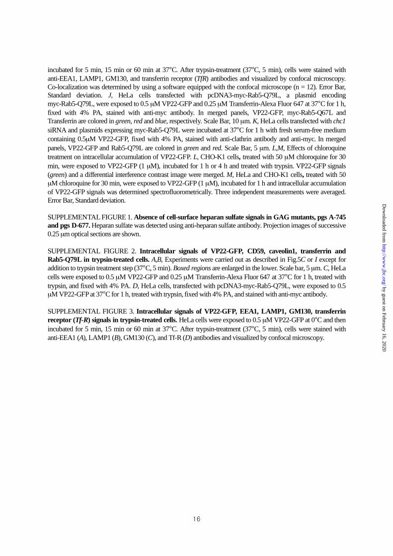
incubated for 5 min, 15 min or 60 min at 37°C. After trypsin-treatment (37°C, 5 min), cells were stained with
anti-EEA1, LAMP1, GM130, and transferrin receptor (TfR) antibodies and visualized by confocal microscopy.
Co-localization was determined by using a software equipped with the confocal microscope (n = 12). Error Bar,
Standard deviation. J, HeLa cells transfected with pcDNA3-myc-Rab5-Q79L, a plasmid encoding
myc-Rab5-Q79L, were exposed to 0.5 μM VP22-GFP and 0.25 μM Transferrin-Alexa Fluor 647 at 37°C for 1 h,
fixed with 4% PA, stained with anti-myc antibody. In merged panels, VP22-GFP, myc-Rab5-Q67L and
Transferrin are colored in green, red and blue, respectively. Scale Bar, 10 μm. K, HeLa cells transfected with chc1
siRNA and plasmids expressing myc-Rab5-Q79L were incubated at 37°C for 1 h with fresh serum-free medium
containing 0.5μM VP22-GFP, fixed with 4% PA, stained with anti-clathrin antibody and anti-myc. In merged
panels, VP22-GFP and Rab5-Q79L are colored in green and red. Scale Bar, 5 μm. L,M, Effects of chloroquine
treatment on intracellular accumulation of VP22-GFP. L, CHO-K1 cells, treated with 50 μM chloroquine for 30
min, were exposed to VP22-GFP (1 μM), incubated for 1 h or 4 h and treated with trypsin. VP22-GFP signals
(green) and a differential interference contrast image were merged. M, HeLa and CHO-K1 cells, treated with 50
μM chloroquine for 30 min, were exposed to VP22-GFP (1 μM), incubated for 1 h and intracellular accumulation
of VP22-GFP signals was determined spectrofluorometrically. Three independent measurements were averaged.
Error Bar, Standard deviation.
SUPPLEMENTAL FIGURE 1. Absence of cell-surface heparan sulfate signals in GAG mutants, pgs A-745
and pgs D-677. Heparan sulfate was detected using anti-heparan sulfate antibody. Projection images of successive
0.25 μm optical sections are shown.
SUPPLEMENTAL FIGURE 2. Intracellular signals of VP22-GFP, CD59, caveolin1, transferrin and
Rab5-Q79L in trypsin-treated cells. A,B, Experiments were carried out as described in Fig.5C or I except for
addition to trypsin treatment step (37°C, 5 min). Boxed regions are enlarged in the lower. Scale bar, 5 μm. C, HeLa
cells were exposed to 0.5 μM VP22-GFP and 0.25 μM Transferrin-Alexa Fluor 647 at 37°C for 1 h, treated with
trypsin, and fixed with 4% PA. D, HeLa cells, transfected with pcDNA3-myc-Rab5-Q79L, were exposed to 0.5
μM VP22-GFP at 37°C for 1 h, treated with trypsin, fixed with 4% PA, and stained with anti-myc antibody.
SUPPLEMENTAL FIGURE 3. Intracellular signals of VP22-GFP, EEA1, LAMP1, GM130, transferrin
receptor (Tf-R) signals in trypsin-treated cells. HeLa cells were exposed to 0.5 μM VP22-GFP at 0°C and then
incubated for 5 min, 15 min or 60 min at 37°C. After trypsin-treatment (37°C, 5 min), cells were stained with
anti-EEA1 (A), LAMP1 (B), GM130 (C), and Tf-R (D) antibodies and visualized by confocal microscopy.
by guest on February 16, 2020http://w
ww
.jbc.org/D
ownloaded from
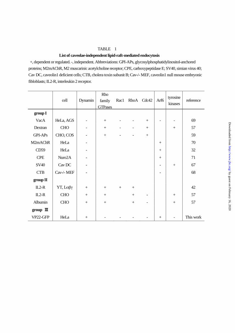
TABLE 1
List of caveolae-independent lipid-raft-mediated endocytosis
+, dependent or regulated. -, independent. Abbreviations: GPI-APs, glycosylphosphatidylinositol-anchored
proteins; M2mAChR, M2 muscarinic acetylcholine receptor; CPE, carboxypeptidase E; SV40, simian virus 40;
Cav DC, caveolin1 deficient cells; CTB, cholera toxin subunit B; Cav-/- MEF, caveolin1 null mouse embryonic
fibloblasts; IL2-R, interleukin 2 receptor.
cell Dynamin
Rho
family
GTPases
Rac1 RhoA Cdc42 Arf6 tyrosine
kinases reference
group I
VacA HeLa, AGS - + - - + - - 69
Dextran CHO - + - - + + 57
GPI-APs CHO, COS - + - - + 59
M2mAChR HeLa - + 70
CD59 HeLa - + 32
CPE Nuro2A - + 71
SV40 Cav DC - - + 67
CTB Cav-/- MEF - - 68
group II
IL2-R YT, L + + + + 42
IL2-R CHO + + + - + 57
Albumin CHO + + + - + 57
group
VP22-GFP HeLa + - - - - + - This work
by guest on February 16, 2020http://w
ww
.jbc.org/D
ownloaded from

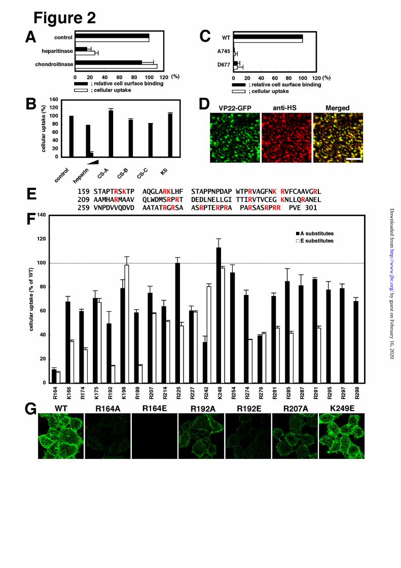

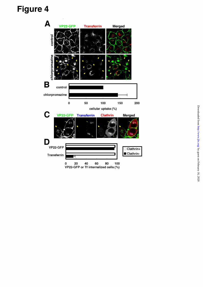
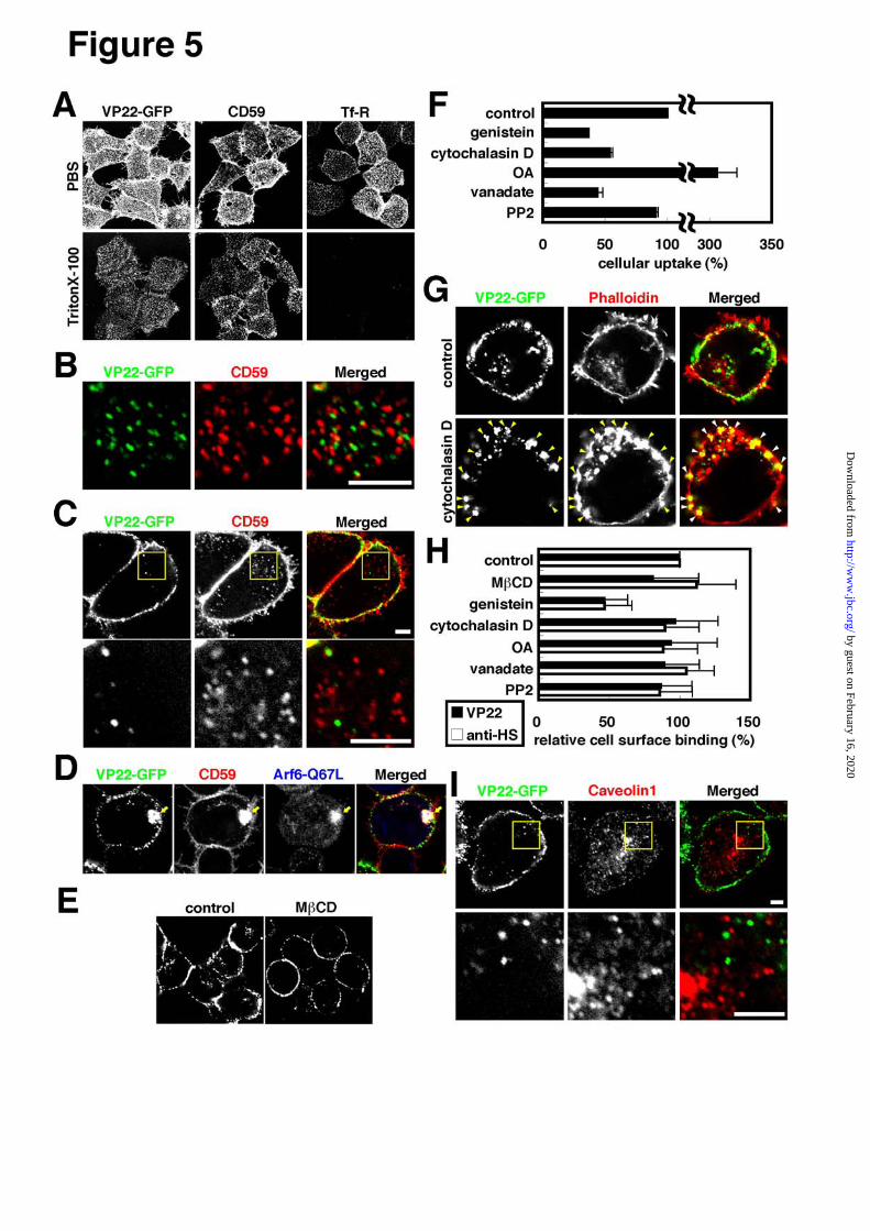
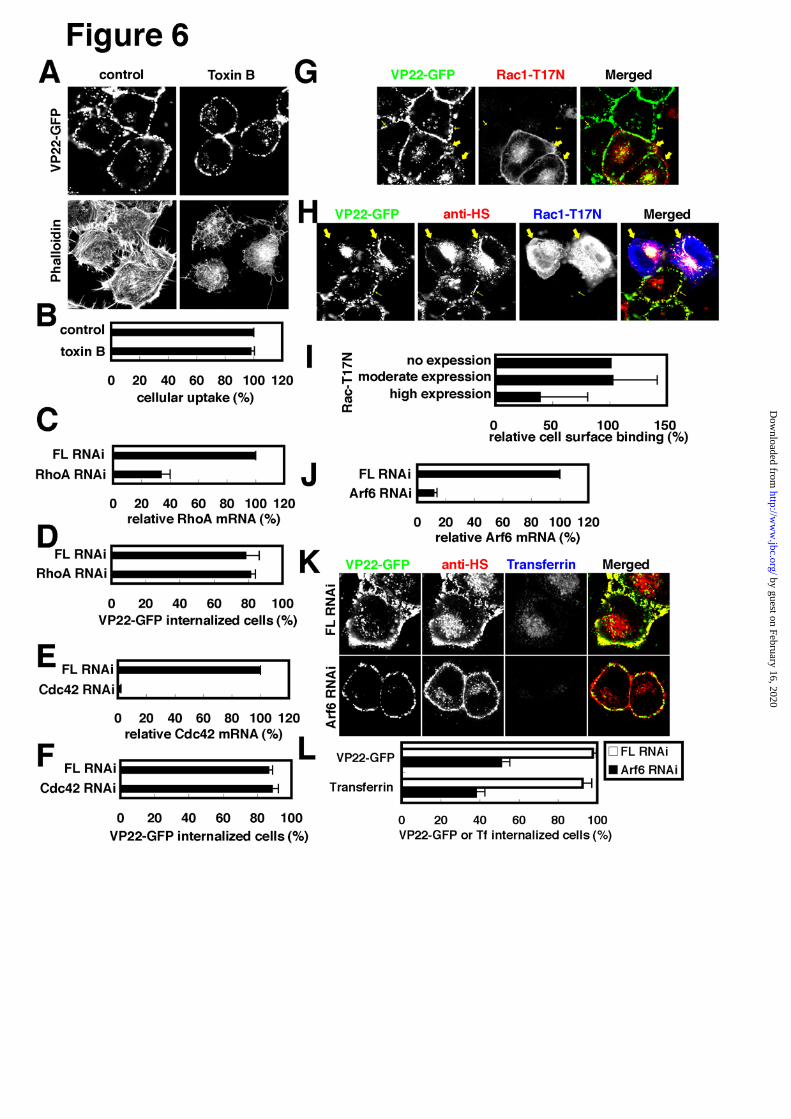
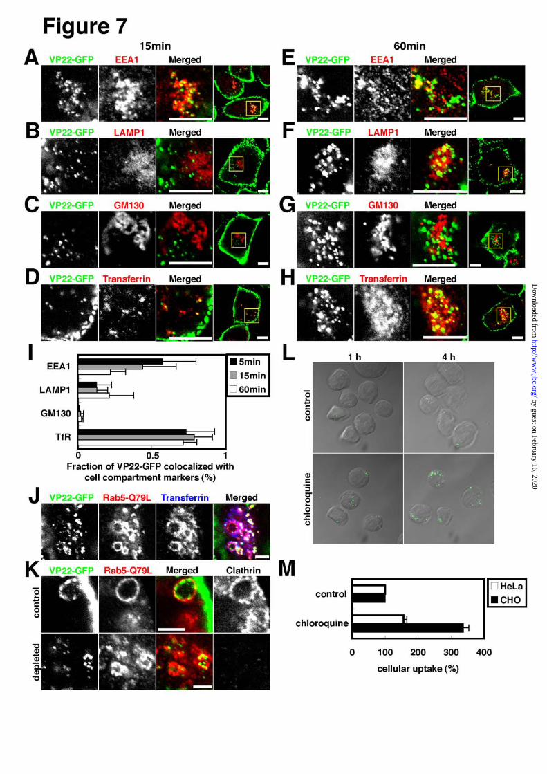
Kenji Nishi and Kaoru Saigocaveolae and rho family GTPases but dependent on dynamin and Arf6
virus protein VP22 via a lipid-raft-mediated endocytic pathway independent of Cellular internalization of green fluorescence protein fused with herpes simplex
published online July 20, 2007J. Biol. Chem.
10.1074/jbc.M703810200Access the most updated version of this article at doi:
Alerts:
When a correction for this article is posted•
When this article is cited•
to choose from all of JBC's e-mail alertsClick here
Supplemental material:
http://www.jbc.org/content/suppl/2007/07/24/M703810200.DC1
by guest on February 16, 2020http://w
ww
.jbc.org/D
ownloaded from