a case of back pain - lieberman's...
TRANSCRIPT

Kimberly Collins, MSIIIGillian Lieberman, MD
A Patient with Low Back PainA Patient with Low Back Pain
Kimberly Collins, Harvard Medical School Year IIIGillian Lieberman, MD
March 2007

2
Kimberly Collins, MSIIIGillian Lieberman, MD
Case PresentationCase Presentation
MJ is a 48 year-old male with a five month history of increased low back pain that radiates down his left leg, left leg weakness, new onset constipation, and slow urination.
MJ is a 48 year-old male with a five month history of increased low back pain that radiates down his left leg, left leg weakness, new onset constipation, and slow urination.
3
Kimberly Collins, MSIIIGillian Lieberman, MD
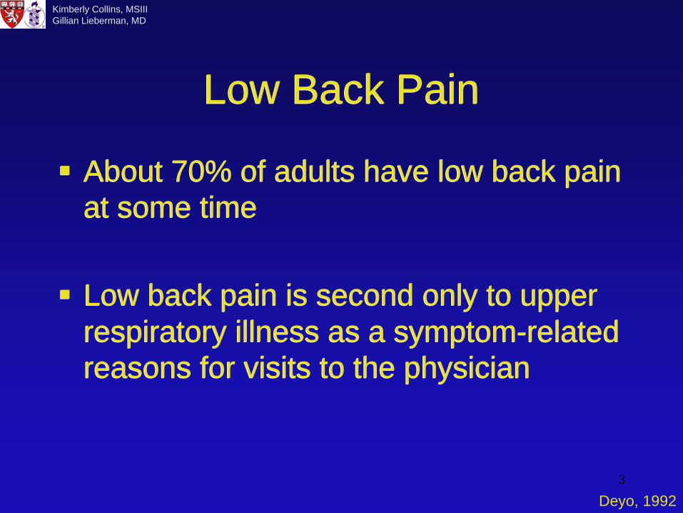
Low Back PainLow Back Pain
About 70% of adults have low back pain at some time
Low back pain is second only to upper respiratory illness as a symptom-related reasons for visits to the physician
About 70% of adults have low back pain at some time
Low back pain is second only to upper respiratory illness as a symptom-related reasons for visits to the physician
Deyo, 1992

4
Kimberly Collins, MSIIIGillian Lieberman, MD
Differential for Back PainDifferential for Back PainMechanical Low Back Pain 97%
• Lumbar strain, sprain 70%• Degenerative processes of disks and facets 10%• Herniated disk 4%• Spinal stenosis 3%• Spondylolisthesis 2%
Non-Mechanical Spinal Conditions ~1%• Neoplasia 0.7%• Infection 0.01%• Inflammatory arthritis 0.3%
Visceral Disease 2%• Disease of pelvic organs• Renal disease• Aortic aneurysm• Gastrointestinal disease
Mechanical Low Back Pain 97%• Lumbar strain, sprain 70%• Degenerative processes of disks and facets 10%• Herniated disk 4%• Spinal stenosis 3%• Spondylolisthesis 2%
Non-Mechanical Spinal Conditions ~1%• Neoplasia 0.7%• Infection 0.01%• Inflammatory arthritis 0.3%
Visceral Disease 2%• Disease of pelvic organs• Renal disease• Aortic aneurysm• Gastrointestinal disease
Deyo, 2001

5
Kimberly Collins, MSIIIGillian Lieberman, MD
Deciding whether to imageDeciding whether to imageBack pain in a patient <50 with no neurological deficits should be followed conservatively with follow-up in 6 weeksPatients with back pain associated with neurological signs should have imagingBack pain lasting more than 3 months is considered chronic and should be imaged
Back pain in a patient <50 with no neurological deficits should be followed conservatively with follow-up in 6 weeksPatients with back pain associated with neurological signs should have imagingBack pain lasting more than 3 months is considered chronic and should be imaged
Jarvik, 2002
6
Kimberly Collins, MSIIIGillian Lieberman, MD
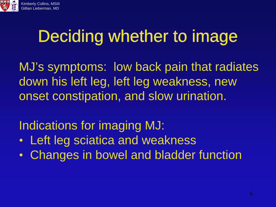
Deciding whether to imageDeciding whether to image
MJ’s symptoms: low back pain that radiates down his left leg, left leg weakness, new onset constipation, and slow urination.
Indications for imaging MJ:• Left leg sciatica and weakness• Changes in bowel and bladder function

7
Kimberly Collins, MSIIIGillian Lieberman, MD
Imaging modalities for patients with low back pain
Imaging modalities for patients with low back pain
IMAGING MODALITY
ADVANTAGES DISADVANTAGES
Plain film Allows visualization of bony structures, low cost
Soft tissue is not well visualized, radiation exposure
CT Detailed visualization of bony structures, some visualization of soft tissue structures
Not great for evaluating soft tissue, radiation exposure, expensive
MRI Best modality for soft tissue visualization, no radiation exposure. Test of choice for back pain with neurological signs!
Difficult to evaluate cortical bone and calcifications, very expensive
Bone scan Can screen the entire skeleton, good for detecting diffuse bone processes, metastases
Low specificity

8
Kimberly Collins, MSIIIGillian Lieberman, MD
Imaging modalities for patients with low back pain
Imaging modalities for patients with low back pain
IMAGING MODALITY
ADVANTAGES DISADVANTAGES
CT Myelography
Used when MRI is contraindicated, allows visualization of spinal cord and nerve roots, can evaluate for lesions within the spinal canal
Invasive, involves the injection of contrast into the thecal sac
Discography Evaluate for disc disease, such as tears
Invasive, rarely used
Bone densitometry/DEXA
Measures bone marrow density, good for determining fracture risk
Spinal angiography
Evaluate for AVM, vascular tumors of spinal cord
Invasive, rarely used

9
Kimberly Collins, MSIIIGillian Lieberman, MD
Anatomy of the lower spineAnatomy of the lower spine
Moore, 2002

10
Kimberly Collins, MSIIIGillian Lieberman, MD
T1W Gad+CT w/o contrast
Patient MJ: Axial CT and MRIPatient MJ: Axial CT and MRI
A gadolinium-enhancing mass with central calcification is seen within
the spinal canal. The mass is better visualized on MRI.
Courtesy Fabio Komlos, MDCourtesy Fabio Komlos, MD

11
Kimberly Collins, MSIIIGillian Lieberman, MD
T1W gad+
Patient MJ: Sagittal CT and MRIPatient MJ: Sagittal CT and MRI
CT
A gadolinium-enhancing mass with central calcification is seen within the spinal canal. The mass is better visualized on MRI.
Courtesy Fabio Komlos, MDCourtesy Fabio Komlos, MD

12
Kimberly Collins, MSIIIGillian Lieberman, MD
Patient MJ: T2 Sagittal MRI of Lumbar Spine
Patient MJ: T2 Sagittal MRI of Lumbar Spine
The mass is located within the spinal column below the level of the cona medullaris and is seen compressing the cauda equina. The location of the mass can explain MJ’s symptoms, which are consistent with cauda equina syndrome.
Courtesy of Fabio Komlos, BIDMC

13
Kimberly Collins, MSIIIGillian Lieberman, MD
Cauda Equina SyndromeCauda Equina SyndromeSymptoms include: • urinary retention• saddle anesthesia - sensory loss occurring
over the buttocks, posterior-superior thighs, and perianal regions
• unilateral or bilateral sciatica• leg weakness• diminished anal sphinctor tone
Usually caused by a tumor or massive midline disk herniation.
Symptoms include: • urinary retention• saddle anesthesia - sensory loss occurring
over the buttocks, posterior-superior thighs, and perianal regions
• unilateral or bilateral sciatica• leg weakness• diminished anal sphinctor tone
Usually caused by a tumor or massive midline disk herniation.
Deyo, 1992

14
Kimberly Collins, MSIIIGillian Lieberman, MD
Intraspinal MassesIntraspinal Masses
Extradural Intradural Extramedullary
Intramedullary
Courtesy of Fabio Komlos, BIDMC

15
Kimberly Collins, MSIIIGillian Lieberman, MD
Differential for Intraspinal MassesDifferential for Intraspinal MassesExtradural Intradural
ExtramedullaryIntramedullary
• Disk disease • Metastases, myeloma and lymphoma deposits
• Hematoma
• Abscess
• Meningioma
• Neurofibroma• Metastases = “drop mets”
• Ependymoma
• Astrocytoma
• Infarct
• Hematoma
• AVM
http://intl.elsevierhealth.com/e-books/viewbook.cfm?ID=576

16
Kimberly Collins, MSIIIGillian Lieberman, MD
Patient MJ: T2 Sagittal MRIPatient MJ: T2 Sagittal MRI
A second intraspinal mass can be seen on this sagittal MRI.
Courtesy of Fabio Komlos, MD
17
Kimberly Collins, MSIIIGillian Lieberman, MD
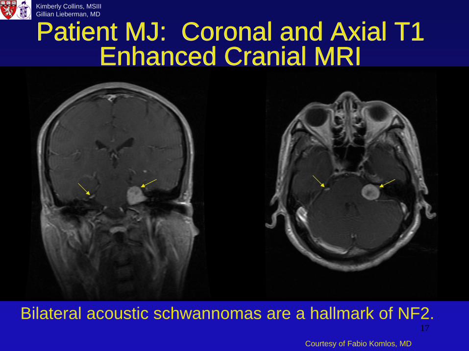
Patient MJ: Coronal and Axial T1 Enhanced Cranial MRI
Patient MJ: Coronal and Axial T1 Enhanced Cranial MRI
Bilateral acoustic schwannomas are a hallmark of NF2.Courtesy of Fabio Komlos, MD

18
Kimberly Collins, MSIIIGillian Lieberman, MD
MJ’s DiagnosisMJ’s Diagnosis
The multiple intraspinal masses and bilateral acoustic schwannomas seen on MRI was suggestive of neurofibromatosis 2.The diagnosis was confirmed by pathology after removal of the intraspinal masses.
The multiple intraspinal masses and bilateral acoustic schwannomas seen on MRI was suggestive of neurofibromatosis 2.The diagnosis was confirmed by pathology after removal of the intraspinal masses.

19
Kimberly Collins, MSIIIGillian Lieberman, MD
NeurofibromatosisNeurofibromatosis
There are two types: Neurofibromatosis Types 1 and 2Neurocutaneous syndromesDevelopment of neoplasms primarily in organs derived from embryonic mesoderm (skin, central and peripheral nervous systems, eyes)Inherited as autosomal dominant conditions with variable penetrance
There are two types: Neurofibromatosis Types 1 and 2Neurocutaneous syndromesDevelopment of neoplasms primarily in organs derived from embryonic mesoderm (skin, central and peripheral nervous systems, eyes)Inherited as autosomal dominant conditions with variable penetrance

20
Kimberly Collins, MSIIIGillian Lieberman, MD NeurofibromatosisNeurofibromatosis
Chromosome 17Gene product: GTPaseactivating proteinOne of the most common autosomal dominant disordersCutaneous features: peripheral neurofibromas, café au lait spots, freckling of axillaNervous system tumors: plexiform neurofibromas, gliomas, ependymomas, meningiomas, astrocytomas, pheochromocytomas
Chromosome 17Gene product: GTPaseactivating proteinOne of the most common autosomal dominant disordersCutaneous features: peripheral neurofibromas, café au lait spots, freckling of axillaNervous system tumors: plexiform neurofibromas, gliomas, ependymomas, meningiomas, astrocytomas, pheochromocytomas
Chromosome 22Gene product: cytoskeletal proteinLess common autosomaldominant disorderCutaneous features: caféau lait spots and peripheral neurofibromasoccur rarelyNervous system tumors: schwannomas, meningiomas, ependymomas
Chromosome 22Gene product: cytoskeletal proteinLess common autosomaldominant disorderCutaneous features: caféau lait spots and peripheral neurofibromasoccur rarelyNervous system tumors: schwannomas, meningiomas, ependymomas
NF1 NF2
Ruggieri, 1999

21
Kimberly Collins, MSIIIGillian Lieberman, MD
Courtesy of Jim Wu, MD
Cutaneous neurofibroma seen as a soft tissue swelling on plain radiograph.
Companion Patient 1: Plain radiographCompanion Patient 1: Plain radiograph

22
Kimberly Collins, MSIIIGillian Lieberman, MD
Plexiform neurofibroma seen in a patient with NF1.STIR T1W Gad+
Companion Patient 2: Chest MRICompanion Patient 2: Chest MRI
Courtesy of Jim Wu, MD

23
Kimberly Collins, MSIIIGillian Lieberman, MD
Radiographs taken before and after the onset of leg pain in a patient with NF1. Fibular fracture is seen in the second radiograph.
Companion Patient 3: Plain radiographCompanion Patient 3: Plain radiograph
Courtesy of Jim Wu, MD

24
Kimberly Collins, MSIIIGillian Lieberman, MD
The fibular fracture occurred due due to mass effect from a neurofibrosarcoma.
Companion Patient 3: MRICompanion Patient 3: MRI
T1W Gad+ T2W
Courtesy of Jim Wu, MD
25
Kimberly Collins, MSIIIGillian Lieberman, MD
SummarySummaryThe most common causes of back pain are mechanical.Not all back pain requires imaging. MRI is the best modality for imaging back pain with neurological signs.The differential for intraspinal masses can be divided into extradural, intraduralextramedullary, and medullary.Neurofibromatosis is a neurocutaneoussyndrome characterized by tumors of the skin and nervous system.
The most common causes of back pain are mechanical.Not all back pain requires imaging. MRI is the best modality for imaging back pain with neurological signs.The differential for intraspinal masses can be divided into extradural, intraduralextramedullary, and medullary.Neurofibromatosis is a neurocutaneoussyndrome characterized by tumors of the skin and nervous system.

26
Kimberly Collins, MSIIIGillian Lieberman, MD
ReferencesReferencesDeyo RA, Weinstein JN. Low Back Pain. N Engl J Med
2001;344(5):363-370. Deyo RA, Rainville J, Kent DL. What Can the History and Physical
Examination Tell Us About Low Back Pain? JAMA 1992;268: 760-764.
Moore KL, Agur AMR. Essentials of Clinical Anatomy. 2nd edition. Baltimore: Lippincott Williams & Wilkins, 2002. p 307.
Jarvik JG, Deyo RA. Diagnostic evaluation of low back pain with emphasis on imaging. Ann Intern Med 2002;137:593.
Kasper, Braunwald, et al. Harrison’s Principles of Internal Medicine. 16th edition. New York: McGraw-Hill, 2005. p 2457.
Ruggieri M. The different forms of neurofibromatosis. Child’s Nerv Sys 1999;15:295-308.
http://www.theuniversityhospital.com/healthlink/archives/articles/ne urofibromatosis.htm
http://intl.elsevierhealth.com/e-books/viewbook.cfm?ID=576
Deyo RA, Weinstein JN. Low Back Pain. N Engl J Med 2001;344(5):363-370.
Deyo RA, Rainville J, Kent DL. What Can the History and Physical Examination Tell Us About Low Back Pain? JAMA 1992;268: 760-764.
Moore KL, Agur AMR. Essentials of Clinical Anatomy. 2nd edition. Baltimore: Lippincott Williams & Wilkins, 2002. p 307.
Jarvik JG, Deyo RA. Diagnostic evaluation of low back pain with emphasis on imaging. Ann Intern Med 2002;137:593.
Kasper, Braunwald, et al. Harrison’s Principles of Internal Medicine. 16th edition. New York: McGraw-Hill, 2005. p 2457.
Ruggieri M. The different forms of neurofibromatosis. Child’s Nerv Sys 1999;15:295-308.
http://www.theuniversityhospital.com/healthlink/archives/articles/ne urofibromatosis.htm
http://intl.elsevierhealth.com/e-books/viewbook.cfm?ID=576

27
Kimberly Collins, MSIIIGillian Lieberman, MD
AcknowledgementsAcknowledgements
Fabio Komlos, MDJim Wu, MDGillian Lieberman, MDPamela LepkowskiLarry Barbaras, webmaster
Fabio Komlos, MDJim Wu, MDGillian Lieberman, MDPamela LepkowskiLarry Barbaras, webmaster