a cascade of gene action controlling heart asymmetry and torsion in embryonic development
TRANSCRIPT

TCM Vol. 8, No. 5, 1998
215
In all vertebrate embryos, including thehuman, incorrectly lateralized cardiacstructure is a rare event that follows re-versal of the normal right-hand loopingof the early heart tube or from indefi-niteness of this looping. Among eventhese rare cases, it is currently unclearhow many are due to gene mutation ormalfunction of a normal complement ofthe relevant genes or are an entirely en-vironmentally triggered developmentalaccident. In all events, most cases ofsuch incorrect laterality, except perhapsthe very rare simple mirror-reversal ofheart structure without other abnormal-ities, lead to severe cardiac malfunction(Burn 1991, Winer-Muram 1995). Eluci-
dation of the mechanism that normallyreliably controls heart laterality is thusof great theoretical and, conceivably,clinical interest.
Heart-tube looping is the earliestreadily visible left–right asymmetry inhigher vertebrate embryos, shortly fol-lowed by a coordinated torsion of thewhole embryo axis. It occurs very earlyduring organogenesis, when, at most,some dozen somite segments have beenproduced and more posterior regionsare still undergoing gastrulation, inwhich the three-layered tissues of thebody are laid down in a front-to-back se-quence. The heart forms from bilateraltracts of the mesoderm or middle layer,initially situated some way from themidline and among the first to be laiddown in gastrulation. These progres-sively converge and fuse to form an ini-tially paired, longitudinal tubular struc-ture beneath the primitive pharyngealcavity. This fusion and tube formationagain occurs in a front-to-back se-quence, and shortly after the future in-flow or atrial heart regions have beenincluded within the tube, the now bi-laterally fused anterior part loops out to
the right. The asymmetrical events lead-ing to right looping are almost certainlyinitiated within this last-formed, moreposterior atrial region, which may in-deed kink more subtly leftward as partof the process. A considerable pattern ofspecific gene activity has already oc-curred by this stage, and heart-tubelooping is just the first major organasymmetry to take its cue from a geneactivation cascade that propagates thedifferent identities of right and lefthalves of the body throughout develop-ment. In this article, we concentrate oninformation deriving from experimentand observation in the chick embryo,providing correlations with, or apparentdifferences from, other vertebrates. Inparticular, the
cSnR
(snail-related) tran-scription factor gene, which we discussextensively, is so far only characterizedin chick.
• Asymmetrical Gene Activity
There have recently been exciting ad-vances in elucidating the gene activationcascade between an early point in gas-trulation, some 18 h before heart-tubeformation in the chick embryo (equiva-lent to a day or two in the human case),and a point just before the inception ofheart looping itself. Many of these ge-netic steps are likely to be evolutionarily“conserved” among the various verte-brates, especially if account is taken ofthe possibility that different members ofa “gene family” may fulfill a particularrole in different but related embryotypes. The mechanism of the left–rightcascade has been revealed mostly in bird(chick) development to date (Levin et al.1995 and 1997, Isaac et al. 1997) by insitu demonstration of asymmetrical ac-tivations of successive genes in thenormal embryo, by experimental inter-ference through ectopic (left–right dis-placed) expression of particular geneproducts at particular stages, and by ob-servation of abnormal expression pat-terns in members of conjoined, paralleltwin embryos at early stages (Levin et al.1996). This later aspect is of particularclinical interest, as heart (and other ma-jor organ) situs abnormalities are rela-tively frequent in one member (althoughseldom in both) of identical twin pairswho give evidence of having sharedstructure in the early conceptus (Burn1991).
A Cascade of Gene Action Controlling Heart Asymmetry and Torsion in Embryonic Development
Jonathan Cooke* and Alison Isaac
Lateralized looping of the heart tube, with associated torsion of theembryo axis, is the first structural sign of the consistent left–rightasymmetrical organization that characterizes all vertebrate embryos.Rare failures or reversals of this asymmetry in humans lead to clini-cally important syndromes of malformation in heart and great bloodvessels. Recently, elements of the genetic control sequence underlyingthis left–right aspect of development have been uncovered. The normalsequence of transmission for asymmetry information can now betraced to a point close to the actual execution of right-hand looping inthe heart tube, with the hope that the further sequence of gene activity,within the heart itself and directing these events, may soon be uncov-ered.
(Trends Cardiovasc Med 1998;8:215–220)
© 1998, ElsevierScience Inc.
Jonathan Cooke is at the National Institutefor Medical Research, The Ridgeway, Lon-don; and Alison Isaac is at the DevelopmentalBiology Group, Department of Anatomy andEmbryology, University College, London,United Kingdom.
* Address correspondence to: Dr. JonathanCooke, National Institute for Medical Re-search, The Ridgeway, Mill Hill, London NW71AA, United Kingdom.
© 1998, Elsevier Science Inc., 1050-1738/98/$19.00.

216
TCM Vol. 8, No. 5, 1998
We present here this gene action cas-cade and the evidence for it, using thesingle diagrammatic representation ofthe relevant period of development givenin Figure 1. In this figure, the axial planof the bird embryo at successive stagesis seen as from above (the mammal be-ing essentially similar in layout), and thecolors show the distributions of the vari-ous gene activities to be described asseen in plan form.
The following discussion touches oneach relevant cell layer, finally focusingon the zinc finger transcription factor-encoding gene
cSnR
(for chicken gene,sequence-related to the
Snail
gene origi-nally named in the fruitfly
Drosophila
).This right-asymmetrically expressed geneis thus far the most “downstream” one,that is, the latest acting of those that
nevertheless may have a controlling,rather than “executive” role. By this, wemean that the gene’s product is probablynot, itself, part of the machinery of theasymmetrical growth or movementswithin left and right components of theheart tube itself. Rather, it is a strongcandidate to control directly any suchasymmetrical executive gene activitywithin looping heart tissue. The stepsfor the future in elucidating the normalmechanism of heart laterality may be tofind and characterize such executivegenes and to understand how they con-trol the mechanical asymmetry.
Thus far, the earliest observed resultof left–right symmetry breaking in termsof gene activity, in chick embryos, is for
activin receptor IIa
(Levin et al. 1995).This transmembrane receptor is know to
be inducible by at least one of itsligands, the transforming growth factor(TGF)
b
–related signaling protein activin,but almost certainly can mediate signal-ing from an unknown number of otherrelated peptides. The mRNA is expressed,clearly preferentially to the right of theanterior primitive streak in ectoderm,by midgastrulation (Figure 1, first, lefthand diagram). Although the
activin B
mRNA has subsequently been reportedas right-expressed in the same region(Levin et al. 1997), this is not so clear,and any conserved, necessary role inright–left asymmetry for this or any ac-tivin is not borne out by targeted mu-tagenesis in mice (Matzuk et al. 1995).Furthermore, results from
Xenopus
(frog)development propose a different but re-lated TGF
b
family protein, one that couldwell signal through this same receptorsystem, as controlling the left–right cas-cade by normal accentuation on the
left
from the very earliest developmentalstages (Hyatt et al. 1996). At present, asecure interpretation, for chick at least,is that signaling via an “activin receptor”pathway to the right of the node, at mid-gastrulation (the full-length primitivestreak stage), determines subsequentsteps. This is because activin protein,applied experimentally on the left from abead source, wipes out left–right infor-mation as evidenced by randomizationof later heart looping (Levin et al. 1995,Isaac et al. 1997).
Next, a localized left-hand, backwardextension of
Sonic hedgehog
mRNA ex-pression occurs at Hensen’s node. Thisgene, encoding another intercellularsignaling protein, is otherwise symmet-rically expressed, but the localized leftasymmetry persists for several hours asthe node “regresses” in gastrulation,leaving in front of it a strip of hedgehogexpression in the future dorsal midlineof the body (Figure 1, second and thirddiagrams). Because experimental left-hand placement of activin sources, asdescribed, leads to reversal or elimina-tion of this
hedgehog
asymmetry (Levinet al. 1995, Isaac et al. 1997), this stepin the normal cascade is thought to beone of repression, by the activin-typesignaling on the right, of a default stateof
Sonic hedgehog
expression in poste-rior node. The following two steps,however, positively propagating the in-formation for “leftness” down the ap-propriate side of the midline as gastru-
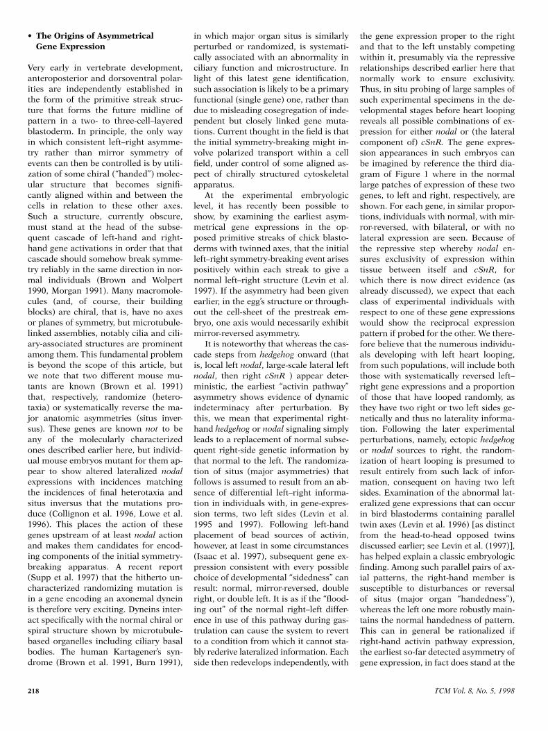
Figure 1. The cascade of laterality information for heart looping. The four schematic dia-grams are of successive stages in the (chick) embryo seen from above, anterior at top, begin-ning at left with the primitive streak and Hensen’s node, going on to a short head process stageat which the node has begun “regression” (see the text), continuing with a late headfold or neu-rula stage (four segmented somites in mesoderm), and ending with the stage of the right-looped heart tube and onset of torsion. The color key identifies the distributions of transcrip-tion for the respective genes at each stage: green, activin receptor IIa; blue, sonic hedgehog;orange, nodal; and red, cSnR. Looping and torsion are normally coordinated as shown, andtheir directions remain significantly linked among embryos where looping direction is ran-domized after disruption of cSnR function. But genetically normal embryos, whose heart-forming tissues have been surgically excised before the asymmetry of looping starts, go on totwist as shown with almost normal reliability. Therefore, the cSnR gene is probably involved inthe control of embryo torsion, as well as of heart looping, via partially independent “executive”mechanisms.

TCM Vol. 8, No. 5, 1998
217
lation proceeds, are of an “activation”nature. The
nodal
gene, encoding yetanother signaling protein from a differ-ent subgroup of the TGF
b
superfamily,becomes expressed first in a small re-gion of the deeper portion of Hensen’snode to the left, then in a much broader,axially extended lateral mesodermalregion on the same side (Figure 1, sec-ond and third diagrams). This lateralstrip and its anatomically symmetrical-appearing partner on the right, oppositethe first-segmenting anterior somites,include much of the cardiogenic meso-derm itself. The major left
nodal
expres-sion domain almost certainly fate-mapsin part to the heart-forming territory ofthat side. Placement of experimentalhedgehog protein sources to the right ofthe node demonstrates the normal posi-tive role of this gene on the left in con-trolling first local, then the broad lateral
nodal
expression. In such experimentalembryos, such expression develops onthe right as well.
Sonic hedgehog
in the mouse has thusfar failed to reveal expression asymme-try at the node (Collignon et al. 1996),and, indeed, targeted mutagenesis offersno evidence that this gene itself is re-quired for normal situs development inmouse. But this is a member of a
hedge-hog
gene family, and there is some evi-dence that other gene disruptions thatmay interfere with the common “hedge-hog signaling pathway” do destabilizeinformation for developmental lateralityin mouse and in fish embryos [for exam-ple, Collignon et al. (1996)]. The dramat-ically left-specific expression of chick
nodal
during late gastrulation, however,is indeed conserved for the orthologousgenes in mouse and
Xenopus
(Collignonet al. 1996, Lowe et al. 1996). Provision-ally then, involvement of
hedgehog
and
nodal
genes in positive left-hand propa-gation of information for laterality is aconserved vertebrate feature.
The next and latest-acting gene thusfar discovered in the left–right controlcascade itself,
cSnR
from chicken, is of adifferent type from the preceding onesin encoding a zinc-finger containing pro-tein, expected to function as a transcrip-tion factor (Isaac et al. 1997). Membersof the
Drosophila
snail gene family, shar-ing a highly conserved DNA-binding re-gion as well as one other sequence-related stretch with this gene, are thoughtto function as transcriptional repressors.
Certainly, activation of
cSnR
would beexpected to control further gene activitywithin the same cells that produce theprotein, rather than, as for the steps al-ready discussed, controlling such activ-ity in neighboring cells via receptor-mediated signaling.
cSnR
is activatedearly and widely during gastrulation, inmesenchymal mesoderm leaving theposterior streak region (destined as pos-terolateral body wall), and also anteri-orly and ventrally in pharyngeal endo-derm including that overlying theforming heart tube. For clarity, these bi-laterally symmetrical expressions areomitted from Figure 1, but the expres-sion in parts of the forming somite seg-ments, also bilateral, is shown (thirdand fourth diagrams). Expression in thelateral cardiogenic mesoderms beginsearly in their sequence of forward move-ment after leaving the primitive streakin gastrulation and, in some individuals,is accentuated on the right from the out-set (Figure 1, second diagram). But thisexpression is never as exclusively right-sided as
nodal
is left-sided, and it onlycomes strongly to mirror the extendedpatch of left
nodal
expression at timeswhen the latter is well established (Fig-ure 1, third diagram).
cSnR
on the righttends to peak in intensity somewhat af-ter, and in a slightly more posterior posi-tion than, the peak
nodal
expression atleft. The latest lateralized expression canbe detected just in the right-hand mem-ber of the paired future atrial or inflowregions, at the posterior of the primitiveheart, after normal right looping has al-ready begun anteriorly in the fused ven-tricular-arterial loop at the 10–12 somitestage (Figure 1, fourth diagram). Al-though the right-hand tissue contribut-ing to this more anterior loop may haveexpressed the gene when at earlier pre-fusion stages, such expression has disap-peared from all except the inflow part bythe stage of lateral looping.
With the use of antisense oligodeoxy-nucleotides (thioated for protectionagainst exonuclease degradation), ap-plied with lipofection in a whole embryoculture system, it has been possible todisrupt
cSnR
function during a period ofseveral hours during its right-lateralizedexpression (Isaac et al. 1997). Gene-sequence specificity of effects was evidentfrom observation of specific loss of
cSnR
mRNA, as well as specific phenotypic ef-fects, after some hours of culture with
oligos of two different
cSnR
-antisensesequences but not those of various con-trol sequences. The distinctive effectswere near-randomization of the direc-tion of heart looping and of embryo tor-sion, without evidence of general toxic-ity. Directions of looping and of torsionremained highly correlated within indi-viduals, in which the form of the loopedheart tube (whether normal or mirror-reversed) looked normal at gross ana-tomic level.
Treatment was most effective whenapplied so that the gene’s function wouldbe most disrupted in the hours preced-ing heart-tube formation, around thetime when normal expression peaks infuture right posterior cardiac tissue andsomewhat later than peak left
nodal
ex-pression. Embryos antisense-treated for
cSnR
and then harvested within the left
nodal
expression period, as well as simi-lar control oligo-treated embryos, showednormal late expression for
nodal.
Be-cause left lateralization of
nodal
hasotherwise been shown to be critical toheart-loop asymmetry (Levin et al. 1995and 1997), this and
cSnR
’s somewhatlater expression peak would place
cSnR
downstream of
nodal
in the cascade ofgene activity, propagating left–right in-formation that is used to direct asym-metrical flexion of the heart tube. Thatis, normal deployment of
cSnR
woulddepend on that of
nodal.
The dorsalmidline appears to act as a barrier tointercellular signaling in this cascade,so an expectation might be that
nodal
expression in left lateral mesoderm(due to left
hedgehog
and then
nodal
signaling at Hensen’s node) represseswhat would otherwise be a defaultpathway of
cSnR
expression in this tis-sue. Lateral
cSnR
expression, occur-ring only or mainly in right mesodermincluding precardiac tissue, might di-rectly control heart looping by control-ling differential executive gene activitywithin that tissue. It has recently beenpossible to confirm this cascade rela-tionship in that experimental ectopic,right-lateral expression of
nodal
pro-tein appears specifically to downregu-late the normal right-lateral
cSnR
ex-pression (A. Isaac, K. Patel, J. Cooke,unpublished). The possibility remains,however, that left
nodal
and right
cSnR
have reciprocal direct influences on ex-ecutive gene activity within their re-spective heart territories.
218
TCM Vol. 8, No. 5, 1998
• The Origins of Asymmetrical Gene Expression
Very early in vertebrate development,anteroposterior and dorsoventral polar-ities are independently established inthe form of the primitive streak struc-ture that forms the future midline ofpattern in a two- to three-cell–layeredblastoderm. In principle, the only wayin which consistent left–right asymme-try rather than mirror symmetry ofevents can then be controlled is by utili-zation of some chiral (“handed”) molec-ular structure that becomes signifi-cantly aligned within and between thecells in relation to these other axes.Such a structure, currently obscure,must stand at the head of the subse-quent cascade of left-hand and right-hand gene activations in order that thatcascade should somehow break symme-try reliably in the same direction in nor-mal individuals (Brown and Wolpert1990, Morgan 1991). Many macromole-cules (and, of course, their buildingblocks) are chiral, that is, have no axesor planes of symmetry, but microtubule-linked assemblies, notably cilia and cili-ary-associated structures are prominentamong them. This fundamental problemis beyond the scope of this article, butwe note that two different mouse mu-tants are known (Brown et al. 1991)that, respectively, randomize (hetero-taxia) or systematically reverse the ma-jor anatomic asymmetries (situs inver-sus). These genes are known
not
to beany of the molecularly characterizedones described earlier here, but individ-ual mouse embryos mutant for them ap-pear to show altered lateralized
nodal
expressions with incidences matchingthe incidences of final heterotaxia andsitus inversus that the mutations pro-duce (Collignon et al. 1996, Lowe et al.1996). This places the action of thesegenes upstream of at least
nodal
actionand makes them candidates for encod-ing components of the initial symmetry-breaking apparatus. A recent report(Supp et al. 1997) that the hitherto un-characterized randomizing mutation isin a gene encoding an axonemal dyneinis therefore very exciting. Dyneins inter-act specifically with the normal chiral orspiral structure shown by microtubule-based organelles including ciliary basalbodies. The human Kartagener’s syn-drome (Brown et al. 1991, Burn 1991),
in which major organ situs is similarlyperturbed or randomized, is systemati-cally associated with an abnormality inciliary function and microstructure. Inlight of this latest gene identification,such association is likely to be a primaryfunctional (single gene) one, rather thandue to misleading cosegregation of inde-pendent but closely linked gene muta-tions. Current thought in the field is thatthe initial symmetry-breaking might in-volve polarized transport within a cellfield, under control of some aligned as-pect of chirally structured cytoskeletalapparatus.
At the experimental embryologiclevel, it has recently been possible toshow, by examining the earliest asym-metrical gene expressions in the op-posed primitive streaks of chick blasto-derms with twinned axes, that the initialleft–right symmetry-breaking event arisespositively within each streak to give anormal left–right structure (Levin et al.1997). If the asymmetry had been givenearlier, in the egg’s structure or through-out the cell-sheet of the prestreak em-bryo, one axis would necessarily exhibitmirror-reversed asymmetry.
It is noteworthy that whereas the cas-cade steps from
hedgehog
onward (thatis, local left
nodal
, large-scale lateral left
nodal
, then right
cSnR
) appear deter-ministic, the earliest “activin pathway”asymmetry shows evidence of dynamicindeterminacy after perturbation. Bythis, we mean that experimental right-hand
hedgehog
or
nodal
signaling simplyleads to a replacement of normal subse-quent right-side genetic information bythat normal to the left. The randomiza-tion of situs (major asymmetries) thatfollows is assumed to result from an ab-sence of differential left–right informa-tion in individuals with, in gene-expres-sion terms, two left sides (Levin et al.1995 and 1997). Following left-handplacement of bead sources of activin,however, at least in some circumstances(Isaac et al. 1997), subsequent gene ex-pression consistent with every possiblechoice of developmental “sidedness” canresult: normal, mirror-reversed, doubleright, or double left. It is as if the “flood-ing out” of the normal right–left differ-ence in use of this pathway during gas-trulation can cause the system to revertto a condition from which it cannot sta-bly rederive lateralized information. Eachside then redevelops independently, with
the gene expression proper to the rightand that to the left unstably competingwithin it, presumably via the repressiverelationships described earlier here thatnormally work to ensure exclusivity.Thus, in situ probing of large samples ofsuch experimental specimens in the de-velopmental stages before heart loopingreveals all possible combinations of ex-pression for either
nodal
or (the lateralcomponent of)
cSnR.
The gene expres-sion appearances in such embryos canbe imagined by reference the third dia-gram of Figure 1 where in the normallarge patches of expression of these twogenes, to left and right, respectively, areshown. For each gene, in similar propor-tions, individuals with normal, with mir-ror-reversed, with bilateral, or with nolateral expression are seen. Because ofthe repressive step whereby
nodal
en-sures exclusivity of expression withintissue between itself and
cSnR
, forwhich there is now direct evidence (asalready discussed), we expect that eachclass of experimental individuals withrespect to one of these gene expressionswould show the reciprocal expressionpattern if probed for the other. We there-fore believe that the numerous individu-als developing with left heart looping,from such populations, will include boththose with systematically reversed left–right gene expressions and a proportionof those that have looped randomly, asthey have two right or two left sides ge-netically and thus no laterality informa-tion. Following the later experimentalperturbations, namely, ectopic
hedgehog
or
nodal
sources to right, the random-ization of heart looping is presumed toresult entirely from such lack of infor-mation, consequent on having two leftsides. Examination of the abnormal lat-eralized gene expressions that can occurin bird blastoderms containing paralleltwin axes (Levin et al. 1996) [as distinctfrom the head-to-head opposed twinsdiscussed earlier; see Levin et al. (1997)],has helped explain a classic embryologicfinding. Among such parallel pairs of ax-ial patterns, the right-hand member issusceptible to disturbances or reversalof situs (major organ “handedness”),whereas the left one more robustly main-tains the normal handedness of pattern.This can in general be rationalized ifright-hand activin pathway expression,the earliest so-far detected asymmetry ofgene expression, in fact does stand at the

TCM Vol. 8, No. 5, 1998
219
head of this cascade. A pattern on theright of a parallel pair, although not thaton the left, would then be susceptible totriggering of a reversed or confused cas-cade by breakthrough from the activinpathway onto its own left side.
• The Execution of Heart Looping; Future Prospects
The idea that left looping of the heartmight follow either actively reversedgene cues, or an absence of such differ-ential cues, raises the interesting ques-tion of the precise degrees of structuralnormality among reversed hearts. Togross anatomic inspection, such heartloops look to be good mirror-reversals ofthe normal process and to occur at thenormal developmental time, or perhapson average very slightly later in relationto other developmental indices such assomite number. The complete normalmechanism therefore seems to involve abuilt-in mechanical instability of growthor morphogenetic movement wherebythe heart is constrained to loop and thento superimposed cues that normally reli-ably trigger the direction of that looping,cues that the experiments discussedhere either eliminate or reverse. To un-derstand further whether randomly di-rected looping (in the absence of left–right cues extrinsic to the heart) is in-deed closely similar in mechanism tonormal looping will require very exactdevelopmental anatomic work. It ishoped that such work can soon be cou-pled with examination of genes asym-metrically active within the normallooping rudiment itself, that is, the exec-utive genes that actually direct the cre-ation of the mechanical asymmetry. Inencoding a likely transcription factor,
cSnR
could be viewed as such a gene,but attention should focus on any asym-metrically expressed genes, controlledperhaps by
cSnR
or reciprocally by
cSnR
and
nodal
, that encode products likely tobe involved in control of force genera-tion in growing tissue or in growth rateitself.
Subtle asymmetry in the sizes andcell division rates, or in timing and in-tensity of bending force productionwithin left and right cardiac precursorregions, have been proposed for chick[for example, Stalsberg (1969), Itasaki etal. (1991), and Manasek (1981)], and re-cently a subtle growth rate and very lo-
calized structural asymmetry have beendescribed within the inflow tracts justbefore onset of the main looping inmouse heart formation (D. Bellomo, N.Brown, unpublished observations). Thesehave been determined to be abnormal inmice genetically disposed toward re-versed looping. A large, cytoskeleton-and/or matrix-associated protein ofunknown function, flectin, has beenstudied immunocytochemically in mouseand chick (Tsuda et al. 1996). It is prefer-entially expressed on the left-hand sidein the inflow tract, precisely at the stagesat which these subtle asymmetries thatprecede main looping have been re-corded. The normal time of maximalleft-accentuated expression of flectin inchick would correspond closely with thetime when right-accentuated
cSnR
ap-pears to be controlling the direction offuture heart looping, on the basis of thatgene’s expression timing and antisenseinterference (Isaac et al. 1997). Flectin isthus a candidate for an executive geneproduct directly controlled by the puta-tive
cSnR
transcription factor, and inthis case, the relationship in the cascadewould once more be a repressive one,whereby right
cSnR
normally ensuresleft flectin by repressing a “default” stateof
flectin
gene expression (or, conceiv-ably, downregulating its protein synthe-sis) on the right. Close examination offlectin distribution within
cSnR
-dis-rupted embryos during the narrow rele-vant time-window is indicated, a demand-ing experiment that might or might notprove possible.
The presence of excess retinoids dur-ing early development (usually via provi-sion of exogenous retinoic acid) hasbeen documented as producing a varietyof teratogenic effects and abnormalitiesof embryo pattern. Many of these are in-terpretable in terms of the idea that thelocal available concentrations of someendogenous retinoid that control thenormal spatio–temporal sequence of de-ployment of the Hox-type clusteredhomeobox-containing genes in develop-ment are disrupted. Hox-type homeoboxgenes are central to regionalization ofthe axial body plan at early stages of ver-tebrate development (Hunt and Krum-lauf 1992). Insufficiency of retinoids alsoproduces pattern abnormalities, and oneof these, in a retinoid-deficient bird em-bryo model (Dersh and Zile 1993), israndomization of heart-looping direc-
tion. Retinoids can, in principle, be re-stored to embryos at chosen develop-mental points in this system; so withincertain limits, the time of necessary ac-tion for retinoids, within a normal path-way of right–left genetic information,could be established. Present indicationsare that this point lies downstream fromthe asymmetrical structure of Hensen’snode with its local posterior left exten-sion of
Sonic hedgehog
activity (Chen etal. 1996) but possibly upstream of flectinexpression (Tsuda et al. 1996).
References
Brown NA, Wolpert L: 1990. The develop-ment of handedness in left/right asymme-try. Development 109:1–9.
Brown NA, McCarthy A, Wolpert L: 1991.Development of handed body asymmetryin mammals.
In
CIBA Foundation Sympo-sium 162: Biological Asymmetry andHandedness. London, Wiley & Sons, pp182–196.
Burn J: 1991. Disturbance of morphologicallaterality in humans.
In
CIBA FoundationSymposium 162: Biological Asymmetryand Handedness. London, Wiley & Sons,pp 282–295.
Chen Y, Dong D, Kostetskii I, Zile MH: 1996.Hensen’s node from vitamin A-deficientquail embryo induces chick limb budduplication and retains its normal asym-metric expression of
Sonic hedgehog
(Shh).Dev Biol 173:256–264.
Collignon J, Varlet I, Robertson, EJ: 1996.Relationship between asymmetic nodalexpression and the direction of embryonicturning. Nature 381:155–158.
Dersh H, Zile MH: 1993. Induction of normalcardiovascular development in the vitaminA-deprived quail embryo by natural reti-noids. Dev Biol 160:424–433.
Hunt P, Krumlauf R: 1992. Hox codes andpositional specification in vertebrateembryonic axes. Annu Rev Cell Biol 8:227–256.
Hyatt BA, Lohr JL, Yost HJ: 1996. Initiationof vertebrate left-right axis formation bymaternal Vg1. Nature 384:62–65.
Isaac A, Sargent MG, Cooke J: 1997. Controlof left-right asymmetry by a Snail-relatedzinc finger gene. Science 275:1301–1304.
Itasaki N, Nakamura H, Sumida H, YasudaM: 1991. Actin bundles on the right side inthe caudal part of the heart tube play a rolein dextro-looping in the embryonic chickheart. Anat Embryol 183:29–39.

220
TCM Vol. 8, No. 5, 1998
Levin M, Johnson R, Stern C, Kuehn M,Tabin C: 1995. A molecular pathway deter-mining left-right asymmetry in chick em-bryogenesis. Cell 82:1–20.
Levin M, Roberts DJ, Holmes LB, Tabin C:1996. Laterality defects in conjoined twins.Nature 384:321.
Levin M, Pagan S, Roberts DJ, Cooke J,Kuehn MR, Tabin C: 1997. Left/right pat-terning signals and the independent regu-lation of different aspects of situs in thechick embryo. Dev Biol 189:57–67.
Lowe L, Supp D-M, Sampath K, et al.: 1996.Conserved left-right asymmetry of nodalexpression and alterations in murine situsinversus. Nature 381:158–161.
Manasek FJ: 1981. Determinants of heartshape in early embryos. Fed Proc 40:2011–2016.
Matzuk MM, Kumar TR, Vassalli A, et al.:1995. Functional analysis of activins dur-ing mammalian development. Nature 374:354–356.
Hunter 1995, Cano and Mahadevan 1995).From yeast to mammals, MAP kinasesshare genetically conserved regulatorymechanisms that enable them to be acti-vated by dual phosphorylation of con-served threonine and tyrosine residuesupon receiving extracellular signals. Thisoccurs via their direct upstream activa-tors, the so-called MAP kinase kinases(MKKs). Subsequently, MAP kinasestransmit the signals by phosphorylatingdownstream substrates on threonine orserine residues that are adjacent to pro-line residues. The overall regulation ofeach MAP kinase is tightly controlled invivo, both with respect to activation andspecificity. Based on their structure andfunctional reactivity, mammalian MAPkinases can be arbitrarily categorizedinto at least three distinct groups, whichare implicated in parallel signaling cas-cades. These subsets include the extra-cellular signal regulated kinases (ERKs),jun- or stress-activated protein kinases(JNK/SAPK), and the p38 group of pro-tein kinases. This review summarizes re-cent progress of in the understanding ofp38, and related studies regarding theother members of p38 group.
• Structural Characteristics of the p38 Group of Mitogen-Activated Protein Kinases
To date, four members of the p38 groupof MAP kinases have been cloned andcharacterized: p38 (or p38
a
) (Han et al.1994, Lee et al. 1994), p38
b
(Jiang et al.1996), p38
g
(or ERK6, SAPK3) (Lechneret al. 1996, Li et al. 1996, Cuenda et al.1997), and p38
d
(or SAPK4) (Jiang et al.1997a, Kumar et al. 1997). p38 is the ar-chetypal member of this group. It wasfirst isolated in the course of a study de-signed to identify proteins that were ty-rosine phosphorylated in macrophagesstimulated with bacterial endotoxin (li-popolysaccharide, or LPS) (Han et al.1993 and 1994). p38 was also purified,and its cDNA cloned, by another groupof researchers as a specific target of py-ridinyl imidazole derivatives that inhibitthe production of proinflammatory cy-tokines by monocytes. In this indepen-dent effort, the protein was termed cytok-ine suppressive anti-inflammatory drugs–binding protein (CSBP) (Lee et al. 1994).Peptide sequence comparisons of p38group kinases and other MAP kinaseshave shown that each p38 isoform has
Morgan MJ: 1991. The asymmetrical geneticdetermination of laterality: flatfish, frogsand human handedness.
In
CIBA Founda-tion Symposium 162 Biological Asymme-try and Handedness. London, Wiley &Sons, pp 234–247.
Stalsberg H: 1969. The origin of heart asymme-try: right and left contributions to the earlychick embryo heart. Dev Biol 19:109–127.
Supp D-M, Witte DP, Potter SS, Brueckner M:1997. Mutation of an axonemal dynein inthe left-right mouse mutant
inversus vis-cerum.
Nature 389:963–966.
Tsuda T, Philp N, Zile MH, Linask, K: 1996.Left-right asymmetric localization of Flec-tin in the extracellular matrix during heartlooping. Dev Biol 173:39–50.
Winer-Muram HT: 1995. Adult presentationof heterotaxic syndromes and related com-plexes. J Thorac Imaging 10:43–57.
PII S1050-1738(98)00009-7 TCM
The p38 MAP Kinase Pathway and Its Biological Function
Liguo New and Jiahuai Han*
p38 is a mitogen-activated protein (MAP) kinase with structural andfunctional characteristics that distinguish it from JNK and ERK MAPkinases. p38 activity is upregulated when cells are exposed to a varietyof stimuli including bacterial pathogens, proinflammatory cytokines,certain growth factors, and other forms of environmental stress. Byregulating downstream substrates that include protein kinases andtranscription factors, p38 participates in transmission, amplification,and diversification of the extracellular signal, initiating several differ-ent cellular responses. Studies have revealed that activation of p38pathway is related to many pathological changes that occur in thecourse of inflammatory/immunologic and cardiovascular diseases.
(Trends Cardiovasc Med 1998;8:220–229)
© 1998, Elsevier ScienceInc.
Liguo New and Jiahuai Han are at theDepartment of Immunology, The ScrippsResearch Institute, La Jolla, California, USA.
* Address correspondence to: Jiahuai Han,PhD, Scripps Research Institute, Departmentof Immunology, IMM-12, 10666 N. TorreyPines Road, La Jolla, CA 92037, USA.
© 1998, Elsevier Science Inc., 1050-1738/98/$19.00.
p38 is a member of mitogen-activatedprotein (MAP) kinase superfamily. TheMAP kinases are ubiquitous signalingmolecules whose activation transducessignals initiated by a wide array of extra-cellular stimuli, such as growth factors,cytokines, or physical–chemical stresses,into intracellular responses (Karin and