9.1 introduction neurons: masses of nerve cells. structural and functional units of the nervous...
TRANSCRIPT
9.1 INTRODUCTION• Neurons: masses of nerve cells.• Structural and functional units of the nervous
system.• Specialized to react to physical and chemical
changes in their surroundings.• Nerve impulses: electrochemical changes
neurons transmit.
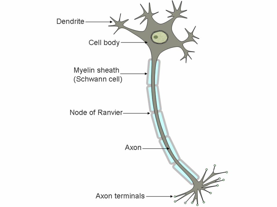
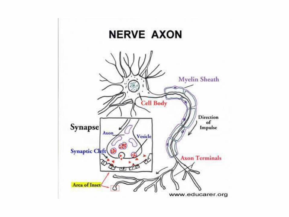
NEURONS• Cell body: rounded area• Extensions: dendrites and axons• Dendrites: receive electrochemical messages.• Axons: extensions that send information in the
form of nerve impulses.• Neurons usually have only one axon.
NERVES• Bundles of axons.• Neuroglial cells: provide physical support, insulation,
and nutrients for neurons.ORGANS OF NERVOUS SYSTEM• Central nervous system: brain, spinal cord.• Peripheral nervous system: nerves that connect the
central nervous system to other body parts.3 FUNCTIONS1. Sensory2. Integrative3. motor
9.2 GENERAL FUNCTIONS OF THE NERVOUS SYSTEM
• Sensory receptors: ends of peripheral neurons.
• Gather information by detecting changes inside and outside the body.
• Monitor external environmental factors, such as light and sound intensities, and conditions of the body’s internal environment, such as temp. and oxygen level.

• Convert environmental information into nerve impulses.
• Transmitted over peripheral nerves to the CNS.
• Signals are integrated; brought together, creating sensations, adding to memory, or helping to produce thoughts that translate sensations into perceptions.
• Integrative function: we make conscious or subconscious decisions.
• Motor functions: to act on them.
• Effectors: carry impulses from the CNS to responsive structures.
• Outside the nervous system, include muscles that contract and glands that secrete when stimulated by nerve impulses.
MOTOR FUNCTIONS OF THE PERIPHERAL NERVOUS SYSTEM
2 categories• Somatic NS: consciously controlled, controls skeletal
muscle.• Autonomic NS: controls effectors that are involuntary,
hear, smooth muscle in blood vessels, and various glands.
9.3 NEUROGLIAL CELLS• Fill spaces between neurons, provide
structural frameworks, produce the fatty lipoprotein myelin, and carry on phagocytosis.
• Neuroglial cells greatly outnumber neurons.
NEUROGLIAL CELLS1. Microglial cells: scattered throughout the CNS.
They support neurons and phagocytize bacterial cells and cellular debris
2. Oligodendrocytes: align along nerve fibers. They provide insulating layers of myelin, called a myelin sheath around axons within the brain and spinal cord.
3. Astrocytes: commonly found between neurons and blood vessels, provide structural support, join parts by their abundant cellular processes, and help regulate the concentrations of nutrients and ions within the tissue. Also form scar tissue.
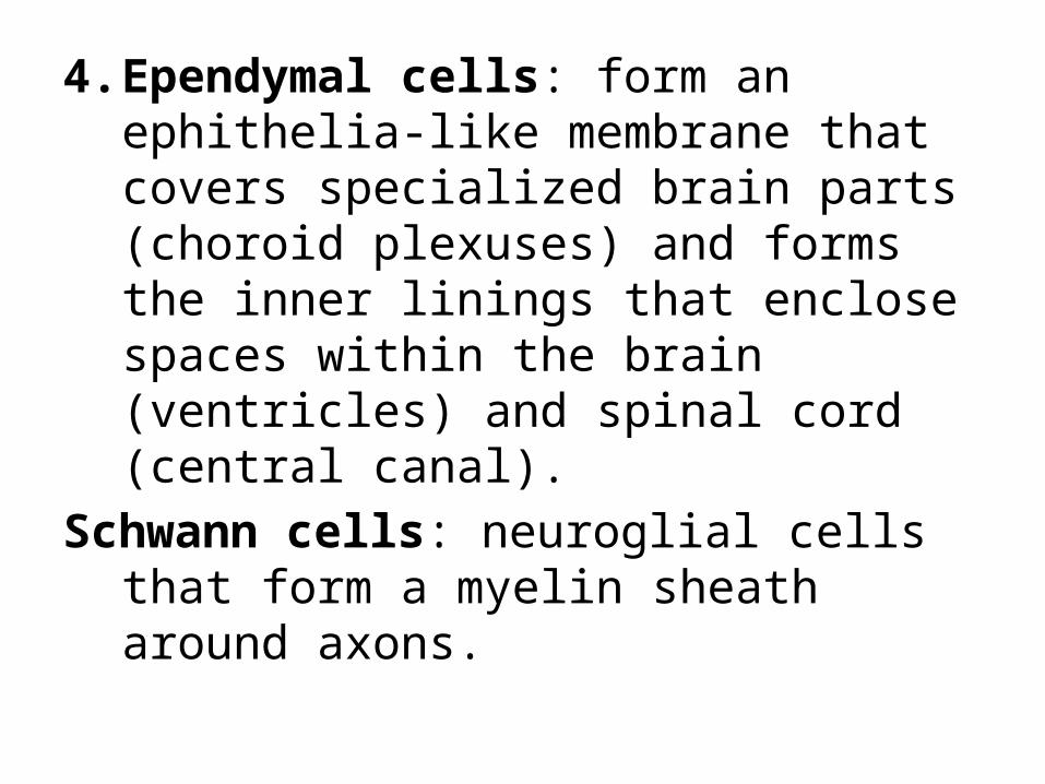
4. Ependymal cells: form an ephithelia-like membrane that covers specialized brain parts (choroid plexuses) and forms the inner linings that enclose spaces within the brain (ventricles) and spinal cord (central canal).
Schwann cells: neuroglial cells that form a myelin sheath around axons.
9.4 NEURONS• Neurofibrils: fine threads, extend into the axons (in
the cell body).• Neuron cell body: granular cytoplasm, cell
membrane, organelles.• Chromatophilic substance: membranous sacs,
similar to rough endoplasmic reticulum.• Axon: conducts nerve impulses away from the cell
body.• Schwann cells: contain myelin, neurilemma sheath
that surrounds the myelin sheath.• Nodes of Ranvier: gaps between Schwann cells
• Axons with myelin sheaths are called myelinated.
• Unmyelinated: lack sheaths.• Peripheral nerve Axons can regenerate• Schwann cells help do this.• CNS axons usually cannot regenerate
themselves.
CLASSIFICATION OF NEURONS• Neurons differ in structure, size, and shape of
their cell bodies.• Trigger zone: sensitive region of the axon,
send a nerve impulse.
THREE MAJOR GROUPS OF NEURONS1. Multipolar neurons: many processes arising from
their cell bodies. Only one process of each neuron is an axon; the rest are dendrites.
2. Bipolar neurons: only two proceses, one an axon and the other a dendrite. Ex: eyes, nose, and ears
3. Unipolar neurons: single process, divides into two branches, single axon. One branch is associated w/ dendrites near a peripheral body part. The other branch enters the brain or spinal cord.
• Ganglia: masses of nervous tissue.• Located outside the brain and spinal cord
• Different functions of neurons:• Carry impulses into the brain or spinal cord• Transmit impulses out of the brain or spinal
cord• Conduct impulses from neuron to neuron
within the brain or spinal cord
GROUPS OF DIFFERENT NEURONS1. Sensory neurons: (afferent neurons) carry
nerve impulses from peripheral body parts into the brain or spinal cord. Either have specialized receptor ends at the tips of their dendrites, or they have dendrites that are closely associated with receptor cells in the skin or in sensory organs. (most unipolar, some bipolar)
2. Interneurons: (association or internuncial neurons) lie within the brain or spinal cord. Multipolar, link other neurons.
3. Motor neurons: (efferent neurons) are multipolar and carry nerve impulses out of the brain or spinal cord to effectors. Stimulate muscles to contract and glands to release secretions.
9.5 CELL MEMBRANE POTENTIAL• Polarized: surface of the cell membrane,
electrically charged, unequal distribution of positive and negative ions
• Action potential: change in charge of the membrane, forms a nerve impulse
DISTRIBUTION OF IONS• Active transport of sodium and potassium
ions.• More sodium ions are outside and more
potassium ions inside.• Cytoplasm: PO4-3, SO4-2, protein, K+• Potassium pass through cell membrane easier
than sodium ions.• K+ a major contributor to membrane
polarization.
RESTING POTENTIAL• Resting cell membrane is more permeable to K+
that to Na+, K+ diffuse out of the cell more rapidly.
• Result, outside of cell gains a slight surplus of + charge.
• Inside – charge• Potential difference: difference in electrical
charge between two regions.• Resting potential: potential difference between
the region inside the membrane and the region outside the membrane.
• As long as a nerve cell membrane is undisturbed, the membrane remains in this polarized state.
POTENTIAL CHANGES• Nerve cells are excitable, they can respond to
changes in their surroundings.• Some detect changes in: temp, light, pressure• Many neurons respond to other neurons.• depolarizing: inside membrane becomes less
negative compared to outside.• Change in potential is proportional to the
intensity of stimulation.• Summation: change in potential is increases as
more stimulation comes to cell
Threshold potential: action potential occursACTION POTENTIAL• When threshold potential occurs: permeability
changes at the trigger zone of the neuron being stimulated.
• Channels highly selective for Na+ open and allow Na+ to diffuse freely inward.
• Aided by the negative electrical condition on the inside of the membrane, which attracts the positively charged sodium ions.

• As sodium ions diffuse inward, the membrane loses its negative electrical charge and becomes depolarized.
• Membrane channels open that allow potasssium ions to pass through, inside of membrane becomes negatively charged once more.
• Membrane returns to the resting potential.
• Rapid sequence of depolarization and repolarization, 1/1000 of a second, called action potential.
• http://www.youtube.com/watch?v=U0NpTdge3aw
9.6 NERVE IMPULSES• Wave of action potentials along a nerve axon –nerve
impulse.• Pg. 213IMPULSE CONDUCTION• Unmyelinated axon: conducts an impulse over its entire
surface.• Myelinated axon prevents almost all ion flow through the
membrane it encloses.• Though, nodes of Ranvier between schwann cells have
sodium and potassium channels, nerve impulse traveling along a myelinated axon appears to jump from node to node.
• Many times faster than conduction on an unmyelinated axon.
• Speed of nerve impulse conduction is proportional to the diameter of the axon.
• 120m/s nuerons of skeletal muscle• 0.5m/s unmyelinated axon neuron in skin

• http://www.youtube.com/watch?v=xx--f9Y8wjg
EVENTS LEADING TO THE CONDUCTION OF A NERVE IMPULSE
1. Neuron membrane maintains resting potential2. Threshold stimulus is received3. Sodium channels in the trigger zone of the
neuron open4. Sodium ions diffuse inward, depolarizing the
membrane5. Potassium channels in the membrane open6. Potassium ions diffuse outward, repolarizing
the membrane
7. The resulting action potential causes a local bioelectric current that stimulates adjacent portions of the membrane.
8. A wave of action potentials travels the length of the axon as a nerve impulse.
ALL OR NONE RESPONSE• If a neuron responds at all, it responds
completely.• A greater intensity of stimulation does not
produce a stronger impulse, but rather, more impulses per second.
• Refractory period: limits the frequency of impulses in a neuron, a threshold stimulus will not trigger another impulse on an axon.
• Common 100 impulses/second, 700 could happen

Homework
Pg. 246Ques. 1-14

• http://highered.mcgraw-hill.com/sites/0072495855/student_view0/chapter14/animation__the_nerve_impulse.html
9.7 THE SYNAPSE• Nerve pathways: pathway of nerve impulses.• Synapse: junction between any two
communicating neurons• Gap, synaptic cleft
SYNAPTIC TRANSMISSION• Neuron carrying the impulse: presynaptic
neuron• Neuron that receives this input, pstsynaptic
neuron• Synaptic transmission: process of crossing the
synaptic cleft with this message.
• Neurotransmitters: biochemicals that aid the synaptic transmission
• Distal end of axon have extensions called synaptic knobs.
• In dendrites they contain synaptic vesicles• When a nerve impulse reaches a synaptic
knob, some of the synaptic vesicles release neurotransmitters.
• Neurotransmitters diffuses across the synaptic cleft and reacts with specific receptors ont eh postsynaptic neuron membrane.
EXCITATORY AND INHIBITORY ACTIONS• Neurotransmitters that increase postsynaptic
membrane permeability to sodium ions will bring the postsynaptic membrane closer to threshold and may trigger nerve impulses.
• Excitatory: these neurotransmitters
• Inhibitory: neurotransmitters that make it less likely that threshold will be reached. Lessens the chance that a nerve impulse will occur.
• If more excitatory than inhibitory neurotransmitters are released, the postsynaptic neuron’s threshold may be reached, and a nerve impulse will be triggered.
NEUROTRANSMITTERS• Fifty types of neurotransmitters have been
identified in the nervous system.• Neurotransmitters: acetylcholine,
monoamines (epinephrine, norepinephrine, dopamine, serotonin)
• Amino acids (glycine, glutamic acid, aspartic acid, gamma-aminobutyric acid)
• Neuropeptides (short chains of amino acids)
• Acetylcholine and norepinephrine are excitatory
• Dopamine, GABA and glycine are inhibitory• Neurotransmitters are made in the cytoplasm
of the synaptic knobs, stored in the synaptic vesicles
• When an action potential reaches the membrane of a synaptic knob, it increases the membrane’s permeability to calcium ions by opening the membrane’s calcium ion channels.
• Calcium ions diffuse inward.• Some neurotransmitters are decomposed by
enzymes in the synaptic cleft (acetylcholinesterase)
• Decomposition or removal of neurotransmitters prevents continuous stimulation of postsynaptic neurons.
EVENTS LEADING TO THE RELEASE OF A NEUROTRANSMITTER
1. Action potential passes along an axon and over the surface of its synaptic knob
2. Synaptic knob membrane becomes more permeable to calcium ions, and they diffuse inward.
3. In the presence of calcium ions, synaptic vesicles fuse to synaptic knob membrane
4. Synaptic vesicles release their neurotransmitter into synaptic cleft
5. Synaptic vesicles reenter cytoplasm of axon and pick up more neurotransmitter