79: ' # '6& *#7 & 8 - opencdn.intechopen.com/pdfs-wm/15674.pdf · the biomimetic...
TRANSCRIPT

3,350+OPEN ACCESS BOOKS
108,000+INTERNATIONAL
AUTHORS AND EDITORS114+ MILLION
DOWNLOADS
BOOKSDELIVERED TO
151 COUNTRIES
AUTHORS AMONG
TOP 1%MOST CITED SCIENTIST
12.2%AUTHORS AND EDITORS
FROM TOP 500 UNIVERSITIES
Selection of our books indexed in theBook Citation Index in Web of Science™
Core Collection (BKCI)
Chapter from the book Advances in BiomimeticsDownloaded from: http://www.intechopen.com/books/advances-in-biomimetics
PUBLISHED BY
World's largest Science,Technology & Medicine
Open Access book publisher
Interested in publishing with IntechOpen?Contact us at [email protected]

4
The Biomimetic Approach to Design Apatites for Nanobiotechnological Applications
Norberto Roveri and Michele Iafisco Alma Mater Studiorum, Università di Bologna
Italy
1. Introduction
Mimicking Nature and designing bioinspired materials represents a promising way to reach technological innovations in many interdisciplinary scientific fields, since biological materials exhibit a high degree of sophistication, hierarchical organisation, hybridisation, efficiency, resistance and adaptability. These properties, which biogenic materials have achieved through specific building principles selected by evolution, can only be partially possessed in man-made materials by present synthetic processes. For this reason Nature is a school for material scientists, in view of the fact that living organisms can produce different amazing high-performance materials (Sanchez et al., 2005). Nature produces soft and hard materials exhibiting remarkable functional properties by controlling the hierarchical assembly of simple molecular building blocks from the nano- to the macro-scale. Biogenic materials are nucleated in defined nano–micro dimensioned sites inside the biological environments, in which chemistry can be spatially controlled (Mann et al., 1993; Weiner & Addadi 1997). The spatial delimitation is essential to biological mechanisms for controlling the size, shape and structural organisation of biomaterials. Recently, with the development of nanotechnology, this strategy employing natural material genesis has attracted a lot of attention in designing bioinspired materials such as polymeric micelles, nanoparticles, dendrimers and nanocrystals synthesised in nanoscale dimensions (Sarikaya et al., 2003; Tamerler & Sarikaya 2007; Vriezema et al., 2005). One of the most exciting and economical rewarding research areas of materials science involves the applications of materials to health care, especially to reconstructive surgery. In the past, many implantations failed due to infections or a lack of knowledge about toxicity of the selected materials. In this case, the use of calcium phosphates, since they are the most important inorganic constituents of hard tissues in vertebrates, is valid due to their chemical similarity to the mineral component of mammalian bones and teeth. Thus, calcium phosphate based biomaterials are now used in many different applications throughout the body, covering all areas of the skeleton. These applications include dental implants, percutaneous devices and use in periodontal treatment, treatment of bone defects, fracture treatment, total joint replacement (bone augmentation), orthopaedics, cranio-maxillofacial reconstruction, otolaryngology and spinal surgery (Dorozhkin 2010). In this chapter we will give details about the principal characteristics of bone, tooth and pathological calcifications where the calcium phosphates are present (section 2); the
www.intechopen.com
Advances in Biomimetics
76
chemical-physical characteristics and the methods to synthesize biomimetic hydroxyapatites (section 3); and the main applications of these in the nanobiotechnology field (section 4).
2. Biogenic hydroxyapatite
In biological systems, calcium phosphates are the principal inorganic constituent of normal (bones, teeth, fish enameloid, deer antlers and some species of shells) and pathological (dental and urinary calculus and stones, atherosclerotic lesions) calcifications. Except for small portions of the inner ear, all hard tissue of the human body is made of calcium phosphates. Structurally, they occur mainly in the form of poorly crystallized non-stoichiometric F, Na, Mg and carbonate substituted hydroxyapatite [Ca10(PO4)6(OH)2] (HA) (Dorozhkin 2010). We will thus give details in this section about the principal characteristics of bone, tooth and pathological calcifications, highlighting the fact that the living organisms can produce different amazing high-performance materials.
2.1 Bone Bones are rigid organs that form part of the endoskeleton of vertebrates. Their function is to move, support and protect the various organs of the body, produce red and white blood cells and store minerals (Loveridge 1999; Reddi 1994). Bones appear in a variety of shapes and have a complex internal and external structure, they are lightweight, yet strong and hard, in addition to satisfying their many other functions. One of the types of tissues that constitutes bone is the mineralized osseous tissue, also called bone tissue, that gives it rigidity and honeycomb-like three-dimensional structure (Cowles et al., 1998). Other types of tissue found in bones include marrow, endosteum and periosteum, nerves, blood vessels and cartilage (Tzaphlidou 2008). There are 206 bones in the adult body and about 300 bones in the infant body. Bone tissue consists of cells embedded in a fibrous, organic matrix, the osteoid, which is primarily constituted by type I collagen (90%) and 10% amorphous ground substance (primarily glycosaminoglycans and glycoproteins). Osteoid comprises approximately 50% of bone by volume and 25% by weight (Urist 2002). Osteoblasts are mono-nucleate cells responsible for the secretion of osteoid and subsequent bone formation through the mineralization of the osteoid matrix. They strongly produce alkaline phosphatase, an enzyme that has a role in the mineralisation of bone, as well as many matrix proteins. When osteoblasts are trapped in the bone matrix, which they themselves produced, they become star-shaped cells named osteocytes, the most abundant cells found in bone (Wozney 1992). Osteocytes are mature bone cells, networked to each other via long processes that occupy tiny canals called canaliculi, which are used for exchange of nutrients and waste. They are actively involved in the maintenance of bony matrix, through various mechanosensory mechanisms regulating the bone's response to stress. Bone is a dynamic tissue constantly being reshaped by osteoblasts, cells which build bone, and osteoclasts, cells which resorb it (Manolagas 2000). Osteoclasts are multi-nucleated cells responsible for the resorption of bone through the removal of the bone's mineralized matrix. The removal process begins with the attachment of the osteoclast to the osteon (predominant structures found in compact bone); the osteoclast then induces an infolding of its cell membrane and secretes collagenase and other enzymes important in the resorption process, such as tartrate resistant acid phosphatase,
www.intechopen.com

The Biomimetic Approach to Design Apatites for Nanobiotechnological Applications
77
secreted against the mineral substrate. During childhood, bone formation exceeds resorption but, as the aging process occurs, resorption exceeds formation (Legeros & Craig 1993). The characteristic rigidity and strength of bone derives from the presence of mineral salts, that permeate the organic matrix, formed by the osteoid mineralization, due to the secretion of vesicles containing alkaline phosphatase, by the osteoblasts (Boskey 2007). The mineral phase comprises approximately 50% of bone by volume and 75% by weight. The principal constituents of bone mineral are calcium phosphate, mainly carbonated hydroxyapatite, amorphous calcium phosphate and calcium carbonate, with lesser quantities of sodium, magnesium, silicon and fluoride (Palmer et al., 2008). The whole architecture of bone is very complex: starting from the smallest constituting elements to the largest scales, the bone is not only organized in an anisotropic manner, but the arrangement of its constituting elements is hierarchical; this means that its structural units are organized at increasing size levels, and this feature confers unique properties to the whole bone structure. To better understand the complex bone architecture, several hierarchical models have been proposed. Weiner and Wagner have identified seven discrete levels of hierarchical organization in bone (Figure 1), which we describe here (Weiner & Wagner 1998). In their model, bone is considered as a family of materials with the mineralized collagen fibre as the primary building block for subsequent higher order architectures.
Fig. 1. Seven hierarchical levels of organization of the bone family of materials as proposed by Weiner and Wagner. Reproduced with permission from (Weiner & Wagner 1998).
www.intechopen.com

Advances in Biomimetics
78
The structure of bone differs greatly among the different locations in the skeleton, but the basic nanoscale structure consisting of mineralized collagen remains the same throughout. The first hierarchical level consists of the molecular components: water, HA, collagen and other proteins. The crystals of HA are plate-shaped and are among the smallest known biological crystals (Table 1).
Composition Enamel Dentin Bone Calcium [wt %][a] 36.5 35.1 34.8
Phosphorus (as P) [wt %][a] 17.7 16.9 15.2 Ca/P (molar ratio) [a] 1.63 1.61 1.71
Sodium [wt %][a] 0.5 0.6 0.9 Magnesium [wt %][a] 0.44 1.23 0.72 Potassium [wt %][a] 0.08 0.05 0.03
Carbonate (as CO32-) [wt %][b]
3.5 5.6 7.4
Fluoride [wt %][a] 0.01 0.06 0.03 Chloride [wt %][a] 0.30 0.01 0.13
Pyrophosphate (as P2O74-) [wt %][b]
0.022 0.100 0.070
Total inorganic [wt %][b] 97 70 65 Total organic [wt %][b] 1.5 20 25
Water [wt %][b] 1.5 10 10 a axis [Å][c] 9.441 9.421 9.410 c axis [Å][c] 6.880 6.887 6.890
Crystallinity index, (HA=100)
70 – 75 33 - 37 33 - 37
Typical crystal sizes [nm] 100 × 50 × 50 μm 35 × 25 × 4 50 × 25 ×
4
Ignition products (800 °C) β-TCP + HA β-TCP + HA
HA + CaO
Elasticity modulus (GPa) 80 15 0.34 - 13.8 Compressive strength (MPa) 10 100 150
Table 1. Comparative composition and structural parameters of inorganic phases of adult-human calcified tissues ([a]Ashed samples. [b]Unashed samples. [c]Lattice parameters: ± 0.003 Å).
Though the size of biological apatite crystals reported in the literature varies due to the different treatment methods and analysis techniques utilized, it is generally around the nanometric level, with values in the ranges of 30–50 nm (length), 15–30 nm (width) and 2–10 nm (thickness) (Fernandez-Moran & Engstrom 1957). In early studies, apatite needles were observed, but other studies suggest that platelets are the dominant morphology and that the apparent needles are most likely to be platelets viewed edge-on (Traub et al., 1989). Noncollagenous proteins (NCPs) are also present but make up 10% or less of the total protein content in the bone matrix. The specific functions of the NCPs are still not completely understood. In addition to influencing crystal nucleation and growth, NCPs also play roles in cell signalling and ion homeostasis (Feng et al., 2006).
www.intechopen.com
The Biomimetic Approach to Design Apatites for Nanobiotechnological Applications
79
The second level is formed by the mineralization of collagen fibrils. Tiny plate-like crystals of biological apatite in bone occur within the discrete spaces within the collagen fibrils and grow with specific crystalline orientation along the c-axes, which are roughly parallel to the long axes of the collagen fibrils (Termine et al., 1981). The HA growth is somehow limited by these small intercollagenous spaces, which are approximately 50 nm in length, 25 nm in width and 2–3 nm thick (Cui et al., 2007). The location of these crystals in the fibril was demonstrated in a study by Traub et al. that showed that mineralized collagen fibrils had the same banded pattern as negatively stained collagen fibrils (Traub et al., 1989). This indicated that mineral is concentrated in the hole zones of the fibril. It was proposed that these mineral platelets were arranged in parallel like a stack of cards within the interstices of the fibril. Olszta et al. concluded from electron diffraction studies that the mineral plates are not quite as ordered as previously assumed (Olszta et al., 2007). This imperfect arrangement of nearly parallel crystals has been supported by recent small-angle X-ray scattering (SAXS) and transmission electron microscopy (TEM) data from Burger et al. (Burger et al., 2008). The third level of hierarchy is composed of arrays of these mineralized collagen fibrils. These fibrils are rarely found isolated, but rather almost always associated as bundles or other arrangements, often aligned along their long axis. Type I collagen molecules are self-assembled into fibrils with a periodicity of 67 nm and with 40 nm gaps between the end and the head of their molecules, into which the apatite crystals are placed. A composite of these two constituents forms mineralized fibrils (Wahl & Czernuszka 2006; Bigi et al., 1991; Bigi et al., 1996; Bigi et al., 1997). The fourth level is the patterns of arrays that are formed. These include parallel arrays, woven arrangements, plywood like structures and radial arrays like those found in dentin. Cylindrical structures called osteons make up the fifth level. Osteons are formed with significant cellular activity and remodeling; osteoclasts resorb bone and form a tunnel and osteoblasts subsequently lay down lamellae in stacked layers until only a small channel (Haversian canal) is left behind. These channels serve as a conduit for nerves and blood supply to the bone cells. The sixth level of bone organization is the classification of osseous tissue as either spongy (trabecular or cancelous) or compact (cortical). Cancellous bone is extremely porous (75-95% porosity), providing space for marrow and blood vessels, but has much lower compressive strength. Cortical bone is the dense outer layer (5-10% porosity) that allows many of the support functions of bone (Fratzl et al., 2004). Therefore, the mechanical properties of cortical bone represent the benchmark for synthetic bone. The seventh level is simply the whole bone on the macroscopic scale, incorporating all of the lower levels of hierarchy.
2.2 Tooth The mammalian body contains numerous mineralized tissues as reported in the previous paragraph, but the tissue with the most robust mechanical properties is enamel. Enamel is the hardest material formed by vertebrates and is the most highly mineralized skeletal tissue present in the body (Whittaker 1982). Mature enamel is composed of 95-97% carbonated HA by weight with less than 1% organic material. The high degree of mineralization makes enamel a fascinating model for understanding fundamental mineralization processes and processes that occur within an extracellular matrix. It is distinct from bone in terms of architecture, pathology and the biological mechanisms mediating its formation. Understanding the biological formation of different mineralized structures could lead to innovative approaches toward engineering novel scaffolds and providing new therapeutics. Additionally, unlike other biomineralized tissues, such as bone
www.intechopen.com
Advances in Biomimetics
80
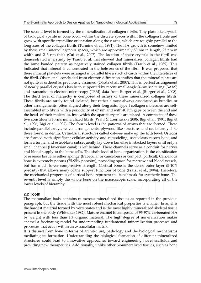
and dentin, mature enamel is acellular and does not resorb or remodel. As a result, enamel regeneration cannot occur in vivo following failure and is therefore an attractive target for future biomimetic and therapeutic approaches. The mammalian tooth is made up of four distinct structures: enamel, dentin, pulp and cementum (Figure 2).
Fig. 2. Hierarchical architecture of mammalian enamel. Enamel (E) is the outermost layer at the crown of the tooth and resides above the dentin (D). The pulp (P) contains nerves and blood vessels, while the cementum (C) is the outermost layer of mineralized tissue surrounding the root of the tooth allowing the tooth to be anchored to the jawbone through the periodontal ligament (PDL). The bulk image depicts the E organ, the transition across the D-E junction, and the D below. On the mesoscale level, prismatic E consisting of weaving of rods (or prisms) that range from 3 to 5 μm in diameter can be visualized. Upon further magnification, the micrometer scale shows the composition of a single rod. The nanometer scale reveals a highly organized array of individual HA crystallites (approximately 30 nm thick, 60 nm wide, and several millimeters in length), which are preferentially aligned along the c-axis. Reproduced with permission from (Tamerler & Sarikaya 2008).
The pulp contains nerves, blood vessels, fibroblasts and lymphocytes, while the mineralized organs of the tooth include enamel, dentin and cementum (Tamerler & Sarikaya 2008). Enamel makes up the uppermost 1-2 mm of the tooth crown and contains a high mineral content, giving it a high modulus but also making it susceptible to cracking. Dentin lies below the enamel and is tougher, forming the bulk of the tooth and absorbing stresses from enamel, preventing its fracture (Arsenault & Robinson 1989). The composition of dentin is similar to that of bone. The cementum is the mineralized layer that surrounds the root of the tooth covering the dentin layer and some of the enamel layer. The cementum allows for the anchoring of the tooth to the alveolar bone (jawbone) through the periodontal ligament. The
www.intechopen.com
The Biomimetic Approach to Design Apatites for Nanobiotechnological Applications
81
primary function of the tooth is for mastication of food; however, some species use them for attacking prey and for defence. It also faces the lifelong challenge of maintaining robust mechanical properties in a bacteria-filled environment. The enamel and dentin tissues give rise to a tough, crack-tolerant and abrasion-resistant tissue through their unique architectures and mineral compositions. Enamel is highly patterned and consists of organized interweaving bundles of crystallites (called rods or prisms). It has a higher reported toughness than that of crystalline HA, indicating that the organization of the crystallites is essential for enamel function (White et al., 2001). Because of the high mineral content and minimal organic, enamel is brittle. Interestingly, the architecture of the enamel crystallites can deflect a propagating crack preventing it from reaching the dentin-enamel junction (DEJ), which also has been shown to resist delamination of the tissues despite their differences in composition (Imbeni et al., 2005). The mechanical properties of enamel, dentin and the DEJ are not completely understood and are a significant area of research. Understanding the properties of these tissues could serve to motivate further engineering of more robust dental materials as well as to inspire fabrication of nonbiological materials. Similar to bone, enamel possesses a complex architecture, which can be broken into several hierarchical levels from the nanoscale to the macroscale (Paine et al., 2001). On the nanoscale, the protein-protein and protein-mineral interactions in the presence of supersaturated ions create a highly organized array of HA crystallites that grow preferentially along the c-axis (Wen et al., 2000). The sizes of these crystallites depend on the stage of the mineralization. The crystallites grow primarily in length during the secretory stage and continue to grow in width and thickness during the maturation stage. The assembly of amelogenin has been shown to be crucial for the proper development of enamel crystallites. Disruption of the assembly alters formation on the nanoscale, subsequently affecting larger length scales and giving rise to a diseased or malformed enamel phenotype. On the mesoscale level, there are three main structural components: the rod, the interrod and the aprismatic enamel. The main component of enamel on the mesoscale includes rods, which are bundles of aligned crystallites that are “woven” into intricate architectures that are approximately 3-5 μm in diameter (Cui & Ge 2007). The second structural component of the enamel matrix is the interrod (or interprismatic) enamel, which surrounds and packs between the rods. The difference between the rod and the interrod is the orientation of HA crystals; the rod contains aligned crystallites, whereas the mineral in the interrod is less ordered. These structures coalesce to form the tough tissue of enamel, which can withstand high forces and resist damage by crack deflection. The third structure, aprismatic enamel, refers to the structures containing HA crystals that show no mesoscale or macroscale alignment. The macroscale architecture includes specific zones of enamel that have unique characteristics, which contribute to the whole tissue. The enamel adjacent to the DEJ exhibits a gradual transition from dentin to enamel. Aprismatic regions of enamel have been proposed to be primitive areas of the tooth serving as a toughening mechanism due to their flexible nature (Wang & Weiner 1998). Several authors have identified these aprismatic areas to be located adjacent to the DEJ and at the incisal surface of both deciduous and permanent human enamel (Kodaka et al., 1989). The Tomes’ process, a unique structure present at the secretory pole of an enamel-forming cell, is responsible for aligned mineral formation in the prismatic enamel. The absence of this process may give rise to the aprismatic zone in the tooth.
www.intechopen.com
Advances in Biomimetics
82
2.3 Pathological calcifications In the body of mammals, osteoblasts and odontoblasts fix ions of calcium and orthophosphate and then precipitate biological apatite onto an organic matrix. This is the process of physiological biomineralization that is restricted to the specific sites in skeletal tissues, including growth plate cartilage, bones and teeth. Unfortunately, owing to ageing, to various diseases and under certain pathological conditions, blood vessels and some internal organs are calcified as well (Daculsi et al., 1997). This process is called pathological calcification or ectopic mineralization and leads to a morbidity and a mortality. In general, any type of abnormal accumulation of calcium phosphates in wrong places is accounted for by a disruption of systemic defense mechanism against calcification (Kazama et al., 2006). Unwanted depositions always lead to various diseases, for instance: soft tissue calcification (in damaged joints, blood vessels, dysfunctional areas in the brain, diseased organs, scleroderma, prostate stones) (Brancaccio & Cozzolino 2005), kidney and urinary stones (Achilles et al., 1995), dental pulp stones and dental calculus (Hayashizaki et al., 2008), salivary stones, gall stones, pineal gland calcification, atherosclerotic arteries and veins (Marra et al., 2006), coronary calcification, cardiac skeleton, damaged cardiac valves, calcification on artificial heart valves (Weissen-Plenz et al., 2008; Bigi et al., 1988), carpal tunnel, cataracts, malacoplakia, calcified menisci, dermatomyositis and other. All these cases are examples of a calcinosis, which might be described as a formation of calcium phosphate deposits in any soft tissue (Laird et al., 2006). Contrary to the mineral phases of the normal calcifications (bone, dentin, enamel, cementum, antlers), which consist of only one type of calcium phosphate (namely, biological apatite), the mineral phases of abnormal and/or pathological calcifications are found to occur as single or mixed phases of other types of calcium phosphates and/or other phosphatic and non-phosphatic compounds, in addition to or in place of biological apatite (Wesson & Ward 2007). This happens because the solution pH is often relatively low in the places of pathological calcifications. However, in some cases, the chemical composition of an unwanted inorganic phase might depend on the age of the pathological calcification and its location (Legeros et al., 1988). It is interesting to note that the mineral phases of animal calculus (e.g., from dog) was found to consist of calcium carbonate and biological apatite, while human calculi do not contain calcium carbonate. Some findings suggested that the mechanisms and factors regulating the physiological biomineralization might be similar to those influencing the ectopic mineralization: both were initiated by various organics (i.e., membrane-enclosed particles released from the plasma membrane of mineralization-competent cells), that were present (Kirsch 2006).
3. Synthetic biomimetic hydroxyapatites
Biomimetism of synthetic calcium phosphates can be carried out at different levels, such as composition, structure, morphology, bulk and surface chemical–physical properties. Biomaterials can be turned into biomimetic imprinting all these characteristics in order not only to optimise their interaction with biological tissues, but also to mimic biogenic materials in their functionalities. Detailed information on calcium phosphates, their synthesis, structure, chemistry, other properties and biomedical application have been comprehensively reviewed recently (Dorozhkin 2010; LeGeros 2008). In this section the chemical-physical characteristics and some methods to synthesize biomimetic calcium phosphates will be described.
www.intechopen.com

The Biomimetic Approach to Design Apatites for Nanobiotechnological Applications
83
3.1 Chemical-physical characteristics of nanocrystalline hydroxyapatites The study of nanocrystalline calcium phosphate physical-chemical characteristics and, thereafter, the possibility to imitate bone mineral for the development of new advanced biomaterials is constantly growing. The possibility to synthesize in the laboratory synthetic biomimetic compounds makes it possible to consider these systems as a bone mineral “model”, enabling to investigate on the one hand the interaction between bone-like apatite nanocrystals and the components of surrounding fluids (ions, proteins…) and, on the other hand, to follow surface interactions with drugs aimed at being delivered in vivo. Among the peculiarities of these compounds, their plate-like morphology (elongated towards the c-axis) is one obvious specificity as compared to regular HA. Another difference is of course the mean crystallite size, of the order of 15-30 nm in length and ca. 6-9 nm in width. From a chemical point of view, the composition of nanocrystalline apatites also strongly differs from that of HA. Although it has been the object of much controversy during several decades in the middle of the 20th Century, the global chemical composition of biological apatites (or their synthetic analogues) can generally be described as:
Ca10-x (PO4)6-x (HPO4 or CO3)x (OH or ½ CO3)2-x with 0 ≤ x ≤ 2
(except maybe for very immature nanocrystals which may depart from this generic formula). In particular, this expression underlines the presence of vacancies in both Ca and OH sites. For example, Legeros et al. (Legros et al., 1987) analyzed various cortical bone specimens, suggesting the following relatively homogeneous composition, unveiling high vacancy contents:
Ca8.3 (PO4)4.3 (HPO4 or CO3)1.7 (OH or ½ CO3)0.3
Minor substitutions are also found in biological apatites involving for example monovalent cations (especially Na+) in cationic sites. In this case, charge compensation mechanisms have to be taken into account. It should however be kept in mind that such chemical formulas only enable to have a “global” insight on the nature and amount of ions present in the compound, but it does not reflect possible local variations that may be observed on the nanocrystals or in vivo within the osteons (Paschalis et al., 1996), linked to the local apatite crystal formation and, in vivo, to continuous bone remodelling. The characterization of nanocrystalline apatites is a relatively arduous task due to their poor crystallinity and metastability. But despite the complexity of these systems, recent in-depth investigations have succeeded to reveal their particular surface features (and related reactivity in wet media) that may be exploited for the setup of new biomaterials, or for a better understanding of natural or pathological biomineralization phenomena. Recent advances in the characterization of apatite nanocrystals were obtained thanks to the use of spectroscopic techniques and, in particular, Fourier Transform Infrared (FT-IR) spectroscopy. FT-IR method is useful for drawing conclusions on the local chemical environment of phosphate, carbonate and hydroxide ions as well as water molecules in such systems. Detailed analyses of the phosphate groups by FT-IR have enabled to distinguish in nanocrystalline apatites the presence of additional bands that cannot be attributed to phosphate groups in a regular apatitic environment (Rey et al., 1990; Rey et al., 1989). These chemical environments have been referred to by Rey as “non-apatitic” environments.
www.intechopen.com

Advances in Biomimetics
84
Taking into account all the above data, nanocrystalline apatites (whether biological or their synthetic analogues prepared under close-to-physiological conditions) may thus most probably be described as the association of an apatitic core (often non stoichiometric) and a structured by fragile surface hydrated layer containing water molecules and rather labile ions (e.g. Ca2+, HPO42-, CO32-…) (Drouet et al., 2009) occupying non-apatitic crystallographic sites (although in the case of biomimetic apatites the layer is directly exposed on the surface and not included in a “sandwich-like” structure between two “apatitic” layers). A schematic model for such nanocrystalline apatites is given in Figure 3 (Eichert et al., 2007).
Fig. 3. Schematic modelling of a biomimetic apatite nanocrystal (a) and interaction with surrounding fluids (b) (from reference (Eichert et al., 2007)), with permission.
The presence of this hydrated surface layer (Sakhno et al., 2010) is thought to be responsible for most of the properties of biomimetic apatites and, in particular, their high surface reactivity in relation with surrounding fluids (which is probably directly linked to a high mobility of ionic species contained within this layer) may explain, from a physical-chemical viewpoint, the role of bone mineral in homeostasis in vivo. This layer indeed contains labile ions that can potentially be exchanged by other ions from the surrounding solution, or by small molecules, which may be exploited for couplings with proteins or drugs. It is interesting to remark that the typical non-apatitic features mentioned above tend to progressively disappear during the ageing of the nanocrystals in solution (Cazalbou et al., 2005; Drouet et al., 2009). This process is referred to as “maturation” and has been related to the progressive growth of apatite domains at the expense of the surface hydrated layer (Cazalbou et al., 2004). This maturation process is thought to be linked to the metastability of such poorly-crystallized non stoichiometric apatites, which steadily evolve in solution towards stoichiometry and better crystallinity (Cazalbou et al., 2005; Cazalbou et al., 2004). This evolution can be for example witnessed by the decrease of the amount of non-apatitic HPO42- ions upon ageing, or else by the decreased potentialities to undergo ion exchanges (Cazalbou et al., 2005). One illustration of such effects can be given for example by the decreased exchangeability of HPO42- by CO32- observed on carbonated apatites matured for incremented amounts of time. Additionally, beside this compositional evolution, some structural and microstructural features also tend to evolve such as the mean crystallite size which increases upon maturation. The control of synthesis parameters such as pH, temperature or maturation time can thus enable one to tailor the physical-chemical
www.intechopen.com
The Biomimetic Approach to Design Apatites for Nanobiotechnological Applications
85
properties of biomimetic apatites (Drouet et al., 2009) and, in particular, their surface reactivity, so to mimic for example mature bone mineral or else newly-formed bone apatite.
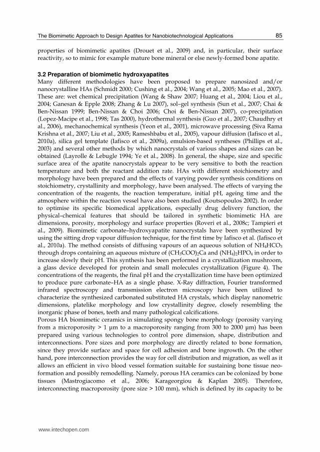
3.2 Preparation of biomimetic hydroxyapatites Many different methodologies have been proposed to prepare nanosized and/or nanocrystalline HAs (Schmidt 2000; Cushing et al., 2004; Wang et al., 2005; Mao et al., 2007). These are: wet chemical precipitation (Wang & Shaw 2007; Huang et al., 2004; Liou et al., 2004; Ganesan & Epple 2008; Zhang & Lu 2007), sol–gel synthesis (Sun et al., 2007; Chai & Ben-Nissan 1999; Ben-Nissan & Choi 2006; Choi & Ben-Nissan 2007), co-precipitation (Lopez-Macipe et al., 1998; Tas 2000), hydrothermal synthesis (Guo et al., 2007; Chaudhry et al., 2006), mechanochemical synthesis (Yeon et al., 2001), microwave processing (Siva Rama Krishna et al., 2007; Liu et al., 2005; Rameshbabu et al., 2005), vapour diffusion (Iafisco et al., 2010a), silica gel template (Iafisco et al., 2009a), emulsion-based syntheses (Phillips et al., 2003) and several other methods by which nanocrystals of various shapes and sizes can be obtained (Layrolle & Lebugle 1994; Ye et al., 2008). In general, the shape, size and specific surface area of the apatite nanocrystals appear to be very sensitive to both the reaction temperature and both the reactant addition rate. HAs with different stoichiometry and morphology have been prepared and the effects of varying powder synthesis conditions on stoichiometry, crystallinity and morphology, have been analysed. The effects of varying the concentration of the reagents, the reaction temperature, initial pH, ageing time and the atmosphere within the reaction vessel have also been studied (Koutsopoulos 2002). In order to optimise its specific biomedical applications, especially drug delivery function, the physical–chemical features that should be tailored in synthetic biomimetic HA are dimensions, porosity, morphology and surface properties (Roveri et al., 2008c; Tampieri et al., 2009). Biomimetic carbonate–hydroxyapatite nanocrystals have been synthesized by using the sitting drop vapour diffusion technique, for the first time by Iafisco et al. (Iafisco et al., 2010a). The method consists of diffusing vapours of an aqueous solution of NH4HCO3 through drops containing an aqueous mixture of (CH3COO)2Ca and (NH4)2HPO4 in order to increase slowly their pH. This synthesis has been performed in a crystallization mushroom, a glass device developed for protein and small molecules crystallization (Figure 4). The concentrations of the reagents, the final pH and the crystallization time have been optimized to produce pure carbonate–HA as a single phase. X-Ray diffraction, Fourier transformed infrared spectroscopy and transmission electron microscopy have been utilized to characterize the synthesized carbonated substituted HA crystals, which display nanometric dimensions, platelike morphology and low crystallinity degree, closely resembling the inorganic phase of bones, teeth and many pathological calcifications. Porous HA biomimetic ceramics in simulating spongy bone morphology (porosity varying from a microporosity > 1 μm to a macroporosity ranging from 300 to 2000 μm) has been prepared using various technologies to control pore dimension, shape, distribution and interconnections. Pore sizes and pore morphology are directly related to bone formation, since they provide surface and space for cell adhesion and bone ingrowth. On the other hand, pore interconnection provides the way for cell distribution and migration, as well as it allows an efficient in vivo blood vessel formation suitable for sustaining bone tissue neo-formation and possibly remodelling. Namely, porous HA ceramics can be colonized by bone tissues (Mastrogiacomo et al., 2006; Karageorgiou & Kaplan 2005). Therefore, interconnecting macroporosity (pore size > 100 mm), which is defined by its capacity to be
www.intechopen.com
Advances in Biomimetics
86
Fig. 4. Crystallization mushroom sketch. Carbonate-hydroxyapatite crystals were grown in sitting droplets on microbridges. NH3(g) and CO2(g) slowly diffused from the reservoir to the droplet through the small opening in the plate at bottom.
colonized by cells, is intentionally introduced in solid ceramics (Lu et al., 1999). Porous HA ceramics processed by high-temperature treatment present a significant reduction of bioreactivity and growth kinetics of new bone due to the lack of resorbability (Rodriguez-Lorenzo et al., 2001). However, the low resorbability of sintered HA-ceramics appears useful when they have to be implanted with a defined 3D form. Macroporosity is usually formed due to release of various volatile materials and, for that reason, incorporation of pore creating additives (porogens) is the most popular technique to create macroporosity. Among porogens additives we can mention paraffin, naphthalene, fluor, hydrogen peroxide, polyvinyl butyral, etc (Tancret et al., 2006; Walsh & Tanaka 2001; Chevalier et al., 2008). These additives are admixed to HA powders or slurries. After moulding, the organics burn away from the moulding body during sintering. This approach allows direct control of the pore characteristics, which are a function of the amount and properties of the volatile phase. Several other techniques, such as replication of polymer foams by impregnation, dual-phase mixing technique, freeze casting, strereolithography and foaming of gel-casting suspensions and transformation of porous calcium carbonate from biological origin (marine corals or echinoderms skeletons) into porous calcium phosphates by treatment with phosphate solutions under hydrothermal conditions have been applied to fabricate porous calcium phosphate ceramics (Gonzalez-McQuire et al., 2005; Liu 1997; Walsh et al., 2005; Charriere et al., 2003). By using the latter technique the interconnected porosity of the aragonitic or calcitic skeletons of corals or echinoderms is maintained after transformation to HA or mixtures of HA and other calcium phosphates (Araiza et al., 1999). The preparation methods of porous bioceramics has been recently reviewed by Sopiana et al. (Sopyan et al., 2007). Many studies have demonstrated that HA ceramics can be used to deliver steroids, antibiotics, proteins, hormones, anticancer drugs. Porous ceramics closely mimicking spongy bone morphology have been synthesized by impregnation of cellulosic sponges with poorly crystalline HA water suspension (Figure 5) (Palazzo et al., 2005). These porous ceramics have been tested as controlled drug delivery bone grafts to evaluate the fundamental parameters that control release kinetics. A theoretical approach, based on the use of the Finite Element Method, was adopted to describe the ibuprofen-lysine and hydrocortisone Na-succinate release kinetics, comparing the numerical results with the experimental ones.
www.intechopen.com

The Biomimetic Approach to Design Apatites for Nanobiotechnological Applications
87
Fig. 5. Scanning electron microscopy image of porous hydroxyapatite
4. Applications of biomimetic hydroxyapatites
The main driving force behind the use of calcium phosphates as bone substitute materials is their chemical similarity to the mineral component of mammalian bones and teeth, as previously explained. As a result, in addition to being non-toxic, they are biocompatible, not recognized as foreign materials in the body and, most importantly, they exhibit bioactive behavior and integrate into living tissue by the same processes active in remodeling healthy bone. This leads to an intimate physical-chemical bond between the implants and bone, termed osteointegration (Puleo 2004). In fact, materials can interact with biomolecules as well as with living systems and these interactions can be used to develop new materials and technologies. In particular, surface interaction of proteins and peptides with calcium phosphates based biomaterial represents the first and pivotal event when materials are implanted within an organism and must integrate in the host tissues, affecting the accompanying biological responses, namely cell attachment and activation (Iafisco et al., 2008; Iafisco et al., 2010b; Palazzo et al., 2009). More to the point, calcium phosphates are also known to support osteoblast adhesion and proliferation (Hong et al., 2003; Sader et al., 2009). Even so, the major limitations to use calcium phosphates as load-bearing biomaterials are their mechanical properties; namely, they are brittle with a poor fatigue resistance (Dorozhkin 2009). The poor mechanical behavior is even more evident for highly porous ceramics and scaffolds because porosity greater than 100 µm is considered a requirement for proper vascularization and bone cell colonization (Sader et al., 2009). That is why, in biomedical applications, calcium phosphates are used primarily as fillers and coatings (de Groot 1993).
4.1 Scaffolds Following the biomimetic approach, inspiring to Nature, natural wood templates have been selected as a starting point to obtain open-pore geometries with high surface area and microstructure allowing cell in-growth and reorganization and providing the necessary space for vascularisation (Li et al., 2006; Zimmerman et al., 2002). In fact, the alternation of fibre bundles and channel-like porous areas makes the wood an elective material to be used as template in starting the development of new bone substitute biomaterials by an ideal biomimetic hierarchic structure (Singh et al., 2003). Hydroxyapatite bone scaffolds characterized by highly organized hierarchical structures have been recently obtained by chemically transforming native woods through a sequence of thermal and hydrothermal processes. The whole chemical conversion has been carried out through five chemical steps
www.intechopen.com

Advances in Biomimetics
88
from native wood to porous hydroxyapatite: i) pyrolysis of ligneous raw materials to produce carbon templates characterized by the natural complex anisotropic pore structure; ii) carburization process by vapour or liquid calcium permeation to yield calcium carbide; iii) oxidation process to transform calcium carbide into calcium oxide; iv) carbonation by hydrothermal process under CO2 pressure for the further conversion into calcium carbonate; v) phosphatization process through hydrothermal treatment to achieve the final hydroxyapatite phase. The five steps of the phase transformation process have been set up in order to achieve total phase conversion and purity maintaining the original native microstructure. An innovative biomimetic apatite hierarchically structured in parallel fastened hollow microtubules has been synthesized, structurally characterized and proposed as new inorganic biomorphic scaffold providing a biomimetic nanostructured surface for fascinating bone engineering applications (Tampieri et al., 2009). The SEM image reported in Figure 6A demonstrates that the structured anisotropy typical of the native woods was preserved on the macro-scale, exhibiting in the case of rattan pore sizes in the range 100–300 μm, revealing an ordered fastening of parallel micro-tubes 100–150 μm long and 15–30 μm wide with a hollow core of about 10–25 μm in diameter, organized like the cell morphology of the natural wood used as the starting template for its synthesis. SEM image reported in Figure 6B of the newly formed HA surface morphology shows typical needle-like nuclei grown on the surface, proving the concomitant occurrence of a dissolution/precipitation process at sub-micron level, in agreement with hypotheses previously reported. This surface nanostructured morphology of the unidirectional fastened hollow HA microtubules allows biological systems like cells to utilize biomorphic scaffolds on the micrometer level which are also biomimetic for composition and structure on the nanometer scale.
Fig. 6. Detailed SEM images of pine wood-derived hydroxyapatite: a) microstructure of wood-derived parallel fastened hydroxyapatite microtubes; b) typical needle-like HA nuclei grown on the micro-tube surface; inset in b shows an higher magnification of picture b. Reproduced with permission from (Tampieri et al., 2009).
4.2 Drug delivery Over the past few decades, the rise of modern pharmaceutical technology and the amazing growth of the biotechnology industry have revolutionized the approach to drug delivery systems development. For most of the industry’s existence, pharmaceuticals have primarily consisted of relatively simple, fast-acting chemical compounds that are dispensed orally (as
www.intechopen.com

The Biomimetic Approach to Design Apatites for Nanobiotechnological Applications
89
solid pills and liquids) or injected. During the past three decades, however, complex formulations that control the rate and period of drug delivery (i.e., time-release medications) and that target specific areas of the body for treatment have become increasingly common. A controlled release drug delivery system should be able to achieve the following benefits: (i) maintenance of optimum therapeutic drug concentration in the blood with minimum fluctuation; (ii) predictable and reproducible release rates for extended duration; (iii) enhancement of activity duration for short half-life drugs; (iv) elimination of side effects, frequent dosing and wastage of drug; and (v) optimized therapy and improved patient compliance. Hydroxyapatites and other calcium phosphates may find several applications in implant drug devices. In fact, as drug carriers, calcium phosphate nanoparticles have some advantageous properties. They are dissolved at low pH (around 4), e.g. in lysosomes after the cellular intake or in the environment of solid tumours, thereby releasing incorporated drugs or biomolecules (Roveri et al., 2008b; Epple et al., 2010). Their size can easily be controlled by stabilizing agents, such as polymers or nucleic acids. The nanoparticles can be made to fluoresce by the incorporation of lanthanide ions and they can also act as carriers for different drugs (Al-Kattan et al., 2010). For example, one of the most efficient ways to improve the bone forming ability of biomaterials is their association with bone morphogenetic proteins (Autefage et al., 2009). Thus the choice of the process used to prepare implantable bone biomaterials has an influence on the release of the associated bioactive molecules. Various techniques associating drugs with a calcium phosphate biomaterial have been reported: adsorption and impregnation allow the therapeutic agent to be incorporated at the surface of the biomaterial, whereas centrifugation and vacuum based-techniques enable it to enter into pores of biomaterials (Gautier et al., 2001; Gautier et al., 2000). Generally bioactive molecules, such as growth factors, are incorporated in biomaterials by simple impregnation, followed by drying, and the type of bonding with the substrate and the release rate are often undetermined (Alam et al., 2001). It is suspected that such associations do not allow the chemical bonding of the growth factor to the biomaterial and thus the release rate is often difficult to control. For example, precipitation and clustering of the growth factor molecules may occur and the release is only determined by local dissolution and diffusion rules. The uncontrolled release of growth factors has, in some instances, been related to an accelerated resorption of bone tissue and of the implant (Autefage et al., 2009). Since growth factors agents can stimulate the degradation as well as the formation of bone (depending on their local concentrations), they could impair the osteoconductivity of the coated-implant surface (Liu et al., 2007). Similarly, bisphosphonates molecules (BPs), by affecting bone remodeling, could also block the bone repair process: the drug at too high concentration could have detrimental effects on the fixation of the implant over longer periods of time. Zoledronate grafted to HA coating on titanium implants shows a dose-dependent effect on the inhibition of resorption activity according to the amount of zoledronate loaded (Peter et al., 2005). Local and slow administration of antineoplasic drug as methotraxate (MTX) is also useful to avoid systemic side effects and, because its time effect (the sensitivity of cells to this drug increases with time) is greater than dose effect (Lebugle et al., 2002). Adsorption, on the contrary, leads to stable association and control of the amount of bioactive molecules contained in the solid implant and thus of the dose released. Generally,
www.intechopen.com
Advances in Biomimetics
90
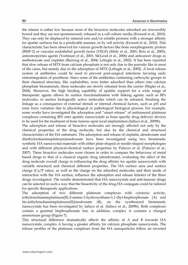
the release is rather low because most of the bioactive molecules adsorbed are irreversibly bound and they are not spontaneously released in a cell culture media (Errassif et al., 2010). They can only be displaced by mineral ions and/or soluble proteins with a stronger affinity for apatite surfaces but in a predicable manner, or by cell activity (Errassif et al., 2010). This characteristic has been observed for various growth factors like bone morphogenetic protein (BMP-2) or vascular endothelial growth factor (VEGF) (Midy et al., 2001; Boix et al., 2005), antiosteoporisis agents (Yoshinari et al., 2001; McLeod et al., 2006) and anticancer drugs as methotraxate and cisplatin (Barroug et al., 2004; Lebugle et al., 2002). It has been reported that slow release of MTX from calcium phosphate is not only due to the porosity like in most of the cases, but mainly due to the adsorption of MTX (Lebugle et al., 2002). A local release system of antibiotics could be used to prevent post-surgical infections favoring early osteointegration of prosthesis. Since some of the antibiotics containing carboxylic groups in their chemical structure, like cephalothin, were better adsorbed than others into calcium phosphate biomaterials, these molecules are slowly released from the carrier (Stigter et al., 2004). Moreover, the high binding capability of apatitic support for a wide range of therapeutic agents allows its surface functionalisation with linking agents, such as BPs molecules, to anchor biologically active molecules which can be released, breaking the linkage as a consequence of external stimuli or internal chemical factors, such as pH and ionic force variation due to physiological or pathological biological process. For example, some works have investigated the adsorption and “smart release” of antitumoral platinum complexes containing BPs onto apatitic nanocrystals as bone specific drug delivery devices to be used for the treatment of bone tumors upon local implantation (Iafisco et al., 2009b). The adsorption and release of bioactive molecules are strongly affected not only by the chemical properties of the drug molecule, but also by the chemical and structural characteristics of the HA substrates. The adsorption and release of cisplatin, alendronate and di(ethylendiamineplatinum)medronate have been investigated using two biomimetic synthetic HA nanocrystal materials with either plate-shaped or needle-shaped morphologies and with different physical-chemical surface properties by Palazzo et al. (Palazzo et al., 2007). These bioactive molecules were chosen in order to compare the behaviour of metal based drugs to that of a classical organic drug (alendronate), evaluating the effect of the drug molecule overall charge in influencing the drug affinity for apatite nanocrystals with variable structural and chemical different properties. The HA surface area and surface charge (Ca/P ratio), as well as the charge on the adsorbed molecules and their mode of interaction with the HA surface, influence the adsorption and release kinetics of the three drugs investigated. The results demonstrated that HA nanocrystals and anti-tumour drugs can be selected in such a way that the bioactivity of the drug-HA conjugate could be tailored for specific therapeutic applications. The adsorption of two different platinum complexes with cytotoxic activity, {ethylenediamineplatinum(II)-2-amino-1-hydroxyethane-1,1-diyl-bisphosphonate (A) and bis-{ethylenediamineplatinum(II)}medronate (B), on the synthesized biomimetic nanocrystals has been investigated by Iafisco et al. (Iafisco et al., 2009b). Both complexes contain a geminal bisphosphonate but, in addition, complex A contains a charged ammonium group (Figure 7). This structural difference dramatically affects the affinity of A and B towards HA nanocrystals, complex A having a greater affinity for calcium phosphate nanocrystals. The release profiles of the platinum complexes from the HA nanoparticles follow an inverted
www.intechopen.com
The Biomimetic Approach to Design Apatites for Nanobiotechnological Applications
91
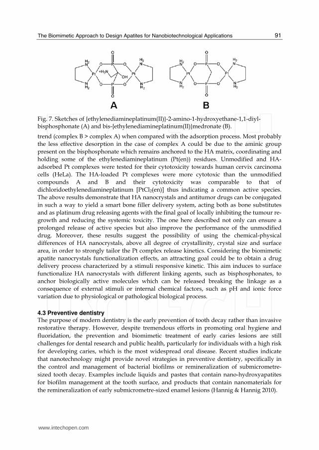
Fig. 7. Sketches of {ethylenediamineplatinum(II)}-2-amino-1-hydroxyethane-1,1-diyl-bisphosphonate (A) and bis-{ethylenediamineplatinum(II)}medronate (B).
trend (complex B > complex A) when compared with the adsorption process. Most probably the less effective desorption in the case of complex A could be due to the aminic group present on the bisphosphonate which remains anchored to the HA matrix, coordinating and holding some of the ethylenediamineplatinum (Pt(en)) residues. Unmodified and HA-adsorbed Pt complexes were tested for their cytotoxicity towards human cervix carcinoma cells (HeLa). The HA-loaded Pt complexes were more cytotoxic than the unmodified compounds A and B and their cytotoxicity was comparable to that of dichloridoethylenediamineplatinum [PtCl2(en)] thus indicating a common active species. The above results demonstrate that HA nanocrystals and antitumor drugs can be conjugated in such a way to yield a smart bone filler delivery system, acting both as bone substitutes and as platinum drug releasing agents with the final goal of locally inhibiting the tumour re-growth and reducing the systemic toxicity. The one here described not only can ensure a prolonged release of active species but also improve the performance of the unmodified drug. Moreover, these results suggest the possibility of using the chemical-physical differences of HA nanocrystals, above all degree of crystallinity, crystal size and surface area, in order to strongly tailor the Pt complex release kinetics. Considering the biomimetic apatite nanocrystals functionalization effects, an attracting goal could be to obtain a drug delivery process characterized by a stimuli responsive kinetic. This aim induces to surface functionalize HA nanocrystals with different linking agents, such as bisphosphonates, to anchor biologically active molecules which can be released breaking the linkage as a consequence of external stimuli or internal chemical factors, such as pH and ionic force variation due to physiological or pathological biological process.
4.3 Preventive dentistry The purpose of modern dentistry is the early prevention of tooth decay rather than invasive restorative therapy. However, despite tremendous efforts in promoting oral hygiene and fluoridation, the prevention and biomimetic treatment of early caries lesions are still challenges for dental research and public health, particularly for individuals with a high risk for developing caries, which is the most widespread oral disease. Recent studies indicate that nanotechnology might provide novel strategies in preventive dentistry, specifically in the control and management of bacterial biofilms or remineralization of submicrometre-sized tooth decay. Examples include liquids and pastes that contain nano-hydroxyapatites for biofilm management at the tooth surface, and products that contain nanomaterials for the remineralization of early submicrometre-sized enamel lesions (Hannig & Hannig 2010).
www.intechopen.com
Advances in Biomimetics
92
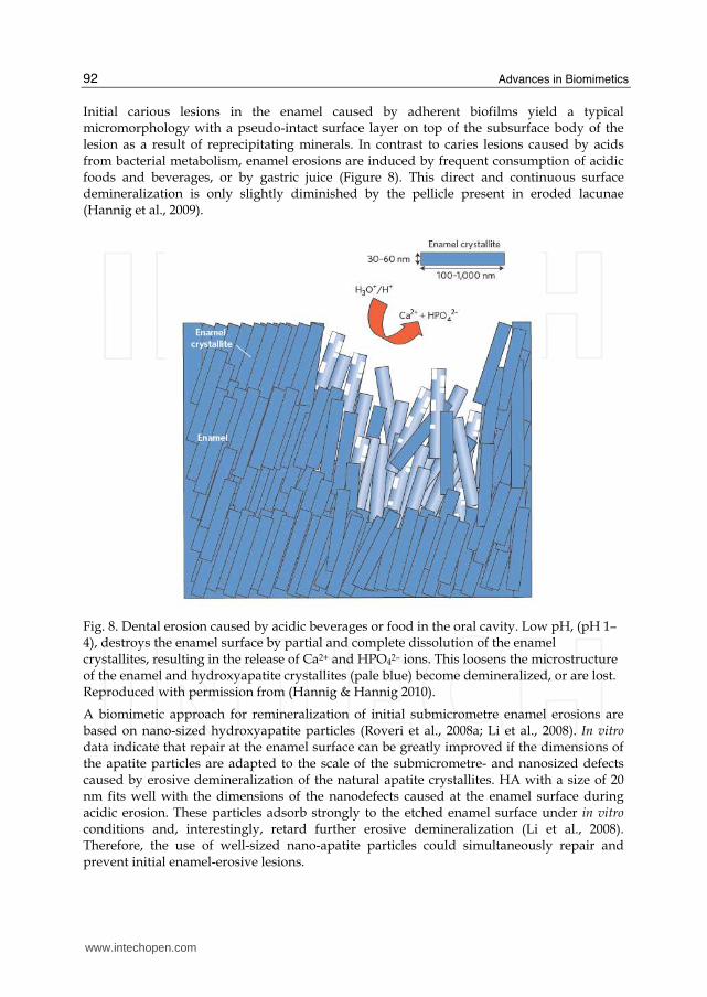
Initial carious lesions in the enamel caused by adherent biofilms yield a typical micromorphology with a pseudo-intact surface layer on top of the subsurface body of the lesion as a result of reprecipitating minerals. In contrast to caries lesions caused by acids from bacterial metabolism, enamel erosions are induced by frequent consumption of acidic foods and beverages, or by gastric juice (Figure 8). This direct and continuous surface demineralization is only slightly diminished by the pellicle present in eroded lacunae (Hannig et al., 2009).
Fig. 8. Dental erosion caused by acidic beverages or food in the oral cavity. Low pH, (pH 1–4), destroys the enamel surface by partial and complete dissolution of the enamel crystallites, resulting in the release of Ca2+ and HPO42– ions. This loosens the microstructure of the enamel and hydroxyapatite crystallites (pale blue) become demineralized, or are lost. Reproduced with permission from (Hannig & Hannig 2010).
A biomimetic approach for remineralization of initial submicrometre enamel erosions are based on nano-sized hydroxyapatite particles (Roveri et al., 2008a; Li et al., 2008). In vitro data indicate that repair at the enamel surface can be greatly improved if the dimensions of the apatite particles are adapted to the scale of the submicrometre- and nanosized defects caused by erosive demineralization of the natural apatite crystallites. HA with a size of 20 nm fits well with the dimensions of the nanodefects caused at the enamel surface during acidic erosion. These particles adsorb strongly to the etched enamel surface under in vitro conditions and, interestingly, retard further erosive demineralization (Li et al., 2008). Therefore, the use of well-sized nano-apatite particles could simultaneously repair and prevent initial enamel-erosive lesions.
www.intechopen.com

The Biomimetic Approach to Design Apatites for Nanobiotechnological Applications
93
In another approach, biomimetic carbonate HA nanoparticles that mimic the size of natural dentinal HA (20 nm) or enamel apatite (100 nm) were used to repair micrometre-sized tooth-surface defects in vitro (Roveri et al., 2008a; Roveri et al., 2009). Clusters of these nanocrystals have been incorporated into toothpastes or mouth-rinsing solutions to promote the repair of demineralised enamel or dentin surfaces by depositing apatite nanoparticles in the defects. Commercially available dental prophylactic products containing carbonate HA nanoparticles to fill microdefects at the etched enamel surface (for example, BioRepair from Coswell Laboratories, Italy and from Dr. Wolff, Germany) have been proved to be effective in vitro after a ten-minute application onto enamel or dentin surfaces (Roveri et al., 2009). Recently an in vivo study has been documented that a toothpaste containing zinc–carbonate HA nanocrystals significantly reduced dentinal hypersensitivity after 4 and 8 weeks, supporting its utility in clinical practice (Orsini et al., 2010). Nano-hydroxyapatite toothpaste with either spheroidal or needle-like particles as an active component was shown to enhance the remineralization of etched enamel better than sodium fluoride solutions. However, the 5–10 day in vitro study had neglected the conditions of the oral cavity. In another in vitro study, nano-sized amorphous calcium carbonate particles that were applied twice a day for a period of 20 days promoted remineralization of artificial white-spot enamel lesions (Nakashima et al., 2009). Reconstitution and remineralization of dentin using nano-sized bioactive glass particles and beta-tricalcium phosphate was also tested in vitro, however, the mechanical properties of original dentin could not be reproduced (Shibata et al., 2008; Vollenweider et al., 2007). Owing to the complex organic and inorganic structure of the dentin, remineralizing dentin into a functional state remains one of the most difficult challenges in dental research (Bertassoni et al., 2009).
5. Conclusion
The biomimetism of biomaterials based hydroxyapatite could be carried out at different levels: composition, structure, morphology and surface reactivity. The aim of a researcher is to realise a biomaterial that is biomimetic in all these characteristics, in order not only to optimise the interaction of synthetic materials with biological materials but also, and more ambitiously, to mimic the biogenic materials in its functionality. This concept should be utilised in designing and preparing synthetic inorganic biomaterials in replacing hard and soft tissues. Nanotechnology has great potential in the biomimetism field to do just that: increase in efficacy by orders of magnitude. In fact, the nano-size of biological tissues building blocks is one of the bases of their self-organisational ability and one that needs to be replaced by synthetic materials in the synthesis of structured architectures with controlled organisation on a multiple-length scale.
6. Acknowledgements
We thank the University of Bologna, (funds for selected research topics) and the Inter University Consortium for Research on Chemistry of Metals in Biological Systems (C.I.R.C.M.S.B). This chapter book is in honour of Professor Alberto Ripamonti in the occurrence of the 80th birthday.
Declaration of interest The authors state no conflict of interest and have received no payment in the preparation of this manuscript.
www.intechopen.com
Advances in Biomimetics
94
7. References
Achilles, W.; Jockel, U.; Schaper, A.; Burk, M. & Riedmiller, H. (1995). In-Vitro Formation of Urinary Stones - Generation of Spherulites of Calcium-Phosphate in Gel and Overgrowth with Calcium-Oxalate Using a New Flow Model of Crystallization. Scanning Microscopy, 9, 2, 577-586.
Al-Kattan, A.; Dufour, P.; Dexpert-Ghys, J. & Drouet, C. (2010). Preparation and Physicochemical Characteristics of Luminescent Apatite-Based Colloids. Journal of Physical Chemistry C, 114, 7, 2918-2924.
Alam, M. I.; Asahina, I.; Ohmamiuda, K.; Takahashi, K.; Yokota, S. & Enomoto, S. (2001). Evaluation of ceramics composed of different hydroxyapatite to tricalcium phosphate ratios as carriers for rhBMP-2. Biomaterials, 22, 12, 1643-51.
Araiza, M. A.; Gomez-Morales, J.; Clemente, R. R. & Castano, V. M. (1999). Conversion of the echinoderm Mellita eduardobarrosoi calcite skeleton into porous hydroxyapatite by treatment with phosphated boiling solutions. Journal of Materials Synthesis and Processing, 7, 4, 211-219.
Arsenault, A. L. & Robinson, B. W. (1989). The Dentino-Enamel Junction - a Structural and Microanalytical Study of Early Mineralization. Calcified Tissue International, 45, 2, 111-121.
Autefage, H.; Briand-Mesange, F.; Cazalbou, S.; Drouet, C.; Fourmy, D.; Goncalves, S.; Salles, J. P.; Combes, C.; Swider, P. & Rey, C. (2009). Adsorption and Release of BMP-2 on Nanocrystalline Apatite-Coated and Uncoated Hydroxyapatite/beta-Tricalcium Phosphate Porous Ceramics. Journal of Biomedical Materials Research Part B-Applied Biomaterials, 91B, 2, 706-715.
Barroug, A.; Kuhn, L. T.; Gerstenfeld, L. C. & Glimcher, M. J. (2004). Interactions of cisplatin with calcium phosphate nanoparticles: in vitro controlled adsorption and release. J Orthop Res, 22, 4, 703-8.
Ben-Nissan, B. & Choi, A. H. (2006). Sol-gel production of bioactive nanocoatings for medical applications. Part 1: an introduction. Nanomedicine, 1, 3, 311-319.
Bertassoni, L. E.; Habelitz, S.; Kinney, J. H.; Marshall, S. J. & Marshall, G. W. (2009). Biomechanical Perspective on the Remineralization of Dentin. Caries Research, 43, 1, 70-77.
Bigi, A.; Compostella, L.; Fichera, A. M.; Foresti, E.; Gazzano, M.; Ripamonti, A. & Roveri, N. (1988). Structural and chemical characterization of inorganic deposits in calcified human mitral valve. J Inorg Biochem, 34, 2, 75-82.
Bigi, A.; Gandolfi, M.; Koch, M. H. & Roveri, N. (1996). X-ray diffraction study of in vitro calcification of tendon collagen. Biomaterials, 17, 12, 1195-201.
Bigi, A.; Gandolfi, M.; Roveri, N. & Valdre, G. (1997). In vitro calcified tendon collagen: an atomic force and scanning electron microscopy investigation. Biomaterials, 18, 9, 657-65.
Bigi, A.; Ripamonti, A.; Cojazzi, G.; Pizzuto, G.; Roveri, N. & Koch, M. H. (1991). Structural analysis of turkey tendon collagen upon removal of the inorganic phase. Int J Biol Macromol, 13, 2, 110-4.
Boix, T.; Gomez-Morales, J.; Torrent-Burgues, J.; Monfort, A.; Puigdomenech, P. & Rodriguez-Clemente, R. (2005). Adsorption of recombinant human bone morphogenetic protein rhBMP-2m onto hydroxyapatite. J Inorg Biochem, 99, 5, 1043-50.
www.intechopen.com
The Biomimetic Approach to Design Apatites for Nanobiotechnological Applications
95
Boskey, A. L. (2007). Mineralization of bones and teeth. Elements, 3, 6, 385-391. Brancaccio, D. & Cozzolino, M. (2005). The mechanism of calcium deposition in soft tissues.
Contrib Nephrol, 149, 279-86. Burger, C.; Zhou, H. W.; Wang, H.; Sics, I.; Hsiao, B. S.; Chu, B.; Graham, L. & Glimcher, M.
J. (2008). Lateral packing of mineral crystals in bone collagen fibrils. Biophysical Journal, 95, 4, 1985-1992.
Cazalbou, S.; Combes, C.; Eichert, D.; Rey, C. & Glimcher, M. J. (2004). Poorly crystalline apatites: evolution and maturation in vitro and in vivo. Journal of Bone and Mineral Metabolism, 22, 4, 310-317.
Cazalbou, S.; Eichert, D.; Ranz, X.; Drouet, C.; Combes, C.; Harmand, M. F. & Rey, C. (2005). Ion exchanges in apatites for biomedical application. Journal of Materials Science-Materials in Medicine, 16, 5, 405-409.
Chai, C. S. & Ben-Nissan, B. (1999). Bioactive nanocrystalline sol-gel hydroxyapatite coatings. Journal of Materials Science-Materials in Medicine, 10, 8, 465-469.
Charriere, E.; Lemaitre, J. & Zysset, P. (2003). Hydroxyapatite cement scaffolds with controlled macroporosity: fabrication protocol and mechanical properties. Biomaterials, 24, 5, 809-817.
Chaudhry, A. A.; Haque, S.; Kellici, S.; Boldrin, P.; Rehman, I.; Fazal, A. K. & Darr, J. A. (2006). Instant nano-hydroxyapatite: a continuous and rapid hydrothermal synthesis. Chemical Communications, 21, 2286-2288.
Chevalier, E.; Chulia, D.; Pouget, C. & Viana, M. (2008). Fabrication of porous substrates: A review of processes using pore forming agents in the biomaterial field. Journal of Pharmaceutical Sciences, 97, 3, 1135-1154.
Choi, A. H. & Ben-Nissan, B. (2007). Sol-gel production of bioactive nanocoatings for medical applications. Part II: current research and development. Nanomedicine, 2, 1, 51-61.
Cowles, E. A.; DeRome, M. E.; Pastizzo, G.; Brailey, L. L. & Gronowicz, G. A. (1998). Mineralization and the expression of matrix proteins during in vivo bone development. Calcified Tissue International, 62, 1, 74-82.
Cui, F. Z. & Ge, J. (2007). New observations of the hierarchical structure of human enamel, from nanoscale to microscale. Journal of Tissue Engineering and Regenerative Medicine, 1, 3, 185-191.
Cui, F. Z.; Li, Y. & Ge, J. (2007). Self-assembly of mineralized collagen composites. Materials Science & Engineering R-Reports, 57, 1-6, 1-27.
Cushing, B. L.; Kolesnichenko, V. L. & O'Connor, C. J. (2004). Recent advances in the liquid-phase syntheses of inorganic nanoparticles. Chemical Reviews, 104, 9, 3893-3946.
Daculsi, G.; Bouler, J. M. & LeGeros, R. Z. (1997). Adaptive crystal formation in normal and pathological calcifications in synthetic calcium phosphate and related biomaterials. International Review of Cytology - a Survey of Cell Biology, Vol 172, 172, 129-191.
de Groot, K. (1993). Clinical applications of calcium phosphate biomaterials: A review. Ceramics International, 19, 5, 363-366.
Dorozhkin, S. V. (2009). Calcium orthophosphate-based biocomposites and hybrid biomaterials. Journal of Materials Science, 44, 9, 2343-2387.
Dorozhkin, S. V. (2010). Bioceramics of calcium orthophosphates. Biomaterials, 31, 7, 1465-85.
www.intechopen.com
Advances in Biomimetics
96
Drouet, C.; Bosc, F.; Banu, M.; Largeot, C.; Combes, C.; Dechambre, G.; Estournes, C.; Raimbeaux, G. & Rey, C. (2009). Nanocrystalline apatites: From powders to biomaterials. Powder Technology, 190, 1-2, 118-122.
Eichert, C.; Drouet, C.; Sfihi, H.; Rey, C. & Combes, C. (2007). Nanocrystalline apatite-based biomaterials: synthesis, processing and characterization. In Biomaterials Research Advances, ed. J. Kendall, 93-143. Nova Science Publishers.
Epple, M.; Ganesan, K.; Heumann, R.; Klesing, J.; Kovtun, A.; Neumann, S. & Sokolova, V. (2010). Application of calcium phosphate nanoparticles in biomedicine. Journal of Materials Chemistry, 20, 1, 18-23.
Errassif, F.; Menbaoui, A.; Autefage, H.; Benaziz, L.; Ouizat, S.; Santran, V.; Sarda, S.; Lebugle, A.; Combes, C.; Barroug, A.; Sfihi, H. & Rey, C. 2010. Adsorption on Apatitic Calcium Phosphates: Applications to Drug Delivery In Advances in Bioceramics and Biotechnologies, eds. R. Narayan, M. Singh & J. McKittrick. Wiley-VCH Verlag GmbH & Co. KGaA
Feng, J. Q.; Ward, L. M.; Liu, S.; Lu, Y.; Xie, Y.; Yuan, B.; Yu, X.; Rauch, F.; Davis, S. I.; Zhang, S.; Rios, H.; Drezner, M. K.; Quarles, L. D.; Bonewald, L. F. & White, K. E. (2006). Loss of DMP1 causes rickets and osteomalacia and identifies a role for osteocytes in mineral metabolism. Nat Genet, 38, 11, 1310-5.
Fernandez-Moran, H. & Engstrom, A. (1957). Electron microscopy and x-ray diffraction of bone. Biochim Biophys Acta, 23, 2, 260-4.
Fratzl, P.; Gupta, H. S.; Paschalis, E. P. & Roschger, P. (2004). Structure and mechanical quality of the collagen-mineral nano-composite in bone. Journal of Materials Chemistry, 14, 14, 2115-2123.
Ganesan, K. & Epple, M. (2008). Calcium phosphate nanoparticles as nuclei for the preparation of colloidal calcium phytate. New Journal of Chemistry, 32, 8, 1326-1330.
Gautier, H.; Daculsi, G. & Merle, C. (2001). Association of vancomycin and calcium phosphate by dynamic compaction: in vitro characterization and microbiological activity. Biomaterials, 22, 18, 2481-7.
Gautier, H.; Merle, C.; Auget, J. L. & Daculsi, G. (2000). Isostatic compression, a new process for incorporating vancomycin into biphasic calcium phosphate: comparison with a classical method. Biomaterials, 21, 3, 243-249.
Gonzalez-McQuire, R.; Green, D.; Walsh, D.; Hall, S.; Chane-Ching, J. Y.; Oreffo, R. O. C. & Mann, S. (2005). Fabrication of hydroxyapatite sponges by dextran sulphate/amino acid templating. Biomaterials, 26, 33, 6652-6656.
Guo, X.; Gough, J. E.; Xiao, P.; Liu, J. & Shen, Z. (2007). Fabrication of nanostructured hydroxyapatite and analysis of human osteoblastic cellular response. J Biomed Mater Res A, 82, 4, 1022-32.
Hannig, C.; Berndt, D.; Hoth-Hannig, W. & Hannig, M. (2009). The effect of acidic beverages on the ultrastructure of the acquired pellicle--an in situ study. Arch Oral Biol, 54, 6, 518-26.
Hannig, M. & Hannig, C. (2010). Nanomaterials in preventive dentistry. Nature Nanotechnology, 5, 8, 565-569.
Hayashizaki, J.; Ban, S.; Nakagaki, H.; Okumura, A.; Yoshii, S. & Robinson, C. (2008). Site specific mineral composition and microstructure of human supra-gingival dental calculus. Archives of Oral Biology, 53, 2, 168-174.
www.intechopen.com
The Biomimetic Approach to Design Apatites for Nanobiotechnological Applications
97
Hong, J.-Y.; Kim, Y. J.; Lee, H.-W.; Lee, W.-K.; Ko, J. S. & Kim, H.-M. (2003). Osteoblastic cell response to thin film of poorly crystalline calcium phosphate apatite formed at low temperatures. Biomaterials, 24, 18, 2977-2984.
Huang, J.; Best, S. M.; Bonfield, W.; Brooks, R. A.; Rushton, N.; Jayasinghe, S. N. & Edirisinghe, M. J. (2004). In vitro assessment of the biological response to nano-sized hydroxyapatite. Journal of Materials Science-Materials in Medicine, 15, 4, 441-445.
Iafisco, M.; Marchetti, M.; Morales, J. G.; Hernandez-Hernandez, M. A.; Ruiz, J. M. G. & Roveri, N. (2009a). Silica Gel Template for Calcium Phosphates Crystallization. Crystal Growth & Design, 9, 11, 4912-4921.
Iafisco, M.; Morales, J. G.; Hernandez-Hernandez, M. A.; Garcia-Ruiz, J. M. & Roveri, N. (2010a). Biomimetic Carbonate-Hydroxyapatite Nanocrystals Prepared by Vapor Diffusion. Advanced Engineering Materials, 12, 7, B218-B223.
Iafisco, M.; Palazzo, B.; Falini, G.; Di Foggia, M.; Bonora, S.; Nicolis, S.; Casella, L. & Roveri, N. (2008). Adsorption and conformational change of myoglobin on biomimetic hydroxyapatite nanocrystals functionalized with alendronate. Langmuir, 24, 9, 4924-4930.
Iafisco, M.; Palazzo, B.; Marchetti, M.; Margiotta, N.; Ostuni, R.; Natile, G.; Morpurgo, M.; Gandin, V.; Marzano, C. & Roveri, N. (2009b). Smart delivery of antitumoral platinum complexes from biomimetic hydroxyapatite nanocrystals. Journal of Materials Chemistry, 19, 44, 8385-8392.
Iafisco, M.; Sabatino, P.; Lesci, I. G.; Prat, M.; Rimondini, L. & Roveri, N. (2010b). Conformational modifications of serum albumins adsorbed on different kinds of biomimetic hydroxyapatite nanocrystals. Colloids Surf B Biointerfaces, 81, 1, 274-84.
Imbeni, V.; Kruzic, J. J.; Marshall, G. W.; Marshall, S. J. & Ritchie, R. O. (2005). The dentin-enamel junction and the fracture of human teeth. Nature Materials, 4, 3, 229-232.
Karageorgiou, V. & Kaplan, D. (2005). Porosity of 3D biomaterial scaffolds and osteogenesis. Biomaterials, 26, 27, 5474-91.
Kazama, J. J.; Amizuka, N. & Fukagawa, M. (2006). Ectopic calcification as abnormal biomineralization. Therapeutic Apheresis and Dialysis, 10, S34-S38.
Kirsch, T. (2006). Determinants of pathological mineralization. Curr Opin Rheumatol, 18, 2, 174-80.
Kodaka, T.; Nakajima, F. & Higashi, S. (1989). Structure of the So-Called Prismless Enamel in Human Deciduous Teeth. Caries Research, 23, 5, 290-296.
Koutsopoulos, S. (2002). Synthesis and characterization of hydroxyapatite crystals: a review study on the analytical methods. J Biomed Mater Res, 62, 4, 600-12.
Laird, D. F.; Mucalo, M. R. & Yokogawa, Y. (2006). Growth of calcium hydroxyapatite (Ca-HAp) on cholesterol and cholestanol crystals from a simulated body fluid: A possible insight into the pathological calcifications associated with atherosclerosis. Journal of Colloid and Interface Science, 295, 2, 348-363.
Layrolle, P. & Lebugle, A. (1994). Characterization and Reactivity of Nanosized Calcium Phosphates Prepared in Anhydrous Ethanol. Chemistry of Materials, 6, 11, 1996-2004.
Lebugle, A.; Rodrigues, A.; Bonnevialle, P.; Voigt, J. J.; Canal, P. & Rodriguez, F. (2002). Study of implantable calcium phosphate systems for the slow release of methotrexate. Biomaterials, 23, 16, 3517-22.
www.intechopen.com
Advances in Biomimetics
98
LeGeros, R. Z. (2008). Calcium Phosphate-Based Osteoinductive Materials. Chemical Reviews, 108, 11, 4742-4753.
Legeros, R. Z. & Craig, R. G. (1993). Strategies to Affect Bone Remodeling - Osteointegration. Journal of Bone and Mineral Research, 8, S583-S596.
Legeros, R. Z.; Orly, I.; Legeros, J. P.; Gomez, C.; Kazimiroff, J.; Tarpley, T. & Kerebel, B. (1988). Scanning Electron-Microscopy and Electron-Probe Microanalyses of the Crystalline Components of Human and Animal Dental Calculi. Scanning Microscopy, 2, 1, 345-356.
Legros, R.; Balmain, N. & Bonel, G. (1987). Age-related changes in mineral of rat and bovine cortical bone. Calcif Tissue Int, 41, 3, 137-44.
Li, L.; Pan, H.; Tao, J.; Xu, X.; Mao, C.; Gu, X. & Tang, R. (2008). Repair of enamel by using hydroxyapatite nanoparticles as the building blocks. Journal of Materials Chemistry, 18, 34, 4079-4084.
Li, X. F.; Fan, T. X.; Liu, Z. T.; Ding, J.; Guo, Q. X. & Zhang, D. (2006). Synthesis and hierarchical pore structure of biomorphic manganese oxide derived from woods. Journal of the European Ceramic Society, 26, 16, 3657-3664.
Liou, S. C.; Chen, S. Y.; Lee, H. Y. & Bow, J. S. (2004). Structural characterization of nano-sized calcium deficient apatite powders. Biomaterials, 25, 2, 189-196.
Liu, D. M. (1997). Fabrication of hydroxyapatite ceramic with controlled porosity. Journal of Materials Science-Materials in Medicine, 8, 4, 227-232.
Liu, J. B.; Li, K. W.; Wang, H.; Zhu, M. K.; Xu, H. Y. & Yan, H. (2005). Self-assembly of hydroxyapatite nanostructures by microwave irradiation. Nanotechnology, 16, 1, 82-87.
Liu, Y. L.; Enggist, L.; Kuffer, A. F.; Buser, D. & Hunziker, E. B. (2007). The influence of BMP-2 and its mode of delivery on the osteoconductivity of implant surfaces during the early phase of osseointegration (vol 28, pg 2677, 2007). Biomaterials, 28, 35, 5399-5399.
Lopez-Macipe, A.; Gomez-Morales, J. & Rodriguez-Clemente, R. (1998). Nanosized hydroxyapatite precipitation from homogeneous calcium/citrate/phosphate solutions using microwave and conventional heating. Advanced Materials, 10, 1, 49-+.
Loveridge, N. (1999). Bone: More than a stick. Journal of Animal Science, 77, 190-196. Lu, J. X.; Flautre, B.; Anselme, K.; Hardouin, P.; Gallur, A.; Descamps, M. & Thierry, B.
(1999). Role of interconnections in porous bioceramics on bone recolonization in vitro and in vivo. Journal of Materials Science-Materials in Medicine, 10, 2, 111-120.
Mann, S.; Archibald, D. D.; Didymus, J. M.; Douglas, T.; Heywood, B. R.; Meldrum, F. C. & Reeves, N. J. (1993). Crystallization at Inorganic-organic Interfaces: Biominerals and Biomimetic Synthesis. Science, 261, 5126, 1286-92.
Manolagas, S. C. (2000). Birth and death of bone cells: Basic regulatory mechanisms and implications for the pathogenesis and treatment of osteoporosis. Journal of Aging and Physical Activity, 8, 3, 248-248.
Mao, Y.; Park, T. J.; Zhang, F.; Zhou, H. & Wong, S. S. (2007). Environmentally friendly methodologies of nanostructure synthesis. Small, 3, 7, 1122-1139.
Marra, S. P.; Daghlian, C. P.; Fillinger, M. F. & Kennedy, F. E. (2006). Elemental composition, morphology and mechanical properties of calcified deposits obtained from abdominal aortic aneurysms. Acta Biomaterialia, 2, 5, 515-520.
www.intechopen.com

The Biomimetic Approach to Design Apatites for Nanobiotechnological Applications
99
Mastrogiacomo, M.; Scaglione, S.; Martinetti, R.; Dolcini, L.; Beltrame, F.; Cancedda, R. & Quarto, R. (2006). Role of scaffold internal structure on in vivo bone formation in macroporous calcium phosphate bioceramics. Biomaterials, 27, 17, 3230-3237.
McLeod, K.; Kumar, S.; Smart, R. S. C.; Dutta, N.; Voelcker, N. H.; Anderson, G. I. & Sekel, R. (2006). XPS and bioactivity study of the bisphosphonate pamidronate adsorbed onto plasma sprayed hydroxyapatite coatings. Applied Surface Science, 253, 5, 2644-2651.
Midy, V.; Hollande, E.; Rey, C.; Dard, M. & Plouet, J. (2001). Adsorption of vascular endothelial growth factor to two different apatitic materials and its release. J Mater Sci Mater Med, 12, 4, 293-8.
Nakashima, S.; Yoshie, M.; Sano, H. & Bahar, A. (2009). Effect of a test dentifrice containing nano-sized calcium carbonate on remineralization of enamel lesions in vitro. Journal of oral science, 51, 1, 69-77.
Olszta, M. J.; Cheng, X. G.; Jee, S. S.; Kumar, R.; Kim, Y. Y.; Kaufman, M. J.; Douglas, E. P. & Gower, L. B. (2007). Bone structure and formation: A new perspective. Materials Science & Engineering R-Reports, 58, 3-5, 77-116.
Orsini, G.; Procaccini, M.; Manzoli, L.; Giuliodori, F.; Lorenzini, A. & Putignano, A. (2010). A double-blind randomized-controlled trial comparing the desensitizing efficacy of a new dentifrice containing carbonate/hydroxyapatite nanocrystals and a sodium fluoride/potassium nitrate dentifrice. Journal of Clinical Periodontology, 37, 6, 510-517.
Paine, M. L.; White, S. N.; Luo, W.; Fong, H.; Sarikaya, M. & Snead, M. L. (2001). Regulated gene expression dictates enamel structure and tooth function. Matrix Biology, 20, 5-6, 273-292.
Palazzo, B.; Iafisco, M.; Laforgia, M.; Margiotta, N.; Natile, G.; Bianchi, C. L.; Walsh, D.; Mann, S. & Roveri, N. (2007). Biomimetic hydroxyapatite-drug nanocrystals as potential bone substitutes with antitumor drug delivery properties. Advanced Functional Materials, 17, 13, 2180-2188.
Palazzo, B.; Sidoti, M. C.; Roveri, N.; Tampieri, A.; Sandri, M.; Bertolazzi, L.; Galbusera, F.; Dubini, G.; Vena, P. & Contro, R. (2005). Controlled drug delivery from porous hydroxyapatite grafts: An experimental and theoretical approach. Materials Science & Engineering C-Biomimetic and Supramolecular Systems, 25, 2, 207-213.
Palazzo, B.; Walsh, D.; Iafisco, M.; Foresti, E.; Bertinetti, L.; Martra, G.; Bianchi, C. L.; Cappelletti, G. & Roveri, N. (2009). Amino acid synergetic effect on structure, morphology and surface properties of biomimetic apatite nanocrystals. Acta Biomater, 5, 4, 1241-52.
Palmer, L. C.; Newcomb, C. J.; Kaltz, S. R.; Spoerke, E. D. & Stupp, S. I. (2008). Biomimetic systems for hydroxyapatite mineralization inspired by bone and enamel. Chemical Reviews, 108, 11, 4754-83.
Paschalis, E. P.; DiCarlo, E.; Betts, F.; Sherman, P.; Mendelsohn, R. & Boskey, A. L. (1996). FTIR microspectroscopic analysis of human osteonal bone. Calcified Tissue International, 59, 6, 480-487.
Peter, B.; Pioletti, D. P.; Laib, S.; Bujoli, B.; Pilet, P.; Janvier, P.; Guicheux, J.; Zambelli, P. Y.; Bouler, J. M. & Gauthier, O. (2005). Calcium phosphate drug delivery system: influence of local zoledronate release on bone implant osteointegration. Bone, 36, 1, 52-60.
www.intechopen.com

Advances in Biomimetics
100
Phillips, M. J.; Darr, J. A.; Luklinska, Z. B. & Rehman, I. (2003). Synthesis and characterization of nano-biomaterials with potential osteological applications. Journal of Materials Science-Materials in Medicine, 14, 10, 875-882.
Puleo, D. A. (2004). Bone–Implant Interface. Encyclopedia of Biomaterials and Biomedical Engineering, 190 - 198.
Rameshbabu, N.; Rao, K. P. & Kumar, T. S. S. (2005). Acclerated microwave processing of nanocrystalline hydroxyapatite. Journal of Materials Science, 40, 23, 6319-6323.
Reddi, A. H. (1994). Symbiosis of Biotechnology and Biomaterials - Applications in Tissue Engineering of Bone and Cartilage. Journal of Cellular Biochemistry, 56, 2, 192-195.
Rey, C.; Collins, B.; Goehl, T.; Dickson, I. R. & Glimcher, M. J. (1989). The Carbonate Environment in Bone-Mineral - a Resolution-Enhanced Fourier-Transform Infrared-Spectroscopy Study. Calcified Tissue International, 45, 3, 157-164.
Rey, C.; Shimizu, M.; Collins, B. & Glimcher, M. J. (1990). Resolution-enhanced Fourier transform infrared spectroscopy study of the environment of phosphate ions in the early deposits of a solid phase of calcium-phosphate in bone and enamel, and their evolution with age. I: Investigations in the upsilon 4 PO4 domain. Calcif Tissue Int, 46, 6, 384-94.
Rodriguez-Lorenzo, L. M.; Vallet-Regi, M. & Ferreira, J. M. (2001). Fabrication of hydroxyapatite bodies by uniaxial pressing from a precipitated powder. Biomaterials, 22, 6, 583-8.
Roveri, N.; Battistella, E.; Bianchi, C. L.; Foltran, I.; Foresti, E.; Iafisco, M.; Lelli, M.; Naldoni, A.; Palazzo, B. & Rimondini, L. (2009). Surface Enamel Remineralization: Biomimetic Apatite Nanocrystals and Fluoride Ions Different Effects. Journal of Nanomaterials, -.
Roveri, N.; Battistella, E.; Foltran, I.; Foresti, E.; Iafisco, M.; Lelli, M.; Palazzo, B. & Rimondini, L. (2008a). Synthetic Biomimetic Carbonate-Hydroxyapatite Nanocrystals for Enamel Remineralization. Advanced Materials Research, 47-50, 821-824.
Roveri, N.; Palazzo, B. & Iafisco, M. (2008b). The role of biomimetism in developing nanostructured inorganic matrices for drug delivery. Expert Opinion on Drug Delivery, 5, 8, 861-877.
Sader, M.; LeGeros, R. & Soares, G. (2009). Human osteoblasts adhesion and proliferation on magnesium-substituted tricalcium phosphate dense tablets. Journal of Materials Science: Materials in Medicine, 20, 2, 521-527.
Sakhno, Y.; Bertinetti, L.; Iafisco, M.; Tampieri, A.; Roveri, N. & Martra, G. (2010). Surface Hydration and Cationic Sites of Nanohydroxyapatites with Amorphous or Crystalline Surfaces: A Comparative Study. The Journal of Physical Chemistry C, 114, 39, 16640-16648.
Sanchez, C.; Arribart, H. & Guille, M. M. G. (2005). Biomimetism and bioinspiration as tools for the design of innovative materials and systems. Nature Materials, 4, 4, 277-288.
Sarikaya, M.; Tamerler, C.; Jen, A. K.; Schulten, K. & Baneyx, F. (2003). Molecular biomimetics: nanotechnology through biology. Nature Materials, 2, 9, 577-85.
Schmidt, H. K. (2000). Nanoparticles for ceramic and nanocomposite processing. Molecular Crystals and Liquid Crystals, 353, 165-179.
www.intechopen.com
The Biomimetic Approach to Design Apatites for Nanobiotechnological Applications
101
Shibata, Y.; He, L. H.; Kataoka, Y.; Miyazaki, T. & Swain, M. V. (2008). Micromechanical property recovery of human carious dentin achieved with colloidal nano-beta-tricalcium phosphate. J Dent Res, 87, 3, 233-7.
Singh, M.; Martinez-Fernandez, J. & de Arellano-Lopez, A. R. (2003). Environmentally conscious ceramics (ecoceramics) from natural wood precursors. Current Opinion in Solid State & Materials Science, 7, 3, 247-254.
Siva Rama Krishna, D.; Siddharthan, A.; Seshadri, S. K. & Sampath Kumar, T. S. (2007). A novel route for synthesis of nanocrystalline hydroxyapatite from eggshell waste. J Mater Sci Mater Med, 18, 9, 1735-43.
Sopyan, I.; Mel, M.; Ramesh, S. & Khalid, K. A. (2007). Porous hydroxyapatite for artificial bone applications. Science and Technology of Advanced Materials, 8, 1-2, 116-123.
Stigter, M.; Bezemer, J.; de Groot, K. & Layrolle, P. (2004). Incorporation of different antibiotics into carbonated hydroxyapatite coatings on titanium implants, release and antibiotic efficacy. J Control Release, 99, 1, 127-37.
Sun, W. B.; Chu, C. L.; Wang, J. & Zhao, H. T. (2007). Comparison of periodontal ligament cells responses to dense and nanophase hydroxyapatite. Journal of Materials Science-Materials in Medicine, 18, 5, 677-683.
Tamerler, C. & Sarikaya, M. (2007). Molecular biomimetics: utilizing nature's molecular ways in practical engineering. Acta Biomater, 3, 3, 289-99.
Tamerler, C. & Sarikaya, M. (2008). Molecular biomimetics: Genetic synthesis, assembly, and formation of materials using peptides. Mrs Bulletin, 33, 5, 504-510.
Tampieri, A.; Sprio, S.; Ruffini, A.; Celotti, G.; Lesci, I. G. & Roveri, N. (2009). From wood to bone: multi-step process to convert wood hierarchical structures into biomimetic hydroxyapatite scaffolds for bone tissue engineering. Journal of Materials Chemistry, 19, 28, 4973-4980.
Tancret, F.; Bouler, J. M.; Chamousset, J. & Minois, L. M. (2006). Modelling the mechanical properties of microporous and macroporous biphasic calcium phosphate bioceramics. Journal of the European Ceramic Society, 26, 16, 3647-3656.
Tas, A. C. (2000). Synthesis of biomimetic Ca-hydroxyapatite powders at 37 degrees C in synthetic body fluids. Biomaterials, 21, 14, 1429-1438.
Termine, J. D.; Kleinman, H. K.; Whitson, S. W.; Conn, K. M.; McGarvey, M. L. & Martin, G. R. (1981). Osteonectin, a bone-specific protein linking mineral to collagen. Cell, 26, 1 Pt 1, 99-105.
Traub, W.; Arad, T. & Weiner, S. (1989). Three-dimensional ordered distribution of crystals in turkey tendon collagen fibers. Proc Natl Acad Sci U S A, 86, 24, 9822-6.
Tzaphlidou, M. (2008). Bone Architecture: Collagen Structure and Calcium/Phosphorus Maps. Journal of Biological Physics, 34, 1-2, 39-49.
Urist, M. R. (2002). Bone: Formation by autoinduction (Reprinted from Science, vol 150, pg 893-899, 1965). Clinical Orthopaedics and Related Research, 395, 5-10.
Vollenweider, M.; Brunner, T. J.; Knecht, S.; Grass, R. N.; Zehnder, M.; Imfeld, T. & Stark, W. J. (2007). Remineralization of human dentin using ultrafine bioactive glass particles. Acta Biomater, 3, 6, 936-43.
Vriezema, D. M.; Aragonès, M. C.; Elemans, J. A. A. W.; Cornelissen, J. J. L. M.; Rowan, A. E. & Nolte, R. J. M. (2005). Self-assembled nanoreactors. Chemical Reviews, 105, 4, 1445-1489.
www.intechopen.com

Advances in Biomimetics
102
Wahl, D. A. & Czernuszka, J. T. (2006). Collagen-hydroxyapatite composites for hard tissue repair. European Cells & Materials, 11, 43-56.
Walsh, D.; Boanini, E.; Tanaka, J. & Mann, S. (2005). Synthesis of tri-calcium phosphate sponges by interfacial deposition and thermal transformation of self-supporting calcium phosphate films. Journal of Materials Chemistry, 15, 10, 1043-1048.
Walsh, D. & Tanaka, J. (2001). Preparation of a bone-like apatite foam cement. Journal of Materials Science-Materials in Medicine, 12, 4, 339-343.
Wang, J. W. & Shaw, L. L. (2007). Morphology-enhanced low-temperature sintering of nanocrystalline hydroxyapatite. Advanced Materials, 19, 17, 2364-+.
Wang, R. Z. & Weiner, S. (1998). Strain-structure relations in human teeth using Moire fringes. Journal of Biomechanics, 31, 2, 135-141.
Wang, X.; Zhuang, J.; Peng, Q. & Li, Y. D. (2005). A general strategy for nanocrystal synthesis. Nature, 437, 7055, 121-124.
Weiner, S. & Addadi, L. (1997). Design strategies in mineralized biological materials. Journal of Materials Chemistry, 7, 5, 689-702.
Weiner, S. & Wagner, H. D. (1998). The material bone: Structure mechanical function relations. Annual Review of Materials Science, 28, 271-298.
Weissen-Plenz, G.; Nitschke, Y. & Rutsch, F. (2008). Mechanisms of Arterial Calcification: Spotlight on the Inhibitors. Advances in Clinical Chemistry, Vol 46, 46, 263-293.
Wen, H. B.; Moradian-Oldak, J. & Fincham, A. G. (2000). Dose-dependent modulation of octacalcium phosphate crystal habit by amelogenins. Journal of Dental Research, 79, 11, 1902-1906.
Wesson, J. A. & Ward, M. D. (2007). Pathological biomineralization of kidney stones. Elements, 3, 6, 415-421.
White, S. N.; Luo, W.; Paine, M. L.; Fong, H.; Sarikaya, M. & Snead, M. L. (2001). Biological organization of hydroxyapatite crystallites into a fibrous continuum toughens and controls anisotropy in human enamel. Journal of Dental Research, 80, 1, 321-326.
Whittaker, D. K. (1982). Structural variations in the surface zone of human tooth enamel observed by scanning electron microscopy. Archives of Oral Biology, 27, 5, 383-92.
Wozney, J. M. (1992). The Bone Morphogenetic Protein Family and Osteogenesis. Molecular Reproduction and Development, 32, 2, 160-167.
Ye, F.; Guo, H. F. & Zhang, H. J. (2008). Biomimetic synthesis of oriented hydroxyapatite mediated by nonionic surfactants. Nanotechnology, 19, 24, -.
Yeon, K. C.; Wang, J. & Ng, S. C. (2001). Mechanochemical synthesis of nanocrystalline hydroxyapatite from CaO and CaHPO4. Biomaterials, 22, 20, 2705-12.
Yoshinari, M.; Oda, Y.; Ueki, H. & Yokose, S. (2001). Immobilization of bisphosphonates on surface modified titanium. Biomaterials, 22, 7, 709-715.
Zhang, Y. J. & Lu, J. J. (2007). A simple method to tailor spherical nanocrystal hydroxyapatite at low temperature. Journal of Nanoparticle Research, 9, 4, 589-594.
Zimmerman, A. F.; Palumbo, G.; Aust, K. T. & Erb, U. (2002). Mechanical properties of nickel silicon carbide nanocomposites. Materials Science and Engineering a-Structural Materials Properties Microstructure and Processing, 328, 1-2, 137-146.
www.intechopen.com
Advances in BiomimeticsEdited by Prof. Marko Cavrak
ISBN 978-953-307-191-6Hard cover, 522 pagesPublisher InTechPublished online 26, April, 2011Published in print edition April, 2011
InTech EuropeUniversity Campus STeP Ri Slavka Krautzeka 83/A 51000 Rijeka, Croatia Phone: +385 (51) 770 447 Fax: +385 (51) 686 166www.intechopen.com
InTech ChinaUnit 405, Office Block, Hotel Equatorial Shanghai No.65, Yan An Road (West), Shanghai, 200040, China
Phone: +86-21-62489820 Fax: +86-21-62489821
The interaction between cells, tissues and biomaterial surfaces are the highlights of the book "Advances inBiomimetics". In this regard the effect of nanostructures and nanotopographies and their effect on thedevelopment of a new generation of biomaterials including advanced multifunctional scaffolds for tissueengineering are discussed. The 2 volumes contain articles that cover a wide spectrum of subject matter suchas different aspects of the development of scaffolds and coatings with enhanced performance and bioactivity,including investigations of material surface-cell interactions.
How to referenceIn order to correctly reference this scholarly work, feel free to copy and paste the following:
Norberto Roveri and Michele Iafisco (2011). The Biomimetic Approach to Design Apatites forNanobiotechnological Applications, Advances in Biomimetics, Prof. Marko Cavrak (Ed.), ISBN: 978-953-307-191-6, InTech, Available from: http://www.intechopen.com/books/advances-in-biomimetics/the-biomimetic-approach-to-design-apatites-for-nanobiotechnological-applications