5-in-5 the effect of ankle bracing on kinematics in
TRANSCRIPT

The Effect of Ankle Bracing on Kinematicsin Simulated Sprain and Drop Landings
A Double-Blind, Placebo-Controlled Study
Alison N. Agres,*y PhD, Marios Chrysanthou,y BSc, and Peter C. Raffalt,y PhDInvestigation performed at the Julius Wolff Institute, Charite-Universitatsmedizin Berlin,Berlin, Germany
Background: The efficacy of external ankle braces to protect against sudden inversion sprain has yet to be determined whiletaking into account the possible placebo effect of brace application.
Purpose: To assess the protective effect of an external ankle brace on ankle kinematics during simulated inversion sprain andsingle-legged drop landings among individuals with a history of unilateral lateral ankle sprain.
Hypothesis: The primary hypothesis was that active and placebo external braces would reduce inversion angle during simulatedinversion sprain.
Study Design: Controlled laboratory study.
Methods: Sixteen participants with ankle instability and previous sprain performed single-legged drop landings and suddeninversion tilt perturbations. Kinematics of the affected limb were assessed in 3 conditions (active bracing, passive placebo brac-ing, and unbraced) across 2 measurement days. Participators and investigators were blinded to the brace type tested. The effectof bracing on kinematics was assessed with repeated measures analysis of variance with statistical parametric mapping, withpost hoc tests performed for significant interactions.
Results: Only active bracing reduced inversion angles during a sudden ankle inversion when compared with the unbraced con-dition. This reduction was apparent between 65 and 140 milliseconds after the initial fall. No significant differences in inversionangle were found between the passive placebo brace and unbraced conditions during sudden ankle inversion. Furthermore,no significant differences were found among all tested conditions in the sagittal plane kinematics at the knee and ankle.
Conclusion: During an inversion sprain, only the actively protecting ankle brace limited inversion angles among participants.These results do not indicate a placebo effect of external bracing for patients with ankle instability and a history of unilateral anklesprain. Furthermore, sagittal plane knee kinematics appear to remain unaffected by bracing during single-legged landing, owingto the limited effects of bracing on sagittal ankle kinematics. These results highlight the role of brace design on biomechanicalfunction during sports-related and injury-prone movements.
Clinical Relevance: Athletes prone to reinjury after lateral ankle sprain may benefit from brace designs that allow for full sagittalrange of motion but restrict only frontal plane motion.
Keywords: kinematics; ankle brace; ankle inversion; lateral ankle sprain
During participation in sports, the ankle is the most fre-quently injured joint in the body.16,31 Eighty-five percent ofthese injuries are sprains, which are often inversion sprainsof the lateral ligaments.26 Although incidence is high, lateralankle sprain (LAS) is typically seen in the clinic as being rel-atively benign,3 which may contribute to lasting complica-tions,3 such as chronic ankle instability, higher mechanicallaxity,9 and higher reinjury risk.28 As a result, there is persis-tent disability in the ankle after LAS27 regardless of initialtreatment,23 and external supports have been employed toresist further inversion injury in this population.
Ideally, a prophylactic ankle support would reduceeither the maximal angle or the angular velocity of a sud-den ankle inversion to minimize the chance of injury.
*Address correspondence to Alison N. Agres, PhD, Julius Wolff Insti-tute, Charite–Universitatsmedizin Berlin, Augustenburger Platz 1, 13353Berlin, Germany (email: [email protected]).
yJulius Wolff Institute, Charite-Universitatsmedizin Berlin, Berlin,Germany.
One or more of the authors has declared the following potential con-flict of interest or source of funding: This study was funded by the FederalMinistry of Education and Research in Germany (BMBF 13GW0104C).AOSSM checks author disclosures against the Open Payments Database(OPD). AOSSM has not conducted an independent investigation on theOPD and disclaims any liability or responsibility relating thereto.
The American Journal of Sports Medicine2019;47(6):1480–1487DOI: 10.1177/0363546519837695� 2019 The Author(s)
5-in-5
1480

Bracing and taping are often interchangeably used in prac-tice; however, external bracing was shown to offer a slowerinversion velocity when directly compared with taping.21
Another added benefit of bracing is the relative ease of use,as ankle taping takes comparatively more time and is moreuser dependent for correct application. The mechanical sup-port offered by bracing suggests that it may be more effectivein preventing reinjury in the frontal plane.
An optimal external ankle support should allow unre-stricted sagittal plane movement. Current designs aim toaddress this with various semirigid or hinged variants.Given that these injuries commonly occur during sportswhile an athlete is landing from a height, single-leggeddrop landing tests offer an ideal model for assessing anklefunction within an ankle brace. However, most bracedesigns continuously limit frontal and sagittal plane mobil-ity during noninjury movements, such as running,38 whichmay lead to diminished athletic performance. An idealbrace would selectively restrict inversion at higher veloci-ties only when the existing mechanical structures areunable to support the ankle.
An inherent limitation in the assessment of an externalankle brace is the confounding effect of cognitive bias,where it remains unclear if the outcomes were affectedby the device or by the user’s expectation.5 The inclusionof a placebo within a blinded study is warranted to investi-gate the effects of an external support5; yet to date, fewstudies assessing ankle bracing have incorporated thisinto their study design.z In a recent blinded study, Saw-kins and colleagues35 found no differences among real tap-ing, placebo taping, and no taping during hopping anda star excursion test, suggesting that for these tasks, thecombination of patient confidence and taping is importantfor injury prevention but that the placebo effect for tapingremains unclear. To date, no study investigating externalankle supports has considered the placebo effect on inves-tigators and users in their assessments.
The purpose of the present study was to assess the pro-tective effect of an active external ankle brace that selec-tively restricts inversion velocity during possiblereinjury, taking into account the possible placebo effect ofbrace application. To investigate this, a repeated meas-ures, double-blind, placebo-controlled study was conductedwithin a cohort of patients with previous unilateral LAS.We hypothesized that active and placebo external braceswould both be capable of reducing angles during a suddeninversion when compared with no bracing. A further aim ofthis study was to determine if an external ankle braceaffects sagittal plane kinematics during single-limb droplandings among patients with a history of unilateral LAS.
METHODS
Participants
The present study was a part of a larger experimental pro-tocol that aimed to assess the effect of external ankle
bracing on movement, which also included gait analysisand balance assessment. The experimental protocol com-pleted by the participants comprised an ankle inversiontest, walking, jogging, a unilateral balance test, and single-legged landing. However, the present study included onlythe kinematic and kinetic data obtained during the inver-sion test and the single-legged landing.
Sixteen participants with a history of unilateral LASwere recruited for this study (7 female and 9 male; mean6 SD age, 30.9 6 4.7 years; body mass, 73.4 6 11.9 kg;body height, 176.4 6 9.5 cm). Participants had self-reported unilateral ankle instability and ‘‘giving way,’’a history of initial acute LAS that was at least 1 year prior,and regular participation in sports for a minimum of 3hours per week. Exclusion criteria included a history ofother lower extremity injuries or surgery, injury withinthe past 4 weeks, vestibular or balance disorders, footdeformities, generalized hypermobility, or any further con-dition that could affect test performance. Foot and AnkleOutcome Scores34 were assessed before measurement. Allprotocols were developed with reference to the Declarationof Helsinki. The local ethics committee (EA1/335/16)approved the study, and all patients gave their informedconsent before participation.
Study Design and Protocol
All participants were required to attend 2 measurementsessions in a motion capture laboratory setting within 1week. At each measurement, participants were testedunder 2 shod conditions: first, a baseline condition withoutan orthotic brace, followed by an intervention conditionwith an orthotic brace. The functional test protocol wasidentical for both conditions and completed in the sameorder, with single-legged drop landings first, followed byinversion plate tests (Appendix Figure 1, available in theonline version of this article). Participants wore the sameathletic shoes at both sessions. Data collection was limitedto the LAS-affected limb for all tests.
The braces used for this investigation (BetterguardsGmbH) (Figure 1) were manufactured to be identical, savefor an exchangeable module that would resist frontal planemotion at high inversion velocity (hereafter, an ‘‘active’’brace) or passively allow frontal plane motion at all veloci-ties (‘‘passive’’ brace). The active module contained anunpowered energy-absorbing system with a dilatant fluidto reduce inversion angles, whereas the passive brace hadno such module and had an identical, flexible elastic band.The module is placed laterally within the brace and is con-nected to the customized insole with a band. The connectionbetween the brace and the insole is placed in a way that themodule is pulled when the foot is inverted. The brace wasput on the patient’s affected foot by external investigatorsfor proper fit. Since the brace required a physical connectionto an insole within the athletic shoe, identical insoles wereplaced in both shoes for both conditions (with and withoutbrace) to exclude the influence of the insole on test results.
Two external investigators were unblinded and ran-domized the brace intervention so that the active bracewas worn during one session and the passive brace duringzReferences 2, 8, 10, 19, 20, 22, 24, 30, 36, 39.
AJSM Vol. 47, No. 6, 2019 Effect of Ankle Bracing on Kinematics 1481

the other. The participants and the primary investigatorswere both blinded to which orthotic was being tested ateach session. Furthermore, both blinded groups (partici-pants and investigators) were not allowed to apply or touchthe orthotic during and after measurement sessions. All par-ticipants performed a standardized warm-up protocol thatincluded walking and jogging for a minimum of 15 minutes.
Single-Legged Drop Landing
For each condition, participants performed a set of 3 single-legged drop landings from a 0.2 m–high platform, similar tothe task described by Gardner and colleagues.19 While bal-ancing near the edge of the platform standing only on thetested leg, participants were asked to lean forward andland on the same leg on a forceplate (sampling frequency,1000 Hz; AMTI). Drop landings were included for analysisif participants were able to stand with an outstretchedknee after maximal flexion. Before the first data collection,all participants were given oral instructions, a demonstra-tion of the task, and at least 1 practice test for familiariza-tion and to ensure proper performance.
Three-dimensional kinematic data of the lower extrem-ities were gathered with a set of 67 reflective markersplaced on the limb and shoes, tracked by an infrared motioncapture system operating at 200 Hz (10 MX-S cameras;VICON). Markers were placed as recommended by Kratzen-stein and colleagues,25 and functional movements, includinga star-arc motion6 and flexion-extension of the knee, wereused to identify the hip joint center13 and tibiofemoralaxes of rotation,14 respectively. The optimal common shapetechnique40 was employed to minimize soft tissue artifact.Markers were placed at bony landmarks palpated on thepelvis, tibia, ankle, and foot with double-sided adhesivetape. Bony landmarks included the anterior and posteriorsuperior iliac spine, greater trochanter, tibial tuberosity,
fibular head, first and fifth metatarsals, and the medialand lateral aspects of the following: knee epicondyles, anklemalloli, and calcaneus. Given its design, the brace did notobstruct the malleoli, so the bony aspects were directlymarked for assessment. Further markers were symmetri-cally attached to the thigh, shank, and foot. Sagittal kneeangles were calculated with the OSSCA projection,40 andankle angles were calculated with ISB standards42 fromthe time of initial ground contact to 250 millisecondsafterward.
Single-Legged Inversion Tilt
Following the single-legged drop landing test, a series oftests were performed whereby an unexpected unilateralfoot inversion was induced on a custom-built tilting plat-form (Figure 2) controlled by an external motor (ServomotorEMMS-AS-140-L-HS-RSB; Festo AG & Co KG) with a max-imal inversion angle of 30� (Figure 2). A set of 10 random-ized trials were performed on the tested leg, with 5 trialsat 400 deg/s and 5 trials at 150 deg/s, simulating a ‘‘fast’’and ‘‘physiological’’ inversion movement, respectively, aboveand below a previously suggested threshold of 300 deg/s.7
All patients were asked to adopt a particular stance(Figure 2A), with the knee on the testing leg outstretchedand with the contralateral foot on a small forceplate (sam-pling frequency, 100 Hz; FP4, Biometrics Ltd), which wasbuilt onto the nontilting portion of the platform. Patientsplaced 80% of their bodyweight onto the tilting platform.An investigator with real-time monitoring of the forceplatevalues guided patients to 20% bodyweight on the contralat-eral limb. Once this measurement was steady, the platformwas triggered to fall by an external control (Figure 2B)(Festo Configuration Tool, v 1.2.3.6; Festo AG & Co KG).
Kinematic data of the tested leg were gathered with thesame camera system, with an increased sampling fre-quency of 400 Hz. Within this measurement, only theinversion angle of the ipsilateral limb was calculated foreach trial. The inversion angle was cropped to a predeter-mined time of 200 milliseconds starting from the initialmovement of the dropping platform. The vertical displace-ment of a reflective marker placed at the far edge of theinversion platform was used to identify the initialmovement.
Figure 1. Depiction of the external ankle brace used for test-ing (left), with the exchangeable module indicated by thedashed line, which is visible only upon lifting the cushioningpads (right). The brace was designed to ensure that partici-pants and investigators were blinded to the module used(active or passive).
Figure 2. (A) Initial and (B) end positions of the single-leggedinversion tilt as performed on the custom-built tilting platform.
1482 Agres et al The American Journal of Sports Medicine

Statistical Analysis
To investigate the effect of the brace on (1) the ankle andknee sagittal angles during the single-legged drop landingand (2) the ankle frontal angles during the single-leggedinversion tilt, statistical parametric mapping (SPM) fora 1-way repeated measures analysis of variance (ANOVA)design was applied to the 2 joints in both planes, with thebrace as the independent variable.18 SPM offers a statisti-cal analysis where the entire time series is considered andtime regions of significant effect of the independent variablecan be identified.18 Thus, when SPM is applied to a time-nor-malized period, significant group or brace-related effectscan be identified within the time domain. Briefly, eachmean joint angle trajectory from the patients during eachtrial was regarded as a single vector field, r(q) = {rx(q)ry(q)}, where q represents time and F statistics were com-puted for separately at each time point q. The analyticaldescription of Gaussian field behavior by random field the-ory was used to calculate the critical threshold F* thatidentically smooth Gaussian fields would reach in only5% of identical repeated experiments. F trajectoriesexceeding F* would indicate a significant effect of the inde-pendent variable.1 In case of an overall significant effect,post hoc paired SPM t tests were applied to compare thejoint angles among the different braces. Details of the
SPM analysis are reported elsewhere.18,32 All SPM analy-ses were implemented in the MATLAB environment withopen-source spm1d code (v M.0.4.5).
RESULTS
Foot and Ankle Outcome Scores
Self-reported scores indicated that previous unilateral LASmost affected the quality of life (subscore, 71.5 6 20.3 outof maximal 100). The cohort also exhibited a number ofnegative symptoms (78.3 6 17) and some minor effectson sports participation (87.5 6 13.4). There were limitedreports of ankle pain (90.5 6 10.4), and activities of dailyliving appeared to be least affected (97.5 6 3).
Single-Legged Drop Landing
Sagittal plane angles of the ankle and knee for all condi-tions are presented in Figure 3A and B, respectively. Arepeated measures ANOVA yielded no significant differen-ces throughout the initial landing phase, with all SPM{F}values remaining below the critical significance thresholdsfor the ankle and knee (Figure 3, C and D).
Figure 3. Mean curves (top row) and SPM results (bottom row) for the repeated measures analyses of variance comparing (A)ankle and (B) knee sagittal angles during the single-legged drop landing across all conditions. The significant threshold forSPM was not exceeded for (C) the ankle, SPM{F} = 4.646 (thin dotted line), nor for (D) the knee, SPM{F} = 4.524, for within-and between-subjects analyses. Pooled SD of the group means is indicated by the gray clouds in panels A and B for eachtime point. SPM, statistical parametric mapping.
AJSM Vol. 47, No. 6, 2019 Effect of Ankle Bracing on Kinematics 1483

Single-Legged Inversion Tilt
A repeated measures ANOVA of the ankle inversion anglecomparing all conditions for the fast tilt speed of 400 deg/sindicated a significant effect between 60 and 125 millisec-onds after initial drop (Figure 4). No differences werefound across all conditions in tests that were performedat the slow drop speed of 150 deg/s.
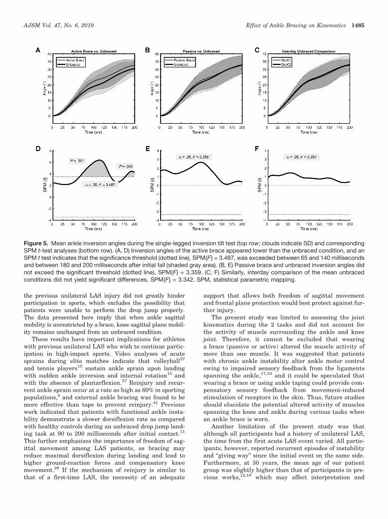
Comparison of the active brace and the unbraced condi-tion (Figure 5A) yielded a significant effect (P \ .001)between 65 and 140 milliseconds after the initial drop (Fig-ure 5D), indicating lower angles on the ankle equippedwith the active brace. At 180 to 200 milliseconds afterthe initial drop, another significant effect (P = .013) indi-cated a similar result. Comparison of the passive braceand the unbraced condition did not yield any differencesduring the entire drop time (Figure 5, B and E). The posthoc t test found no interday differences between theunbraced measurements (Figure 5, C and F).
DISCUSSION
This study assessed the effect of a selectively restrictiveankle brace on sagittal kinematics during simulated sprain
and drop landings among patients with a history of unilat-eral ankle sprain, while accounting for a possible placeboeffect. Our first hypothesis was partially supported, asthe active external brace was able to reduce angles duringa sudden inversion, but the placebo brace did not exhibitany differences when compared with an unbraced condi-tion. Furthermore, we did not find any differences in theknee and ankle sagittal angles across all conditions duringdrop landings. These results indicate that for the designtested, the brace effectively protects the ankle from inver-sion while allowing unrestricted sagittal movements of theankle and knee, and these effects are not due to the mereact of placing an external device on the ankle.
First, participants with a history of LAS were able toreduce inversion angles during a simulated inversion sprainwhen wearing an actively protecting brace, in comparisonwith both the unbraced and placebo brace conditions. Theseresults are in agreement with similar experimental setupsthat found reduced angles among healthy participantswith semirigid,8,39 hinged,2 and lace-up8 supports as com-pared with unbraced conditions. The resultant reductionof maximal inversion angle by 5� is comparable with previ-ous reports from Tang et al,39 who found 3� reduction whencomparing the Aircast brace and unbraced barefoot condi-tions in a similar inversion tilt test. These collective resultsare lower than the higher reductions found by Alfuth et al2
and Cordova et al,8 who found roughly 17� and 16.4� in dif-ferent brace settings, respectively. These differences may bedue to various brace designs, such as semirigid versus rigid.Furthermore, all of these previous reports were performedwith healthy participants and not participants with previ-ous LAS injury. Given the brace’s minimal design, directaccess to both malleoli was available for marker placement,ensuring that kinematics of the ankle were measured andnot those of the brace itself, as previously reported.39 Previ-ous tests either did not specifically control for weightbearingon the tested leg or assumed that weight distribution waseven between legs.2,8 Since we ensured that participantsalways placed 80% of their body weight on the testedlimb, we could ensure that the inversion tilt was performedsimilarly across patients and measurement days. By con-trolling these parameters, in addition to the motor-con-trolled velocity of the inversion plate and the potentialeffect of a placebo, we can be confident in the kinematicresults presented here.
Second, participants with a history of LAS showed no par-ticular differences in knee and ankle sagittal angles duringa single-legged drop landing among all tested conditions,which implies that freedom of movement in the sagittal planeis unaffected by this brace design. Gardner et al19 found thata more restrictive brace reduced the eccentric energyabsorbed at the ankle but not in braces that allow sagittalplane movement. Yet Cordova et al10 found that closed bas-ket weave taping and semirigid bracing led to lower kneeand ankle mobility during single-legged landing. These col-lective results indicate the importance of brace design onlanding lower extremity kinematics and that device-dependent sagittal plane restriction at the ankle may nega-tively affect the knee.29 Within the investigated cohort,self-reported Foot and Ankle Outcome Scores indicate that
Figure 4. Mean (A) curves and (B) SPM results from the anal-ysis of variance comparing inversion angles across all condi-tions for the sudden inversion plate tilt. The significantthreshold, indicated by the dotted line at SPM{F} = 6.498,was exceeded for within-subjects analysis between 60 and125 milliseconds after the initial fall (shaded gray area).Pooled SD of the group means is indicated by the gray cloudin panel A for each time point. SPM, statistical parametricmapping.
1484 Agres et al The American Journal of Sports Medicine
the previous unilateral LAS injury did not greatly hinderparticipation in sports, which excludes the possibility thatpatients were unable to perform the drop jump properly.The data presented here imply that when ankle sagittalmobility is unrestricted by a brace, knee sagittal plane mobil-ity remains unchanged from an unbraced condition.
These results have important implications for athleteswith previous unilateral LAS who wish to continue partic-ipation in high-impact sports. Video analyses of acutesprains during elite matches indicate that volleyball37
and tennis players15 sustain ankle sprain upon landingwith sudden ankle inversion and internal rotation15 andwith the absence of plantarflexion.37 Reinjury and recur-rent ankle sprain occur at a rate as high as 60% in sportingpopulations,4 and external ankle bracing was found to bemore effective than tape to prevent reinjury.41 Previouswork indicated that patients with functional ankle insta-bility demonstrate a slower dorsiflexion rate as comparedwith healthy controls during an unbraced drop jump land-ing task at 90 to 200 milliseconds after initial contact.11
This further emphasizes the importance of freedom of sag-ittal movement among LAS patients, as bracing mayreduce maximal dorsiflexion during landing and lead tohigher ground-reaction forces and compensatory kneemovement.29 If the mechanism of reinjury is similar tothat of a first-time LAS, the necessity of an adequate
support that allows both freedom of sagittal movementand frontal plane protection would best protect against fur-ther injury.
The present study was limited to assessing the jointkinematics during the 2 tasks and did not account forthe activity of muscle surrounding the ankle and kneejoint. Therefore, it cannot be excluded that wearinga brace (passive or active) altered the muscle activity ofmore than one muscle. It was suggested that patientswith chronic ankle instability alter ankle motor controlowing to impaired sensory feedback from the ligamentsspanning the ankle,17,33 and it could be speculated thatwearing a brace or using ankle taping could provide com-pensatory sensory feedback from movement-inducedstimulation of receptors in the skin. Thus, future studiesshould elucidate the potential altered activity of musclesspanning the knee and ankle during various tasks whenan ankle brace is worn.
Another limitation of the present study was thatalthough all participants had a history of unilateral LAS,the time from the first acute LAS event varied. All partic-ipants, however, reported recurrent episodes of instabilityand ‘‘giving way’’ since the initial event on the same side.Furthermore, at 30 years, the mean age of our patientgroup was slightly higher than that of participants in pre-vious works,12,19 which may affect interpretation and
Figure 5. Mean ankle inversion angles during the single-legged inversion tilt test (top row; clouds indicate SD) and correspondingSPM t-test analyses (bottom row). (A, D) Inversion angles of the active brace appeared lower than the unbraced condition, and anSPM t test indicates that the significance threshold (dotted line), SPM{F} = 3.487, was exceeded between 65 and 140 millisecondsand between 180 and 200 milliseconds after initial fall (shaded gray area). (B, E) Passive brace and unbraced inversion angles didnot exceed the significant threshold (dotted line), SPM{F} = 3.359. (C, F) Similarly, interday comparison of the mean unbracedconditions did not yield significant differences, SPM{F} = 3.342. SPM, statistical parametric mapping.
AJSM Vol. 47, No. 6, 2019 Effect of Ankle Bracing on Kinematics 1485

direct comparison of these results. However, to ensurehomogeneity within the investigated cohort, we ensuredthat all participants were free of any other injury, didnot sustain recent injury, and continued to participate inathletic activities regularly.
In conclusion, this study investigated the protectiveeffect of an external ankle brace while accounting for a pos-sible placebo effect of brace application. The results indi-cate that during a sudden inversion movement, there isno placebo effect for patients with previous LAS and thatonly an actively protecting brace protects from reinjury.These results further suggest that a brace design thatrestricts motion in only the frontal plane and not in thesagittal plane of the ankle may be ideal for use in high-impact sports, particularly for landing on a single leg. Fur-thermore, these results indicate that sagittal knee andankle kinematics remain unaffected when an ankle sup-port is worn that allows sagittal plane mobility.
ACKNOWLEDGMENT
The authors acknowledge Lena Castel-Wohnlich for herassistance with data collection in the laboratory.
REFERENCES
1. Adler RJ, Taylor JE. Random Fields and Geometry. New York, NY:
Springer; 2007.
2. Alfuth M, Klein D, Koch R, Rosenbaum D. Biomechanical comparison
of 3 ankle braces with and without free rotation in the sagittal plane. J
Athl Train. 2014;49(5):608-616.
3. Anandacoomarasamy A, Barnsley L. Long term outcomes of inver-
sion ankle injuries. Br J Sports Med. 2005;39(3):e14-e14.
4. Attenborough AS, Hiller CE, Smith RM, Stuelcken M, Greene A, Sin-
clair PJ. Chronic ankle instability in sporting populations. Sports Med.
2014;44(11):1545-1556.
5. Balsamo B, Geil MD, Ellis R, Wu J. Confirmation bias affects user
perception of knee braces. J Biomech. 2018;75:164-170.
6. Camomilla V, Cereatti A, Vannozzi G, Cappozzo A. An optimized pro-
tocol for hip joint centre determination using the functional method.
J Biomech. 2006;39(6):1096-1106.
7. Chu VW-S, Fong DT-P, Chan Y-Y, Yung PS-H, Fung K-Y, Chan K-M.
Differentiation of ankle sprain motion and common sporting motion
by ankle inversion velocity. J Biomech. 2010;43(10):2035-2038.
8. Cordova ML, Dorrough JL, Kious K, Ingersoll CD, Merrick MA. Pro-
phylactic ankle bracing reduces rearfoot motion during sudden inver-
sion. Scand J Med Sci Sports. 2007;17(3):216-222.
9. Cordova ML, Sefton JM, Hubbard TJ. Mechanical joint laxity associ-
ated with chronic ankle instability: a systematic review. Sports
Health. 2010;2(6):452-459.
10. Cordova ML, Takahashi Y, Kress GM, Brucker JB, Finch AE. Influ-
ence of external ankle support on lower extremity joint mechanics
during drop landings. J Sport Rehabil. 2010;19(2):136-148.
11. Delahunt E, Monaghan K, Caulfield B. Changes in lower limb kine-
matics, kinetics, and muscle activity in subjects with functional insta-
bility of the ankle joint during a single leg drop jump. J Orthop Res.
2006;24(10):1991-2000.
12. Doherty C, Bleakley C, Hertel J, Caulfield B, Ryan J, Delahunt E.
Single-leg drop landing motor control strategies following acute
ankle sprain injury. Scand J Med Sci Sports. 2015;25(4):525-533.
13. Ehrig RM, Heller MO, Kratzenstein S, Duda GN, Trepczynski A, Taylor
WR. The SCoRE residual: a quality index to assess the accuracy of
joint estimations. J Biomech. 2011;44(7):1400-1404.
14. Ehrig RM, Taylor WR, Duda GN, Heller MO. A survey of formal methods
for determining functional joint axes. J Biomech. 2007;40(10):2150-2157.
15. Fong DT-P, Ha SC-W, Mok K-M, Chan CW-L, Chan K-M. Kinematics
analysis of ankle inversion ligamentous sprain injuries in sports: five
cases from televised tennis competitions. Am J Sports Med. 2012;
40(11):2627-2632.
16. Fong DT-P, Hong Y, Chan L-K, Yung PS-H, Chan K-M. A systematic
review on ankle injury and ankle sprain in sports. Sports Med.
2007;37(1):73-94.
17. Freeman MA, Dean MR, Hanham IW. The etiology and prevention of
functional instability of the foot. J Bone Joint Surg Br. 1965;
47(4):678-685.
18. Friston KJ, Holmes AP, Worsley KJ, Poline J-PB, Frith CD, Fracko-
wiak RSJ. Statistical parametric maps in functional imaging: a general
linear approach. Hum Brain Mapp. 1995;2(4):189-210.
19. Gardner JK, McCaw ST, Laudner KG, Smith PJ, Stafford LN. Effect
of ankle braces on lower extremity joint energetics in single-leg land-
ings. Med Sci Sports Exerc. 2012;44(6):1116-1122.
20. Greene AJ, Stuelcken MC, Smith RM, Vanwanseele B. The effect of
external ankle support on the kinematics and kinetics of the lower
limb during a side step cutting task in netballers. BMC Sports Sci
Med Rehabil. 2014;6(1):42.
21. Hall EA, Simon JE, Docherty CL. Using ankle bracing and taping to
decrease range of motion and velocity during inversion perturbation
while walking. J Athl Train. 2016;51(4):283-290.
22. Jerosch J, Hoffstetter I, Bork H, Bischof M. The influence of orthoses
on the proprioception of the ankle joint. Knee Surg Sports Traumatol
Arthrosc. 1995;3(1):39-46.
23. Kemler E, van de Port I, Schmikli S, Huisstede B, Hoes A, Backx F.
Effects of soft bracing or taping on a lateral ankle sprain: a non-
randomised controlled trial evaluating recurrence rates and residual
symptoms at one year. J Foot Ankle Res. 2015;8:13.
24. Klem NR, Wild CY, Williams SA, Ng L. Effect of external ankle sup-
port on ankle and knee biomechanics during the cutting maneuver
in basketball players. Am J Sports Med. 2017;45(3):685-691.
25. Kratzenstein S, Kornaropoulos EI, Ehrig RM, Heller MO, Popplau BM,
Taylor WR. Effective marker placement for functional identification of
the centre of rotation at the hip. Gait Posture. 2012;36(3):482-486.
26. Liu SH, Nguyen TM. Ankle sprains and other soft tissue injures. Curr
Opin Rheumatol. 1999;11:132-137.
27. Maffulli N, Ferran NA. Management of acute and chronic ankle insta-
bility. J Am Acad Orthop Surg. 2008;16(10):608-615.
28. Malliaropoulos N, Ntessalen M, Papacostas E, Longo UG, Maffulli N.
Reinjury after acute lateral ankle sprains in elite track and field ath-
letes. Am J Sports Med. 2009;37(9):1755-1761.
29. Mason-Mackay AR, Whatman C, Reid D. The effect of ankle bracing
on lower extremity biomechanics during landing: a systematic
review. J Sci Med Sport. 2016;19(7):531-540.
30. McCaw ST, Cerullo JF. Prophylactic ankle stabilizers affect ankle
joint kinematics during drop landings. Med Sci Sports Exerc.
1999;31(5):702-707.
31. McKay GD, Goldie PA, Payne WR, Oakes BW, Watson LF. A prospec-
tive study of injuries in basketball: a total profile and comparison by gen-
der and standard of competition. J Sci Med Sport. 2001;4(2):196-211.
32. Pataky TC. Generalized n-dimensional biomechanical field analysis using
statistical parametric mapping. J Biomech. 2010;43(10):1976-1982.
33. Richie DH. Functional instability of the ankle and the role of neuro-
muscular control: a comprehensive review. J Foot Ankle Surg.
2001;40(4):240-251.
34. Roos EM, Brandsson S, Karlsson J. Validation of the Foot and Ankle
Outcome Score for ankle ligament reconstruction. Foot Ankle Int.
2001;22(10):788-794.
35. Sawkins K, Refshauge K, Kilbreath S, Raymond J. The placebo effect
of ankle taping in ankle instability. Med Sci Sports Exerc.
2007;39(5):781-787.
36. Simpson KJ, Yom JP, Fu Y, Arnett SW, Rourke SO, Brown CN. Does
wearing a prophylactic ankle brace during drop landings affect lower
extremity kinematics and ground reaction forces? J Appl Biomech.
2013;29(2):205-213.
1486 Agres et al The American Journal of Sports Medicine

37. Skazalski C, Kruczynski J, Bahr MA, Bere T, Whiteley R, Bahr R.
Landing-related ankle injuries do not occur in plantarflexion as
once thought: a systematic video analysis of ankle injuries in
world-class volleyball. Br J Sports Med. 2018;52(2):74-82.
38. Tamura K, Radzak KN, Vogelpohl RE, et al. The effects of ankle braces
and taping on lower extremity running kinematics and energy expen-
diture in healthy, non-injured adults. Gait Posture. 2017;58:108-114.
39. Tang YM, Wu ZH, Liao WH, Chan KM. A study of semi-rigid support
on ankle supination sprain kinematics. Scand J Med Sci Sports.
2010;20(6):822-826.
40. Taylor WR, Kornaropoulos EI, Duda GN, et al. Repeatability and
reproducibility of OSSCA, a functional approach for assessing the
kinematics of the lower limb. Gait Posture. 2010;32(2):231-236.
41. Verhagen EA, van Mechelen W, de Vente W. The effect of preventive
measures on the incidence of ankle sprains. Clin J Sport Med.
2000;10:291-296.
42. Wu G, Siegler S, Allard P, et al. ISB recommendation on definitions of
joint coordinate system of various joints for the reporting of human
joint motion—part I: ankle, hip, and spine. International Society of
Biomechanics. J Biomech. 2002;35(4):543-548.
For reprints and permission queries, please visit SAGE’s Web site at http://www.sagepub.com/journalsPermissions.nav.
AJSM Vol. 47, No. 6, 2019 Effect of Ankle Bracing on Kinematics 1487