4d statistical shape model of the heart for x-ray ... · pdf file4d statistical shape model of...
TRANSCRIPT

4D Statistical Shape Model of the Heartfor X-Ray Projection Imaging
Mathias Unberath, Andreas Maier, Dominik Fleischmann, JoachimHornegger, Rebecca Fahrig
April, 17. 2015
Pattern Recognition Lab, FAU Erlangen-NürnbergRadiological Sciences Lab, Stanford University
Context
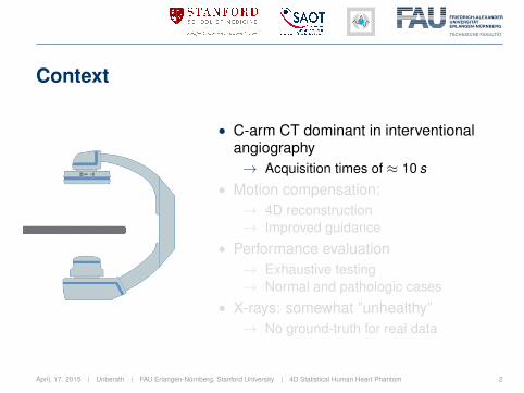
• C-arm CT dominant in interventionalangiography→ Acquisition times of ≈ 10 s
• Motion compensation:→ 4D reconstruction→ Improved guidance
• Performance evaluation→ Exhaustive testing→ Normal and pathologic cases
• X-rays: somewhat ”unhealthy”→ No ground-truth for real data
April, 17. 2015 | Unberath | FAU Erlangen-Nürnberg, Stanford University | 4D Statistical Human Heart Phantom 2

Context
• Need for artificial data→ Simulation frameworks→ Numerical phantoms
• Enable comparison:→ Framework: CONRAD
• XCAT1
→ 3D from Visible Human→ Motion from one male patient→ Developed for ET→ Licensing fee (small)
1Segars et al., “4D XCAT phantom for multimodality imaging research”.April, 17. 2015 | Unberath | FAU Erlangen-Nürnberg, Stanford University | 4D Statistical Human Heart Phantom 3
Goals
A new phantom should be:• available
• dynamic (temporal variation)
• versatile (inter-subject variation)
• clinically relevant
Dynamic statistical shape model of the heart.
April, 17. 2015 | Unberath | FAU Erlangen-Nürnberg, Stanford University | 4D Statistical Human Heart Phantom 4
Contents
Training set generationRegistration PipelineResults and conclusions
Model-building and simulationAlignment and principal component analysisResults and conclusions
April, 17. 2015 | Unberath | FAU Erlangen-Nürnberg, Stanford University | 4D Statistical Human Heart Phantom 5
Problem statement
Learn valid behavior from training setMany shapes from diverse anatomies. 2
Point correspondence must be established/preserved.
? Data-driven segmentation (incl. manual)
! Registration-based segmentation
2How ”many” and how ”diverse”?
April, 17. 2015 | Unberath | FAU Erlangen-Nürnberg, Stanford University | 4D Statistical Human Heart Phantom 6

General idea
Propagate landmarks from atlas to new images.3,4
What is needed?
• Landmarked atlas segmentation
• Registration pipeline
What atlas? What pipeline?
3Frangi et al., “Automatic construction of multiple-object three-dimensional statistical shape models: Application to cardiacmodeling”.
4Ordas et al., “A statistical shape model of the heart and its application to model-based segmentation”.
April, 17. 2015 | Unberath | FAU Erlangen-Nürnberg, Stanford University | 4D Statistical Human Heart Phantom 7
Atlas segmentation
1. Manual segmentation in ITK Snap
2. Mesh generation (coarsening and smoothing)
Data set used:
• 45 y/o female, 78% phase
• 512× 512× 241 pixels
• 0.29× 0.29× 0.5 mm spacing
April, 17. 2015 | Unberath | FAU Erlangen-Nürnberg, Stanford University | 4D Statistical Human Heart Phantom 8
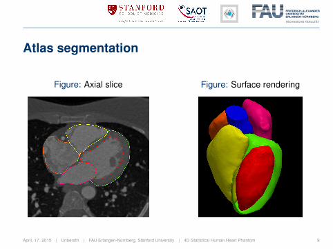
Atlas segmentation
Figure: Axial slice Figure: Surface rendering
April, 17. 2015 | Unberath | FAU Erlangen-Nürnberg, Stanford University | 4D Statistical Human Heart Phantom 9

Registration
B-Spline-based registration pipeline:
Rigid:• Similarity transform
• Mutual information
Non-rigid: multi-resolution• B-spline transforms
• Mutual information
April, 17. 2015 | Unberath | FAU Erlangen-Nürnberg, Stanford University | 4D Statistical Human Heart Phantom 10

Mutual information
Reduce uncertainty in X by knowing YNo explicit form of dependency needed
Mutual information∫ ∫pfm (f (x),m(y)) log
(pfm (f (x),m(y))
pf (f (x)) pm (m(y))
)dxdy
f , m are the fixed and moving imagepf and pm, and pfm are the marginal and joint histograms
April, 17. 2015 | Unberath | FAU Erlangen-Nürnberg, Stanford University | 4D Statistical Human Heart Phantom 11

B-Spline transforms
Smooth transforms defined on control gridWeighted sum of points in finite support region
1D B-Spline
T (x) =d∑
n=0
Bn(u)Φk+n,
u = xnx− b x
nxc ∈ [0, 1] k = b x
nxc − 1
Bn(u): B-Spline basis function (≡ weights)
Φi : control points in grid
April, 17. 2015 | Unberath | FAU Erlangen-Nürnberg, Stanford University | 4D Statistical Human Heart Phantom 12
Evaluation
Procedure
• Fix registration parameters
• Register to data at all cardiac phases
Quality assessmentRepresentative female and male patient data
• Visual evaluation
• Expert ranking
April, 17. 2015 | Unberath | FAU Erlangen-Nürnberg, Stanford University | 4D Statistical Human Heart Phantom 13
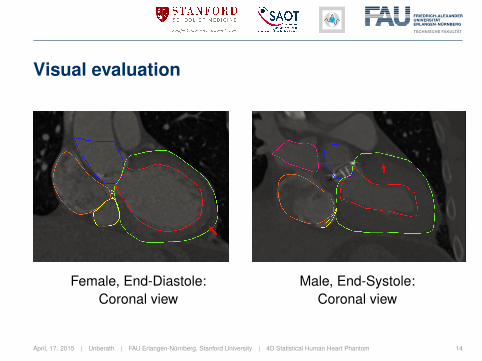
Visual evaluation
Female, End-Diastole:Coronal view
Male, End-Systole:Coronal view
April, 17. 2015 | Unberath | FAU Erlangen-Nürnberg, Stanford University | 4D Statistical Human Heart Phantom 14

Expert ranking: Results & Discussion
Average scores throughout the cardiac cycle
• 3 experts: Grades ∈ [0, 5], 5 ≡ best• Overall score: 3.33± 0.51• Atlas segmentation is at 78% phase (end-diastole)
→ Induces bias.
April, 17. 2015 | Unberath | FAU Erlangen-Nürnberg, Stanford University | 4D Statistical Human Heart Phantom 15

Conclusions
• Reduce bias: create atlas from ”mean heart”
• Automatic registration: parameters fixed
→ Automatic parameter tuning at run-time?
Training set generation is not time-sensitive but crucial.→Refinement of the automatic segmentation (e.g. local adaptation).
April, 17. 2015 | Unberath | FAU Erlangen-Nürnberg, Stanford University | 4D Statistical Human Heart Phantom 16

Best case scenario
Atlas segmentation:Axial view
Female, End-Diastole:Axial view
April, 17. 2015 | Unberath | FAU Erlangen-Nürnberg, Stanford University | 4D Statistical Human Heart Phantom 17

Procedure
Statistical shape model generation
Four step process:
1. Obtain training shapes
2. Establish point correspondence
3. Align shapes
4. Extract principal modes of variation
April, 17. 2015 | Unberath | FAU Erlangen-Nürnberg, Stanford University | 4D Statistical Human Heart Phantom 18
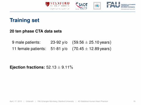
Training set
20 ten phase CTA data sets
9 male patients: 23-92 y/o (59.56± 25.10 years)
11 female patients: 51-81 y/o (70.45± 12.89 years)
Ejection fractions: 52.13± 9.11%
April, 17. 2015 | Unberath | FAU Erlangen-Nürnberg, Stanford University | 4D Statistical Human Heart Phantom 19

Alignment
Generalized Procrustes AnalysisPose (and scale) is not part of shape.
1. Center and scale input samples Xi
2. Rotate all n shapes Xi to fit X1
3. Calculate consensus shape Y4. Until convergence:
• Rotate and scale Xi to consensus Y• Reassure proper scaling• Calculate residual change
April, 17. 2015 | Unberath | FAU Erlangen-Nürnberg, Stanford University | 4D Statistical Human Heart Phantom 20
PCA
Goals of Principal Component Analysis
• Extract most important information from the data
• Reduce dimensionality of the data
• Simplify description of shapes
April, 17. 2015 | Unberath | FAU Erlangen-Nürnberg, Stanford University | 4D Statistical Human Heart Phantom 21
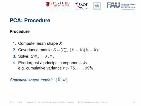
PCA: Procedure
Procedure
1. Compute mean shape X̄
2. Covariance matrix: S =∑n
i=1(Xi − X̄ )(Xi − X̄ )T
3. Solve: S Φk = λk Φk
4. Pick largest c principal components Φk
e.g. cumulative variance r > 75, · · · , 99%
Statistical shape model: {X̄ ,Φ}
April, 17. 2015 | Unberath | FAU Erlangen-Nürnberg, Stanford University | 4D Statistical Human Heart Phantom 22
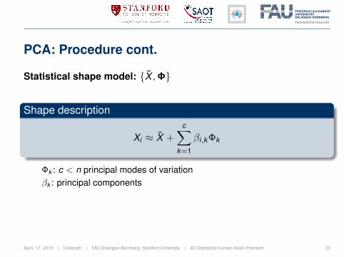
PCA: Procedure cont.
Statistical shape model: {X̄ ,Φ}
Shape description
Xi ≈ X̄ +c∑
k=1
βi,k Φk
Φk : c < n principal modes of variationβk : principal components
April, 17. 2015 | Unberath | FAU Erlangen-Nürnberg, Stanford University | 4D Statistical Human Heart Phantom 23
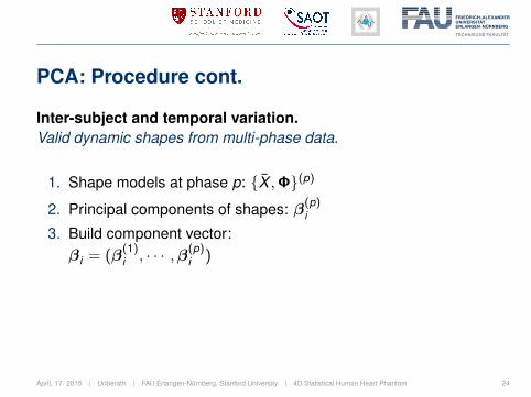
PCA: Procedure cont.
Inter-subject and temporal variation.Valid dynamic shapes from multi-phase data.
1. Shape models at phase p: {X̄ ,Φ}(p)
2. Principal components of shapes: β(p)i
3. Build component vector:βi = (β
(1)i , · · · ,β(p)
i )
April, 17. 2015 | Unberath | FAU Erlangen-Nürnberg, Stanford University | 4D Statistical Human Heart Phantom 24
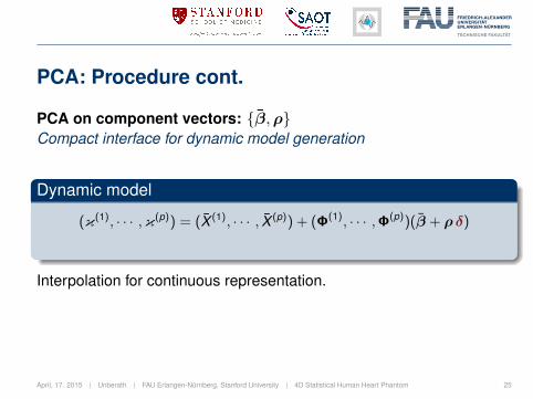
PCA: Procedure cont.
PCA on component vectors: {β̄,ρ}Compact interface for dynamic model generation
Dynamic model
(κ(1), · · · ,κ(p)) = (X̄ (1), · · · , X̄ (p)) + (Φ(1), · · · ,Φ(p))(β̄ + ρδ)
Interpolation for continuous representation.
April, 17. 2015 | Unberath | FAU Erlangen-Nürnberg, Stanford University | 4D Statistical Human Heart Phantom 25

Results: Generalization
Cross validation: leave-one-out testCapability to represent unseen instances.
• Exclude shape from model
• Fit model to shape
• Compute error
90% variation: 5.00± 0.93 mm95% variation: 4.89± 0.90 mm
April, 17. 2015 | Unberath | FAU Erlangen-Nürnberg, Stanford University | 4D Statistical Human Heart Phantom 26

Results: Specificity
Random shape samplingValidity of new instances.
• Generate random component vectors
• Compute distance to nearest training shape
Static: 1000 samples 7.18± 0.45 mmDynamic: 100 samples 7.30± 0.97 mm
April, 17. 2015 | Unberath | FAU Erlangen-Nürnberg, Stanford University | 4D Statistical Human Heart Phantom 27

Results: Variability at diastole
Decreasing variance µ from left to right: δb = −µb/2 top,δb = µb/2 bottom, and δb = 0 mid.
April, 17. 2015 | Unberath | FAU Erlangen-Nürnberg, Stanford University | 4D Statistical Human Heart Phantom 28
Results: Projection imaging
Volume rendering Projection image
April, 17. 2015 | Unberath | FAU Erlangen-Nürnberg, Stanford University | 4D Statistical Human Heart Phantom 29
Conclusions
• Large variation among few samples• Deteriorates specificity→ Revisit training set generation
• Currently: anatomy at rest determines contraction→ Multi-linear PCA
→ Thorax model, Breathing motion, ...
To our knowledge the first open-source,dynamic statistical shape model of the heart. 5
5Available at: conrad.stanford.edu
April, 17. 2015 | Unberath | FAU Erlangen-Nürnberg, Stanford University | 4D Statistical Human Heart Phantom 30