3d bioprinters - future of implants biofabrication
TRANSCRIPT
52 VOLUME 17, No. 1, 2013
BIOGRAPHICAL NOTESdoc. Ing. Radovan Hudák, PhD. was born in 1976 in Košice, Slovakia. He received his MS and PhD degree in biomedical engineering at Technical University in Košice in 2000 and 2008. Today he is an associative professor and head of prosthetics and orthotics study program and head of Biomedical Engineering Division at the Department of Biomedical Engineering and Measurement, Technical University of Košice. His research interests include additive manufacturing in medicine, medical thermography, indus-trial thermography, human biomechanics and rehabilitation technologies. From 2004 to 2009 he was a technical assistant at the Center of Refractive Surgery in Kosice. Mr. Hudak has more than 120 publications in home and foreign journals. He is co-author of 3 monographies and 7 books.Dr.h.c. prof. Ing. Jozef Živčák, PhD. is a professor of biomedical engineering at Tech-nical University in Košice, Slovakia. He was born in 1958. He received his MS and PhD degrees from Technical University in Košice in 1995. Prof. Živčák is from 2009 Doctor Honoris Causa of Uzhorod National University. His research interests include human biomechanics, medical sensorics, medical thermography and rehabilitation technol-ogy. Today Mr. Zivcak is head of Department of Biomedical Engineering and Mea-surement at the Faculty of Mechanical Engineering, Technical University of Kosice. He attended at more than 15 international stays, e.g. at CTU in Prague, Polytechnika Bialystok, Poland, Dresden, Germany, Tokyo, Japan, etc. Since 1998 he is an expert wit-ness in machine and electrical technology member of international committee TC 18 and head of IMEKO in Slovakia - Measurement of Human Functions. Professor Zivcak has more than 200 publications in home and foreign journals. He is an author and co-author of 8 monographies and 11 books.Ing. Martin Šarik was born in 1987 in Michalovce, Slovakia. He received his master degree in biomedical engineering at Technical University of Košice in 2011. Today Mr. Šarik is a doctoral student at the Department of Biomedical Engineering and Measure-ment, Technical University of Košice. His research interests include laser sintering, ad-ditive manufacturing, and software thermal analysis. From 2012 he is co-investigator of project Center for research of control of technical, environmental and human risks for permanent development of production and products in mechanical engineering supported by the Research & Development Operational Programme funded by the ERDF. In 2012/ 2013 he attended the meeting and software training of RadTherm ap-plication in Paris, France and Munich, Germany. He is an author and co-author of 3 publications in home and in foreign scientific journal. Ing. Robert Dadej was born in 1988 in Prešov, Slovakia. He received his MS degree
3D Bioprinters – Future of Implants Biofabrication
Radovan Hudák 1*, Jozef Živčák 2, Martin Šarik 3, Robert Dadej 4, Richard Raši 5
1,2,3,4 Department of Biomedical Engineering and Measurement, Faculty of Mechanical Engineering, Technical University of Košice, Letná 9, 042 00 Košice, Slovak Republic
5 Louis Pasteur University Hospital, Rastislavová 43, 041 90 Košice, Slovak Republic

Acta Mechanica SlovacaJournal published by Faculty of Mechanical Engineering - Technical University of Košice
53
in biomedical engineering at Technical University of Košice, at the Department of Biomedical Engi-neering and Measurements, in 2012. He is since 2012 active as a PhD. student at the Department of Biomedical Engineering and Measurements, at Technical University of Košice. Today he continues his research interests in direct metal laser sintering and additive manufacturing of implants. He is an author and co-author of 2 publications in home and in foreign scientific journal.MUDr. Richard Raši, PhD., MPH was born in 1971 in Košice, Slovakia. He received his doctor´s degree in general medicine at the Pavol Jozef Šafárik Uni-versity in Košice in 1995. In 1998 he finished his first specialization study in surgery and in 2004 his sec-ond degree specialization study in traumatology. He received the MPH - Master of Public Health de-gree at the Slovak Medical University in Bratislava in 2004 and his PhD degree at the Technical Uni-versity in Košice in 2010. Today Mr. Raši is a Mem-ber of the National Council of the Slovak Republic, Chairman of the Committee of Health of the Slovak National Council and Mayor of the City of Košice. He has been working as a Physician in the Clinic of Traumatology at the University Hospital of Louis Pasteur in Košice since 1995 until now. From 2004 to 2007 he was Vice Director for surgical depart-ments at the University Hospital of L. Pasteur in Košice. From 2007 to 2008 he was Director of the University Hospital in Bratislava. He served as Min-ister of Health of the Slovak Republic from 2008 to 2010. He participated in internships and medical trainings in Stockholm (Sweden), Worcester (USA), Basel (Switzerland), Reims (France) and in Bratislava (Slovakia).
KEY WORDSAdditive manufacturing, bioprint, bioprinter, scaf-fold, organs, tissues.
ABSTRACTIn a last decade has additive manufacturing passed a long way, where was reached an impressive ad-vance in rapid prototyping technology of fabrica-tion. From plastic and ceramic materials through metals to at the moment most interesting technol-ogy of bioprint, where the material which is used for building, directly consist of human tissues. Or-gan printing, which is based on computer-aided 3D tissue engineering, offers the wide range for
research and development in this area. This article summarizes the present advance in this new and not entirely explored field of bio-additive manu-facturing. With the help of this technology can be produced the real 3D models of organs and tissues, that should help surgeons in preopera-tive planning or can be used like spare “parts” for transplantations. The main emphasis is placed on tissue engineering technology which has the best assumptions to solve transplantation issues. Also this article includes the comparison of devices and materials which are possible to engage within the bio-manufacturing.
1. Introduction The change in the field of biology and bio-engi-neering has built an background in which the im-provements in the life sciences are not only more accessible, but call for the active fellowship of en-gineering design and fabrication to reach solutions for complex biological problems. This progress, along with the development of new design and fabrication, biomaterials, biology and biomedicine, has advanced the additive manufacturing technol-ogy to a broad application in biomedical engineer-ing [1]. Tissue engineering has reached more attention in the past decade, owing to its success in enabling tissue regeneration for therapeutic purposes [2]. Tissue engineering introduces the interdisciplinary area in which are applied the principles of biology and engineering to the evolution of substitutes that repair, improve or restore function of tissue. The main target is to fabricate patient-specific bio-logical substitutes in an attempt to sidestep the restrictions of existing clinical treatments for dam-aged tissue or organs. The main regenerative tissue engineering approach involves transplantation of cells onto scaffolds [2]. These restrictions include deficiency of donor organs, chronic refusal and cell morbidity. The principal approach includes injec-tion of cells alone, evolution of encapsulated sys-tems and implantations of cells onto scaffolds [2]. The utilization of additive manufacturing (AM) in combination with computer-aided (CA) technolo-gies has offered new possibilities for medical mod-eling, with using of computer models or additive manufacturing fabricated physical models for rep-resentation of patient specific anatomical geom-etry [1].

54 VOLUME 17, No. 1, 2013
Fig. 1: Example of rapid prototyping fabrication. DMLS method which uses the additive principle to build the real parts. a) .STL model of
part, b) technology used for manufacture, c) final physical model
ments for printing the living human tissue.
3. Bio-Additive Manufacturing Bio-additive manufacturing (BAM) utilize the principles of standard additive manufacturing to build the physical model which have the same or nearly the same properties like real tissue or organ. BAM should be described as biofabrication using cells, biologics or biomaterials as building blocks to fabricate biological and bio-application orient-ed substances, devices and therapeutic products through a broad range of engineering, physical, chemical and/or biological processes [1].The re-search and development (R&D) of anatomical and biological models represents the most significant area which is necessary for integration of RP in bio-logical engineering. On the Fig. 2 is shown the ex-ample procedure of BAM process.
2. Additive ManufacturingA. Rapid Prototyping Rapid prototyping also called as solid freeform fabrication and layered (additive) manufacturing technology allow scientist to create physical part-sin a short period, directly from 3D models created via computer-aided design (CAD),computer-aided engineering (CAE), and computer-aided manufac-turing (CAM) programs as is shown on a Fig. 1 [3, 4, 5]. The additive fabrication refers to a class of manu-facturing processes, in which a part is built by add-ing layers of material upon one another [6]. It offers several advantages as speed, part complexity, wide range of materials to use (plastics, metals, ceramics, composites and even material with properties sim-ilar to wood) and low-volume production because there is no need to produce custom tooling. Gen-erally the AM technologies bring the special and unique possibilities for customization, upgrades in product performance, multi-dependence, and lower overall manufacturing costs. These possibilities include [4]: Shape complexity ,Material complexity, Hierarchical complexity. B. Standard 3D printing methods Between 3D printing methods which are up to now known, and they differ among themselves by used technology, we classify for example: direct metal laser sintering (DMLS), selective laser sin-tering (SLS), jetted photopolymer (JP), laminated object manufacturing (LOM), fused deposition modeling (FDM), stereolithography (SLA), three-di-mensional printing (3DP) by TheriForm fabrication, precision extruding deposition (PED) and micro-syringe based polymer deposition. Several of these methods are used for the bio-additive manufactur-ing, because they meet the principles and require-
Damaged organ - tissue Scan (MRI,CT)
BioCAD
(create CAD model)
Bioprinter (hydrogel+cells)
Substitute organ (implant)
Fig. 2: Example of BAM process which can be modified according
to real needs of biofabrication.
Classical tissue engineering refers to seeding isolated cells on solid scaffolds as introduced by Langer and Vacanti almost two decades ago and is still a cutting-edge technology [7]. Biomaterials can offer ideal biocompatible prop-erties for scaffolding and generating cell-scaffold constructs demonstrate promising alternatives for autologous grafting and organ replacement

Acta Mechanica SlovacaJournal published by Faculty of Mechanical Engineering - Technical University of Košice
55
[7]. Biofabrication allows cells to be situated in a controlled way in and together with biomaterials. Biofabrication includes various techniques: bio-printing, bioplotting, inkjet printing and stereo-lithography. Definition is described as production of com-plex living and non-living biological products by placing proteins, peptides, DNA, cells, hormones or ECM molecules together with biomaterials [7]. During the BAM process is necessary to ensure ful-fillment requirements which are shown on Fig. 3.
Fig. 3: Requirements for bio-additive manufacturing.
(USA) bioprinter is schematically shown on Fig. 5. One of the most complex challenges in the devel-opment of the bio-printer was to perfect a means to consistently position the cell dispensing capil-lary tip attached to the print head within microns [10]. Invetech (USA) developed a computer-con-trolled, laser-based calibration system to achieve the required repeatability. The process uses inkjet based 3D printers and an ink made of human cells (as with cloning, using the patient‘s own cells are the best bet) mixed with a dissolvable gel, often cellulose [10].
As part of the requirements is necessary, to pro-vide taxonomy of communication between scien-tists from different disciplines (engineering, biol-ogy, materials and science) validation of biological tissue for production, biological function and sta-bility of bio-material before and after the manufac-ture of the product. A. Rapid Prototyping Inkjet 3D printing is a non-contact method which uses digital data from a computer and reproduces it layer-by-layer by putting ink drops on previously printed successive layers [7]. Bioprinters may be constructed in various configurations. However, all bioprinters output cells from a bioprint head that moves in three dimensional (3D) space (x,y,z) in or-der to place the cells precisely where required. In addition to outputting cells, most bioprinters also output dissolvable water based hydrogel to sup-port and protect cells during printing [10]. Several experimental bioprinters have already been built. The Fig. 4 presents the timeline of bioprinting evo-lution and its pioneers over the years. The NovoGen MMX Bioprinter (USA) includes two robotically controlled accuracy print heads: one for placing human cells, the other for placing a hydrogel, scaffold, or support matrix. The NovoGen
Fig. 4: 2002 – utilization of inkjet principle to bioprint, 2006 – arti-
ficial bladder, 2008 – bioprinter NovoGen MMX, 2010 – creation of
blood vessels by bioprinter.
Fig. 5: NovoGen MMX Bioprinter (USA) [15].
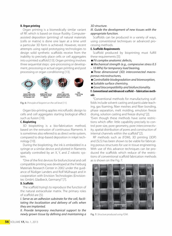
The fabrication process is similar to other (AM) techniques. The printer puts down a layer, which is then cured with heat, chemicals or ultra-violet (UV) light, before moving on to the next layer. To create its output, the NovoGen (USA) first lays down a single layer of a water-based bio-paper made from collagen, gelatin or other hydrogels [12]. Bioink spheroids are then injected into this water-based material. This process is shown on the Fig. 6. After that bioink spheroids slowly fuse to-gether. As this occurs, the bio-paper dissolves away or is removed, than leaving a final bioprinted body part or tissue [10].
56 VOLUME 17, No. 1, 2013
B. Organ printing Organ printing is a biomedically similar variant of RP, which is based on tissue fluidity. Computer-assisted deposition (printing) of natural materials (cells or matrix) is done one layer at a time until a particular 3D form is achieved. However, recent attempts using rapid prototyping technologies to design solid synthetic scaffolds receive from the inability to precisely place cells or cell aggregates into a printed scaffold [13]. Organ printing involves three sequential steps: -pre-processing or develop-ment, processing or actual organ printing and post processing or organ conditioning [13].
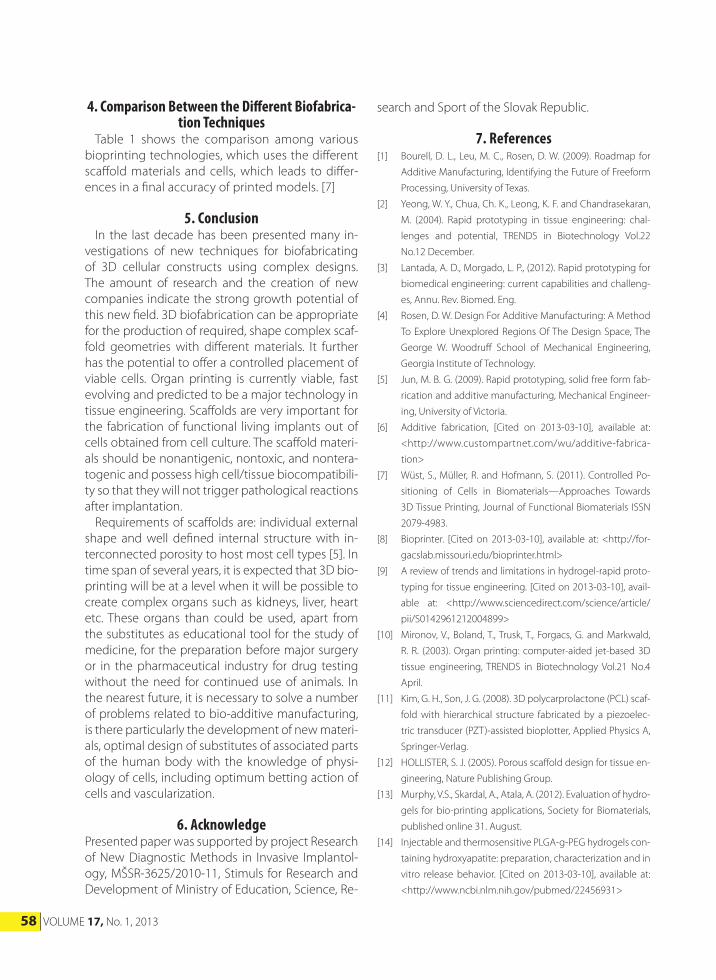
3D structure.III. Guide the development of new tissues with the appropriate function. Scaffolds can be produced in a variety of ways, using conventional techniques or advanced pro-cessing methods.E. Scaffolds Requirements Scaffold produced by bioprinting must fulfill these requirements [5]:Fit complex anatomic defects,Mechanical strength (e.g., compressive stress 0.5 – 10 MPa) for temporary load bearing,Three dimensional (3D) interconnected macro-porous microstructures,Controllable biodegradation and bioresorption,Suitable surface chemistry,Good biocompatibility and biofunctionality.F. Conventional and Advanced scaffold – fabrication meth-ods “Conventional methods for manufacturing scaf-folds include solvent casting and particulate leach-ing, gas foaming, fiber meshes and fiber bonding, phase separation, melt molding, emulsion freeze drying, solution casting and freeze drying” [2]. “Even though these methods have some restric-tions which offer: little capability precisely to con-trol pore size, pore geometry, pore interconnectiv-ity, spatial distribution of pores and construction of internal channels within the scaffold” [2]. RP methods such as (FDM), 3D printing (3DP) and (SLS) has been shown to be viable for fabricat-ing porous structures for use in tissue engineering. With use of this advance techniques can be pro-duced the scaffolds which reduce of the restric-tions of conventional scaffold fabrication methods as is shown on the Fig. 7.
Fig. 6: Principle of bioprint on the cell level [11].
Fig. 7: Structure produced using FDM.
Organ bio-printing applies microfluidic design to cells and cell aggregates starting biological effect such as fusion [10].C. Bioplotting 3D bioplotting is a bio-fabrication method, based on the extrusion of continuous filaments. It is sometimes also referred to as direct write system, compared to drop-based deposition in inkjet tech-nology [10]. During the bioplotting, the ink is embedded in a syringe or a similar device and plotted in filaments spatially controlled by an X, Y, and Z robotic sys-tem. “One of the first devices for biofunctional and cell compatible printing was developed at the Freiburg Materials Research Center in 2002 under the guid-ance of Rüdiger Landers and Rolf Mülhaupt and in cooperation with Envision Technologies (Envision-tec GmbH, Gladbeck, Germany)” [7].D. Scaffolds The scaffold trying’s to reproduce the function of the natural extracellular matrix. The primary roles of scaffold are [5]:I. Serve as an adhesion substrate for the cell, facili-tating the localization and delivery of cells when they are implanted. II. Provide temporary mechanical support to the newly grown tissue by defining and maintaining a
Acta Mechanica SlovacaJournal published by Faculty of Mechanical Engineering - Technical University of Košice
57
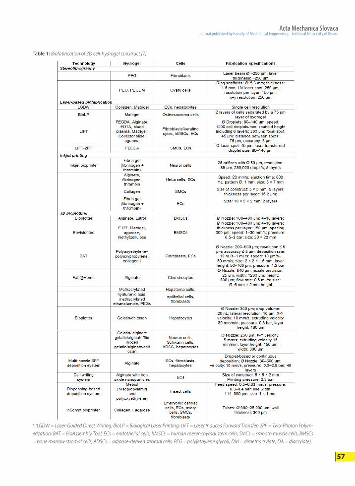
Table 1: Biofabrication of 3D cell-hydrogel construct [7]
a (LGDW = Laser-Guided Direct Writing; BioLP = Biological Laser Printing; LIFT = Laser Induced Forward Transfer; 2PP = Two-Photon Polym-
erization; BAT = BioAssembly Tool; ECs = endothelial cells; hMSCs = human mesenchymal stem cells; SMCs = smooth muscle cells; BMSCs
= bone marrow stromal cells; ADSCs = adipose-derived stromal cells; PEG = poly(ethylene glycol); DM = dimethacrylate; DA = diacrylate).
58 VOLUME 17, No. 1, 2013
4. Comparison Between the Different Biofabrica-tion Techniques
Table 1 shows the comparison among various bioprinting technologies, which uses the different scaffold materials and cells, which leads to differ-ences in a final accuracy of printed models. [7]
5. Conclusion In the last decade has been presented many in-vestigations of new techniques for biofabricating of 3D cellular constructs using complex designs. The amount of research and the creation of new companies indicate the strong growth potential of this new field. 3D biofabrication can be appropriate for the production of required, shape complex scaf-fold geometries with different materials. It further has the potential to offer a controlled placement of viable cells. Organ printing is currently viable, fast evolving and predicted to be a major technology in tissue engineering. Scaffolds are very important for the fabrication of functional living implants out of cells obtained from cell culture. The scaffold materi-als should be nonantigenic, nontoxic, and nontera-togenic and possess high cell/tissue biocompatibili-ty so that they will not trigger pathological reactions after implantation. Requirements of scaffolds are: individual external shape and well defined internal structure with in-terconnected porosity to host most cell types [5]. In time span of several years, it is expected that 3D bio-printing will be at a level when it will be possible to create complex organs such as kidneys, liver, heart etc. These organs than could be used, apart from the substitutes as educational tool for the study of medicine, for the preparation before major surgery or in the pharmaceutical industry for drug testing without the need for continued use of animals. In the nearest future, it is necessary to solve a number of problems related to bio-additive manufacturing, is there particularly the development of new materi-als, optimal design of substitutes of associated parts of the human body with the knowledge of physi-ology of cells, including optimum betting action of cells and vascularization.
6. AcknowledgePresented paper was supported by project Research of New Diagnostic Methods in Invasive Implantol-ogy, MŠSR-3625/2010-11, Stimuls for Research and Development of Ministry of Education, Science, Re-
search and Sport of the Slovak Republic.
7. References[1] Bourell, D. L., Leu, M. C., Rosen, D. W. (2009). Roadmap for
Additive Manufacturing, Identifying the Future of Freeform
Processing, University of Texas.
[2] Yeong, W. Y., Chua, Ch. K., Leong, K. F. and Chandrasekaran,
M. (2004). Rapid prototyping in tissue engineering: chal-
lenges and potential, TRENDS in Biotechnology Vol.22
No.12 December.
[3] Lantada, A. D., Morgado, L. P., (2012). Rapid prototyping for
biomedical engineering: current capabilities and challeng-
es, Annu. Rev. Biomed. Eng.
[4] Rosen, D. W. Design For Additive Manufacturing: A Method
To Explore Unexplored Regions Of The Design Space, The
George W. Woodruff School of Mechanical Engineering,
Georgia Institute of Technology.
[5] Jun, M. B. G. (2009). Rapid prototyping, solid free form fab-
rication and additive manufacturing, Mechanical Engineer-
ing, University of Victoria.
[6] Additive fabrication, [Cited on 2013-03-10], available at:
<http://www.custompartnet.com/wu/additive-fabrica-
tion>
[7] Wüst, S., Müller, R. and Hofmann, S. (2011). Controlled Po-
sitioning of Cells in Biomaterials—Approaches Towards
3D Tissue Printing, Journal of Functional Biomaterials ISSN
2079-4983.
[8] Bioprinter. [Cited on 2013-03-10], available at: <http://for-
gacslab.missouri.edu/bioprinter.html>
[9] A review of trends and limitations in hydrogel-rapid proto-
typing for tissue engineering. [Cited on 2013-03-10], avail-
able at: <http://www.sciencedirect.com/science/article/
pii/S0142961212004899>
[10] Mironov, V., Boland, T., Trusk, T., Forgacs, G. and Markwald,
R. R. (2003). Organ printing: computer-aided jet-based 3D
tissue engineering, TRENDS in Biotechnology Vol.21 No.4
April.
[11] Kim, G. H., Son, J. G. (2008). 3D polycarprolactone (PCL) scaf-
fold with hierarchical structure fabricated by a piezoelec-
tric transducer (PZT)-assisted bioplotter, Applied Physics A,
Springer-Verlag.
[12] HOLLISTER, S. J. (2005). Porous scaffold design for tissue en-
gineering, Nature Publishing Group.
[13] Murphy, V.S., Skardal, A., Atala, A. (2012). Evaluation of hydro-
gels for bio-printing applications, Society for Biomaterials,
published online 31. August.
[14] Injectable and thermosensitive PLGA-g-PEG hydrogels con-
taining hydroxyapatite: preparation, characterization and in
vitro release behavior. [Cited on 2013-03-10], available at:
<http://www.ncbi.nlm.nih.gov/pubmed/22456931>
Acta Mechanica SlovacaJournal published by Faculty of Mechanical Engineering - Technical University of Košice
59
[15] Bioprinting. [Cited on 2013-03-10], available at: <http://
www.explainingthefuture.com/bioprinting.html>
[16] The Amazing History and Future of Bioprinting. [Cited on
2013-03-10], available at: <http://www.webpronews.com/
the-amazing-history-and-future-of-bioprinting-infograph-
ic-2012-07>
[17] Traction Stresses and Translational Distortion of the Nu-
cleus during Fibroblast Migration on a Physiologically
Relevant ECM Mimic. [Cited on 2013-03-10], available
at: <http://www.sciencedirect.com/science/article/pii/
S0006349509006018#>
[18] Tsang, V. L., Chen, A. A, Cho, L.M., Jadin, K. D., Sah, R. L., et al.
(2007). Fabrication of 3D hepatic tissues by additive pho-
topatterning of cellular hydrogels, The FASEB Journal vol. 21
no. 3, March.
[19] Fedorovich, N. E., Alblas, J., Henning, W. E., Öner, F. C. and
Dhert, W. J. A. (2011). Organ printing: the future of bone
regeneration?, Trends in Biotechnology, Vol. 29, No. 12, De-
cember.
[20] Moroni, L., Wijn, de J., Blitterswijk, van C. A. (2004). 3D Plot-
ted Scaffolds for Tissue Engineering: Dynamical Mechanical
Analysis, European Cells and Materials Vol. 7. Suppl. 1.
[21] Vozda, M., Sekora, M., Penhaker, M. “ Precise Temperature
Stabilizing System of Liquids for the Purpose Biomedical
Applications” In Journal Electronics and Electrical Engineer-
ing, vol.18, Iss 10., pp. 29 – 32, Received 3rd MArch 2012,
Accepted 12th May 2012; Published October 2012. ISSN
1392 – 1215 (print), ISSN 2029-5731 (online)
[22] Sidun, J., Dabrowski, J.R.: Bone Ingrowth Processes on Po-
rous Metalic Implants, 2009, Solid State Phenomena, 147-
149, 776 <http://www.scientific.net/SSP.147-149.776>