3-1 copyright 2005 mcgraw-hill australia pty ltd ppts t/a biology: an australian focus 3e by knox,...
TRANSCRIPT
3-1Copyright 2005 McGraw-Hill Australia Pty Ltd PPTs t/a Biology: An Australian focus 3e by Knox, Ladiges, Evans and Saint
Chapter 3: Functioning cells
3-2Copyright 2005 McGraw-Hill Australia Pty Ltd PPTs t/a Biology: An Australian focus 3e by Knox, Ladiges, Evans and Saint
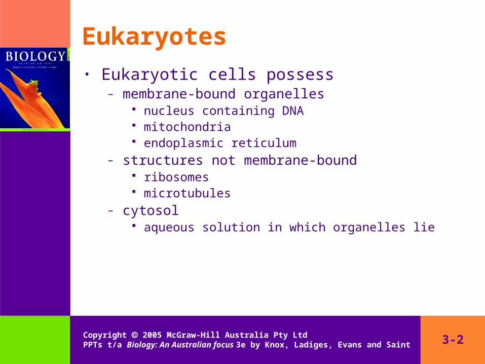
Eukaryotes• Eukaryotic cells possess
– membrane-bound organelles nucleus containing DNA mitochondria endoplasmic reticulum
– structures not membrane-bound ribosomes microtubules
– cytosol aqueous solution in which organelles lie

3-3Copyright 2005 McGraw-Hill Australia Pty Ltd PPTs t/a Biology: An Australian focus 3e by Knox, Ladiges, Evans and Saint
Fig. 3.3: Animal cell
3-4Copyright 2005 McGraw-Hill Australia Pty Ltd PPTs t/a Biology: An Australian focus 3e by Knox, Ladiges, Evans and Saint
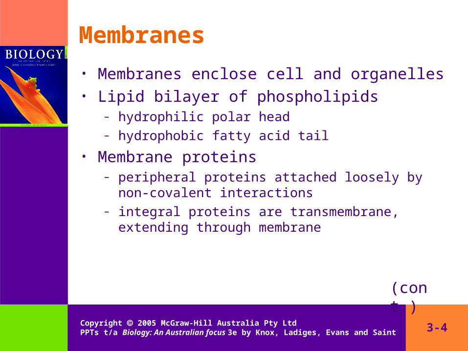
Membranes
• Membranes enclose cell and organelles• Lipid bilayer of phospholipids
– hydrophilic polar head– hydrophobic fatty acid tail
• Membrane proteins– peripheral proteins attached loosely by non-covalent
interactions– integral proteins are transmembrane, extending through
membrane
(cont.)
3-5Copyright 2005 McGraw-Hill Australia Pty Ltd PPTs t/a Biology: An Australian focus 3e by Knox, Ladiges, Evans and Saint
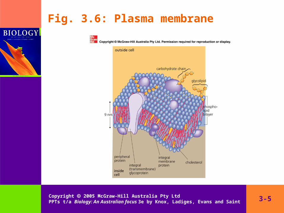
Fig. 3.6: Plasma membrane

3-6Copyright 2005 McGraw-Hill Australia Pty Ltd PPTs t/a Biology: An Australian focus 3e by Knox, Ladiges, Evans and Saint
Membranes (cont.)• Membranes usually with different molecules on
each side• Carbohydrates may be attached to lipids or
proteins– glycolipids– glycoproteins
• Carbohydrates occur on non-cytosolic side of membrane
– lumen (inside) of organelles– outer surface of plasma membrane to form glycocalyx
(cont.)

3-7Copyright 2005 McGraw-Hill Australia Pty Ltd PPTs t/a Biology: An Australian focus 3e by Knox, Ladiges, Evans and Saint
Membranes (cont.)
• Membrane are fluid mosaics– lipid (and some protein) molecules can move laterally– proteins embedded in irregular arrangement
• Membranes are selectively permeable– H2O, O2, CO2 cross freely
– ions and other polar molecules can only cross at selective pores formed by transmembrane proteins
3-8Copyright 2005 McGraw-Hill Australia Pty Ltd PPTs t/a Biology: An Australian focus 3e by Knox, Ladiges, Evans and Saint
Nucleus
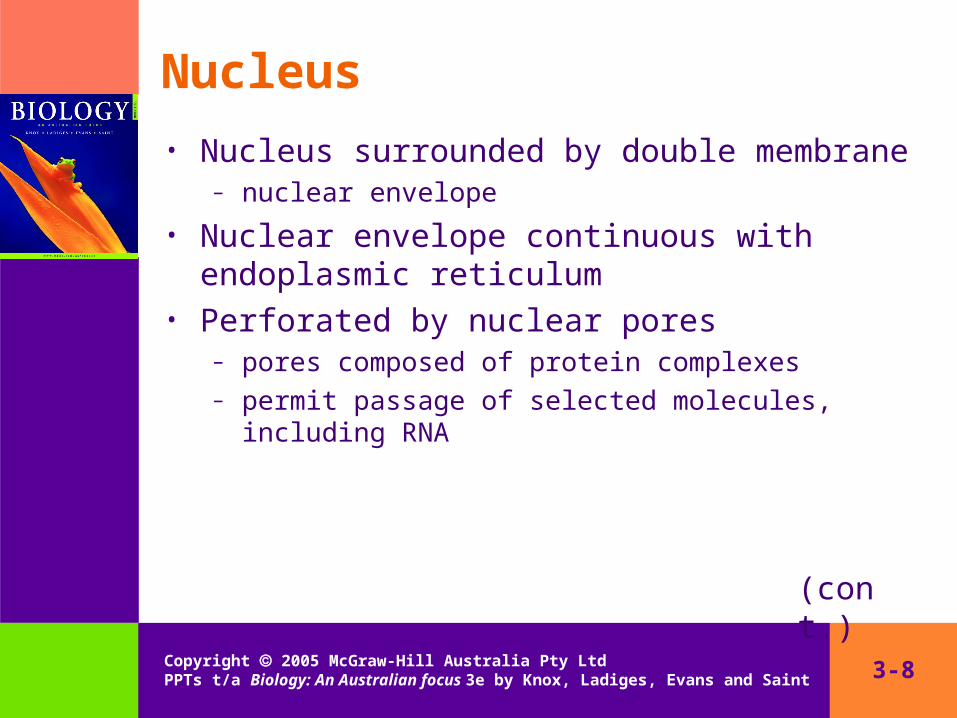
• Nucleus surrounded by double membrane – nuclear envelope
• Nuclear envelope continuous with endoplasmic reticulum
• Perforated by nuclear pores– pores composed of protein complexes– permit passage of selected molecules, including RNA
(cont.)

3-9Copyright 2005 McGraw-Hill Australia Pty Ltd PPTs t/a Biology: An Australian focus 3e by Knox, Ladiges, Evans and Saint
Nucleus (cont.)• Nucleus contains DNA• DNA molecules winds around histone molecules to
form nucleosomes– DNA twists into helical chromatin strands
• When cell is not dividing, chromatin strands – aggregate to form densely staining heterochromatin– disperse to form lightly staining euchromatin
• When cell divides, chromatin strands condense to form chromosomes
(cont.)

3-10Copyright 2005 McGraw-Hill Australia Pty Ltd PPTs t/a Biology: An Australian focus 3e by Knox, Ladiges, Evans and Saint
Nucleus (cont.)
• Nucleus usually contains one or several nucleoli (sing. nucleolus)
– densely-staining area of DNA, RNA and protein
• Nucleolus size depends on level of protein synthesis in cell
– site of ribosomal RNA synthesis– site of assembly of ribosomal subunits
3-11Copyright 2005 McGraw-Hill Australia Pty Ltd PPTs t/a Biology: An Australian focus 3e by Knox, Ladiges, Evans and Saint
Ribosomes
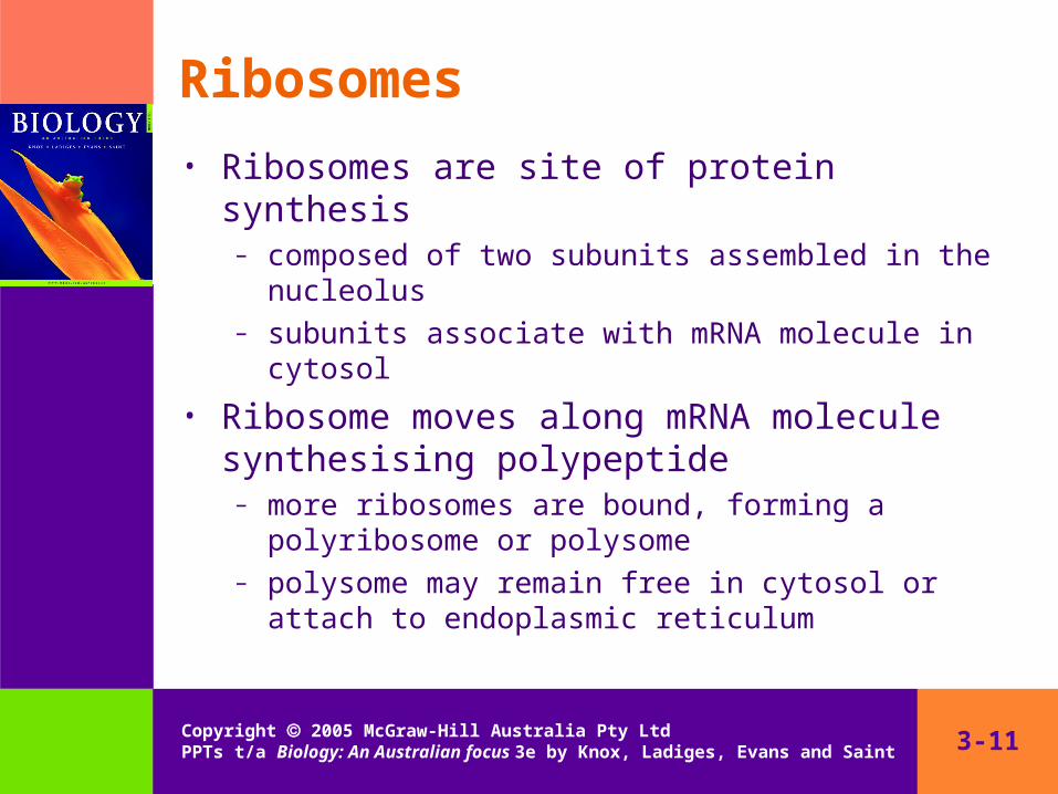
• Ribosomes are site of protein synthesis– composed of two subunits assembled in the nucleolus– subunits associate with mRNA molecule in cytosol
• Ribosome moves along mRNA molecule synthesising polypeptide
– more ribosomes are bound, forming a polyribosome or polysome
– polysome may remain free in cytosol or attach to endoplasmic reticulum

3-12Copyright 2005 McGraw-Hill Australia Pty Ltd PPTs t/a Biology: An Australian focus 3e by Knox, Ladiges, Evans and Saint
Endomembrane system• Cell and nucleus enclosed in membranes
– plasma membrane, nuclear envelope
• Membranes enclose components inside cell– endomembrane system
• Cell components– endoplasmic reticulum– Golgi apparatus– lysosomes– endosomes– vacuoles

3-13Copyright 2005 McGraw-Hill Australia Pty Ltd PPTs t/a Biology: An Australian focus 3e by Knox, Ladiges, Evans and Saint
Endoplasmic reticulum
• Endoplasmic reticulum (ER) extends through cytosol
– network of sacs (cisternae)– continuous with outer membrane of nuclear envelope
• Cisternae usually flat, sheet-like– linked by tubular cisternae– extensive surface area
(cont.)
3-14Copyright 2005 McGraw-Hill Australia Pty Ltd PPTs t/a Biology: An Australian focus 3e by Knox, Ladiges, Evans and Saint
Endoplasmic reticulum (cont.)
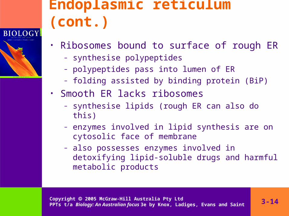
• Ribosomes bound to surface of rough ER– synthesise polypeptides– polypeptides pass into lumen of ER– folding assisted by binding protein (BiP)
• Smooth ER lacks ribosomes– synthesise lipids (rough ER can also do this)– enzymes involved in lipid synthesis are on cytosolic face
of membrane– also possesses enzymes involved in detoxifying lipid-
soluble drugs and harmful metabolic products

3-15Copyright 2005 McGraw-Hill Australia Pty Ltd PPTs t/a Biology: An Australian focus 3e by Knox, Ladiges, Evans and Saint
Golgi apparatus
• Golgi apparatus composed of stacks of cisternae– 4 to 10– disc-shaped, flat or curved
• Golgi apparatus has distinct orientation– cis face towards ER
lacks ribosomes cytosol between Golgi apparatus and ER filled with small
vesicles
– trans face outwards associated with tubular membranes of the trans-Golgi
network
(cont.)

3-16Copyright 2005 McGraw-Hill Australia Pty Ltd PPTs t/a Biology: An Australian focus 3e by Knox, Ladiges, Evans and Saint
Golgi apparatus (cont.)
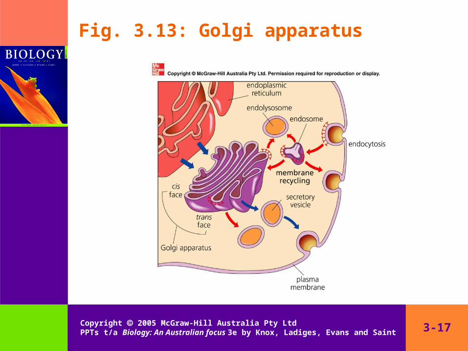
• Golgi apparatus processes and packages glycoproteins and polysaccharides
• cis face– proteins and glycoproteins enter from ER via vesicles– modified as they pass through stack of cisternae
• trans face– sorting and packaging of products in trans-Golgi network
3-17Copyright 2005 McGraw-Hill Australia Pty Ltd PPTs t/a Biology: An Australian focus 3e by Knox, Ladiges, Evans and Saint
Fig. 3.13: Golgi apparatus
3-18Copyright 2005 McGraw-Hill Australia Pty Ltd PPTs t/a Biology: An Australian focus 3e by Knox, Ladiges, Evans and Saint
Sorting and transport• Products of Golgi apparatus transported to target
organelles or exported from cell• Sorting
– products localised by association with ‘cargo’ receptors on inner face of membrane of cisternae or trans-Golgi network
• Transport– vesicle-marker proteins (v-SNARE) on outer membrane
identify different vesicle types– v-snare proteins attach to target docking proteins
(t-SNARE) on target membrane
3-19Copyright 2005 McGraw-Hill Australia Pty Ltd PPTs t/a Biology: An Australian focus 3e by Knox, Ladiges, Evans and Saint
Lysosomes
• Lysosomes contain hydrolytic enzymes for breaking down old organelles
• Enzymes for lysosomes manufactured in Golgi apparatus
– enzymes marked in cis cisternae for sorting in trans-Golgi network
– markers recognised by endolysosome– enzymes released into endolysosome– active uptake of H+ decreases pH– endolysosome matures into lysosome
3-20Copyright 2005 McGraw-Hill Australia Pty Ltd PPTs t/a Biology: An Australian focus 3e by Knox, Ladiges, Evans and Saint
Transport vesicles
• Materials can be exported from or imported into the cell by vesicles fusing with the plasma membrane
• Exocytosis (exportation)– continual (constitutive secretion) or intermittent (regulated
secretion)
• Endocytosis (importation)– vesicles fuse with endosomes– some materials recycled, others broken down

3-21Copyright 2005 McGraw-Hill Australia Pty Ltd PPTs t/a Biology: An Australian focus 3e by Knox, Ladiges, Evans and Saint
Mitochondria
• Mitochondria are thought to have evolved from engulfed prokaryotes
• Mitochondria are the site of cellular respiration– release energy by oxidation of sugars and fats (oxidative
phosphorylation)– released energy stored in ATP– cells with high level of metabolic activity have large
numbers of mitochondria
(cont.)

3-22Copyright 2005 McGraw-Hill Australia Pty Ltd PPTs t/a Biology: An Australian focus 3e by Knox, Ladiges, Evans and Saint
Mitochondria (cont.)
• Double membrane– outer membrane
permeable to ions and small molecules many transport channels
– inner membrane impermeable transport of ions by transport proteins
– generates electrochemical gradient
(cont.)

3-23Copyright 2005 McGraw-Hill Australia Pty Ltd PPTs t/a Biology: An Australian focus 3e by Knox, Ladiges, Evans and Saint
Mitochondria (cont.)
• Inner membrane of mitochondria folded into cristae– lined with enzyme complexes for ATP synthesis– enzymes use electrochemical gradient to generate ATP
from ATP and inorganic phosphate
• Matrix space of mitochondria– ribosomes– one or more copies of circular mtDNA– mtDNA codes for rRNA, tRNA and mRNA– proteins required for oxidative reactions and DNA
synthesis

3-24Copyright 2005 McGraw-Hill Australia Pty Ltd PPTs t/a Biology: An Australian focus 3e by Knox, Ladiges, Evans and Saint
Plastids
• Plastids occur in plant and protist cells– photosynthetic organelles
• Plastids resemble mitochondria in structure– double membrane
each membrane with different permeability
– ribosomes– circular DNA– RNA
• Evolved from engulfed prokaryotes

3-25Copyright 2005 McGraw-Hill Australia Pty Ltd PPTs t/a Biology: An Australian focus 3e by Knox, Ladiges, Evans and Saint
Chloroplasts
• Chloroplasts contain light-absorbing pigments– mainly chlorophyll
• Well-developed internal membrane system– stacks (grana) of disc-like sacs (thylakoids)– thylakoids continuous with each other and those in
adjacent grana
• Chlorophyll molecules on thylakoid membrane– light energy used to create electrochemical gradient– gradient used to generate ATP

3-26Copyright 2005 McGraw-Hill Australia Pty Ltd PPTs t/a Biology: An Australian focus 3e by Knox, Ladiges, Evans and Saint
Microbodies• Microbodies remove unwanted compounds from
cells• Microbodies contain oxidative enzymes
– remove hydrogen from molecules and couple it to oxygen– generate hydrogen peroxide (H2O2)– catalase breaks down H2O2 into water and oxygen
• Peroxisomes oxidise amino acids and uric acid• Glyoxysomes convert fatty acids to sugars in
germinating seeds
3-27Copyright 2005 McGraw-Hill Australia Pty Ltd PPTs t/a Biology: An Australian focus 3e by Knox, Ladiges, Evans and Saint
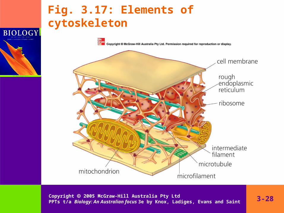
Cytoskeleton
• Cytoskeleton imposes and maintains structure of cell
– fixes organelles in position– moves organelles around cell– maintains and remodels cell shape
• Elements of cytoskeleton– microtubules– microfilaments– intermediate filaments
3-28Copyright 2005 McGraw-Hill Australia Pty Ltd PPTs t/a Biology: An Australian focus 3e by Knox, Ladiges, Evans and Saint
Fig. 3.17: Elements of cytoskeleton
3-29Copyright 2005 McGraw-Hill Australia Pty Ltd PPTs t/a Biology: An Australian focus 3e by Knox, Ladiges, Evans and Saint
Microfilaments• Structure of microfilaments
– diameter 7–8 nm– composed of actin (42 kD)
• Free actin (G-actin) interacts to form chains or filaments of F-actin
– length of F-actin filaments controlled by actin-binding proteins
• Interactions between actin and myosin microfilaments are the basis of many cytoplasmic, organelle and cell movements
3-30Copyright 2005 McGraw-Hill Australia Pty Ltd PPTs t/a Biology: An Australian focus 3e by Knox, Ladiges, Evans and Saint
Microtubules
• Structure of microtubules– diameter 25 nm– composed of α-tubulin and β-tubulin (both 55 kD)
• Microtubule-associated proteins (MAPs) control assembly and disassembly of microtubules
• Microtubule arrays may be radiating, bundled or parallel
– more rigid than microfilaments– support projections from cells, movement of organelles
3-31Copyright 2005 McGraw-Hill Australia Pty Ltd PPTs t/a Biology: An Australian focus 3e by Knox, Ladiges, Evans and Saint
Intermediate filaments
• Structure of intermediate filaments– diameter 8–10 nm– composed of different proteins (40–130 kD)
• Intermediate filament arrays are stable• Provide mechanical support for cell and nucleus
– keratin– desmin– nuclear laminins
3-32Copyright 2005 McGraw-Hill Australia Pty Ltd PPTs t/a Biology: An Australian focus 3e by Knox, Ladiges, Evans and Saint
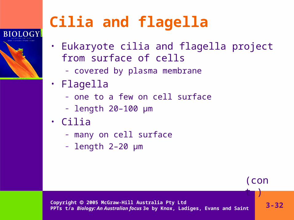
Cilia and flagella
• Eukaryote cilia and flagella project from surface of cells
– covered by plasma membrane
• Flagella– one to a few on cell surface– length 20–100 μm
• Cilia– many on cell surface– length 2–20 μm
(cont.)

3-33Copyright 2005 McGraw-Hill Australia Pty Ltd PPTs t/a Biology: An Australian focus 3e by Knox, Ladiges, Evans and Saint
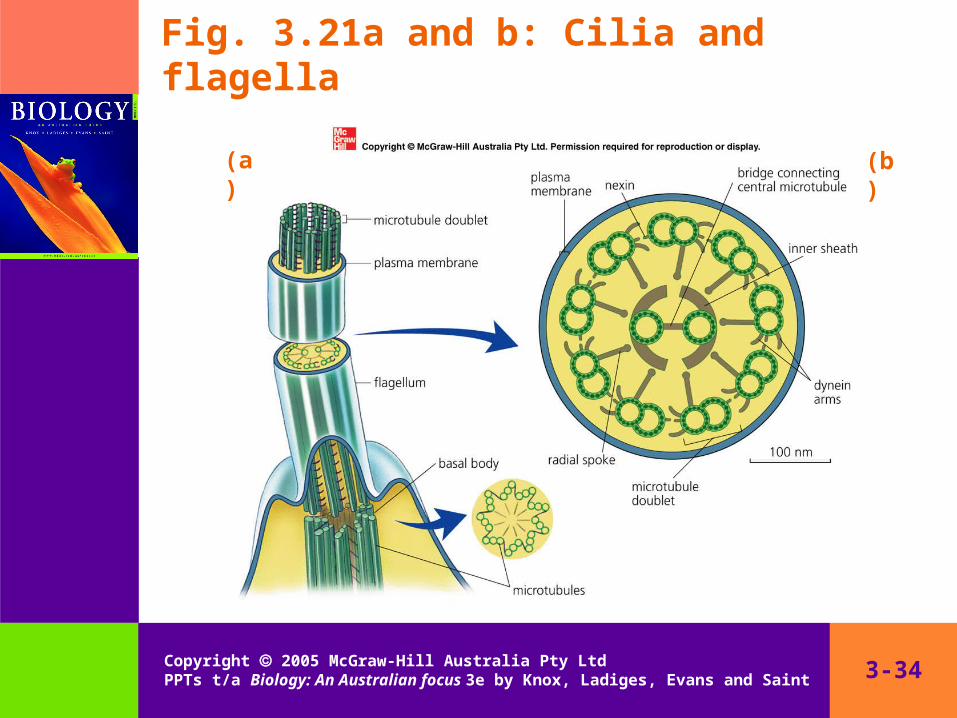
Cilia and flagella (cont.)
• Supported by paired microtubules (doublets) forming axoneme
– microtubules in each pair linked by fibres– two short arms of dynein on one side of each doublet
• Nine doublets surround two central doublets– movement created by doublets sliding relative to each
another– dynein attaches to adjacent doublet, undergoes
conformational change, then releases doublet– energy provided by dynein hydrolysis of ATP
3-34Copyright 2005 McGraw-Hill Australia Pty Ltd PPTs t/a Biology: An Australian focus 3e by Knox, Ladiges, Evans and Saint
Fig. 3.21a and b: Cilia and flagella
(a) (b)
3-35Copyright 2005 McGraw-Hill Australia Pty Ltd PPTs t/a Biology: An Australian focus 3e by Knox, Ladiges, Evans and Saint
Prokaryotic cells
• Prokaryotic cells– semirigid cell wall surrounding plasma membrane– lack membrane-bound organelles– circular DNA in cytosol
ribosomes attach directly to mRNA, even while mRNA is being transcribed
– enzymes on plasma membrane those enzymes occurring in eukaryotic mitochondria
– light-trapping pigments on plasma membrane those pigments occurring in eukaryotic chloroplasts
– rotating flagella of flagellin fibrils