24 lecture presentation pc
TRANSCRIPT
7/27/2019 24 Lecture Presentation PC
http://slidepdf.com/reader/full/24-lecture-presentation-pc 1/105
© 2012 Pearson Education, Inc. Lecture by Edward J. Zalisko
PowerPoint Lectures for
Campbell Biology: Concepts & Connections, Seventh Edition
Reece, Taylor, Simon, and Dickey
Chapter 24 The Immune System

7/27/2019 24 Lecture Presentation PC
http://slidepdf.com/reader/full/24-lecture-presentation-pc 2/105
Introduction
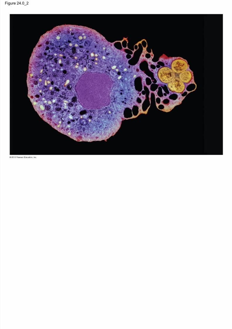
Neutrophils are – a kind of white blood cell,
– capable of recognizing and destroying foreign invaders,and
– part of the body’s immune system.
© 2012 Pearson Education, Inc.
7/27/2019 24 Lecture Presentation PC
http://slidepdf.com/reader/full/24-lecture-presentation-pc 3/105
Introduction
The human body’s immune system
– recognizes agents that cause disease and
– attacks them.
© 2012 Pearson Education, Inc.

7/27/2019 24 Lecture Presentation PC
http://slidepdf.com/reader/full/24-lecture-presentation-pc 4/105
Figure 24.0_1
Chapter 24: Big Ideas
Innate Immunity Adaptive Immunity
Disorders of the
Immune System
7/27/2019 24 Lecture Presentation PC
http://slidepdf.com/reader/full/24-lecture-presentation-pc 5/105
Figure 24.0_2
7/27/2019 24 Lecture Presentation PC
http://slidepdf.com/reader/full/24-lecture-presentation-pc 6/105
INNATE IMMUNITY
© 2012 Pearson Education, Inc.

7/27/2019 24 Lecture Presentation PC
http://slidepdf.com/reader/full/24-lecture-presentation-pc 7/105
24.1 All animals have innate immunity
Nearly everything in the environment teems withpathogens, agents that cause disease.
The immune system is the body’s system of
defenses against agents that cause disease.
Innate immunity is a series of defenses that
– act immediately upon infection and
– are the same whether or not the pathogen has beenencountered before.
© 2012 Pearson Education, Inc.

7/27/2019 24 Lecture Presentation PC
http://slidepdf.com/reader/full/24-lecture-presentation-pc 8/105
24.1 All animals have innate immunity
Invertebrates rely solely on innate immunity, whichmay consist of
– an exoskeleton,
– low pH, – the enzyme lysozyme, and
– immune cells capable of phagocytosis, cellular ingestion
and digestion of foreign substances.
Vertebrates have innate and adaptive immunity.
© 2012 Pearson Education, Inc.

7/27/2019 24 Lecture Presentation PC
http://slidepdf.com/reader/full/24-lecture-presentation-pc 9/105
Figure 24.1A
Innate immunity (24.1 –3)The response is the same whether
or not the pathogen has beenpreviously encountered
Adaptive immunity(24.4 –15)
Found only invertebrates; previous
exposure to thepathogen enhances the
immune response
External
barriers (24.1)
Internal
defenses (24.1 –2)
The lymphatic system (24.3)
• Antibodies (24.8–10)• Lymphocytes
(24.11 –13)
• Phagocytic cells • NK cells • Defensive
proteins• Inflammatory response (24.2)
• Skin/ exoskeleton
• Acidic
environment• Secretions • Mucous
membranes• Hairs • Cilia

7/27/2019 24 Lecture Presentation PC
http://slidepdf.com/reader/full/24-lecture-presentation-pc 10/105
24.1 All animals have innate immunity
Vertebrate innate immunity includes – barriers such as skin and mucous membranes,
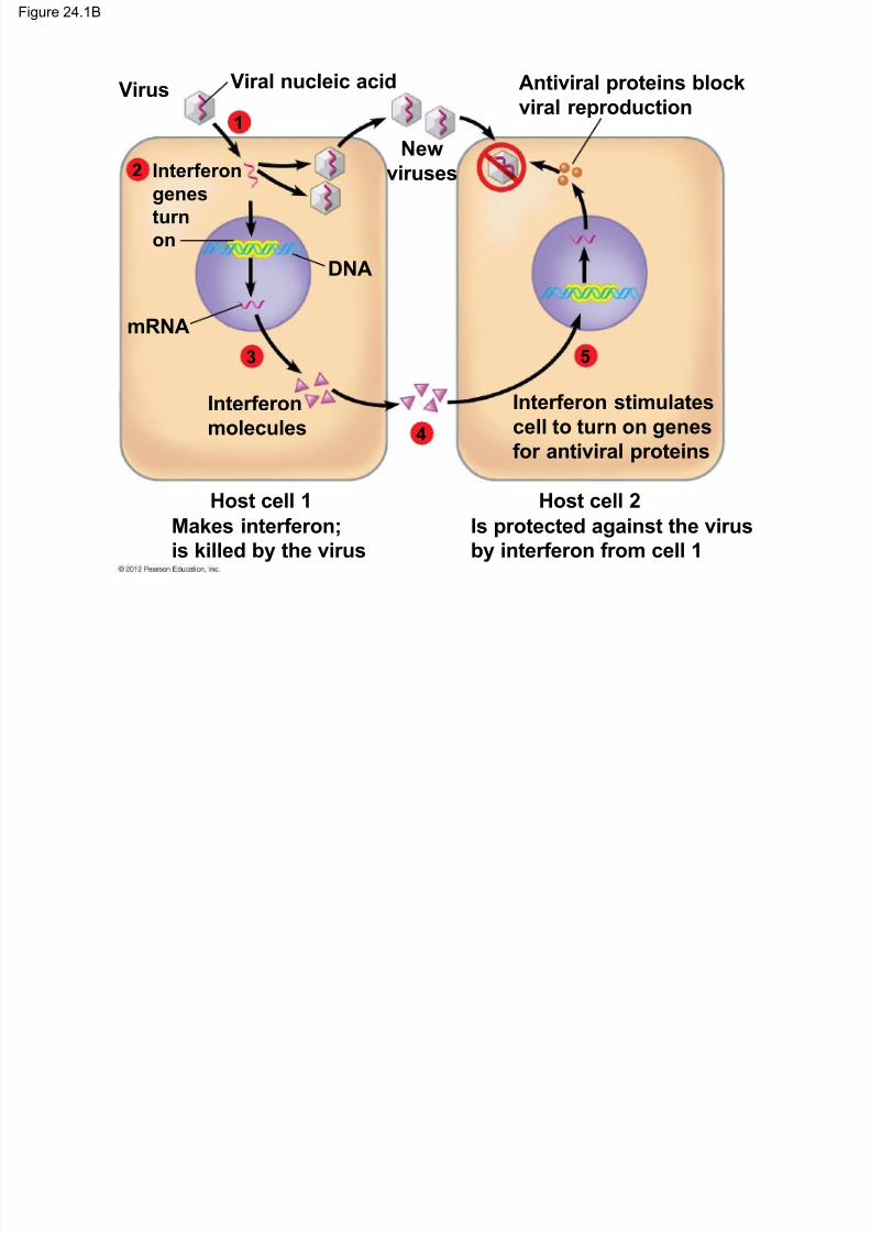
– interferons, proteins produced by virus-infected cells,that help to limit the cell-to-cell spread of viruses,
– neutrophils (phagocytic cells),
– macrophages, large phagocytic cells that wanderthrough the interstitial fluid,
– natural killer cells that attack cancer cells and virus-infected cells, and
– a complement system, a group of about 30 kinds ofproteins that can act with other defense mechanisms.
© 2012 Pearson Education, Inc.
7/27/2019 24 Lecture Presentation PC
http://slidepdf.com/reader/full/24-lecture-presentation-pc 11/105
Figure 24.1B
Viral nucleic acidVirus
mRNA
DNA
Interferon
genes
turn
on
Interferon stimulates
cell to turn on genes
for antiviral proteins
Antiviral proteins block
viral reproduction
Newviruses
Interferon
molecules
Makes interferon;
is killed by the virus
Is protected against the virus
by interferon from cell 1
Host cell 1 Host cell 2
3
4
2
1
5

7/27/2019 24 Lecture Presentation PC
http://slidepdf.com/reader/full/24-lecture-presentation-pc 12/105
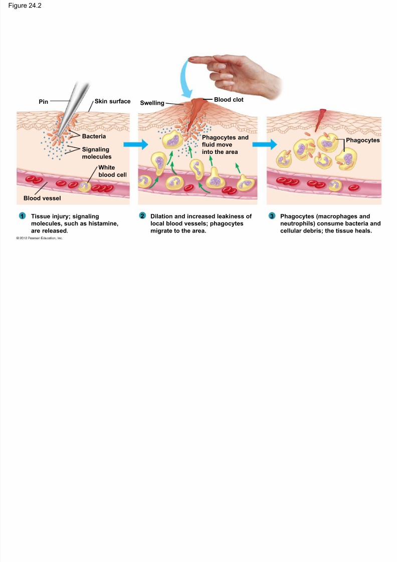
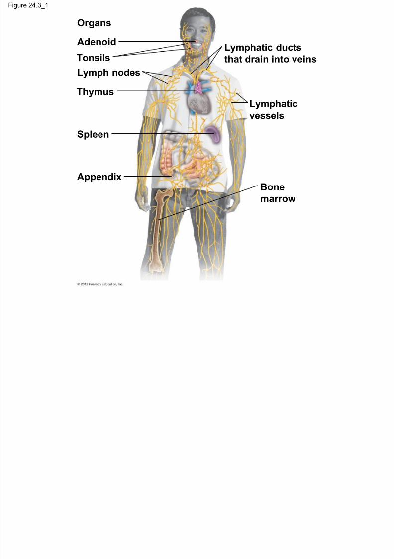
24.2 Inflammation mobilizes the innate immuneresponse
Tissue damage triggers the inflammatoryresponse, a major component of our innate
immunity, which can
– disinfect and clean infected tissues and
– limit the spread of infection to surrounding tissues.
Bacterial infections can bring about an overwhelming
systemic inflammatory response leading to septic
shock, characterized by
– very high fever and
– low blood pressure.
© 2012 Pearson Education, Inc.
Fi 24 2
7/27/2019 24 Lecture Presentation PC
http://slidepdf.com/reader/full/24-lecture-presentation-pc 13/105
Figure 24.2
Tissue injury; signaling
molecules, such as histamine,
are released.
Dilation and increased leakiness of
local blood vessels; phagocytes
migrate to the area.
Phagocytes (macrophages and
neutrophils) consume bacteria and
cellular debris; the tissue heals.
Blood vessel
White
blood cell
Signaling
molecules
Bacteria
Pin Skin surface SwellingBlood clot
Phagocytes andfluid move
into the area
Phagocytes
21 3
Fi 24 2 1

7/27/2019 24 Lecture Presentation PC
http://slidepdf.com/reader/full/24-lecture-presentation-pc 14/105
Figure 24.2_1
Tissue injury; signaling
molecules, such as histamine,
are released.
Blood vessel
White
blood cell
Signaling
molecules
Bacteria
Pin Skin surface
1
Figure 24 2 2
7/27/2019 24 Lecture Presentation PC
http://slidepdf.com/reader/full/24-lecture-presentation-pc 15/105
Figure 24.2_2
2 Dilation and increased leakiness
of local blood vessels;
phagocytes migrate to the area.
SwellingBlood clot
Phagocytes and
fluid move
into the area
Figure 24 2 3

7/27/2019 24 Lecture Presentation PC
http://slidepdf.com/reader/full/24-lecture-presentation-pc 16/105
Figure 24.2_3
3 Phagocytes (macrophages andneutrophils) consume bacteria
and cellular debris; the tissue
heals.
Phagocytes

7/27/2019 24 Lecture Presentation PC
http://slidepdf.com/reader/full/24-lecture-presentation-pc 17/105
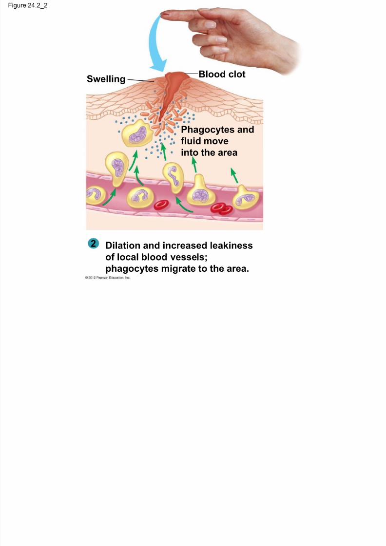
24.3 The lymphatic system becomes a crucialbattleground during infection
The lymphatic system is – involved in innate and adaptive immunity and
– consists of a network of
– lymphatic vessels, – lymph nodes, and
– lymph.
© 2012 Pearson Education, Inc.

7/27/2019 24 Lecture Presentation PC
http://slidepdf.com/reader/full/24-lecture-presentation-pc 18/105
24.3 The lymphatic system becomes a crucialbattleground during infection
Lymphatic vessels – collect fluid from body tissues and
– return it as lymph to the blood.
Lymph organs – include the spleen and lymph nodes and
– are packed with white blood cells that fight infections.
© 2012 Pearson Education, Inc.

7/27/2019 24 Lecture Presentation PC
http://slidepdf.com/reader/full/24-lecture-presentation-pc 19/105
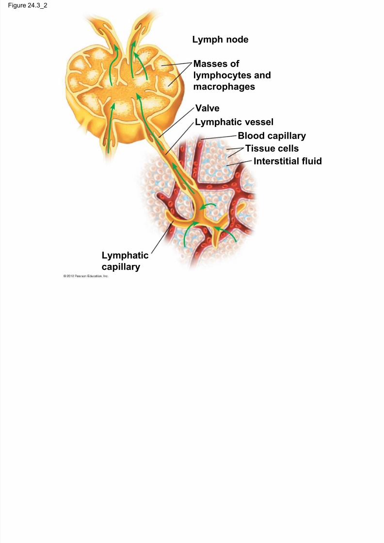
24.3 The lymphatic system becomes a crucialbattleground during infection
As lymph circulates through lymphatic organs it – collects
– microbes,
– parts of microbes, and
– microbial toxins, and
– transports them to lymphatic organs where
– macrophages in lymphatic organs engulf the invaders and
– lymphocytes may mount an adaptive immune response.
© 2012 Pearson Education, Inc.
Figure 24 3

7/27/2019 24 Lecture Presentation PC
http://slidepdf.com/reader/full/24-lecture-presentation-pc 20/105
Figure 24.3
Lymphatic
capillary
Interstitial fluid
Tissue cells
Blood capillary
Lymphatic vessel
Valve
Masses of
lymphocytes and
macrophages
Lymph nodeLymphatic ductsthat drain into veins
Lymphatic
vessels
Bone
marrow
Appendix
Spleen
Thymus
Lymph nodes
Tonsils
Adenoid
Organs
Figure 24 3 1
7/27/2019 24 Lecture Presentation PC
http://slidepdf.com/reader/full/24-lecture-presentation-pc 21/105
Figure 24.3_1
Lymphatic ducts
that drain into veins
Lymphatic
vessels
Bone
marrow
Appendix
Spleen
Thymus
Lymph nodes
Tonsils
Adenoid
Organs
Figure 24 3 2
7/27/2019 24 Lecture Presentation PC
http://slidepdf.com/reader/full/24-lecture-presentation-pc 22/105
Figure 24.3_2
Lymph node
Masses oflymphocytes and
macrophages
Valve
Lymphatic vessel
Blood capillaryTissue cells
Interstitial fluid
Lymphatic
capillary
7/27/2019 24 Lecture Presentation PC
http://slidepdf.com/reader/full/24-lecture-presentation-pc 23/105
ADAPTIVE IMMUNITY
© 2012 Pearson Education, Inc.
24 4 Th d i i

7/27/2019 24 Lecture Presentation PC
http://slidepdf.com/reader/full/24-lecture-presentation-pc 24/105
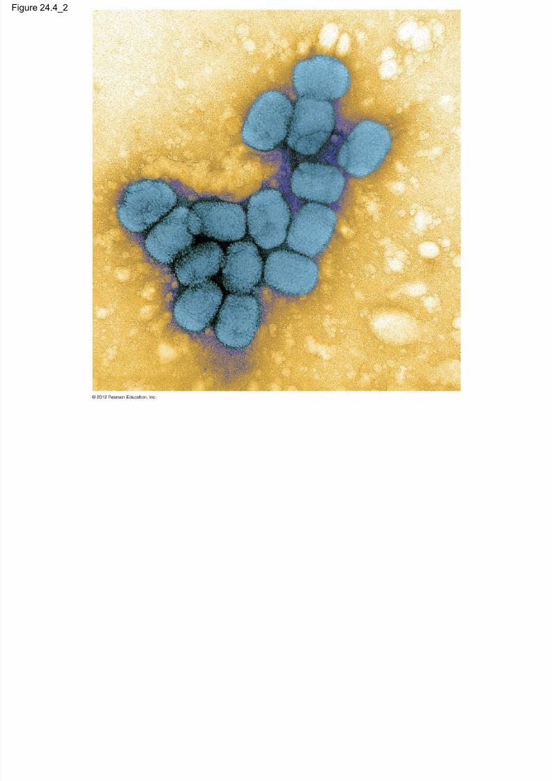
24.4 The adaptive immune response countersspecific invaders
Our immune system responds to foreign moleculescalled antigens, which elicit the adaptive immune
response.
The adaptive immune system
– is found only in the vertebrates,
– reacts to specific pathogens, and
– “remembers” an invader.
© 2012 Pearson Education, Inc.
24 4 Th d i i

7/27/2019 24 Lecture Presentation PC
http://slidepdf.com/reader/full/24-lecture-presentation-pc 25/105
24.4 The adaptive immune response countersspecific invaders
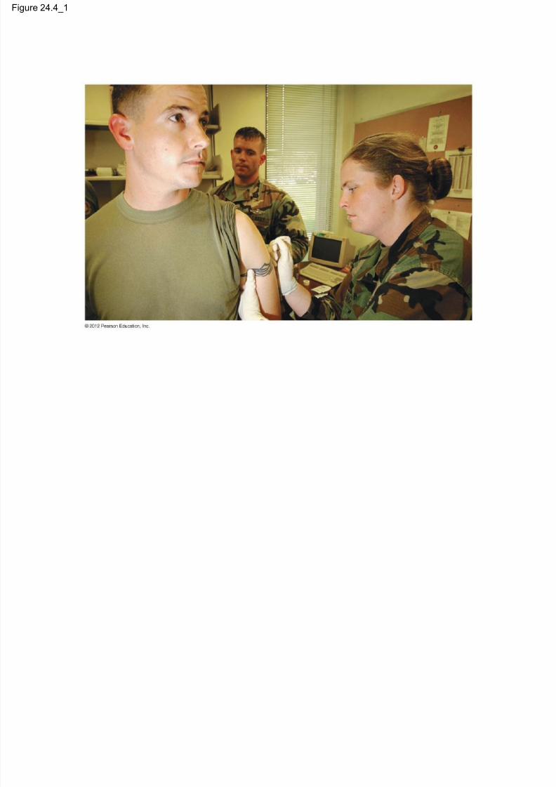
Infection or vaccination triggers active immunity.
Vaccination, or immunization, exposes the
immune system to a vaccine,
– a harmless variant or
– part of a disease-causing microbe.
We can temporarily acquire passive immunity by
receiving premade antibodies.
© 2012 Pearson Education, Inc.
Figure 24.4

7/27/2019 24 Lecture Presentation PC
http://slidepdf.com/reader/full/24-lecture-presentation-pc 26/105
g
Figure 24.4_1
7/27/2019 24 Lecture Presentation PC
http://slidepdf.com/reader/full/24-lecture-presentation-pc 27/105
g _
Figure 24.4_2
7/27/2019 24 Lecture Presentation PC
http://slidepdf.com/reader/full/24-lecture-presentation-pc 28/105
24 5 L h t t d l d f

7/27/2019 24 Lecture Presentation PC
http://slidepdf.com/reader/full/24-lecture-presentation-pc 29/105
24.5 Lymphocytes mount a dual defense
Lymphocytes – are white blood cells that spend most of their time in the
tissues and organs of the lymphatic system,
– are responsible for adaptive immunity, and
– originate from stem cells in the bone marrow.
– B lymphocytes or B cells continue developing in bone marrow.
– T lymphocytes or T cells develop further in the thymus.
© 2012 Pearson Education, Inc.
24 5 L h t t d l d f

7/27/2019 24 Lecture Presentation PC
http://slidepdf.com/reader/full/24-lecture-presentation-pc 30/105
24.5 Lymphocytes mount a dual defense
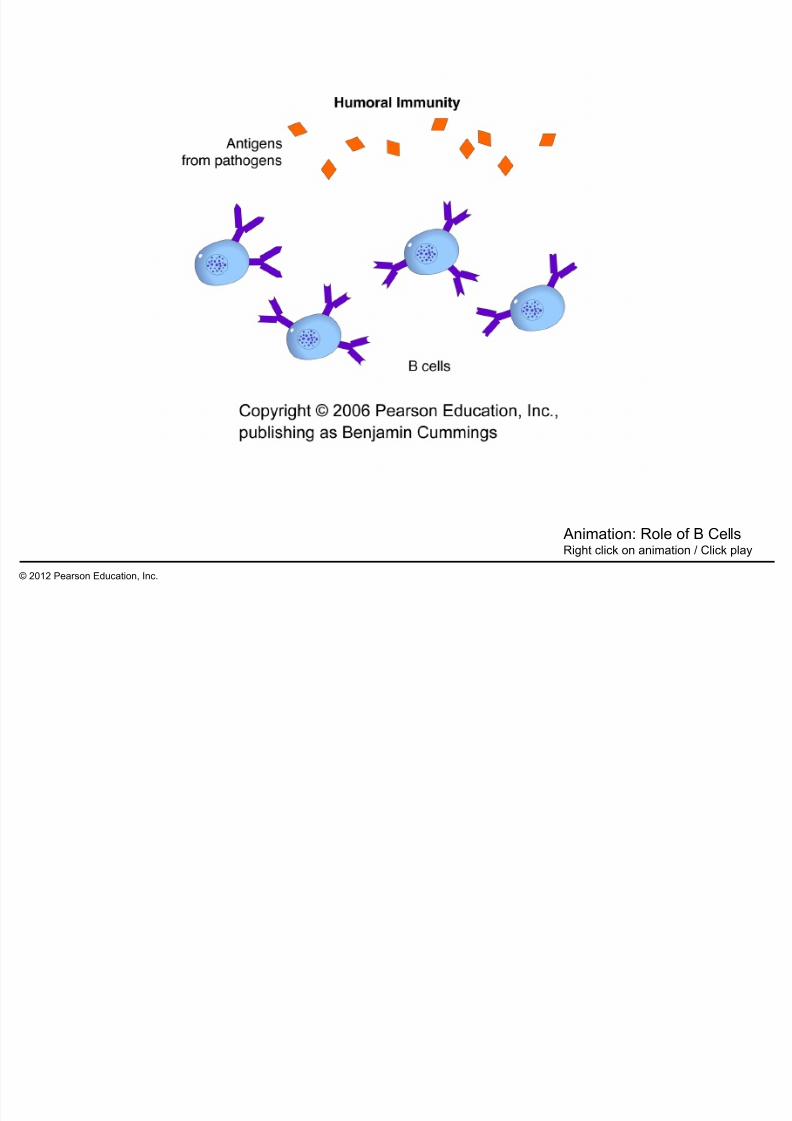
B cells – participate in the humoral immune response and
– secrete antibodies into the blood and lymph.
T cells – participate in the cell-mediated immune response,
– attack cells infected with bacteria or viruses, and
– promote phagocytosis by other white blood cells and bystimulating B cells to produce antibodies.
© 2012 Pearson Education, Inc.
Figure 24.5A

7/27/2019 24 Lecture Presentation PC
http://slidepdf.com/reader/full/24-lecture-presentation-pc 31/105
Bone
marrow
Stem cell
Immature lymphocytes
Viablood
Antigen
receptors
Thymus
T cellB cellVia
blood
Final maturation
of B and T cells in a
lymphatic organ
Lymph
nodes,
spleen, and
other
lymphatic
organs
Humoral
immune response
Cell-mediated
immune response
Figure 24.5A_1

7/27/2019 24 Lecture Presentation PC
http://slidepdf.com/reader/full/24-lecture-presentation-pc 32/105
Bone
marrow
Stem cell
Immature lymphocytes
Via
blood
Antigenreceptors
Thymus
T cellB cell
Figure 24.5A_2

7/27/2019 24 Lecture Presentation PC
http://slidepdf.com/reader/full/24-lecture-presentation-pc 33/105
Antigen
receptors
T cellB cellVia
blood
Final maturation
of B and T cells in a
lymphatic organ
Lymph
nodes,
spleen, and
otherlymphatic
organs
Humoral
immune response
Cell-mediated
immune response
24 5 Lymphocytes mount a dual defense
7/27/2019 24 Lecture Presentation PC
http://slidepdf.com/reader/full/24-lecture-presentation-pc 34/105
24.5 Lymphocytes mount a dual defense
Millions of kinds of B cells and T cells – each with different antigen receptors, capable of binding
one specific type of antigen,
– wait in the lymphatic system, – where they may respond to invaders.
© 2012 Pearson Education, Inc.
Figure 24.5B

7/27/2019 24 Lecture Presentation PC
http://slidepdf.com/reader/full/24-lecture-presentation-pc 35/105
24 6 Antigens have specific regions where

7/27/2019 24 Lecture Presentation PC
http://slidepdf.com/reader/full/24-lecture-presentation-pc 36/105
24.6 Antigens have specific regions whereantibodies bind to them
Antigens – are molecules that elicit the adaptive immune response,
– usually do not belong to the host animal, and
– are proteins or large polysaccharides on the surfaces ofviruses or foreign cells.
© 2012 Pearson Education, Inc.
24 6 Antigens have specific regions where

7/27/2019 24 Lecture Presentation PC
http://slidepdf.com/reader/full/24-lecture-presentation-pc 37/105
24.6 Antigens have specific regions whereantibodies bind to them
Antigenic determinants are specific regions on anantigen where antibodies bind.
– An antigen usually has several different determinants.
– The antigen-binding site of an antibody and an antigenicdeterminant have complementary shapes.
© 2012 Pearson Education, Inc.
Figure 24.6

7/27/2019 24 Lecture Presentation PC
http://slidepdf.com/reader/full/24-lecture-presentation-pc 38/105
Two differentantibody
molecules
Antigen-
binding
site
Antigen
molecule
Antigenic
determinant
24 7 Clonal selection musters defensive forces

7/27/2019 24 Lecture Presentation PC
http://slidepdf.com/reader/full/24-lecture-presentation-pc 39/105
24.7 Clonal selection musters defensive forcesagainst specific antigens
When an antigen enters the body it activates only asmall subset of lymphocytes that have
complementary receptors.
In clonal selection, the selected lymphocyte cells
– multiply into clones of short-lived effector cells,
specialized for defending against the antigen that
triggered the response, and
– multiply into memory cells, which confer long-termimmunity.
– Plasma cells are the effector cells produced during
clonal selection of B cells.
© 2012 Pearson Education, Inc.
24 7 Clonal selection musters defensive forces

7/27/2019 24 Lecture Presentation PC
http://slidepdf.com/reader/full/24-lecture-presentation-pc 40/105
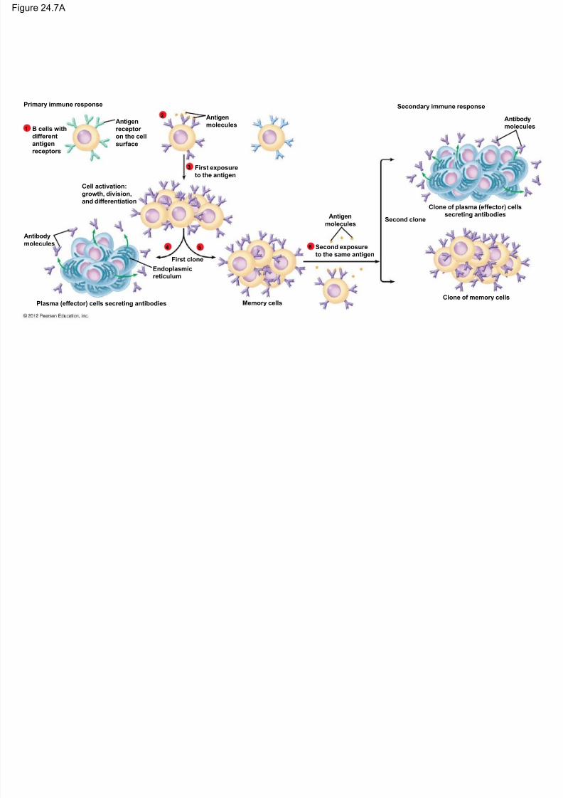
24.7 Clonal selection musters defensive forcesagainst specific antigens
The clonal selection of B cells occurs in tworesponses.
– In the primary immune response, clonal selection
produces
– effector cells and
– memory cells that may confer lifelong immunity.
– In the secondary immune response, memory cells are
activated by a second exposure to the same antigen.
© 2012 Pearson Education, Inc.
7/27/2019 24 Lecture Presentation PC
http://slidepdf.com/reader/full/24-lecture-presentation-pc 41/105
© 2012 Pearson Education, Inc.
Animation: Role of B CellsRight click on animation / Click play
Figure 24.7A
7/27/2019 24 Lecture Presentation PC
http://slidepdf.com/reader/full/24-lecture-presentation-pc 42/105
Primary immune response
B cells with
different
antigen
receptors
1
2
3
4 5 6
Antigen
receptor
on the cell
surface
Cell activation:
growth, division,
and differentiation
Antigen
molecules
First exposure
to the antigen
Antibody
molecules
First clone
Endoplasmic
reticulum
Plasma (effector) cells secreting antibodies Memory cells
Second exposure
to the same antigen
Antigen
moleculesSecond clone
Secondary immune response
Antibody
molecules
Clone of memory cells
Clone of plasma (effector) cells
secreting antibodies
Figure 24.7A_s1

7/27/2019 24 Lecture Presentation PC
http://slidepdf.com/reader/full/24-lecture-presentation-pc 43/105
1
Primary immune response
B cells with
different
antigenreceptors
Antigen
receptor
on the cell
surface
Figure 24.7A_s2

7/27/2019 24 Lecture Presentation PC
http://slidepdf.com/reader/full/24-lecture-presentation-pc 44/105
1
2
Primary immune response
B cells with
different
antigenreceptors
Antigen
receptor
on the cell
surface
Antigen
molecules
Figure 24.7A_s3
P i i

7/27/2019 24 Lecture Presentation PC
http://slidepdf.com/reader/full/24-lecture-presentation-pc 45/105
1
2
3
Primary immune response
B cells with
different
antigenreceptors
Antigen
receptor
on the cell
surface
Cell activation:
growth, division,
and differentiation
Antigen
molecules
First exposure
to the antigen
Figure 24.7A_s4
P i i

7/27/2019 24 Lecture Presentation PC
http://slidepdf.com/reader/full/24-lecture-presentation-pc 46/105
1
2
3
4 5
Primary immune response
B cells with
different
antigenreceptors
Antigen
receptor
on the cell
surface
Cell activation:
growth, division,
and differentiation
Antigen
molecules
First exposure
to the antigen
Antibody
molecules
First clone
Endoplasmic
reticulum
Plasma (effector) cells secreting antibodies Memory cells
Figure 24.7A_s5

7/27/2019 24 Lecture Presentation PC
http://slidepdf.com/reader/full/24-lecture-presentation-pc 47/105
6
Memory cells
Second
exposure
to the same
antigen
Antigen
molecules
Figure 24.7A_s6

7/27/2019 24 Lecture Presentation PC
http://slidepdf.com/reader/full/24-lecture-presentation-pc 48/105
6
Memory cells
Second
exposure
to the same
antigen
Antigen
molecules
Second clone
Secondary immune responseAntibody
molecules
Clone of memory cells
Clone of plasma (effector) cellssecreting antibodies
24.7 Clonal selection musters defensive forces

7/27/2019 24 Lecture Presentation PC
http://slidepdf.com/reader/full/24-lecture-presentation-pc 49/105
24.7 Clonal selection musters defensive forcesagainst specific antigens
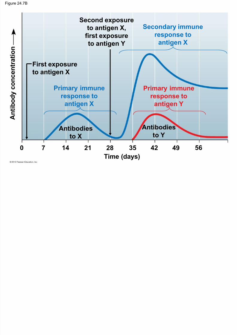
Primary vs. secondary immune responses – The primary immune response
– occurs upon first exposure to an antigen and
– is slower than the secondary immune response.
– The secondary immune response
– occurs upon second exposure to an antigen and
– is faster and stronger than the primary immune response.
© 2012 Pearson Education, Inc.
Figure 24.7B
7/27/2019 24 Lecture Presentation PC
http://slidepdf.com/reader/full/24-lecture-presentation-pc 50/105
Time (days)
5649423528211470
A n t i b o d y c o
n c e n t r a t i o n
Antibodiesto X
Antibodiesto Y
Second exposure
to antigen X,
first exposure
to antigen Y
First exposure
to antigen X
Primary immune
response to
antigen X
Secondary immune
response to
antigen X
Primary immune
response to
antigen Y
24.8 Antibodies are the weapons of the humoral

7/27/2019 24 Lecture Presentation PC
http://slidepdf.com/reader/full/24-lecture-presentation-pc 51/105
24.8 Antibodies are the weapons of the humoralimmune response
Antibodies are secreted – by plasma (effector) B cells,
– into the blood and lymph.
© 2012 Pearson Education, Inc.
Figure 24.8A
Light chain

7/27/2019 24 Lecture Presentation PC
http://slidepdf.com/reader/full/24-lecture-presentation-pc 52/105
Light chain
Heavy chain
24.8 Antibodies are the weapons of the humoral

7/27/2019 24 Lecture Presentation PC
http://slidepdf.com/reader/full/24-lecture-presentation-pc 53/105
24.8 Antibodies are the weapons of the humoralimmune response
An antibody molecule – is Y-shaped and
– has two antigen-binding sites specific to the antigenic
determinants that elicited its secretion.
© 2012 Pearson Education, Inc.
Figure 24.8B

7/27/2019 24 Lecture Presentation PC
http://slidepdf.com/reader/full/24-lecture-presentation-pc 54/105
Antigen
Lightchain
Heavy
chain
Antigen-binding
sites
C C
24.9 Antibodies mark antigens for elimination

7/27/2019 24 Lecture Presentation PC
http://slidepdf.com/reader/full/24-lecture-presentation-pc 55/105
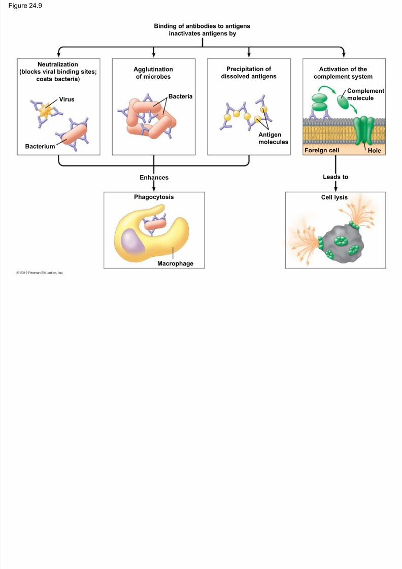
24.9 Antibodies mark antigens for elimination
Antibodies promote antigen elimination throughseveral mechanisms:
1. neutralization, binding to surface proteins on a virus or
bacterium and blocking its ability to infect a host,
2. agglutination, using both binding sites of an antibody to
join invading cells together into a clump,
© 2012 Pearson Education, Inc.
24.9 Antibodies mark antigens for elimination

7/27/2019 24 Lecture Presentation PC
http://slidepdf.com/reader/full/24-lecture-presentation-pc 56/105
g
3. precipitation, similar to agglutination, except that theantibody molecules link dissolved antigen molecules
together, and
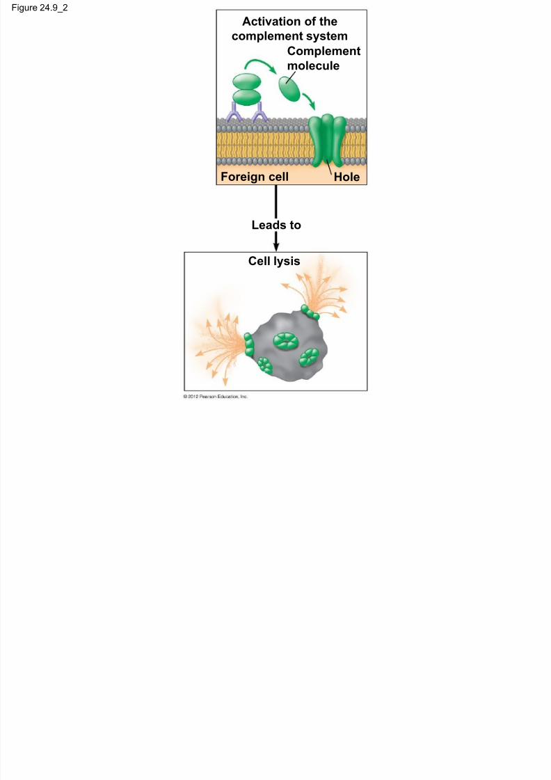
4. activation of the complement system by antigen-antibody
complexes.
© 2012 Pearson Education, Inc.

7/27/2019 24 Lecture Presentation PC
http://slidepdf.com/reader/full/24-lecture-presentation-pc 57/105
© 2012 Pearson Education, Inc.
Animation: AntibodiesRight click on animation / Click play
Figure 24.9
7/27/2019 24 Lecture Presentation PC
http://slidepdf.com/reader/full/24-lecture-presentation-pc 58/105
Bacterium
Virus
Neutralization
(blocks viral binding sites;coats bacteria)
Binding of antibodies to antigensinactivates antigens by
Agglutination
of microbes
Precipitation of
dissolved antigens
Activation of the
complement system
Bacteria
Antigen
molecules
Complement
molecule
Foreign cell Hole
Leads to
Cell lysis
Enhances
Phagocytosis
Macrophage
Figure 24.9_1
Neutralization

7/27/2019 24 Lecture Presentation PC
http://slidepdf.com/reader/full/24-lecture-presentation-pc 59/105
Bacterium
Virus
Neutralization
(blocks viral binding
sites; coats bacteria)
Agglutination
of microbes
Precipitation of
dissolved antigens
Bacteria
Antigen
molecules
Enhances
Phagocytosis
Macrophage
Figure 24.9_2
Activation of the
7/27/2019 24 Lecture Presentation PC
http://slidepdf.com/reader/full/24-lecture-presentation-pc 60/105
complement system
Complement
molecule
Foreign cell Hole
Leads to
Cell lysis
24.10 CONNECTION: Monoclonal antibodies are

7/27/2019 24 Lecture Presentation PC
http://slidepdf.com/reader/full/24-lecture-presentation-pc 61/105
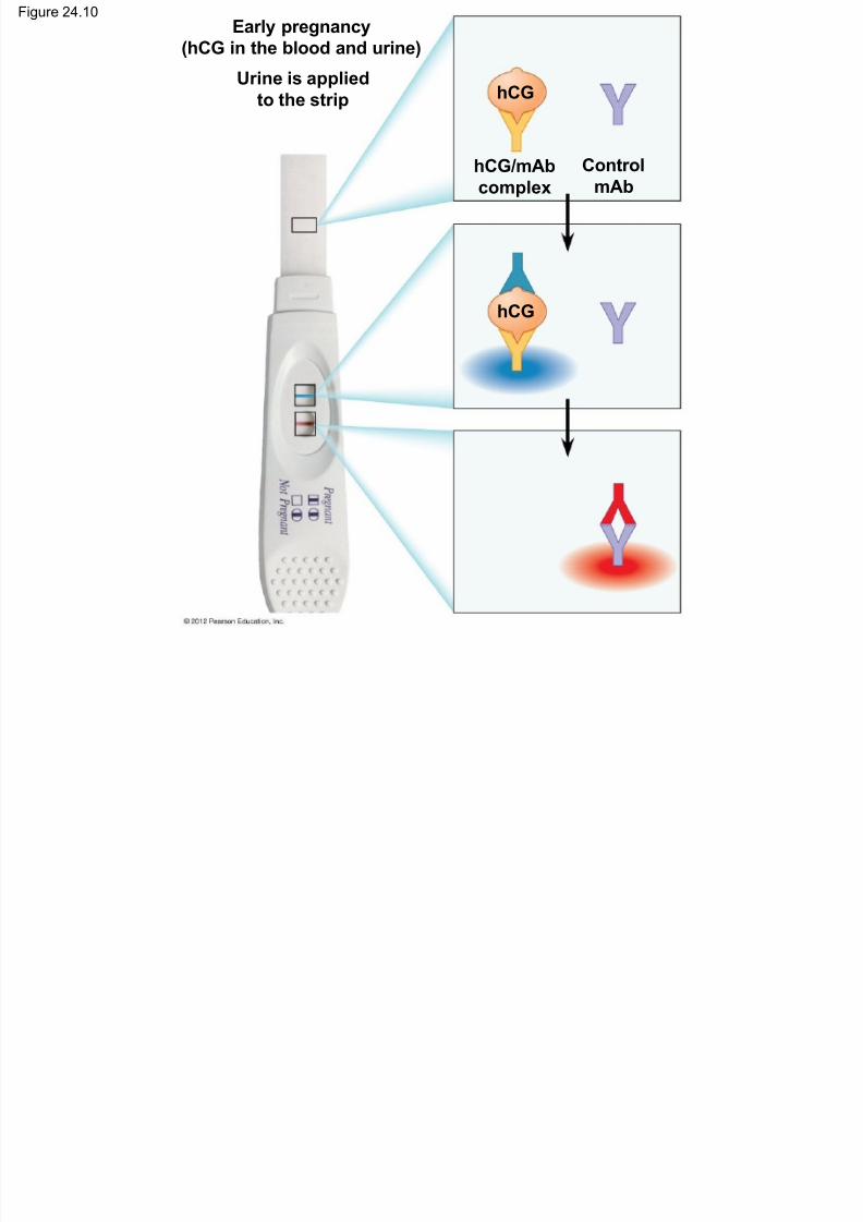
powerful tools in the lab and clinic
Monoclonal antibodies (mAb) are – identical antibodies
– produced by cells that are all descendants of a single,
hybrid cell.
To make the hybrid cell with desirable properties,
two cells are fused.
1. A cancerous tumor cell, able to multiply indefinitely, isfused to
2. a normal antibody-producing B cell, which is producing
the desired antibody.
© 2012 Pearson Education, Inc.
Figure 24.10Early pregnancy
(hCG in the blood and urine)
7/27/2019 24 Lecture Presentation PC
http://slidepdf.com/reader/full/24-lecture-presentation-pc 62/105
(hCG in the blood and urine)
Urine is applied
to the strip
hCG/mAbcomplex
ControlmAb
hCG
hCG
24.10 CONNECTION: Monoclonal antibodies are

7/27/2019 24 Lecture Presentation PC
http://slidepdf.com/reader/full/24-lecture-presentation-pc 63/105
powerful tools in the lab and clinic
Monoclonal antibodies are useful in – research,
– diagnosis (such as home pregnancy tests), and
– treatment of certain cancers.
© 2012 Pearson Education, Inc.
24.11 Helper T cells stimulate the humoral and

7/27/2019 24 Lecture Presentation PC
http://slidepdf.com/reader/full/24-lecture-presentation-pc 64/105
pcell-mediated immune responses
In the cell-mediated immune response, an antigen-presenting cell displays
– a foreign antigen (a nonself molecule) and
– one of the body’s own self proteins
– to a helper T cell.
© 2012 Pearson Education, Inc.
24.11 Helper T cells stimulate the humoral and
7/27/2019 24 Lecture Presentation PC
http://slidepdf.com/reader/full/24-lecture-presentation-pc 65/105
pcell-mediated immune responses
The helper T cell’s receptors
– recognize the self –nonself complexes and
– the interaction activates the helper T cells.
The helper T cell can then activate
– cytotoxic T cells, which attack body cells that are
infected with pathogens, and
– B cells.
© 2012 Pearson Education, Inc.

7/27/2019 24 Lecture Presentation PC
http://slidepdf.com/reader/full/24-lecture-presentation-pc 66/105
© 2012 Pearson Education, Inc.
Animation: Helper T CellsRight click on animation / Click play
7/27/2019 24 Lecture Presentation PC
http://slidepdf.com/reader/full/24-lecture-presentation-pc 67/105
© 2012 Pearson Education, Inc.
Video: T Cell ReceptorsUse window controls to play
Figure 24.11

7/27/2019 24 Lecture Presentation PC
http://slidepdf.com/reader/full/24-lecture-presentation-pc 68/105
Antigen from the microbe
(nonself molecule)
Antigen-presenting
cell
Self protein
Microbe Macrophage
12
3
4
5 6
7
Self-nonself
complex
Phagocytic cell
(yellow) engulfing
a foreign cellT cellreceptor
Interleukin-1
stimulates thehelper T cell
Helper
T cell
Binding
site for the
antigen
Binding
site for the
self protein
Interleukin-2
stimulates
cell division
B cell
Cytotoxic
T cell
Interleukin-2
activates B cells
and other T cells
Cell-mediatedimmune
response
(attack on
infected cells)
Humoral
immune
response
(secretion of
antibodies by
plasma cells)
Figure 24.11_1

7/27/2019 24 Lecture Presentation PC
http://slidepdf.com/reader/full/24-lecture-presentation-pc 69/105
Antigen from the microbe(nonself molecule)
Antigen-presentingcell
Self protein
Microbe Macrophage
Self-nonself
complex
3
21
Figure 24.11_2

7/27/2019 24 Lecture Presentation PC
http://slidepdf.com/reader/full/24-lecture-presentation-pc 70/105
3
2
4
5 6
7
Antigen-presenting
cell
Self-nonself
complex T cellreceptor
Interleukin-1stimulates the
helper T cell
Helper
T cell
Binding
site for the
antigen
Binding
site for theself protein
Interleukin-2
stimulates
cell division
B cell
Cytotoxic
T cell
Interleukin-2
activates B cells
and other T cells
Figure 24.11_3

7/27/2019 24 Lecture Presentation PC
http://slidepdf.com/reader/full/24-lecture-presentation-pc 71/105
Phagocytic cell(yellow) engulfing
a foreign cell
24.12 Cytotoxic T cells destroy infected body cells
7/27/2019 24 Lecture Presentation PC
http://slidepdf.com/reader/full/24-lecture-presentation-pc 72/105
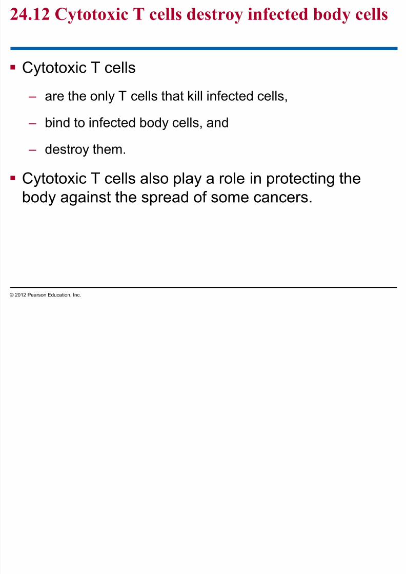
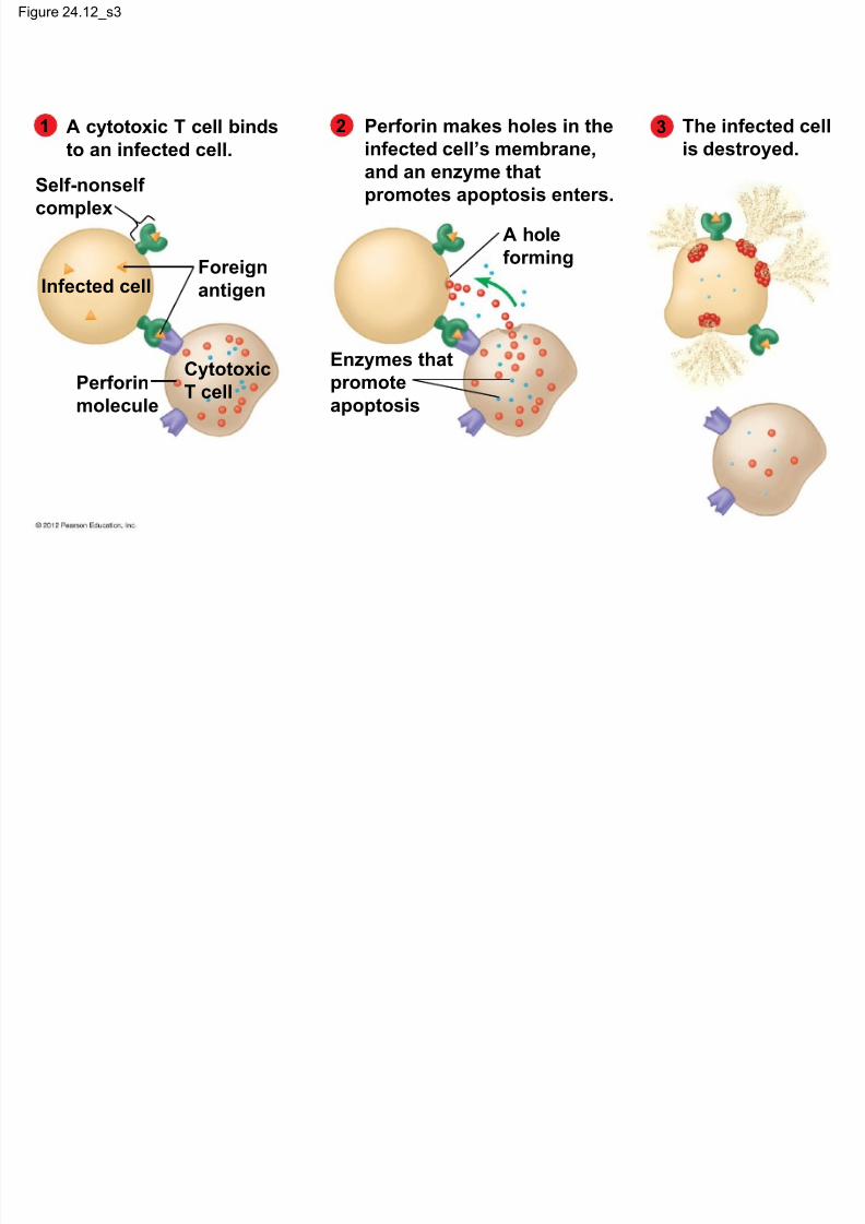
Cytotoxic T cells
– are the only T cells that kill infected cells,
– bind to infected body cells, and
– destroy them.
Cytotoxic T cells also play a role in protecting the
body against the spread of some cancers.
© 2012 Pearson Education, Inc.

7/27/2019 24 Lecture Presentation PC
http://slidepdf.com/reader/full/24-lecture-presentation-pc 73/105
© 2012 Pearson Education, Inc.
Animation: Cytotoxic T CellsRight click on animation / Click play
Figure 24.12_s1

7/27/2019 24 Lecture Presentation PC
http://slidepdf.com/reader/full/24-lecture-presentation-pc 74/105
1 A cytotoxic T cell binds
to an infected cell.
Self-nonself
complex
Foreign
antigenInfected cell
Perforin
molecule
Cytotoxic
T cell
Figure 24.12_s2

7/27/2019 24 Lecture Presentation PC
http://slidepdf.com/reader/full/24-lecture-presentation-pc 75/105
21 A cytotoxic T cell binds
to an infected cell.
Perforin makes holes in the
infected cell’s membrane,
and an enzyme that
promotes apoptosis enters.Self-nonself
complex
Foreign
antigenInfected cell
Perforin
molecule
Cytotoxic
T cell
Enzymes that
promote
apoptosis
A hole
forming
Figure 24.12_s3
7/27/2019 24 Lecture Presentation PC
http://slidepdf.com/reader/full/24-lecture-presentation-pc 76/105
321 A cytotoxic T cell binds
to an infected cell.
Perforin makes holes in the
infected cell’s membrane,
and an enzyme that
promotes apoptosis enters.
The infected cell
is destroyed.
Self-nonself
complex
Foreign
antigenInfected cell
Perforin
molecule
Cytotoxic
T cell
Enzymes that
promote
apoptosis
A hole
forming
24.13 CONNECTION: HIV destroys helper Tll i i th b d ’ d f
7/27/2019 24 Lecture Presentation PC
http://slidepdf.com/reader/full/24-lecture-presentation-pc 77/105
cells, compromising the body’s defenses
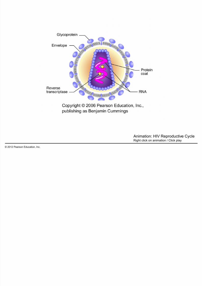
AIDS (acquired immunodeficiency syndrome),
results from infection by HIV, the human
immunodeficiency virus.
– Since 1981 AIDS has killed more than 27 million people,
and more than 33 million people live today with HIV.
– In 2008,
– 2.7 million people were newly infected with HIV, and
– over 2 million died, including 300,000 children under age 15.
– Most AIDS infections and deaths occur in nonindustrialized
nations of southern Asia and sub-Saharan Africa.
© 2012 Pearson Education, Inc.
24.13 CONNECTION: HIV destroys helper Tll i i th b d ’ d f

7/27/2019 24 Lecture Presentation PC
http://slidepdf.com/reader/full/24-lecture-presentation-pc 78/105
cells, compromising the body’s defenses
The AIDS virus usually attacks helper T cells,
impairing the
– cell-mediated immune response and
– humoral immune response, and – opening the way for opportunistic infections.
© 2012 Pearson Education, Inc.
24.13 CONNECTION: HIV destroys helper Tll i i th b d ’ d f
7/27/2019 24 Lecture Presentation PC
http://slidepdf.com/reader/full/24-lecture-presentation-pc 79/105
cells, compromising the body’s defenses
AIDS patients typically die from
– opportunistic infections and
– cancers
– that would normally be resisted by a person with ahealthy immune system.
Until there is a vaccine or a cure, the best way to
stop AIDS is to educate people about how the virusis transmitted.
© 2012 Pearson Education, Inc.
7/27/2019 24 Lecture Presentation PC
http://slidepdf.com/reader/full/24-lecture-presentation-pc 80/105
© 2012 Pearson Education, Inc.
Animation: HIV Reproductive CycleRight click on animation / Click play
Figure 24.13

7/27/2019 24 Lecture Presentation PC
http://slidepdf.com/reader/full/24-lecture-presentation-pc 81/105
24.14 EVOLUTION CONNECTION: The rapidl ti f HIV li t AIDS

7/27/2019 24 Lecture Presentation PC
http://slidepdf.com/reader/full/24-lecture-presentation-pc 82/105
evolution of HIV complicates AIDStreatment
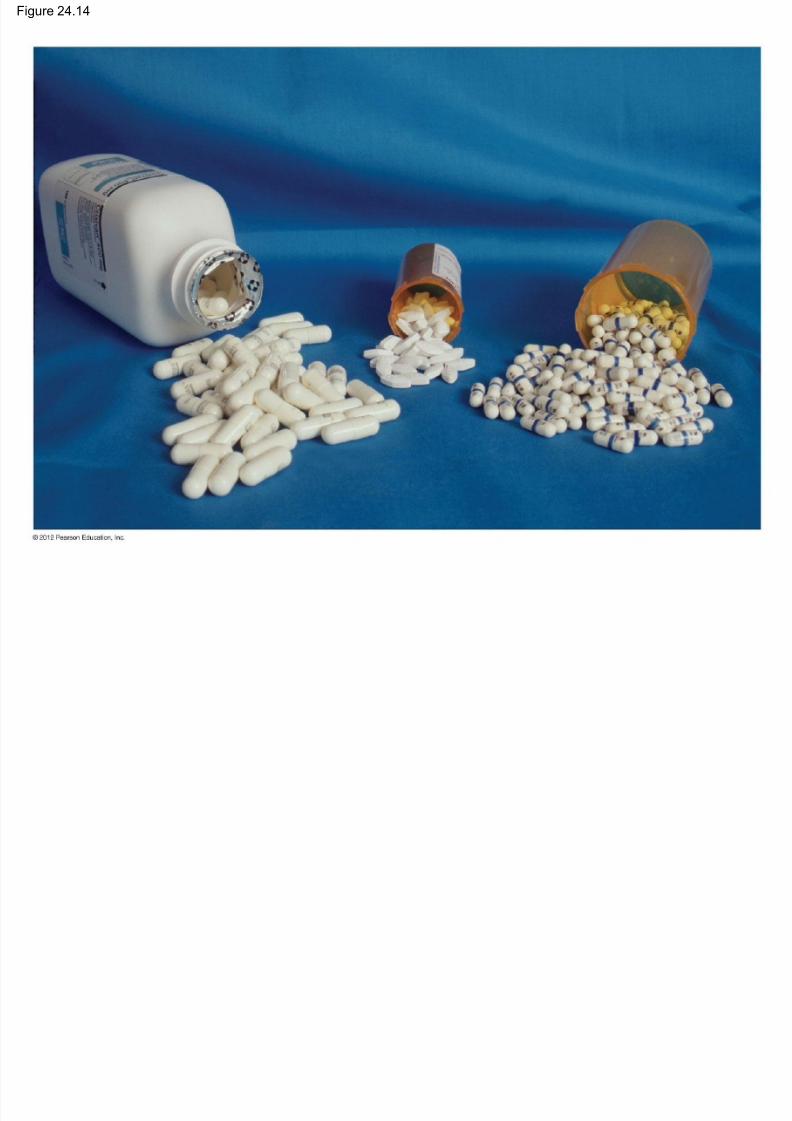
HIV mutates very quickly.
New strains are resistant to AIDS drugs.
Drug-resistant strains now infect new patients.
© 2012 Pearson Education, Inc.
Figure 24.14
7/27/2019 24 Lecture Presentation PC
http://slidepdf.com/reader/full/24-lecture-presentation-pc 83/105
24.15 The immune system depends on ourmolecular fingerprints

7/27/2019 24 Lecture Presentation PC
http://slidepdf.com/reader/full/24-lecture-presentation-pc 84/105
molecular fingerprints
The immune system normally reacts
– only against nonself substances and
– not against self.
© 2012 Pearson Education, Inc.
24.15 The immune system depends on ourmolecular fingerprints
7/27/2019 24 Lecture Presentation PC
http://slidepdf.com/reader/full/24-lecture-presentation-pc 85/105
molecular fingerprints
Transplanted organs may be rejected because the
transplanted cells lack the unique “fingerprint” of the
patient’s self proteins, called major
histocompatibility complex (MHC) molecules.
Donors are used that most closely match the
patient’s tissues.
Transplants between identical twins do not typically
have this problem.
© 2012 Pearson Education, Inc.
7/27/2019 24 Lecture Presentation PC
http://slidepdf.com/reader/full/24-lecture-presentation-pc 86/105
DISORDERS OF THEIMMUNE SYSTEM
© 2012 Pearson Education, Inc.
24.16 CONNECTION: Malfunction or failure ofthe immune system causes disease
7/27/2019 24 Lecture Presentation PC
http://slidepdf.com/reader/full/24-lecture-presentation-pc 87/105
the immune system causes disease
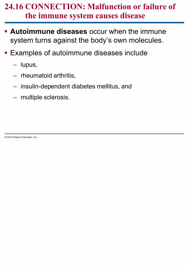
Autoimmune diseases occur when the immune
system turns against the body’s own molecules.
Examples of autoimmune diseases include
– lupus,
– rheumatoid arthritis,
– insulin-dependent diabetes mellitus, and
– multiple sclerosis.
© 2012 Pearson Education, Inc.
Figure 24.16

7/27/2019 24 Lecture Presentation PC
http://slidepdf.com/reader/full/24-lecture-presentation-pc 88/105
24.16 CONNECTION: Malfunction or failure ofthe immune system causes disease

7/27/2019 24 Lecture Presentation PC
http://slidepdf.com/reader/full/24-lecture-presentation-pc 89/105
the immune system causes disease
Immunodeficiency diseases occur when an
immune response is
– defective or
– absent.
The immune system may be weakened by
– physical stress or
– emotional stress.
– Students are more likely to be sick during a week of
exams.
© 2012 Pearson Education, Inc.
24.17 CONNECTION: Allergies are overreactionsto certain environmental antigens

7/27/2019 24 Lecture Presentation PC
http://slidepdf.com/reader/full/24-lecture-presentation-pc 90/105
to certain environmental antigens
Allergies are hypersensitive (exaggerated)
responses to otherwise harmless antigens in our
surroundings.
Antigens that cause allergies are called allergens.
© 2012 Pearson Education, Inc.
24.17 CONNECTION: Allergies are overreactionsto certain environmental antigens

7/27/2019 24 Lecture Presentation PC
http://slidepdf.com/reader/full/24-lecture-presentation-pc 91/105
to certain environmental antigens
Allergic reactions typically occur
– very rapidly and
– in response to tiny amounts of an allergen.
Allergic reactions can occur in many parts of thebody, including
– nasal passages,
– bronchi, and
– skin.
© 2012 Pearson Education, Inc.
24.17 CONNECTION: Allergies are overreactionsto certain environmental antigens

7/27/2019 24 Lecture Presentation PC
http://slidepdf.com/reader/full/24-lecture-presentation-pc 92/105
to certain environmental antigens
The symptoms of an allergy result from a two-stage
reaction.
1. The first stage, called sensitization, occurs when a person
is first exposed to an allergen.
2. The second stage begins when the person is exposed to
the same allergen later.
– The allergen binds to mast cells.
– Mast cells release histamine, causing irritation, itchy skin, andtears.
© 2012 Pearson Education, Inc.
Figure 24.17

7/27/2019 24 Lecture Presentation PC
http://slidepdf.com/reader/full/24-lecture-presentation-pc 93/105
1 2 3 4 5
Sensitization: Initial exposure to an allergen Later exposure to the same allergen
B cell
(plasma cell)
Antigenic determinant
Mastcell
Histamine
An allergen (pollen
grain) enters the
bloodstream.
B cells make
antibodies.
Antibodies
attach to a
mast cell.
The allergen binds
to antibodies on
a mast cell.
Histamine is
released, causing
allergy symptoms.
Figure 24.17_1

7/27/2019 24 Lecture Presentation PC
http://slidepdf.com/reader/full/24-lecture-presentation-pc 94/105
321
Sensitization: Initial exposure to an allergen
B cell
(plasma cell)
Antigenic determinant
Mastcell
Histamine
An allergen (pollengrain) enters the
bloodstream.
B cells makeantibodies.
Antibodiesattach to a
mast cell.
Figure 24.17_2

7/27/2019 24 Lecture Presentation PC
http://slidepdf.com/reader/full/24-lecture-presentation-pc 95/105
4 5
Later exposure to the same allergen
The allergen bindsto antibodies on
a mast cell.
Histamine isreleased, causing
allergy symptoms.
24.17 CONNECTION: Allergies are overreactionsto certain environmental antigens

7/27/2019 24 Lecture Presentation PC
http://slidepdf.com/reader/full/24-lecture-presentation-pc 96/105
to certain environmental antigens
Antihistamines
– interfere with histamine’s action,
– provide temporary relief, but
– often make people drowsy.
Anaphylactic shock
– is an extreme life-threatening allergic reaction and
– can be treated with injections of epinephrine.
© 2012 Pearson Education, Inc.
You should now be able to
7/27/2019 24 Lecture Presentation PC
http://slidepdf.com/reader/full/24-lecture-presentation-pc 97/105
1. Describe the functions of neutrophils.
2. Describe the nature of innate defenses ininvertebrates and vertebrates.
3. Describe the steps of the inflammatory response
and explain how they help to prevent the spread ofdisease.
4. Describe the specific nature of adaptive immune
system responses.5. Describe the development and functions of B
lymphocytes and T lymphocytes.
© 2012 Pearson Education, Inc.
You should now be able to

7/27/2019 24 Lecture Presentation PC
http://slidepdf.com/reader/full/24-lecture-presentation-pc 98/105
6. Define and distinguish between the humoral
immune response and the cell-mediated immune
response.
7. Describe the nature of antigens. Explain how an
antigen and an antibody interact.
8. Describe the process of clonal selection and
compare a primary immune response to a
secondary immune response.
9. Describe the specific structure of an antibody and
relate its shape to its functions.© 2012 Pearson Education, Inc.
You should now be able to
7/27/2019 24 Lecture Presentation PC
http://slidepdf.com/reader/full/24-lecture-presentation-pc 99/105
10. Describe four effector mechanisms of the humoral
immune system.
11. Describe the production and uses of monoclonal
antibodies.
12. Describe the specific functions of helper T cells
and how they interact with other cells.
13. Explain how cytotoxic T cells destroy infected body
cells.
14. Explain how HIV infects cells, multiplies, and
causes disease.
© 2012 Pearson Education, Inc.
You should now be able to
7/27/2019 24 Lecture Presentation PC
http://slidepdf.com/reader/full/24-lecture-presentation-pc 100/105
15. Explain why it has been difficult to develop a
successful treatment for AIDS.
16. Explain how the immune system identifies the
body’s own molecules and how this system
complicates organ transplantations.
17. Describe how the malfunction or failure of the
immune system can cause disease.
18. Explain why allergies occur and what causesanaphylactic shock.
© 2012 Pearson Education, Inc.
Figure 24.UN01

7/27/2019 24 Lecture Presentation PC
http://slidepdf.com/reader/full/24-lecture-presentation-pc 101/105
The humoral immune response:
B cell
T cell
makes which bind to
Antibodies Antigens in
body fluid
The cell-mediated immune response:
Infected
body cell
Self-nonself complex
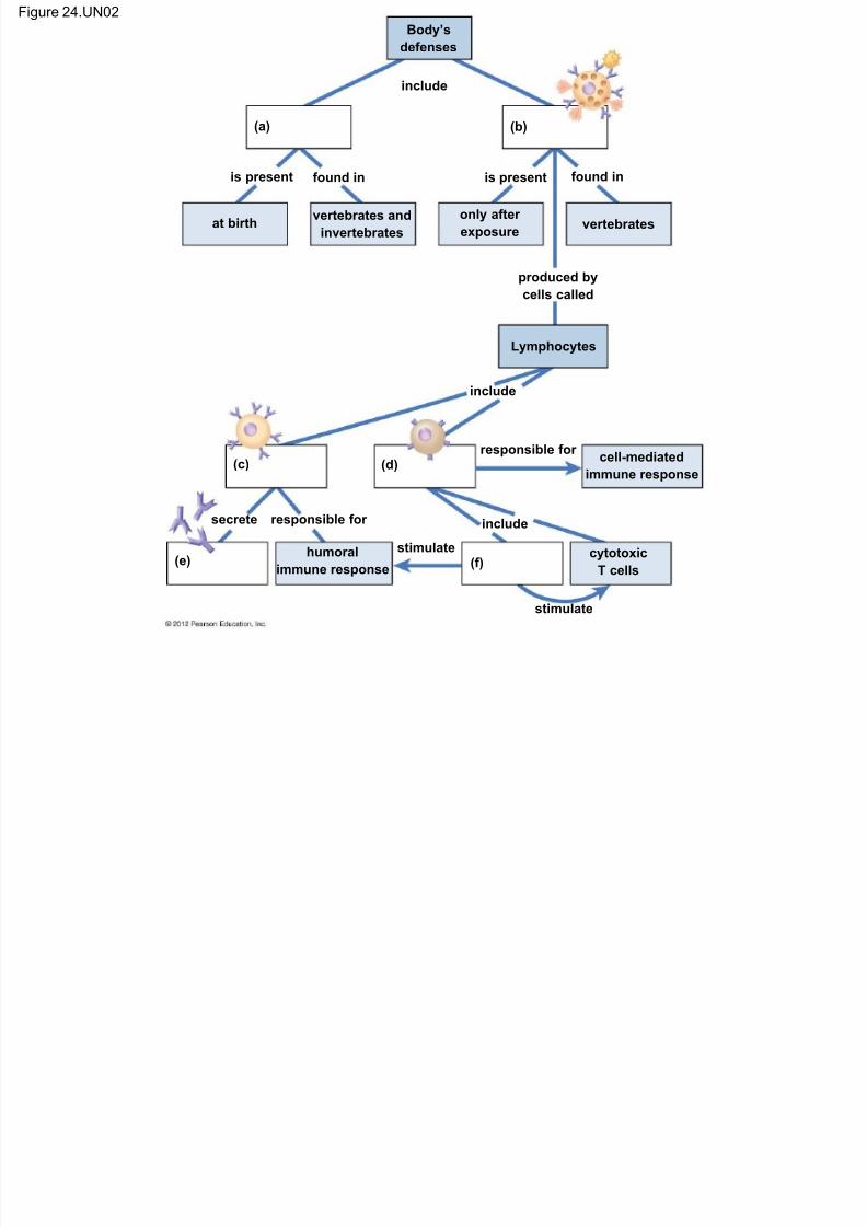
Figure 24.UN02 Body’s
defenses
i l d
7/27/2019 24 Lecture Presentation PC
http://slidepdf.com/reader/full/24-lecture-presentation-pc 102/105
at birthvertebrates and
invertebrates
only after
exposurevertebrates
include
is present found in is present found in
produced by
cells called
Lymphocytes
include
responsible forcell-mediated
immune response
include
stimulate
stimulate
secrete responsible for
humoral
immune response
cytotoxic
T cells
(a) (b)
(c) (d)
(e) (f)
Figure 24.UN02_1

7/27/2019 24 Lecture Presentation PC
http://slidepdf.com/reader/full/24-lecture-presentation-pc 103/105
Body’s
defenses
at birthvertebrates and
invertebrates
only after
exposurevertebrates
include
is present found in is present found in
produced by
cells called
Lymphocytes
(a) (b)
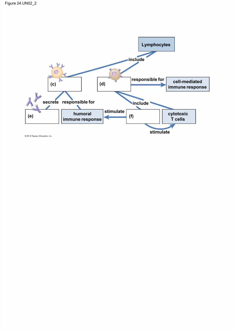
Figure 24.UN02_2
7/27/2019 24 Lecture Presentation PC
http://slidepdf.com/reader/full/24-lecture-presentation-pc 104/105
Lymphocytes
include
responsible forcell-mediated
immune response
include
stimulate
stimulate
secrete responsible for
humoral
immune response
cytotoxic
T cells
(c) (d)
(e) (f)
Figure 24.17_UN01

7/27/2019 24 Lecture Presentation PC
http://slidepdf.com/reader/full/24-lecture-presentation-pc 105/105