208462orig1s000 - food and drug administration · i concur with dr. sheth’s conclusion that...
TRANSCRIPT

CENTER FOR DRUG EVALUATION AND RESEARCH
APPLICATION NUMBER:
208462Orig1s000
PHARMACOLOGY REVIEW(S)

MEMORANDUM
Ninlaro (ixazomib)
Date: November 13, 2015To: File for NDA 208462From: John K. Leighton, PhD, DABT
Director, Division of Hematology Oncology ToxicologyOffice of Hematology and Oncology Products
I have examined pharmacology/toxicology supporting and labeling reviews for Ninlaro conducted by Dr. Place, and secondary memorandum and labeling provided by Dr. Sheth. I concur with Dr. Sheth’s conclusion that Ninlaro may be approved for the proposed indication.
Reference ID: 3846536

---------------------------------------------------------------------------------------------------------This is a representation of an electronic record that was signedelectronically and this page is the manifestation of the electronicsignature.---------------------------------------------------------------------------------------------------------/s/----------------------------------------------------
JOHN K LEIGHTON11/13/2015
Reference ID: 3846536

MEMORANDUM
Date: November 09, 2015From: Christopher Sheth, PhD
Division of Hematology Oncology Toxicology (DHOT)Office of Hematology and Oncology Products (OHOP)
Re: Approvability for Pharmacology and ToxicologyNDA: 208462Drug: Ninlaro (ixazomib)Indication: Treatment of patients with multiple myeloma who have received at least one prior
therapyApplicant: Millennium Pharmaceuticals, Inc.
Ixazomib is a small molecule reversible inhibitor of the chymotrypsin-like activity of the beta 5 subunit of the 20S proteasome, being developed as a treatment for patients with multiple myeloma who have received at least one prior therapy. Ixazomib will be formulated into capsules of Ninlaro of 2.3, 3, and 4 mg strengths. Ninlaro will be administered orally once a week on Days 1, 8, and 15 of a 28-day treatment cycle, in combination with lenalidomide and dexamethasone.
The pharmacology and toxicology studies reviewed included primary pharmacodynamics, genotoxicity, safety pharmacology, repeat dose toxicology (6-month rat and 9-month dog), and embryo-fetal developmental toxicity in rats and rabbits. With regards to the pharmacology of ixazomib, the drug induced apoptosis of multiple myeloma cell lines in vitro and demonstrated cytotoxicity against myeloma cells from patients who had relapsed after multiple prior therapies, including bortezomib, lenalidomide, and dexamethasone. The combination of ixazomib and lenalidomide demonstrated synergistic cytotoxic effects in multiple myeloma cell lines. Additionally, ixazomib demonstrated antitumor activity in vivo in a mouse multiple myeloma tumor xenograft model. The Established Pharmacological Class of “proteasome inhibitor” was determined to be both scientifically valid and clinically meaningful for ixazomib.
In multi-cycle general toxicity studies conducted in dogs, the principal target organs included the nervous system. Nervous system effects were primarily seen in dogs at oral doses greater than or equal to 0.1 mg/kg (2 mg/m2) and included microscopic findings of minimal to mild neuronal degeneration of the sympathetic, dorsal root, peripheral autonomic (salivary gland), end organ ganglia, and minimal secondary axonal/nerve fiber degeneration of the peripheral nerves and ascending tracts in the dorsal columns of the spinal cord. In the 9-month study (10 cycles) in dogs where the dosing regimen mimics the clinical regimen (28-day cycle), microscopic neuronal effects were generally minimal in nature and only observed at 0.2 mg/kg (4 mg/m2; AUC0-168 = 1940 hr*ng/mL). The majority of target organ findings (e.g., in the gastrointestinal tract, lymphoid tissue, and nervous system) partially or completely recovered following discontinuation of treatment, except for the neuronal findings in the lumbar dorsal root ganglion and dorsal column.
The Applicant’s proposal for Section 8 of the label is consistent with the Pregnancy and Lactation Labeling Rule. Ninlaro can cause fetal harm
Reference ID: 3844455
(b) (4)


---------------------------------------------------------------------------------------------------------This is a representation of an electronic record that was signedelectronically and this page is the manifestation of the electronicsignature.---------------------------------------------------------------------------------------------------------/s/----------------------------------------------------
CHRISTOPHER M SHETH11/09/2015
Reference ID: 3844455

1
DEPARTMENT OF HEALTH AND HUMAN SERVICESPUBLIC HEALTH SERVICE
FOOD AND DRUG ADMINISTRATIONCENTER FOR DRUG EVALUATION AND RESEARCH
PHARMACOLOGY/TOXICOLOGY NDA REVIEW AND EVALUATION
Application number: 208462
Supporting document/s: 1
Applicant’s letter date: July 10, 2015
CDER stamp date: July 10, 2015
Product: Ninlaro (ixazomib)
Indication: Patients with multiple myeloma who have
received at least one prior therapy
Applicant: Millenium Pharmaceuticals Inc.
Review Division: Division of Hematology Oncology Toxicology
(DHOT) for Division of Hematology Products
(DHP)
Reviewer: Emily Place PhD MPH
Supervisor/Team Leader: Chris Sheth PhD
Division Director: John Leighton PhD, DABT (DHOT)
Ann Farrell MD (DHP)
Project Manager: Jacquin Jones MS BSN
Disclaimer
Except as specifically identified, all data and information discussed below and necessary for approval of NDA 208462 are owned by Millenium Pharmaceuticals Inc. or are data for which Millenium Pharmaceuticals Inc. has obtained a written right of reference. Any information or data necessary for approval of NDA 208462 that Millenium Pharmaceuticals Inc. does not own or have a written right to reference constitutes one of the following: (1) published literature, or (2) a prior FDA finding of safety or effectiveness for a listed drug, as reflected in the drug’s approved labeling. Any data or information described or referenced below from reviews or publicly available summaries of a previously approved application is for descriptive purposes only and is not relied upon for approval of NDA 208462.
Reference ID: 3844453
NDA 208462 Emily Place PhD MPH
2
TABLE OF CONTENTS
1 EXECUTIVE SUMMARY...........................................................................................81.1 INTRODUCTION .....................................................................................................81.2 BRIEF DISCUSSION OF NONCLINICAL FINDINGS .......................................................81.3 RECOMMENDATIONS ...........................................................................................10
2 DRUG INFORMATION............................................................................................102.1 DRUG ................................................................................................................102.2 RELEVANT INDS, NDAS, BLAS AND DMFS..........................................................112.3 DRUG FORMULATION ..........................................................................................112.4 COMMENTS ON NOVEL EXCIPIENTS ......................................................................112.5 COMMENTS ON IMPURITIES/DEGRADANTS OF CONCERN ........................................112.6 PROPOSED CLINICAL POPULATION AND DOSING REGIMEN .....................................122.7 REGULATORY BACKGROUND ...............................................................................12
3 STUDIES SUBMITTED...........................................................................................133.1 STUDIES REVIEWED ............................................................................................133.2 STUDIES NOT REVIEWED.....................................................................................143.3 PREVIOUS REVIEWS REFERENCED.......................................................................20
4 PHARMACOLOGY .................................................................................................204.1 PRIMARY PHARMACOLOGY ..................................................................................204.2 SECONDARY PHARMACOLOGY .............................................................................334.3 SAFETY PHARMACOLOGY ....................................................................................33
5 PHARMACOKINETICS/ADME/TOXICOKINETICS ...............................................345.1 PK/ADME .........................................................................................................34
6 GENERAL TOXICOLOGY......................................................................................426.1 SINGLE-DOSE TOXICITY ......................................................................................426.2 REPEAT-DOSE TOXICITY .....................................................................................42
7 GENETIC TOXICOLOGY........................................................................................667.1 IN VITRO REVERSE MUTATION ASSAY IN BACTERIAL CELLS (AMES) ........................667.2 IN VITRO ASSAYS IN MAMMALIAN CELLS ...............................................................707.3 IN VIVO CLASTOGENICITY ASSAY IN RODENT (MICRONUCLEUS ASSAY) ..................747.4 OTHER GENETIC TOXICITY STUDIES.....................................................................79
8 CARCINOGENICITY...............................................................................................85
9 REPRODUCTIVE AND DEVELOPMENTAL TOXICOLOGY.................................859.1 FERTILITY AND EARLY EMBRYONIC DEVELOPMENT................................................859.2 EMBRYONIC FETAL DEVELOPMENT.......................................................................859.3 PRENATAL AND POSTNATAL DEVELOPMENT ........................................................107
10 SPECIAL TOXICOLOGY STUDIES..................................................................107
Reference ID: 3844453

NDA 208462 Emily Place PhD MPH
3
11 INTEGRATED SUMMARY AND SAFETY EVALUATION................................107
12 APPENDIX/ATTACHMENTS ............................................................................109
Reference ID: 3844453

NDA 208462 Emily Place PhD MPH
4
Table of Tables
Table 1. Ixazomib drug formulation (capsules)...............................................................11Table 2. Dosing schedule for Ninlaro with lenalidomide and dexamethasone ...............12Table 3. Ixazomib protease selectivity............................................................................21Table 4. Summary of enzymology comparison of ixazomib and bortezomib .................22Table 5. Ixazomib proteasome inhibition in cultured cells ..............................................23Table 6. LD50 values for ixazomib treated human myeloma cell lines ............................25Table 7. Ixazomib in combination with lenalidomide in vitro ...........................................29Table 8. Summary pharmacokinetic parameters in human xenograft mouse model of multiple myeloma............................................................................................................30Table 9. Summary pharmacokinetics in nonclinical testing species following single dose oral administration of ixazomib or ixazomib citrate .........................................................35Table 10. Pharmacokinetic parameters of MLN2238 following oral dosing in dogs .......36Table 11. Mean Plasma concentrations of MLN2238 in beagle dogs following a single oral dose.........................................................................................................................36Table 12. Study design for tissue radioactivity analysis of Long Evans rats following a single oral dose of MLN9708..........................................................................................38Table 13. Pharmacokinetic parameters of tissue radioactivity following single oral dose of MLN9708 in male Long Evans rats ............................................................................38Table 14. Test articles used in a pharmacokinetic study of metabolism, mass balance and excretion in rats following oral administration..........................................................40Table 15. Study design of metabolism, mass balance and excretion study in rats following single oral administration.................................................................................40Table 16. Sample collection in metabolism, mass balance and excretion study in rats following single oral administration.................................................................................41Table 17. Mean percent excretion of radioactivity in urine and feces in intact rats following a single oral dose of [14C]MLN9708 .................................................................41Table 18. Mean percent excretion of radioactivity in urine and feces in bile duct cannulated rats following a single oral dose of [14C] MLN9708 ......................................42Table 19. Study design for 6 month repeat dose toxicology study in rats ......................43Table 20. Cause of mortality in 6 month repeat dose toxicology study in rat .................45Table 21. Supplemental information: summary of historical control data pertaining to calculi formation in kidney and urinary bladder in male..................................................47Table 22. Hematology findings for 6 month repeat dose toxicity study in rats (percent change from control).......................................................................................................49Table 23. Histological findings from 6 month repeat dose toxicology study in rats ........50Table 24. Summary toxicokinetics for 6 month repeat dose toxicology study in rat .......54Table 25. Dosing solution analysis (concentration) for 6 month repeat dose toxicology study in rats ....................................................................................................................55Table 26. Study design for 9 month repeat dose toxicology study in dogs ....................57Table 27. Hematology findings for 9 month repeat dose toxicity study in dogs (percent change from control).......................................................................................................58Table 28. Clinical chemistry findings for 9 month repeat dose toxicity study in dogs (percent change from control).........................................................................................59Table 29. Histological findings for 9 month repeat dose toxicity study in dogs ..............59
Reference ID: 3844453
NDA 208462 Emily Place PhD MPH
5
Table 30. Histological findings for 9 month repeat dose toxicity study in dogs (nervous system-Week 38)............................................................................................................61Table 31.Histological findings for 9 month repeat dose toxicity study in dogs (nervous system-recovery Week 40) .............................................................................................62Table 32. Time points of conduct of FOB assessments in 10 cycle study with MLN9708 in beagle dogs. ...............................................................................................................62Table 33. Parameters evaluated in FOB assessments in beagle dogs. .........................63Table 34. Summary toxicokinetics for 9 month repeat dose toxicology study in dogs ...64Table 35. Evidence for steep dose-toxicity relationship with MLN9708 in chronic rat and dog toxicology.................................................................................................................65Table 36. Dosing solution analysis (concentration) for 9 month repeat dose toxicology study in dogs...................................................................................................................65Table 37. Summary results of initial assay for MLN2238 in the Salmonella/E.Coli mammalian microsome reverse mutation assay ............................................................68Table 38. Summary results of confirmatory assay for MLN2238 in the Salmonella/E. coli mammalian microsome reverse mutation assay ............................................................69Table 39. Concentrations of MLN-9708 evaluated for chromosomal aberrations assay in HPBL ..............................................................................................................................71Table 40. Cytotoxicity and aberration summary: 3 hour incubation without metabolic activation.........................................................................................................................72Table 41. Cytotoxicity and aberration summary: 22 hour incubation without metabolic activation.........................................................................................................................73Table 42. Cytotoxicity and aberration summary: 3 hour incubation with metabolic activation.........................................................................................................................74Table 43. Study design for in vivo clastogenic micronucleus assay in mice ..................75Table 44. Summary evaluation of definitive phase bone marrow micronucleus assay in mice ................................................................................................................................77Table 45. Individual and mean data for definitive phase bone marrow micronucleus assay in mice 24-48 hours after dosing..........................................................................78Table 46. Individual and mean data for definitive phase bone marrow micronucleus assay in mice 44-48 hours after dosing..........................................................................79Table 47. Study design for comet assay of MLN-9708 in mice ......................................81Table 48. Summary results from comet assay in liver tissue from MLN-9708 testing in mice ................................................................................................................................83Table 49. Summary results from comet assay in stomach tissue from MLN-9708 testing in mice ............................................................................................................................84Table 50. Summary toxicokinetics following single oral dose of MLN-9708 for injection in mice (Study 13-039/tk)....................................................................................................85Table 51. Toxicokinetic parameters in embryonic fetal development dose range finding toxicity study in rats (daily administration) ......................................................................87Table 52. Toxicokinetic parameters in embryonic fetal development dose range finding toxicity study in rats (administration/ 3 days) ..................................................................87Table 53. Dosing solution stability analysis in a embryofetal toxicity dose range finding study in rats ....................................................................................................................88Table 54. Dosing solution concentration analysis in a embryofetal toxicity dose range finding study in rats.........................................................................................................89
Reference ID: 3844453
NDA 208462 Emily Place PhD MPH
6
Table 55. Placental gross pathology findings in rats ......................................................89Table 56. Pregnancy parameters in an embryofetal toxicity dose range finding study in rats..................................................................................................................................90Table 57. Fetal malformations, abnormalities, and variations in dose range finding study in rats ..............................................................................................................................90Table 58. Study design for embryonic fetal development toxicity study in rats ..............93Table 59. Cause of maternal death in embryonic fetal development toxicity study in rats........................................................................................................................................93Table 60. Toxicokinetic parameters in embryonic fetal development toxicity study in rats........................................................................................................................................95Table 61. Maternal gross pathology findings in rats .......................................................96Table 62. Placental gross pathology findings in rats ......................................................96Table 63. Pregnancy parameters in rats ........................................................................96Table 64. Fetal malformations, abnormalities, and variations in rats .............................96Table 65. Study design for dose range finding embryonic fetal development toxicity study in rabbits................................................................................................................99Table 66. Summary toxicokinetics ................................................................................100Table 67. Dosing solution concentration analysis in a embryofetal toxicity dose range finding study in rabbits ..................................................................................................100Table 68. Maternal gross pathology findings in a dose range finding study in rabbits .101Table 69. Pregnancy parameters in a dose range finding study in rabbits...................101Table 70. Fetal malformations, abnormalities, and variations in a dose range finding study in rabbits..............................................................................................................101Table 71.Study design for embryonic fetal development toxicity study in rabbits ........103Table 72. Toxicokinetic parameters in embryonic fetal development toxicity study in rabbits ...........................................................................................................................104Table 73. Dosing solution concentration analysis in an embryofetal toxicity study in rabbits ...........................................................................................................................105Table 74. Pregnancy parameters in rabbits..................................................................106Table 75. Fetal malformations, abnormalities, and variations in rabbits.......................106
Reference ID: 3844453

NDA 208462 Emily Place PhD MPH
7
Table of Figures
Figure 1. Effect of ixazomib on the viability of human multiple myeloma cell lines ........25Figure 2. Ixazomib induces caspase-mediated cell death in NCI-H929 multiple myeloma cell line............................................................................................................................26Figure 3. Ixazomib induces caspase-mediated cell death in MM.1S multiple myeloma cell line............................................................................................................................27Figure 4. Effect of ixazomib on the viability of human multiple myeloma patient cells ex-vivo .................................................................................................................................28Figure 5. Proteasome inhibition following treatment with ixazomib (IV, PO) in xenograft mouse model of multiple myeloma .................................................................................31Figure 6. Immunoblotting analysis following treatment with ixazomib (IV, PO) in xeonograft mouse model of multiple myeloma ...............................................................32Figure 7. Mean tumor volume (mm3) over time following treatment with ixazomib (biweekly, PO) in a human xenograft mouse model of multiple myeloma......................33Figure 8. Body weights in male rats in 6 month repeat dose toxicology study ...............48Figure 9. Body weights in female rats in 6 month repeat dose toxicology study ............48Figure 10.Plasma MLN2238 concentration vs. time curves after MLN9708 administration to SD rats ................................................................................................54
Reference ID: 3844453
NDA 208462 Emily Place PhD MPH
8
1 Executive Summary1.1 IntroductionIxazomib is a second-generation proteasome inhibitor that reversibly binds and inhibits the chymotrypsin-like activity of the β5 subunit of the 20S proteasome. Nonclinical pharmacology and toxicology studies have been submitted and reviewed to support the approval of Ninlaro for the treatment of patients who have received at least one prior therapy. The drug will be administered at a recommended dose of 20 mg once a week on Days 1, 8, and 15 of a 28-day treatment cycle by oral administration.
1.2 Brief Discussion of Nonclinical FindingsIxazomib is a proteasome inhibitor similar to the previously approved Velcade (bortezomib). Upon the citrate ester form of the drug, MLN9708, is hydrolyzed into the active boronic acid moiety, MLN2238. Ixazomib reversibly binds to the active threonine reside in the β5 portion of the 20S proteasome, inhibiting its chymotrypsin like activity. A series of non-GLP studies have been conducted with ixazomib to investigate its pharmacology. In vitro cell culture-based assays confirmed that ixazomib does target the β5 subunit at its cymotryptic site and prevents proteasome mediated degradation. Ixazomib also has activity against the β5i isoform, of the immunoproteasome expressed in hematological cells. In vitro, ixazomib induces caspase mediated apoptosis in multiple myeloma cell lines. In multiple myeloma cell lines, ixazomib also had a synergistic effect on cytotoxicity in combination with lenalidomide. Ixazomib also induces cytotoxicity in cells from patients with multiple myeloma refractory to lenalidomide and dexamethasone. In vivo pharmacology studies using a human myeloma xenograft SCID mouse model following treatment with ixazomib confirmed dose-dependent proteasome inhibition by modulation of β5 subunit activity, tumor cell protein levels of GADD34 and caspase-3 (protein upregulated as parts of the unfolded protein response pathway and apoptosis). Ixazomib treatment of SCID mice inoculated with human myeloma tumor xenografts also reduces tumor volumes.
Stand-alone safety pharmacology studies were not conducted for the respiratory or nervous system. In the repeat-dose toxicology studies, ixazomib exposure did not result in clinical signs of toxicity, there were no toxicologically significant effects on the respiratory systems of rats or dogs, and there were no acute nervous system-related effects (as assessed in part by a functional observational battery (FOB) incorporated into the 9 month dog study). There were chronic toxicities of the nervous system in dogs (see Section 6). The IC50 in the hERG assay for ixazomib was 59.6 μM suggesting weak inhibition of the potassium channel. No adverse effects on cardiovascular parameters were observed in dogs administered oral ixazomib.
Based on the data collected in general toxicology studies, there were no gender differences in exposure to oral ixazomib, and increases in Cmax and AUC values were dose proportional. Intravenous administration of 2 mg/kg ixazomib and oral administration of 6 mg/kg ixazomib resulted in similar plasma area under the
Reference ID: 3844453
(b) (4)

NDA 208462 Emily Place PhD MPH
9
concentration-time curves from the time 0 to 24 hours (AUC0-24hr). Tissue distribution was extensive following administration of oral ixazomib. The highest exposure (by Cmax or AUC) was in the urinary bladder, lymph node, spleen, adrenal glands, kidney (renal cortex and medulla), bile, GI tract, and liver. The longest observed half-life was for the urinary bladder with a t1/2 of up to 946 hours. Upon , ixazomib citrate ester (MLN9708) is metabolized to the active boronic acid form of the drug, (MLN2238). The elimination half-life after oral dosing in nonclinical species ranged from 30 to 72 hours.
The general toxicology studies were conducted in the rat and dog via oral (gavage). All studies relevant to an approvability determination were conducted in compliance with Good Laboratory Practices (GLP).
General Toxicology studiesNonclinical findings in the rat and dog show that ixazomib targets the gastrointestinal tract, hematopoietic/lymphatic systems, and nervous system (dog only). Findings include:
Hematopoietic/lymphatic systems: lymphoid necrosis and depletion in lymph nodes and spleen correlating with increased absolute counts for total white blood cells, neutrophils, monocytes, and lymphocytes (rats only).
Gastrointestinal tract: epithelial hyperplasia, neutrophilic infiltrates (stomach, large and small intestines, peyers patch, mesenteric lymph nodes), single-cell necrosis in the lamina propria of the small and large intestines, and erosion in the stomach.
Nervous system (dogs only): neuronal degeneration of the sympathetic, dorsal root, peripheral autonomic, and end organ ganglia, and nerve fiber degeneration of the peripheral nerves and ascending tracts in the dorsal columns of the spinal cord. Changes present at recovery in high dose animals included nerve fiber degeneration of the dorsal root ganglia and an increase in axonal degeneration in the dorsal columns of the spinal cord.
Safety concerns stem from the steep dose-toxicity relationship with ixazomib in the chronic rat and dog toxicology studies. There was a less than 1.5-fold separation in systemic exposure between the limit dose and a dose associated with mortality due to drug-induced injury of the liver and gastrointestinal tract in the rat. In dogs, there was an approximately 2-fold separation in exposure between the no observable adverse effect level (NOAEL) and doses associated with extensive neuronal pathology in the central nervous system (CNS) and peripheral nervous system (PNS).
GenotoxicityIxazomib was non-mutagenic in the in vitro bacterial reverse mutation assay (AMES test). Ixazomib was not clastogenic in a bone marrow micronucleus assay in mice. Ixazomib showed clastogenic activity (structural chromosomal aberrations) in the in vitro chromosomal aberration assay in human peripheral blood lymphocytes in the presence
Reference ID: 3844453
(b) (4)

NDA 208462 Emily Place PhD MPH
10
or absence of an exogenous metabolic activation system. Ixazomib was negative for DNA damage in a Comet assay in mouse lymphoma cells.
Reproductive and Development ToxicityIxazomib was teratogenic in the rat and rabbit. In pregnant rat (0.6 mg/kg; 3.6 mg/m2) dose range-finding studies, there were decreases in fetal weights, decreased fetal viability, and increased post-implantation losses (at 2.5 times the human AUC when given 4 mg orally, in the rat respectively). In pregnant rabbit (1.0 mg/kg; 12 mg/m2) dose range-finding studies, there were decreases in fetal weights, decreased fetal viability, and increased post-implantation losses (at 3.2 times the human AUC when given 4 mg orally, in the rabbit). In the definitive rabbit study, increases in fetal skeletal variations/abnormalities (caudal vertebrae, number of lumbar vertebrae, full supernumerary ribs, asymmetric sternebrae, shortened tail) were observed at doses greater than or equal to 0.3 mg/kg (3.6 mg/m2), which were also associated with maternal toxicity. The dose of 0.3 mg/kg (3.6 mg/m2) in rabbits is approximately 1.9 times the human AUC when given 4mg orally. Thus, administration of ixazomib during pregnancy may pose a risk to the human fetus. The Applicant proposed a conservative post treatment use of 90 days contraception, based on potential effects of the drug and taking into consideration its long half-life. The agency does not object to the proposal.
1.3 Recommendations
1.3.1 ApprovabilityRecommended for approval. The nonclinical studies submitted to this NDA provide sufficient information to support the use of Ninlaro for the treatment of patients with multiple myeloma who have received at least one prior therapy.1.3.2 Additional Non Clinical RecommendationsNone1.3.3 LabelingThe content for the labeling of ixazomib is contained in this review. Based on the nonclinical data submitted in the NDA, the Established Pharmacological Class (EPC) of “proteasome inhibitor” was determined to be both clinically meaningful and scientifically valid for ixazomib.
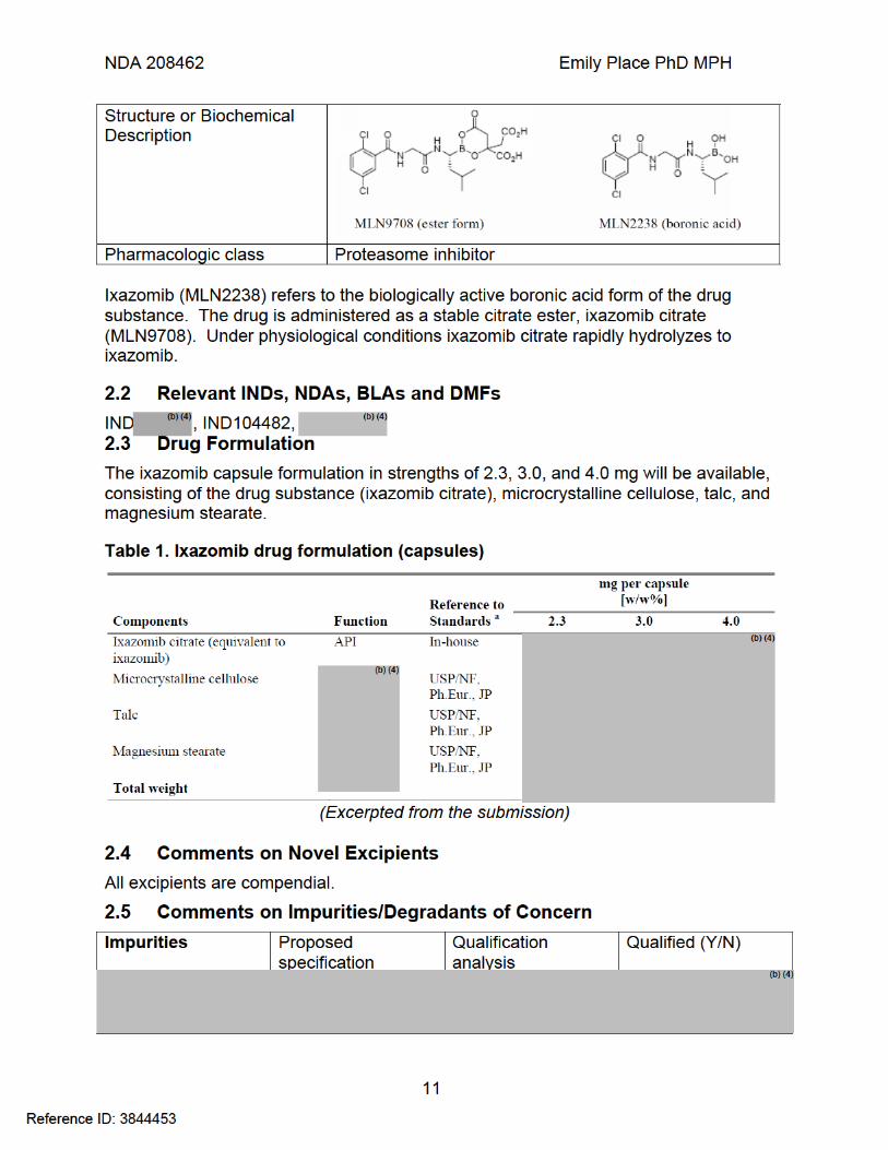
2 Drug Information2.1 DrugCAS Registry Number 1201902-20-3Generic Name Ixazomib citrateCode Names MLN9708 (ixazomib citrate); MLN2238 (ixazomib) Chemical Name B-[(1R)-1-[[2-[(2,5-dichlorobenzoyl)amino]acetyl]amino]-
3-methylbutyl] boronic acidMolecular formula/ Molecular Weight
C20H23BCl2N2O9/ 517.12 g/mol; C14H19BCl2N2O4 /361.03 g/mol
Reference ID: 3844453


NDA 208462 Emily Place PhD MPH
13
3 Studies Submitted3.1 Studies Reviewed Study Title Study No. ModulePrimary Pharmacodynamics
In vitro Pharmacology
Enzymology and Cell Biology of MLN2238RPT-01200Amendment 2
4.2.1.1
Proteasome beta5- and beta2-Site Inhibition by Ixazomib and Bortezomib in HCT116 Cells Measured by Proteasome-Glo Assay
MLN9708-28351 4.2.1.1
Effects on in vitro viability of human myeloma cell lines MLN9708-27528 4.2.1.1
Effects of ixazomib in combination with lenalidomide on in vitro viability of human myeloma cell lines
MLN9708-30663 4.2.1.1
In vivo PharmacologyA Pharmacokinetic/Pharmacodynamic Study of Ixazomib After A Single Oral or Intravenous Administration to MM.1S Multiple Myeloma Xenograft-Bearing Female CB-17 SCID Mice
MLN9708-24699 4.2.1.1
Antitumor Activity of Ixazomib (MLN2238) Administered Orally Twice Weekly in MM.1S Multiple Myeloma Xenograft-Bearing Female CB-17 SCID Mice
MLN9708-24176 4.2.1.1
Safety PharmacologyIn Vitro Binding Study of ML00604174 (MLN2238) to the hERG Channel inHEK-293 Cells
RPT-01141 4.2.1.3
Safety pharmacology studies of MLN9708: effects on the cardiovascular system in conscious dogs
MLN9708-24953 4.2.1.3
Pharmacokinetics
AbsorptionPharmacokinetics of MLN2238 after oral administration of MLN9708 capsules to non-naive male beagle dogs RPT-01218 4.2.2.2
DistributionTissue Distribution via Quantitative Whole-Body Autoradiography in Male Long- Evans Rats Following a Single Oral Gavage Administration of [14C] MLN9708 and Human Radiation Dosimetry Estimation
96N-1206 4.2.2.2
MetabolismPharmacokinetics, Metabolism, Mass Balance, And Excretion Routes Of [14C]MLN9708 In Male Sprague-Dawley Rats Following A Single PO Dose
MILR1800R2A 4.2.2.2
Repeat dose toxicology: (including supportive toxicokinetics)A 6-Month Oral Gavage Toxicity Study of MLN9708 with a 2-Week Recovery Period in Sprague Dawley Rats -416165 4.2.3.2
A 9-month oral gavage toxicity study of MLN9708 with a 2- week recovery period in Beagle dogs -416164 4.2.3.2
Genotoxicity
Reference ID: 3844453
(b) (4)
(b) (4)

NDA 208462 Emily Place PhD MPH
14
Study Title Study No. Module
Salmonella E.coli/Mammalian Microsome Reverse Mutation Assay MBR08-152 4.2.3.3
In vitro Chromosome Aberration Test in Cultured Human Peripheral Blood Lymphocytes MBR12-367 4.2.3.3
MLN9708: A Single-Dose Oral (Gavage) Bone Marrow Micronucleus Evaluation in CD-1 Mice WIL-416152 4.2.3.3
Comet assay of MLN-9708 for injection in mice B130357 4.2.3.3
Reproductive and development ToxicityRange finding study for effects of MLN9708 on Embryo-fetal development in rats 13-07/te 4.2.2.4
Effects of MLN 9708 on Embryo-fetal development in Rats 13-155/te 4.2.2.4An Oral (Gavage) Dose Range-Finding Study of MLN9708 on Embryo/Fetal Development in Rabbits -416156 4.2.2.4
Effects of MLN 9708 on Embryo-fetal development in Rabbits 14-027/te 4.2.2.4
3.2 Studies Not Reviewed Study Title Study No. Module
Pharmacology
In vitro Pharmacology
Customized Profile Screening Profile for Millennium Pharmaceuticals T0-07-4203 4.2.1.1In Vitro Inhibition of Proteasome Activity by ML00701207, an MLN9708 Enantiomer
MLN9708-31263 4.2.1.1
In vivo pharmacology
Antitumor Activity of Ixazomib (MLN2238) Administered IntravenouslyTwice Weekly and Once Weekly in MM.1S Multiple MyelomaXenograft-Bearing Female CB-17 SCID Mice
MLN9708-24177 4.2.1.1
Effect of MLN9708 on antibody production in a rat model of T cell-dependent antigenresponse
MLN9708-24528 4.2.1.1
A Pharmacokinetic/Pharmacodynamic Study of Ixazomib After A Single Oral or Intravenous Administration to MM.1S Multiple Myeloma Xenograft-Bearing Female CB-17 SCID Mice
MLN9708-24699 4.2.1.1
Effect of MLN9708 on autoantibody production in MRL/lpr lupus mice MLN9708-24739 4.2.1.1
In Vivo Antitumor Activity and Pharmacodynamic Response of Bortezomib and MLN2238 in WSU-DLCL2 Subcutaneous Diffuse Large B Cell Lymphoma Xenografts in Female SCID Mice
RPT-01121 4.2.1.1
A Pharmacokinetic/Pharmacodynamic Study of MLN2238 (18 mg/kg) After an Intravenous Administration to CWR22 Tumor-Bearing Male SCID Mice
RPT-01161 4.2.1.1
A Pharmacokinetic/Pharmacodynamic Study of MLN2238 (14 mg/kg) After an Intravenous Administration to Male CWR22 Tumor-Bearing SCID Mice
RPT-01162 4.2.1.1
A Pharmacokinetic/Pharmacodynamic Study of MLN2238 (2 and 4 mg/kg) After a Subcutaneous Administration to CWR22 Tumor-Bearing Male SCID Mice
RPT-01163 4.2.1.1
In Vivo Antitumor Activity and Pharmacodynamic Response of MLN2238 and Bortezomib in WSU-DLCL2 Subcutaneous Diffuse RPT-01165 4.2.1.1
Reference ID: 3844453 5 Page(s) has been Withheld in Full as b4 (CCI/TS) immediately following this page
(b) (4)

NDA 208462 Emily Place PhD MPH
20
3.3 Previous Reviews ReferencedIND , IND104482,
4 Pharmacology4.1 Primary PharmacologyMLN9708 is a second-generation proteasome inhibitor similar to the previously approved Velcade (bortezomib). Upon the citrate ester form of the drug, MLN9708, is hydrolyzed into the active boronic acid moiety, MLN2238. Ixazomib reversibly binds to the in the β5 portion of the 20S proteasome, inhibiting its chymotrypsin like activity. A series of non-GLP studies have been performed with ixazomib to investigate its pharmacology. In vitro cell culture-based assays confirmed that ixazomib does target the β5 subunit at its cymotryptic site and prevents proteasome mediated degradation. Ixazomib also has activity against the β5i isoform, of the immunoproteasome expressed in hematological cells. In vitro ixazomib induces caspase mediated apoptosis in multiple myeloma cell lines. Ixazomib also induces cytotoxicity in cells from patients with multiple myeloma refractory to lenalidomide and dexamethasone. Ixazomib was cytotoxic in culture to primary cultures of cells harvested from multiple myeloma patients who had relapsed after prior therapies. In multiple myeloma cell lines, ixazomib also had a synergistic effect on cytotoxicity in combination with lenalidomide as determined by both combination index and non-linear blending. In vivo assays looking at β5 subunit activity, tumor cell protein levels of GADD34 and caspase-3 (protein upregulated as parts of the unfolded protein response pathway and apoptosis) further confirm that ixazomib treatment results in dose-dependent proteasome inhibition. Furthermore, the studies indicate that ixazomib treatment of SCID mice inoculated with human myeloma tumor xenografts reduces tumor volumes.
Ixazomib enzymology: ixazomib inhibits the chymotrypsin-like activity of the β5 subunit of the 20S proteasomeStudy no: MLN9708-28351, RPT-01200 Amendment 2
Methods The activity of ixazomib against the 20S proteasome subunits, β1 (caspase-like), β2 (trypsin-like), and β5 (chymotrypsin-like) and β5i (isoform for the immunoproteasome) proteolytic sites was determined using fluorogenic substrates in biochemical microtiter plate-based assays. IC50 values and inhibition dissociation constant were determined for ixazomib. The activity of ixazomib was also screened against 7 serine proteases and 2 cysteine proteases, as this class of peptidyl boronic acids are known to serine proteases with sequence-dependent activity.
Reference ID: 3844453
(b) (4)
(b) (4)
(b) (4)
(b) (4)
(b) (4)

NDA 208462 Emily Place PhD MPH
21
ResultsIxazomib inhibited 20S proteasome activity in vitro; it had preferential activity by binding the β5 site of the 20S proteasome (IC50 of 3.4 nM: [CI] = 2.8 - 4.1 nM; n = 3). Ixazomib was approximately 10- and 1000-fold less potent against the β1 and β2 sites, respectively (IC50 = 31 and 3500 nM, respectively; n = 1 for both). Ixazomib also showed inhibition of the β5i subunit of the 20S immunoproteasome (an isoform of the 20S proteasome found in hematopoietic cells). The dissociation constant (Ki) was 0.4 nM for the β5i site which is similar to the Ki for the β5 site, 0.93 nM. The concentrations of activity and differences in selectivity of ixazomib for the proteasome active sites is similar to that of bortezomib, however bortezomib does have more preferential activity towards the β1 and β2 subunits of the 20S proteasome. In measuring selectivity against screened serine proteases, elastase was inhibited by ixazomib (IC50 = 19 µM), at a concentration more than 5000-fold that of the IC50 for the 20S proteasome β5 site (3.4 nM). The overall selectivity for elastase by ixazomib is related to elastases preference for a leucine at the P1 position.
Table 3. Ixazomib protease selectivity
(Excerpted from the submission)
Reference ID: 3844453

NDA 208462 Emily Place PhD MPH
22
Table 4. Summary of enzymology comparison of ixazomib and bortezomib
(Excerpted from the submission)
Ixazomib inhibits the proteasome in cultured human tumor cellsStudy no: MLN9708-28351, RPT-01200 Amendment 2
MethodsThree separate cell based assays were conducted to determine the ability of ixazomib to inhibit the proteasome and prevent proteasome dependent degradation. This was carried out using cell-based assay to measure the kinetics of inhibition and recovery of chymotrypsin-like (β5 site) activity in live cells; by examining the effects of ixazomib on a direct proteasome substrate (NF- ΚB Luciferase assay); and by using the 4 x ubiquitin-luciferase reporter to directly monitor the degradation of polyubiquitinated luciferase by the proteasome.
ResultsUsing the Proteasome-Glo cell based assay, the IC50 for ixazomib and bortezomib after 1 hour of treatment in Calu-6 cells was 9.7 and 3.0 nM respectively which was approximately comparable. Recovery of proteasome activity after the brief exposure to and following washout (4 hours) of ixazomib and bortezomib (1 µM) was performed in the same assay system. Prior to washout, MLN2238 and bortezomib inhibited proteasome activity 92.9% and 96.55% respectively compared to untreated controls, but while bortezomib treated cells recovered only 20% of their activity, MLN2238 recovery was 69%. Ixazomib was active when used in an assay with the 4 x Ub-Luc reporter with producing a stimulatory maximum effect (Emax) of 265-fold in the MDA-MB-231-4 x Ub-Luc cell line, yielding a half-maximal response (EC50) of 525 nM. In comparison, bortezomib yielded an Emax of 370-fold and an EC50 of 310 nM. When examining the effect of ixazomib on a proteasome substrate, using NF- ΚB activation by TNF-α, ixazomib almost completely inhibited (99.3%) activation in the NF- ΚB - Luc assay in HEK-293 cells, with an inhibitory concentration (IC50) of 55 nM, compared to IC50 of 33 nM for bortezomib.
Reference ID: 3844453
(b) (4)


NDA 208462 Emily Place PhD MPH
24
Ixazomib inhibited cell viability dose dependently in all cell lines tested using the with concentrations causing cell death for 50% of cells in culture
(LD50) in the nanomolar range (Table 6). Similarly using the MTT assay, cells cultured with ixazomib showed a dose dependent decrease in cell viability upon exposure (Figure 1). At concentrations of 12.5 nM in culture, MM.1S, MM.1R, and NCI-H929 cells exhibited over a 50% loss in cell viability and over 90% loss in cell viability at 25 nM. OPM1 and OPM2 were not as sensitive and only showed 40-50% loss in cell viability at 25 nM in culture. Immunoblot analysis showed increase in cleavage of caspase-3, caspase-8, caspase-9, and PARP, indicating that ixazomib induced both intrinsic mitochondria-dependent (caspase-9) and extrinsic mitochondria-independent (caspase-8) apoptosis (Figures 2 and 3).
Reference ID: 3844453
(b) (4)


NDA 208462 Emily Place PhD MPH
27
Figure 3. Ixazomib induces caspase-mediated cell death in multiple myeloma cell line
(Excerpted from Chauhan et al.2011)
Ixazomib induces cytotoxicity in multiple myeloma cells ex vivoSource: Chauhan et al.2
2 Chauhan D, Tian Z, Zhou B, Kuhn D, Orlowski R, Raje N, et al. In vitro and in vivo selective antitumor activity of a novel orally bioavailable proteasome inhibitor MLN9708 against multiple myeloma cells. Clin Cancer Res. 2011;17(16):5311-21
Reference ID: 3844453
COPYRIGHT MATERIAL WITHHELD
(b) (4)


NDA 208462 Emily Place PhD MPH
29
Ixazomib in combination with lenalidomide in vitro Study no: MLN9708-30663
MethodsMM.1S, ANBL-6, RPMI-8226, and NCI-H929 were cultured in suspension for 120 hours in ixazomib, lenalidomide or a combination of the two up to 25 µM maximum concentrations. Culture plating densities were determined to ensure optimal linear growth over the experimental period. Cell proliferation was used as a measure of viability and was determined following lysis by assaying cellular adenosine 5′-triphosphate (ATP) with the Isobolograms were constructed to visualize the effect of combining ixazomib and lenalidomide treatments. The combination index (based on the isobologram) and non-linear blending values (see Peterson and Novick, J Recept Signal Transduct Res 2007; 27:125-146) were used to determine treatment synergy (additivity to antagonism).
ResultsIxazomib was cytotoxic in all multiple myeloma cell lines. Lenalidomide was cytotoxic to ANBL-6, NCI-H929, and RPMI-8226 cell lines and had no effect on cell viability in MM1.S cells at concentrations up to 25 µM. In combination, ixazomib and lenalidomide were synergistic in ANBL-6 and NCI-H929 cells and additive in MM.1S and RPMI-8226 cells as determined by both combination index and non-linear blending.
Table 7. Ixazomib in combination with lenalidomide in vitro
(Excerpted from the submission)
Reference ID: 3844453
(b) (4)
NDA 208462 Emily Place PhD MPH
30
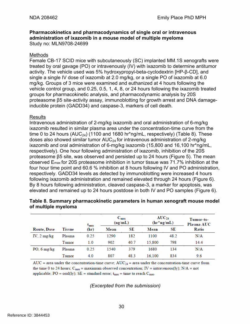
Pharmacokinetics and pharmacodynamics of single oral or intravenous administration of ixazomib in a mouse model of multiple myelomaStudy no: MLN9708-24699
MethodsFemale CB-17 SCID mice with subcutaneously (SC) implanted MM.1S xenografts were treated by oral gavage (PO) or intravenously (IV) with ixazomib to determine antitumor activity. The vehicle used was 5% hydroxypropyl-beta-cyclodextrin [HP-β-CD], and single a single IV dose of ixazomib at 2.0 mg/kg, or a single PO of ixazomib at 6.0 mg/kg. Groups of 3 mice were examined and euthanized at 4 hours following the vehicle control group, and 0.25, 0.5, 1, 4, 8, or 24 hours following the ixazomib treated groups for pharmacokinetic analysis, and pharmacodynamic analysis by 20S proteasome β5 site-activity assay, immunoblotting for growth arrest and DNA damage-inducible protein (GADD34) and caspase-3, markers of cell death.
ResultsIntravenous administration of 2-mg/kg ixazomib and oral administration of 6-mg/kg ixazomib resulted in similar plasma area under the concentration-time curve from the time 0 to 24 hours (AUC24) (1100 and 1680 hr*ng/mL, respectively) (Table 8). These doses also showed similar tumor AUC24 for intravenous administration of 2-mg/kg ixazomib and oral administration of 6-mg/kg ixazomib (15,800 and 16,100 hr*ng/mL, respectively). One hour following administration of ixazomib, inhibition of the 20S proteasome β5 site, was observed and persisted up to 24 hours (Figure 5). The mean observed Emax for 20S proteasome inhibition in tumor tissue was 71.7% inhibition at the four hour time point and 60.6 % inhibition at 8 hours following IV and PO administration, respectively. GADD34 levels as detected by immunoblotting were increased 4 hours following ixazomib administration and remained elevated through 24 hours (Figure 6). By 8 hours following administration, cleaved caspase-3, a marker for apoptosis, was elevated and remained up to 24 hours postdose in both IV and PO samples (Figure 6).
Table 8. Summary pharmacokinetic parameters in human xenograft mouse model of multiple myeloma
(Excerpted from the submission)
Reference ID: 3844453

NDA 208462 Emily Place PhD MPH
31
Figure 5. Proteasome inhibition following treatment with ixazomib (IV, PO) in xenograft mouse model of multiple myeloma
(Excerpted from the submission)
Reference ID: 3844453

NDA 208462 Emily Place PhD MPH
32
Figure 6. Immunoblotting analysis following treatment with ixazomib (IV, PO) in xeonograft mouse model of multiple myeloma
(Excerpted from the submission)
In vivo activity of ixazomib in a mouse model of multiple myelomaStudy no: MLN9708-24176
MethodsUsing a mouse model of human multiple myeloma xenograft cells (MM.1S) in female CB-17 SCID mice vehicle (5% HP-β-CD) or ixazomib at 1, 2, 4, 6, 8, or 10 mg/kg was administered orally (PO) twice weekly for 18 days; a total of 5 doses. Day 1, mean tumor volumes (MTVs) measured approximately 100 to 350 mm3. On Day 19, tumor growth inhibition (TGI) was calculated by using the percent TGI ([MTV of the control group - MTV of a treated group]*100 / [MTV of the control group]). Change in AUC was calculated by the change in the area under the tumor volume-versus-time curve, p values were calculated using a linear mixed effects regression model, with p < 0.05 considered statistically significant.
ResultsFollowing treatment with ixazomib at 6, 8, and 10 mg/kg, there was a statistically significant decrease in mean tumor volume (Y-axis, mm3) compared to vehicle control (TGI = 99.3%, 99.7%, and 100%, respectively). At 10 mg/kg, there were no measurable tumors on Day 19. At 6 and 8 mg/kg, tumor volume at Day 19 was less than that on Day 0. Changes in antitumor activity were statistically significant at both the 1 mg/kg and 4 mg/kg dose groups (TGI = 26.3% and 30.8%, respectively; change in AUC, p > 0.005 for both dosesbut not at the 2 mg/kg dose group. (TGI = 9.1%).
Reference ID: 3844453

NDA 208462 Emily Place PhD MPH
33
Figure 7. Mean tumor volume (mm3) over time following treatment with ixazomib (biweekly, PO) in a human xenograft mouse model of multiple myeloma
(Excerpted from the submission)
4.2 Secondary PharmacologyStudies not conducted.4.3 Safety PharmacologyCardiovascular (GLP): hERG patch-clamp HEK293 cells: The effect of MLN9708 (100, 33.3, 11.1, 3.70, 1.23, 0.412, 0.137, 0.0457, 0.0152, and 0.00508 μM) on in vitro hERG current was evaluated in study RPT-04111 to assess the potential for delayed repolarization and prolongation of the QT interval. HERG (human-Ether-a-go-go Related Gene) is a gene encoding the pore forming subunit of a human delayed rectifying potassium channel, and blockade of hERG current has been associated clinically with delayed repolarization and proarrythmic responses in humans.
Reference ID: 3844453

NDA 208462 Emily Place PhD MPH
34
Binding assay was performed in a 96 well plate. Competition assays were performed using the ligand 3H-dofetilide at 3.5nM (50% occupancy of receptor sites). The drug was added at a range of concentrations. Concentrations of 100, 33.3, 11.1, 3.70, 1.23, 0.412, 0.137, 0.0457, 0.0152, and 0.00508 μM MLN9708 were chosen to determine concentration response. Assays were initiated by adding membrane homogenates. Assay plates were placed on a shaking platform and incubated at room temperature for 80 minutes. The assay reaction was terminated by vacuum filtration onto polyethylenimine (PEI) treated glass fiber/barex (GF/B)-grade filters.
Results The IC50 for inhibition due to ixazomib was 59.6 μM, Ki value of 24.9 μM. The positive control, terfenadine, inhibited hERG potassium current with an IC50
value of 0.09 μM, mean Ki value of 0.04 μM.
Telemetry study in Dogs/Beagle for oral administration (GLP):The effect of MLN9708 on cardiovascular parameters were assessed in telemetered dogs (n=4 males) in study B130356. Dogs were given single oral dose of vehicle or MLN9708 at 0.03, 0.2, and 0.3 mg/kg (0.021, 0.14, and 0.21 mg/kg as MLN2238; the biologically active form of MLN9708). The vehicle was 0.5 w/v% methylcellulose solution. Cardiovascular parameters included arterial blood pressures, pulse pressure, heart rate and electrocardiogram measurements 30 minutes prior to dosing and following dosing at 30 minutes 1, 2, 4, 8, and 24 hours. Parameters at different timepoints were compared to the animal’s own baseline values.
Results There were no significant clinical signs. No affects were observed on any cardiovascular parameters.
5 Pharmacokinetics/ADME/Toxicokinetics5.1 PK/ADMEToxicokinetic assessments were included in the toxicology reports and are discussed in the toxicology section of this review.
Reference ID: 3844453

NDA 208462 Emily Place PhD MPH
35
Table 9. Summary pharmacokinetics in nonclinical testing species following single dose oral administration of ixazomib or ixazomib citrate
(Excerpted from the submission)
AbsorptionPharmacokinetics of MLN2238 after oral administration of MLN9708 capsules to non-naive male beagle dogsKey study findings
Absorption of MLN2238 was moderate following oral dosing (tmax 0.5 h). The mean plasma Cmax value was 279 ng/mL for MLN2238 following oral dosing. The mean plasma AUC(0-24h) value for MLN2238 was 973 hr*ng/mL following oral
dosing.
Study no.: RPT-01218Study report location: eCTD 4.2.2.2Conducting laboratory and location:
Millenium Pharmaceuticals Inc,Cambridge, MA, USA
Date of study initiation: April 25, 2008GLP compliance: NoQA statement: NoDrug, Batch #, and % purity: MLN9708, Batch 8620-02, 99%
Methods Liquid chromatography with tandem mass spectrophotometry (LC/MS/MS)
Species/strain: Beagle dogsN: 4 malesDose: 1.48 mg Frequency: single doseRoute: oral capsule (powder-in-capsule formulation)
Reference ID: 3844453

NDA 208462 Emily Place PhD MPH
36
Volume: capsule given with 10 mL distilled water; 5 mL distilled water flush
Observations and times: Samples collected from a peripheral vessel at 0, 15, 30 minutes and 1, 2, 4, 6, 8, and 24 hr post dose. The lower limit of quantitation (LLOQ) was 1.00 ng/mL.
Results
Table 10. Pharmacokinetic parameters of MLN2238 following oral dosing in dogs
(Excerpted from the submission)
Table 11. Mean Plasma concentrations of MLN2238 in beagle dogs following a single oral dose
(Excerpted from the submission)
Reference ID: 3844453

NDA 208462 Emily Place PhD MPH
37
Distribution Tissue Distribution via Quantitative Whole-Body Autoradiography in Male Long-Evans Rats Following a Single Oral Gavage Administration of [14C] MLN9708 andHuman Radiation Dosimetry Estimation
Key study findings Tissue half-life for radioactivity was longest in the urinary bladder (t1/2= 946 h)
and sciatic nerve (887 h). In addition, the highest Cmax of radioactivity was observed in the urinary bladder.
In addition to these organs, exposure based on Cmax and/ or AUC was high in lymph node, spleen, adrenal glands, kidney (renal cortex and medulla), bile colon, small intestine, cecum and liver, consistent with renal and hepatobiliary routes of excretion.
Relatively low amount of radioactivity in the brain indicates that drug-related materials don’t likely cross the blood-brain barrier.
Study no.: 96N-1206Study report location: eCTD 4.2.2.2Conducting laboratory and location:
Millenium Pharmaceuticals Inc,Cambridge, MA, USA
Date of study initiation: April 1, 2012GLP compliance: NoQA statement: NoDrug, lot #, and % purity: [14C] MLN9708, Lot # MP-E2763-1, > 95%;
MLN9708, Lot # 100895, 99.7%
Methods Tissue levels of [14C] radioactivity detected by whole body radiography ranged from 0.003 to 4.412 µg equiv/g. [14C] MLN9708 and MLN9708 were formulated in 55mM citrate, 1% propylene glycol, pH 5.2. The concentration of radioactivity of [14C] MLN9708 in formulation was 13.11 μCi/g and the specific activity of the [14C] MLN9708 in formulation was 112.25 μCi/mg.
Species/strain: Long Evans (n=9) pigmented ratsN: 9 male Dose: 0.6 mg/kg (approximately 65 μCi/kg)Frequency: single doseRoute: oral gavageVolume: 5 ml/kg Observations and times: 0.5, 1, 4, 12, 24, 72, 168, 336 and 672h post-dose
Reference ID: 3844453


NDA 208462 Emily Place PhD MPH
41
Table 16. Sample collection in metabolism, mass balance and excretion study in rats following single oral administration
(Excerpted from the submission)
Results
Table 17. Mean percent excretion of radioactivity in urine and feces in intact rats following a single oral dose of [14C]MLN9708
(Excerpted from the submission)
Reference ID: 3844453

NDA 208462 Emily Place PhD MPH
42
Table 18. Mean percent excretion of radioactivity in urine and feces in bile duct cannulated rats following a single oral dose of [14C] MLN9708
6 General Toxicology6.1 Single-Dose ToxicityStudies not reviewed.6.2 Repeat-Dose ToxicityStudy title: A 6-month Oral Gavage Toxicity Study of MLN9708 with a 2-Week Recovery Period in Sprague Dawley RatsThe following study was initially reviewed under IND by Matt Whittaker PhD and parts of it were reformatted to fit this NDA review.
Study no.: 416165Study report location: 4.2.3.2
Conducting laboratory and location:Date of study initiation: September 3, 2013
GLP compliance: Signed and IncludedQA statement: Signed and Included
Drug, lot #, and % purity: MLN9708, Lot # 101296; 99.1%
Key Study Findings There were 5 early mortalities in the high dose group (0.6/0.8 mg/kg) and 2
mortalities in the mid dose group (0.4 mg/kg). The cause of death in the high dose group was attributed to toxicities in the intestine, liver, and/or lymphoid depletion. Mortalities at 0.4 mg/kg were attributed to renal failure (non-test article related).
Clinical signs include decreased defecation, soft feces, small feces, yellow material around the urogenital and anogenital areas and forelimbs, hypoactivity, thin appearance, diarrhea, and clear discharge from the eyes.
Reference ID: 3844453
(b) (4)
(b) (4)
(b) (4)


NDA 208462 Emily Place PhD MPH
44
Mortality
Table 20. Cause of mortality in 6 month repeat dose toxicology study in rat
Reference ID: 3844453


NDA 208462 Emily Place PhD MPH
46
Research, studies conducted between 2007 - 2014) are summarized in the following table:
Table 21. Supplemental information: summary of historical control data pertaining to calculi formation in kidney and urinary bladder in male
(Excerpted from the submission)
It is noted that while there was evidence for multiple sporadic kidney findings of minimalseverity (e.g. tubular basophilia, mononuclear cell infiltrate, tubular mineralization) in animals in all dose groups, there were no dose-related renal pathologies observed in this study. Therefore, it appears that the kidney is not a target organ of toxicity for oral ixazomib in rats.Clinical SignsAnimals in the 0.8/0.6 mg/kg exhibited decreased defecation, soft feces, small feces (females only), yellow material around the urogenital and anogenital areas and forelimbs, hypoactivity, thin appearance, diarrhea, and clear discharge from the eyes. Body WeightsStatistically significant changes in body weight were observed in the 0.8/0.6 mg/kg dose group at 25 weeks. At Week 26, males and females had 11.1% and 10.1% lower mean body weight relative to controls, respectively.
Reference ID: 3844453

NDA 208462 Emily Place PhD MPH
47
Figure 8. Body weights in male rats in 6 month repeat dose toxicology study
(Excerpted from the submission)
Figure 9. Body weights in female rats in 6 month repeat dose toxicology study
(Excerpted from the submission)
Reference ID: 3844453

NDA 208462 Emily Place PhD MPH
48
Feed ConsumptionFeed consumption was reduced in animals in the 0.8/0.6 mg/kg group males and females, beginning during Week 4 to 5 and persisted throughout the study. OphthalmoscopyVisual examinations were not performed. HematologyHematology and coagulation were collected on all animals at the scheduled necropsies (Weeks 26 and 28, main study and recovery period respectively).
Table 22. Hematology findings for 6 month repeat dose toxicity study in rats (percent change from control)
(Excerpted from the submission)WBC: white blood cells; NEU: neutrophils; LYMPH: lymphocytes; MONO: monocytes; RDW: red cell distribution width; APTT: activated partial thromboplastin time* Significantly different from the control group at 0.05** Significantly different from the control group at 0.01Clinical ChemistryClinical chemistry parameters were collected on all animals at the scheduled necropsies (Weeks 26 and 28, main study and recovery period respectively). All clinical chemistry parameters were unremarkable.
Reference ID: 3844453

NDA 208462 Emily Place PhD MPH
49
UrinalysisUrinalysis was not conductedGross PathologyUnremarkableOrgan WeightsUnremarkableHistopathologyAdequate Battery: Yes
Peer Review: Yes
Histological Findings
Table 23. Histological findings from 6 month repeat dose toxicology study in rats
Reference ID: 3844453

NDA 208462 Emily Place PhD MPH
50
Reference ID: 3844453

NDA 208462 Emily Place PhD MPH
51
Reference ID: 3844453

NDA 208462 Emily Place PhD MPH
52
ToxicokineticsBlood samples (~0.5 ml) for toxicokinetic evaluation were taken from 3 animals per sexper group at each time point. Blood was processed to plasma. 250 μl of each plasma sample was mixed with 15 μl of 30% ascorbic acid and stored frozen at -65 to -85°C. Frozen samples were shipped to for analysis. MLN9708 is a prodrug for the active MLN2238. Toxicokinetic values reflect measurement of MLN2238. MLN238 concentration was determined using a validated HPLC/MS/MS method and toxicokinetic analysis was conducted using Phoenix™ WinNonlin®, Version 6.2.
Reference ID: 3844453
(b) (4)

NDA 208462 Emily Place PhD MPH
53
MLN9708 was rapidly absorbed, with Tmax values generally between 0.5 – 1 h post-dose at all time points. MLN9708 exposure increased approximately proportionally with dose in males and females after both the first dose of cycle 1 and cycle 7 (Figure 10, Table 24). Evidence for modest accumulation was observed in LD males and females only (accumulation factor: 1.3 – 1.9) at Day 168. Mean Cmax values were ~3 –fold higher in HD females vs. HD males after the initial MLN9708 dose. AUC0-168 values were 1.4-fold higher in females at this time point. Mean Cmax values were also increased in females vs. males at the Day 14 and Day 42 time points (data not shown). The Applicant’s notion that the 4 premature deaths observed in HD females are related to elevated Cmax appears to be reasonable. No clear gender differences were observed in Cmax or AUC0-168 at Day 168.
Figure 10.Plasma MLN2238 concentration vs. time curves after MLN9708 administration to SD rats
(Excerpted from the submission)
Table 24. Summary toxicokinetics for 6 month repeat dose toxicology study in rat
(Excerpted from the submission)
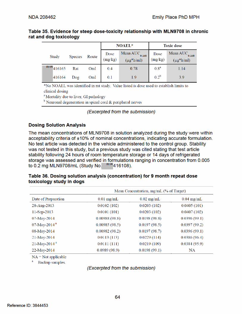
Dosing Solution AnalysisThe mean concentrations of MLN9708 in solution analyzed during the study were within acceptability criteria of ±10% of nominal concentrations, indicating accurate formulation. No test article was detected in the vehicle administered to the control group. Stability was not tested in this study, but a previous study was cited stating that test article
Reference ID: 3844453

NDA 208462 Emily Place PhD MPH
54
stability following 24 hours of room temperature storage or 14 days of refrigerated storage was assessed and verified in formulations ranging in concentration from 0.005 to 0.2 mg MLN9708/mL (Study No. -416108).
Table 25. Dosing solution analysis (concentration) for 6 month repeat dose toxicology study in rats
(Excerpted from the submission)
Reference ID: 3844453
(b) (4)

NDA 208462 Emily Place PhD MPH
55
Study title: A 9-month oral gavage toxicity study of MLN9708 with a 2-week recovery period in Beagle dogsThe following study was initially reviewed under IND by Matt Whittaker PhD and parts of it were reformatted to fit this NDA review
Study no.: 416164Study report location: 4.2.3.2
Conducting laboratory and location:Date of study initiation: August 29, 2013
GLP compliance: Signed and IncludedQA statement: Signed and Included
Drug, lot #, and % purity: MLN9708; Lot #101296; 69.3% purity
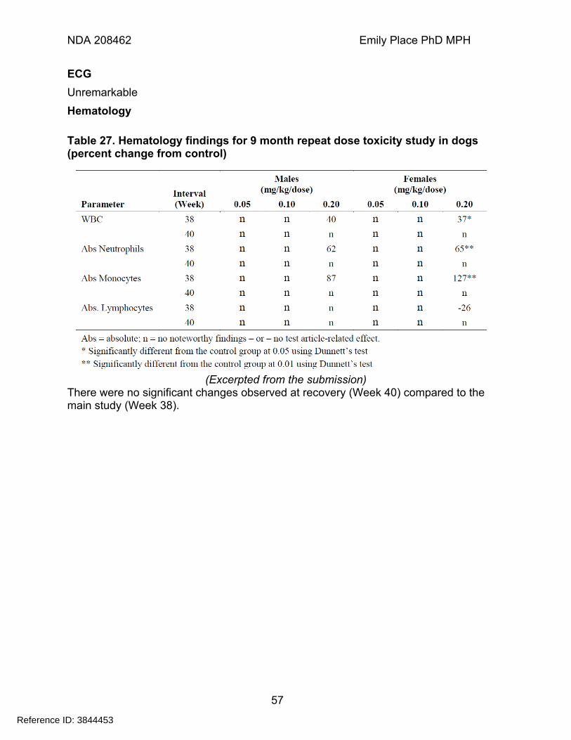
Key Study Findings There were no mortalities during the study. Toxicologically significant ixazomib hematology findings included higher mean
values of absolute counts for total white blood cells, neutrophils, and monocytes and lower mean counts for lymphocytes at 0.2 mg/kg dose group.
Toxicologically significant ixazomib related changes in clinical chemistry parameters included elevated AST levels and lower phosphorous levels at the 0.20 mg/kg/dose group.
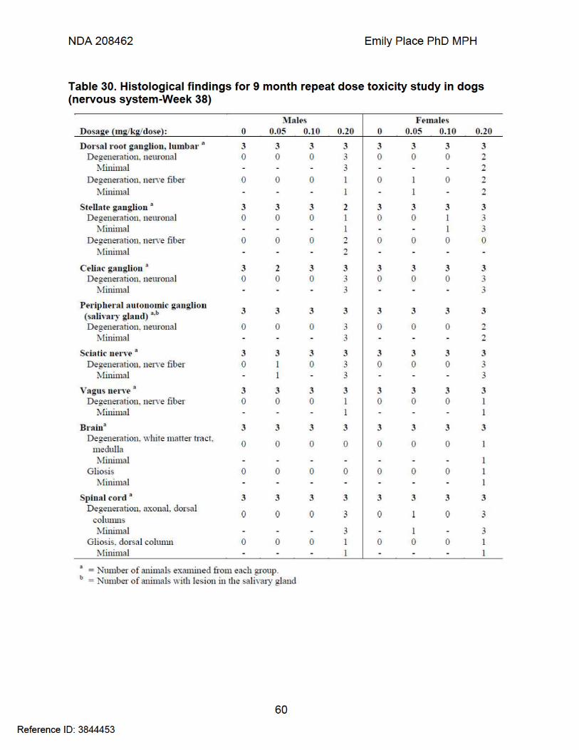
Histopathology findings included neutrophil infiltration in the gastrointestinal tract (stomach, intestines, peyers patch) and mesenteric lymph nodes and erosion in the stomach at 0.2 mg/kg dose group. Findings in the nervous system primarily at 0.2 mg/kg dose group included neuronal degeneration of the sympathetic (celiac and stellate), dorsal root, and end organ (salivary gland) ganglia, and minimal secondary axonal/nerve fiber degeneration of the peripheral nerves (vagus and sciatic nerves, dorsal roots and mixed spinal nerves) and ascending tracts in the dorsal columns of the spinal cord, and in white matter tracts in the medulla oblongata of the brain, gliosis of the dorsal column of the spinal cord and the white matter tracts in the brain.
Histopathology findings still present at recovery were limited to the nervous system and included nerve fiber degeneration of the dorsal root ganglia and an increase in axonal degeneration in the dorsal columns of the spinal cord.
Reference ID: 3844453
(b) (4)
(b) (4)
(b) (4)

NDA 208462 Emily Place PhD MPH
56
MethodsDoses: 0.05, 0.1, 0.2 mg/kg
Frequency of dosing: Once weekly for 3 weeks with each cycle separated by a 13-day nondosing period. 10 x 28 day cycles.
Route of administration: Oral gavageDose volume: 5 mL/kg
Formulation/Vehicle: 55 mM citrate buffer and 0.45% NaCl, pH 5.8 Species/Strain: Canis lupus familiaris, Beagle dogs
Number/Sex/Group: 3/sex/group, 3/sex per recovery excluding 0.05 dose group
Age: 12-14 monthsWeight: 9.1 kg to 12.5 kg (males) or 7.1 kg to 8.9 kg
(females)Satellite groups: N/A
Unique study design: N/ADeviation from study protocol: None impacted study results or objectives
Table 26. Study design for 9 month repeat dose toxicology study in dogs
(Excerpted from the submission)
Observations and Results
MortalityThere were no mortalities during the study. Clinical SignsUnremarkable. Body WeightsUnremarkableFeed ConsumptionUnremarkable. OphthalmoscopyVisual examinations were not performed.
Reference ID: 3844453
NDA 208462 Emily Place PhD MPH
57
ECGUnremarkableHematology
Table 27. Hematology findings for 9 month repeat dose toxicity study in dogs (percent change from control)
(Excerpted from the submission)There were no significant changes observed at recovery (Week 40) compared to the main study (Week 38).
Reference ID: 3844453

NDA 208462 Emily Place PhD MPH
58
Clinical Chemistry
Table 28. Clinical chemistry findings for 9 month repeat dose toxicity study in dogs (percent change from control)
(Excerpted from the submission)
There were no significant changes observed at recovery (Week 40) compared to the main study (Week 38).UrinalysisUrinalysis was not conductedGross PathologyDark red areas in the stomach of animal 4156 (0.20 mg/kg/dose group female). In animal 4156 dark red areas correlated microscopically with neutrophilic infiltrates in the stomach. Three high dose male animals (0.2 mg/kg) had reported dark red discoloration of the colon without microscopic correlates. Organ WeightsUnremarkableHistopathologyAdequate Battery: Yes
Peer Review: Yes
Reference ID: 3844453

NDA 208462 Emily Place PhD MPH
59
Histological Findings
Table 29. Histological findings for 9 month repeat dose toxicity study in dogs Histopathology findings in lymphoid and GI after 10 cycles
Reference ID: 3844453

NDA 208462 Emily Place PhD MPH
61
(Excerpted from the submission)
Table 31.Histological findings for 9 month repeat dose toxicity study in dogs (nervous system-recovery Week 40)
(Excerpted from the submission)
Special EvaluationFunctional observation battery- nervous system
Table 32. Time points of conduct of FOB assessments in 10 cycle study with MLN9708 in beagle dogs.
(Excerpted from the submission)
Motor, sensory, and autonomic pathways were examined to assess neurological function. The parameters evaluated are summarized below. Each parameter was evaluated according to an established scoring system. The conduct of the FOB was consistent with the proposal submitted by the Applicant on 2/11/14.
Reference ID: 3844453

NDA 208462 Emily Place PhD MPH
62
Table 33. Parameters evaluated in FOB assessments in beagle dogs.
(Excerpted from the submission)
(Excerpted from the submission)
There was no evidence for treatment related effects on any parameter. A diminished orabsent perineal reflex was observed in a small number (1-2) of both control and high dose male and female dogs animals throughout the study period. All other open field and functional observations were scored as present or normal for males and females of all dose groups.
Reference ID: 3844453

NDA 208462 Emily Place PhD MPH
63
There was no evidence for MLN9708-evoked functional deficits in any dogs in the study.ToxicokineticsOn Days 0, 14 and 252, whole blood samples from all animals were collected predose and at approximately 0.25, 0.5, 1, 4, 8, 24, 72, 120, and 168 hours postdose. Samples were also collected at the conclusion of the recovery period on Day 266 at approximately 0.25, 0.5, 1, 4, 8, and 24 hours postdose. MLN9708 is a prodrug for the active MLN2238. Toxicokinetic values reflect measurement of MLN2238. Plasma MLN2238 concentrations were determined using a validated HPLC/MS/MS method. The lower limit of quantitation was 2.0 ng/ml. Toxicokinetic analysis was conducted using Phoenix™ WinNonlin®, Version 6.2. There were no differences between males and females with respect to Cmax or AUC0-168.Mean plasma Tmax values were less than 1 hour throughout the duration of the study.Mean plasma Cmax and AUC0-168 increased in an approximately dose-proportional manner at all time points (Table 34). There was no evidence for significant accumulation over the course of the 9 month study at any dose level.
Table 34. Summary toxicokinetics for 9 month repeat dose toxicology study in dogs
(Excerpted from the submission)
Safety concerns stem from the steep dose-toxicity relationship with MLN9708in the chronic rat and dog toxicology studies. There was a less than 1.5-fold separation in systemic exposure between the limit dose and a dose associated with mortality due to drug-induced injury of the liver and GI tract in the rat (Table 35). In dogs, there was an approximately 2-fold separation in exposure between the NOAEL and doses associated with extensive neuronal pathology in the CNS and PNS.
Reference ID: 3844453

NDA 208462 Emily Place PhD MPH
65
7 Genetic Toxicology7.1 In vitro Reverse Mutation Assay in Bacterial Cells (Ames)Study title: Salmonella E.coli/Mammalian Microsome Reverse Mutation Assay
Study no.: 08-152Study report location: 4.2.3.3.
Conducting laboratory and location:Date of study initiation: April 2, 2008
GLP compliance: Signed and IncludedQA statement: Signed and Included
Drug, lot #, and % purity: MLN-2238, Lot# 001-D, purity 98.24%
Key Study Findings MLN-2238 was negative in the in tester strains of Salmonella or E.
coli in the presence and absence of S-9 mix up to 5000 µg/plate, under the conditions tested. Methods
Strains: Salmonella typhimurium :TA98, TA100, TA1535 and TA1537Escherichia coli: WP2 uvrA
Concentrations in definitive study: 100, 250, 500, 1000, 2500, and 5000 μg/plate with TA98; 50, 100, 250, 500, 1000 and 2500 μg/plate WP2 uvrA; 25, 50, 100, 250, 500 and 1000 with TA1537 and TA1535, and at 10, 25, 50, 100, 250 and 500 μg/plate with TA100.
Basis of concentration selection: Cytotoxicity observed in the initial assay or up to the limit dose (5000 µg/plate) recommended by regulatory guidelines.
Negative control: Dimethyl sulfoxide (DMSO)Positive control: With S9: 2-Aminoanthracene,
Without S9: Sodium Azide, ICR-191 acridine, 2-Nitrofluorene, 4-Nitroquinoline-N-oxide
Formulation/Vehicle: DMSOIncubation & sampling time: Plate incorporation: 48 hours at 37±1°C
Analysis After incubation, colonies were counted electronically for bacterial colony formation using a ProtoCol automatic colony counter, Model number 60000, software version 2.05. Cytotoxicity was assessed by examining bacterial lawn density and number of spontaneous revertants per plate.
Reference ID: 3844453
(b) (4)
(b) (4)

NDA 208462 Emily Place PhD MPH
66
Criteria for Positive Response:
Results were considered positive if the data for any treatment level showed a dose dependent a response with at least 2 times the concurrent vehicle control for TA100 and 3 times the concurrent vehicle control for TA1535, TA1537, TA98 and WP2 uvrA.Study Validity
The vehicle and the positive control values (± S9 mix) for each tester strain were within the laboratory historical ranges.
Vehicle control plates displayed normal growth (i.e., normal background lawn) in the ± S9 mix.
Selection of the tester strains was adequate based upon Guideline for Industry: Specific Aspects of Regulatory Genotoxicity Tests for Pharmaceuticals (ICHS2A, April 1996).
The highest concentration tested was 5000 μg/plate, which allowed maximum exposure.
The appropriate positive control compounds (± S9 mix) produced clear, unequivocal increases in the number of revertant colonies.
Tester strains demonstrated their appropriate genetic markers for strain integrity.Results
MLN 9708 was cytotoxic (i.e., decrease in spontaneous revertants and/or background lawn) to all strains tested at different concentrations. MLN 9708 concentrations ≤ 2500 μg/plate showed no signs of bacterial mutagenicity. As shown in the table below, MLN 9708 was not mutagenic in tester strains of Salmonella or E. coli in the presence or absence of S-9 mix, under the conditions tested.
Reference ID: 3844453

NDA 208462 Emily Place PhD MPH
67
Table 37. Summary results of initial assay for MLN2238 in the Salmonella/E.Coli mammalian microsome reverse mutation assay
(Excerpted from the submission)
Reference ID: 3844453

NDA 208462 Emily Place PhD MPH
68
Table 38. Summary results of confirmatory assay for MLN2238 in the Salmonella/E. coli mammalian microsome reverse mutation assay
(Excerpted from the submission)
Reference ID: 3844453

NDA 208462 Emily Place PhD MPH
69
7.2 In Vitro Assays in Mammalian CellsStudy title: In vitro Chromosome Aberration Test in Cultured Human Peripheral Blood Lymphocytes
Study no.: 12-367Study report location: 4.2.3.3.1
Conducting laboratory and location:Date of study initiation: February 12, 2013
GLP compliance: Signed and IncludedQA statement: Signed and Included
Drug, lot #, and % purity: MLN-9708; lot # 101296; purity 99.1%
Key Study Findings MLN9708 was positive for inducing structural aberrations in HPBL with and
without metabolic activation, under the conditions tested.
MethodsCell line: Human peripheral blood lymphocytes
(HPBL) Concentrations in definitive study: Non-activated treatment (-S9):
3 hour s: 0.010, 0.030, 0.050, 0.10, 0.25, 0.50, 1.0, 2.0, 3.0, 4.0, 5.0, 6.0, 7.0, 8.0, 9.0, 10, 12, 15, and 20 µg/mL22 hours: 0.010, 0.020, 0.030, 0.040, 0.050, 0.055, 0.060, 0.065, 0.070, 0.075,0.080, 0.085, 0.090, 0.095, 0.10, 0.20, 0.30, 0.40, and 0.50 μg/mLS9-activated treatment (+S9):3 hours: 0.10, 0.30, 0.50, 1.0, 2.0, 3.0, 4.0, 5.0, 6.0, 7.0, 8.0, 9.0, 10, 12, 15, 20, 25, 30, 35, 40, 45, and 50 µg/mL
Basis of concentration selection: Cytotoxicity tests were performed to select doses for the definitive assay. The concentrations tested ranging from 0.00305 to 50 μg/mL. Cytotoxicity was observed at MLN-9708 at concentrations of 0.0977 (3 hour and 22 hour) and 6.25 μg/mL with or without S9 mix, respectively;
Negative control: Sterile distilled waterPositive control: Mitomycin C for –S9 3 hour and 22 hour
treatmentCyclophosphamide for +S9 3 hour treatment
Formulation/Vehicle: Sterile distilled waterIncubation & sampling time: 3 hour treatment with 19 hour recovery for
-S9/+S9
Reference ID: 3844453
(b) (4)
(b) (4)

NDA 208462 Emily Place PhD MPH
70
22 hour treatment with no recovery for –S9
Table 39. Concentrations of MLN-9708 evaluated for chromosomal aberrations assay in HPBL
(Excerpted from the submission)
AnalysisApproximately 2 hours prior to harvest, Colcemid® (0.1 μg/mL, final concentration in culture) was added to each culture to arrest cells in metaphase. Cells were treated with hypotonic solutions and fixed with 3:1 methanol: glacial acetic acid. Slides were prepared, and stained analysis. CytotoxicityThe number of mitotic cells in at least 500 cells from each culture was enumerated.Chromosome aberrations Coded slides were analyzed for structural chromosomal aberrations from a total of 200 metaphase cells and from a total of 400 cells for numerical aberrations from each concentration.Study ValidityThe study was considered valid based on meeting the following criteria for a valid test set in the protocol.
The frequency of cells with structural chromosome aberrations in the vehicle control groups were within the historical range for vehicle controls.
The percentage of cells with chromosome aberrations in each positive control group were statistically increased (p ≤ 0.05, Fisher's Exact test), relative to the appropriate vehicle controls.
Criteria for Positive Response:
The test article would be considered positive for inducing chromosomal aberrations if a significant increase (p ≤ 0.01) in the number of cells with chromosomal aberrations is observed at one or more dose levels, or if a significant increase (p ≤ 0.05) in the number of cells with chromosomal aberrations is observed at two or more dose levels.
Reference ID: 3844453
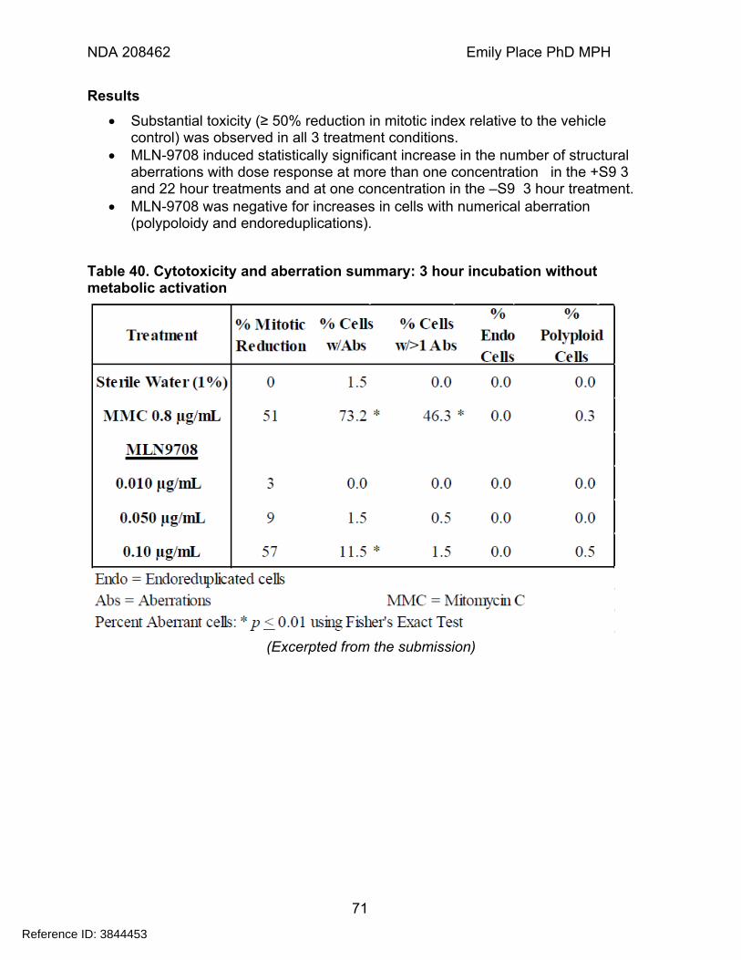
NDA 208462 Emily Place PhD MPH
71
Results Substantial toxicity (≥ 50% reduction in mitotic index relative to the vehicle
control) was observed in all 3 treatment conditions. MLN-9708 induced statistically significant increase in the number of structural
aberrations with dose response at more than one concentration in the +S9 3 and 22 hour treatments and at one concentration in the –S9 3 hour treatment.
MLN-9708 was negative for increases in cells with numerical aberration (polypoloidy and endoreduplications).
Table 40. Cytotoxicity and aberration summary: 3 hour incubation without metabolic activation
(Excerpted from the submission)
Reference ID: 3844453

NDA 208462 Emily Place PhD MPH
72
Table 41. Cytotoxicity and aberration summary: 22 hour incubation without metabolic activation
(Excerpted from the submission)
Reference ID: 3844453

NDA 208462 Emily Place PhD MPH
73
Table 42. Cytotoxicity and aberration summary: 3 hour incubation with metabolic activation
(Excerpted from the submission)
7.3 In Vivo Clastogenicity Assay in Rodent (Micronucleus Assay)Study title: MLN9708: A Single-Dose Oral (Gavage) Bone Marrow Micronucleus Evaluation in CD-1 Mice
Study no: -416152Study report location: 4.2.3.3.2
Conducting laboratory and location:Date of study initiation: October 25, 2012
GLP compliance: Signed and IncludedQA statement: Signed and Included
Drug, lot #, and % purity: MLN-9708; Lot # 21E004; purity 99.0%
Key Study Findings• MLN-9708 was negative in the micronucleus test in the bone marrow cells of male mice.
Reference ID: 3844453
(b) (4)
(b) (4)

NDA 208462 Emily Place PhD MPH
74
MethodsDoses in definitive study: 2, 4, and 8 mg/kg
Frequency of dosing: Single doseRoute of administration: Oral gavage
Dose volume: 5 mL/kg; cyclophosphamide at 10 mL/kgFormulation/Vehicle: Sterile water for injection, diluted with 55 mM
citrate buffer and 3% (w:w) glycine, pH 5.8Species/Strain: CD-1 mice
Number/Sex/Group: 6 male mice/group at 2 and 4 mg/kg, and 14 male mice at 8 mg/kg
Satellite groups: 2 preliminary dose finding studies; groups of 3 mice/sex/dose
Basis of dose selection: Dose selection for the main micronucleus assaywas based on the range finding study.
Negative control: Vehicle: sterile water for injection, diluted with 55 mM citrate buffer and 3% (w:w) glycine, pH 5.8
Positive control: Cyclophosphamide monohydrate (CPS) 60 mg/kg administered once on Day 0
Table 43. Study design for in vivo clastogenic micronucleus assay in mice
(Excerpted from the submission)
Analysis For the range finding study, groups of 3 mice/sex/dose were administered
MLN9708 by oral gavage, single dose at 10, 20, and 30 mg/kg. All 3 doses caused severe toxicities and mortality. A second range-finding study was conducted at doses of 5, 6, and 8 mg/kg with 3 male mice/group. The majority of animals tolerated 8 mg/kg in the second range finding study (1/3 mortalities at 8 mg/kg).
Reference ID: 3844453

NDA 208462 Emily Place PhD MPH
75
All mice were sacrificed 24 hours after receiving their final dose and femoral bone marrow smears prepared. Smears were stained with acridine orange prior to analysis with fluorescence microscopy to determine the proportion of polychromatic erythrocytes in the total erythrocyte count (% PCE) and the number of micronucleated polychromatic erythrocytes (MPCE) per 2000 PCE analysed.
Criteria for Positive ResponseThe test article may be considered positive for induction of micronuclei if a dose-related increase in the percentage of PCEs with micronuclei is observed and a statistically significant increase relative to the vehicle is seen at one or more dose levels.Study ValidityThe vehicle group mean must lie within the historical control. The positive control response must be higher than the vehicle group and be consistent with historical positive control data.
The toxicokinetic assessments (study number 13-039/tk) in a single dose study of MLN9708 for injection at 2, and 4 mg/kg in mice demonstrated systemic exposure, dosing appeared to be adequate. The Cmax and AUC0-24h values of MLN2238 increased dose-dependently when MLN9708 was administered orally to mice at 2 and 4 mg/kg as MLN2238. In the 8 mg/kg group, a decrease in locomotor activity and hypothermia were observed in all 5 animals, and all animals in this group were moribund and euthanized at 6 hours after dosing. The Cmax and AUC0-24h values were not calculated from 8 mg/kg group.
Results One male in the 8 mg/kg dose group was found dead on Day 2. Clinical signs
included the body was cool to touch on Day 1; no significant macroscopic changes were observed. A maximum tolerated doses (MTD) was achieved (6 mg/kg) based clinical signs of toxicity and death.
MLN-9708 did not produce an increase in the percent mean number of micronucleated polychromatic erythrocytes (%MN-PCEs) compared to the vehicle control group.
Bone marrow cytotoxicity (decreases in the ratio of polychromatic to total erythrocytes, PCE:TE ratio) was not observed any of the treatment groups.
Reference ID: 3844453

NDA 208462 Emily Place PhD MPH
76
Table 44. Summary evaluation of definitive phase bone marrow micronucleus assay in mice
(Excerpted from the submission)
Reference ID: 3844453
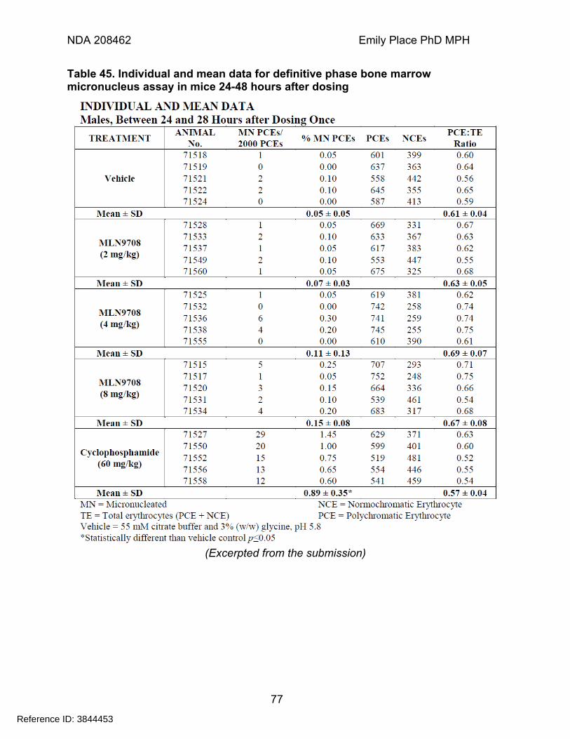
NDA 208462 Emily Place PhD MPH
77
Table 45. Individual and mean data for definitive phase bone marrow micronucleus assay in mice 24-48 hours after dosing
(Excerpted from the submission)
Reference ID: 3844453

NDA 208462 Emily Place PhD MPH
78
Table 46. Individual and mean data for definitive phase bone marrow micronucleus assay in mice 44-48 hours after dosing
7.4 Other Genetic Toxicity StudiesStudy title: Comet assay of MLN-9708 for injection in miceStudy no: B130357Study report location: 4.2.3.3.2Conducting laboratory: Takeda Pharmaceutical Company Limited.
Sunayama, JapanDate of study initiation: July 10, 2013GLP compliance: Signed and IncludedQA statement: Signed and IncludedDrug, lot #, and % purity: MLN-9708, Lot #21E004, 99.0% pure
Key Study Findings: MNL9708 was negative in the Comet assay in the liver and glandular stomach, under
the conditions tested.
Reference ID: 3844453
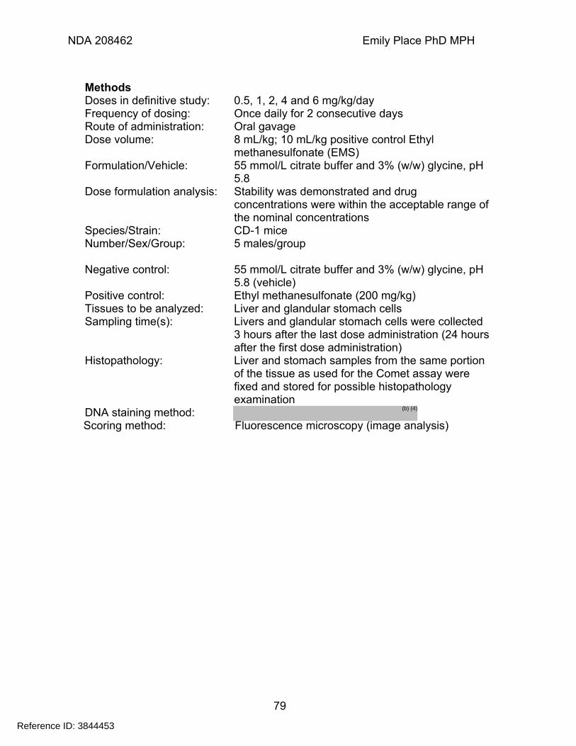
NDA 208462 Emily Place PhD MPH
79
MethodsDoses in definitive study: 0.5, 1, 2, 4 and 6 mg/kg/dayFrequency of dosing: Once daily for 2 consecutive daysRoute of administration: Oral gavageDose volume: 8 mL/kg; 10 mL/kg positive control Ethyl
methanesulfonate (EMS)Formulation/Vehicle: 55 mmol/L citrate buffer and 3% (w/w) glycine, pH
5.8Dose formulation analysis: Stability was demonstrated and drug
concentrations were within the acceptable range of the nominal concentrations
Species/Strain: CD-1 miceNumber/Sex/Group: 5 males/group
Negative control: 55 mmol/L citrate buffer and 3% (w/w) glycine, pH 5.8 (vehicle)
Positive control: Ethyl methanesulfonate (200 mg/kg)Tissues to be analyzed: Liver and glandular stomach cellsSampling time(s): Livers and glandular stomach cells were collected
3 hours after the last dose administration (24 hours after the first dose administration)
Histopathology: Liver and stomach samples from the same portion of the tissue as used for the Comet assay were fixed and stored for possible histopathology examination
DNA staining method: Scoring method: Fluorescence microscopy (image analysis)
Reference ID: 3844453
(b) (4)

NDA 208462 Emily Place PhD MPH
80
Table 47. Study design for comet assay of MLN-9708 in mice
(Excerpted from the submission)AnalysisEach animal was anesthetized and euthanized to remove liver and stomach. Part of liver and stomach was used to prepare single cell suspensions to comet assay and the remaining parts of the tissues were processed for histopathological examinations. The slides specimens were processed for cell lysing, unwinding of DNA, electrophoresis for 20 minutes and neutralization. The slides were stained with staining solution
and covered with a cover glass.
Comets of the cell nuclei were measured under a fluorescent microscope. For each animal, 100 cellular nuclei (50 cellular nuclei/slide) were analyzed for % tail DNA. Heavily damaged cells, commonly referred to as “hedgehogs” and consisting of small or non-existent head and largely diffused tails, were excluded from the data collection.Study ValidityIndividual values in the negative control group ranged within the historical control data (mean ± 3SD) at the test facility.The mean value obtained from the positive control group was significantly higher than that from the negative control group statistically.
Reference ID: 3844453
(b) (4)

NDA 208462 Emily Place PhD MPH
81
Criteria for Positive Response
The comet assay was judged positive if the test substance caused a significant increase in the % tail DNA not caused by the cytotoxic effects.ResultsNo statistical significances were observed in the mean % tail DNA between the test substance groups and the negative control group for both liver and the glandular stomach. No abnormal necropsy findings were observed in any treated groups in the liver and the glandular stomach.
Reference ID: 3844453

NDA 208462 Emily Place PhD MPH
82
Table 48. Summary results from comet assay in liver tissue from MLN-9708 testing in mice
(Excerpted from the submission)
Reference ID: 3844453

NDA 208462 Emily Place PhD MPH
83
Table 49. Summary results from comet assay in stomach tissue from MLN-9708 testing in mice
(Excerpted from the submission)
Reference ID: 3844453
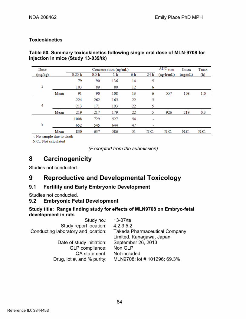
NDA 208462 Emily Place PhD MPH
84
Toxicokinetics
Table 50. Summary toxicokinetics following single oral dose of MLN-9708 for injection in mice (Study 13-039/tk)
(Excerpted from the submission)
8 CarcinogenicityStudies not conducted.
9 Reproductive and Developmental Toxicology9.1 Fertility and Early Embryonic DevelopmentStudies not conducted.9.2 Embryonic Fetal DevelopmentStudy title: Range finding study for effects of MLN9708 on Embryo-fetal development in rats
Study no.: 13-07/teStudy report location: 4.2.3.5.2
Conducting laboratory and location: Takeda Pharmaceutical Company Limited, Kanagawa, Japan
Date of study initiation: September 26, 2013GLP compliance: Non GLP
QA statement: Not includedDrug, lot #, and % purity: MLN9708; lot # 101296; 69.3%
Reference ID: 3844453

NDA 208462 Emily Place PhD MPH
85
Key Study Findings Three maternal mortalities observed at 0.8 mg/kg/3 days (3/6 in this dose group).
All other dams survived the duration of the study cycle. Fetal malformations at 0.6 mg/kg/3 days include increased percentage of
biparate ossification of the thoracic centrum, and shortened 13th rib. Upon daily administration at 0.3 mg/kg/day fetal malformations included left umbilical artery, malpositioned subclavian branch, malpositioned common carotid artery, and retroesophageal aortic arch.
Maternal toxicity and embryo-fetal effects were observed at ≥0.6 mg/kg (Gestation Day 19 AUC0-72hr of 1081 hr*ng/mL) and included post implantation loss, decreased fetal weight, and reduced fetal viability.
MethodsDoses: 0.4, 0.6, 0.8 mg/kg every 3 days (4 total doses);
0.1 and 0.3 mg/kg/dailyFrequency of dosing: Every 3 days (Days 6, 9, 12 and 15 of
gestation); and once daily for 0.1 and 0.3 mg/kg dose groups (Day 6-17 of gestation).
Dose volume: 5mL/kgRoute of administration: Oral gavage
Formulation/Vehicle: 55-mM citrate buffer containing 0.45 weight/volume percent (w/v %) NaCl in sterile water for injection
Species/Strain: Crl:CD Sprague DawleyNumber/Sex/Group: 6/group
Satellite groups: 3 animals per dose group were used for toxicokinetics.
Study design: N/ADeviation from study protocol: N/A
Observations and Results
MortalityAt 0.8 mg/kg/3 days 3 animals died (one found dead #5F03, two sacrificed moribund # 5F01, 5F02) on Day 7. They presented with the following clinical signs: decreased food consumption, locomotor activity, and hypothermia. Gross pathology findings included: pale liver and stomach distention in one animal sacrificed moribund (# 5F02). One of the sacrificed animals was not pregnant (#5F01). Due to the severe maternal toxicity at 0.8 mg/kg, 3 non-treated animals in this group were reassigned to 0.4 mg/kg/3days dose group. Clinical SignsAdditional clinical signs observed at 0.6 mg/kg/3 days included: soiled fur, decreased feces, abnormal gait, and sanguineous discharge from vagina. At 0.3 mg/kg/ day decreased feces and black stool was reported.
Reference ID: 3844453

NDA 208462 Emily Place PhD MPH
86
Body WeightBody weight gain was statistically significantly reduced at 0.6 mg/kg/3 days from Day 8 throughout the treatment and post treatment recovery period (Gestational Day 20). Body weight gain was also reduced significantly in the 0.3 mg/kg/day dose group from Day 8 through the post treatment period (Gestational Day 18). Feed ConsumptionFood consumption was statistically significantly reduced at 0.6 mg/kg/3 days from Day 6 throughout the treatment period (Gestational Day 16). Food consumption was also reduced significantly in the 0.3 mg/kg/day dose group on Gestational Day 6 and Day 12.Toxicokinetics
Table 51. Toxicokinetic parameters in embryonic fetal development dose range finding toxicity study in rats (daily administration)
(Excerpted from the submission)
Table 52. Toxicokinetic parameters in embryonic fetal development dose range finding toxicity study in rats (administration/ 3 days)
(Excerpted from the submission)
Dosing Solution AnalysisThe mean concentrations of MLN9708 in solution at 0.002 w/v% after storage for 11 days in a refrigerator (control range: 2°C to 8°C)and for 24 hours at room temperature was measured and these results were within acceptability criteria of ±10% of nominal concentrations, indicating accurate formulation. Given the results, dosing solution for the study was prepared at once and stored in a refrigerator. The concentrations of
Reference ID: 3844453

NDA 208462 Emily Place PhD MPH
87
MLN2238 in the 0.002, 0.006, .008 and 0.012 w/v% dosing solutions of MLN9708 were assayed three times, during the dosing period. The concentrations in the solutions were reported to be within the acceptability criteria ± 10%.
Table 53. Dosing solution stability analysis in a embryofetal toxicity dose range finding study in rats
(Excerpted from the submission)
Table 54. Dosing solution concentration analysis in a embryofetal toxicity dose range finding study in rats
(Excerpted from the submission)
Reference ID: 3844453

NDA 208462 Emily Place PhD MPH
88
NecropsyGross pathology was in the dams was unremarkable with the exception of the pale liver and stomach distention in one animal sacrificed moribund (# 5F02) on Day 7.
Table 55. Placental gross pathology findings in ratsMacroscopic findings- Daily administration Placental findings
Dose (mg/kg/day) 0 0.1 0.3No. placenta 91 74 80
Enlarged Placenta (% frequency) - 3.7 -(-): no change/no toxicologically significant change
Gross pathology in the placenta was unremarkable for dose groups with intermittent dosing (up to 0.6 mg/kg/ 3 days).
Cesarean Section Data
Table 56. Pregnancy parameters in an embryofetal toxicity dose range finding study in rats
Dose (mg/kg/3days) 0 0.4 0.6No. Dams 3 3 3
Corpora lutea 16.8 16.7 15.8Implantation sites 15.5 16.0 14.2Live fetuses 15.2 15.7 10.7Dead fetuses 0 0 0Total resorptions 0 0 0Pre-implantation loss 7.1 4.2 7.9Post-implantation loss 2.1 2.4 25.2Mean fetus weight (g) 3.6/3.76 3.47/3.54 3.02*/3.20*
Mean fetus weight (g) given in female/male*p<0.05
Dose (mg/kg/day) 0 0.1 0.3No. Dams 3 3 3
Corpora lutea 16.8 15.5 15.8Implantation sites 15.5 13.2 14.5Live fetuses 15.2 12.3* 13.3*Dead fetuses 0 0 0Total resorptions 0 0 0Pre-implantation loss 7.1 15.6 8.3Post-implantation loss 2.1 5.6 7.8Mean fetus weight (g) 3.6/3.76 3.38/3.67 3.24/3.49
Mean fetus weight (g) given in female/male*p<0.05
Reference ID: 3844453
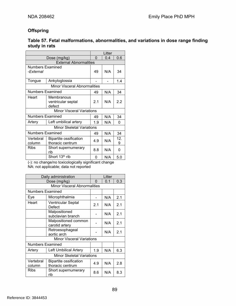
NDA 208462 Emily Place PhD MPH
89
Offspring
Table 57. Fetal malformations, abnormalities, and variations in dose range finding study in rats
LitterDose (mg/kg) 0 0.4 0.6
External AbnormalitiesNumbers Examined-External 49 N/A 34
Tongue Ankyloglossia - - 1.4Minor Visceral Abnormalities
Numbers Examined 49 N/A 34Heart Membranous
ventricular septal defect
2.1 N/A 2.2
Minor Visceral VariationsNumbers Examined 49 N/A 34Artery Left umbilical artery 1.9 N/A 0
Minor Skeletal Variations Numbers Examined 49 N/A 34Vertebral column
Bipartite ossification thoracic centrum 4.9 N/A 12.
9Ribs Short supernumerary
rib 8.8 N/A 0
Short 13th rib 0 N/A 5.0(-): no change/no toxicologically significant changeNA: not applicable; data not reported
Daily administration LitterDose (mg/kg) 0 0.1 0.3
Minor Visceral AbnormalitiesNumbers ExaminedEye Microphthalmia - N/A 2.1Heart Ventricular Septal
Defect 2.1 N/A 2.1
Malpositioned subclavian branch - N/A 2.1
Malpositioned common carotid artery - N/A 2.1
Retroesophageal aortic arch - N/A 2.1
Minor Visceral VariationsNumbers ExaminedArtery Left Umbilical Artery 1.9 N/A 6.3
Minor Skeletal VariationsVertebral column
Bipartite ossification thoracic centrum 4.9 N/A 2.8
Ribs Short supernumerary rib 8.6 N/A 8.3
Reference ID: 3844453

NDA 208462 Emily Place PhD MPH
90
Study title: Effects of MLN 9708 on Embryo-fetal development in RatsStudy no.: 13-155/TE
Study report location: 4.2.3.5.2Conducting laboratory and location: Takeda Pharmaceutical Company
Limited, Kanagawa, JapanDate of study initiation: February 24, 2014
GLP compliance: Signed and Included; JapanQA statement: Signed and Included
Drug, lot #, and % purity: MLN9708; lot # 102031; 99.3%
Key Study Findings Two maternal mortalities at 0.6 mg/kg/3 days (2/20 in this dose group). All other
dams survived the duration of the study cycle. A slight but not statistically increase in post implantation loss was observed at 0.6
mg/kg/3 days. Maternal toxicity was observed at ≥0.6 mg/kg (Gestation Day 15 AUC0-72hr of
1103 hr*ng/mL).
MethodsDoses: 0.2, 0.4, 0.6 mg/kg every 3 days (4 total doses)
Frequency of dosing: Every 3 days (Days 6, 9, 12 and 15 of gestation)Dose volume: 5mL/kg
Route of administration: Oral gavageFormulation/Vehicle: 55-mM citrate buffer containing 0.45
weight/volume percent (w/v%) NaCl in sterile water for injection
Species/Strain: Crl:CD Sprague DawleyNumber/Sex/Group: 20/group
Satellite groups: Groups 5-8 were used for toxicokinetics (see Table 58).
Study design: N/ADeviation from study protocol: N/A
Reference ID: 3844453
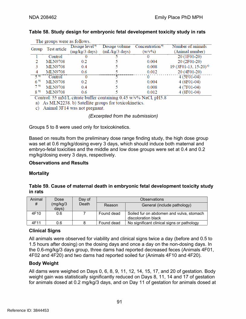
NDA 208462 Emily Place PhD MPH
91
Table 58. Study design for embryonic fetal development toxicity study in rats
(Excerpted from the submission)
Groups 5 to 8 were used only for toxicokinetics.
Based on results from the preliminary dose range finding study, the high dose group was set at 0.6 mg/kg/dosing every 3 days, which should induce both maternal and embryo-fetal toxicities and the middle and low dose groups were set at 0.4 and 0.2 mg/kg/dosing every 3 days, respectively. Observations and Results
Mortality
Table 59. Cause of maternal death in embryonic fetal development toxicity study in rats
ObservationsAnimal #
Dose(mg/kg/3
days)
Day of Death Reason General (include pathology)
4F10 0.6 7 Found dead Soiled fur on abdomen and vulva, stomach discoloration black
4F11 0.6 8 Found dead No significant clinical signs or pathology
Clinical SignsAll animals were observed for viability and clinical signs twice a day (before and 0.5 to 1.5 hours after dosing) on the dosing days and once a day on the non-dosing days. In the 0.6-mg/kg/3 days group, three dams had reported decreased feces (Animals 4F01, 4F02 and 4F20) and two dams had reported soiled fur (Animals 4F10 and 4F20).Body WeightAll dams were weighed on Days 0, 6, 8, 9, 11, 12, 14, 15, 17, and 20 of gestation. Body weight gain was statistically significantly reduced on Days 8, 11, 14 and 17 of gestation for animals dosed at 0.2 mg/kg/3 days, and on Day 11 of gestation for animals dosed at
Reference ID: 3844453

NDA 208462 Emily Place PhD MPH
92
0.4 mg/kg/3days. Animals dosed at 0.6 mg/kg/3days group showed decreased body weight gain on Days 8, 11, 14, and 17 of gestation and increased on Days 12 and 15 of gestation.Feed Consumption24-hour food consumption was measured on Days 0, 6, 8, 9, 11, 12, 14, 15, 17, and 19 of gestation. There were statistically significant decreases in food consumption on Days 6, 9, 12, and 15 of gestation in the 0.2 mg/kg/3days and 0.4 mg/kg/3days groups. There were statistically significant decreases in food consumption between Days 6 and 9, and Days 12 and 15 of gestation in the 0.6 mg/kg/3days group.ToxicokineticsBlood samples (0.3 mL) were collected from the jugular vein of 4 animals in each satellite group (See Table 58) with heparinized syringes at 1, 2, 4, 8, 24, and 72 hours after dosing on the first and last day of dosing (Days 6 and 15 of gestation) and before dosing on the last dosing day (Day 15).Increases in the Cmax of MLN9708 were greater than dose proportional to the increases in the doses between 0.2 and 0.6 mg/kg/3days. Increases in the AUC of MLN9708 were proportional to the increases in the doses between 0.2 and 0.6 mg/kg/3days.
Reference ID: 3844453

NDA 208462 Emily Place PhD MPH
93
Table 60. Toxicokinetic parameters in embryonic fetal development toxicity study in rats
(Excerpted from the submission)
Dosing Solution AnalysisConcentration and Stability
The mean concentrations of MLN9708 in solution at 0.004, 0.008, and 0.012 w/v% after storage for 10 days in a refrigerator (control range: 2°C to 8°C)and for 24 hours at room temperature was measured and these results were within acceptability criteria of ±10% of nominal concentrations, indicating accurate formulation. Given the results, dosing solution for the study was prepared at once and stored in a refrigerator. The concentrations of MLN2238 in the 0.004, 0.008, and 0.012 w/v% dosing solutions of MLN9708 were assayed twice, on the first and the last day of preparation, during the
Reference ID: 3844453

NDA 208462 Emily Place PhD MPH
94
dosing period. The concentrations in the solutions were reported to be within the acceptability criteria ± 10%.Necropsy
Table 61. Maternal gross pathology findings in ratsMacroscopic findings DamsDose (mg/kg/3days) 0 0.2 0.4 0.6
No. animals 20 20 19 20Thymus small - - 1 11Stomach Dark discoloration - - - 1(-): no change/no toxicologically significant change
Table 62. Placental gross pathology findings in ratsMacroscopic findings Placental findingsDose (mg/kg/3days) 0 0.2 0.4 0.6
No. animals 20 20 19 20Placenta Fused placenta - - 1 -(-): no change/no toxicologically significant change
Cesarean Section Data
Table 63. Pregnancy parameters in ratsDose (mg/kg/3days) 0 0.2 0.4 0.6
No. Dams 20 20 19 20Corpora lutea 17.1 15.8 15.9 16.2Implantation sites 15.5 14.9 15.2 14.8Live fetuses 14.8 14.2 14.6 13.7Dead fetuses 0.1 0.0 0.0 0.1Total resorptions 0.0 0.0 0.0 0.0Pre-implantation loss 9.5 5.4 3.5 8.9Post-implantation loss 0.5 0.8 0.6 1.1Mean fetus weight (g) 3.5/3.67 3.51/3.68 3.44/3.71 3.35/3.53
Mean fetus weight (g) given in female/male
Reference ID: 3844453

NDA 208462 Emily Place PhD MPH
95
Offspring
Table 64. Fetal malformations, abnormalities, and variations in ratsLitter
Dose (mg/kg) 0 0.2 0.4 0.6Major Malformations
Numbers Examined-External 295 283 278 246
Head Domed head 3 1 -Cleft palate 3 - -Macroglossia 3 - -Mandibular micrognathia 3 - - -
Maxillary micrognathia 3 - - -Skin, tissue
Anasarca 1 - - -
Neural tube
Meningocele - 1 - -
GI Umbilical hernia - - 1 -Heart Membranous
ventricular septal defect
1 - - 1
Minor Visceral AnomaliesNumbers ExaminedArtery Left umbilical artery 4 NA NA -Adrenal gland
discolored 1 NA NA -
Minor Skeletal Variations and AnomaliesNumbers Examinedmandible small 2 NA NA -Vertebral column
Supernumerary lumbar vertebra - NA NA 1
Bipartite ossification thoracic centrum 7 NA NA 1
Bipartite ossification of lumbar centrum 3 NA NA 2
Ribs Short supernumerary rib 9 NA NA 5
(-): no change/no toxicologically significant changeNA: not applicable; data not reported
Reference ID: 3844453

NDA 208462 Emily Place PhD MPH
96
Study title: An Oral (Gavage) Dose Range-Finding Study of MLN9708 on Embryo/Fetal Development in Rabbits
Study no.: -416156Study report location: 4.2.3.5.2
Conducting laboratory and location:Date of study initiation: December 6, 2012
GLP compliance: Signed and IncludedQA statement: Signed and Included
Drug, lot #, and % purity: MLN9708; lot # 21E004; 99.0%
Key Study Findings Three maternal mortalities observed at 1.2 mg/kg/3 days (3/6 in this dose group).
All other dams survived the duration of the study cycle. Fetal malformations at 1.2 mg/kg/3 days include omphalocele. Embryo-fetal effects were observed at 1.0 mg/kg (Gestation Day 19 AUC0-72hr of
1340 hr*ng/mL) and included post implantation loss, total resorptions, and reduced fetal viability; increased pre-implantation loss was observed at a dose of 0.5 mg/kg (Gestation Day 19 AUC0-72hr of 956 hr*ng/mL).
MethodsDoses: 0.25, 0.5, 1.0, 1.2 mg/kg/3 days
Frequency of dosing: Every 3 days (Days 7,10,13, 16 and 19 of gestation)
Dose volume: 5mL/kgRoute of administration: Oral gavage
Formulation/Vehicle: 55-mmol/L citrate solution containing 3 weight/volume percent (w/v%) glycine
Species/Strain: New Zealand White RabbitNumber/Sex/Group: 6/group
Satellite groups: N/A Study design: N/A
Deviation from study protocol: N/A
Reference ID: 3844453
(b) (4)
(b) (4)

NDA 208462 Emily Place PhD MPH
97
Table 65. Study design for dose range finding embryonic fetal development toxicity study in rabbits
(Excerpted from the submission)
Observations and Results
MortalityOne female (# 68522) in the 1.2 mg/kg group was found dead on Gestation Day 11; two females in the 1.2 mg/kg group (# 68517 and #68513) were sacrificed on Gestation Days 13 and 19, respectively, following body weight losses (16.4% and 17.7% of Gestation Day 7 body weight, respectively) and reduced food consumption. Additional clinical signs for these animals starting at Gestational Day 8 included decreased feces, diarrhea, soft stool, mucoid feces, and/or brown material on various body surfacesClinical SignsDecreased feces, diarrhea, and/or soft stool were reported in the 0.5, 1.0, and 1.2 mg/kg dose groups starting on Gestational Day 8 up to Gestation Day 24.Body WeightThrough the overall treatment period of Gestational Day 7 to 22, significantly decreased mean body weight gain was reported at 0.5 mg/kg dose group and a mean body weight loss was reported at 1.0 and 1.2 mg/kg groups. Feed ConsumptionStatistically significant decreased food consumption was reported during the entire treatment period (Gestational Day 7 to 22) in 1.0 and 1.2 mg/kg dose groups. Food consumption was also reported statistically significantly lower at 0.5 mg/kg dose groups from Gestational Day 7 to 13. Toxicokinetics
Reference ID: 3844453

NDA 208462 Emily Place PhD MPH
98
Table 66. Summary toxicokinetics
(Excerpted from the submission)Dosing Solution AnalysisThe mean concentrations of MLN9708 in solution at 1.00 and 50.0 µg/mL after storage for 5 days in a refrigerator (control range: 2°C to 8°C) and for 17 hours at room temperature was measured and these results were within acceptability criteria of ±10% of nominal concentrations, indicating accurate formulation. Given the results, dosing solution for the study was prepared at once and stored in a refrigerator. The concentrations of MLN9708 in the 0.05, to 1.0 mg/mL dosing solutions were assayed three times, during the dosing period. The concentrations in the solutions were reported to be within the acceptability criteria ± 10%.
Table 67. Dosing solution concentration analysis in a embryofetal toxicity dose range finding study in rabbits
(Excerpted from the submission)
NecropsyTwo of the animals found dead had cystic oviducts reported upon necropsy (# 68517 Gestational Day13, #68513 Gestational Day 19). Additionally animal #68513 had an accessory spleen. Animal #68522 found dead on Gestational Day 11 was unremarkable upon necropsy. Gross pathology in the placenta was not reported.
Reference ID: 3844453

NDA 208462 Emily Place PhD MPH
99
Table 68. Maternal gross pathology findings in a dose range finding study in rabbits
Macroscopic findings DamsDose (mg/kg/3days) 0 0.25 0.5 1.0 1.2
No. animals 5 6 5 6 6Female -reproductive
Cystic Oviducts 1 1 1 - 2
Spleen Accessory spleen - - 1 - 1Lung Dark areas 1(-): no change/no toxicologically significant change
Cesarean Section Data
Table 69. Pregnancy parameters in a dose range finding study in rabbitsDose (mg/kg/3days) 0 0.25 0.5 1.0 1.2
No. Dams 5 6 5 6 3Corpora lutea 50 62 53 60 27Implantation sites 49 58 46 60 25Live fetuses 47 56 45 51 24Dead fetuses 0 0 0 0 0Total resorptions 2 2 1 9 1Pre-implantation loss 1 4 7 0 2Post-implantation loss 2 2 1 9 1Mean fetus weight (g) 41.0 41.5 41.6 39.1 41.4
Mean fetus weight (g) average for males and females combinedOffspring
Table 70. Fetal malformations, abnormalities, and variations in a dose range finding study in rabbits
LitterDose (mg/kg) 0 0.25 0.5 1.0 1.2
Major MalformationsNumbers Examined-External
Abdomen Omphalocele - - - - 1(-): no change/no toxicologically significant change
Reference ID: 3844453
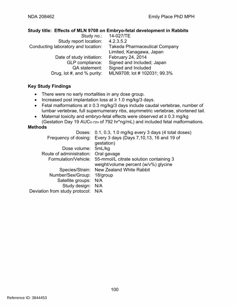
NDA 208462 Emily Place PhD MPH
100
Study title: Effects of MLN 9708 on Embryo-fetal development in RabbitsStudy no.: 14-027/TE
Study report location: 4.2.3.5.2Conducting laboratory and location: Takeda Pharmaceutical Company
Limited, Kanagawa, JapanDate of study initiation: February 24, 2014
GLP compliance: Signed and Included; JapanQA statement: Signed and Included
Drug, lot #, and % purity: MLN9708; lot # 102031; 99.3%
Key Study Findings There were no early mortalities in any dose group. Increased post implantation loss at ≥ 1.0 mg/kg/3 days. Fetal malformations at ≥ 0.3 mg/kg/3 days include caudal vertebrae, number of
lumbar vertebrae, full supernumerary ribs, asymmetric vertebrae, shortened tail. Maternal toxicity and embryo-fetal effects were observed at ≥ 0.3 mg/kg
(Gestation Day 19 AUC0-72hr of 792 hr*ng/mL) and included fetal malformations.Methods
Doses: 0.1, 0.3, 1.0 mg/kg every 3 days (4 total doses)Frequency of dosing: Every 3 days (Days 7,10,13, 16 and 19 of
gestation)Dose volume: 5mL/kg
Route of administration: Oral gavageFormulation/Vehicle: 55-mmol/L citrate solution containing 3
weight/volume percent (w/v%) glycineSpecies/Strain: New Zealand White Rabbit
Number/Sex/Group: 18/groupSatellite groups: N/A
Study design: N/ADeviation from study protocol: N/A
Reference ID: 3844453

NDA 208462 Emily Place PhD MPH
101
Table 71.Study design for embryonic fetal development toxicity study in rabbits
(Excerpted from the submission)
Observations and Results
MortalityNo test article related deaths in any group.Clinical SignsHaving decreased feces was reported in all dose groups and was showed a dose dependent effect (19/19 animals at 1.0 mg/kg, 13/19 animals at 0.3 mg/kg, 4/19 animals at 0.1 mg/kg/3days). At 1.0 mg/kg/3 days soiled fur was reported in 3/19 animals and loose stool were noted in 2/19 animals.Body WeightAt 0.3 mg/kg/3 days and 1.0 mg/kg/3 days, body weight gain was decreased during Days 7 through 9 and Days 10 through 12 of gestation. Additionally at 1.0 mg/kg/3days body weight gains were decreased during Days 13 through 15 of gestation.Feed ConsumptionFood consumption was significantly decreased on dosing days (Days 7, 10, 13, 16, and 19) in the 0.3 mg/kg/3 days and 1.0-mg/kg/day/3days groups. Food consumption was only decreased on Day 19 in the 0.1 mg/kg/3 days group.
Reference ID: 3844453

NDA 208462 Emily Place PhD MPH
102
Toxicokinetics
Table 72. Toxicokinetic parameters in embryonic fetal development toxicity study in rabbits
(Excerpted from the submission)
Dosing Solution AnalysisThe mean concentrations of MLN9708 in solution at 0.02 and .002 w/v% after storage for 10 days in a refrigerator (control range: 5.6°C to 6.7°C) and for 24 hours at room temperature (21.7°C to 22.9°C) was measured and these results were within acceptability criteria of ±10% of nominal concentrations, indicating accurate formulation. Given the results, dosing solution for the study was prepared at once and stored in a refrigerator. The concentrations of MLN9708 in 0.002, 0.006, and 0.02 w/v% dosing
Reference ID: 3844453

NDA 208462 Emily Place PhD MPH
103
solutions were assayed two times, during the dosing period. The concentrations in the solutions were reported to be within the acceptability criteria ± 10%.
Table 73. Dosing solution concentration analysis in an embryofetal toxicity study in rabbits
(Excerpted from the submission)NecropsyGross pathology was in the dams was unremarkable. Gross pathology in the placenta was unremarkable.
Reference ID: 3844453

NDA 208462 Emily Place PhD MPH
104
Cesarean Section Data
Table 74. Pregnancy parameters in rabbitsDose (mg/kg/3days) 0 0.1 0.3 1.0
No. Dams 16 18 18 19Corpora lutea 9.9 9.4 10.7 8.8Implantation sites 8.5 7.7 9.7 7.4Live fetuses 8.3 7.3 9.3 6.7Dead fetuses 0.2 0.2 0.1 0.1Total resorptions 0 0 0 0Pre-implantation loss 14.1 18.2 8.9 14.2Post-implantation loss 2.7 5.8 4.6 8.6Mean fetus weight (g) 32.6/33.2 34.7/35.6 32.4/32.8 32.8/34.6
Mean fetus weight (g) given in female/male
Reference ID: 3844453
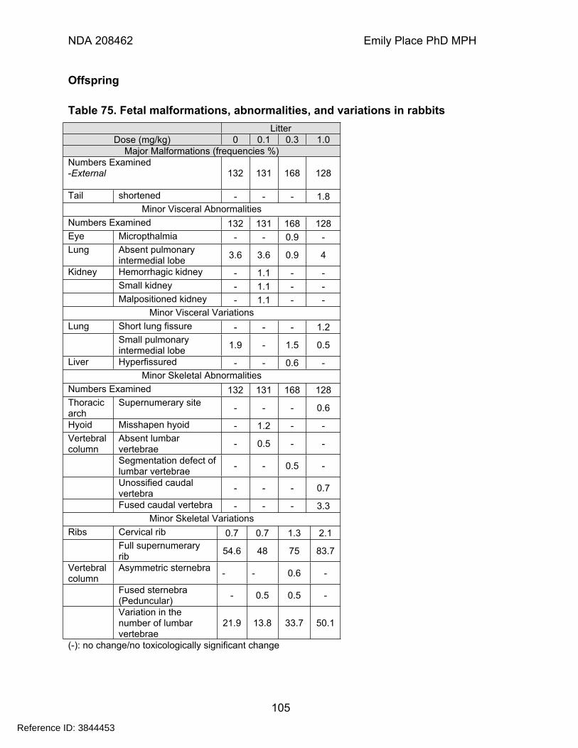
NDA 208462 Emily Place PhD MPH
105
Offspring
Table 75. Fetal malformations, abnormalities, and variations in rabbitsLitter
Dose (mg/kg) 0 0.1 0.3 1.0Major Malformations (frequencies %)
Numbers Examined-External 132 131 168 128
Tail shortened - - - 1.8Minor Visceral Abnormalities
Numbers Examined 132 131 168 128Eye Micropthalmia - - 0.9 -Lung Absent pulmonary
intermedial lobe 3.6 3.6 0.9 4
Kidney Hemorrhagic kidney - 1.1 - -Small kidney - 1.1 - -Malpositioned kidney - 1.1 - -
Minor Visceral VariationsLung Short lung fissure - - - 1.2
Small pulmonary intermedial lobe 1.9 - 1.5 0.5
Liver Hyperfissured - - 0.6 -Minor Skeletal Abnormalities
Numbers Examined 132 131 168 128Thoracic arch
Supernumerary site - - - 0.6
Hyoid Misshapen hyoid - 1.2 - -Vertebral column
Absent lumbar vertebrae - 0.5 - -
Segmentation defect of lumbar vertebrae - - 0.5 -
Unossified caudal vertebra - - - 0.7
Fused caudal vertebra - - - 3.3Minor Skeletal Variations
Ribs Cervical rib 0.7 0.7 1.3 2.1Full supernumerary rib 54.6 48 75 83.7
Vertebral column
Asymmetric sternebra - - 0.6 -
Fused sternebra (Peduncular) - 0.5 0.5 -
Variation in the number of lumbar vertebrae
21.9 13.8 33.7 50.1
(-): no change/no toxicologically significant change
Reference ID: 3844453

NDA 208462 Emily Place PhD MPH
106
9.3 Prenatal and Postnatal DevelopmentStudies not conducted.
10 Special Toxicology StudiesStudies not conducted.
11 Integrated Summary and Safety EvaluationThe nonclinical studies submitted to this NDA provide sufficient information to support the use of Ninlaro (ixazomib) for the treatment of patients with multiple myeloma who have received at least one prior therapy.
Ixazomib is a second-generation proteasome inhibitor that reversibly binds and inhibits the chymotrypsin-like activity of the β5 subunit of the 20S proteasome. In vitro, ixazomib induces caspase mediated apoptosis in multiple myeloma cell lines. In multiple myeloma cell lines, ixazomib also had a synergistic effect on cytotoxicity in combination with lenalidomide. Ixazomib was cytotoxic in culture to primary cells harvested from multiple myeloma patients who had relapsed after prior therapies. In vivo pharmacology studies using a human myeloma xenograft SCID mouse model following treatment with ixazomib confirmed dose-dependent proteasome inhibition by modulation of β5 subunit activity, other pharmacodynamics markers of cell death and also showed reduced tumor volumes.
Stand-alone safety pharmacology studies were not conducted for the respiratory or nervous system. In the reviewed GLP-compliant repeat-dose toxicology studies: ixazomib exposure did not result in obvious clinical signs of toxicity; no adverse effects were observed on the respiratory systems of rats or dogs; and there were no acute nervous system-related effects, as assessed in part through incorporation of a functional observational battery in the 9 month toxicology study in dogs. There were chronic toxicities in the nervous system in dogs and these findings are outlined in the general toxicology section. When ixazomib was given orally to dogs in a cardiovascular telemetry study, no adverse effects were observed on any cardiovascular parameters.
Based on the data collected in general toxicology studies, there were no gender differences in exposure to oral ixazomib, and increases in Cmax and AUC values were dose proportional. Intravenous administration of 2 mg/kg ixazomib and oral administration of 6 mg/kg ixazomib resulted in similar plasma area under the concentration-time curves from the time 0 to 24 hours (AUC0-24hr). Tissue distribution was extensive following administration of oral ixazomib. The highest exposure (by Cmax or AUC) was in the urinary bladder, lymph node, spleen, adrenal glands, kidney (renal cortex and medulla), bile, GI tract, and liver. The longest observed half-life was for the urinary bladder with a t1/2 of up to 946 hours. Upon , ixazomib citrate ester (MLN9708) is metabolized to the active boronic acid form of the drug, (MLN2238). The elimination half-life after oral dosing in nonclinical species ranged from 30 to 72 hours.
Reference ID: 3844453
(b) (4)

NDA 208462 Emily Place PhD MPH
107
The general toxicology studies were conducted in the rat and dog via oral (gavage). Nonclinical findings in the rat and dog show that ixazomib targets the gastrointestinal tract, hematopoietic and lymphatic systems, nervous system (dog only). Mortalities in the rat toxicology study were attributable to gastrointestinal toxicity and lymphoid depletion. Toxicities in the gastrointestinal system included epithelial hyperplasia, neutrophilic infiltrates (stomach, large and small intestines, peyers patch, mesenteric lymph nodes), single-cell necrosis in the lamina propria of the small and large intestines, and erosion in the stomach. Toxicities in the hematopoietic and lymphatic systems included lymphoid necrosis and depletion in lymph nodes and spleen correlating with increased absolute counts for total white blood cells, neutrophils, monocytes, and elevated lymphocytes in the rats only. At doses below those associated with a dose limiting toxicity, white blood cell parameters were altered consistent with what is observed during an inflammatory response. Toxicities in the nervous system were only observed in dogs and included neuronal degeneration of the sympathetic, dorsal root, peripheral autonomic, and end organ ganglia, and nerve fiber degeneration of the peripheral nerves and ascending tracts in the dorsal columns of the spinal cord. Changes present at recovery in high dose animals included nerve fiber degeneration of the dorsal root ganglia and an increase in axonal degeneration in the dorsal columns of the spinal cord.
Safety concerns stem from the steep dose-toxicity relationship with ixazomib in the chronic rat and dog toxicology studies. There was a less than 1.5-fold separation in systemic exposure between the limit dose and a dose associated with mortality due to drug-induced injury of the liver and gastrointestinal tract in the rat. In dogs, there was an approximately 2-fold separation in exposure between the NOAEL and doses associated with extensive neuronal pathology in the nervous system.
Ixazomib was not mutagenic in the Ames assay nor was it clastogenic in the bone marrow micronucleus assay in mice. It was negative for DNA damage in Comet study. However it showed clastogenic activity (structural chromosomal aberrations) in the in vitro chromosomal aberration assay in human peripheral blood lymphocytes in the presence or absence of an exogenous metabolic activation system.
Ixazomib was teratogenic in the rat and rabbit. In pregnant rat (0.6 mg/kg; 3.6 mg/m2) dose range-finding studies, there were decreases in fetal weights, decreased fetal viability, and increased post-implantation losses (at 2.5 times the human AUC when given 4 mg orally, in the rat respectively). In pregnant rabbit (1.0 mg/kg; 12 mg/m2) dose range-finding studies, there were decreases in fetal weights, decreased fetal viability, and increased post-implantation losses (at 3.2 times the human AUC when given 4 mg orally, in the rabbit). In the definitive rabbit study, increases in fetal skeletal variations/abnormalities (caudal vertebrae, number of lumbar vertebrae, full supernumerary ribs, asymmetric sternebra, shortened tail) were observed at doses greater than or equal to 0.3 mg/kg (3.6 mg/m2), which were also associated with maternal toxicity. The dose of 0.3 mg/kg (3.6 mg/m2) in rabbits is approximately 1.9 times the human AUC when given 4mg orally. Thus, administration of ixazomib during pregnancy may pose a risk to the human fetus.
Reference ID: 3844453

NDA 208462 Emily Place PhD MPH
108
.
12 Appendix/AttachmentsNone
Reference ID: 3844453

---------------------------------------------------------------------------------------------------------This is a representation of an electronic record that was signedelectronically and this page is the manifestation of the electronicsignature.---------------------------------------------------------------------------------------------------------/s/----------------------------------------------------
EMILY J PLACE11/09/2015
CHRISTOPHER M SHETH11/09/2015
Reference ID: 3844453