2 cell growth, cycle and differentiation
DESCRIPTION
ÂTRANSCRIPT


All references made are to the unit 1 Scholar book. Page numbers may vary each year as different versions of the book are released.

• new cells can only arise from reproduction of existing cells
• time between division of a mother cell to when it’s daughter cells divide is known as cell cycle
• length of cycle varies depending on the cell
• bacteria cells ~ 20 minutes
• liver cells ~ once a year
• human cells in culture ~ 20 hours
• frog embryo ~ 20 minutes

The cycle is split into 2 parts:
1.Interphase
2.Mitosis
Copy diagram of cell cycle from Scholar – Figure 2.1 p.25

1. Interphase
• sometimes called rest phase
• period where cell growth happens
• split into 3 stages:
1.G1 – duplication of cell organelles (not chromosomes
2.S (synthesis) phase – DNA replication
3.G2 – duplicated chromosomes are checked for errors and repaired
• G used to stand for ‘gap’ until research showed growth taking place
2. M-Phase M = mitosis
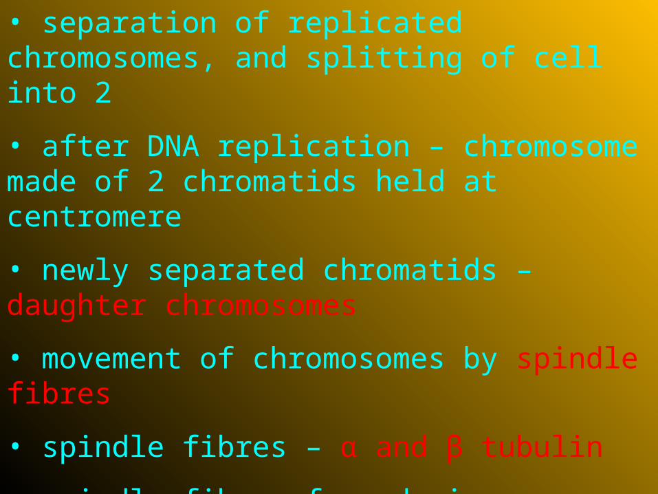
• separation of replicated chromosomes, and splitting of cell into 2
• after DNA replication – chromosome made of 2 chromatids held at centromere
• newly separated chromatids – daughter chromosomes
• movement of chromosomes by spindle fibres
• spindle fibres – α and β tubulin
• spindle fibres form during prophase of mitosis, and are organised by spindle poles
• guide and pull chromosomes during mitosis

Mitosis consists of several stages:
1.Prophase
2.Metaphase
3.Anaphase
4.Telophase
5.Cytokinesis
Use Scholar to describe briefly what is happening during each stage of mitosis named above (p.26 + 27) Try and draw a diagram of the stage if you can remember what it looks like from Higher.

Mitotic index - %cells in a sample undergoing mitosis
No. cells undergoing mitosis
Total no. cells X 100

Controlling the Cell Cycle
3 main checkpoints:
1. G1 checkpoint
2. G2 checkpoint
3. M checkpoint
Make sure these checkpoints are added onto your cell cycle diagram. (Figure 2.3 p.29)

G1 checkpoint
• end of G1 phase
• cell size assessed, if it is large enough => entry into ‘S’ phase
• if not – non-growing G0 phase
• most human cells are in G0 phase e.g. muscle, liver and nerve cells

G2 Checkpoint
• assesses if DNA has been successfully replicated
• if OK => M phase
• dubbed ‘restriction point’
• if cell continues after G1, it usually completes the full cycle
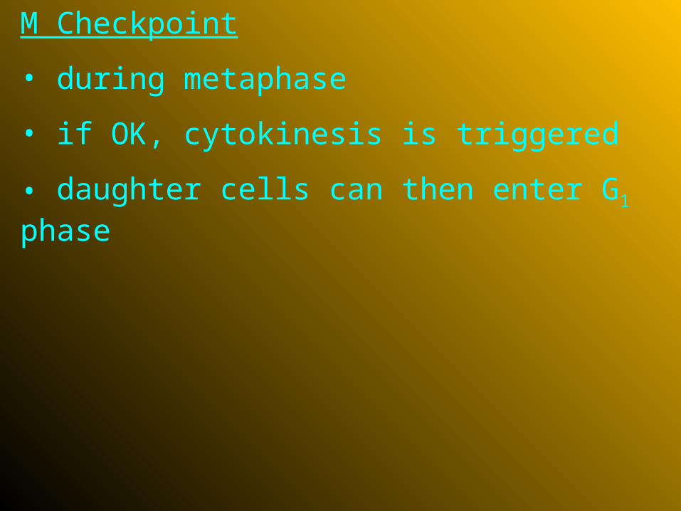
M Checkpoint
• during metaphase
• if OK, cytokinesis is triggered
• daughter cells can then enter G1 phase

• each checkpoint has a STOP signal until it is overridden by a GO signal
• GO signals are usually protein kinases
• protein kinases are inactive until activated by cyclin molecules => they are cyclin-dependent kinases (Cdks)
• G2 checkpoint signal was first discovered

• MPF – ‘Maturation/Mitosis/M-phase Promoting Factor
• triggers passage from G2 to M phase
• MPF results from cyclin and Cdk complex
• MPF causes nuclear membrane to fragment, and initiates enzymes to break down proteins holding chromatids together at anaphase
• MPF also triggers enzymes to break down the cyclin component of itself to switch off
• resulting Cdk remains in cell to associate with a cyclin protein in next cycle
• no conclusive theory for G1 checkpoint yet
• recent research shows possibly 3 Cdks, cyclins and their complexes control all parts of the cycle
• cells in which control of the cell cycle has been lost
• results in excessive cell growth
=> tumours form
• tumours – benign or malignant
• malignant tumours are cancerous and can undergo metastasis
• metastasis – spreading of cancer cells from primary tumour

• proteins controlling cell cycle stimulate or inhibit cell growth and division
• altered proteins have no control over cell cycle => cancer cells develop
• proto-onco (proliferation) genes code for proteins which stimulate normal cell division
• mutations form dominant oncogenes
• oncogenes cause excessive production of proteins stimulating cell division leading to excessive growth => tumours

3 types of mutation responsible:
1.Translocation (non-homologous chromosomes swapping segments - promoters are swapped to wrong genes, or wrong genes to a promoter)
2.Gene amplification (leading to multiple copies of proto-oncogene)
3.Point mutation (protein produced becomes hyperactive or resistant to degradation)
• Translocation and amplification lead to excess protein
• All 3 lead to tumour formation

• anti-proliferation (tumour suppressing) genes produce proteins which inhibit cell growth or division
• act at cell cycle check points
• mutations in these genes are recessive
• if both genes are affected control of cycle is lost
• tumours form as cell growth is not inhibited

Your task:
Research a type of cancer, and produce a presentation which you will present to the class. You should include information about your chosen cancer, the side effects, likelihood of metastasis, if there are any lifestyle choices which can lead to it, current treatment, and a prognosis.
You may or may not get a single period to start your research, the majority of this work will be completed in your own time as homework.
Cell differentiation – cells becoming specialised
Cells become tissues
Tissues become organs
Organs arranged in systems

• cells all develop from undifferentiated stem cells
• differentiation occurs as a result of changes in gene expression
• i.e. some genes are ‘switched on’ and others are ‘switched off’
• known as Jacob and Monod operon/hypothesis
Jacob Monod Operon
• discovered lac operon in bacteria
• on the same chromosome – regulator, operator, and structural genes
• regulator gene makes repressor molecule which joins to operator gene to ‘switch off’ structural gene

• if repressor joins with substrate (for enzyme coded for by structural gene) structural gene ‘switched on’, and produces enzyme
• when substrate used up, repressor molecule is freed, and joins with operator to ‘switch off’ structural gene
• gene expression is not fully understood in humans yet.