1,2 ; kjell olafsen,3 md.; caroline schnakers,4,5 phd.;
TRANSCRIPT

“Cognitive event related potentials during the sub-acute phase of severe traumatic brain
injury and their relationship to outcome”
Solveig L. Hauger,1,2 Cand. Psychol.; Kjell Olafsen,3 MD.; Caroline Schnakers,4,5 PhD.;
Nada Andelic,6,7 MD, PhD.; Kristian Bernhard Nilsen,8,9 MD, PhD.; Eirik Helseth,10,11
MD, PhD.; Ingrid Funderud,2 PhD.; Stein Andersson,2 PhD.; Anne-Kristine Schanke,1,2
PhD.; Marianne Løvstad1,2 PhD.
Author Affiliations: 1 Department of Research, Sunnaas Rehabilitation Hospital, Norway 2 Department of Psychology, University of Oslo, Norway 3 Department of Neurointensive treatment, Oslo University Hospital, Norway 4 Neurosurgery Department, University of California, Los Angeles, CA, USA 5 Research Institute, Casa Colina Hospital and Centers of Healthcare, Pomona, CA, USA 6 Department of Physical Medicine and Rehabilitation, Oslo University Hospital, Norway 7 Institute of Health and Society, CHARM (Center for habilitation and rehabilitation Models
and Services), Faculty of Medicine, University of Oslo, Norway 8 Department of Neurology, Oslo University Hospital, Norway 9 Department of Neuroscience, Norwegian University of Science and Technology, Norway 10 Department of Neurosurgery, Oslo University Hospital, Norway 11 Faculty of Medicine, University of Oslo, Norway
Corresponding author:
Mrs. Solveig Lægreid Hauger
Department of Research, Sunnaas Rehabilitation Hospital, 1450 Nesoddtangen, Norway
([email protected]/ [email protected]), Phone: +47 91310358.
Fax: +47 66963631

2
Abstract
Predicting outcome in the early phase after severe traumatic brain injury (sTBI) is a major
clinical challenge, particularly identifying patients with potential of good cognitive outcome.
The current single center prospective study aimed to explore presence and normalization of
electroencephalography (EEG)-based event related potentials (ERPs) in the early phase
following sTBI, and their relationship to functional and cognitive outcome 6 months post-
injury. Fourteen adult patients with sTBI were recruited from the neurointensive care unit
(mean age=38.2 years (SD=14.7); 8 males; mean lowest Glasgow Coma Scale score within
first 24 hours=5.4, SD=1.87). EEG-recordings were conducted biweekly at three time-points
applying an ERP paradigm encompassing a passive condition involving hearing their own
name randomly interspersed between an unfamiliar name, and an active condition with
instruction to count their own name. Functional and cognitive outcome 6 months post-injury
was measured with Glasgow Outcome Scale-Extended (GOSE) and neuropsychological tests
of attention and memory. Ten patients demonstrated a significantly enhanced cognitive P3 in
the active counting task compared to passive listening across recordings, and 6 presented with
normalization of P3 in the counting task. Moreover, P3-amplitude to the counting task at the
third time-point was positively correlated with both functional outcome (GOSE) and
cognition (verbal learning, attentional set-shifting and switching) 6 months post-injury. ERP
can index cognitive capacities in the early phase following sTBI, and the cognitive P3
component in an active design is associated with functional and cognitive outcome,
demonstrating that the cognitive P3 may yield valuable information of residual cognition and
provide supplementary prognostic information.
Key words: brain injury, prognosis, cognition, EEG, event-related potentials

3
Introduction
Substantial long-lasting disability with cognitive deficits,1-3 impaired overall health,4,5
and difficulties with community integration and work6 are common after moderate to severe
traumatic brain injury (sTBI). However, the severity of chronic symptoms and the rate of
recovery is highly variable. Some patients regain good cognitive capacities and functional
independence, while a minority of patients remain in a state of prolonged disorder of
consciousness (DoC).7-9 Early and reliable recognition of those who might regain good
cognitive outcome is a great challenge, but essential for treatment planning.
Major effort has been put into developing prognostic models based on clinical and laboratory
parameters from the acute phase following TBI. Both the IMPACT (International-Mission-
For-Prognosis-And-Clinical-Trial) and CRASH (Corticosteroid-Randomisation-After-
Significant-Head injury) models are based on large, prospective patient cohorts. Studies of the
two prognostic models have demonstrated that high age, low Glasgow Coma Scale (GCS),10
absent pupillary reactivity, and certain CT characteristics are associated with poor outcome or
death.11-13 However, the psychometric properties of the methods and their limitations in
mainly focusing on predicting mortality have been criticized.14,15 Also, the fact that they do
not take newer advancements in critical care management into consideration causes
overestimation of the risk of mortality or unfavorable outcome.16
Neurophysiological techniques such as somatosensory evoked potentials (SEPs) have
shown some promise in aiding prognostication.17 Specifically, SEPs have demonstrated high
specificity in predicting poor outcome in anoxic coma, but presence of SEPs is not necessarily
predictive of good recovery.18,19 Importantly, for TBI-related coma with initial absence of
cortical responses, recovery of bilaterally abolished SEP followed by favorable outcome may
occur more often than after other etiologies.18-20 Also, most studies lack serial SEP recordings
with potential to demonstrate sub-acute SEP-normalization, they are restricted by coarse
outcome measures, and often use follow-up time-points as early as one month post
injury.18,19,21
ERPs are based on time-locked encephalographic (EEG) activity elicited by external
or internal events, providing a neurophysiological correlate of cognitive processing at the
millisecond level. ERPs range from early components representing largely sensory and
automatic brain processes, i.e. the mismatch negativity component (MMN), to later

4
components mediating increasingly more complex cognitive processes, such as the well-
established P3 component, reflecting allocation of attentional and memory resources.22,23 A
P3 response in healthy persons typically peaks between 300 and 600 ms post stimulus, while
brain injury is associated with prolonged latencies and attenuated amplitudes.22,24-26 A meta-
analysis concluded that the MMN and the P3 appear to be reliable predictors of awakening
from TBI-induced coma,27 but few ERP-studies have explored the relationship to recovery
levels. One exception is Lew and colleagues,28 whom summarize a review of existing
literature in stating that despite promise, EEG-based techniques currently have limited clinical
application beyond the prediction of negative outcomes in the acute phase. The P3 component
elicited in active cognitive tasks has also been widely investigated as a marker of
consciousness in patients with DoC.29-31 The probability of eliciting cortical
neurophysiological responses in patients with severe brain injury increases with the use of
salient, self-referential stimuli.32 Thus, including the subject’s own name (SON) randomized
with unfamiliar name (UN), in an active condition instructing subjects to count the number of
SON (denoted as active or effortful ERP tasks), has proven robust and sensitive in healthy
subjects as well as patients with DoC.31,33-35 Compared to patients with non-traumatic
disorders, patients with TBI are also more likely to follow commands in active paradigms.36
There is however scarce knowledge of the prognostic utility of ERPs beyond mere awakening
from coma and detection of consciousness.
The main aim of the present study was to investigate residual cognitive capacity using
ERPs in the sub-acute phase after very sTBI, and to explore their association to outcome.
Presence and normalization of cognitively mediated P3 responses was explored, along with
investigation of the relationship between sub-acute P3 and 6 months outcome. It was
anticipated that cognitive P3 elicited in an active task could be detected at an individual level
in the sub-acute phase. It was furthermore expected that P3 elicited to SON in an active task
would be related to better functional and cognitive outcome 6 months post-injury.
Methods
Study setting
This single center prospective study included adult patients admitted to the intensive
care unit at Oslo University Hospital (OUH), a level 1 trauma center for the southeast of
Norway, between September 2013 and June 2015. For inclusion, patients needed to be adults

5
aged between 18-65, fluent Norwegian speakers prior to their injury, admitted with sTBI
defined according to the International Classification of Diseases, Tenth Revision, criteria
(S06.1-S06.9: intracranial brain injury presenting as traumatic cortical edema; focal or diffuse
TBI; epidural, subdural, or subarachnoid hemorrhage, and/or other specified or unspecified
intracranial injury) within 24 hours of injury, and had GCS score 3-8 during the first 24 hours
after injury, representing the severe range of TBI.37 In order to recruit the most severe end of
the sTBI population, additional criterions were in need of at least five days of neurointensive
care. Patients were excluded if they had severe comorbidities prior to injury such as
progressive neurological disorders, severe psychiatric and substance abuse disorders in need
of treatment, or lack of Norwegian residency. Patients treated with hemicraniectomy were
excluded due to problems with EEG-recordings. Patients were excluded if they had a
bilaterally absent brainstem auditory evoked potentials (BAEP) along with absent auditory N1
ERP component, indicating primary sensory processing disorders. Patient lists were screened
on a weekly basis. 76 with GCS lower than 9 were considered for study inclusion. Of
excluded patients, two were excluded due to lack of consent from next of kin, 7 died during
the initial phase, 10 had hemicraniectomies, 11 lacked Norwegian residency or were not prior
fluent Norwegian speakers, and the rest were not eligible due to high age, severe premorbid
psychiatric or substance abuse disorders, or had early transfer to neurointensive care unit
(ICU) at a local hospital. The study was conducted in agreement with the Helsinki
Declaration and approved by the Regional Committee for Medical Research Ethics in South
East Norway (2013/407). Written informed consent was obtained from the next of kin at
inclusion, and from those patients capable to provide consent at follow-up.
Procedures and design
Demographic variables (age, gender) and injury-related characteristics were extracted
from medical charts. The Injury Severity Score (ISS)38 and the Abbreviated Injury Scale (AIS
head)39 were used as indicators of injury severity.
EEG-recordings were performed biweekly three times (T1, T2 & T3). Intravenous
sedation was ended at least four days before the first EEG-recording. The average time
between injury-onset and day of first EEG-recording was 19 days (SD= 5.9), and then the
second and third recording were conducted biweekly. Average time between T1 and T2 was
16.4 days and between T2 and T3 15.8 days. The recordings were thus conducted in the
transition from acute to sub-acute phase following sTBI.40,41 GCS was scored at the day of

6
each EEG-recording. Level of consciousness was assessed with the Coma Recovery Scale-
Revised (CRS-R)42,43 on the day of EEG-recording at T1 and T2, and also at T3 for those with
DoC at previous EEG-recordings. Vegetative State (VS) and Minimally Conscious State
(MCS) were classified according to established criteria.44,45 The distinction between MCS+
and MCS- was also established, with MCS+ defined as presence of reproducible response to
command (CRS-R auditory subscale score ≥ 3), and MCS- as no reproducible response to
command (CRS-R auditory subscale score < 3).46 Patients were classified with Post-
Traumatic Confusional State (PTCS),47 if emerged from DoC (i.e. showing functional use of
objects or functional communication at the CRS-R), but presenting with disorientation and
confusion, that is a verbal score of 4 at the GCS. Patients emerged from DoC and a maximum
verbal score of 5 at the GCS were classified as fully conscious and oriented. At follow-up, the
first author conducted a clinical interview and assessed the patients with the GOSE and a
neuropsychological test battery.
ERP acquisition
EEG-data were acquired bedside with a 32-electrode cap (Quik-Cap; Compumedics
Neuroscan) with electrode positions according to the 10-20 system, connected to a portable
digital NuAmp EEG amplifier (Compumedics Neuroscan). Electro-oculogram was recorded
using the electrodes located above and below the left eye and at the outer canthi of the two
eyes. The ground electrode was placed near Fz with a nasal reference. EEG signals were
recorded (NeuroScan Inc.) in DC with a 500 Hz sampling rate. Impedance was kept below 10
kΩ. Stimuli were presented binaurally with a maximum 90 dB sound pressure level. The
procedure lasted approximately 25-30 minutes including breaks.
The auditory ERP paradigm (see figure 1) included two-stimuli conditions with subject`s own
name (SON) and an unfamiliar name (UN) in a passive and an active effortful task, with 50
SON randomly interspersed in between 50 UN. The passive condition was presented first
(each condition contained four sets of consecutive blocks of 25 stimuli). In the passive
condition, the patients were instructed to do nothing but stay awake. In the active condition,
they were instructed to count the number of times they heard SON. Instructions were repeated
between each block of 25 stimuli. A short break and, if needed, brief auditory or deep
pressure stimulation according to CRS-R protocol were applied between conditions in order
to ensure optimal arousal levels. All names were digitally recorded from a female, middle-
aged native Norwegian speaker (stimulus duration range: approximately 500–600 ms), with
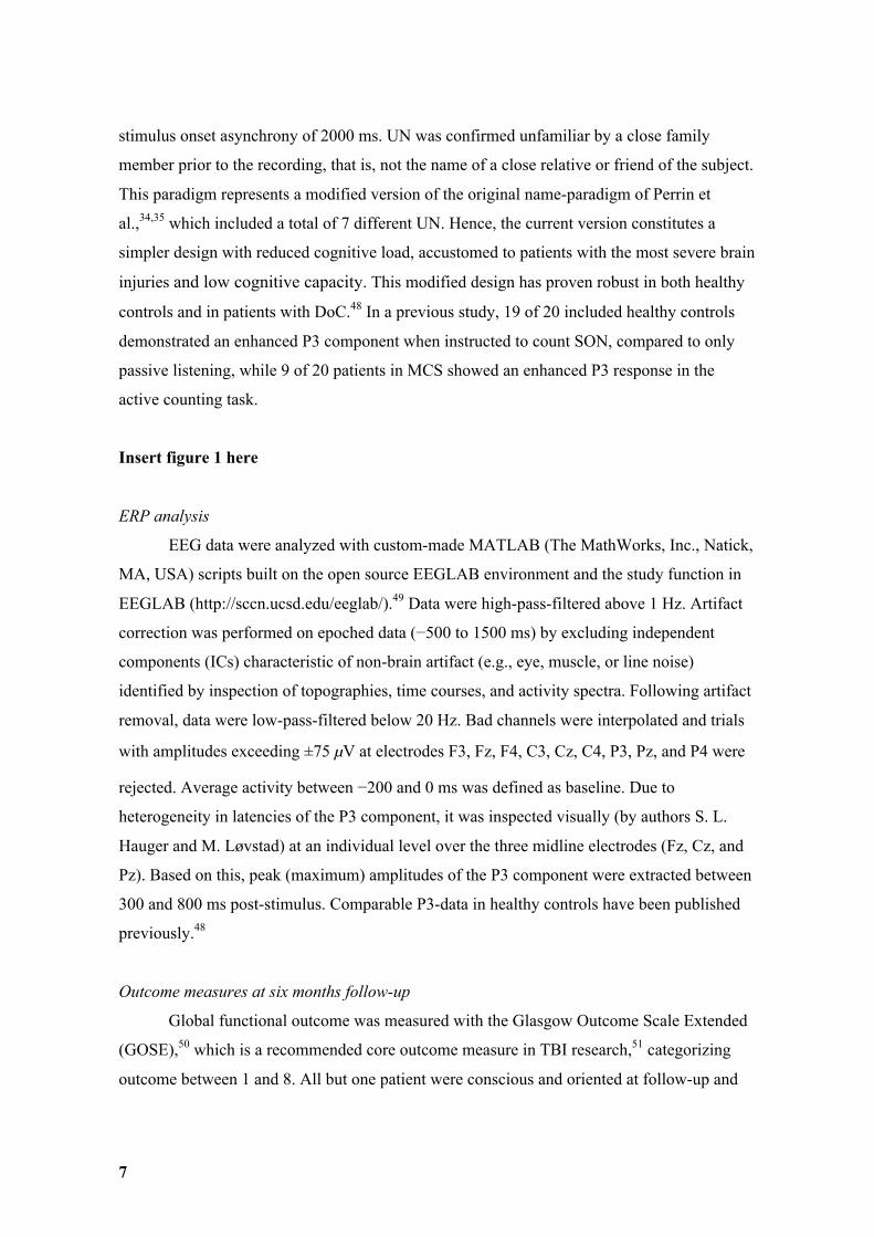
7
stimulus onset asynchrony of 2000 ms. UN was confirmed unfamiliar by a close family
member prior to the recording, that is, not the name of a close relative or friend of the subject.
This paradigm represents a modified version of the original name-paradigm of Perrin et
al.,34,35 which included a total of 7 different UN. Hence, the current version constitutes a
simpler design with reduced cognitive load, accustomed to patients with the most severe brain
injuries and low cognitive capacity. This modified design has proven robust in both healthy
controls and in patients with DoC.48 In a previous study, 19 of 20 included healthy controls
demonstrated an enhanced P3 component when instructed to count SON, compared to only
passive listening, while 9 of 20 patients in MCS showed an enhanced P3 response in the
active counting task.
Insert figure 1 here
ERP analysis
EEG data were analyzed with custom-made MATLAB (The MathWorks, Inc., Natick,
MA, USA) scripts built on the open source EEGLAB environment and the study function in
EEGLAB (http://sccn.ucsd.edu/eeglab/).49 Data were high-pass-filtered above 1 Hz. Artifact
correction was performed on epoched data (−500 to 1500 ms) by excluding independent
components (ICs) characteristic of non-brain artifact (e.g., eye, muscle, or line noise)
identified by inspection of topographies, time courses, and activity spectra. Following artifact
removal, data were low-pass-filtered below 20 Hz. Bad channels were interpolated and trials
with amplitudes exceeding ±75 µV at electrodes F3, Fz, F4, C3, Cz, C4, P3, Pz, and P4 were
rejected. Average activity between −200 and 0 ms was defined as baseline. Due to
heterogeneity in latencies of the P3 component, it was inspected visually (by authors S. L.
Hauger and M. Løvstad) at an individual level over the three midline electrodes (Fz, Cz, and
Pz). Based on this, peak (maximum) amplitudes of the P3 component were extracted between
300 and 800 ms post-stimulus. Comparable P3-data in healthy controls have been published
previously.48
Outcome measures at six months follow-up
Global functional outcome was measured with the Glasgow Outcome Scale Extended
(GOSE),50 which is a recommended core outcome measure in TBI research,51 categorizing
outcome between 1 and 8. All but one patient were conscious and oriented at follow-up and

8
could go through a neuropsychological assessment including IQ (Wechsler Abbreviated Scale
of Intelligence (WASI)),52 verbal memory and learning (Hopkins Verbal Learning Test-
Revised (HVLT-R)),53 , attentional set-shifting (Letter-Number Switching condition from the
Trail Making Test (TMT), Delis-Kaplan Executive Function System (D-KEFS)),54 inhibition
and switching (Color-Word Interference Test (CWIT 3 and 4 from D-KEFS)),54 and working
memory (Digit Span backwards, from the Wechsler Adult Intelligence Scale-Fourth Edition
(WAIS-IV)).55
Statistical analysis
Firstly, P3 to SON in the active task was identified by visual inspection at an
individual level in each recording. In cases where a P3 to SON was established, P3-amplitude
differences in the passive versus the active condition to SON in T1, T2 and T3 were
investigated with two-tailed paired t-tests on a trial-by-trial basis in each individual for each
sampling point (in the individual P3 time-window to SON) at the midline electrodes, using
the EEGLAB study function. Signs of normalization of P3 elicited in the active task, that is, a
development towards a clearly identifiable P3 component with increasing amplitudes across
the 3 EEG-recordings, was explored by visual inspection, resulting in a group of 6 patients (1,
4, 7, 8, 10, 12) whom displayed normalization, and a group of 8 without detectable
normalization. Normalization was then primarily analyzed as amplitude change over time for
each condition, comparing peak P3-amplitudes at each midline electrode between T1 and T2,
T2 and T3, and T1 and T3 in each group and experimental condition, using paired-samples t-
tests. Secondarily, changes in amplitude difference between active and passive conditions
were explored in each group, time-point and midline electrode.
The relationship between P3 to SON in the active task and outcome at 6 months was
examined with Pearson`s correlations, including the following sub-set of outcome measures;
functional outcome (GOSE), verbal learning (HVLT-R, total words learned), working
memory (Digit Span backwards), attentional set-shifting (TMT) and inhibition and switching
(CWIT 3 and 4). The effect of GCS on these associations was examined in partial correlations
where acute GCS was controlled for. Analyses involving neuropsychological variables were
conducted on raw scores.
Normality distribution was investigated with the Shapiro-Wilk's test (p < .05), and in
cases of violation, analyses were repeated with non-parametric tests, with any subsequent
9
changes in results being reported. P-values ≤ .05 were considered statistically significant.
Effect size is reported with r for correlations and Cohen´s d for t-tests, with values of 0.1, 0.3,
and 0.5 and 0.2, 0.5, and 0.8, being considered small, medium and large effect sizes,
respectively.56 Statistical analyses were performed using SPSS for Macintosh, version 23
(SPSS Inc, Chicago, IL).
Results
The initial included sample consisted of 19 patients. Five were excluded due to lack of
cooperation, low EEG-quality or medical complications. The remaining 14 patients (mean age
=38.2 (SD=14.7); 8 males) completed the experimental procedure, included follow-up.
Demographic, injury-related and clinical characteristics are presented in table 1. The patients
had very severe injuries with a mean lowest GCS score within the first 24 hours after injury of
5.43 (SD=1.87), all had an AIS-head score of 5 and intracranial pressure (ICP) monitoring.
Mean days in ICU was 21 (SD=6.4). Thus, the patient sample was in the most severe range of
TBI.
Insert table 1 here
Insert table 2 here
Insert table 3 here
Presence of P3 in the active task
For all patients except one (patient 6), a P3 component to SON in the active task was
identified at Pz, Cz and/or Pz in at least one of the three recordings. Ten of the 14 patients had
a significant P3-amplitude difference between the active and the passive condition in at least
one of the sub-acute EEG-recordings, but only two demonstrated this P3-amplitude difference
across all three recordings (patient 7 and 10, see table 2). Patients with significant P3-
amplitude differences to SON between the active and passive conditions are presented in
Figure 2A. Of the 4 patients (patient 5, 6, 11 and 12) without significant P3 difference at any
recording, 2 (patient 6 and 12) were the most severely injured, both being in a VS at the first
recording, evolving to MCS during later recordings. Five patients had a significant P3-
amplitude difference at T1 and T3 (patient 3, 4, 8, 13 and 14), patient 9 only at T1, patient 2
only at T2, and patient 1 only at T3. Also, of all 23 recordings with no significant P3-
amplitude difference (see example figure 2B), 2 were in patients in a VS at T1, 4 recordings

10
were in patients in a MCS-, 2 in patients diagnosed as MCS+, 6 in patients with PTCS, and 9
recordings were in conscious and oriented patients.
Normalization of P3 across ERP recordings
The 6 patients identified as the "normalization group" (see figure 3A) showed a
significantly larger P3-amplitude to SON in the active task at T3 compared to T1 at Cz (t(5) =
-3,75, p <.03, d = 1.53), and a borderline significant difference at Pz (t(5) = -2,47, p <.06, d =
1.0). No significant differences were seen neither in the non-normalization group, nor in the
passive condition. There was also a significantly larger peak P3-amplitude in the active
compared to the passive condition at T3 at Cz (t(1,5) = -3.52, p < .02, d = 1.44), while
borderline significantly larger at Pz (t(5) = -2,47, p <.06, d = 1.0), not seen in the non-
normalization group (see figure 3A and B).
Insert figure 2 A&B here
Insert figure 3 A&B here
Relationship between P3 in active task and outcome
Of the 14 patients, one achieved a good recovery level (GOSE 7–8), 9 had moderate
disability (GOSE 5–6) and 4 had severe disability (GOSE 3–4). All but one patient had
emerged from DoC at follow-up. Patient 12 still presented with PTCS, and could not be
assessed neuropsychologically. A strong positive correlation was found at T3 between the P3-
amplitude to SON in the active task and global outcome (GOSE; r(12) =.54, p<.05), while no
significant relationships were evident at T1 or T2. After controlling for the role of acute GCS,
P3 elicited in the active task was still significantly correlated with GOSE at T3: r(12) = .61, p
< .05. There was also a strong positive correlation between P3-amplitude and learning at T3
r(11) =.60, p<.03, but not at T1 and T2. Also, P3 elicited in the active task at T3 was
significantly correlated to better performance on inhibition (CWIT condition 3; rs(11) = -.65,
p < .02), inhibition and switching (CWIT condition 4; r(11) = -.56, p < .05), as well as set-
shifting (TMT; r(11) = -.57, p < .05). When controlling for acute GCS, all previous
significant associations were still significant, and as with GOSE, some even more strongly
associated (learning; r(11) = .61, p < .05, and set-shifting; r(11) = -.58, p < .05). No
significant associations were found between P3 elicited in the active task and working
memory (Digit Span backwards), and GCS alone was not significantly associated with GOSE
or any of the neuropsychological tests.

11
Discussion
To our awareness, this study is the first to investigate both the presence and
normalization of sub-acutely recorded cognitive ERP as well as it`s association to global and
cognitive functioning 6 months after very sTBI.
Ten of the 14 patients displayed a significant difference in the P3-amplitude to SON
between the active and the passive task, demonstrating that neurophysiological indices of
cognitive capacity can be identified even in the early phases after sTBI, and that
neurophysiological methods encompassing active experimental conditions can tap into
residual cognitive functioning in patients with sTBI, even long before standardized
neuropsychological assessment is feasible. This P3-amplitude difference even preceded overt
evidence of command-following in 2 non-communicating patients in MCS- (patient 8 and
10). However, a significant difference in P3-amplitude between the active and passive task
could not be established in 23/42 recordings across the 3 time-points. Surprisingly, the
majority of negative findings was in patients presenting with PTCS or regained orientation.
This highlights that cognitive ERPs have low sensitivity, understood in this context as the
ability of the neurophysiological assessment to detect effortful cognition in patients
behaviorally displaying such capacity. It has previously been shown that the P3 component
elicited in active paradigms can be a marker of consciousness in patients with DoC, but a
wide variety in sensitivity rates have been found in ERP-studies, ranging from 100% to
14%.31,57,58 It is, however, difficult to disentangle whether negative ERP-recordings can be
explained by underlying patient deficits, or by methodological limitations inherent to the
paradigm or recording technique. Patients with sTBI may suffer from severe cognitive
impairments, such as deficits in language, attention, memory, executive functioning, cognitive
drive, as well as behavioral impairments characterized by reduced motivation and lack of
cooperation, all potentially preventing them from responding in active tasks despite having
regained consciousness and cognitive resources. Issues related to compliance to task
instructions and high levels of movement artifacts can be particularly prominent in the PTCS
phase, as severe symptoms of PTCS is associated with poor cooperation.59
Six patients showed normalization of P3 to SON in the active task. However, the
remaining eight did not display such normalization within the time frame of this study.
Studies investigating test-retest reliability of the P3-amplitude in healthy subjects, have shown
12
variable results, ranging from 0.31 to 0.93, although the majority of studies reported
moderate-to-strong reliability estimates over periods ranging from weeks to years.60,61
Conducting bedside EEG-recordings in the early phase after severe brain injury is challenging
and involve dealing both with environmental noise from the recording site, technical and
movement artifacts, as well as potential underlying cognitive deficits. All these factors may
contribute to low signal-to-noise ratios, resulting in negative findings as well as low ERP
stability. In the normalization-group, the P3-amplitude was significantly larger in the active
task compared to the passive at T3. Wijnen and colleagues26 investigated ERPs in severely
brain-injured patients during recovery from VS to consciousness with a paradigm
encompassing regular and irregular tones, including comparison of passive listening to an
active condition. They did not find differences in P3-amplitudes between patients who
recovered consciousness and those who did not, nor did they see a P3-difference between
active and passive tasks within the group that regained consciousness. This emphasizes the
importance of including robust experimental paradigms in addition to using salient stimuli in
ERP studies of patients with the most severe brain injuries.
Importantly, the P3-amplitude to SON in the active task at T3 was related to outcome,
both in terms of global functioning and with regard to verbal learning, cognitive switching,
and attentional set-shifting six months post-injury. Also, the P3 in the active task showed a
specific relation to outcome superior to that of the acute GCS scores. A P3 component elicited
in an active condition is associated with allocation of attentional and memory resources, and
the task instructions require language comprehension.22,23 To our knowledge, this study is the
first to investigate the positive association between cognitive P3 elicited in an active task and
global and cognitive outcome following the most severe range of TBI, indicating prognostic
utility. A previous study has shown high correlations between amplitudes of the MMN
component and recovery from VS to consciousness in patients with DoC after severe brain
injuries of mixed ethiologies.62 Yet, study outcome was limited to recovery of consciousness.
Houlden and colleagues21 found that a normalized SEP obtained within the first week after
injury was positively correlated with higher levels of functioning as well as better attentional
performance one-year post-injury. However, SEP, even more so than the MMN component, is
a highly automatic, bottom-up driven somatosensory evoked potential, but does not imply
effortful cognitive processing, as does a P3 component elicited in an active task. Importantly,
a previous study recorded ERP and SEP within 8 days post sTBI. When comparing SEP and
ERP in relation to outcome, ERP was found superior to SEP for prognosticating good

13
functional outcome.63 The fact that ERP can tap into cognitive capacities sub-acutely, and
even more so, that the cognitive P3 component significantly relates to outcome both measured
by GOSE, verbal learning, and attentional set-shifting and switching capacity, is highly
noteworthy.
All patients included in the current study were in the most severe spectrum of TBI.
Still, global functioning at 6 months post-injury showed large variation in outcome, with
GOSE scores ranging from 3 to 8. A score of 8 represents “Good Recovery-Upper Level”,
encompassing patients with the full functional recovery after the TBI. A score of 3, on the
other hand, represents “Severely Disabled-Lower Level”, including patients that are
completely dependent on others.50,64 Total IQ at follow-up varied from 69 to 115 (see table 3).
In sum, the outcomes indicated substantial heterogeneity in functional level 6 months post-
injury, as has been previously established in studies of outcome following sTBI.4,65-67 Hence,
there is a need for more precise prognostication at an individual level in the early phase even
in the most severe category of TBI, as the initial severity of the TBI, as routinely measured by
the GCS, does not sufficiently predict patient outcome. Also, there is a need to not only make
prognostic estimations regarding unfavorable outcomes or death, but also to early on identify
those who have a good chance of positive outcome. This is potentially helpful both in guiding
early therapeutic decisions, and to better inform the families.
Study limitations
Several limitations should be considered when interpreting the results of this study.
Firstly, it was limited to a small consecutively included patient sample, both due to strict
inclusion criteria, as well as low prevalence of the most severe injuries. This limits the
statistical analysis that could be performed in the study, and there is a need for precaution
with regard to representativeness of the included patients with sTBI. Also, performing
repeated ERPs in the sub-acute clinical setting is complicated. For instance in ICU, the EEG-
recordings might be interrupted by critical medical events, arousal may be difficult to
maintain, or the patient is characterized by motor restlessness, all hazards to EEG quality. The
current ERP study results are thus in need of replication in larger studies. The passive-active
ERP paradigm included two names, that is, the patients own name and an unfamiliar name.
14
Linguistic features of the name-stimuli, such as gender or frequencies of syllables, were
not controlled for.
Conclusions
In summary, there is a need to identify early signs of cognitive restoration, and to
recognize those patients with prospect of good outcome. This study shows that ERPs elicited
in an active task requiring mental effort in the early phase after sTBI may yield valuable
information of residual cognitive capacities and their association to outcome. Today, there is
no prognostic tool with perfect sensitivity for predicting outcome after sTBI. Combining ERP
components with other clinical methods may optimize prognosis of functional recovery.
Acknowledgments
The authors wish to thank all the patients and their families, and the staff of the
Department of Intensive Care, OUH, in contributing to this study. This research is supported
by the Norwegian Extra Foundation for Health and Rehabilitation through EXTRA funds
(Grant 2012/2/0084), and Sunnaasstiftelsen funded NeuroScan equipment.
Author Disclosure Statement
No competing financial interests exist.

15
References
1. Sigurdardottir, S., Andelic, N., Wehling, E., Roe, C., Anke, A., Skandsen, T., Holthe,
O., Jerstad, T., Aslaksen, P.M., Schanke, A-K. (2015). J. Head Trauma Rehabil.
Neuropsychological functioning in a national cohort of severe traumatic brain injury:
demographic and acute injury-related predictors. 30, E1-12.
2. Dikmen, S.S., Corrigan, J.D., Levin, H.S., Machamer, J., Stiers, W., Weisskopf, M.G.
(2009). Cognitive outcome following traumatic brain injury. J. Head Trauma Rehabil.
24, 430-438.
3. Spitz, G., Ponsford, J.L., Rudzki, D., Maller, J.J. (2012). Association between
cognitive performance and functional outcome following traumatic brain injury: a
longitudinal multilevel examination. Neuropsychology. 26, 604-612.
4. Soberg, H.L., Roe, C., Anke, A., Arango-Lasprilla, J.C., Skandsen, T., Sveen, U., von
Steinbuchel, N., Andelic, N. (2013). Health-related quality of life 12 months after
severe traumatic brain injury: a prospective nationwide cohort study. J. Rehabil. Med.
45, 785-791.
5. Andelic, N., Sigurdardottir, S., Schanke, AK., Sandvik, L., Sveen, U., Roe, C. (2010).
Disability, physical health and mental health 1 year after traumatic brain injury.
Disabil. Rehabil. 32, 1122-1131.
6. Dahm, J., Ponsford, J. (2015). Long-term employment outcomes following traumatic
brain injury and orthopaedic trauma: a ten-year prospective study. J. Rehabil. Med. 47,
932-940.
7. Beaumont, J.G., Kenealy, P.M. (2005). Incidence and prevalence of the vegetative and
minimally conscious states. Neuropsychol. Rehabil. 15, 184-189.
8. Leonardi, M., Sattin, D., Raggi, A. (2013). An Italian population study on 600 persons
in vegetative state and minimally conscious state. Brain Inj. 27, 473-484.
9. Lovstad, M., Andelic, N., Knoph, R., Jerstad, T., Anke, A., Skandsen, T., Hauger,
S.L., Giacino, J.T., Roe, C., Schanke, AK. (2014). Rate of disorders of consciousness
in a prospective population-based study of adults with traumatic brain injury. J. Head
Trauma Rehabil. 29, E31-43.
10. Teasdale, G., Jennett, B. (1974). Assessment of coma and impaired consciousness. A
practical scale. Lancet. 2, 81-84.
11. Jacobs, B., Beems, T., van der Vliet, T.M., van Vugt, A.B., Hoedemaekers, C., Horn,
J., Franschman, G., Haitsma, I., van der Naalt, J., Andriessen, T.M., Borm, G. F., Vos,

16
P.E. (2013). Outcome prediction in moderate and severe traumatic brain injury: a
focus on computed tomography variables. Neurocrit. Care. 19, 79-89.
12. Collaborators M.C.T., Perel, P., Arango, M., Clayton, T., Edwards, P., Komolafe, E.,
Poccock, S., Roberts, I., Shakur, H., Steyerberg, E.,Yutthakasemsunt, S. (2008).
Predicting outcome after traumatic brain injury: practical prognostic models based on
large cohort of international patients. Bmj. 336, 425-429.
13. Steyerberg, E.W., Mushkudiani, N., Perel, P., Butcher, I., Lu, J., McHugh, G.S.,
Murray, G.D., Marmarou, A., Roberts, I., Habbema, J.D., Maas, A.I. (2008).
Predicting outcome after traumatic brain injury: development and international
validation of prognostic scores based on admission characteristics. PLoS med. 5, e165.
14. Castano-Leon, A.M., Lora, D., Munarriz, P.M., Cepeda, S., Paredes, I., de la Cruz, J.,
Gomez Lopez, P.A., Lagares, A. (2016). Predicting outcomes after severe and
moderate traumatic brain injury: an external validation of impact and crash prognostic
models in a large Spanish cohort. J. Neurotrauma. 33, 1598-1606.
15. Sandsmark, D.K. (2016). Clinical outcomes after traumatic brain injury. Curr. Neurol.
Neurosci. Rep. 16, 52.
16. Olivecrona, M., Olivecrona, Z. (2013). Use of the CRASH study prognosis calculator
in patients with severe traumatic brain injury treated with an intracranial pressure-
targeted therapy. J. Clin. Neurosci. 20, 996-1001.
17. Wang, J.T., Young, G.B., Connolly, J.F. (2004). Prognostic value of evoked responses
and event-related brain potentials in coma. Can. J. Neurol. Sci. 31, 438-450.
18. Carter, B.G., Butt, W. (2001). Review of the use of somatosensory evoked potentials
in the prediction of outcome after severe brain injury. Crit. Care Med. 29, 178-186.
19. Robinson, L.R., Micklesen, P.J., Tirschwell, D.L., Lew, H.L. (2003). Predictive value
of somatosensory evoked potentials for awakening from coma. Crit. Care Med. 31,
960-967.
20. Schorl, M., Valerius-Kukula, S.J., Kemmer, T.P. (2014). Median-evoked
somatosensory potentials in severe brain injury: does initial loss of cortical potentials
exclude recovery? Clin. Neurol. Neurosurg. 123, 25-33.
21. Houlden, D.A., Taylor, A.B., Feinstein, A., Midha, R., Bethune, A.J., Stewart, C.P.,
Schwartz, M.L. (2010). Early somatosensory evoked potential grades in comatose
traumatic brain injury patients predict cognitive and functional outcome. Crit. Care
Med. 38, 167-174.
17
22. Soltani, M., Knight, R.T. Neural origins of the P300. (2000). Crit. Rev. Neurobiology.
14, 199-224.
23. Polich, J. Updating P300: an integrative theory of P3a and P3b. (2007). Clin.
Neurophysiol. 118, 2128-2148.
24. Solbakk, A.K., Reinvang, I., Andersson, S. (2002). Assessment of P3a and P3b after
moderate to severe brain injury. Clin. Electroencephalogr. 33, 102-110.
25. Duncan, C.C., Barry, R.J., Connolly, J.F., Fischer, C., Michie, P.T., Naatanen, R.,
Polich, J., Reinvang, I., Van Petten, C. (2009). Event-related potentials in clinical
research: guidelines for eliciting, recording, and quantifying mismatch negativity,
P300, and N400. Clin. Neurophysiol. 120, 1883-1908.
26. Wijnen, V.J., Eilander, H.J., de Gelder, B., van Boxtel, G.J. (2014). Repeated
measurements of the auditory oddball paradigm is related to recovery from the
vegetative state. J. Clin. Neurophysiol. 31, 65-80.
27. Daltrozzo, J., Wioland, N., Mutschler, V., Kotchoubey, B. (2007). Predicting coma
and other low responsive patients outcome using event-related brain potentials: a
meta-analysis. Clin. Neurophysiol. 118, 606-614.
28. Lew, H.L., Poole, J.H., Castaneda, A., Salerno, R.M., Gray, M. (2006). Prognostic
value of evoked and event-related potentials in moderate to severe brain injury. J.
Head Trauma Rehabil. 21, 350-360.
29. Noirhomme, Q., Brecheisen, R., Lesenfants, D., Antonopoulos, G., Laureys, S.
(2017). "Look at my classifier's result": disentangling unresponsive from (minimally)
conscious patients. Neuroimage. 15, 288-303.
30. Lehembre, R., Gosseries, O., Lugo, Z., Jedidi, Z., Chatelle, C., Sadzot, B., Laureys, S.,
Noirhomme, Q. (2012). Electrophysiological investigations of brain function in coma,
vegetative and minimally conscious patients. Arch. Ital. Biol. 150, 122-139.
31. Hauger, S.L., Schanke, A.K., Andersson, S., Chatelle, C., Schnakers, C., Lovstad, M.
(2017). The clinical diagnostic utility of electrophysiological techniques in assessment
of patients with disorders of consciousness following acquired brain injury: a
systematic review. J. Head Trauma Rehabil. 23.
32. Laureys, S., Perrin, F., Bredart, S. (2007). Self-consciousness in non-communicative
patients. Conscious. Cogn. 16, 722-741.
33. Schnakers, C., Perrin, F., Schabus, M., Majerus, S., Ledoux, D., Damas, P., Boly, M.,
Vanhaudenhuyse, A., Bruno, M.A., Moonen, G., Laureys, S. (2008). Voluntary brain
processing in disorders of consciousness. Neurology. 71, 1614-1620.

18
34. Perrin, F., Schnakers, C., Schabus, M., Degueldre, C., Goldman, S., Bredart, S.,
Faymonville, M. E., Lamy, M., Moonen, G., Luxen, A., Maquet, P., Laureys, S.
(2006). Brain response to one's own name in vegetative state, minimally conscious
state, and locked-in syndrome. Arch. Neurol. 63, 562-569.
35. Perrin, F., Garcia-Larrea, L., Mauguiere, F., Bastuji H. (1999). A differential brain
response to the subject's own name persists during sleep. Clin. Neurophysiol. 110,
2153-2164.
36. Kondziella, D., Friberg, C. K., Frokjaer, V.G., Fabricius, M., Moller, K. (2016).
Preserved consciousness in vegetative and minimal conscious states: systematic
review and meta-analysis. J. Neurol. Neurosurg. Psychiatry. 87, 485-492.
37. Chesnut, R.M. (1997). The management of severe traumatic brain injury. Emerg. Med.
Clin. North Am.15, 581-604.
38. Baker SP, S.P., O'Neill, B., Haddon, W.Jr., Long, W.B. (1974). The injury severity
score: a method for describing patients with multiple injuries and evaluating
emergency care. J. Trauma. 14, 187-196.
39. Association for the Advancement of Automative Medicine. (1998). The abbreviated
injury scale 1990. Revision update 98. Des Plaines, IL.
40. Malec, J.F., Basford, J.S. (1996). Postacute brain injury rehabilitation. Arch. Phys.
Med. Rehabil. 77, 198-207.
41. Mazaux, J.M., Richer, E. (1998). Rehabilitation after traumatic brain injury in adults.
Disabil. Rehabil. 20, 435-447.
42. Giacino, J.T., Kalmar, K., Whyte, J. (2004). The JFK Coma recovery scale-revised:
measurement characteristics and diagnostic utility. Arch. Phys. Med. Rehabil. 85,
2020-2029.
43. Lovstad, M., Froslie, K.F., Giacino, J.T., Skandsen, T., Anke, A., Schanke, A.K.
(2010). Reliability and diagnostic characteristics of the JFK coma recovery scale-
revised: exploring the influence of rater's level of experience. J. Head Trauma Rehabil.
25, 349-356.
44. Giacino, J.T., Ashwal, S., Childs, N., Cranford, R., Jennett, B., Katz, D.I., Kelly, J.P.,
Rosenberg, J.H., Whyte, J., Zafonte, R.D., Zasler, N.D. (2002). The minimally
conscious state: definition and diagnostic criteria. Neurology. 58, 349-353.
45. Giacino, J.T., Kalmar, K. (2005). Diagnostic and prognostic guidelines for the
vegetative and minimally conscious states. Neuropsychol. Rehabil. 15, 166-174.
19
46. Bruno, M.A., Majerus, S., Boly, M., Vanhaudenhuyse, A., Schnakers, C., Gosseries,
O., Boveroux, P., Kirsch, M., Demertzi, A., Bernard, C., Hustinx, R., Moonen, G.,
Laureys S. (2012). Functional neuroanatomy underlying the clinical subcategorization
of minimally conscious state patients. J. Neurol. 259,1087-1098.
47. Sherer, M., Nakase-Thompson, R., Yablon, S.A., Gontkovsky, S.T. (2005).
Multidimensional assessment of acute confusion after traumatic brain injury. Arc.
Phys. Med. Rehabil. 86, 896-904.
48. Hauger, S.L., Schnakers, C., Andersson, S., Becker, F., Moberget, T., Giacino, J.T.,
Schanke, A.K., Lovstad, M. (2015). Neurophysiological indicators of residual
cognitive capacity in the minimally conscious state. Behav. Neurol. 145913.
49. Delorme, A., Makeig, S. (2004). EEGLAB: an open source toolbox for analysis of
single-trial EEG dynamics including independent component analysis. J. Neurosci.
Methods. 134, 9-21.
50. Wilson, J.T., Pettigrew, L.E., Teasdale, G.M. (1998). Structured interviews for the
Glasgow Outcome Scale and the extended Glasgow Outcome Scale: guidelines for
their use. J. Neurotrauma. 15, 573-585.
51. Wilde, E.A., Whiteneck, G.G., Bogner, J., Bushnik, T., Cifu, D.X., Dikmen, S.,
French, L., Giacino, J.T., Hart, T., Malec, J.F., Millis, S.R., Novack, T.A., Sherer, M.,
Tulsky, D.S., Vanderploeg, R.D., von Steinbuechel, N. (2010). Recommendations for
the use of common outcome measures in traumatic brain injury research. Arc. Phys.
Med. Rehabil. 91, 1650-1660.
52. Wechsler, D. Wechsler abbreviated scale of intelligence. (1999). San Antonio, TX:
The Psychological Corporation.
53. Brandt, J.B.R. (2001). Hopkins verbal learning test-revised. Professional Manual.
Lutz, F.L.: Psychological Assessment Resources Inc.
54. Delis, D.C., Kramer, J. (2001). Delis-Kaplan executive function system. San Antonio,
TX: The Psychological Corporation.
55. Wechsler, D. (2008). Wechsler adult intelligence scale-fourth edition; WAIS-IV. San
Antonio, TX: The Psychological Corporation.
56. Cohen, J. (1992). A power primer. Psychol. Bull. 112, 155-159.
57. Bekinschtein, T.A., Dehaene, S., Rohaut, B., Tadel, F., Cohen, L., Naccache, L.
(2009). Neural signature of the conscious processing of auditory regularities. PNAS.
106, 1672-1677.

20
58. Faugeras, F., Rohaut, B., Weiss, N., Bekinschtein, T., Galanaud, D., Puybasset, L.,
Bolgert, F., Sergent, C., Cohen, L., Dehaene, S., Naccache, L. (2012). Event related
potentials elicited by violations of auditory regularities in patients with impaired
consciousness. Neuropsychologia. 50, 403-418.
59. Silva, M.A., Nakase-Richardson, R., Sherer, M., Barnett, S.D., Evans, C.C., Yablon,
S.A. (2012). Posttraumatic confusion predicts patient cooperation during traumatic
brain injury rehabilitation. AJPMR. 91, 890-893.
60. Walhovd, K.B., Fjell, A.M. (2002). One-year test-retest reliability of auditory ERPs in
young and old adults. Int. J. Psychophysiol. 46, 29-40.
61. Cassidy, S.M., Robertson, I.H., O'Connell, R.G. (2012). Retest reliability of event-
related potentials: evidence from a variety of paradigms. Psychophysiology. 49, 659-
664.
62. Wijnen, V.J., van Boxtel, G.J., Eilander, H.J., de Gelder, B. (2007). Mismatch
negativity predicts recovery from the vegetative state. Clin. Neurophysiol. 118, 597-
605.
63. Lew, H.L., Dikmen, S., Slimp, J., Temkin, N., Lee, E.H., Newell, D., Robinson, L.R.
(2003). Use of somatosensory-evoked potentials and cognitive event-related potentials
in predicting outcomes of patients with severe traumatic brain injury. AJPMR. 82, 53-
61.
64. Teasdale, G.M., Pettigrew, L.E., Wilson, J.T., Murray, G., Jennett, B. (1998).
Analyzing outcome of treatment of severe head injury: a review and update on
advancing the use of the Glasgow Outcome Scale. J. Neurotrauma. 15, 587-597.
65. Anke, A., Andelic, N., Skandsen, T., Knoph, R., Ader, T., Manskow, U.,
Sigurdardottir, S., Roe, C. (2015). Functional recovery and life satisfaction in the first
year after severe traumatic brain injury: a prospective multicenter study of a
Norwegian national cohort. J. Head Trauma Rehabil. 30, E38-49.
66. Ponsford, J., Draper, K., Schonberger, M. (2008). Functional outcome 10 years after
traumatic brain injury: its relationship with demographic, injury severity, and
cognitive and emotional status. JINS. 14, 233-242.
67. Jourdan, C., Bayen, E., Pradat-Diehl, P., Ghout, I., Darnoux, E., Azerad, S., Vallat-
Azouvi, C., Charanton, J., Aegerter, P., Ruet, A., Azouvi, P. (2016). A comprehensive
picture of 4-year outcome of severe brain injuries. Results from the Paris-TBI study.
Ann. Phys. Rehabil. Med. 59, 100-106.

Figure 1. Experimental task design.
Abbreviations: SON: subject´s own name; UN: unfamiliar name.
ERPtask
PASSIVECONDITIONSON/UNPassive
(50SON/50UNrandomized)
ACTIVECONDITIONSON/UNActive
(50SON/50UNrandomized)"Countthenumberoftimesyou
hearyourownname"

Table 1. D
emographic, injury-related and clinical characteristics
Patients
1 2
3 4
5 6
7 8
9 10
11 12
13 14
Dem
ographic
A
ge
53
47 50
32 27
20 17
57 42
19 31
30 58
52 G
ender
F M
F
F M
M
M
M
F
M
M
F M
F
Injury-related factors
C
ause of injury
Traffic Fall
Fall Traffic
Traffic Traffic
Traffic Fall
Fall Fall
Fall Traffic
Traffic Traffic
GC
S at site of injury 6
3 11
12 6
3 6
7 14
4 9
3 6
8 Low
est GC
S w
ithin first 24 hours 6
3 8
3 6
3 6
7 8
4 7
3 6
6 A
IShead score
5
5 5
5 5
5 5
5 5
5 5
5 5
5 ISS score
43
25 29
34 43
35 50
38 34
26 42
43 25
45 Isolated injury head yes/no
no yes
no no
no no
no no
no yes
no no
yes no
ICP m
onitoring yes/no yes
yes yes
yes yes
yes yes
yes yes
yes yes
yes yes
yes D
ays on respirator 14
16 9
17 17
30 13
24 16
23 22
28 12
14 D
ays sedated
13 4
7 14
12 20
12 10
10 17
19 14
4 8
Days before eye opening
11 6
7 13
8 26
11 14
14 25
19 15
6 9
Total days in ICU
20
17 11
21 20
30 18
25 21
20 33
29 13
15 Total days in rehabilitation
16
92 4
42 127
149 25
71 95
158 78
119 111
65 D
ata related to EEG-recording
D
ays between sedation
and first EEG-rec.
4 12
5 4
6 10
7 8
4 6
8 14
8 4
Time since injury
and first EEG-rec.
17 16
14 18
18 30
19 18
14 23
27 28
12 12
Clinical status 1st. EEG
-recording
Level of consciousness M
CS+
PTCS
PTCS
PTCS
MC
S- V
S PTC
S M
CS-
MC
S+ M
CS-
PTCS
VS
MC
S+ PTC
S G
CS score
12
13 14
14 8
4 14
8 11
6 13
5 10
14 C
RS-R
total score 16
22 23
23 12
2 23
10 18
6 23
6 14
23 C
linical status 2nd. EEG-recording
Level of consciousness C
&O
C
&O
C
&O
C
&O
PTC
S M
CS-
C&
O
PTCS
C&
O
MC
S- C
&O
M
CS-
PTCS
C&
O
GC
S score
15 15
15 15
14 8
15 14
15 10
15 9
13 15
CR
S-R total score
23 23
23 23
23 12
23 23
23 10
23 8
23 23
Clinical status 3rd. EEG
-recording
Level of consciousness
C&
O
C&
O
C&
O
C&
O
PTCS
MC
S+ C
&O
C
&O
C
&O
PTC
S C
&O
M
CS-
PTCS
C&
O
GC
S score
15 15
15 15
14 12
15 15
15 11
15 8
14 15
CR
S-R total score
N.P
N.P
N.P
N.P
N.P
18 N
.P N
.P N
.P 20
N.P
9 N
.P N
.P A
bbreviations: GC
S, Glasgow
Com
a Scale; AIS
head , Abbreviated Injury Scale head score; ISS, Injury Severity Score; IC
P, intracranial pressure; ICU
, Intensive care unit; EEG
, electroencephalogram; C
RS-R
, Com
a Recovery Scale-R
evised; M, m
ale; F, female; V
S, vegetative state; MC
S+, minim
ally conscious state pluss; M
CS-, m
inimally conscious state m
inus; PTCS, post-traum
atic confusional state; C&
O, patients that are both conscious and oriented; N
.P, not performed.

Table 2. L
evel of consciousness and presence of significant P3-difference in active compared to passive E
RP task post-acutely at each tim
e point Patients 1
2 3
4 5
6 7
8 9
10 11
12 13
14 T
1 M
CS+/n.s. PTC
S/n.s PTC
S/* PTC
S/*** M
CS-/n.s
VS/n.s
PTCS/*
MC
S-/* M
CS+/**
MC
S-/* PTC
S/n.s V
S/n.s M
CS+/*
PTCS/**
T2
C&
O/n.s.
C&
O/*
C&
O/n.s
C&
O/n.s
PTCS/n.s
MC
S-/n.s C
&O
/* PTC
S/n.s C
&O
/n.s M
CS-/*
C&
O/n.s
MC
S-/n.s PTC
S/n.s C
&O
/n.s T
3 C
&O
/* C
&O
/n.s C
&O
/* C
&O
/*** PTC
S/n.s M
CS+/n.s
C&
O/*
C&
O/*
C&
O/n.s
PTCS/***
C&
O/n.s
MC
S-/n.s PTC
S/** C
&O
/* A
bbreviations: VS, vegetative state; M
CS+, m
inimally conscious state plus; M
CS-, m
inimally conscious state m
inus; PTCS, post-traum
atic confusional state; C
&O
, patients that are conscious and oriented. n.s. = no significant P3-difference * = significant P3-difference present at p<.01 ** = significant P3-difference present at p<.02 *** = significant P3-difference present at p<.05

Table 3. Functional and cognitive outcom
e 6 months post injury
Patients
1 2
3 4
5 6
7 8
9 10
11 12
13 14
GO
SE score
6 5
5 6
4 3
8 6
5 3
6 3
5 6
Total IQ
108
100 93
112 69
98 100
113 89
85 100
N.P
115 111
Verbal learning
(Hopkins, trials 1-3;
raw score/T-score)
32/60 28/50
21/33 31/57
6/≤20 21/30
31/57 20/32
20/30 15/≤20
24/40 N
.P 11/≤20
20/32 D
igit Span backwards
(maxim
um digits*/
cumulative percent)
5/53,2 5/53,2
5/53,2 5/53,2
3/98,7 4/86,4
5/66,2 4/98,6
5/56,5 3/100
5/52,4 N
.P 3/100
5/53,2 TM
T condition 4 (raw
score/ scaled score)
91/10
155/3 67/12
40/13 221/1
156/1 66/10
84/11 60/12
110/5 179/1
N.P
146/6 80/11
CW
IT condition 3 (raw
score/ scaled score)
45/13
42/13 41/14
42/13 126/1
69/5 57/9
66/9 44/13
123/1 75/4
N.P
108/1 75/7
CW
IT condition 4 (raw
score/ scaled score)
46/14
51/12 52/13
39/14 160/1
136/1 73/7
98/6 52/12
88/4 75/6
N.P
82/8 62/11
Abbreviations: G
OSE, G
lasgow O
utcome Scale Extended; IQ
, Intelligence Quotient; C
WIT, C
olor-Word Interference Test; N
.P, not performed.
*maxim
um num
ber of digits reported.

Figure 2. A: Significant P3 in active task in individual patients
µ
V
Patient 1, T3 Patient 2, T2 Patient 3, T3 Pz Fz Pz
Time (ms) Patient 4, T1 Patient 7, T3 Patient 8, T1 Fz Pz Fz
Patient 9, T1 Patient 10, T3 Patient 13, T1 Fz Pz Fz
Patient 14, T1 Fz
—ACTIVECOUNTING—PASSIVELISTENING
B: Example of patient without significant P3 difference between active and passive task
Figure 2. A: Presence of P3 to SON in active task in all 10 individuals presenting with a significant P3-difference between active and passive task, exemplified with one electrode. Recording time-point is indicated above each curve. The averaged ERPs in the active counting (red) versus passive (black) condition (y-axis, amplitude in µV; x-axis, time in ms) are illustrated. Observed significant differences of P3-amplitude between conditions (values < .05 to .001) are marked with grey line at the P3 curve. B: Example of patient presenting with no significant P3-difference between active and passive task. Recording time-point and electrodes are indicated above the curves.
µ
V
Patient 6, T2
Fz Cz Pz
Time (ms)
P3
—ACTIVECOUNTING—PASSIVELISTENING

Figure 3A. Grand average ERPs in the patient group (n=6) with normalization of P3
Cz Pz T1
µ
V
T2 Time (ms)
T3
Figure 3A. Grand averaged ERPs in the patient group with normalization of P3 in active task across the three sub-acute recordings, showing significantly larger P3-amplitude to SON in active task at T3 compared to T1 at Cz, while borderline significant at Pz, and a significantly larger peak P3-amplitude in active compared to passive condition at T3 at Cz, also borderline significant at Pz (p < .05). The P3 curve elicited in active task is marked with grey line. The active counting condition marked in red and the passive condition marked in black (y axis, amplitude in µV; x axis, time in ms).
—ACTIVECOUNTING—PASSIVELISTENING

Figure 3B: Grand average ERPs in the patient group (n=8) without normalization of P3
Cz Pz T1
µ
V
T2 Time (ms)
T3
Figure 3B: Grand averaged ERPs in patient group without normalization of P3 in active task across the three time-points sub-acutely. The active counting condition marked in red versus the passive condition marked in black is illustrated (y axis, amplitude in µV; x axis, time in ms). The P3 curve elicited in active task is marked with grey line.
—ACTIVECOUNTING—PASSIVELISTENING