12-4 _ act
TRANSCRIPT

5/17/2018 12-4 _ Act - slidepdf.com
http://slidepdf.com/reader/full/12-4-act 1/110

5/17/2018 12-4 _ Act - slidepdf.com
http://slidepdf.com/reader/full/12-4-act 2/110
Journal of the Arab Board of Health Specializations
General SupervisorPresident of the Higher Council of the Arab Board of Health Specializations
Faisal Radi Al-Moussawi, MD.
Editor-in-Chief Secretary General of the Arab Board of Health Specializations
Mohammad Hisham Al-Sibai, MD.
Co-EditorSamir Al-Dalati, MD.
Editorial Board
Abdullah Issa, MD. (Bahrain)Mohamed Swehli, MD. (Libya)
Ehtuish Farag Ehtuish, MD. (Libya)Faleh Albayaty, MD. (Iraq)
Faisal Al-Nasir, MD. (Bahrain)Mohammad Hasan Zaher, MD. (Egypt)
Mahdi Abomdeni, MD. (Saudi Arabia)Abdul Wahab Fouzan, MD. (Kuwait)
Omar Dardiri, MD. (Sudan)Jamal Bleik, MD. (Lebanon)
Salah Mansour, MD. (Lebanon)Ibrahim Zetoon, DDS. (Egypt)
Bassam Al-Sawaf, MD. (Syria)Abdul Wahab Musleh, MD. (Qatar)
Mohsen Jadallah, MD. (Egypt)Ghazi S. Zaatari, MD. (Lebanon)
Mario Pianesi, MD. (Italy)Salih Al-Mohsen, MD. (Saudi Arabia)
Aly Elyan, MD. (Egypt)Robert F. Harrison, MD. (Ireland)
Zaid Baqain, MD. (Jordan)Salwa Al-Sheikh, MD. (Syria)
Anis Baraka, MD. (Lebanon)Abed Alhameed Ateya, MD. (Egypt)
Editorial Assistants
Lama Al-Trabulsi Lina Al-Kallas Lina Jeroudi Lana Souman, Phar
Advisory Board
Mahmoud Bozo, MDMaysoon Jabir, MDSamir Faouri, MDAkbar M. Mohammad, MD
MHD. Elbagir Ahmed, MDDhar Alkhudairi, MDMuawyah Albdour, MDHyam Bashour, MD
Ahmed Alamadi, MDZayed Atef, MDSabeha Albayati, MDSuhaila Ghuloum, MD
Mohsen Naom, MDMohammed Alkatta'a, MDMustafa Giaan, MDMHD.Awadalla Sallam, MD
The Journal of the Arab Board of Health Specializations is a Medical Journal, Issued quarterly, encompassing
all medical specializations. It will strive to publish researches of the Arab physicians in order to strengthen the
communication and exchange of scientic and medical information within the Arab Countries.Besides, the Journal publishes selected important medical abstracts which have recently been accepted for
publication elsewhere, along with their Arabic translation to facilitate communication. The Journal will also
publish the activities and news of the Arab Board of Health Specializations.
Correspondence to: Journal of the Arab Board of Health SpecializationsThe Arab Board of Health Specializations
P.O. Box 7669, Damascus, Syria.
Tel: +963-11-6119741/6119740 Fax: +963-11-6119739/6119259.
E-mail: [email protected]

5/17/2018 12-4 _ Act - slidepdf.com
http://slidepdf.com/reader/full/12-4-act 3/110
Requirements for Authors Submitting Manuscripts
to the Journal of the Arab Board of Health Specializations
arab-board

5/17/2018 12-4 _ Act - slidepdf.com
http://slidepdf.com/reader/full/12-4-act 4/110
LETTER FROM
THE EDITOR
O R I G I N A L A R T I C L E S
Does The C-Peptide Level Predict Remission Phase
In Type1 Diabetic Children
ف اؤ طور ادأة د اطل اصن ط اول داء اكري C دور وى اد
Tawfeeq F. R. Al-Auqbi, et al. (Iraq). ....................................................................................................P 2
Potts Puffy Tumor: Rare Complication of Frontal Sinusitis
جا بجا ب ةردا تطخا ىدإ :خا سو مرو
AbdulMohsin Y Saleem, et al. (Iraq). ..................................................................................................P 10
Chronic Obstructive Lung Disease and Osteoporosis
اداء ارئوي اد ازن وهشش اظم
Mohamad El-Desoky Abu Shehata, et al (Egypt). ..............................................................................P 16
Laparoscopic Versus Open Appendectomy for Perforated Appendicitis
ائصل ازائدة اجرة طرق اوح ر ظر اطن
Ahmed Subhy Alsheikhly, et al. (Qatar ). .............................................................................................P 24
Serum Levels of Cytokines (TNF-α, IFN-γ and IL-10) in Type 2
Diabetic Patients with HCV Infection
ر ىد (TNF-α, IFN-γ, IL-10) اوت اص وكت
C ورا دكا ب نصا يركا ءاد ثا طا
May Saour, et al. (Iraq). ......................................................................................................................P 29
Incidental Renal Tumours Identied During Living-donor Nephrectomy
ا عرا ن كا لصئا ءثأ فدصا ض شكا كا ماروأ
Assem Nasser. (Syria). ........................................................................................................................P 34
Mohammad Hisham Al-Sibai, MD
Editor-in-Chief, Secretary General of the Arab Board of Health Specializations...........................P 1
CONTENTS
JABHS Vol. 12, No. 4, 2011
Journal of the Arab Board of Health Specializations
A Medical Journal Encompassing all Health Specializations
Issued Quarterly
5/17/2018 12-4 _ Act - slidepdf.com
http://slidepdf.com/reader/full/12-4-act 5/110
CASE REPORT
SELECTED ABSTRACTS
CONTENTS
JABHS Vol. 12, No. 4, 2011
PH Monitoring in Syria from 2004-2010
2004و2010 ن رو لطا د يرا و س ت ل Mahmoud Bozo. (Syria). .................................................................................................P 44
........................................................................................................................................P 59
Diclofenac-Induced Acute Respiratory Distress Syndrome (ARDS)
ز ا ا ادة ار دوكMohamad A Bajubair, et al. (Yemen). ...............................................................................P 50
Multiple Magnet Ingestion in a Child (Gastro-colic Fistula)
وو يد رو - ط مأ ةد عا Abdulqadir Maghded Zangana. (Iraq). .............................................................................P 54
MRI Study Of Tethered Spinal Cord in Pediatric Population
درا ال ا ادود رن اط د اطلMuhammad Joumma Muhammad. (Syria). .....................................................................P 37
ORIGINAL ARTICLES
MEDICAL CASECysticercosis
ذا تا ءاد ......................................................................................................P 57
Journal of the Arab Board of Health Specializations
A Medical Journal Encompassing all Health Specializations
Issued Quarterly

5/17/2018 12-4 _ Act - slidepdf.com
http://slidepdf.com/reader/full/12-4-act 6/110
1
Journal of the Arab Board of Health Specializations Vol.12, No 4, 2011
Letter from the Editor
Medical Malpractice
When illness or injury forces you to see a physician or go to the hospital, you can generally be assured
that medical professional’s years of experience and training will result in excellent treatment. But in truth,
medical care providers are only human, and errors are always possible. Medical malpractice occurs when a
negligent act or omission by a doctor or other medical professional results in damage or harm to a patient.
Negligence by a medical professional can include an error in diagnosis, treatment, or illness management. If such
negligence results in injury to a patient, a legal case for medical malpractice can arise against the doctor, if his or her
actions deviated from generally accepted standards of practice; or the hospital for improper care or inadequate training,
such as problems with medications or sanitation; or local, state or federal agencies that operate hospital facilities.
Medical malpractice is a broad category that encompasses any injury occurring to a patient because a doctor, nurse, psychologist or other medical professional failed to perform his or her duties according to acceptable medical practices
or standards of care. The denition of medical malpractice, in short, is an act of medical negligence that results inundue injury to a patient. Medical negligence comes in a wide variety of forms that range from misdiagnosis, failure
to diagnose, surgical errors, failure to follow up with treatment, acquired infections or failure to monitor patient vital
signs.
In order to have a valid medical malpractice case, there are typically four fundamental elements that must be
present. They are:
1. Duty: Duty is a legal element that establishes a requirement between a doctor, nurse or other medical professional
and their patient to treat said patient to the accepted medical standard of care.
2. Medical negligence: Medical negligence is a medical professional’s deviation from the accepted medical
standard of care. For example, if a 50 year old male patient comes into a hospital with complaints of chest pain, thereare standards of care that would require the doctor to look into the symptoms further in order to rule out potential
heart disease and heart attack. These standards have been developed over hundreds of years of medical study to give
doctors “guidelines” on how best to treat patients with certain symptoms in order to minimize patient mortality and
maximize recovery. If a doctor or other medical professional does not adhere to these standards, then he or she is said
to be negligent.
3. Patient injury: Undue injury is a requisite of a meritorious medical malpractice claim. Medical malpractice is
a form of civil tort, and all tort claims require some form of damages for which to seek compensation or other legal
remedy.
4. Causation: The fourth part of building a good medical malpractice case is showing how the medical negligence
caused undue injury to the patient. For example, if a doctor fails to order further testing for a man complaining of
chest pains, then that man goes home and has a heart attack, it must be shown that the doctors negligence in failing
to diagnose heart disease lead to his heart attack.
Of course, in reality, medical malpractice cases are very complex. The laws are fairly straight forward, but proving
the entire element in practice is very difcult. It is important for anyone considering a medical malpractice lawsuit totalk to an experienced medical malpractice attorney to evaluate the case and get real legal advice and options.
Professor M. Hisham Al-Sibai
Editor-in-chief
Secretary General of the Arab Board of Health Specializations

5/17/2018 12-4 _ Act - slidepdf.com
http://slidepdf.com/reader/full/12-4-act 7/110
2
Journal of the Arab Board of Health Specializations Vol.12, No 4, 2011
Original Article
DOES THE C-PEPTIDE LEVEL PREDICT REMISSIONPHASE IN TYPE 1 DIABETIC CHILDREN
ف اتبؤ بطور اهدأة عد اطل امصبن بمط اول داء اسكري C بتد دور مستوى اب
Tawfeeq F. R. Al-Auqbi, MB, ChB, FICMS; Maan A. J. Bahrani, MB, ChB, DCH, FICMS
Noor T.T Al-Khalidy, BSc, MSc
ور ثئ طه اي ،احبا لجا بع نم د. ،با خف قفوت د.
ABSTRACT
Objective: To study the C-peptide level for prediction
of remission phase and its role in management of newly
diagnosed type 1 diabetic children.
Methods: A cohort study was conducted on 64 newly
diagnosed type 1 diabetic children. Children were
classied into Group 1 (35 patients) who experienced
remission and Group 2 (29 patients) who did not
experience remission. Fasting plasma C-peptide,
fasting plasma glucose (FPG) and glycated hemoglobin
(HbA1c) were done for all patients.
Results: Prevalence of remission was 0.546 (35/64).
Fasting serum C-peptide of Group 1 at rst, last visits
and Group 2 were 163.25±120.84, 358.25±184.67 and
86.70±63.51 pmol/l respectively; p<0.05. Receiver
Operating Characteristic (ROC) showed that the
prevalence of remission phase was 0.594. Plotting ROC
curve for C-peptide among patients of both groups
showed Area Under the Curve (AUC=0.82). The ROC
decision plot curve of best C-peptide true predictive
values was 100-200 pmol/l.
Conclusions: Remission phase is prevalent in
newly diagnosed type 1 diabetes pediatric patients.
مخص البحث
.ح ا يا ءا لوا ا ض لطا و ةأا ر ؤا C ا ى رود ارد هدف البحث:
: إ ضا ف ،يا ءا لوا ح ا لطا 64 ش اأ ارد ءاجإ طرق البحث:
،ام اح واب اي C ا ت س .را اذ اث ) 29( او ةأا رط اخد ) 35( وا
.ضا عج
وا ا ض ى C ا ت .)0.546 )ر غ 64 أ 35 ةأا ا تر النئج:
ا ى /ل 63.51±86.70 ،ا /ل و184.67±358.25 120.84±163.25 ازرة او واخة
)ROC( ا ر و ،)%59.4( غ ةأا رط را ل نأ )ROC( ئحا ا صئخ تظأ .)0.05p( ،ا
ل/ل(. 200-100( C ا ؤا ا أ ن ا ك ،0.82 ا ا ا
اخر حس C ا ى ،يا ءا لوا ح ا لطا أ ةأا ر لا نإ اسنت:
.)ل/ل 200-100( ؤ ةا تاءاا أ ع .ح ا ضا ةأا ر ؤ ه قثا و.يا ءا لوا ا ص ك و ئا ا تج ءطا نأ C ا ى
*Tawfeeq F.R. Al-Auqbi, MB, ChB, FICMS, Head of Department of Nutrition, National Diabetes Center )NDC(, Al-Mustansiriyah University, Al-Yarmook Teaching Hospital Campus, Al-Yarmook, Baghdad, Iraq. E-mail: [email protected]/[email protected].*Maan A.J. Bahrani, MB, ChB, DCH, FICMS, Head of Department of Pediatric, Al-Karkh General Hospital, Part-time Consultant NDC, Baghdad, Iraq.*Noor T.T Al-Khalidy, BSc, MSc, Clinical Biochemistry Lab, National Diabetes Center )NDC(, Al-Mustansiriyah University, Baghdad, Iraq.

5/17/2018 12-4 _ Act - slidepdf.com
http://slidepdf.com/reader/full/12-4-act 8/110
3
Journal of the Arab Board of Health Specializations Vol.12, No 4, 2011
The C-peptide level is sensitive and specic test to
certain degree that we can trust its use to predict the
remission phase in newly diagnosed patients. The most
considerable readings found to be 100-200 pmol/l, as
prediction cut-off limit for diagnosis of honey moon
period. The C-peptide level may assist physician in
adjusting the initial insulin dosage early after diagnosis
of type 1 diabetes mellitus.
INTRODUCTION
Type 1 diabetes mellitus is characterized by selective
and progressive autoimmune destruction of beta-cells of
the pancreas, before the development of clinical DM in
genetically susceptible individuals. Over the course of
the disease, some patients regain their ability to secreteendogenous insulin to some extent for a period of few
months to years.1 Moreover, the initial period of type
1 diabetes mellitus is of great importance, since early
metabolic adjustment has profound impact on long term
control.2 Partial remission was dened as a requirementof insulin <0.5 U/kg/day.3
C-peptide concentration was found to be within
normal levels in the patients under clinical remission
phase and in 75% of children during the rst 6 months of
disease development.4
Moreover, basal and stimulatedC-peptide secretion was higher in patients in clinical
remission than in those who were not.5
Endogenous insulin secretion is assessed and
determined by measurement of C-peptide,6 which is
co-secreted with insulin in one-to-one molar ratio but
unlike insulin experiences little rst pass clearance bythe liver. Measurement of C-peptide under standardized
conditions provides a sensitive, well accepted, and
clinically validated assessment of β-cell function.6
A better understanding of the remission phase is very
important because of the potential for pharmacological
intervention to preserve this function and to evaluate
the natural course and characteristics of the remission
phase.7,8
We tried to study the C-peptide level in newly
diagnosed type 1 diabetic children for prediction of the
remission phase and its role in management of diabetic
children.
METHODS
All patients and their families were informed about
the aim and the suspected benet of the study beforeobtaining their agreements for participation according
to the medical research and ethical regulations, thus an
oral consent was taken from all enrolled participants
and their families.
All the medical research ethics rules and instructions
adopted in National Diabetes Center (NDC) regarding
patient’s privacy, humanity and security; as well as the
medical research, laboratory data and investigationresults were strictly considered throughout all the steps
of study.
A six month cohort study, from 4 th January to 30th
June 2009, was conducted for the newly diagnosed type
1 diabetes pediatric patients registered in the National
Diabetes Center (NDC)/Al-Mustansiriyah University,
Baghdad-Iraq.
During the study period, the total number of pediatric
diabetic patients visits to the National Diabetes Center (NDC)/Al-Mustansiriyah University, were 1555 visits.
Since the remission lasts from 2-3 months up to a year
or more and the study period was six months; for this
limitation of the study, our team tried in his design to
enroll only the newly diagnosed patients who consulted
NDC for treatment of type 1 diabetes for the rst timewithin the rst week after diagnosis and followed up untilthe end of study. Even the registered, newly diagnosed
type 1 diabetes mellitus, patients during the same period
were 96 patients; but only 64 children and their families
agreed to participate in the study. Consents of childrenand their families were obtained prior to be enrolled
in the study. The newly diagnosed diabetic patients
were thoroughly interviewed, examined and followed
up during the period of the study by a team formed
of consultant pediatrician and clinical nutritionist
according to the standard medical and laboratory work
up which is adopted in the NDC. Participants were
asked, every visit, about any associated disease, side

5/17/2018 12-4 _ Act - slidepdf.com
http://slidepdf.com/reader/full/12-4-act 9/110
4
Journal of the Arab Board of Health Specializations Vol.12, No 4, 2011
effect, complications, hypo and hyperglycemic events;
also they were examined physically and their height,
weight and BMI were measured.
All the newly diagnosed pediatric diabetic patientsenrolled in the study were started on Neutral Protamine
Hagedorn insulin (NPH insulin) therapy based on
several factors to match the patients needs and lifestyle,
keeping in mind the patients age, weight, blood glucose
target and initial C-peptide levels. Insulin therapy was
started, 0.5-1 U/kg/day in three divided doses which was
adjusted later according to blood sugar home readings
and attacks of hypo and/or hyperglycemia. Patients were
seen after one week from the rst visit and then monthly by the same team; all patients were capable to consult
the NDC out of the usual follow up appointments whenneeded.
Patients were classied into two groups; Group 1 (35 patients) experienced remission phase criteria (daily
required insulin dose <0.5 U/kg/day) and Group 2 (29
patients) who did not experience remission phase criteria
(daily required insulin dose >0.5 U/kg/day) during the
study period.
Fasting blood sampels were taken from all patients
during their visits for laboratory analysis to measure thefasting plasma C-peptide, fasting plasma glucose (FPG)
and glycated hemoglobin (HbA1c).
The C-peptide IRMA kit, Immunotech, A Beckman
Coulter Company, immunoradiometric assay kit for the
in vitro determination of C-peptide in human used for the
measurement of C-peptide directly in serum and plasma;
depending on the principle of immunoradiometric assay
of C-peptide, “sandwich” type assay.9
Statistical analysis and reporting of obtained datawere carried out by using Microsoft Excel - Windows XP
professional program. Statistical tests were performed
using a null hypothesis of no difference with F-test; the
p-values were ≤0.05 for the levels of signicance.
Receiver Operating Characteristic Curves (ROC)
analysis was used for assessment of predictive accuracy
of test (sensitivity and specicity) for evaluation and
comparing data that produce the predictions. Analyse-
it for Microsoft Excel statistical software was used to
conduct the ROC related statistical tests.
RESULTS
The male/female ratio was 1/1.2. Mean age of the
newly diagnosed patients was 9.45±4.5 years, with age
range 2-17 years. Patients’ BMI was 18.153±4.12 kg/m2.
Frequency of patients with positive family history of
diabetes was 14.06%. The prevalence of patients who
experienced partial remission was 0.546 (35/64) and total
remission (insulin treatment was completely stopped)
was 0.031 (2/64). Prevalence of diabetic ketoacidosis
(DKA) in all patients, Group 1 and Group 2 before
starting insulin therapy were 23.43% (15/64), 25.71(9/35) and 20.68 (6/29) respectively; no more events of
DKA was recorded after starting insulin therapy in both
groups. In Group 1 frequently experienced attacks of
hypoglycemia; about 40.0%(14/35) of them experienced
at least one or more attacks of hypoglycemia during the
course of study; while there was no such attack noticed
among patients of Group 2. The total daily insulin dose
required to achieve glycemic control for all the patients,
Group 1 and Group 2 at their rst visit was 0.65±0.42,
0.45±0.12and 0.86±0.55U/kg respectively; while at the
last visit after three months, were 0.61±0.31, 0.32±0.16and 0.82±0.20 U/kg respectively, (Table 1).
Fasting plasma glucose (FPG) of all patients
enrolled in the study at their rst and last visits were
190.44±75.58 and 131.20±56.70 mg/dl respectively;
p<0.05, signicant statistical difference. The glycated
hemoglobin (HbA1c) of Group 1 and Group 2 at the rst visit was 9.59±2.46 and 10.62±2.02%, and at the last
visit was 7.81±1.36 and 8.28±1.45% respectively; there
were high statistical signicant differences between rst and last visit of both groups, p<0.05, (Table 2).
Fasting serum C-peptide level of Group 1 at rst and
last visit and Group 2 at rst visit were 163.25±120.84,
358.25±184.67 and 86.70±63.51 pmol/l respectively;
differences between Group 1 and Group 2 were
signicant at rst visit, p<0.05, and highly signicant at
last visit, p<0.001, (Table 2).

5/17/2018 12-4 _ Act - slidepdf.com
http://slidepdf.com/reader/full/12-4-act 10/110
5
Journal of the Arab Board of Health Specializations Vol.12, No 4, 2011
Receiver Operating Characteristic (ROC) Curves
showed that the prevalence of remission phase was
0.594 among the population of our study. Plotting the
ROC curve for the C-peptide level among patients
with and without remission phase showed the diagonal
segment of Area Under the Curve (AUC), true positive
rate (sensitivity) versus false positive rate (1-specicity),
AUC=0.82, was quite good; it was close to the ideal
value of 1.0 and more than worst value of 0.5, (Table
3).
Moreover, ROC decision plot curve of C-peptide
Group 1
(Honeymoon period)
Group 2
(No honeymoon period)All patients
Age (years)a 8.26±4.19 11.19±4.51 9.45±4.5
BMI (kg/m2) a 17.650±0.221 18.983±4.925 18.153±4.12
Family history of diabetes (%) b 10.52 19.23 14.06
DKA (%) b
(before insulin therapy)
25.71%
(9/35)
20.68%
(6/29)
23.43%
(15/64)
Dose of insulin
(U/kg/day)
(First visit) 0.45±0.12 0.86±0.55 0.65±0.42
(Last visit) 0.32±0.16 0.82±0.20 0.61±0.31
BMI: Body mass index, DKA: Diabetes ketoacidosis, a F-test, p-value0.05, insignicant statistical difference, b Chi-
test, p-value0.05, insignicant statistical difference.
Table 1. Descriptive criteria of Group 1 and Group 2.
Group 1
(Honeymoon period)
Group 2
(No honeymoon period)All patients
FPG (mg/dl) a
(First visit) 190.93±82.0 189.75±67.25 190.44±75.58
(Last visit) 133.58±69.18 128.07±37.00 131.2±56.70
HbA1c (%)a(First visit) 9.59±2.46 10.62±2.02 10.18±2.26
(Last visit) 7.81±1.36 8.28±1.45 8.08±1.41
C-peptide
(pmol/l)
(First visit)a 163.25±120.84 86.70±63.51 129.81±116.46
(Last visit) b 358.25±184.67 ------- -------
FPG: Fasting plasma glucose, HbA1c: Glycated hemoglobin, a F-test, p-value<0.05, b F-test, p-value≤0.001.
Table 2. Fasting plasma glucose, glycated hemoglobin and C-peptide level of
patients throughout the study period.

5/17/2018 12-4 _ Act - slidepdf.com
http://slidepdf.com/reader/full/12-4-act 11/110
6
Journal of the Arab Board of Health Specializations Vol.12, No 4, 2011
true predictive value versus different positive test cutoff
values was done and showed prevalence of positive and
negative predictive values; the best predictive values
were between C-peptide levels of 100-200 pmol/l,
(Table 4).
DISCUSSION
Features of diabetes do not become evident until
the majority of β-cells are destroyed (~80%); at this point, residual functioning beta cells still exist but are
insufcient in number to maintain glucose tolerance;C-peptide level reects the state of residual β-cellfunction.10 Lombardo et.al, 2002, found in his study
in Italy that honeymoon frequency and duration are
strictly conditioned by both residual beta-cell function
and insulin resistance at onset of T1DM; he found that
more than 80% of the children experienced partialremission; it lasted more than 12 months in 41.7% andat least 24 months in 16.4%.11 In Turkey, 2001, Bober
found that 56.5% of type 1 diabetics entered partialremission;8 in Kuwait Majedah Abdul-Rasoul, 2006,
found partial remission rate was 68.9% (71/103) andtotal remission rate was 4.22% (3/71);12 while in Saudi
Arabia, Salman 1991, found that partial remission rate
was 30.4% (35/115), total remission rate was 1.7%(2/115).13 In our study we found that the prevalence
of partial remission was 54.6% (35/64) and the totalremission rate was 3.1% (2/64). Early diagnosis andrigorous insulin therapy at the time of diagnosis may
explain these differences between studies and may
have an impact on the beginning and the duration of
Area under curve (AUC) 95% CI SE p-value Conclusion
0.82 0.72 to 0.92 0.052 <0.0001 Honeymoon present
have higher valuesH0: Area ≤ 0.5. H1: Area 0.5.
CI: Condence interval, SE: standard error, H0: nil hypothesis, H1: acceptance hypothesis.
Table 3. Area under the curve of Receiver Operating Characteristic
curve and statistical parameters of C-peptide.
C- peptide
(Positive test≥cutoff)
(pmol/l)
True
Positive rate
(Sensitivity)
95% CI
True
negative rate
(Specicity)
95% CIPredictive
value (+)
Predictive
value (-)
True
positive
True
negative
False
positive
False
negative
0.01 1.0000.907
to 1.0000.000
0.000
to 0.1320.594 - 38 0 26 0
100.00 0.7630.598
to 0.8860.692
0.482
to 0.8570.784 0.667 29 18 8 9
150.00 0.4740.310
to 0.6420.923
0.749
to 0.9910.900 0.545 18 24 2 20
200.00 0.3680.218
to 0.5400.923
0.749
to 0.9910.875 0.500 14 24 2 24
250.00 0.2890.154
to 0.4591.000
0.868
to 1.0001.000 0.491 11 26 0 27
300.00 0.1840.077
to 0.3431.000
0.868
to 1.0001.000 0.456 7 26 0 31
350.00 0.0260.001
to 0.1381.000
0.868
to 1.0001.000 0.413 1 26 0 37
Table 4. Coordinate of receiver operating characteristic curve atdifferent cutoff levels of C-peptide.
CI: condence interval.

5/17/2018 12-4 _ Act - slidepdf.com
http://slidepdf.com/reader/full/12-4-act 12/110
7
Journal of the Arab Board of Health Specializations Vol.12, No 4, 2011
remission; moreover, Heinze et.al. found that age and
sex were another two important variables for remission
which cannot be inuenced at all.14
The prevalence of DKA in both Group 1 and Group2 in our study was nearly equal; while, Majedah (2006)
found that all children who did not present with DKA
entered remission compared with 63.2% of thosewho presented with DKA.12 This difference might
be explained on the bases of early or delayed start of
insulin therapy and seeking medical advice; we noticed
in our study in Group 2 even those who experienced
DKA their subsequent insulin dosage went down from
the initial one but not to the level of partial remission,
suggesting some residual β-cell function; Sochett et. al.
found that median C-peptide concentration at diagnosiswas low reached a maximum at 1-3 months and
declined gradually to 1 year.15 This explains the decline
in glucose levels during the rst several months after diagnosis as insulin therapy removes the toxic effects of
chronic hyperglycemia on β-cells and enhances glucoseuptake.16
Fasting plasma C-peptide levels of Group 1 and
Group 2 showed signicant statistical difference atthe rst visit, F-test, p<0.05. Patients of Group 1 whoexperienced remission showed higher C-peptide level
than the Group 2 which improved and raised up after
starting insulin therapy; similar to what Heinze and
Pelkonen found in their studies,14,17 the higher C-peptide
level of Group 1 and the raise achieved after insulin
therapy showed difference between rst and last visit of high statistical signicance, F-test, p<0.001 due to theimprovement in β-cell function.15,16
Receiver Operating Characteristic (ROC) Curves
analysis of C-peptide test among Group 1 and Group
2 showed good higher values (Tables 3 and 4) and area
under the curve (AUC) be 0.82, with 95% condenceinterval of 0.72 to 0.92, p-value<0.0001, in conclusion
it means that C-peptide test is sensitive and specic testto certain degree that we can trust its use to predict or
suspect development of the remission phase in newly
diagnosed patients; so in order to decide about the cut-
off point of C-peptide level, to differentiate between
positive and negative values and to predict the remission
phase the readings from 0.01 up to 350 Pmol/l were
compared, (Table 4). We found that C-peptide level at
a cut-off point of 100 pmol/l was sensitive (0.763 with
95% C.I 0.598–0.889) and specic (0.692 with 95%C.I 0.482–0.857) near from what Komulainen et.al.18
found in his study; the serum C peptide concentration
of 0.10 nmol/l (100 pmol/l) or more was associated with
a favorable metabolic situation. Agnieszka Zmysłowskaet.al. (2007)19 found that the threshold of C-peptide level
for prediction of clinical remission during rst year of T1DM was 0.141 pmol/mL (141 pmol/l ); however we
decide in our study to examine the cut-off point of 200
Pmol/l, although it was less sensitive (0.389 with 95%C.I 0.218-0.540) but we got high specicity (0.923 with
95% C.I 0.749-0.991); therefore we decided about acut-off limit of 100-200 pmol/l to predict the remission
phase with condence, because this range of cut-off is well-matched with the results obtained in our study
to differentiate between Group 1 and Group 2 and to
predict the remission phase depending on the C-peptide
level.
We considered what Oscar Escobar et al20 and Alice
et.al.21 recommended as an initial insulin dose 0.5-1.0
U/kg/day with close monitoring; but we noticed that the
early insulin doses to achieve full control, of patients who
got remission, high C-peptide level, were ≤0.5 U/kg/day;while those who didn’t suppose to get remission, low
C-peptide level, were ≥ 0.5 U/kg/day. Good control wasachieved by noticing the decline in HbA1c, at last visit,
of both groups which correspond to Chase et.al. criteria
to redene the remission phase, as an insulin dose <0.5U/kg/day and HbA1c below 8%,16 like what happened in
Group 1. Close monitoring and short interval follow up
for patients with high C-peptide level, Group 1, to netune their insulin dosages and decrease or stop attacks
of hypoglycemia helped us to learn that subsequent
insulin dosage coincides with C-peptide level, negative
correlation; the higher the C-peptide level the lower
the insulin requirement was. So we were able safely to
suggest the use of C-peptide level for judging and ne-tuning the insulin dosage during follow ups in remission
phase. These notes, which learned from our trial, helped
us to have a better glycemic control from the start and

5/17/2018 12-4 _ Act - slidepdf.com
http://slidepdf.com/reader/full/12-4-act 13/110
8
Journal of the Arab Board of Health Specializations Vol.12, No 4, 2011
lessen hypo and/or hyperglycemic attacks and to gain
patients and families compliance from the beginning of
treatment. Elizabeth et.al.,22 in her study got the same
conclusion but in different approach they considered the
measurement of urinary C-peptide excretion as a simple
technique that may be useful in assessing endogenous
insulin production in T1DM patients; in group of well
controlled T1DM patients, those receiving high doses of
insulin had low or negligible C-peptide excretion, while
most patients with low exogenous insulin requirement
had near normal urinary C-peptide excretion.
As a limitation, in our study in which we tried to
predict remission phase and to have better understanding
about the whole course of remission, because of the
time limit of the study (six months), we were unable tofollow the patients till the end of the remission phase
and know for how long was it.
CONCLUSIONS
Remission phase is prevalent in newly diagnosed
type 1 diabetes pediatric patients. The C-peptide level
is sensitive and specic test to certain degree that wecan trust its use to predict the remission phase in newly
diagnosed patients.
The most considerable readings found to be 100-200
pmol/l, as prediction cut-off limit for diagnosis of honey
moon period.
The C-peptide level may assist physician in adjusting
the initial insulin dosage early after diagnosis of type 1
diabetes mellitus.
RECOMMENDATIONS
It is important to perform the C-peptide test for allnewly diagnosed pediatric patients to predict those who
supposed to experience remission and decide about their
initial insulin dosage.
ACKNOWLEDGMENT
This research was supported by grant number of
scientists and researchers in research and development
ofce, Ministry of Higher Education and scienticresearch, Baghdad, Iraq.
REFERENCES
1. Büyükgebiz A, Cemeroglu AP, Böber E, et al. Factors
inuencing remission phase in children with type 1
diabetes mellitus. J Pediatr Endocrinol Metab 2001
Nov-Dec;14(9):1585-96.
2. Dost A, Herbst A, Kintzel K, et al. Shorter remission
period in young versus older children with diabetes
mellitus type 1. Exp Clin Endocrinol Diabetes 2007
Jan;115(1):33-7.
3. Muhammad BJ, Swift PG, Raymond NT, et al. Partial
remission phase of diabetes in children younger than age
10 years. Arch Dis Child 1999 Apr;80(4):367-9.
4. Díaz Gómez NM, Doménech Martínez E, Rodríguez
Rodríguez I, et al. Metabolic control, C peptide and
lipids in insulin-dependent diabetes mellitus. An Esp
Pediatr 1991 Apr;34(4):276-82.
5. Bonfanti R, Bognetti E, Meschi F, et al. Residual beta-
cell function and spontaneous clinical remission in type
1 diabetes mellitus: the role of puberty. Acta Diabetol
1998 Jul;35(2):91-5.
6. Palmer JP, Fleming GA, Greenbaum CJ, et al. C-peptide
is the appropriate outcome measure for type 1 diabetes
clinical trials to preserve beta-cell function: report of
an ADA workshop, 21-22 October 2001. Diabetes 2004
Jan;53(1):250-64.
7. Zmyslowska A, Szadkowska A, Andrzejewski W, et al.
Factors affecting C-peptide level during the rst year of
type 1 diabetes in children. Endokrynol Diabetol Chor
Przemiany Materii Wieku Rozw 2004;Jan,10(2):103-11.
8. Bober E, Dundar B, Buyukgebiz A. Partial remission
phase and metabolic control in type 1 diabetes mellitus
in children and adolescents. J Pediatr Endocrinol Metab
2001 Apr;14(4):435-41.9. C-peptide Kit. Ref.3639, Immunoradiometric assay for the
in vitro determination of C-peptide in human. Direction
for use. Immunotech, A Beckman Coulter Company.
Radiová 1 -102 27 Prague 10 -Czech Republic.
10. Powers AC. Diabetes mellitus, pathogenesis. In: Kasper
DL, editors. Harrison’s Principles of Internal Medicine.
16 th Edition. McGraw-Hill, Medical Publishing
Division;2005. p. 2155.

5/17/2018 12-4 _ Act - slidepdf.com
http://slidepdf.com/reader/full/12-4-act 14/110
9
Journal of the Arab Board of Health Specializations Vol.12, No 4, 2011
11. Lombardo F, Valenzise M, Wasniewska M, et al. Two-
year prospective evaluation of the factors affecting
honeymoon frequency and duration in children with
insulin dependent diabetes mellitus: the key-role of age
at diagnosis. Diabetes Nutr Metab 2002 Aug;15(4):246-
51.
12. Abdul-Rasoul M, Habib H, Al-Khouly M. The honeymoon
phase’ in children with type 1 diabetes mellitus:
frequency, duration, and inuential factors. Pediatr
Diabetes 2006:7(2):101–7.
13. Salman H. Remission in diabetic children in Riyadh. Ann
Saudi Med 1991 Sep;11(5):507-9.
14. Heinze E, Thon A. Honeymoon period in insulin-
dependent diabetes mellitus. Pediatrician 1983-
1985;12(4):208-12.
15. Sochett EB, Daneman D, Clarson C, et al. Factors affecting and patterns of residual insulin secretion during the rst
year of type 1 (insulin-dependent) diabetes mellitus in
children. Diabetologia 1987 Jul;30(7):453-9.
16. Chase HP, MacKenzie TA, Burdick J, et al. Redening
the clinical remission period in children with type 1
diabetes. Pediatric Diabetes 2004:5(1):16-9.
17. Pelkonen R, Aro A. Factors predicting remission in type
I diabetes. Ann Clin Res 1984;16(2):94-7.
18. Komulainen J, Lounamaa R, Knip M, et al. Ketoacidosis
at the diagnosis of type 1 (insulin dependent) diabetes
mellitus is related to poor residual beta cell function.
Childhood Diabetes in Finland Study Group. Arch Dis
Child 1996 Nov;75(5):410-5.
19. Zmysłowska A, Młynarski W, Szadkowska A, et al.
Prediction of clinical remission using the C-peptide level in
type 1 diabetes in children. Endokrynologia, diabetologia
i choroby przemiany materii wieku rozwojowego: organ
Polskiego Towarzystwa Endokrynologów Dziecięcych.
01/02/2007;13(2):71-4.
20. Escobar O, Drash AL, Becker DJ. Management of the
child with type 1 diabetes. In: Pediatric endocrinology,
Volume 1, edited by: Fima Lifshitz Pediatric Sunshine
Academics, Inc. and Sansum Medical Research Institute.
2007 by Informa Healthcare USA, Inc. Informa Healthcare is an Informa business.
21. Alice Y, Cheng Y, Zinman B. Principles of Insulin
Therapy. In: Ronald RC, Weir GG, King GL, editors.
Joslin’s Diabetes Mellitus. 14th ed. Ovid Technologies
Inc; 2000-2006.
22. Rappaport EB, Ulstrom RA, Etzwiler DD, et al. Urine
C-peptide, β-cell function, and insulin requirement. Am J
Dis Child 1980;134(12):1129-33.

5/17/2018 12-4 _ Act - slidepdf.com
http://slidepdf.com/reader/full/12-4-act 15/110
10
Journal of the Arab Board of Health Specializations Vol.12, No 4, 2011
Original Article
ABSTRACT
Objective: Potts Puffy Tumor (PPT) is uncommon
serious complication of frontal sinusitis. Our aim
is to study the current status of the incidence of this
complication in Mosul city in North of Iraq, evaluatethe benet of early diagnosis by high index of suspicion
and Computed Tomography with or without contrast
and the effectiveness of simple non-invasive surgical
procedure in management of patients with (PPT) and
protect from serious sequelae and necessity to invasive
surgical intervention.
Methods: In this study, we reviewed the management
of 42 patients with Potts Puffy Tumor in Mosul general
teaching hospital and Al-Rahma Hospital (Mosul-Iraq),
over a period of 17 years (January 1989 to December 2006).
Results: Twenty thousand patients were reported to
have sinusitis in out patients consultant clinic in these
hospital during this period, only 2000 patients were
POTTS PUFFY TUMOR: RARE COMPLICATION
OF FRONTAL SINUSITIS
ا با ب ةردا تطا ىدإ :ا سو مرو AbdulMohsin Y Saleem, MD; Ajib Ali Moho, FRCP
Muna Muneer, MSc; Gassan Mohamed Ahmed, MBCHB
غا مد حد . ،رم ىم. ،وم ع ع . ،س نوي دع .
ص اث
ر ورم وس ا إدى اطت ادرة اطرة ب اب ا .دف ذا اث إ درا دوث ذا ارض دف اث:و ن نود وأ بوا طا ر واو يررا كا ل ن را صا ةد مو ،قا د اول ل ار
.ز ار تاد ءارإ بط د او ا ةرطا لا ب تا هذ رد زا ر طا ا اراءات ار ن ار ان ورم وس ا اول ا و ار اول ووا ل 42 اردا ت :ثا قرط
.2006 1989و ون اول ا نو ن ةرا 20أف رض ن اب وب ادات ار ار ا ا ث ل ذه ادة ،ل ا :أورد ة
42ط ،)%10ون ؤء ص وود ورم وس ا د ( بوا با تطا د ا م ضر 2000.)%2.1 (ات: وظ أن دل ار ورم وس ا أ ذه ادرا ر ادت اظ ادان اد .ود
.ا هذ ةرطا لا روط ب ا رادا ذاو را صا و
*AbdulMohsin Y Saleem, MD; Head of Department of Otolaryngology, Mosul General Teaching Hospital, Mosul, Iraq. Email:[email protected]
*Ajib Ali Moho, FRCP; Consultant in Neurology in Al-Rahma Hospital Mosul, Iraq.
*Muna Muneer, MSc; Department of Community Medicine, College of Medicine, Mosul, Iraq.
*Gassan Mohamed Ahmed, MBCHB; Otolaryngology Department, Mosul Teaching Hospital, Mosul, Iraq.

5/17/2018 12-4 _ Act - slidepdf.com
http://slidepdf.com/reader/full/12-4-act 16/110
11
Journal of the Arab Board of Health Specializations Vol.12, No 4, 2011
admitted to hospital with complication of sinusitis
(10%), only 42 from these patients diagnosed as Potts
Puffy Tumor (2.1%).
Conclusions: The prevalence rate of Potts Puffy
Tumor in our locality is more than observed in developed countries. There is a denite need for early diagnosis
and management in order to save patients and prevent
serious sequelae.
INTRODUCTION
Sinusitis is a common disease in children; fortunately,
serious intracranial complications are relatively
uncommon in the era of antibiotics. Potts Puffy Tumor
(PPT) is one of these complications.
Complication of sinusitis occurs when sinus infection
extends beyond the mucous membrane lining. In pre-
antibiotic era, complications were common, high
mortality and morbidity requires radical surgery on the
involved sinus. The incidence of this complication of
sinusitis (most commonly caused by Streptococcus,
Haemophilus inuenza, Staphylococcus and Klebsiella)has signicantly decreased in the post-antibiotic era. Itmost often presents between the age of 10 and 20 years.
The resultant epidural abscess is thought to be related
to the emissary veins that connect the frontal and dural
sinuses.
Potts Puffy Tumor is uncommon complication of
sinusitis in the era of antibiotic. Clayman et al1,2 reported
(3.7%) incidence of complications in a study of 649 patient with sinusitis. Despite antibiotic, the treatment
failure can occur as a result of lack of local therapy.PPT present with intermittent or progressive soft tissue
swelling of the forehead due to edema,3 accumulation
of pus,4,5 or granulation tissue over the infected bone.
High index of suspicion is important for early and
successful treatment. Computed tomography with or
without contrast is an important and highly specicdiagnostic modality. Management of PPT requires
multidisciplinary approach.
METHODS
Forty two patients with Potts Puffy Tumor were
included in this hospital based study in Mosul general
teaching hospital and Al-Rahma Hospital (Mosul-Iraq),
over a period of 17 years (January 1989 to December
2006).
For every patient included in this study, a detailed
history and physical examination was done. All patients
were presented with fever, bilateral nasal obstruction,
headache, rhinorrhea, uctuant, and expanding softtissue mass on forehead (Figure 1). In most of the
patients, symptoms preceded by u. Table 1 shows
Total p-value
Gender
p-value
Age (years)
Preliminary
diagnosisFemaleMale20><20
No. (%) No. (%) No. (%) No. (%) No. (%)
4300 (21.5)0.0002100 (10.5)2200 (11.0)0.0002800 (14.0)1500 (7.5)Maxillary
2500 (12.5)1.0001100 (5.5)1400 (7.0)0.2831400 (7.0)1100 ( 5.5)Ethmoid
3900 (19.5)0.0001400 (7.0)2500 (12.5)0.0002000 (10.0)1900 (9.5)Frontal
9300 (46.5)0.0024200 (21.0)5100 (25.5)0.0004800 (24.0)4500 (22.5)Pan sinusitis
2000 (100.0)-----8800 (44.0)11200 (56.0)-----11000 (55.0)9000 (45.0)Total
2000 (10.0)0.058920 (46.0)1080 (54.0)0.0001220 (61.0)780 (39.0)Sinusitis with
complication
42 (0.21)0.92019 (0.1)23 (0.12)-----42 (0.21)Potts Puffy Tumor
Table 1. Distribution of patients on study in relation to age, gender and preliminary diagnosis.

5/17/2018 12-4 _ Act - slidepdf.com
http://slidepdf.com/reader/full/12-4-act 17/110
12
Journal of the Arab Board of Health Specializations Vol.12, No 4, 2011
Total
p-value
Gender
p-value
Age (years)
Complication No. (%)FemaleMale20><20 No. (%) No. (%) No. (%) No. (%)
150 (7.5)0.86570 (2.8)80 (4.0)0.00070 (3.5)80 (4.0)Intracranial
800 (40.0)0.099350 (17.5)450 (22.5)0.009460 (23.0)340 (17.0)Extracranial
1050 (52.5)0.127500 (25.0)550 (27.5)0.000690 (34.5)360 (18.0)Extra and
intracranial
2000 (100.0)-----920 (46.0)1080 (54.0)-----1220 (61.0)780 (39.0)Total
42 (2.1)0.92019 (1.0)23 (1.16)-----42 (2.1)Putts Puffy
Tumor
Table 2. Distribution of patients with complication in relation to age and gender.
p-value
Gender Total
Age (years)
Preliminary diagnosis FemaleMale20>20<
No. (%) No. (%) No. (%) No. (%) No. (%)
0.72914 (33.0)18 (43.0)32 (76.0)-----32 (76.0)Extracranial
5 (12.0)5 (12.0)10 (24.0)-----10 (24.0)Extra with intracranial
19 (45.0)23 (55.0)42 (100.0)-----42 (100.0)Putts Puffy Tumor
Table 3. Distribution of patients with Potts Puffy Tumor in relation to age and gender.
distribution of patients in relation to age, gender and
preliminary diagnosis. All patients were examined and
were subjected to computed tomography with or without
contrast depending on the presence of intracranial
lesion or not in order to conrm the diagnosis and planfor surgical intervention, CT scan ndings of different patients shown in (CT scan gures 1, 2, 3, and 4).
Treatment started with heavy antibiotics depending
on the swab for culture and sensitivity taken from the
nose and later from the sinus, and surgical intervention
in all the patients were done under general anesthesia
included bilateral antral wash out, drainage of the
forehead collection and trephination of frontal sinus
(Figures 2, 3, and 4).
Postoperatively, patients kept on same antibiotic
regimen, irrigation with normal saline of frontal sinus
through small catheter inserted in the sinus during the
operation, removed after clear wash out come from
the nose. Only one patient needs frontal craniotomy
in the neurosurgical unit for drainage of intracranial
collection not responding to above measures. After two
weeks the patients reassessed, recovery was complete,
cosmetically excellent scar hardly visible (Figure 4).
RESULTS
During the 17 years period, 20000 patients (constitute
33% of all patients consult the otolaryngologydepartments) where diagnosed in the otolaryngology
outpatient consultation clinics to have sinusitis.
Table 1 shows the preliminary diagnosis depending
on the history, examination and plain X-ray of the
sinuses and the age group of the patients. Nine thousand
patients were below the age of twenty years constitute
(45%) of the patients, (56%) of these patients where
males. Two thousand patients (10%) were admitted tothe hospital with diagnosis of complications of sinusitis,
Table 2 shows the different complication in our patients,
780 patients (39%) of them where below 20 years of age. Forty two patients (2.1%) of those admitted withsinusitis complication was diagnosed as case of Potts
Puffy tumor which constitute (0.21%) of all patientsdiagnosed to have sinusitis, (Table 2 and 3).

5/17/2018 12-4 _ Act - slidepdf.com
http://slidepdf.com/reader/full/12-4-act 18/110
13
Journal of the Arab Board of Health Specializations Vol.12, No 4, 2011
Figure1. Figure 2.
Figure 3. Figure 4.
Figures of one of our patients showing preoperative, operative and postoperative.
DISCUSSION
Complication of sinusitis occur when sinus infection
extends beyond the mucous membrane lining.Complications were common in pre-antibiotic era, high
mortality and morbidity requires radical surgery on the
involved sinus. After antibiotics introduction, incidence
markedly decreased.6 Inappropriate therapy will cause
masking of symptoms with delay in diagnosis and
treatment.7
Pott’s Puffy Tumor was rst described by Sir
Percivall Pott in 1760.8 This initial description included
the subgaleal abscess and associated osteomyelitis
appearing as a puffy, indolent tumor of forehead, (75%)
in all case of osteomyelitis have been observed thatthe condition could be seen secondary to underlying
infection in the sinuses (most often frontal) or after
injury.
Pott’s puffy tumor have been described mostly in
teenagers.9-11 Extension of frontal sinusitis is possible
in three ways namely; the rst route, via posterior plateof frontal bone causing epidural collection which may

5/17/2018 12-4 _ Act - slidepdf.com
http://slidepdf.com/reader/full/12-4-act 19/110
14
Journal of the Arab Board of Health Specializations Vol.12, No 4, 2011
CT scan of different patients showing different ndings.
1. Axial CT with contrast shows epiduralcollection.
2. CT shows complete opacication of the frontalsinus and erosion of the septum separating the right
and left frontal sinuses.
3. Axial CT scan shows an epidural abscess secondaryto acute frontal sinusitis.
4. Axial CT with no contrast extra cranial collection.
progress in to abscess, subdural and meningitis, the
second route is through the anterior table resulting in
superiosteal abscess Potts Puffy Tumor and the third route
which is inferiorly causing intraorbital abscess.3,12-14 In
our patients, the disease extend through rst, second,and/or third route causing epidural, subperiosteal
collection, and/or intraorbital abscess (Figure 5).
Computed tomography scan is the most denitivemodality for diagnosis of Potts Puffy Tumor and
when combined with contrast, it is the best choice for
visualizing both intracranial as well as extracranial
complications.14-17
Management of Potts Puffy Tumor requires
multidisciplinary approach. The liberal use of antibiotics
alone may mask the clinical features that herald the onset
of intracranial complications.1,6,13,18 Different surgical
approaches include frontal trephine, antral wash out, and
external frontal frontoethmoidectomy, compined frontal
sinus trephine and functional sinus endoscopy with or
without frontonasal duct a stent has been reported.18
In our study, some patients developed an early
intracranial and extradural collection, however early
intervention in a form of frontal sinus trephination

5/17/2018 12-4 _ Act - slidepdf.com
http://slidepdf.com/reader/full/12-4-act 20/110
15
Journal of the Arab Board of Health Specializations Vol.12, No 4, 2011
and bilateral antral wash out with proper antibiotics
for fourteen days were adequate to avoid further
deteriorations and a need for invasive surgical procedure.
Recovery acheived in few days by this simple surgical
intervention and no cosmetic deformity occurs.
CONCLUSIONS
Although Potts Puffy Tumor is considered a rare
entity in the era of antibiotics. High index of suspicion,
early diagnosis and management could save patients
from serious sequelae and necessity of invasive surgical
intervention.
REFERENCES
1. Alsanosi A, El-Seyed Y. Pott’s Puffy: A condition still to
be considered. Saudi Med J 2007 June;28(6):949-51.
2. Adams GL, Paugh DR, Koopmann CF Jr. Intracranial
complications of paranasal sinusitis: A combined institutional review. Layngoscope 1991;101:234-9.
3. Bambakidis NC, Cobr AR. Intracranial complications of
frontal sinusitis in children: Potts Puffy Tumors visited.
Pediatr Neurosurg 2001;35:82-9.
4. Fountas KN, Duwayri Y, Dimopovlos VG, et al. Epidural
intracranial abscess as a complication of frontal
sinusitis: Case report and review of the literature. South
Med J 2004;97(3):279-82.
5. Koch SE. Potts puffy tumor. A clinical marker for
osteomyelitis of skull. Arch Dermatol 1985;121:548-9.
6. Carsino NL, Piontkwosky D, Garcia J, et al. Potts
Puffy Tumor: Case report of the complication of frontal
sinusitis and review of literature. J Gen Intern Med
2003;18(suppl):42.
7. Deutsch E, Herron I, Eilon A. Potts puffy tumor treated
by endoscopic sinusotomy. Rhinology 2000;38:177-80.
8. Marshall ALL, Jones NS. Osteomylitis of frontal bone
secondary to frontal sinusitis. J Larygol Otol 2000;
14:144-6.
9. Wells RG, Sty JR, Landers AD. Radiological evaluations
of Potts Puffy Tumor. JAMA 1986;255(10):1331-3.
10. Bagdatoglu C, Guleryus A, Ersoz G, et al. Rare clinical entity Potts Puffy Tumor: case report. Pediatr Neurosurg
2001;34:156-8.
11. Altman KW, Austin MB, Tom LW. Complications of
frontal sinusitis in adolescences: case presentations
and treatment options. Int J Pediatr Otorhinlaryngol
1997;18:41(1):9-20.
12. Chow KM, Szeto CC. Images from headache:
Headache caused by Pott’s Puffy Tumor. Headache
2003;43(8):916.
13. Eustis HS, Mafee MF, Walton C, et al. MR imaging
and CT of orbital infections and complications in acuterhinosinusitis. Radiol Clin North Am 1998;36(6):1165-
83.
14. Hagtvedt T, Aalokken TM, Notthellen J. A new low-dose
CT examination compared with standard-dose CT in the
diagnosis of acute sinusitis. Eur Radiol 2003;13(5): 976-
80.
15. Mucha SM, Baroody FM. Sinusitis update. Curr Opin
Allerg Clin Immunol 2003;3(1):33-8.
16. Skoulas IG, Helidonis E, Kountakis SE. Evaluation of
sinusitis in the intensive care unit patient. Otolaryngol
Head Neck Surg 2003;128(4):503-9.17. Younis RT, Lazar RH, Anand VK. Intracranial
complications of sinusitis: A 15-year review of 39 cases.
Ear Nose Throat J 2002;81(9):636-8, 640-4.
18. Bhattacharyya N, Fried MP. The accuracy of computed
tomography in the diagnosis of chronic rhinosinusitis.
Laryngoscope 2003;113(1):125-9.
Figure 5. One of our cases showing
intraorbital abscess.

5/17/2018 12-4 _ Act - slidepdf.com
http://slidepdf.com/reader/full/12-4-act 21/110
16
Journal of the Arab Board of Health Specializations Vol.12, No 4, 2011
Original Article
CHRONIC OBSTRUCTIVE LUNG DISEASE AND OSTEOPOROSIS
اداء اروي اد ازن و اظمMohamad El-Desoky Abu Shehata, MD; Ahmad Younes El-Sayed, MD; Omima Ibrahim El-Sayed, MD
أمة بره لد . ،دل نوي دحأ . ،تاش وبأ قوسدل دم .
ABSTRACT
Objective: The aim of the work is to study the degree
of declining in bone mineral densities (BMD) in ChronicObstructive Pulmonary Disease COPD patients and
the correlations of decrease in BMD to body mass
indices (BMI), smoking indices, staging of COPD, and
respiratory muscle strengths (RMS).
Methods: This study was performed on fty three
male patients with COPD and 40 male controls. All
patients were subjected to: pulmonary function tests
(FVC% of predicted, FEV 1% of predicted, FEV1/FVC%
ratio), calculation of body mass index, respiratorymuscle strength and Dual Energy X Ray Absorptiometry
(DEXA).
Results: There were signicant negative correlations
between DEXA spine and hip (T score and Z score)
and smoking indices (p=0.020, 0.002, 0.020 and 0.002
ص اثا در اض اظم اد دى ر اداء اروي اد ازن و ذا اض ر ام ،ر دف اث: در
.RMS ا تا ةوو نزا دا يورا ءادا لار ،ندا طرق اث: ت ذه ادرا 53 ن ر اداء اروي اد ازن و 40د م ن اذور. ار راءا ا صا تو ا تا ةو ،مظا ر ب ،)FVC\FEV
1 او FEV
1،FVC( را فظو تارا
.)DEXA( ط ا ودز ) ط Tوط )Zورات وظ وود ارطت ن ت اص ا ا زدو اط ود اري واورك :ا 0.000و0.004 ،0.001 ،0.000 pدل ( ضر ا لاراو ،)برا 0.020و0.002 ،0.002 ،0.020 pدل ( ندا Tوط أرى وود ارطت إ ن س اص ا ا زدو اط ود اري واورك )ط ن ظو .)برا وة ات أ را ظو ،)برا 0.000و0.001 ،0.022 ،0.001 pدل ( BMI )Zور ام
ارب(. ن أن ر اداء اروي اد ازن )ان ور ان 0.003 ،0.000و0.003 ،0.000 pدل ( RMS اظو .دا و ر دا مظا ضا دوو برا 4.29و 9.69ــ أ ا مد )تادور ن ر اداء اروي اد ازن ) %26.4ت ظم و %49.1ت ظم(. %75.5 وود ص اظم اد د ن ر اداء اروي اد ازن ،ود راق ذك رات ادن %75.5 وظ وود ص اظم اد د ات:ال اد رض ،اض ر ام ،اض وة ات ا وادام ارودات ،وذا ب إراء د ار ،ار
.DEXA ا ا صا س ل ن كذو لاوا هذ دوو نزا دا يورا ء ر ادا
*Ahmad Younes El-Sayed, MD, Associate professor of Thoracic Medicine, Mansoura faculty of Medicine, Egypt. E-mail: [email protected]
*Mohamad El-Desoky Abu Shehata, MD, Professor of Thoracic Medicine, Mansoura faculty of Medicine, Egypt.
*Omima Ibrahim El-Sayed, MD, Resident of Thoracic Medicine, Mansoura University Hospital, Egypt.

5/17/2018 12-4 _ Act - slidepdf.com
http://slidepdf.com/reader/full/12-4-act 22/110
17
Journal of the Arab Board of Health Specializations Vol.12, No 4, 2011
respectively) and stages of COPD (p=0.000, 0.001,
0.000 and 0.004 respectively). There were signicant
positive correlations between DEXA spine and hip (T
score and Z score) and BMI (p=0.001, 0.022, 0.000 and
0.001 respectively). There were signicantly positive
correlations between DEXA spine and hip (T score and
Z score) and RMS (p=0.000, 0.000, 0.003 and 0.003
respectively). COPD patients without and with steroid
treatment had 4.29 and 9.69 greater probability of low
BMD than control. About 75.5% of COPD patients
had low BMD (26.4% for osteopenia and 49.1% for
osteoporosis)
Conclusions: Low BMD was found in 75.5% of
COPD patients. Low BMD in COPD patients were
associated with: higher smoking indices, higher stages
of COPD, lower BMI, lower RMS and steroid use, soCOPD patients with theses risk factors must be screened
with DEXA.
INTRODUCTION
In the care of patient with Chronic Obstructive
Pulmonary Disease (COPD), the primary focus of
the physicians is respiratory function. However, as
COPD progresses and the patient becomes debilitated,
osteoporosis is a common nding,1 with progressive
loss of bone mass, the patient is at high risk of vertebral
or hip fractures. Fractures cause signicant morbiditysuch as pain, worsened respiratory functions, decreased
mobility and even mortality.2
Progressive kyphosis due to thoracic vertebral
fractures may decrease lung volumes, causing
restrictive ventilatory defects.3 In addition, respiratory
muscle function signicantly impaired.4 While fractures
occur more often in patients with advanced COPD,5
recognition of the problem early in the course of lung
disease should occur so that preventive measures can be
instituted before fractures develop.6
The aim of the work is to study the degree of declining
in bone mineral densities (BMD) in COPD patients and
the correlations of decrease in BMD in COPD patients
to body mass indices (BMI), smoking indices, staging
of COPD, and respiratory muscle strengths (RMS).
METHODS
This study was performed on fty three male patientswith COPD with mean age (58±7.46) years and mean
BMI 25.34±5.19, and 40 male controls with matchedage and BMI (mean 52.60±9.02 years and 28.88±5.94
respectively) This study was carried out at Thoracic
Medicine Department in collaboration with clinical
Pathology and Radiology departments, Mansoura
University Hospitals, Egypt.
All patients were fullling the criteria of COPDaccording to GOLD, 2004 (irreversible obstructive
airway disease i.e. FEV1/FVC<70% and <12%improvement in FEV1 expressed as percentage of
predicted after inhalation of B2 agonists) and clinicallystable COPD condition.7
Subjects were excluded from the study for coexisting
renal impairment, liver impairment, thyroid diseases,
rheumatologic disease, also subjects were excluded
if they had ever been treated with bisphosphonates,
ergocalciferol, levothyroxine, chronic lithium,
heparin, chronic phosphate binders, phenothiazines,
methotrexate, D-penicillamine, or gold. None of the
subjects were receiving treatment with androgens.8
All patients were subjected to:
- Full history taking with stress on age, special habits
specially smoking and alcohol intake. history of bone
ache, history of fractures, duration of COPD, concurrent
illness, therapeutic history especially steroid intake.
- Physical examinations with stress on central cyanosis,
shape of the chest, breath sounds and chest wheezes,
kyphosis, scoliosis, pathological fractures.
- Laboratory tests including complete blood picture,
liver function tests serum creatinine, arterial bloodgases, random blood sugar.
- Plain chest X-ray.
- Pulmonary function tests: Forced vital capacity
(FVC)% of predicted, forced expiratory volume inthe rst second (FEV
1)% of predicted, FEV1/FVC%
ratio were measured by standard spirometric technique
(Spiro-Jaeger, Germany), the highest value from at least
three spirometric maneuvers was selected.

5/17/2018 12-4 _ Act - slidepdf.com
http://slidepdf.com/reader/full/12-4-act 23/110
18
Journal of the Arab Board of Health Specializations Vol.12, No 4, 2011
- Calculation of body mass index: The used method
for estimation of the body mass index was the weight-
height index according to the following equation.
(BMI =weight (kg)/height2 (m2).
- Respiratory muscle strength: By using the (Micro
Medical Limited P. box 6 Rochester Kent ME, A2
England) for measuring maximal inspiratory pressure
(MIP) and maximal expiratory pressure (MEP). The
respiratory muscle strength (RMS) was calculated as
follows: RMS=(MIP+MEP)/2 according to Aldrich et
al., 1982.
- Dual Energy X ray Absorptiometry (DEXA): Bone
mineral density (BMD) was measured by DEXA using
a densitometer (DPX – IQ – Lunar USA),T score: a standard deviation score for a subjects BMD
relative to the peak BMD attained for his or here gender.Z score: A standard deviation score for a subjects BMD
relative to age and gender.11
BMD status T score
Normal Above –1
Osteopenia – 1 to –2.5
Osteoporosis Below –2.5
Severe
osteoporosis
Below –2.5 and one or more fragility
fractures
Table 1. Shows the World Health Organizationdenition of osteoporosis and interpretation.10
Data was analyzed using SPSS (Statistical Package
for Social Sciences) version 10. Qualitative data was
presented as number and percent. Comparison between
groups was done by Chi-square test. Normally distributed
data was presented as mean±SD. Pearson’s correlation
coefcient was used to test correlation between variables. p<0.05 was considered to be statistically signicant.
RESULTS
This study included 53 clinically stable COPD
patients divided into 4 groups according to GOLD
2004, 6 cases were mild, 16 cases were moderate, 15
cases were severe and 16 cases were very severe. The
mean smoking indices were 39.76±28.54 pack-years
and mean BMI were 25.34±5.19.
There were negative correlations between DEXA
spine and hip (T score and Z score) and the age of COPD patients but with no statistical signicance(p=0.587, 0.985, 0.060, 0.527, respectively). There were
statistically signicant negative correlations betweenthe DEXA spine and hip (T score and Z score) and the
smoking indices and stages of COPD (p=0.000, 0.000,
0.020, 0.002, 0.000, 0.001, 0.000, 0.004 respectively).
There were statistically signicant positive correlations between the DEX-A spine and hip (T score and Z
score) and the BMI (p=0.001, 0.022, 0.000 and 0.001
respectively), Table 2.
There were statistically signicant positivecorrelations between DEXA spine and hip (T score
and Z score) and MIP (p=0.000, 0.001, 0.011, 0.014
respectively) also there were statistically signicant positive correlations between DEXA spine and hip (T
DEXAAge (years)
Smoking indices
(Pack/Years)Stage of COPD BMI (Kg/m2)
r p r p r p r p
SpineT score -0.076 0.587 -0.487 0.000* -0.478 0.000* 0.454 0.001*
Z score -0.003 0.985 -0.474 0.000* -0.377 0.001* 0.314 0.022*
Hip
T score -0.260 0.060 -0.319 0.020* -0.471 0.000* 0.594 0.000*
Z score -0.089 0.527 -0.414 0.002* -0.368 0.004* 0.547 0.001*
Table 2. Correlations of DEXA spine and hip versus demographic data
of all studied COPD patients.

5/17/2018 12-4 _ Act - slidepdf.com
http://slidepdf.com/reader/full/12-4-act 24/110
19
Journal of the Arab Board of Health Specializations Vol.12, No 4, 2011
DEXAMIP MEP RMS
r P r p r p
SpineT score 0.490 0.000* 0.516 0.000* 0.523 0.000*
Z score 0.453 0.001* 0.483 0.000* 0.491 0.000*
HipT score 0.348 0.011* 0.426 0.001* 0.397 0.003*
Z score 0.337 0.014* 0.438 0.001* 0.403 0.003*
Table 3. Correlation of DEXA (spine and hip) and repiratory muscle
power in COPD patients.
T score Normal BMD Osteopenia Osteoporosis Total
No % No % No % No %
T score spine 13 24.5 14 26.4 26 49.1 53 100
T score hip 25 47.2 16 30.2 12 22.6 53 100
T score spine and hip 13 24.5 14 26.4 26 49.1 53 100
Table 4. Prevalence of normal BMD, osteopenia and osteoporosis in COPD patients
according to WHO classication.
score,and Z score) and MEP (p=0.000, 0.000, 0.001,
0.001 respectively). There were statistically signicant positive correlations between DEXA spine and hip (T
score and Z score )and RMS (p=0.000, 0.000, 0.003,
0.003 respectively), Table 3.
The prevalence of osteopenia and osteoporosis in
COPD patients by using T score spine were 26.4% and49.1% respectively while by using T score hip were30.2% and 22.6% respectively, the total prevalence of osteopenia and osteoporosis by using T score spine and
hip was 26.4% and 49.1% respectively, Table 4.
According to T score spine and hip there were
signicant higher percentage of osteopenia and
osteoporosis in COPD patients who received steroidtherapy versus who had not received steroid therapy
(34.4% versus 14.3% and 62.2% versus 28.6%
respectively p=0.000) and (37.5% versus 19.1% and31.3% versus 9.5% respectively p=0.015 ), Table 5.
The total prevalence of low BMD in COPD patients
is 75.5%. COPD patients without and with steroidtreatment had 4.29 and 9.69 greater probability of low
BMD (osteopenia and osteoporosis) than controls,
Table 6.
DISCUSSION
Chronic obstructive pulmonary disease (COPD)
is a progressive disease of adulthood and older age.
While the initial treatment is focused on relieving the
symptoms due to the impairment of the lung function, a
variety of systemic effects become obvious as the disease progresses.13 Osteoporosis has been recognized as one
of the systemic effects of COPD and debate continues

5/17/2018 12-4 _ Act - slidepdf.com
http://slidepdf.com/reader/full/12-4-act 25/110
20
Journal of the Arab Board of Health Specializations Vol.12, No 4, 2011
There were negative correlations between DEXA
spine and hip (T score and Z score) and the age of
COPD patients but with no statistical signicance (p=0.587, 0.985, 0.060, 0.527 respectively). This illustrates
that the age of the studded groups did not signicantlyaffect the degree of BMD.
In our study, there were statistically signifcant
negative correlations between the DEXA spine and hip
(T score and Z score) and the smoking indices of COPD
patients (p=0.000, 0.000, 0.020, 0.002 respectively).This illustrate that osteoporosis is associated with
increase in the smoking indices and this was in
accordance to Seeman et al, 1983 who reported a 2.3-
fold increased risk of vertebral fractures among long
term smokers. Also Slemenda et al, 1989 reported that
lumbar spine BMD was 12% lower in smokers who
have smoked more than 20 pack-years compared to
nonsmokers.
Normal BMD Low BMD TotalRelative risk (RR) No % No % No %
COPD patients 13 24.5 40 75.5 53 100
COPD without steroid
treatment12 57.1 9 42.9 21 100 4.29
COPD with steroid
treatment1 3.1 31 96.9 32 100 9.69
Control 36 90 4 10 40 100
(reduced physical activity due to dyspnea, reduced
skeletal muscle mass and changes in body composition,
systemic inammation), of the therapy used during thedisease (corticosteroid treatment), and of the natural
the chronic inammatory lung disease and lung damage(reduced physical activity due to dyspnea, reduced
skeletal muscle mass and changes in body composition,
systemic inammation), of the therapy used during thedisease (corticosteroid treatment), and of the natural
changes due to ageing (hypogonadism, reduced muscle
mass, inactivity).6 Environmental factors and habitsfrom earlier in life also contribute to the pathogenesis of
osteoporosis. When fractures occur as a complication of
osteoporosis the quality of life of such patients, who are
already restricted because of the lung disease, is further
reduced. Therefore, awareness amongst healthcare
providers and early diagnosis should trigger preventive
and therapeutic measures that could avoid or reduce the
consequences of osteoporosis.14
Normal BMD Osteopenia OsteoporosisTotal
(n = 53) Statistic
No % No % No % No %
T score
spine
No steroid 12 57.1 3 14.3 6 28.6 21 39.6 Chi = 19.996
p=0.000*Steroid 1 3.1 11 34.4 20 62.5 32 59.4
T score
hip
No steroid 15 71.4 4 19.1 2 9.5 21 39.6 Chi = 8.413
p=0.015*Steroid 10 31.3 12 37.5 10 31.3 32 59.4
Table 5. Steroid therapy in different BMD COPD patients according to T score
of the spine and hip.
Table 6. BMD in COPD patients versus control group.

5/17/2018 12-4 _ Act - slidepdf.com
http://slidepdf.com/reader/full/12-4-act 26/110
21
Journal of the Arab Board of Health Specializations Vol.12, No 4, 2011
There were statistically signicant negativecorrelations between the DEXA spine and hip (T score
and Z score) and stages of COPD (p=0.000, 0.001,
0.000 and 0.004 respectively). This was in accordance
to Vrieze et al, 2007 who reported decreasing BMD
with increasing GOLD stages. This illustrate the
intimate relation between progress of COPD and the
development of osteoporosis.
In our study, there were statistically signicant positive correlations between the DEXA spine and hip
(T score and Z score) and the BMI (p=0.001 0.022, 0.000
and 0.001 respectively). This illustrates that decrease
in BMI was associated with increase in the risk of
osteoporosis. This was in accordance to Iqbal et al., 1999
who reported that there was positive correlation betweenBMI and hip Z score (r=0.346, p<0.001). Also between
BMI and spine Z score (r=0.202, p<0.01). Also this was
in accordance to Dubois et al., 2002 who reported that
Pearson correlation coefcients (r) between BMI andthe lumbar spine T score was 0.40, and between BMI
and the T score hip was 0.60, (p<0.001 for all sites).
There were statistically signicant positivecorrelations between the DEXA spine and hip (T
score and Z score) and MIP (p=0.000, 0.001, 0.011,
and 0.014 respectively), MEP (p=0.000 for all) andRMS (p=0.000, 0.000, 0.003, 0.003 respectively). This
illustrate that the decrease in RMS was associated with
increase in osteoporosis. This was supported by the fact
that the decrease in RMS is associated with decrease in
the activity of the patients which is a known risk factor
for osteoporosis.20
The prevalence of osteopenia and osteoporosis in
COPD patients according to WHO classication by
using T score spine were 26.4% and 49.1% respectively,
while by using T score hip were 30.2% and 22.6%respectively. This approach what reported by Dubois et
al., 2002 who reported that the prevalence of osteopenia
and osteoporosis was 27% and 21% at lumbar spine,31% and 22% at hip, respectively. Also Ionescu andSchoon; 2003 reported osteoporosis was found in 21%at lumbar spine, 22% at the hip. In our study the total
prevalence of osteoporosis by using T score spine and
hip was 49.1% and this was in accordance to Katsura
and Kida; 2002 who reported that the total prevalence
of osteoporosis was 50% in COPD patients and inaccordance to Adler et al., 2003 who reported that the
prevalence of osteoporosis was 44.9% in COPD patients
and within the range according to Biskobing, 2002 whoreported that osteoporosis was within the range between
36 to 60% of patients with COPD. In our study, thetotal prevalence of osteopenia by using T score spine
and hip in COPD patients were 26.4%, this was slightlylower than the range 35%-72% according to Biskobing,2002.The total prevalence of low BMD (osteopenia and
osteoporosis) was 75.5% which approach Jorgensenet al 2007 who reported 68%. This illustrate the high
prevalence of low BMD in our study and previous
studies and so highlight the magnitude of the problem
of low BMD in COPD patient and subsequent necessityof need for increasing the awareness of physicians for
searching for low BMD in COPD patients.
According to T score spine there were signicanthigher percentage of osteopenia and osteoporosis in
COPD patients who received steroid therapy versus who
had not received steroid therapy (34.4% versus 14.3%and 62.2% versus 28.6% respectively) (p=0.000 ). Thiswas in accordance to Dubois et al., 2002 who reported that
the percentages of osteopenia and osteoporosis in COPD
patients treated with steroid were higher than those not
treated with steroid (36% versus 34% and 41% versus16%). According to T score hip there were signicanthigher percentages of osteopenia and osteoporosis in
COPD patients who received steroid therapy versus who
had not received steroid therapy (37.5% versus 19.1%and 31.3% versus 9.5% respectively) (p=0.015 ). Thiswas in accordance to Dubois et al., 2002 who reported
that the percentages of osteopenia and osteoporosis in
COPD patients treated with various regimens of steroid
were higher than those not treated with steroid (38%versus 35% and 33% versus 16%). Also in accordanceto F. de Vries et al 2007 who reported a signicantrisk factor for fracture in COPD patients on systemic
steroids. COPD patients without and with steroid
treatment had 4.29 and 9.69 greater probability of low
BMD (osteopenia and osteoporosis) than control. This
illustrate that COPD and steroid use is associated with
increased risk of osteopnia and osteoporosis.

5/17/2018 12-4 _ Act - slidepdf.com
http://slidepdf.com/reader/full/12-4-act 27/110
22
Journal of the Arab Board of Health Specializations Vol.12, No 4, 2011
Limitations of this study include the small number
of studied COPD patients and not surveying the serum
testosterone level as a risk factor for low BMD.
CONCLUSIONS & RECOMMENDATIONS
From this study we can conclude that: 75.5% of COPD patients had low BMD (26.4% for osteopeniaand 49.1% for osteoporosis). Low BMD in COPD
patients were associated with: higher smoking indices,
higher stages of COPD, lower BMI, lower RMS and
steroid use, so COPD patients with theses risk factors
must be screened with DEXA.
From this study we recommend use of calcium
and vitamin D for COPD patients at increased risk of osteopenia and osteoporosis. Also multicenter studies
of COPD patients are needed for better assessment
of the prevalence of osteoporosis in COPD patients
by surveying large number of COPD patients and
for use of bone markers like bone formation markers
(osteocalcin, alkaline phosphatase and its bone
isoenzymes and procollagen peptides), bone resorption
markers (urinary hydroxyproline, pyridinium cross– links, urinary galactosyl hydroxylysine, telopeptides
of type–1 collagen, tartarate resistant acid phosphatase,
bone sialoprotein and urinary calcium) and serumtestosterone for early detection of osteoporosis.
REFERENCES
1. Incalzi RA, Caradonna P, Ranieri P. Correlates of
osteoporosis in chronic obstructive pulmonary disease.
Respir Med 2000 Nov;94(11):1079-84.
2. Block JE, Stubbs H. Hip fracture-associated mortality
reconsidered. Calcif Tissue Int 1997 Jul;61(1):84.
3. Leech JA, Dulberg C, Kellie S. Relationship of lung
function to severity of osteoporosis in women. Am Rev
Respir Dis 1990 Jan;141(1):68-71.
4. Lisboa C, Moreno R, Fava M. Inspiratory muscle
function in patients with severe kyphoscoliosis. Am Rev
Respir Dis 1985 Jul;132(1):48-52.
5. McEvoy CE, Ensrud KE, Bender E. Association between
corticosteroid use and vertebral fractures in older men
with chronic obstructive pulmonary disease. Am J Respir
Crit Care Med 1998 Mar;157(3 pt 1):704-9.
6. Biskobing DM. COPD and osteoporosis. Chest 2002
Feb;(2):609-620.
7. Yoshikawa M, Yoneda T, Kobayashi A, et al.
Body composition analysis by Dual Energy X-ray
Absorptiometry and exercise performance in underweight
patients with COPD. Chest 1999 Feb;115(2):371-5.
8. Iqbal F, Michaelson J, Thaler L, et al. Declining bone mass
in men with chronic pulmonary disease: Contribution of
glucocorticoid treatment, body mass index, and gonadal
function. Chest 1999 Dec;116:1616-24.
9. GOLD: Global initiative for chronic obstructive lung
disease. Global strategy for diagnosis, management and
prevention of chronic obstructive pulmonary disease
2004.
10. Licata A. Osteoporosis in men: Suspect secondary
disease rst. Cleve Clin Med 2003 Mar;70(3):247-54.11. Munoz-Torres M, Jodar E, Jimencz E. BMD measured by
DEXA in spanish patients with IDDM. Calcif Tissue Int
1996 May;58(5):316-9.
12. Aldrich JK, Arora N, Rocheter DF. The inuence of
airway obstruction and respiratory muscle strength on
maximal voluntary ventilation in lung disease. Am Rev
Respir Dis 1982 Aug;126(2):195-9.
13. Wouters EFM, Creutzberg EC, Schols AMWJ. Systemic
effects in COPD. Chest 2002 May;121:127S-30S.
14. Ionescu AA, Schoon E. Osteoporosis in chronic
obstructive pulmonary disease. Eur Respir J 2003 Nov;(Suppl 46):64s-75s.
15. Sin DD, Man JP, Man SF. The risk of osteoporosis in
Caucasian men and women with obstructive airways
disease. Am J Med 2003 Jan;114(1):10-4.
16. Seeman E, Melton LJ, O’Fallon WM. Risk factors for
spinal osteoporosis in men. Am J Med 1983 Dec;75
(6):977-83.
17. Slemenda CW, Hui SL, Longcope C. Cigarette smoking,
obesity, and bone mass. J Bone Miner Res 1989 Oct;
4(5):737-41.
18. Vrieze A, de Greef MH, Wijkstra PJ, et al. Low bonemineral density in COPD patients related to worse
lung function, low weight and decreased fat-free mass.
Osteoporos Int 2007;18:1197-202.
19. Dubois E, Roder E, Dekhuijzen R, et al. Dual energy
X-ray Absorptiometry outcomes in male COPD patients
after treatment with different glucocorticoid regimens.
Chest 2002 May;121:1456-63.
20. de Vries F, van Staa TP, Bracke MS, et al. Severity of

5/17/2018 12-4 _ Act - slidepdf.com
http://slidepdf.com/reader/full/12-4-act 28/110
23
Journal of the Arab Board of Health Specializations Vol.12, No 4, 2011
obstructive airway disease and risk of osteoporotic
fracture. Eur Respir J 2005 May;25(5): 879-84.
21. Bourjeily G, Rochester CL. Exercise training in chronic
obstructive pulmonary disease. Clin Chest Med 2000
Dec;21(4):763-81.
22. Katsura H, Kida K. A comparison of bone mineral density
in elderly female patients with COPD and bronchial
asthma. Chest 2002 Dec;122(6):1949-55.
23. Adler RA, Funkhouser HL, Petkov VI, et al. Osteoporosis
in pulmonary clinic patients: Does point-of-care screening
predict central dual-energy x-ray absorptiometry. Chest
2003 Jun;123(6):2012-8.
24. Jorgensen NR, Schwarz P, Holme I, et al. The prevalence
of osteoporosis in patients with chronic obstructive
pulmonary disease: a cross sectional study. Respir Med
2007;101:177-85.
25. de Vries F, Pouwels S, Lammers JWJ, et al. Use of inhaled
and oral glucocorticoids, severity of inammatory disease
and risk of hip/femur fracture: a population-based case-
control study. J Intern Med 2007;261(2):170-7 .

5/17/2018 12-4 _ Act - slidepdf.com
http://slidepdf.com/reader/full/12-4-act 29/110
24
Journal of the Arab Board of Health Specializations Vol.12, No 4, 2011
Original Article
ABSTRACT
Objective: The aim of this study is to compare
the different outcomes of laparoscopic with open
appendectomy and ascertain their therapeutic benets,
if any, in the overall management of perforated acute
appendicitis.
Methods: Data was collected from 60 cases admitted to a governmental hospital in South Africa over a one
year interval (2009) with an intraoperative diagnosis of
perforated acute appendicitis, 31 of them were managed
laparoscopically (LA) while the other 29 cases were
approached by an open appendectomy (OA).
Results: The procedure duration was signicantly
higher in the laparoscopic group (120 minutes for
LA as opposed to 60 minutes for OA); the rate of
intraabdominal sepsis was found to be lower in the LA
group (6.4% for LA compared to 13.7% for OA). Both
groups showed no signicant statistical difference in
the hospital length of stay. Also it was observed that theage, symptoms’ duration, clinical presentation and the
white blood cell count (WBC) were inuencing factors
to the outcome measures of the OA group.
Conclusions: We conclude that perforated
acute appendicitis can be managed successfully
LAPAROSCOPIC VERSUS OPEN APPENDECTOMYFOR PERFORATED APPENDICITIS
ال ازادة ارة طرق اوح ر ظر اطن Ahmed Subhy Alsheikhly, MD; Naji Aleasawy, MD
ول جان . ،ل دحأ .
ص اثدف اث: دف ذا اث إ ار ن ال ازادة طرق اوح ال ظر اطن دد اواد
ة اد ارة. ا اظر در ت اب ازاد 2009ص اب زادة د راق ووا ا او وب ار ل ام ر 60 اردا ت :ثا قرط
29إ ا ل ازادة اطن. 31ن ل ظر اطن ،ات ار ازادة ،م ج د 60 ا :وظ أن دة اراء ظر ت أطول ول م ن اوت اذي ار ت اطن ) 120د لارب( ،إ أن ات دال اطن د ا را اظر ت أل ر اج ن طرق اطن ) %6.4ل %13.7،ر ر نطا ارا نأ ظو .ا ثوا ةد ث ن تا دوو نوا رظ م .)برا
دة اراض ارر د ارض واظرات اظ و ارع داد رت ادم اء.،ارا رظا ل ن نطا فو لاد ةرا دا ةدازا با رد لوا ثا اذ ل ن ن ات:ن و أل طت ،وذا ل ةو ا ل ررا تادوو ار لأو ،قطا و آ ا هذ نو كذو
ا ا در ذه ات.
*Ahmed Subhy Alsheikhly, MD, Associate Professor of General and Emergency Surgery, Hamad Medical Corporation, Emergency Department, Doha-Qatar.
E-mail: [email protected]
*Naji Aleasawy, MD, Specialist General and Emergency Surgeon, Hamad Medical Corporation, Emergency Department, Doha-Qatar.

5/17/2018 12-4 _ Act - slidepdf.com
http://slidepdf.com/reader/full/12-4-act 30/110
25
Journal of the Arab Board of Health Specializations Vol.12, No 4, 2011
laparoscopically. It is safe, feasible; less inuenced by
preoperative ndings and shows a trend towards less
postoperative complications and can be considered
as the procedure of choice for perforated acute
appendicitis.
INTRODUCTION
Appendicitis was rst recognized as a disease entityin the sixteenth century and was called perityphlitis.
McBurney described the clinical ndings in 1889 andappendectomy remains the most frequently performed
emergency abdominal procedure.1 The lifetime risk of
acute appendicitis for men and women is 8.6% and 6.7%respectively, minimal access surgery has been proved
to be a useful surgical technique, and new standardshave been established for various indications. Patient
comfort is a greater consideration in the 21st century.
The acquisition of recent technology and skills now
affords a better choice of the mode of surgery.2
Although the role of laparoscopic technique in the
treatment of perforated acute appendicitis remains
controversial as many researches have reported higher
rates of intraabdominal sepsis, longer operative time
and increased cost compared to open appendectomy,3
still there is a need for more extended studies; such
cases represent a unique challenge.
The aim of this study was to compare the effectiveness
and safety of laparoscopic and conventional “open”
appendectomy in the treatment of perforated acute
appendicitis. The following parameters were evaluated
for both laparoscopic and open procedures.
1. Method of patients’ selection.
2. Operative technique.3. Operating time.
4. Intra-operative complications.
5. Postoperative pain.
6. Time until resumption of diet.
7. Postoperative morbidity.
8. Intensive care unit and hospital stay.
9. Quality of life analyses.
10. Cost.
METHODS
This study was conducted as a retrospective review
of 60 cases with perforated appendicitis complicated
from acute appendicitis admitted to a governmentalhospital in South Africa over a one year interval (2009),
the exclusion criteria included: all cases of generalized
purulent peritonitis from causes other than acute
appendicitis, cases with localized pus collection and
nally all cases of complicated appendicitis without the presence of generalized purulent peritonitis.
All the 60 cases involved in the study had their clinical
and histopathological records subsequently retrieved
from hospital records.
Thirty one patients had been managed by laparoscopic
appendectomy approach, with each patient; (as it was
done for the 29 cases of the OA group as well) kept
in supine position, after prophylactic antibiotics
(rocephine- ceftriaxone 2 g and agyl-metronidazole 0.5g infused slowly one hour before surgical procedures),
then general anaesthesia induced. The abdomen was
prepared and draped in a sterile fashion so as to expose
the entire abdomen.
Laparoscopic appendectomy was performed using
a 30 degree laparoscope inserted through a 10 mm
infraumbilical port. Pneumoperitoneum was established
by insufating the abdomen with carbon dioxide by anopen technique through the infraumbilical port. Two
additional ports (10 mm and 5 mm) are placed as well,
one at the suprapubic region and the other at the left
lower quadrant respectively.
Pus aspirated, the bowel loops manipulated in cases
of interloop pus collections. Examination of the bowelwas commenced from the ileocaecal junction up to the
ligament of Treitz as this was feasible laparoscopically.
The appendix is removed from the abdominal cavity
through the 10 mm suprapubic port or a retriever bag.
Four quadrants irrigation with warmed isotonic saline
was done to complete the procedure making sure that
recto-vesicle pouch in males or pouch of Douglas in
females were visualized and irrigated.

5/17/2018 12-4 _ Act - slidepdf.com
http://slidepdf.com/reader/full/12-4-act 31/110
26
Journal of the Arab Board of Health Specializations Vol.12, No 4, 2011
In the open approach, the appendix was accessed
through a McBurney or Rocky-Davis right lower
quadrant incision made over the area of maximum
tenderness. The abdominal wall muscles are splitted
in the direction in which the bers ran. Pus evacuated,appendix was then identied, mobilized, ligated andremoved. These were followed by a thorough irrigation
of the abdominal cavity with isotonic saline. Finally the
wound closed in layers.
All cases were managed postoperatively in the
intensive care units if they are haemodynamically
unstable, or in surgical wards for stable cases. Antibiotics
were continued postoperatively in all patients with sepsis
(Rocephine- ceftriaxone 1 gm twice daily in addition
to Flagyl- metronidazole 0.5 gm infusions Three timesdaily) until the patients recovered completely. Drainage
of intra-abdominal pus was achieved by ultrasonic-
guided procedure leaving a drainage tube which was
removed after few days depending on the absence of
fever, return of WBC level to normal and the subsequent
ultrasound studies.
RESULTS
There were 60 cases of appendectomies with
generalized purulent peritonitis involved in this study,all of them had caused by perforated acute appendicitis.
Of these, 29 patients underwent an open appendectomy
and 31 patients had been managed by laparoscopic
appendectomy. No deaths occurred in this study. The
data analysis results of OA and LA groups are shown
in Table 1.
As shown in the table, the study populations were
comparable in both groups: 17 males/12 females for
OA group and 18 males/13 females for LA group; in
addition to that mean duration of symptoms was thesame in both groups as well (3 days).
The mean theater duration was remarkably shorter in
the OA group (one hour) than that in the LA group (two
hours).
There were 2 cases of postoperative intra-abdominal
sepsis in the LA group (6.4%) compared to 4 cases in
the OA group (13.7%). A single case of pneumonia wasrecorded in the OA group as one of the complications,
in addition to two cases of localized wound infection.
Variables Open procedure Laparoscopic procedure
Number of cases 29 31
Average age (years) 20 22
Sex17 Males/12
Females
18 Males/13
Females
Average symptoms
duration (days)3 3
Average WBC 14.7 15.8
Average theater
duration (hours)1.0 2.0
Average ICU duration(days) 3.7 2
Average hospital stay
(days)7 6
Table 1. Data analysis.
As it was found, there was a statistical signicantinuence on the theater duration (p-value=0.005) and
postoperative intra-abdominal sepsis (p-value=0.009)
between the two groups of patients. The outcome
measures and complications are shown in Table 2.
As it has been shown that those patients who
underwent successful laparoscopic appendectomy
have a better postoperative recovery. The reduced
trauma to the anterior abdominal wall is a very
signicant factor in post-surgical discomfort. The better mobility of the abdominal musculature and the earlier
ambulation, reduce the risk of the early post-operative
complications.
The cost of each procedure demonstrated an economic
advantage to the hospital of open appendectomy. Incontrast, laparoscopic appendectomy represents a better
economic choice for the patient.
DISCUSSION
Acute appendicitis is the most common causes of
acute abdomen requiring surgery, if left untreated it can
perforate. The progression from an acute appendicitis to

5/17/2018 12-4 _ Act - slidepdf.com
http://slidepdf.com/reader/full/12-4-act 32/110
27
Journal of the Arab Board of Health Specializations Vol.12, No 4, 2011
a perforated appendicitis is rapid, sometimes occurring
within 6-12 hours. The risk of perforation within 24
hours of symptom onset is less than 3%; after 48 hours,the risk of perforation increases to greater than 70%.4
Open appendectomy is the ‘gold standard’ for the
treatment of acute appendicitis. Laparoscopic approach
came only into play in 1983; described by Kurt Semm5
has now widely practiced and gained universal approval.
Although it is a generally safe operation, postoperative
complications occur in few patients. Reports of early
studies were equivocal with few studies evaluating
analgesic requirements and the length of hospital stay.
Laparoscopic appendectomy has gained lot of
attention around the world. However, the role of laparoscopy for appendectomy, one of the commonest
indications, remains controversial. Several controlled
trials have been conducted, some are in favour of
laparoscopy, others not. The goal of this review was
to ascertain that if the laparoscopic appendectomy is
superior to conventional, and if so what are the benetsand how it could it be instituted more widely. There is
also diversity in the quality of the randomized controlled
trials.6-11 The main variable in these trials are following
parameters:
- Number of patients in trial.
- Withdrawal of cases.
- Exclusion of cases.
- Blinding.
- Intention to treat analysis.
- Publication biases.
- Local practice variation.
- Prophylaxis antibiotic used.
- Follow-up failure.
Without proper attention to the detail of all the
parameters it is very difcult to draw a conclusion.It has been found among the surgeons that; there is a
hidden competition between laparoscopic surgeons andthe surgeons who are still doing conventional surgery,
and this competition inuences the result of study. Oneshould always think of laparoscopic surgery and open
as being complimentary to each other.
A successful outcome requires greater skills from the
operator. The result of many comparative studies have
shown that outcome of laparoscopic appendectomy
was inuenced by the experience and technique of theoperator. Minimal access surgery requires different skills
and technological knowledge. With a clear diagnosisof complicated appendicitis, the skill and experience
of the surgeon should be considered for the selection
of operating method. Surgeons should perform the
procedure with which they are more comfortable.
Acknowledging that appendectomy by virtue of its
small incision is already a type of minimal access surgery,
and any advantage of laparoscopic appendectomy over
open appendectomy is likely to be small and difcult to prove, getting a consensus on the choice of the operative
approach among surgeons will be difcult.
From this study, the outcome measures of open
appendectomy depended on several factors including the
age, the duration of symptoms, the clinical presentation
and the patient’s WBC. However, the outcome measures
of laparoscopic appendectomy were inuenced only bythe patient’s age as shown in Table 2. The implication
of this is that in the interpretation of data comparing
Variables OA group LA group p-value
Theater duration (minutes) 60 (40-80) 120 (90-150) 0.005
Average ICU duration (days) 3.7 2 0.01
Intra-abdominal sepsis (cases) 4 2 0.009
Wound infection 2 0 ----
Pneumonia 1 0 ----
Hospital stay (days) 7 6 0.2
Table 2. Postoperative course and complications.
5/17/2018 12-4 _ Act - slidepdf.com
http://slidepdf.com/reader/full/12-4-act 33/110
28
Journal of the Arab Board of Health Specializations Vol.12, No 4, 2011
the outcome measures between open appendectomy
and laparoscopic appendectomy, we should be fully
aware that these individual outcome measures are
affected by the preoperative parameters, and these
same preoperative parameters exhibit different effects
based on the surgical approach. We do believe that the
skepticism on laparoscopic appendectomy has solely
based on its complications; which unfortunately are
dependant on other factors.
CONCLUSIONS
We conclude that perforated acute appendicitis can
be managed successfully laparoscopically. It is feasible,
safe, less inuenced by preoperative parameters andshows a trend towards less postoperative complications
and should be considered the procedure of choice for
complicated purulent perforated appendicitis.
REFERENCES
1. David RF, Thomas DK. Evaluating diagnostic accuracy
in appendicitis using administrative data. J Surg Res
2005;123:257-61.
2. Shelton T, McKinlay R, Schwartz RW. Acuteappendicitis: Current diagnosis and treatment. Curr
Surg 2003;60:502-5.
3. Cothren CC, Ernest EM. Can we afford to do laparoscopic
appendicectomy in an academic hospital? Am J Surg
2005;190:973-7.
4. Hayden CK Jr, Kuchelmeister J, Lipscomb TS.
Sonography of acute appendicitis in childhood:
Perforation versus nonperforation. J Ultrasound Med
1992;11:209-16.
5. Semm K. Endoscopic appendicectomy. Endoscopy
1983;15:59-64.
6. Barkhausen S, Wullstein C, Gross E. Laparoscopic
versus conventional appendectomy, a comparison
with reference to early postoperative complications.
Zentaralbl Chir 1998;123(7):858-62.
7. Kang KJ, Lim TJ, Kim YS. Laparoscopic appendectomy
is feasible for the complicated appendicitis. Surg
Laparosc Endosc Percutan Tech 2000;10(6):364-7.8. Kathouda N, Friedlander MH, Grant SW, et al. Intra-
abdominal abscess rate after laparoscopic appendectomy.
Am J Surg 2000;180(6):456-9.
9. Piskun G, Kozik D, Rajpal S, et al. Comparison of
laparoscopic, open and converted appendectomy
for perforated appendicitis. Surg Endosc 2001 Jul;
15(7):660-2.
10. Kazemier G, de Zeeuw GR, Lange JF, et al. Laparoscopic
VS open appendectomy. A randomized clinical trial. Surg
Endosc 1997;11:336-40.
11. Stöltzin H, Thon K. Perforated appendicitis: islaparoscopic appendectomy advisable? Dig Surg
2001;17(6):610-6.

5/17/2018 12-4 _ Act - slidepdf.com
http://slidepdf.com/reader/full/12-4-act 34/110
29
Journal of the Arab Board of Health Specializations Vol.12, No 4, 2011
Original Article
SERUM LEVELS OF CYTOKINES (TNF-α, IFN-γ AND IL-10) IN TYPE-2
DIABETIC PATIENTS WITH HCV INFECTION
( دى ر اط ا داء اري TNF-α, IFN-γ, IL-10( تو ا تواC ورا دا ب نا
May Saour, MSC, PhD; Eman Mahdi Saleh, MSC, PhD; Zena Talal, BSc
ط ةي . ،لا دم اإ . ،وعاس سوي م .
ABSTRACT
Objective: The aim of this study is to investigate
the role of HCV infection in the development of type 2
diabetes through the assessment of IFN-γ, TNF-α and
IL-10 serum levels.
Methods: Thirty six known type 2 diabetes patients
attending the endocrine and diabetes center in Baghdad
for check-up were underwent this study. Their age
ranged from 15-70 years. Twenty one patients have type
2 diabetes only while the other fteen were have diabetes
with HCV infection. Thirteen healthy individuals
without any signs or symptoms of disease were alsoincluded in this study as healthy controls. Serum levels
of cytokines including IFN-γ, TNF-α and IL-10 were
analyzed by ELISA immunoassay.
Results:Higher serum levels of IFN-γ and TNF-α were
observed in the investigated HCV patients with diabetes
ص اث
ا توا م ر يرا ءاد ا طا روط C دف اث: دف ذه ادرا إ اء دور ان روس اب اد ررون ،ال ار ورم أ وارون-10.
اد. راوت أر ار ن ن ر اط ا داء اري رز أراض ادد ام واري د 36 طرق اث: ل ام 13 اردا ت .C ن داء ري إن روس اب اد رآ 15 م داء ري ن اط ا ط ،ودى 21 15و ، 70دى
أ مرو را لا ، نورر ا توا م م .د و ر ت وأ ضارأ أ نود ءا ن .ELISA وارون- 10ن ل ا ا ازاز ارط زم
ع ااء اي C وظ وود وت أ ن اررون وال ار ورم أ د ر ان روس اب اد :ا/ماـغ غام/ ى ا (رـ ع ى ا ا ــــاء اي دون ود إن (19.75 غام/ و68.1 43.87)
غام/ ى ا .(أظ ا اصئ ود فرق غام/ و31.4 8.08) هشا و ،(ا ى /ماغ و55.10
ا ى شو ىأ ن 10- صا ا نأ .(0.01=p) ا ثا تا ي10.32 ،28.7) هشا و C ا با سو نا ع ي ا ا ى ع ر لوا يا ءا ا
.(0.05=p ،ا ى /ماغ و15.78
اسجت: ادة ف إج ات طـ ا (افون غــــ وا اخ رم أ) واخض ــــت
وا ةدز ا ا يا ءاا ر د ياوC ا با سو نا اإ ف ه ارود 10-ا
.
*May Saour, MSC, PhD, Lecturer, Department of Microbiology, Al-Kindy College of Medicine, Baghdad University, Iraq.
E-mail: [email protected].
*Eman Mahdi Saleh, MSC, PhD, Assistant Professor, Department of Microbiology, Al-Kindy College of Medicine, Baghdad University, Iraq.
*Zena Talal, BSC, Department of Microbiology, Al-Kindy College of Medicine, Baghdad University, Iraq.

5/17/2018 12-4 _ Act - slidepdf.com
http://slidepdf.com/reader/full/12-4-act 35/110
30
Journal of the Arab Board of Health Specializations Vol.12, No 4, 2011
(43.87 pg/ml, 68.1 pg/ml, respectively) compared to type
2 diabetes patients (19.75 and 55.10 pg/ml, respectively)
and controls (8.08 and 31.4 pg/ml respectively). The
statistical analysis revealed a signicant difference
between these three groups (p=0.01). The mean serum
levels of IL-10 were signicantly elevated in type 2
diabetes group as compared to HCV patients with type
2 diabetes and control groups (28.7, 10.32, 15.78 pg/ml
respectively, p=0.05).
Conclusions:The overproduction of proinammatory
cytokines (IFN-γ, TNF-α) and the low IL-10 level could
play a crucial role in pathogenesis of HCV that leads to
type 2 diabetes via increasing the insulin resistance.
INTRODUCTION
Hepatits C virus (HCV) infection and type 2 diabetes
(T2D) are two common disorders with a high impact on
health worldwide. Hepatitis C virus mainly affects the
liver, but also several tissues outside the liver have been
reported to be involved, resulting in a wide spectrum of
extrahepatic manifestations.1
The most frequent and clinically important endocrine
HCV are thyroid disorders and type 2 diabetes mellitus
(T2D).2 A link between hepatitis C virus infection and
development of diabetes mellitus remains contraversal.Most controlled studies have suggested a signicantassociation, the proportion of HCV-positive persons
among diabetics being two- to seven- fold compared to
controls.3,4 Seropositivity rates of HbsAg and anti-HCV
were 5.1% and 3.2% in diabetic patients, and were 3.8%and 1.3% in control group respectively.5 Mason et al.,
found that the prevalence of HCV antibodies in T2D
population ranges between 1.78 and 12.1%.6 The same
authors found that 52% of persons with both HCV andT2D had risk factors for HCV infection before the onset
of diabetes, whereas none had risk factors for HCVinfection after the onset of diabetes. In another study,
the prevalence rate of HCV infection was 11% among Nigerian type 2 diabetic patients.7
Cross sectional and case-control studies support a
role of hepatitis C as a factor implies in the development
of T2D in high risk patients (male patients, older than
40 years and overweight).8 The high prevalence of
both diabetes and impaired fasting glucose (IFG)
found in HCV-infected patients suggests that they
should be considered a high risk group for diabetes
development.9
Cytokines such as tumor necrosis factor (TNF-α) andinterleukin (IL-6) have been related with inammation.Several reports have shown an increase in serum levels
of TNF-α and its receptors in addition to IL-6 and IL-10levels in HCV-infected patients.10,11
The aim of this study is to investigate the role of
HCV infection in the development of type 2 diabetes
through the assessment of IFN-γ, TNF-α and IL-10 inthe serum.
METHODS
Our study was carried out according to the ethical
standards for human experimentation. After explaining
the aim of the study and the possible need for blood
tests, written informed consent were obtained and
ethical clearance granted. The subjects included in the
research project were 36 known type 2 diabetic patients
attending the center of endocrinology and diabetes in
Baghdad (which is a government center belongs to
ministry of health) for check-up from January to May2009. Their age ranged from 15-70 years. Twenty
one patients have diabetes type 2 only while the other
fteen were have diabetes with HCV infection. Oralcommunication with those patients showed that about
seven of them were experienced HCV before have
diabetes. Thirteen healthy individuals without any signs
or symptoms of disease were also included in this study
as healthy controls.
Five milliliters of venous blood were collected
from each patient and healthy individual. The bloodwas allowed to clot, and aliquot of sera samples was
dispensed into tubes and stored at -20ºc prior use.
The diagnostic rapid test kit was used to analyze
the samples for HCV antibodies. This is a rapid
chromatographic immunoassay for the qualitative
detection of antibody to HCV in serum (Acon
laboratories, USA).

5/17/2018 12-4 _ Act - slidepdf.com
http://slidepdf.com/reader/full/12-4-act 36/110
31
Journal of the Arab Board of Health Specializations Vol.12, No 4, 2011
Circulating plasma levels of IFN-γ, TNF-α, and IL-10were measured by sandwich ELISA using commercial
kits according to the manufacturers instructions. These
kits includes:
- Human IFN-γ Bioassay ELISA Kit: (USA
Biological).- Human Necrotic Factor Alpha TNF-α Bioassay ELISAKit: (USA Biological)
- Human IL-10 Bioassay ELISA Kit: (USA
Biological).
Student t-test was used to measure the differences
between two means. The results were expressed as
means ± standard error (SE), using MiniTab program.
RESULTS
Serum level of hIFN-γ: The mean serum levels of
IFN-γ was observed in the investigated HCV patientswith diabetes who showed higher means (43.87
pg/ml) than Type 2 Diabetic patients (19.75 pg/ml) and
controls (8.08 pg/ml). The statistical analysis revealed
a signicant difference between these three groups(p=0.001, 0.01 and 0.000, respectively), Table 1.
Serum level of TNF-α: The estimated levels of
TNF-α in the sera of the HCV patients with diabeteswere higher than Type 2 Diabetic group (68.1 vs. 55.1
pg/ml, respectively), but these values are not signicant(p=0.306), but when compared with the values of
control group (31.4 pg/ml) they were signicantlyhigher (p=0.002), Table 1.
Serum level of hIL-10: Table 1 demonstrated the
mean serum levels of IL-10 in the studied groups. The
mean value of serum IL-10 for Type 2 Diabetic patients
group was signicantly higher than HCV patients withdiabetes (28.7 vs. 10.32 pg/ml, respectively, p=0.006).
Type 2 Diabetic patients showed also signicantelevation in IL-10 serum levels compared with controls
(15.78 pg/ml) (p=0.047). A statistically differenceof mean IL-10 concentration appears between HCV
patients with diabetes and control groups (p=0.024).
DISCUSSION
Although HCV is hepatotropic virus it has also been
identied in extrahepatic tissues including kidney, lung,testis, peripheral blood mononuclear cells, and also in
the pancreas.12 The present data demonstrated that the
serum levels of TNF-α and IFN-γ were higher in HCV
patients with diabetes. Several reports have shown anincrease in serum levels of TNF-α in HCV patients withdiabetes.13
Two main hypothesis are considered to explain the
mechanism of this disorder, primary cytopathic effect
of the virus and secondary induced autoimmunity.14
Many studies suggest that a possible immunological
TH1 pattern could be the pathophysiological base of
the association of HCV infection with T2D. Activation
of TNF-α system in HCV-infected patients could be
related to the immune response that is characteristicallymediated by the TH1 cells.15 These lymphocytes secrets
IFN-γ as the predominant cytokines which is able toenhance the production of TNF-α and its two receptors(TNFR1 and TNFR2) by macrophages including those
inltrating the liver from systemic circulation and theKupffer cells,16 HCV infection of pancreatic beta cells
may act by upregulating CXCL10 secretion in these cells
that is responsible for TH1 lymphocyte recruitment.
Table 1. Mean serum levels of hIL-10, hTNF-α, and hIFN-γ in type 2 diabetic patients, HCV patients with diabetes and healthy control groups.
ParametersType 2 diabetes patients HCV patients with diabetes Healthy controls
Mean SE Mean SE Mean SE
IFN-γ (pg/ml) 19.75 3.79 43.87 5.34 8.08 1.2
TNF-α (pg/ml) 55.10 6.1 68.1 10.8 31.4 3.1IL-10 (pg/ml) 28.7 5.9 10.32 1.2 15.78 1.9

5/17/2018 12-4 _ Act - slidepdf.com
http://slidepdf.com/reader/full/12-4-act 37/110
32
Journal of the Arab Board of Health Specializations Vol.12, No 4, 2011
TH1 response leads to increase IFN-γ and TNF-α production that in turn stimulates CXCL10 secretion by
the target cells, thus perpetuating the immune cascade,
and may lead to the appearance of T2D in genetically
predisposing subjects.
2
In fact, in chronic hepatitis C patients, an increased
intrahepatic TNF-α response, which results in insulinresistance (IR) and a high risk of developing T2D has been
described.13,10 Hepatic brosis and inammation appear to play key roles in the increase the insulin resistance
in patients with chronic HCV infection.17 Interactions
between the HCV core protein and intracellular lipid
metabolism pathways as well as induction of insulin
resistance are the suspected molecular mechanisms.18
Therefore, it seems that insulin resistance mediated by proinammatory cytokines, but not a decit in insulinsecretion, is the main pathogenic mechanism involved
in the pathogenesis of diabetes associated with HCV
infection.19,20
The results of this study indicated a high level of
serum IL-10 in T2D patients as compared to the HCV
patients with diabetes. On their study, Van Exel et al,
found that low IL-10 production capacity was reported
to be associated with the development of metabolic
syndrome and T2D.21Interestingly, IL-10 is characterized
as a potent B-cell activator and enhances MHC class
II expression on B-cells which in turn accelerates the
disease progress.22 The anti-inammatory cytokineIL-10, is known to exert a protective role in hepatic
damage caused by viruses, alcohol and autoimmunity.
Its main biological function seems to be the limitation
and termination of inammatory responses and theregulation of differentiation and proliferation of several
immune cells.21
CONCLUSIONS
It seems that the overproduction of proinammatory(IFN-γ) and inammatory (TNF-α) cytokines inaddition to low level of IL-10 could play a crucial role
in pathogenesis of HCV that leads to T2D. Knowledge
of the pathogenic mechanisms involved in diabetes
association with HCV infection will enable us not only to
further identify those patients at high risk of developing
diabetes but also select the best therapeutic option.
REFERENCES
1. Nocente R, Ceccanti M, Bertazzoni G, et al. HCV
infection and extrahepatic manifestations (Review).
Hepatogastroentrology 2003;50:1149-54.
2. Antonelli A, Ferri C, Ferrari SM, et al. Immunopathogenesis
of HCV-related endocrine manifestations in chronic
hepatitis and mixed cryoglobulinemia. Autoimmun Rev
2008;8(1):18-23.
3. Okan V, Araz M, Aktaran S, et al. Increased frequency of
HCV but not HBV infection in type 2 diabetic patients in
Turkey. Int J Clin Pract 2002;56:175-7.
4. Chen HF, Li CY, Chen P, et al. Seroprevelance of hepatitis
B and C in type 2 diaetic patients. J Chin Med Assoc
2006;69:146-52.
5. Gulcan A, Gulcan E, Toker A, et al. Evaluation of risk
factors and seroprevalence of hepatitis B and C in
diabetic patients in Kutahya, Turkey. J Investig Med
2008;56(6):858-63.
6. Mason AL, Lau JY, Hoang N, et al. Association of
diabetes mellitus and chronic hepatitis C virus infection.
Hepatology 1999;29:328-33.
7. Ndako JA, Echeonwu GO, Shidali NN, et al. Occurrence
of hepatitis C virus infection in type 2 diabetic patients
attending Plateau state specialist hospital Jos Nigeria.
Virol J 2009;6:98-102.
8. Romero-Gomez M. Hepatitis C and insulin resistance:
steatosis, brosis and non-response. Rev Esp Enferm Dig
2006;98(8):605-15.
9. Hernandez LA, Genesca J, Esteban JI, et al. High
prevalence of glucose abnormalities in patients with
hepatitis C virus infection: a multivariate analysis
considering the liver injury. Diabetes Care 2004;
27:1171-5.10. Knobler H, Schattner A. TNF-alpha, chronic hepatitis C
and diabetes: a novel triad. QJM 2005;98:1-6.
11. Hung CH, Lee CM, Chen CH, et al. Association of
inammatory and anti-inammatory cytokines with
insulin resistance in chronic hepatitis C. Liver Int
2009;29(7):1086-93.
12. Yan FM, Chen AS, Hao F, et al. Hepatitis C virus may
infect extrahepatic tissues in patients with hepatitis C.

5/17/2018 12-4 _ Act - slidepdf.com
http://slidepdf.com/reader/full/12-4-act 38/110
33
Journal of the Arab Board of Health Specializations Vol.12, No 4, 2011
World J Gastroentrol 2002;6:805-11.
13. Knobler H, Zhornicky T, Sandler A, et al. Tumor necrosis
factor-alpha- induced insulin resistance may mediate the
hepatitis C virus-diabetes association. Am J Gastroentrol
2003;98:2751-6.
14. Pisko P, Fleisher K, Sion K, et al. Endocrine disorder
in Hepatitis infection. Pregl Epidemiol 2006;60(4):707-
14.
15. Napoli J, Bishop GA, McGuinness PH, et al. Progressive
liver injury in chronic hepatitis C infection correlates
with increased intrahepatic expression on Th1-associated
cytokines. Hepatology 1996;24:759-65.
16. Del Prete G, Maggi E, Romagnani S. Human Th1 and Th2
cells: functional properties, mechanism of regulation,
and role in disease. Lab Invest 1994;70:299-306.
17. Maeno T, Okumura A, Ishikawa T, et al. Mechanismsof increased insulin resistance in non-cirrhotic patients
with chronic hepatitis C virus infection. J Gastroenterol
Hepatol 2003;18(12):1358-63.
18. Castera L. Steatosis, insulin resistance and brosis
progression in chronic hepatitis C. Minerva Dietol
Gastroentrol 2006;52(2):125-34.
19. Lecube A, Genesca J, Hernandeiz C, et al. Glucose
abnormalities in patients with hepatitis C virus infection.
Epidemiology and pathogenesis (Review). Diabetes Care
2006;29(5):1140-9.
20. Negro F, Alaei M. Hepatitis C virus and type 2 diabetes
(Review). World J Gastroenterol 2009;15(13):1537-47.
21. Van Exel E, Gussekloo J, De Craen AJ, et al. Leiden 85
plus study. Low production capacity of interleukin-10
associates with metabolic syndrome and type 2 diabetes:
the leiden 85-plus study. Diabetes 2002;51:1088-92.
22. Gianani R, Sarvetnick N. Viruses, cytokines, antigensand autoimmunity. Proc Natl Acad Sci 1996;93:2257-9.

5/17/2018 12-4 _ Act - slidepdf.com
http://slidepdf.com/reader/full/12-4-act 39/110
34
Journal of the Arab Board of Health Specializations Vol.12, No 4, 2011
Original Article
INCIDENTAL RENAL TUMOURS IDENTIFIED DURING
LIVING-DONOR NEPHRECTOMY
ا عرا ن ا لا ءأ دا ض ا ا ماروأ Assem Nasser, MD
عا نار .
ص اثا بطا تاد ن ا ما روأ ر :ثا فداء أو اث ءاو نرا ددا صا نأ إ عر ا ء لوا إ ا عرز زار ن را د ا ل ا ةرا إ اردا فد .مارو اا ورام در رع وذك ن ظروف وروط ق
ةدو ا و ذا ر كذو و مروا م ال ادوي ل ازرع.
و 2007 طرق اث: م ذ دا ون ا م زرع ،م ن 288 إراء 2009 ر ون اول مارا ل ل م ءارإ م .)برأ رو برأ( ءأ نر ات ء ا ت .)و ر( ءا نرا
ABSTRACT
Objective: Renal tumours are considered relative contraindications for kidney donation. Severe shortage of donation sources (living or cadaveric) leads to use the marginal organs as kidney containing tumours. The aim of
our study is to evaluate the idea of using tumour-contained kidney for donation on condition that the tumour small-
sized and low-grade. The age of recipient, his performance status and duration of preoperatively hemodialysis, all of
the above mentioned are important factors in taking decision.
Methods: Between January 2007 and December 2009, 288 kidney transplant operations have been done. All of them
are from living donors (relative and non-relative). A comprehensive evaluation has been performed for living donors
preoperatively (clinical examination, laboratory tests and radiologic investigations). Radiologic investigations
include, in addition to ultrasound, traditional angiogram and multislices computed tomography CT.
Results: During the nephrectomy from living donors we have discovered three cases of renal tumours (1% of
donors). All of them were not diagnosed preoperatively. Frozen section has not been performed. We went on with
nephrectomy, partial nephreatomy has been done and the specimens were sent to pathologic analysis. We havetransplanted the kidney after having the informed consent of the patient’s family. Pathologic analysis revealed
malignant tumor with negative surgical margins in the three cases. The rst patient died one week postoperatively.
Graft nephrectomy has been performed for the second patient 24 hour after getting pathology. The third patient
managed by watchful waiting.
Conclusions:As a result of severe shortage of kidneys available for donation and the increased number of patients
suffering from end stage renal failure on the waiting list for transplantation, it may be a good option to transplant
kidneys containing small-sized low-grade tumours keeping in mind that we can not rule out certainly the tumours
recurrence locally or by distant metastases.
*Assem Nasser, MD, Urologist (Arab board certication), Kidney Transplant Specialist (Certication of Kidney transplantation from Ministry of Health),Doctorate in Urology, Faculty of Medicine, Damascus University, Damascus, Syria. E-mail: [email protected].

5/17/2018 12-4 _ Act - slidepdf.com
http://slidepdf.com/reader/full/12-4-act 40/110
35
Journal of the Arab Board of Health Specializations Vol.12, No 4, 2011
ا إ ادرا واج وق او إراء ور.ارا دد يرو ط روو يد لظ نار
و مارو ت ث فا ارا لا ءأ م :ا ر
م .ارا ل ر و )را ن %1 (
از ادة ل اؤت ا ا ون م م ال اوراما تر ز .را را إ تا لر او درا وط ماروأ دوو را را تدأ .لا او ذأ د واف ات اث. و ارض اول د أوع ن 24 ارا واؤت ا ازرو ارض ا د
. ار ت ثا ضرا أ ،را را روظ ن ب اص ادد دد ا ا رع وزدة ات:دد ر ارال ا ن ور ا اظر ،د ون ن اب ال ا ا ورام و رة ام
أن س اورم و أو ت ادة و ردا . د ر رأ
دا
ارت اورام او رة طو ن ازن د اطب طقإ أن اص ادد در ارع 2،1،ا عرز ت عر وزدة دد ر اور اوي اظر أدى إ إدل
ر ددة ل ارع د رور ن ال اورم ود ار ن ادرات واث ا ل 5-3. ف ل طر )ارا لا ءأ وأ ل ( مارو عرز
ار ر اذ و ا ودة ال ادوي. 7،6أن ون ذه اورام رة ام و ادر وان ن
طرق اث
زرع ودة زرع ا ن دا ر 288 م إراء ،2009وم و ر ون أول م 2007 ون م
ن رن أء )أرب ور أرب(.
م إراء م ل ارن رر ور و .ت
وا قو جاو اردا إ ا تاءا ارا دد يرو ط رو وأ يد لظ نار رو )%19.7 ( بر عر 57 ر 288 ن دة ظ .ود ن ن ،)%80.3م إراء ق رة دي ( بر ر ر 231و 11 ( وال ظر اطن د %93.7 ( ر 270 د ت أ لد ر لاو )%3.8 ( ر ،)%2.7ت دة اء د ( ر 8 رج روان د
.طو مأ نرا
1 2ارض رم 3ارض رم ارض رم 57 34 22ر ارض
أ ذر ذر س ارض
ق ارر ربر رب در را ارع
اطراب ود د الا ا رض ءوو ارا
دد اوات ال ادوي ن 3وات 12ل ازرع
را را
Papillary cell carcinoma
م واف0.7
Iورم ،م اورم رد
Clear cell carcinoma
فاو م 1.2
I- IIم اورم رد
Clear cell carcinoma
فاو م 0.8
I- IIم اورم رد
24را ارضادر اذ ال ا ازرو درا را روظ ن ضرا ار
اارا ن عوأ د ضرا ةو ل رو ب
ور ودة ارض إ ال ادوي
ا ن را ت ر وظ و دة د رة أم
ارا لا ن
. ات ا ت ا م زرع ا ا ورم وي. 1 ادول

5/17/2018 12-4 _ Act - slidepdf.com
http://slidepdf.com/reader/full/12-4-act 41/110
36
Journal of the Arab Board of Health Specializations Vol.12, No 4, 2011
ا
( و مارو ت ث ص ارا لا ءأ م ( م ن ءات ا ل ال ارا ب %1
.1 ادول
شا
ةدووا لا لا د ا عرز لا او ذأ م ر م . لا هذ نو نأ ل مإ دو نأ ش م ن ورة ذك ان. frozen section از ا دةا مارو wedge resection م ال ا ورد وا راء 48 وذك طو ارد. ظرت ار ار د
و وودة ادول ارق.
دورت ا ا رض اول وو د رور أوع رو .م ل رو ب ارا لا ار ار ارن ا واث وذوم م واق ارض ا وأ ء ا ازرو د أن نورزا ا لا م و دؤ ل ث مرو دوو ا را أ .ىرأ ةر يودا لا إ ضرا دو ا لا أو قاو م عر او ازرو م ل س اورم ال ن ارار
و س يأ ف رود ءارإ ا ء طاروا سا ن ا ا لد ن .ةد تا وأ ال ار 23 د رور %100 survival free – tumour
.ارا
ك )ذخا( ضرا نك لا اذ ةروشا تا ظم ادر ب احل ادوي رة طو وب ره ادم ووع احة اخض أ ح اذكورة ذه ادرا ن ار صغرة ار 7،د وح ا ح ووو ى احل ادوي ل اراح رة وزة case reportحق ا واوثق. ل ذه ار ح درة
partial nephrectomyا كا لصئا ن ةرشا ن دلح ك ك ةدودحاو ةرغصا ماروا ثو آ ءارإ ر 8-7.وا براو فز ىأ ةروطخ ط ن
ات
ل ل م ط اردا هذ رط ا ةرا ر ا ا ن ةدا ل
ن ،ظا هذ
أرى ن عر وطا ا ددا صا ن مارو ل ن ط طورة اس اور و د ون ددة ة. ل ل ن ار ن اراز وادرات و درا ذه ؤد أن ال ا ا ورام رة ام ادر د ا ارا طر ا ن رد اد ار نو
أي س ور و أو د.
ارا
1. Delmonico F, Dicken SC. Kidney transplantation principles and practice. In :Buckholtz B, editor. Medical
evaluation of the living donor. 6 th ed. Philadelphia:
Sauders Elsevier:2008. p.99-104.
2. Davis CL, Delmonico FL. Living–donor kidney
transplantation: A review of the current practice for live
donor. J Am Soco Nephrol 2005;16:2098-110.
3. Warnecke G, Wood KJ. Living donor kidney
transplantation. Transplantation 2005;80(11):1639.
4. Bycroft JA, Benaragam KS, Green A, et al. Incidental
renal cell carcinoma identied during laparoscopic live-
related donor nephrectomy. J R Soc Med 2010;1(4):
1258.
5. Fuggle SV, Allen JE, Johanson RJ, et al. Factors affecting
graft and patient survival after live donor kidney
transplantation in UK. Transplantation 2010;89:694-
701.
6. Brook NR, Gibbons N, Johnson DW, et al. Outcomes
of transplants from patients with small renal tumours.
Live unrelated donors and dialysis wait-listed patients.
Transplant Int 2009;23:476-83.
7. Sener A, Uberoi V, Bartlett S, et al. Living-donor renal
transplantation of grafts with incidental renal masses after
ex-vivo partial nephrectomy. BJU Int 2009;104:1655-
60.
8. Russo P, Huang WC, EPkin EB, et al. Partial nephrectomyversus radical nephrectomy in patients with small
renal tumours: Is there a difference in mortality and
cardiovascular outcomes? J Urol 2009;181:55-61.

5/17/2018 12-4 _ Act - slidepdf.com
http://slidepdf.com/reader/full/12-4-act 42/110
37
Journal of the Arab Board of Health Specializations Vol.12, No 4, 2011
Original Article
MRI STUDY OF TETHERED SPINAL CORD IN PEDIATRIC POPULATION
درا ال ا ادود رن اط د اطلMuhammad Joumma Muhammad, MD
مد جة مد .
دا
،ا طورا ا ا ن وا عا د 3أر طول اة ار ان ر اد نو ث 16ن اة ا ب او L4-5اوع طا ىوو
ا طورا نو وا عا و ر را ة عرا L1وأ دود د اودة ووى ا ا رة L3 وى . 20ظ أل اروط ا اداد م L2ر ارةا ا نو )ا طا( ك ل نوا
1.وا ا ط
ABSTRACT
Objective: The aim of this work is to study tethered cord syndrome by magnetic resonance imaging (MRI), becauseit is widespread congenital spinal cord malformation in our country.
Methods: The applied research studied 400 children by lumbosacral magnetic resonance imaging (MRI), they
have different problems and symptoms (nervous, urologic and vertebral or dermal anomalies).
Results: The magnetic resonance imaging (MRI) study showed that the most common congenital spinal cord
malformation was tethered cord syndrome (67.4%). The rate of female malformations are higher than males
(56.2%), the highest was between 1 day and 4 year old children (70.3%). Most cases were accompanied with another
malformations (84.5%).
Conclusions: Tethered cord syndrome should be always kept in our minds for every child has dermovertebral
anomalies, local nervous or urologic symptoms. Magnetic resonance imaging (MRI) considered the perfect way to
discover these malformations.
ص اث .د ا ا ا تا ن و طا نر دودا لا ز ارد :ثا فد
،و ،( و تو ضارأ ن نو ط 400 يزا طا دو طا نر اردا ترأ طرق اث:.)...دو ر تو
،%67.4وود رن ا د دودا لا ز و وا عا و را ا هوا نأ ظو :اار ت ا ت مظ نأ ،)%70.3( تاو 4 و مو 1 اث ل ف ) ،)%56.2ا ار ار إ ت ن
وت أرى )%84.5(. د ز ال ادود ن ات ا د اطل د وب أن اذن د ل طل ن وت ات:
اض و أو و .ر اور رن اط او ا ف ن ذه اوت. د أو ر أو أر
*Muhammad Joumma Muhammad, Assistant Professor, Department of Radiology, Faculty of Medicine, University of Aleppo, Aleppo, Syria.
E-mail: [email protected].
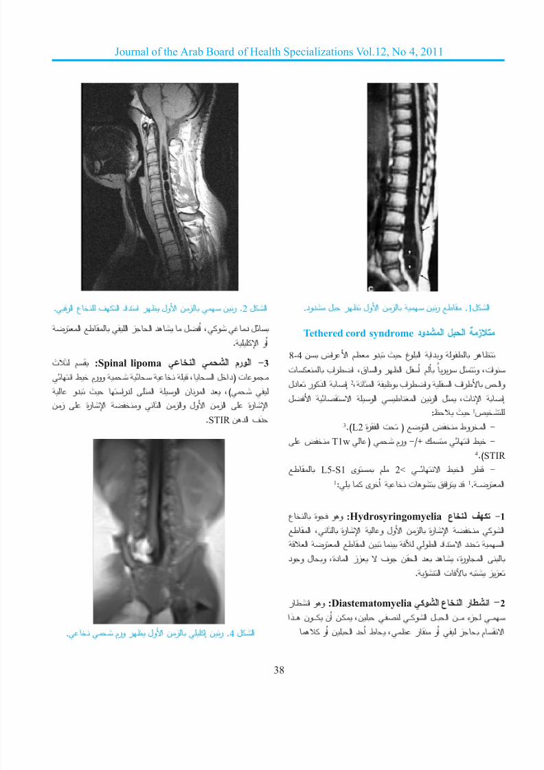
5/17/2018 12-4 _ Act - slidepdf.com
http://slidepdf.com/reader/full/12-4-act 43/110
38
Journal of the Arab Board of Health Specializations Vol.12, No 4, 2011
. ط رن زن اول ظر ل دود. ال1
Tethered cord syndrome ز ال اشدود
8-4 ظر طو ودا اوغ ث دو ظم اراض ن م أل اظر واق ،اطراب ت رر لو ،تاو إ اذور دل 2،ا ظو بارطاو ا فارط ساو
ص 1ث ظ:إ اث ،ل ارن اط او ا ال3.)L2 - اروط ض او ( ت ارة
ض T1w ( مرو -/+ ك ا ط -4.)STIR
ـط L5-S1 م وى 2< ــ ـ ـ ـ ا طا رط - 1: ىرأ تو قار د 1. ـ ـ ـرا
:Hydrosyringomyeliaوو وة ع -1ف اع
طا ، ةرا و لوا نز ةرا واا را طا ن وطا دادا دد ا اورة ،د د ان وف زز ادة ،ول وود
.ؤا ت زز
اشطر اع اشو :Diastematomyelia وو اطر -2
ـــ زء ــــن اـــل اوـــ ـ ن ،ـن أن ـــــون ــــذا وأ نا دأ ط ،ظ ر وأ ز ما
.را ع فا دا ن زن اول ظر اد ر . 2 ا ل
را ط ا زا د لأ ،و د ل .ا وأ
:Spinal lipomaم ث ا شا مروا -3ا ط مروو ،ا لاد( تو ود ث ارد ا وا نرا د ،)
ارة ازن اول وازن ا و ارة زن.STIR ندا فذ
. مرو رظ لوا نز إ نر . 4 ال

5/17/2018 12-4 _ Act - slidepdf.com
http://slidepdf.com/reader/full/12-4-act 44/110
39
Journal of the Arab Board of Health Specializations Vol.12, No 4, 2011
. رن ورض زن اول ظر ل دود اطر ع. 3 ال
. ن: رن زن ا ظر ، ر: رن زن اول ظر ، اوط: ل 5 ال ر واع ات.
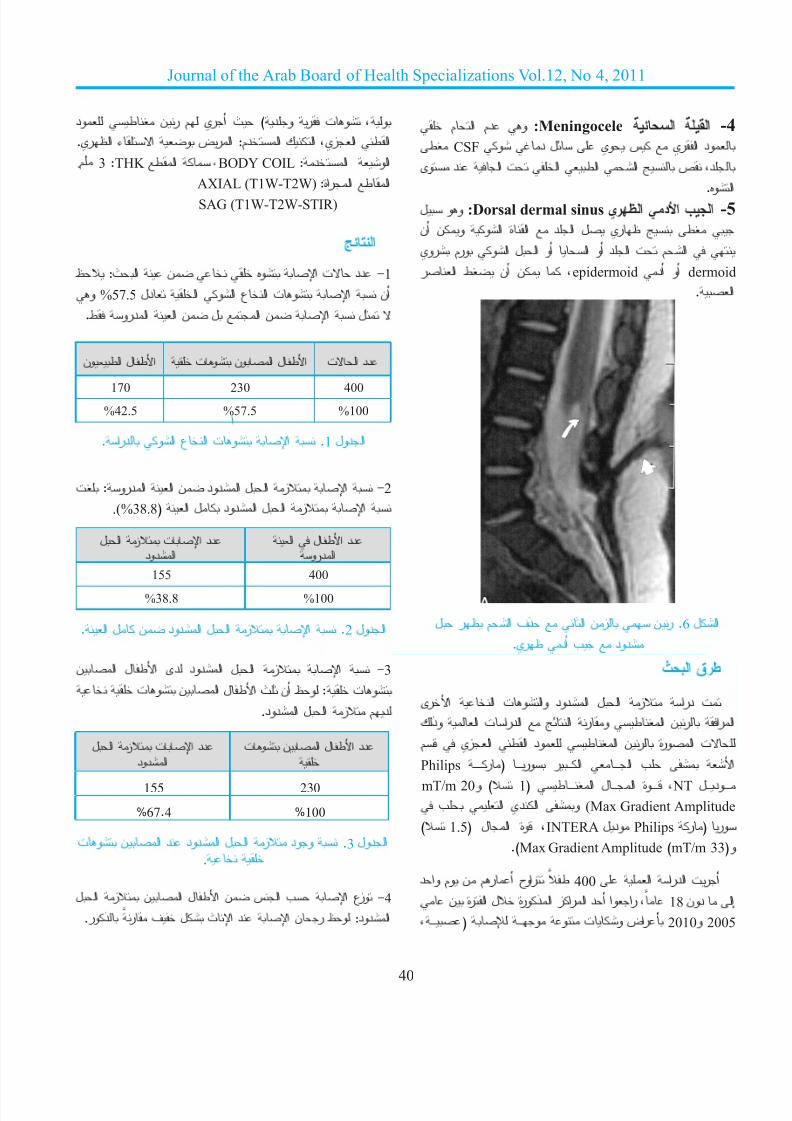
5/17/2018 12-4 _ Act - slidepdf.com
http://slidepdf.com/reader/full/12-4-act 45/110
40
Journal of the Arab Board of Health Specializations Vol.12, No 4, 2011
ما م د و :Meningocele ا ا -4
ط CSF و د ل يو س يرا دو د ،ص ا اط ا ت ا د وى
:Dorsal dermal sinusوو لاوه. اب اد اظري -5 ط ظري ل اد اة او ون أن ام ت اد أو ا أو ال او ورم روي ار ط نأ ن ،epidermoid دأ وأ dermoid
.ا
. رن زن ا ذف ام ظر ل 6 ال دود ب أد ظري.
طرق اث
را ز ال ادود واوت ا ارى د ت ارا رن اط ور ا ادرات ا وذك ت اورة رن اط ود اط ازي م Philips ــ ـ ـ ـ ـ ـ ـ ر( ــ ـ ـ ـ رو رــ ـ ـ ا ــ ـ ـ ـ ـ ا ب اmT/m 20و ) 1( طــ ـ ـ ـ ـ ا لــ ـ ـ ـ ا ةوــ ـ ـ ـ ـ ـ ، NT ل ـ ـ ـ ـدو ـ ـ ـ ـ ـ ـ ب ا يدا و (Max Gradient Amplitude
) 1.5( لا ةو ،INTERA Philipsودل ر( رو .)Max Gradient Amplitude )mT/m 33(و
400ط راوح أرم ن وم واد ا اردا ترأ ن ةرا ل ةروذا زارا دأ اوار ، 18 إ دون، ـ ـ ـ ـ ـ( ـ ـ ـ ـ ـ ـو و تو ضار 2010و 2005
و ،وت ر ود )ث أري م رن ط وداط ازي ،اك ادم: ارض و اء اظري.. 3 :THK طا ،BODY COIL :دا وا
AXIAL (T1W-T2W) اط اراة:SAG (T1W-T2W-STIR)
ا
-1دد ت ا وه ن اث: ظ
و %57.5 أن ا وت اع او ا دل ل ا ن ا ل ن ا ادرو ط.
ا.ارد وا عا تو ا . 1 ادول
- ا ز ال ادود ن ا ادرو :ت 2
.(%38.8( ا ل دودا لا ز ا
دد ات ز الادود
ا لطا ددوردا
155 400
%38.8 %100
.ا ل ن دودا لا ز ا .2 ادول
دى اطل ان دود ا لا ز ا -3 تو نا لطا ث نأ ظو : تو
دم ز ال ادود.
. وود ز ال ادود د ان وت 3 ادول.
-4وزع ا ب اس ن اطل ان ز ال
ذور. ر ف ل ثا د ا نر ظو :دودا
دد ات ز الادود
دد اطل ان وت
155 230
%67.4 %100
ون طا ل طا تو نو ا ل طا ات د د
170 230 400
%42.5 %57.5 %100
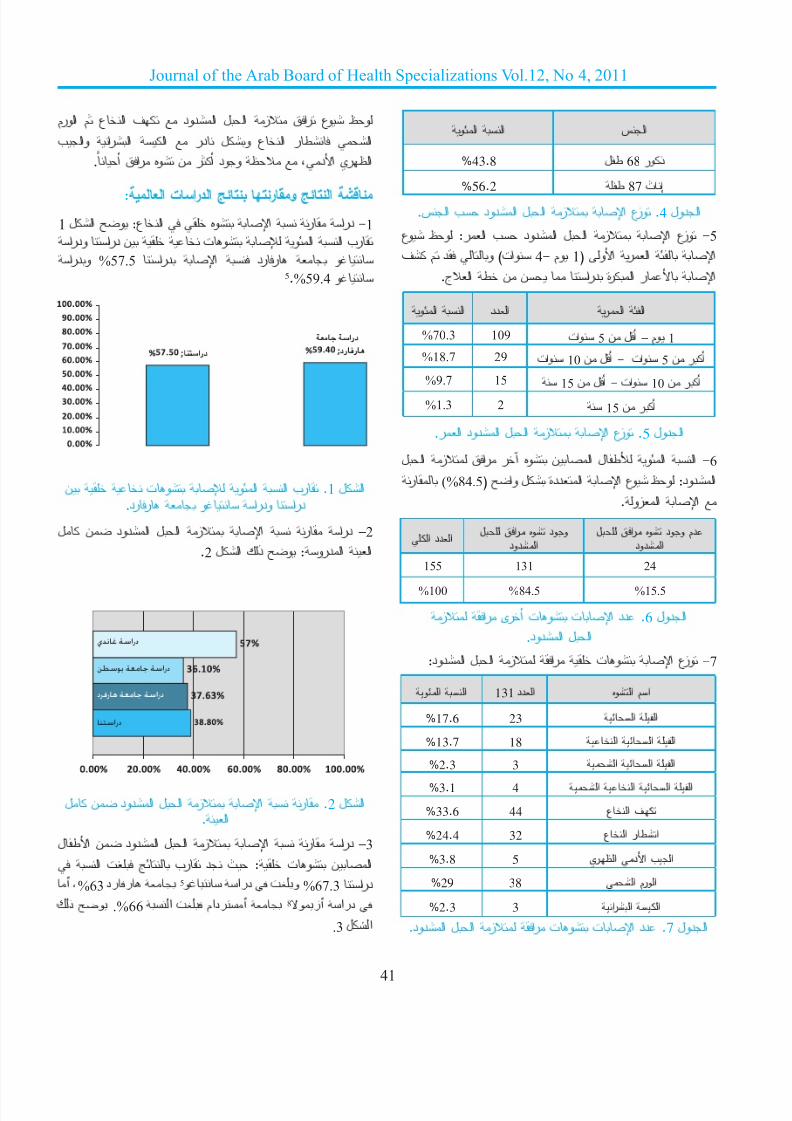
5/17/2018 12-4 _ Act - slidepdf.com
http://slidepdf.com/reader/full/12-4-act 46/110
41
Journal of the Arab Board of Health Specializations Vol.12, No 4, 2011
-5وزع ا ز ال ادود ب ار: وظ وع
ا ار او 1( وم - 4وات( و د م فا ر ارة درا ن ن ط اج.
ادول .5وزع ا ز ال ادود ار. -6ا او طل ان وه آر راق ز ال
ر (%84.5( او ل ةددا ا عو ظو :دودا.وزا ا
ز ار ىرأ تو تا دد .6 لودا ال ادود.
- وزع ا وت را ز ال ادود: 7
وظ وع راق ز ال ادود ف اع م اورمارا واب ا رد لو عا رط ا
.أ قار هو ن رأ دوو ظ ،دا يرظا
:ا تاردا رو ا ش -1درا ر ا وه اع: و ال 1
اردو ارد ن ت و وا ا بر اردو %57.5 ارد ا درر و
5.%59.4 و
ال .1رب ا او وت ن درا ودرا و ررد.
–2درا ر ا ز ال ادود ن ل
.2 ا ادرو :و ذك ال
ال .2ر ا ز ال ادود ن ل.ا
–3درا ر ا ز ال ادود ن اطل
ا ت بر د ث : تو ناأ ،%63 هرفرد 5غ ارد ف و %67.3 ارد أدام ف ا .%66 ح ذك 8زأ ارد ف
.3 شا
وا ا 131 د د ا ه و ا م ا
%17.6 23 ا ا%13.7 18 ا ا ا%2.3 3 ا ا ا%3.1 4 ا ا ا ا%33.6 44 ف اع
%24.4 32 اطر اع
%3.8 5 اب اد اظري%29 38 ا مروا
%2.3 3 ارا ا
ا ددا وود وه راق لادود
دم وود وه راق لادود
155 131 24
%100 %84.5 %15.5
و ا ا د د ا ر ا ا %70.3 109 5وات 1وم – أل ن%18.7 29 10وات 5وات - أل ن أر ن%9.7 15
15
وات - أل ن10
%1.3أر ن 2 15 أر ن
و ا ا س ا
%43.8 ل ط 68 ذور
%
56.2 ط 87 ث إ
. وزع ا ز ال ادود ب اس. 4 ادول
ادول .7دد ات وت را ز ال ادود.
دناغ ةس
سوب ةماج ةس
رفاه ةماج ةس
سا
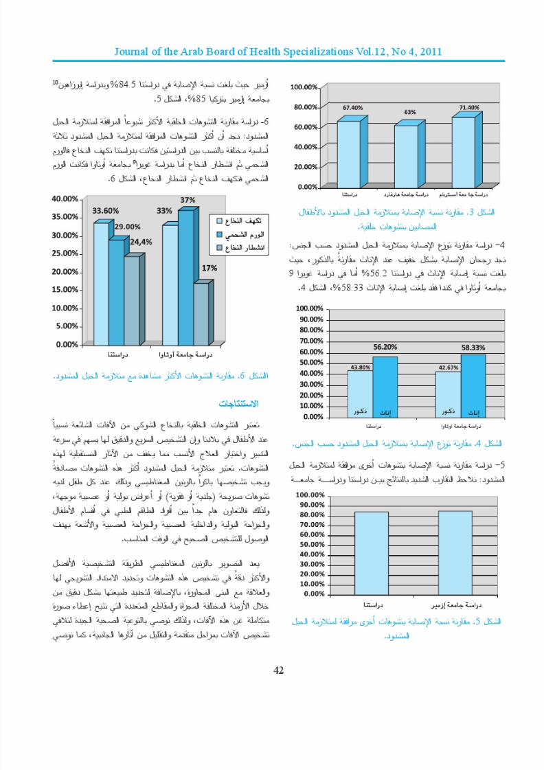
5/17/2018 12-4 _ Act - slidepdf.com
http://slidepdf.com/reader/full/12-4-act 47/110

5/17/2018 12-4 _ Act - slidepdf.com
http://slidepdf.com/reader/full/12-4-act 48/110
43
Journal of the Arab Board of Health Specializations Vol.12, No 4, 2011
تو دوو و ا تا نر ا ءار طا ددو د ل ا م ر ل وا عا ا ،و او ء ات ادث ل اور.اد را اراو ةوا ا طا تاذ ةزا ء نر
المراجع
1. Ross JS, Brant-Zawadzki M, Moore KR, et al. Diagnostic
imaging, spine. 1 st edition. Manitoba-Canada: Friesens,
Altona; 2005. p. 10-59.
2. Weissleder R, Wittenberg J, Harsiinghani M. Primer
of diagnostic imaging. 3rd edition. Philadelphia-USA:
Mosby; 2003. p. 1049, 569-71.
3. Brant W, Helms C. Fundamentals of diagnostic radiology.
3rd edition. Vol:1. Philadelphia-USA: Lippincott; 2007.
p. 1559,308-11.4. Benz-Bohm G. Kinderradiologie. Stuttgart-Germany:
Georg Thieme; 1997. p. 286,244-6.
5. Santiago ML, al-Orfali M, Zurakowski D, et al.
Occult lumbosacral dysraphism in children and young
adults: diagnostic performance of fast screening
and conventional MR imaging. Radiology 1999
Jun;211(3):767-71.
6. Long FR , Hunter JV, Mahboubi S, et al. Tethered cord
and associated vertebral anomalies in children and
infants with imperforate anus: evaluation with MR
imaging and plain radiography. Radiology 1996 Aug;
200(2):377-82.
7. Kumar R, Singh V, Singh SN. Split cord malformation
(Occult spina bida): An indian scenario. JK Sci
2005;7:192-4.
8. Azimullah PC , Smit LM, Rietveld -Knol E, et al.
Malformations of the spinal cord in 53 patients with
spina bida studied by magnetic resonance imaging.
Childs Nerv Syst 1991 Apr;7(2):63-6.
9. Guerra LA, Pike J, Milks J, et al. Outcome in patients
who underwent tethered cord release for occult spinal
dysraphism. J Urol 2006 Oct;176(4 Pt 2):1729-32.10. Erşahin Y, Mutluer S, Kocaman S, et al. Split spinal
cord malformations in children, neurosurgical focus.,
Nov 97. This paper was presented in part at the XXIV
Scientic Meeting of the International Society for
Pediatric Neurosurgery, Ottawa, Ontario, Canada, July
913, 1996.
5/17/2018 12-4 _ Act - slidepdf.com
http://slidepdf.com/reader/full/12-4-act 49/110
44
Journal of the Arab Board of Health Specializations Vol.12, No 4, 2011
Original Article
PH MONITORING IN SYRIA FROM 2004-2010
2004و2010 ن رو لطا د يرا و س ت ل Mahmoud Bozo, MD.
مو بوظو .
ص اث س و اري اطر ا ص د :ثا فد،ا تواو تا ن ةدا را يدا ساا 2004 اد ذ لطا د رو إ دإ م دو إ ا ر د ا ه ذ فد .ا ا 2009 ا وم ل ارات س و اري ،ودرا ارط ات،ا ط ر تاءارا ل ط
وا طء رت ن طب س و اري.ذن م طب س ا لطا اردا ت :ثا قرط
2010-2004ور ل ن مد يرا و ط ىدو را ،بطا ،را ،سا اردو ا
.ا طو )ثإ 19 ،روذ 11( ط 30 اردا ت :اا را تاذ و ا بطا ن ،ار 72.4 أرماررة ) ،)%34أ ارو وات ا ا ا م ل
ABSTRACT
Objective: PH monitoring is considered as the best diagnostic modality of gastro-esophageal reux, introduced in
Syria in 2004 in private and in 2009 in public hospitals. The aim of this pilot study is to analyze the PH monitoring tests cases, the indications, the results and parameters analysis, and do the proposals.
Methods: All cases of PH monitoring from 2004 to 2010 in Syria in pediatric patients were reviewed. Age, gender,
indications, results of the different parameters were analyzed.
Results: Thirty patients, 11 males and 19 females, mean age 72.4 months, the most prevalent indication was the
recurrent pneumonia (34%), the asthma and the recurrent upper respiratory infections presented only 20%. Forty
perecent presented with one symptom, 60% form two symptoms or more. The test was positive in 73% and negative
in 27%. The mean number of reux episodes ≥5 min was 189.3, and the mean of total time pH <4 was 26.6.
Conclusions: The demanded tests are too limited, the most frequent cases are not the asthma or upper respiratory
infections. The parameters analyzed are compatible with the international parameters.
ن اطل ون ن رض واد ط %40 نأ ظو .%20 وى %73ن إ را ن .ر نر ن نو %60و
و طوا سا تار دد ن ،%27 طو تا.26.6 < 4دل و وا او ،189.3
يرا و س را بط ةدز ب :تا ور ،ث ظ دد ات ،ب زدة طب ار.ا ا ا تاو ورا ل ا تطا ا اردا هذ ظا تاءارا قاو إ ا ر
. ةدا
دا لطا د ضارا أ ن را يدا سا ر ف ار ،وف إ ن: اس ار واس ر تط قار يذا سا و را ساو ،را أو رج ، ل اء اد ،ل او ،ارو ،ذات
*Mahmoud Bozo, MD, Department of Pediatrics, Damascus Hospital, Damascus, Syria. E-mail: [email protected].

5/17/2018 12-4 _ Act - slidepdf.com
http://slidepdf.com/reader/full/12-4-act 50/110
45
Journal of the Arab Board of Health Specializations Vol.12, No 4, 2011
ت ا ا ا اررة ،ال ازن ا ،ةررا را.ا،...
ا اورو راض ز ام واذ د اطل د أ 1،ضرا ص لا وا و يرا و س نأ اطرق ارى ل اورة اظ د إ رم ل زام ث ن وت را ءات اطل ،وأ وطأ .لطا د سا ص مرأ د م يرا ورة ص ث ل و وأ د ،نوا ،ذا تاو تا ن اد قرطا هذ ل أ ط ر در دة اس ل ووده ،ورارا را توا ن ثا تاءارا هذ تطا
س.1
وو تاذ ر رط و يرا و س نإ %90ث د رة اص ،ل ا وب رب ا رو ، ن ةدا ر ذا تا عو نأ ل اذو .ررا ا رو أو ات اذ ود إ دة أب ر أ أطء اراءة
)إ إ ت اس اوي ،أو اس اوب ن ام(.
وا د ص اس رأ يرا و س ط
وا ر ت د ا ةءار م ث ،را يداةدا ـ ـ ـ ـ ـ ـ ـ ـا ا نرو ، ـ ـ ـ ـ ـ ـ ـ ـ ـ ـ ـ 24 أل اري دة.)1 )ادول رم
طب س و اري ت ر ددة ل اتا أو رج ا ا و إ ال وود اسأو ر ا را ةدا ر وأ ةدا تا ء ا ل ، ب
ا ا تا ،ةررا را تاذ ،ورا ،و ل ارااء س ر إ د ث 8-3،إ.... نزا لا ،ةررا ا و اري ن أم اطرق ا و و اص ا رض
1
اب رض وال ب ار. طب إراء س و اري را اج اد س ات ا ر اراض د ارة ج مإ س و اري ر إذا ت ك أراض أرىرا تاوذ وأ ورا وأ نزا لا ل( ضر س ار
.)ا .... ةررا
،رم اده دد د إ يرا و س لدإ ر لوأ ار إ ود اذو ،ةروطا دا لوط نز ذ
ور ذا ار. دف ذه ادرا او ن و ور إ دد وا تر ث نو تطا ث ن رو يرا و س ط تا طرا ارد إ ، نا لطااءات ر ط ا ،وا طء ر ا ل
رت ن طب س و اري.
طرق اث
و س ءار مو م نذا لطا اردا ت لطا مو قد طا ا ن ل يرا .2010وم د 2004و زران م دق ذ دا م أي طل أري ار ن ادرا .وم ر ادار ل
.ر ر ارد إ
24 اءة ا د ت ر او أل اري دة ت ر ،وت ر ا ا ادة.
24 24-hour Component
4>و وا ا<4.8%Total time pH <4 (%)
<48 No. of reux episodesدد رات ازر
ئد 5 ≤ <5 No. of reux episodes ≥5 minدد رات ازر ادة
)د( رزا دد لوطأ <28Longest reux episodes (min)
. ر ص اس س و اري.2 1 ادول

5/17/2018 12-4 _ Act - slidepdf.com
http://slidepdf.com/reader/full/12-4-act 51/110
46
Journal of the Arab Board of Health Specializations Vol.12, No 4, 2011
م ادرا رط اراض ارر او ل ال رات اسادث دى اطل ،ب ادت ا :دد رات اس اراقإ ار ر ر ث ،ا سا تار دد\يرر ضر ،%10ودد رات اس ا تزو اذإ ر يد س دوو
ر رو ،ضارا لو تار دد\يرر ضر قارا 2.%75 ا تزو اذإ ر يد س دوو إ ار
زا طورا ن م ،يرا و س ءارإ د ت ات اذ ر ،و ر أ رورة إف اجاد س ل إراء ار دة ( م ن ار ن أل را اج( ،وذك دة أم دات ا ،و48
،H2و 12دات اس. داتا
بط ا تا دد مد ظو ، 30 ا ددا ،2010 و زران 2004 ار دم اوات ذ م ن ن طب ار رة ل أر د أ طب رة ل
.1 أ ،ال رم
،)%63( ار 19 اردا ت د س ا داو ،فو نار ر دا دـــــاو ،ثــــإ )%37( 11
.2 ،ال رم ار 72.4 132را ،ووط ار
)%34( را ما نو ،ةدد ت تطاا ا ا تا ت أ ،ا را تاوذ ت أ .2 %14ط ،ادول رم وارو %10 اررة م ل وى) )%40وا ون ط 12 ن ث دد اراض ،د وظ أن
ط أطل ن رن و11 )%23.3( 7 ،ط داو ضر ن ) )%36.6ن أراض ر.
.2اطت س و اري. ادول
21 ن دد ات ا أظرت إ ر و%( ،ت 9ت )27 ط را ن ،)%73( ض ت ذوات ر ،اءات را ن ا تا ول و وال زن ،ت ل ت ارو واق
.3
وات ا ا ا إ ،ادول رم دل را ن ا تا طوا را طوا را نأ .4 ( ،ادول رم ار 13-2.5( ار 10.5
120-2.5( ار 42.9 ات ا ن ار إ دل.4 ( ،ادول رم ار
وحظ حل ئج اخر اوط أن دد رات اس احت
اطب (%) وا ا
ر تاذ 34
ال ازن 13
ارو 14
اق 13
ا وا ا تا 10
را اج 7
ر ف اص 3
إءات ول و 3
ر ف 3
ا 100
. أر اطل اذن أري م ار. 2 ال .1دد ات ب اوات. ال

5/17/2018 12-4 _ Act - slidepdf.com
http://slidepdf.com/reader/full/12-4-act 52/110
47
Journal of the Arab Board of Health Specializations Vol.12, No 4, 2011
ر ا ت ا د د ر ا ت ا د د ض ر ا 9 3 ا را تاذ
2 1 إءات ول و3 0 اق1 0 ارا د ار 4 0 ارو3 1 ل زن1 0 ر ف اص3 0 إت ررة
. ار ب اطب.3
ادول
أو ار. تا دد .4 ادول
،5أ ات ) ،)625-48ادول رم 189.7 اا أ .)71-2( 35.2 ا ،د ن وط دد رات اس-4.6( %26.6 او و 4< ات ا ت %1.54 ــ ت ـ ـ ا تا ــ ــ ـ ـ أ ،6 ،)%100ادول رم
.)%2.9-0.2(
شا
ا ب ضارا أو مأ ن را يدا سا د اطل ف ار ،و ظرات ل اء وظرات
ل ارو وات ا ا وا ا ووب جر اق ووف اس ور .ود س و اري ن أم ذإ ،را يدا سا ص طو م ا لوا
13-9 ،2، 1.%90 ـاو وو
،وذا ر إ أن ارا 30 ارد لوا ا ددا ن دد ات ا طب ار دود ( رم ان اوظ د م إ دو اذو ،)ةرا ا بوطا ددا
د د ا س ا ت ا ر دد
1 48>
2 100-48
2 200-101
2 300-201
7 400-301
7 500-401
2 625
.ا تا سا تار دد .5 ادول
اد 4> و وا ا8 4.6-9.2
7 11.3-17.3
4 31.3-68.6
2 92.4-100
.ا تا 4< و وا ا .6 ادول
دد ات ذات ار
ا
دد ات ذات ار
اار
1 2 > 6أر3 4 ار 12-6
2 3 ار 24-13
6 0
62-25را
5 0 > 10وات
5/17/2018 12-4 _ Act - slidepdf.com
http://slidepdf.com/reader/full/12-4-act 53/110
48
Journal of the Arab Board of Health Specializations Vol.12, No 4, 2011
ار د ض اراءات ر او اص )لوروا ةرا توا ن ،)ير ظا ةروا وار راض ز ام واذ د اطل ادرة م
ذه اول اد ن اص وأن
2008ر إ أن
اص د س و اري.
،وذا ود ار 72.4 إ أن طب ار م ات اط ل ارو وات ن وط ار ا طب ار. د رأ ثد رو ا ا ا
رم وود رن ذري او ادرو وو واق ض ا ا ،إ أ ن د د إ ذها واق ا ا ب دود ادد ان
14
.ارد ازر ن تطا ن را ما نأ ارد ظ ا ا تا ت أ ،)%34( ا را تاوذ ت %14ط. ول ذا ود وارو %10 ا اررة م ل وىإ ف ا ا ن اطء اث ن اس اديا تاو ورا ت ل و را تا را
15.ا تاردا إ ترأ وو ا ا
ن اطل وا ون ن رن %60 أرت درا إ أن
وس ود اظرات. ر ،وذا أن اس ادي ار رض دد اظرات
%73ن ات ت إ ر إ أ إراء س ون نإ را إ نأ ذإ ، ا تا ل يرا و ر إ رورة طب ذا ار ذه ورا تا ل ةررا را تاوذ ل ضارا ض لطا إ ،تا%70 نأ ت ارد ن ،سا و با نأ ةرور
ن ت ذوات ار ا ن س.
( ،وار اوط ات ا ن ن ار اوط ات ا ن ار دل ار 13-2.5( ار 10.5
نأ اذو .)ار 120-2.5( ار 42.9 دل إ را أب ات ا ود أراض رة س ن أن ون
ار ار وواق إ ر.
طو نأ ث ،ا مرا طوا اءار قاو ،189.7أ ات دد رات اس ات ا ن
ا ت ن .35.2 ا د ن وط دد رات اس ،%26.6أ ات ا تا 4>و وا
.ا ا قاو وو 1.54 ا ت
ات
ظ ث يرا و س را بط ةد ز ب دد ات ،أن ار إ زدة ط تاطت ا ل ارو وات ا ا ا ،ث. ةدا ا ظا تاءارا قاو إ ا ر
ارا
1. Vandenplas Y, Colin D. Rudolph Committee Members:
Di Lorenzo Z, Hassall E, Liptak JJ, et al. Pediatric
gastroesophageal reux clinical practice guidelines:Joint
recommendations of the North American Society for
Pediatric Gastroenterology, Hepatology and Nutrition
(NASPGHAN) and the European Society for Pediatric
Gastroenterology, Hepatology and Nutrition (ESPGHAN)
J Pediatr Gastroenterol Nutr 2009;49:498-547.
2. Fass R, Hell R, Sampliner RE, et al. Effect of ambulatory
24-hour esophageal pH monitoring on reux-provoking
activities. Dig Dis Sci 1999;44(11):2263-9.
3. Fass R, Ofman JJ. Gastroesophageal reux disease-
should we adopt a new conceptual framework? Am J
Gastroenterol 2002;97:1901-9.
4. Sandler AD, Everhart JE, Donowitz M, et al. The burden
of selected digestive diseases in the United States.
Gastroenterology 2002;122:1500-11.
5. Rudolph CD, Mazur LJ, Liptak GS, et al. Guidelines
for evaluation and treatment of gastroesophageal reux
in infants and children. J Pediatr Gastroenterol Nutr
2001;32 Suppl 2:S1-31.
6. DeVault K, Castell D. Practice parameters committee of
the American College of Gastroenterology. Guidelines for the diagnosis and treatment of gastroesophageal
reux disease. Arch Intern Med 1995;155:2165-73.
7. Drumm B. The dangers of consensus. J Pediatr
Gastroenterol Nutr 2001;33:429-30.
8. Colletti RB. Can a good guideline improve patient care?
J Pediatr Gastroenterol Nutr 2002;35:242-3.
9. Bagucka B, Badriul H, Vandemaele K, et al. Normal ranges
of continuous pH monitoring in the proximal esophagus.

5/17/2018 12-4 _ Act - slidepdf.com
http://slidepdf.com/reader/full/12-4-act 54/110
49
Journal of the Arab Board of Health Specializations Vol.12, No 4, 2011
J Pediatr Gastroenterol Nutr 2000;31:244-7.
10. Vandenplas Y, Franckx GA, Pipeleers MM, et al. Area
under pH 4: advantages of a new parameter in the
interpretation of esophageal pH monitoring data in
infants. J Pediatr Gastroenterol Nutr 1989;9:34-9.
11. Friesen CA, Hayes R, Hodge C, et al. Comparison of
methods of assessing 24-hour intraesophageal pH
recordings in children. J Pediatr Gastroenterol Nutr
1992;14:252-5.
12. Rebecchi F, Di Francia I, Giaccone C, et al. Improving
the analysis of esophageal acid exposure by a new
parameter: area under H+. Am J Gastroenterol
2002;97:568-74.
13. Weusten B, Akkermans L, vanBerge-Henegouwen G, et al.
Symptom perception in gastroesophageal reux disease
is dependent on spatiotemporal reux characteristics.
Gastroenterology 1995;108:1739-44.
14. Størdal K, Johannesdottir GB, Bentsen BS, et al. Acid
suppression does not change respiratory symptoms in
children with asthma and gastro-oesophageal reux
disease. Arch Dis Child 2005;90(9):956-60.
15. Thakkar K, Boatright RO, Gilger MA, et al.
Gastroesophageal reux and asthma in children: A
systematic review. Pediatrics 2010;125(4):925-30.

5/17/2018 12-4 _ Act - slidepdf.com
http://slidepdf.com/reader/full/12-4-act 55/110
50
Journal of the Arab Board of Health Specializations Vol.12, No 4, 2011
Case Report
ABSTRACT
Diclofenac is one of the most common drug in use
for relieving the pain and discomfort. It's popular uses
make it's rare side effects of the injectable form appear
more frequently and in a dangerous form. One of theseis the respiratory distress which may be fatal. Our case
may refect that many cases may be lost especially in
rural areas and no one will notice it.
INTRODUCTION
The acute respiratory distress syndrome (ARDS) is a
life threatening condition with mortality rates of about
40-60%.1,2 Treatment strategies, with the exception
of low tidal volume mechanical ventilation, have had
little impact on outcomes.3 ARDS is dened as an
acute condition characterized by bilateral pulmonaryinltrates and severe hypoxemia in the absence of
evidence for cardiogenic pulmonary edema. By these
criteria, the severity of hypoxemia necessary to make
the diagnosis of ARDS is dened by the ratio of the
patient' s arterial oxygen partial pressure (PaO2) to the
fractional concentration of oxygen in the inspired air
(FIO2), PaO2/FIO2. In ARDS, this ratio is less than 200.4 The acute phase of ARDS usually resolves completely.
Less commonly, residual pulmonary brosis occurs, in
which the alveolar spaces are lled with mesenchymal
cells and new blood vessels.
Non-steroidal anti-inammatory drugs (NSAID) are
most commonly used to combat the symptoms associated
with diseases such as rheumatoid arthritis, osteoarthritis,
ulcerative colitis, and collagen diseases. Their analgesic
properties also help to relieve back pain, joint pain, and
other types of musculoskeletal pain, as well as the paincaused by external and surgical wounds.5
DICLOFENAC-INDUCED ACUTE RESPIRATORYDISTRESS SYNDROME (ARDS)
ز ائ اس احدة احر بدوفكMohamad A Bajubair, MSc, MD; Yahia Elezzi, MD; Faker Qobati, MD; Yassin Abdulkader, MD
سن ع الدر د. ،طق كف د. ،يزلا ىح د. ،جب حم د.
مخص الحلة
ادوفك و أحد ادو شئ اسخدام في ج حت ام. وج سخدات ارة ذا ادواء فد أصبحت ارات اجب ادرة وخطورة. شل ز ائ اس إحدى ذه ارات. سرض في ذا ارر أحد ذه ادراو رأ لشب ظح نحب ئ ط ثودحا
.را قطا يف صخو صخش لغ د سا ئا تح ن را نأ دا ع تحا
*Mohamad A. Bajubair, MSc, MD; Associate professor, Faculty of Medicine and Health Sciences, University of Sana'a, Althawra Modern General Hospital,
Department of Medicine, Yemen. E-mail: [email protected]
*Yahia Elezzi, MD; Associate professor, Faculty of Medicine and Health Sciences, University of Sana'a, Althawra Modern General hospital, Department of
Medicine, Yemen.
*Faker Qobati, MD; Associate professor of Medicine, Respirology Division, Faculty of Medicine and Health Sciences, University of Sana'a, Department of
Medicine, Yemen.
*Yassin Abdulkader, MD; Professor of Medicine, Faculty of Medicine and Health Sciences, University of Sana'a, Department of Medicine, Yemen.
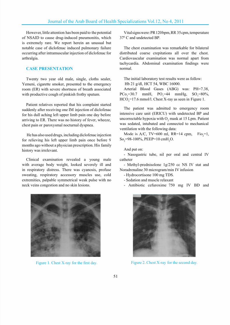
5/17/2018 12-4 _ Act - slidepdf.com
http://slidepdf.com/reader/full/12-4-act 56/110
51
Journal of the Arab Board of Health Specializations Vol.12, No 4, 2011
However, little attention has been paid to the potential
of NSAID to cause drug-induced pneumonitis, which
is extremely rare. We report herein an unusual but
notable case of diclofenac induced pulmonary failure
occurring after intramuscular injection of diclofenac for
arthralgia.
CASE PRESENTATION
Twenty two year old male, single, cloths sealer,
Yemeni, cigarette smoker, presented to the emergency
room (ER) with severe shortness of breath associated
with productive cough of pinkish frothy sputum.
Patient relatives reported that his complaint started
suddenly after receiving one IM injection of diclofenac
for his dull aching left upper limb pain one day before
arriving to ER. There was no history of fever, wheeze,
chest pain or paroxysmal nocturnal dyspnea.
He has also used drugs, including diclofenac injection
for relieving his left upper limb pain once before 9
months ago without a physician prescription. His family
history was irrelevant.
Clinical examination revealed a young male
with average body weight, looked severely ill and
in respiratory distress. There was cyanosis, profusesweating, respiratory accessory muscles use, cold
extremities, palpable symmetrical weak pulse with no
neck veins congestion and no skin lesions.
Vital signs were: PR 120 bpm, RR 35 cpm, temperature
370 C and undetected BP.
The chest examination was remarkable for bilateral
distributed coarse crepitations all over the chest.
Cardiovascular examination was normal apart from
tachycardia. Abdominal examination ndings were
normal.
The initial laboratory test results were as follow:
Hb 21 g/dl, HCT 54, WBC 16000.
Arterial Blood Gases (ABG) was: PH=7.38,
PCo2=30.7 mmH, PO
2=44 mmHg, SO
2=80%,
HCO3=17.6 mmol/l. Chest X-ray as seen in Figure 1.
The patient was admitted to emergency room
intensive care unit (ERICU) with undetected BP anduncorrectable hypoxia with O
2mask at 15 Lpm. Patient
was sedated, intubated and connected to mechanical
ventilation with the following data:
Mode is A/C, TV=600 ml, RR=14 cpm, Fio2=1,
So2=98-100%, PEEP=10 cmH
2O.
And put on:
- Nasogastric tube, nil per oral and central IV
catheter
- Methyl-prednisolone 1g/250 cc NS IV stat and
Noradrenaline 30 microgram/min IV infusion- Hydrocortisone 100 mg TDS.
- Sedation and muscle relaxant
- Antibiotic cefuroxime 750 mg IV BD and
Figure 1. Chest X-ray for the rst day. Figure 2. Chest X-ray for the second day.
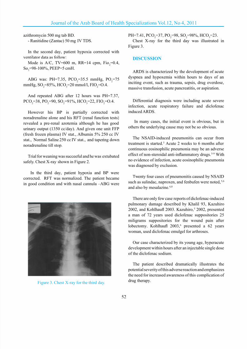
5/17/2018 12-4 _ Act - slidepdf.com
http://slidepdf.com/reader/full/12-4-act 57/110
52
Journal of the Arab Board of Health Specializations Vol.12, No 4, 2011
azithromycin 500 mg tab BD.
- Ranitidine (Zantac) 50 mg IV TDS.
In the second day, patient hypoxia corrected with
ventilator data as follow:
Mode is A/C, TV=600 m, RR=14 cpm, Fio2=0.4,
So2=98-100%, PEEP=5 cmH.
ABG was: PH=7.35, PCO2=35.5 mmHg, PO
2=75
mmHg, SO2=85%, HCO
3=20 mmol/l, FIO
2=O.4.
And repeated ABG after 12 hours was PH=7.37,
PCO2=38, PO
2=90, SO
2=91%, HCO
3=22, FIO
2=O.4.
However his BP is partially corrected with
noradrenaline alone and his RFT (renal function tests)
revealed a pre-renal azotemia although he has good
urinary output (1350 cc/day). And given one unit FFP
(fresh frozen plasma) IV stat., Albumin 5% 250 cc IV
stat., Normal Saline 250 cc IV stat., and tapering down
noradrenaline till stop.
Trial for weaning was succeeful and he was extubated
safely. Chest X-ray shown in Figure 2.
In the third day, patient hypoxia and BP were
corrected. RFT was normalized. The patient became
in good condition and with nasal cannula –ABG were
PH=7.41, PCO2=37, PO
2=98, SO
2=98%, HCO
3=23.
Chest X-ray for the third day was illustrated in
Figure 3.
DISCUSSION
ARDS is characterized by the development of acute
dyspnea and hypoxemia within hours to days of an
inciting event, such as trauma, sepsis, drug overdose,
massive transfusion, acute pancreatitis, or aspiration.
Differential diagnosis were including acute severe
infection, acute respiratory failure and diclofenac
induced ARDS.
In many cases, the initial event is obvious, but inothers the underlying cause may not be so obvious.
The NSAID-induced pneumonitis can occur from
treatment is started.5 Acute 2 weeks to 6 months after
continuous eosinophilic pneumonia may be an adverse
effect of non-steroidal anti-inammatory drugs.5-9 With
no evidence of infection, acute eosinophilic pneumonia
was diagnosed by exclusion.
Twenty four cases of pneumonitis caused by NSAID
such as sulindac, naproxen, and fenbufen were noted,5,6
and also by mesalazine.6,8
There are only few case reports of diclofenac-induced
pulmonary damage described by Khalil 93, Kazuhiro
2002, and Kohlhau 2003. Kazuhiro,5 2002, presented
a man of 72 years used diclofenac suppositories 25
miligrams suppositories for the wound pain after
lobectomy. Kohlhau 2003,6 presented a 62 years
woman, used diclofenac emulgel for arthroses.
Our case characterized by its young age, hyperacutedevelopment within hours after an injectable single dose
of the diclofenac sodium.
The patient described dramatically illustrates the
potential severity of this adverse reaction and emphasizes
the need for increased awareness of this complication of
drug therapy.Figure 3. Chest X-ray for the third day.

5/17/2018 12-4 _ Act - slidepdf.com
http://slidepdf.com/reader/full/12-4-act 58/110
53
Journal of the Arab Board of Health Specializations Vol.12, No 4, 2011
CONCLUSIONS
We conclude that ARDS may be induced by the
injectable forms of diclofenac. It is an acute illness that
may result in acute life-threatening respiratory failure. It
usually affects young healthy individuals and simulates
severe community acquired pneumonia. Due to the
differing treatments, it is important to distinguish ARDS
from infectious pneumonia. Other causes of respiratory
distress should be excluded always. Treatment with
corticosteroids could be life-saving as ARDS responds
easily and uniformly to corticosteroids.
REFERENCES
1. Leaver SK, Evans TW. Acute respiratory distress
syndrome. BMJ 2007;335:389-94.
2. Milberg JA, Davis DR, Steinberg KP, et al. Improved
survival of patients with acute respiratory distress
syndrome (ARDS): 1983- 1993. JAMA 1995;273:306 -9.
3. The ARDS Network Authors for the ARDS Network.
Ventilation with lower tidal volumes as compared with
traditional tidal volumes for acute lung injury and
the acute respiratory distress syndrome. The Acute
Respiratory Distress Syndrome Network. N Engl J Med
2000;342:1301 -8.
4. Abraham E, Matthay MA, Dinarello CA. Consensus
conference denitions for sepsis, septic shock, acute lung
injury, and acute respiratory distress syndrome: time for
a reevaluation. Crit Care Med 2000 Jan;28(1):232-5.
5. Kazuhiro U, Hisashi S, Toshiki T, et al. Diclofenac
(voltaren)-induced pneumonitis after chest operation.
Ann Thorac Surg 2002;74:2176 -7.
6. Kohlhau M, Weber N, Morresi-Hauf A, et al. Pulmonary
inltrates with blood eosinophilia in a 62-year-old
patient. Internist (Berl) 2003 Aug;44(8):1037-41.
7. Goodwin SD, Glenny RW. Non-steroidal anti-
inammatory drug-associated pulmonary inltrates
with eosinophilia: review of the literature and Food and
Drug Administration reaction reports. Arch Intern Med
1992;152:1521 -4.
8. Khalil H, Molinary E, Stoller JK. Diclofenac (Voltaren)-
induced eosinophilic pneumonitis: case report and review
of the literature. Arch Intern Med 1993;153:1649-52.
9. Martinez BM, Domingo P. Acute eosinophilic pneumonia
associated with tenidap. BMJ 1997;314(7077):349.
10. Allen JN, Pacht ER, Gadek JE, et al. Acute eosinophilic
pneumonia as a reversible cause of non-infectious
respiratory failure. N Engl J Med 1989;321:569-74.
11. Umeki S. Re-evaluation of eosinophilic pneumonia
and its diagnostic criteria. Arch Intern Med
1992;152:1913 -9.

5/17/2018 12-4 _ Act - slidepdf.com
http://slidepdf.com/reader/full/12-4-act 59/110
54
Journal of the Arab Board of Health Specializations Vol.12, No 4, 2011
Case Report
ABSTRACT
We report a case of a 12-year mentally retarded
boy presented with clinical features of acute abdomen
admitted to the Emergency Departement Erbil Teaching
Hospital. Explorative laparotomy showed wit gastro-colic stula following ingestion of multiple magnet
pieces before ten days; this case report highlights the
diagnostic challenge and the need for early surgical
intervention in children with foreign bodies ingestion
and especially when multiple magnets are ingested
INTRODUCTION
The accidental ingestion of foreign bodies is
a common clinical problem encountered in early
childhood but ingestion of magnets is extremely rare.
When multiple magnets are ingested, physically they
may attract each other and cause pressure necrosis
through the bowel wall and eventually lead to serious
lethal complications like obstruction, perforation, and
stula formation an example is the present case. Review
of literature shows that multiple magnet ingestion is an
unexpected health hazard in children that can lead to
signicant gastrointestinal morbidity.1,2
CASE PRESENTATION
We report 12-year old mentally retarded boy presented
with a ten days history of diffuse upper abdominal
pain with vomiting and fever. The vomiting eventually
became feculent. On clinical examination, he was
pyrexial with a soft, distended abdomen in the upper
abdominal region. There was no percussion tenderness
or guarding. A plain radiograph of the abdomen revealed
multiple opaque cylindrical structures in the upper
abdomen (Fig 1). On closer questioning it transpired
that he had ingested multiple large powerful magnets,
largest one measuring 3.5×1.5 cm, on separate occasions
a week before his illness. The white blood cell count was
16000; the blood biochemistry was grossly deranged
(sodium 129 mmol/l, potassium 3.3 mmol/l, chloride 82
mmol/l, urea 22.4 mmol/l, creatinine 128 μmol/l).
MULTIPLE MAGNET INGESTION IN A CHILD
(GASTRO-COLIC FISTULA)
ووك يد روس - سطغ مسأ ةد عبا Abdulqadir Maghded Zangana, CABS-FICS-MD; Mohammad Majeed, FICMS
جم حم د. ،هكنز يغم ردقلابع د.
مخص الحلة
12س ظر ب بطن د سم اسف شى إربل ا .أظر ح ابطن ره ق لط ررا اذ درو صخشا تدا ى ءوضا ا هذ طس .مأ 10 اسصئ وود سور دي كوو ج ن ابع دة أسم غطس بل
.سطغا مسا ت صخو لطا د برغا مسا عبا ت ركبا ارا لخادا ءار او
*Abdulqadir M. Zangana, CABS, FICS, MD, Department of surgery, Hawler Teaching Hospital, Erbil, Iraq. E:mail:[email protected]
*Mohammad Majeed, FICMS, Department of surgery, Hawler Teaching Hospital, Erbil, Iraq.

5/17/2018 12-4 _ Act - slidepdf.com
http://slidepdf.com/reader/full/12-4-act 60/110
55
Journal of the Arab Board of Health Specializations Vol.12, No 4, 2011
Following uid resuscitation, antibiotics, and
nasogastric drainage, a laparotomy was performed.
There was no obvious peritoneal contamination. On
careful palpitation multiple metallic pieces inside the
stomach and mid transverse colon were identied. The
magnets had adhered to one another, forming a cylindrical
structure that was moving free in the lumen of stomach
and transverse colon (Figure 2). Exploration through
the lesser sac revealed a large gastro-colic stula, tract
of stula resected and the bowel wall repaired and the
peritoneal cavity was lavaged with normal saline.
In the postoperative period he was seriously ill. He
had a prolonged postoperative ileus requiring total
parenteral nutrition for a week. Ultimately, however, he
made a full recovery and he was discharched home on
the tenth postoperative day, and he has no complaints 3
months after the procedure.
DISCUSSION
Foreign body ingestion is common but multiple
magnet ingestion is rare.1 When more than one magnet
is ingested, gastrointestinal complications may occur.
The magnets are attracted to each other across the bowel
wall and this may lead to pressure necrosis, perforation,
stula formation, or intestinal obstruction.2,3
Foreign body ingestion in 80% of cases involving
children between the ages of 6 months and 3 years.
Commonly ingested objects include coins, toy parts,
jewellery, batteries, needles and pins, and sh and
chicken bones. In 80-90% of cases, usually nonmagnetic
FBs spontaneous passage through the gastrointestinal
tract occurs once the foreign body has entered the small
bowel,3,4 so that surgical intervention is not usually
necessary. Upper gastrointestinal FBs are amenable
to retrieval by endoscopy2,5 or Foley balloon catheter
extraction.6,7
On the other hand multiple magnet ingestion is
especially hazardous, because individual magnets tend
to interact through the bowel wall. A review of the
published literature revealed previous cases of children
aged 2–3 years in whom obstruction, perforation, stula
formation, and adhesions occurred.6,7 Magnets attract
one another forcefully, and through pressure necrosis
can cause serious bowel injury. In all previously reported
cases, surgical intervention was necessary.7
Unless the objects are large or sharp, they usually
pass through a child’s digestive system without health
consequences. However, the U.S. Consumer Product
Safety Commission (CPSC) has reported 20 cases of
severe outcomes following ingestion of powerful rare-
earth magnets.6
CPSC has become aware of toy products containing
small, powerful rare-earth magnets that pose uniquehealth hazards to children. Since 2003, CPSC staff
members have identied one death resulting from
ingestion of these magnets and 19 other cases of
Figure 2. Demonstration on the X-ray the site of gastro-
colic stula, with pressure necrosis of stomach wall and
transverse colon wall by severe magnets traction.
Figure 1. Plain X-ray of the abdomen of 12-year mentally
retarded child revealed multiple opaque cylindrical
structures in the upper abdomen.

5/17/2018 12-4 _ Act - slidepdf.com
http://slidepdf.com/reader/full/12-4-act 61/110
56
Journal of the Arab Board of Health Specializations Vol.12, No 4, 2011
injuries requiring gastrointestinal surgery.4,6 All children
swallowed more than one magnet, except one child who
ingested a magnet and another metal object. Most of the
magnets had been dislodged from toys. One patient died
following volvulus, bowel necrosis, and sepsis. The
other 19 patients had some combination of the following:
perforations, volvulus, bowel resection, ulcerations,
peritonitis, and magnets embedded in the stomach
lining. The CPSC has recalled some toys (e.g., magnetic
building sets) and is working with the American Societyfor Testing and Materials International Toy Safety
Standard subcommittee to address these risks.8
If the ingested FB is revealed on a plane radiograph
and the position becomes xed on serial radiographs,
there should be suspicious of ingestion of multiple
magnets. In multiple magnet ingestion, once symptoms
of increasing abdominal pain or signs of intestinal
obstruction or perforation develop then prompt
exploratory laparotomy should be performed.7,8
CONCLUSIONS
Multiple magnet ingestion, compared to a single
magnet, is a distinct clinical situation that needs to
be managed differently. Two X-ray views should be
considered. Multiple magnet ingestion can lead to
serious gastrointestinal complications, and awareness
among clinicians and the public of the health hazards
may improve the outcomes.
REFERENCES
1. Hachimi-Idrissi S, Corne S, Vandenplas Y. Management
of ingested foreign bodies in childhood: our experience
and review of the literature. Eur J Emerg Med 1998;
5:319-23.
2. Laurance Hill J, Voigt RW. Foreign bodies. In: Ashcraft
KW, editor. Pediatric surgery. 3rd ed. Philadelphia: WB
Saunders Company; 2000. p. 146-52.
3. Honzumi M, Shigemori C, Ito H, et al. An intestinal
fstula in a 3-year-old child caused by the ingestion of
magnets: report of a case. Surg Today1995;25:552-3.
4. Kay M, Wyllie R. Pediatric foreign bodies and their
management. Curr Gastroenterol Rep 2005;7:212-8.
5. Haraguchi M, Matsuo S, Tokail H, et al. Surgical
intervention for the ingestion of multiple magnets by
children. J Clin Gastroenterol 2004;38:915-6.
6. Tay ET, Weinberg G, Levin TL. Ingested magnets: the
force within. Pediatr Emerg Care 2004;20:466-7.7. Centers for Disease Control and Prevention (CDC).
Gastrointestinal injuries from magnet ingestion in
children, United States, 2003–2006. MMWR Morb
Mortal Wkly Rep 2006 Dec 8; 55:1296-300.
8. Ahmed AM, Hassab MH, Al Hussaini AA, et al. Magnetic
toy ingestion leading to jejunocecal fstula in a child.
Saudi Med J 2010 Apr;31(4):47-51.
.
Figure 3. Postoperatve reforming picture of the magnets
swallowed showing magnets attract one another
forcefully between stomach and transverse colon
with stula formation.
Figure 4. Postoperative magnet pieces separated
from each other after removal with some
other nonmagnetic foreign bodies.

5/17/2018 12-4 _ Act - slidepdf.com
http://slidepdf.com/reader/full/12-4-act 62/110
57
Journal of the Arab Board of Health Specializations Vol.12, No 4, 2011
Cysticercosis
داء الكيت المذنبة
A 67-year-old man was referred to the hematology department for evaluation of monoclonal gammopathy.
As part of his workup, a skeletal survey was performed. The study did not show any lytic bone lesions, but
numerous “rice grain” calcications were seen throughout the body (with the exception of the hands and feet),
with their long axes oriented in the plane of the muscle bers (Panels A and B). This appearance is highly
suggestive of cysticercosis. The patient was asymptomatic at the time of presentation but reported that as an
adolescent he had had muscle pain for which he had never been treated. The patient grew up without indoor
plumbing, in a region where the domestic breeding of pigs was a frequent practice and the likely cause of
his infection. The patient reported no past or present neurologic symptoms. Cysticercosis is caused by the
hematogenous dissemination of larvae from the pork tapeworm, Taenia solium. The eggs are typically ingested
through contaminated food or water. Common sites of involvement include the central nervous system, which
can lead to neurologic symptoms, and the muscles, which can lead to muscle pain. No therapy
for cysticercosis was given.
ءارإ م د ما ط ن ءزو ،د ا دو لا م مدا ضا 67إ م أر ول رض ره .م ظر اور وود أ آت ظم ،ون وظ وود ت ددة ) ت ارز( ل ام )ء ادن
Medical Case

5/17/2018 12-4 _ Act - slidepdf.com
http://slidepdf.com/reader/full/12-4-act 63/110
58
Journal of the Arab Board of Health Specializations Vol.12, No 4, 2011
ذا تا ءاد ص ةدو رظا اذ حر .) Bو A وادن( ،ث و ور ذه ات وى اف ا )ال م ارا ةر مأ دوو دروأ أ إ ا رظ د ضا .Cysticercosisم و ارض ن أ أر
.ررع ارض ط ود ددات ه دال ازل ،ر ر ت أ ن ازر ث
.Taenia soliumم اع ر ذه اوات ادر او ذه ا دى ارض. م ورد ارض وود أو راض .داء ات اذ ن ار ادوي رت دودة رط ازر )ارط ا )أو ارط اودة، ضار دو وو زرا ا ا ا نا ل .ثوا ءا وأ مطا ر ةدودا هذ ضو
أم .م م إطء أ ذا ارض دى ارض. تاو .....................................................................................................................
Ivo Ricardo da Silva Ferreira, M.D, Sara Pinto Magalhaes, M.D.
Centro Hospitalar do Porto, Porto, Portugal
N Engl J Med 2011; 365:e41. November 24, 2011. Images in Clinical Medicine
Translated by Samir Aldaldati, MD
.

5/17/2018 12-4 _ Act - slidepdf.com
http://slidepdf.com/reader/full/12-4-act 64/110
59
Journal of the Arab Board of Health Specializations Vol.12, No 4, 2011
Selected Abstracts
Public Health..........................................................(P60)
*Antioxidants in the chemoprevention of colorectal cancer
and colorectal adenomas in the general population.
Pediatrics................................................................(P61)
*Molecular assays in the diagnosis of neonatal sepsis.
*Systematic review of gastrointestinal injury caused
by magnetic foreign body ingestions in children and
adolescence.
*Pharmacotherapy review of chronic pediatric hypertension.
*Usefulness of magnetic resonance cholangiopancreatography
in pancreatobiliary abnormalities in pediatric patients.
Obstetrics and Gynecology....................................(P65)*Coenzyme Q10 content in follicular uid and its relationshipwith oocyte fertilization and embryo grading.
*Exercise during pregnancy and the gestational age
distribution.
*Effect of progestin compared with combined oral
contraceptive pills on lactation.
*Maternal caffeine intake and its effect on pregnancy
outcomes.
*Postpartum screening for diabetes among women with a
history of gestational diabetes mellitus.
Surgery...................................................................(P70)*Recurrence of inguinal herniae following removal of
infected prosthetic meshes.
*Video-assisted mediastinoscopy in superior vena cava
obstruction: To fear or not to fear?
*Nonoperative treatment for acute scaphoid fractures.
*Systematic review and meta-analysis of antibiotic
prophylaxis to prevent infections from chest drains in blunt
and penetrating thoracic injuries.
Cardiovascular Diseases........................................(P73)
*The «smoker’s paradox» in patients with acute coronary
syndrome.
*Sudden cardiac death in athletes: what is the role of
screening?
Pulmonary Diseases...............................................(P75)
*uPAR regulates bronchial epithelial repair in vitro and is
elevated in asthmatic epithelium.
Gastroenterology....................................................(P77)
*Imbalance of CD4(+) T cell subgroups in ulcerative colitis.
*Meta-analysis: ursodeoxycholic acid for primary sclerosing
cholangitis.
Endocrinology, Metabolism, & Diabetes Mellitus(P79)
*The effect of metformin therapy on vitamin D and B12
levels in patients with diabetes mellitus type 2.
*Clinical pharmacology of incretin therapies for type 2
diabetes mellitus: implications for treatment.
Hematology and Oncology....................................(P81)
*A review of tumour lysis syndrome with targeted therapies
and the role of rasburicase.
Neurology...............................................................(P82)
*Serum lipid levels and the risk of intracerebral hemorrhage.
Rheumatology and Orthopedics.............................(P83)
*Prognostic interest of bone turnover markers in the
management of postmenopausal osteoporosis.
Allergic and Immunologic Diseases......................(P84)
*The effect of perinatal omega-3 fatty acid supplementation
on inammatory markers and allergic diseases.Urology and Nephrology.......................................(P85)
*The efcacy of ascorbic acid in suboptimal responsiveanemic hemodialysis patients receiving erythropoietin.
Psychiatry...............................................................(P86)
*Psychological treatments for depression and anxiety
disorders in low- and middle- income countries.
Anaesthesia and Intensive Care Medicine.............(P87)
*Intravenous arginine vasopressin infusion in refractory
vasodilatory shock.
*Systematic review and meta-analysis of the effect of
warming local anesthetics on injection pain.
Otorhinolaryngology..............................................(P90)
*Effects of Asian sand dust on mucin gene expression and
activation of nasal polyp epithelial cells.
Laboratory Medicine..............................................(P91)
*Wide variation in reference values for aluminum levels in
children.
Dermatology...........................................................(P92)*Treatment of granuloma annulare with the 595-nm pulsed
dye laser.
Ophthalmology.......................................................(P93)
*Nonselective β-blocker propranolol for orbital and periorbitalhemangiomas in infants: a new rst-line of treatment?*Long-term follow-up of changes in corneal endothelium
after primary and secondary intraocular lens implantations in
children.

5/17/2018 12-4 _ Act - slidepdf.com
http://slidepdf.com/reader/full/12-4-act 65/110
60
Journal of the Arab Board of Health Specializations Vol.12, No 4, 2011
Public Health
Antioxidants in the chemoprevention of colorectal cancer and colorectal adenomas
in the general population
دا مارواو ماو نووا نطر ن وا وا ةدا تاد رود اوون وام د اس
Papaioannou D, et al.
Colorectal Dis 2011 Oct;13(10):1085-99.
Aim: Antioxidants, such as vitamin A, C and E, selenium and β-carotene, have been proposed as possible agents in
the chemoprevention of colorectal cancer and have been the subject of recent trials and reviews. This review aimedto assess the present evidence on the effect of antioxidants on the incidence of colorectal neoplasms in the general
population.
Methods: A systematic review of randomized controlled trials was undertaken comparing antioxidants alone or in
combination with other agents vs placebo. The following databases were searched for published and unpublished
literature: Cochrane Library, MEDLINE, PreMEDLINE, CINAHL, EMBASE, Web of Science, and Biological
Abstracts and Research Registers. Studies were quality appraised and extracted. Meta-analysis was performed.
Results: Twelve studies were identied as relevant. In the nine comparing antioxidants with no antioxidants(n=148922), there was no difference in the incidence of colorectal cancer [relative risk (RR) 1.00, 95% condenceinterval (CI) 0.88-1.13]. One study assessed the effect of antioxidants on adenoma formation (n=15 538) and did
not demonstrate a statistically signicant effect (RR 1.47, 95% CI 0.97-2.23). Of 14 discrete analyses for different
combinations of antioxidants, only one reported a statistically signicant increase in relative risk of adenomaformation in participants receiving vitamin E (RR 1.74, 95% CI 1.09-1.79, P=0.02) or vitamin E plus β-carotene (RR 1.63, 95% CI 1.01-2.63, P=0.04). Effectiveness did not seem to differ between healthy populations, participants withcardiovascular risk factors or populations exposed to smoking or asbestos.
Conclusion:The review demonstrates that antioxidants (vitamin A, C and E, selenium and β-carotene), as single agents,in combination with other antioxidants or in combination with other agents, are not effective in the chemoprevention
of colorectal neoplasia in the general population. This questions their involvement in future randomized controlled
trials of chemoprevention in colorectal cancer.
،اوم وا رون( وال م او او ن Eو C ،A د ارح وود دور دات ادة )ل ات دف اث:
. دف ذه ارا إ م ادل ا ول ر دات ادة دوث ؤات اوون وام د وم اس. رطن اوون وام ث ت دة دد ن ادرات وارات اراة ؤرا
طرق اث: م إراء را درات اوا اوط اراة ر ن ادام دات ادة ود أو ر وال،EMBASE ،CINAHL ،PreMEDLINE ،MEDLINE ،Cochrane ت داو ثا م .را ا ىرأ ،ات ا ا او وت اوث ا ن ات اورة وا م ر د ول ذا اووع. م Web of Science
. ل ءارإ تطا صاو تاردا ةدو م )148922 9درات ر ن ادام دات ادة ودم ادا( دد ات 12درا وا ووع ،ت ا :م دد .)1.13-0.88ت درا وادة م ،%95 لاو ،1.00 ا رطا( ماو نووا نطر ثود قر دوو مد تدروأ ث

5/17/2018 12-4 _ Act - slidepdf.com
http://slidepdf.com/reader/full/12-4-act 66/110
61
Journal of the Arab Board of Health Specializations Vol.12, No 4, 2011
،1.74 ا رطا( ا ا ن م ر دوو رظ مو )15538 ر دات ادة ل اورام اد( دد ات ةداو تدروأ د ،ةدا تاد ن طا ترا ف لو ا تا ن 14 .)2.23-0.97،%95ون ن لاو ،1.79-1.09 ،%95 لاو ،1.74 ا رطا( E ط زدة إ اطر ا ل اورام اد دى و ان أرى م ظ وود
ن .)0.04=p ،2.63 -1.01،%95 لاو ،1.63 ا رطا( نور ا E ،)0.02= pأو ان
روت ا ن اص اء ،اص ذوو وال اطورة ا او أو أوك ارن دن أو اوز. ،اوم وا رون( وال ردة ،أو ر دات Eو C ،A ات: ظر ذه ارا أن دات ادة )اتت اوون وام د وم ا ،ار اذي ر وك رة ول ة ارى أو وال أرى ر او ا ن ؤا اد
ادرات اوا اوط ا و ا ن رطن اوون وام ال.
Pediatrics
طب اطلMolecular assays in the diagnosis of neonatal sepsis
ات از شص إن ادم اوديPammi M, et al.
Pediatrics 2011 Oct;128(4):e973-85.
Background: Microbial cultures for diagnosis of neonatal sepsis suffer from low sensitivity and reporting delay.
Advances in molecular microbiology have fostered new molecular assays that are rapid and may improve neonatal
outcomes.
Objectives: We assessed whether molecular assays have sufcient sensitivity (0.98) and specicity (0.95) toreplace microbial cultures in the diagnosis of neonatal sepsis and explored heterogeneity by use of subgroup analyses
based on the type of assay, gestational age of the neonate, and type of sepsis onset.Methods: We performed the systematic review as recommended by the Cochrane Diagnostic Test Accuracy Working
Group. Electronic bibliographic databases, conference abstracts, personal les, and reference lists of identiedarticles were searched. We included studies of case-control or consecutive series design, which evaluated molecular
assays (index test) in neonates with suspected sepsis (participants) in comparison with microbial cultures (reference
standard). Two reviewers independently assessed the methodologic quality of the studies and extracted data.
Results: A bivariate random-effects model was used for meta-analysis of the 23 included studies, and summary
estimates of sensitivity and specicity with 95% condence intervals (CIs) were generated. Mean sensitivity andspecicity were 0.90 (95% CI: 0.78-0.95) and 0.96 (95% CI: 0.94-0.97), respectively. Real-time polymerase chainreaction (PCR) and broad-range conventional PCR had higher sensitivity and specicity than other assays. Sufcientdata were not available to evaluate gestational-age and sepsis-type subgroups.
Conclusion: Molecular assays do not have sufcient sensitivity to replace microbial cultures in the diagnosis of neonatal sepsis but may perform well as “add-on” tests.
ازروع ارو اد ص إن ادم دى د اودة ن ور إراد ا .زز ادم اث:ال م اء اد از دور ات از اد وار ن ا ارر اظ دى د اودة.
دف اث: م ذه ادرا م دى ات از( وق 0.98( وو )وق )0.95ن دا دل ازروعإ وع ادا را توا ت ا ل ن را ف ءارإ م ،ةدوا د ىد مدا نإ ص ورا
ا اد ،ار ا د اودة وط دء ان.

5/17/2018 12-4 _ Act - slidepdf.com
http://slidepdf.com/reader/full/12-4-act 67/110
62
Journal of the Arab Board of Health Specializations Vol.12, No 4, 2011
،ورا با ت داو ثا م .ا تارا د Cochrane وت رق ل ار ترأ طرق اث: ت اؤرات ا ،ات ا واوام ار ت اددة. م ن درات ن ط ات واواد أو درات اتورا عورزا ر )نرا( مد مد نإ دوو ا ةدوا د د )را( زا تا مادا ت او ا
.را تاو اردا ةدا ا ةء م ل لو نا نار م .)را را(واو ا لو تارد و م ،ارد 23 ا لا را اوا تارا جذو مادا م :ا)0.97-0.94 ،%95 لاو( 0.96و )0.95-0.78 ،%95 لاو( 0.90 واو طوا ما ت .% 95 لاو ارب. ل ن ل اوراز زن ا )Real-time PCR ( ول اوراز ادي وا اطقا ر را توا م ت راو م .تا واو ا م ) broad-range PCR (
ووع ان. ءادأ يد د أ إ ،ةدوا د ىد مدا نإ ص ورا عورزا ل ل زا تا ك ات:
دا ر إ د.
Systematic review of gastrointestinal injury caused
by magnetic foreign body ingestions in children and adolescence
را ذت اد او ا ن اع ام ار اط د اطل واران
Liu SQ, et al.
Zhonghua Wei Chang Wai Ke Za Zhi 2011 Oct;14(10):756-61.
Objective: To study the diagnosis and treatment of gastrointestinal injury caused by magnetic foreign body ingestions
in children.
Methods: A literature search was performed to identify all the studies related to gastrointestinal tract injury caused by
ingesting magnetic foreign body using databases including Google, Medline, ISI Web of Knowledge, Ovid, Wanfang
data, VIP, CNKI, degree dissertation, meeting abstracts, and request for document delivery. Language was limitedto English, Chinese, Japanese, and Korean. Parameters studies were age at diagnosis, gender, country, regional
distribution, number of magnetic foreign bodies, source of magnetic foreign bodies, clinical features, diagnosis, and
method for foreign body removal.
Results: A total of 98 cases of magnet ingestion were identied from 17 countries and regions. There were 94 patientsunder the age of 18, with most children younger than 5 years old (62.2%,61/98). The age at peak incidence was 3 yearsold (16.3%, 16/98). Magnetic foreign bodies ingested included toys (74.5%), medical apparatus (8.2%), accessories(4.1%), and others (6.2%). The number of bodies ranged from 2 to 100. Eleven (11.2%) patients were complicatedwith allotriophagia or autism. Delay diagnosis and treatment existed in all the patients to varying extents, of whom
one died from severe infection. Exploratory laprotomy showed a wide range of bowel damage from the esophagus
to the colon, including perforation and intestinal stula. Intestinal damage was the most common injury (51.0%),
followed by intestine-colon stula (15.3%). All the patients required bowel resection with anastomosis or stularepair except for 2 children who were managed by endoscopic removal of the foreign bodies.
Conclusion: Ingesting more than one magnet will lead to severe gastrointestinal injury. Early diagnosis and surgical
intervention are important. More precautious measures should be taken for children aged younger than 5 years old.
دف اث: درا ص و اذت اد او ا ن اع ام ار اط د اطل.طا را ما عا ن ا وا دا تذ ا تاردا دد طا بدا ث ءارإ م :ثا قرط ،Wanfang، VIP، CNKIرل ادوراه ،ت أل ،Ovidت ،را ISI ،Google، Medline دام دة ت

5/17/2018 12-4 _ Act - slidepdf.com
http://slidepdf.com/reader/full/12-4-act 68/110
63
Journal of the Arab Board of Health Specializations Vol.12, No 4, 2011
وردا را ت .رواو ا ،ا ،زا ت ةدراوا ثا را م .قوا م تطو ا تاءاازإ رطو صا ،ررا رظا ،ردو ا طا ما دد ،طا عزوا ،دا ،سا ،صا د ضرا ر
.ا ما ،18ون ظم ات دى أطل 94ر دون ن وط .ود اد 17 ط م عا 98 ا :م ل دد.)%16.3 98 3وات ) 16ن أل .)%62.2 وظ أن ذروة ادوث ت ر 98 دون ن ا 61( ن أل،)%6.2( ىرأ تاودأو )%4.1( ا ءا ،)%8.2( طا ةزا ،)%74.5 ( با ن ا طا ما ت allotriophagia ) )%11.2 رة ن اط ن را ال ر 11 ىد ا ت .100 إ 2 راوح دد ن رظأ .دد نإ ةداو ةو تد ، ترد را ىد او صا ر دوو ظو .دوا وأ ر او .وظ أن اذ او أر اواو با ن نووا و ءيرا ن وا تذا ن او فط ا نط ا)%51.0( ،اوار او اوو ) .)%15.3طت ا ال ء اررة إراء رة أو إح ور و تذا
وذك ات ء ن م ال ام ا ن ل اظر. د و ددة. ر اص ار وا راء ادال ارا ازم تذأ إ ط أ م ن رأ عا دو : ات
ن اور ددة ا ذا اووع. ب طق زد ن ادار او طل دون ا ن ار ب ذه ات.
Pharmacotherapy review of chronic pediatric hypertension
را ادوا رط اور اشر ازن دى اطل
Meyers RS, et al.
Clin Ther 2011 Oct 7.
Background: The number of antihypertensive agents on the market has increased dramatically over the past 20
years. Many of these agents are used to treat children and adolescents with hypertension despite there being relatively
limited data available supporting such use. Recent legislation has helped to increase the number of studies conducted
in children, but many clinical questions remain unanswered.
Objective: The goals of this article were to review the currently available antihypertensive agents used in the treatment
of pediatric hypertension and to assist clinicians in selecting the most appropriate treatment.
Methods: Searches of MEDLINE and International Pharmaceutical Abstracts through July 2011 were conducted.
Search terms used included child, pediatric, hypertension, and the following drugs: captopril, enalapril, lisinopril,
fosinopril, losartan, valsartan, irbesartan, candesartan, olmesartan, amlodipine, nifedipine, isradipine, felodipine,
propranolol, metoprolol, labetalol, minoxidil, furosemide, spironolactone, chlorothiazide, hydrochlorothiazide,
hydralazine, and prazosin. Clinical trial data were reviewed and evaluated and were limited to English-language
articles.
Results: A total of 45 observational and randomized controlled trials were identied and summarized in this review.The angiotensin-converting enzyme (ACE) inhibitors, angiotensin receptor blockers (ARBs), and calcium channel
antagonists (CCAs) had the strongest data to support their use in pediatric patients. ACE inhibitors and ARBs
are preferred agents for children with renal disease and have a favorable safety prole. Many trials, including 2comparative trials, supported the use of CCAs, particularly amlodipine, in children.
Conclusions: Trials in all 3 classes suggested their efcacy as well as a tolerable adverse-effect prole. More trialsin children are needed, particularly with newer antihypertensive agents. Comparative trials of different agents are the
most lacking.
: دت زدة وظ دد اوال ادة رع اور ار ل اوام ارن ا .دم ار ن ذه اوال اث

5/17/2018 12-4 _ Act - slidepdf.com
http://slidepdf.com/reader/full/12-4-act 69/110
64
Journal of the Arab Board of Health Specializations Vol.12, No 4, 2011
دم ادا دم. ت ارت ةدود تط دوو ن مرا ناراو لطا ىد را روا طر اودا.إ نود لاز ررا ا ن را نأ إ ،عووا اذ لو لطا د ةارا تاردا دد ةدز دا
واد ت ارع اور ار د ةراوا را روا عر ةدا لاوا ا دف اث: دف ذه ا إ راطل وذك دة اررن ار ا اب ذه ات.
ا تا مادا م .2011 وت اث ا ادوا ا ر وز MEDLINE ث ءارإ م :ثا قرط captopril, enalapril, lisinopril, fosinopril, losartan, valsartan,ا ودا ءأو ،را روا عرا ،لطا بط ،لط :ثاirbesartan, candesartan, olmesartan, amlodipine, nifedipine, isradipine, felodipine, propranolol, metoprolol, labetalol,
و . prazosinت را وم minoxidil, furosemide, spironolactone, chlorothiazide, hydrochlorothiazide, hydralazine
.زا ة طت ادرات ارر ار ات اوارد 45ن درات ارا وادرات اوا اوط وم ذه ارا .ن أن طت ازم اول ون ا :م دد CCAsز وى اطت ا دم ادا د اطل. ر ARBsورات وات اس ،ACEرات ت اون
اوال ا دى اطل د وود أراض و ث ARBs ،ACEرات ت اون طت ازم اول ون و CCAs ز ش ل ذه ات. د دت ار ن ادرات –و درا ر -ادام رات وات اس
د اطل. amlodipineات: رح ادرات دة وآر ل ف اث دو ادة رع اور ار .ب إراء ازد ن دو اددة ادة رع اور ار ،و ر درات ار ن و لطا د لاوا هذ مادا لو تاردا
وود ص ر ذه ادرات. ارظ ةرور را ا لاوا
Usefulness of magnetic resonance cholangiopancreatography
in pancreatobiliary abnormalities in pediatric patients
دة ور ارس واطرق اراو رن اط شف اشذوذات اراو ار دى اطل
Huang CT, et al.
Pediatr Neonatol 2011 Dec;52(6):332-6.
Background: Magnetic resonance cholangiopancreatography (MRCP) is an innovative and noninvasive technique
for evaluating the biliary tree and pancreatic duct in children. The aim of this study was to assess the usefulness of
MRCP as a noninvasive method to evaluate the biliary system in children.
Methods: We retrospectively reviewed the records of patients undergoing MRCP between October 2002 and May
2007 for suspected biliary system abnormalities. MRCP ndings were compared with other imaging modalities,operative ndings, and clinical endpoints.Results: Complete data were available for 60 patients (35 girls, 25 boys; mean age 2 years, 33 children less than 1
year old). Ultrasound was performed in all 60 patients. Twenty-two patients had choledochal cyst, and 19 had a thin
or invisible gall bladder. Endoscopic retrograde cholangiopancreatography was done in two patients. The sensitivities
and specicities of MRCP for diagnosing choledochal cyst and biliary atresia were 100.0% and 100.0% and 86.7%and 100.0%, respectively. Surgery was performed in 37 patients, including 21 with a choledochal cyst, 14 with biliaryatresia, and 1 with a pancreatic duct stone.
Conclusion: MRCP is useful method for evaluation of the pancreaticobiliary system in pediatric patients. It yields a
high degree of accuracy in the diagnosis of biliary atresia and choledochal cyst.
و وادة ور را م ارة اراو واة MRCP طا نر وارا قرطاو سرا رو ر :ثا فد

5/17/2018 12-4 _ Act - slidepdf.com
http://slidepdf.com/reader/full/12-4-act 70/110
65
Journal of the Arab Board of Health Specializations Vol.12, No 4, 2011
ار د اطل. دف ذه ادرا إ م دة ذه ا طر ر را م ا اراو د اطل. MRCP طا نر وارا قرطاو سرا رو نا ر طا تا ار ار ارد م :ثا قرط او .ت ر اوودات اظ ور ارس واطرق وود ك ذوذ ا ار 2007 2002وأر ارة ن رن اول
.رراو ارا تادووا ،ىرا ا لوا تادوو MRCP
طا نر واراا نود او اردا لطا ن 33 ) 35ة و 25وط أر ن ،أن ر 60 ن تو تراو :ا ،وظ وود ول راوي ط 22 اء ور واج وق او ار .وظ وود ة راو د ن ار(. م إر 19ط .م إراء ور رس وطرق راو طرق ارا ر اظر ن. ت وو ور رق أو ر ر د%86.7 ،%100 ،%100 وارا قرطا )دادا( قرو وارا ةا ت ص طا نر وارا قرطاو سرا ة ةداو و يوار دادا 14و وار ة ت 21 و %100ارب. م إراء را ـ 37 ر ن م
.را ةاط طر دة م ا اراو وار د اطل ،ث ا نر وارا قرطاو سرا رو ل : ات
.وارا ةا تو وارا ا دادا ص دا ن رد ط أ
Obstetrics And Gynecology
ا ضاراو دواCoenzyme Q10 content in follicular uid and its relationship
with oocyte fertilization and embryo grading
ال ار وه ب ا ا ودر ان Q10 زا ما ىو
Turi A, et al.
Arch Gynecol Obstet 2011 Dec 3.
Background: No data are available on the presence and content of Coenzyme Q10 (CoQ10) in human follicular uidand its role.
Objective: To assess the presence and concentration of CoQ10 in human follicular uid in relation to oocytefertilization.
Methods: CQ10 content was measured in follicular uid obtained from 20 infertile women undergoing ovarianstimulation program for in vitro fertilization. CoQ10 levels were assayed by high-performance liquid chromatography
system and normalized for follicular cholesterol and protein levels. Oocyte morphology and embryo grading were
assessed.
Results: CoQ10/Protein levels resulted signicantly in mature versus dysmorphic oocytes. Similarly, CoQ10/Cholesterol was signicantly higher in grading I-II versus grading III-IV embryos.Conclusions: This study is the rst demonstration of the presence of CoQ10 in the human follicular uid. Althoughthe biological and endocrine mechanism of CoQ10 in the follicular uid and its correlation with oocyte and embryodevelopment is unclear, a new step may be the administration of CoQ10 in infertile women to evaluate the biological
and reproductive outcomes.
ال ار دى ان ودوره ال. Q10 زا ما ىوو دوو لو تو راو : اث.ا ا ب و را ىد را لا Q10 زا ما زرو دوو م : دف اث

5/17/2018 12-4 _ Act - slidepdf.com
http://slidepdf.com/reader/full/12-4-act 71/110
66
Journal of the Arab Board of Health Specializations Vol.12, No 4, 2011
ضا ضر ر تا تا ءا ن 20 Q10ال ار اوذ ن زا ما ىو س م : طرق اث Q10ن ل اراب ا ال اداء وت و وت زا ما تو ت .جزا ب ءارإ
أرى م اص ا ا ودد در ان. ن م .برا لورواو نورا
\ارون إ طور ر طور ذ د دم ووده ،ول د وظ أن وىCoQ10
: أدت وتا
.IVو III درت ر IIو I \اورول أ ول م ادرت CoQ10
ا حوو مد ن مرا و .نا د را لا Q10 زا ما دوو رظ ا وا اردا هذ ر :تا و ا وطور ان ،إ أن اطوة اددة ل ط ء ت ام م Q10 او واد او م
.ظا او وا ا
Exercise during pregnancy and the gestational age distribution
ر ارن ل ال وزع ر ال
Owe KM, et al
Med Sci Sports Exerc 2011 Dec 2.
Purpose: To examine the associations between exercise performed at different time points during pregnancy and
gestational age in a population based cohort study.
Methods: Data included 61,098 singleton pregnancies enrolled between 2000 and 2006 in the Norwegian Mother
and Child Cohort Study (MoBa), conducted by the Norwegian Institute of Public Health. Self-reported exercise
was collected from two questionnaires in pregnancy weeks 17 and 30. Gestational age was determined based on
expected date of delivery according to ultrasound, as registered in the Medical Birth Registry of Norway. We used
logistic regression to analyze preterm (<37 completed weeks) and post-term birth (≥42 weeks). Comparison of mean
gestational age (GA) by exercise levels were estimated by general linear model.Results: Mean GA for women exercising 3-5 times a week in week 17 was 39.51 (95% CI 39.48-39.54) comparedto 39.34 (39.30-39.37) com pleted weeks for non-exercisers (p<0.001). Mean differences remained for all
categories of exercise after adjusting for confounding with the greatest mean difference between exercising
3-5 times per week in week 17 and non-exercisers (equals 1 day). Similar mean differences in GA were
observed by exercise levels in week 30. The greatest protective effect on risk of preterm birth were observed
for women exercising 3-5 times a week in weeks 17 or 30 (aOR=0.82; 95% CI 0.73-0.91; and 0.74; 0.65-0.83, respectively) compared to non-exercisers. Whereas, women exercising 1-2 or 3-5 times per week in
week 17, were slightly more likely to have a post-term birth (aOR=1.14;1.04-1.24; aOR=1.15;1.04-1.26,
respectively). Mean GA did not differ by type of exercise performed during pregnancy.
Conclusion: Exercise performed during pregnancy shifted the gestational age distribution slightly upwards resulting
in reduced preterm births and slightly increased post-term births.
. ارأ ارد لا رو لا ن تار ل را را ن را ن ط را ارد :ثا فد لطاو م ارا MoBa ارد 2009و 2000 ن ةرا ل ةدرا لوا ن 61098 طرق اث: ت ادرا ت ناوا ر كذ ل ما دارإ ل ن نرا ر دد م .ا ورا زر ل ن ترأ او ورا واج وق او .م ادام ار اط ل ةدو وا دوا د لا ر دد م 17و30. ا ا 42أو .)ت ر وط ار ا وت ارن ن ل وذج ( واودات د اوان )≤ وأ 37 نود( ناوا ل تادوا
ط م.

5/17/2018 12-4 _ Act - slidepdf.com
http://slidepdf.com/reader/full/12-4-act 72/110
67
Journal of the Arab Board of Health Specializations Vol.12, No 4, 2011
لاو( وأ 39.51 اوع ا ر وأ تار 5-3 نرا نر اوا ءا ىد ا را طو :اطوا تورا ء ظو .)0.001< p( نر ترا ر د )39.37-39.30( 39.34 ر )39.54-39.48 ،%95
اظ دى ل ت ر ارن د إراء ادت وال ارا وود ارق اوط ار ر ارن 5-3رات نرا ر تو ا را طو تور تظو .)مو 1 اوع 17ودم ر ارن )دار. ن أن ار او ار طر اودة ل اوان ظر دى اء اوا رن ارن 5-3رات أو اوع 17 30 اوع 0.83-0.65 ،%95ارب( ر دم لاو 0.74و 0.91-0.73 ،%95 لاو ،0.82 دا را ( أو30 اوع 17د وظ دن ال أ ل دوث وأ تار 5-3 وأ 2-1 لد نرا نر اوا ءا أ .نرا ر ودة رة ) ار اد ،1.24-1.04 ،1.14 و 1.26-1.04 ،1.15ارب(. م ل وود ات وط ر ال
ط ارن اراة ل ال.: ؤدي ر ارن ل ال إ ازح وزع ر ال و ام وو ل ن دل اودات ل اوان وزد ل طف ات
ن اودات د اوان.
Effect of progestin compared with combined oral contraceptive pills on lactation
ر ارون ر وب ال او اشر ارع
Espey E, et al.
Obstet Gynecol 2011 Dec 2.
Objective: To estimate the effect of progestin-only compared with combined hormonal contraceptive pills on
rates of breastfeeding continuation in postpartum women. Secondary outcomes include infant growth parameters,
contraceptive method continuation, and patient satisfaction with breastfeeding and contraceptive method.
Methods: Postpartum breastfeeding women who desired oral contraceptives were randomly assigned to progestin-
only and combined hormonal contraceptive pills. At 2 and 8 weeks postpartum, participants completed in-person
questionnaires that assessed breastfeeding continuation and contraceptive use. Infant growth parameters includingweight, length, and head circumference were assessed at 8 weeks postpartum. Telephone questionnaires assessing
breastfeeding, contraceptive continuation, and satisfaction were completed at 3-7 weeks and 4 and 6 months.
Breastfeeding continuation was compared between groups using Cox proportional hazards regression. Differences
in baseline demographic characteristics and in variables between the two intervention groups were compared using χ tests, Fisher exact test, or two-sample t tests as appropriate.
Results: Breastfeeding continuation rates at 8 weeks (progestin-only 63.5%; combined hormonal 64.1%), contraceptivecontinuation, and infant growth parameters did not differ between users of progestin-only and combined hormonal
contraceptive pills. Infant formula supplementation and maternal perception of inadequate milk supply were associated
with decreased rates of breastfeeding in both groups.
Conclusion: Choice of combined hormonal or progestin-only contraceptive pills administered 2 weeks postpartum
did not adversely affect breastfeeding continuation.
: م ر ادام ارون ل رد ر وب ال ارو او ار دت ارار ارع دف اثاط د اء د اودة. ت ا او رات او د ار ،ارار ادام طر ال ودى ر ارت ن
ع وو ال. را طرق اث: ت ادرا و ن اء طور ار اط د اودة ارات طق طر و ال ،م ن2 ز لاو د ترا تأ .را وا ورا لا بو ماد وأ در ل نورا ماد او ل او را د وا تار م م .ند لا ماداو طا عر رارا م باوا ةدوا د أ 8و

5/17/2018 12-4 _ Act - slidepdf.com
http://slidepdf.com/reader/full/12-4-act 73/110
68
Journal of the Arab Board of Health Specializations Vol.12, No 4, 2011
أ ن اودة. م ر اف إم اواب م ار اط وادام ال ،ودى 8 ل اوزن ،اطول وط ار أس دا Cox 4و 6أر. ت ر ارار رع اط ن اون دام ر ر ارت ن ذك د 7-3أ ود Fisher exact طورة. ت ر ارات اد اص ا وارات ن و ادال ن ل ار ي ،ار
ن.t
دى ادام ارون ل رد و %64.1دى ادام اروت وار %63.5( أ 8 ا :ت دت ارار رع اط درا اذا تارا ءطإ قار .نا نوا ن ف م را د وا تار ،لا رارا أ ،)را
ر وور ام دم اب ر ص دت ارع اط اون.: إن ار ن ت ال ارو ار أو ك او ارون ل رد اطة د أون ن اودة ؤر ات
.طا عرا رارا ل
Maternal caffeine intake and its effect on pregnancy outcomes
ول ام ن وراه ال
Jarosz M, et al.
Eur J Obstet Gynecol Reprod Biol 2011 Dec 3.
Objective: Estimation of the maternal caffeine intake during pregnancy and its inuence on pregnancy duration, birthweight and Apgar score of the newborn.
Study Design:The research was conducted on pregnant women who gave birth at the Clinic of Obstetrics, Gynaecology
and Oncology, 2nd Faculty of Medicine, Medical University of Warsaw. It covered unifetal pregnancies without
diseases (N=509). Research data were collected by direct questionnaire supplemented with data from patients’
records. Statistical analysis employed a multivariate logistic regression model and a non-parametric Spearman’s rank
correlation coefcient.Results: 98.4% of pregnant women consume no more than 300mg of caffeine per day. Pregnant women who smokeand older women consume more caffeine than non-smokers and younger women [β 1.95 (95% CI: 1.02-2.88)] and[β 0.68 (95% CI: 0.05-1.3)] respectively. There is no association between maternal caffeine intake during pregnancyand the risk of premature birth, the birthweight or the Apgar score of newborns.
Conclusion: Caffeine intake of no more than 300mg per day during pregnancy does not affect pregnancy duration
and the condition of the newborn.
دى دث اودة. Apgar و ةدوا نزو ،لا ةد ارو لا ل ما د نا لو م :ثا فد ا بطا مارواو ءا ضارأو دوا ةد ندو او لاوا ءا ن و ثا ءارإ م :ثا ط
(. م اطت ن ل اواب ار 509 ،ث ل اث اول اردة دون وود أراض را( دد ات Warsaw
ل د طت اوودة اط .م إراء ال ا ن ل وذج ار اط دد ارات ول ارط.ا ر ا Spearman’s
. أ اوال ادت أو ار را د ان و نا ن 300 % ن اء اوال ان وز 98.4 ا :ن أن 0.68 2.88-1.02({ و}) βدل ،%95 لاو( 1.95 لد β({ ار را لاواو تدا ر ر نا ن رأ وأ ةدوا نزو ،ةرا ةدوا رطو لا ل ن ما لو ن طرا دوو ظ م .برا })1.3-0.05 ،% 95 لاو(
دى دث اودة. Apgar
دة ال أو ا ا دث اودة. و 300 ات: ؤر ول ان ل ال ت أل ن
5/17/2018 12-4 _ Act - slidepdf.com
http://slidepdf.com/reader/full/12-4-act 74/110
69
Journal of the Arab Board of Health Specializations Vol.12, No 4, 2011
Postpartum screening for diabetes among women with a history
of gestational diabetes mellitus
ا يرا ءادا ت ةدوا د يرا ءادا ن حا
Tovar A, et al.
Prev Chronic Dis 2011 Nov;8(6):A124.
Introduction: To make recommendations for future clinical, public health, and research practices for women with
abnormal glucose tolerance during pregnancy, we reviewed the latest evidence regarding rates of postpartum diabetes
screening and types of screening tests.
Methods: We searched PubMed for journal articles published from January 2008 through December 2010 that
reported on postpartum screening and studies designed to prevent progression to type 2 diabetes among women with
gestational diabetes mellitus (GDM). Two authors independently reviewed titles and abstracts from 265 articles.
Results: From 34% to 73% of women with GDM completed postpartum glucose screening. Predictors of higher screening rates included older age, nulliparity, and higher income or education. Screening rates varied by race/
ethnicity; Asian women were more likely to be screened than were other racial/ethnic minorities. Women who
received prenatal care, who were treated with insulin during pregnancy, or who completed a 6-week postpartum visit
were also more likely to receive screening. A moderate proportion of women screened had type 2 diabetes (1.2%-4.5%) or prediabetes (12.2%-36.0%).Conclusion: Rates of postpartum screening among women with a history of GDM are low; only half of women in
most populations are screened. Our ndings can inform future screening initiatives designed to overcome barriers toscreening for both providers and patients. Well-designed lifestyle interventions specic to women with a history of abnormal glucose tolerance during pregnancy and also studies to determine the efcacy and safety of pharmacologicalinterventions will be important to help prevent progression to diabetes among these high-risk women.
ار ت د لا ل ءا د را ل تاذوذ ت او ا ،ررا ت تو و : د ادل اد ول دت ا ن اداء اري د اودة وط اوت ا ازم إراؤ ذه ات.
2010وا أوردت ا د 2008و ون اول ا نو ن ةرا ل ةروا تا ن PubMed طرق اث: م اث ر .GDMم ن ا يرا ءادا ت ءا د يرا ءاد ا طا روط ن و ا تاردا ،ةدوا
. 265 ان ول ل را اون وات ا نا مازا تد تار .ةدوا د مدا ر ن ا را ا ير تا ءا ن %73-34 تأ : ا ر ا ار ار ،دم وود ودات وادل ادي واوى ا ا .وظ وود روت دت ا فأ 6 ڊ ارق وا .وظ أن اء ات ل اودة ،أو ات ون ل ال ،أو اوا أن ازرات د اودة
،)%4.5-1.2( تا ءا ن طو ىد ا طا ن يرا ءادا دوو ظو .او أ لا ند وظت ل اداء اري دى %36.0-12.2.
اء ا د اودة د اء اوا ن ن داء ري دت ،ث أن ف ات: وظ أن دت إررا ن ل د ا او ا قاوا لو ا ا تا اء ط د ن .ن ذه اوودات أن و ادرإ لا ل را ل ذوذ ء او ةا ط ا تاد ما نإ .ا را ناو ا إ دد و ادات ادوا أور اد ن طور اداء اري د اء ذوات اطورة ا داء. ادرات ار

5/17/2018 12-4 _ Act - slidepdf.com
http://slidepdf.com/reader/full/12-4-act 75/110
70
Journal of the Arab Board of Health Specializations Vol.12, No 4, 2011
Surgery
اراRecurrence of inguinal herniae following removal of infected prosthetic meshes
س اق ار د إزا اشت ا ا ن
Rehman S, et al.
Hernia 2011 Aug 20.
Purpose: Late-onset mesh infection, occurring months to years following hernia repair, is a rare complication of
hernia surgery. Its management usually requires removal of the mesh. The aim of this paper was to assess the rate of
recurrence of inguinal herniae following removal of the mesh for late onset deep mesh infection.Methods: We performed a literature review to assess the rate of recurrence of herniae following the removal of a
delayed onset infected mesh. Relevant studies from January 1966 to June 2010 were identied from a Medline, PubMed,Embase, Scopus and Cochrane database search. Studies that reported recurrence of the hernia following removal of
delayed onset infected meshes were included. Results were tabulated and analysed to derive conclusions.
Results: Some 153 abstracts were reviewed and 12 potential studies initially identied; of these, only 7 were nallyincluded in this review. The included studies were six case series and one case report and comprised a total of 40
patients in whom delayed-onset infected mesh was removed. The median follow-up period after the removal of the
infected mesh was 26 months. Of these 40 patients, only 2 (<5%) developed a recurrent hernia. Removal of the meshresulted in resolution of symptoms in the majority of the patients.
Conclusion: Based on this review, removal of a late-onset infected mesh results in resolution of symptoms in the
majority of cases, whereas recurrence of hernia is not common. However, further research is required as the number
of patients in our review is relatively small.
دف اث: ر ان ار ا( ار ا )واذي دث د رور أر إ وات ن اح ارا ق إدىإزا ا اد اق. دف ذه اور ا إ م دل س ةد ا هذ ج بط .قوا ار ةردا تطا
. را قا نا ثود ىد ا ازإ د را قا طرق اث: م إراء اراض دب اط م دل س اق د إزا ا دى دوث إن ر ،م دد ادرات ذاتا تاردا تردأ .Cochraneو Scopus ،Embase ،PubMed ،Medline 2010ن واد ت وزران 1966 ا نو ن ا
ا ات دوث إن ر ا .ت دو ا و اص ات ول اووع. أوردت س اق د إز 6 ارا ا تاردا ت .ارا هذ ط 7 ،أدرت
اد تدد ارد 12و
153 ار ت :اطو .ا ر نإ ثود ىد مد ا ازإ ت ر 40 و ت ط داو ررو ت تارد
ت ا تا عو ن )%5 26را .طور س اق ن ط ) دون رة ا د إزا ا ا ن.را ىد ضارا ار إ ا ازإ تدأ .) 40( ر نإ روط ا ازإ
ادا ذه ارا ا ن اول ن إزا ا د طور إن ر اظور ؤدي إ را اراض ظم ات:ات ،دون دوث س اق. و ذك ب إراء ازد ن اث ول ذا اووع ذك أن دد ار ذه ارا ر
. ل

5/17/2018 12-4 _ Act - slidepdf.com
http://slidepdf.com/reader/full/12-4-act 76/110
71
Journal of the Arab Board of Health Specializations Vol.12, No 4, 2011
Video-assisted mediastinoscopy in superior vena cava obstruction: To fear
or not to fear?
ل ل ظر اف دة ادو ت اداد اورد اوف اوي دة ق؟
Pop D, et al.
J Thorac Oncol 2011 Dec 2.
Introduction: We investigate the safety and efcacy of video-assisted mediastinoscopy (VAM) used for diagnosis of the superior vena cava syndrome.
Methods: In a 7-year period, we have done 447 VAM for malignant causes. We have compared, in a retrospective
study from a prospectively maintained database, the differences between the groups with (rst group) or without(second group) superior vena cava syndrome in terms of operative time, preoperative and postoperative complications,
and results of pathologic examination. Statistical differences between the groups were calculated by χ2 test.Results: Mean operative time for rst (31 patients) and second (416 patients) groups was 18 and 34 minutes,respectively (p<0.00). Mean hospital stay was 1.2 days in both the groups. Mortality rates for the rst and second
groups were 0 and 0.5% (p=0.31); major morbidity rates were 0 and 0.7% (p=0.50); and minor morbidity rates were6.4 and 1.7% (p=0.27), respectively. In the superior vena cava syndrome group, most of the lymph nodes biopsiedwere in paratracheal superior sites (81%); histology showed small cell lung cancer in 51.6%, non-small cell lungcancer in 25.8%, and lymphoma in 22.6%.Conclusions: VAM represents an important diagnostic tool in superior vena cava syndrome. Once decided to be used,
we think that video technique is a good option with high rate of efcacy and low rates of mortality and morbidity.
ادم ص ز اورد اوف اوي. VAM د :م ذا اث اء و ظر اف دة ادو ظر ف دة ادو ت .م درا را ر ات ن 447 طرق اث: م ل رة 7وات إراء و وود ز اورد اوف اوي )او او( وو دم وود )او ا )ن ث دة اراء ،اطت ل
.ر ي را ل ن توا ن ا تورا ب م .را را صا و ءا ود ار،)0.00< p( برا د 34و 18 )ر 416( ا واو )ر 31( وا و ءارا ةد طو :ا %0.5و 0 او وا وا توا لد ىرأ ن .نوا و 1.2 ا ءا ةد طو 0و0.50= p( %0.7( ،دت ارا اط 6.4 و%1.7 ) )0.27= pارب. وظ دى ا ارا تد ،)0.31= p( برا و ز اورد اوف اوي أن ظم اد او ا م اول زت اوا ب ار ،)81%( ث أظر
.%22.6 وو %25.8 ا ر ر ر نطر ،تا ن %51.6 ا ر ر نطر دوو را راا هذ نأ ث ،يوا فوا دروا ز ت ةادأ VAM ات: ل ظر اف دة ادو
ل را دا دت ءة و ودت را واوت.
Nonoperative treatment for acute scaphoid fracturesةدا روزا مظا رو ارا رغ ا
Doornberg JN, et al.
J Trauma 2011 Oct;71(4):1073-81.
Background: Recommendations for cast immobilization of acute scaphoid fractures vary substantially. We reviewed
data from randomized controlled trials comparing nonoperative treatment methods for acute scaphoid fractures to
determine the best available evidence.

5/17/2018 12-4 _ Act - slidepdf.com
http://slidepdf.com/reader/full/12-4-act 77/110
72
Journal of the Arab Board of Health Specializations Vol.12, No 4, 2011
Methods: A systematic search of the medical literature from 1966 to 2010 was performed. Two authors
independently screened titles and abstracts, reviewed articles, assessed methodological quality according to the
Grading of Recommendations Assessment Development and Evaluation system, and extracted data. The primary
outcome parameter was nonunion. Data were pooled using random-effects models with standard mean differences
for continuous and risk ratios for dichotomous variables, respectively. Heterogeneity across studies was assessedwith calculation of the I statistic.
Resuts: The search resulted in ve potentially eligible trials of which four met our inclusion criteria. In total, 523 patients were included in four trials including two evaluating below-elbow casting versus above-elbow casting;
one trial comparing below-elbow casting including the thumb versus excluding the thumb; and one trial comparing
fractures with a below-elbow cast with the wrist in 20-degrees exion to 20-degrees extension, with both typesexcluding the thumb. There were no signicant differences in union rate, pain, grip strength, time to union, or osteonecrosis for the various nonoperative treatment methods.
Conclusions: There is no evidence from randomized controlled trials on physician-based or patient-based outcome
to favor any nonoperative treatment method for acute scaphoid fractures.
ف اوت ول ادام ارة ت اور ادة اظم ازور ات رة. م ذا اث را طت اث:دا دد روزا مظا ةدا روا ت ا ارا ر قرطا ن ر ت او طوا اوا تا ادر
ال ول ذه ات. ،2010ث م ن ول ل راض اون و 1966 ن ةرا طا بدا ار ءارإ م :ثا قرط ا ا ت .توا رد روطو م مظ تاردا ةدا قرطا ةدو م ارا تارد ،ا تاو ت دم ام ار. ت اطت ن ل ذج ارات اوا اروت ار اوط ارار وب اطورة
.I رات ا ارب. م م ار ن ادرات ن ل ب إت 4درات ان ول م رة ت ر 523 5درات ت ار ا .م ل ن ا :ود ن ل اثارق ر رة وق ارق ،درا وادة ر ن رة ت ارق ا م ارة اء ام ،ودرا وادة ول
20در اء ام . م واز طا و ر رد 20 را فط و قرا ت ةر و ن را.ا ارا ر قرط مظا وأ ا ةو ،ما ،ما تد تور دوو ظ أ دوو مد ضراو بطا رظ و ن ظا ا لو ةا ود دل ن ل ادرات اوا اوط ار ات:
ي ن ات ر ارا ور اظم ازور ادة.
Systematic review and meta-analysis of antibiotic prophylaxis to prevent infections
from chest drains in blunt and penetrating thoracic injuries
را ول دور او دات او اد ن اتا ن رات ادر أذت ادر ا واذة
Bosman A, et al.
Br J Surg 2011 Dec 2.
Background: No consensus exists as to whether antibiotic prophylaxis in tube thoracostomy as primary treatment
for traumatic chest injuries reduces the incidence of surgical-site and pleural cavity infections.
Methods: A systematic literature search was performed according to PRISMA guidelines to identify randomized
clinical trials on antibiotic prophylaxis in tube thoracostomy for traumatic chest injuries. Data were extracted by
two reviewers using piloted forms. Mantel-Haenszel pooled odds ratios (ORs) were calculated with 95 per cent
condence intervals (c.i.).

5/17/2018 12-4 _ Act - slidepdf.com
http://slidepdf.com/reader/full/12-4-act 78/110
73
Journal of the Arab Board of Health Specializations Vol.12, No 4, 2011
Results: Eleven articles were included, encompassing 1241 chest drains in 1234 patients. Most patients (84.7 per
cent) were men, and a penetrating injury mechanism was most common (856, 69.4 per cent). A favourable effect
of antibiotic prophylaxis on the incidence of pulmonary infection was found, with an OR for the overall infectious
complication rate of 0.24 (95 per cent c.i. 0.12 to 0.49). Patients who received antibiotic prophylaxis had an almost
three times lower risk of empyema than those who did not receive antibiotic treatment (OR 0.32, 0.17 to 0.61). Asubgroup analysis in patients with penetrating chest injuries showed that antibiotic prophylaxis in these patients
reduced the risk of infection after tube thoracostomy (OR 0.28, 0.14 to 0.57), whereas in a relatively small blunt
trauma subgroup no effect of antibiotic prophylaxis after blunt thoracic injury was found.
Conclusion: Infectious complications are less likely to develop when antibiotic prophylaxis is administered to
patients with thoracic injuries requiring chest drains after penetrating injury.
ود إع ول دور ادات او او ال ن دوث ات اق ارا ووف اب ت ر اث:.را ردا تذ وا ا ق ر بوأ و ردا
PRISMAدد ادرات ارر اوا ا و دات طرق اث: م إراء ث اورات اط وت
.%95او ل ادام أوب ر ادر أذت ادر ار .م ران ان ص اطت دام ذج إرد ،ون لاو Mantel-Haenszel ارا را ب ب م م ،)%84.7( لرا ن را مظ ن . ر 1234 1241ن ت طق ر ادر د و ن 11 ا :م نورا تا ثود وا تاد و إ ر دوو ظو .)%69.4 856( ةذا را تا عو وا تاد و نا را نأ ظو .)0.49-0.12 ،% 95 لاو( 0.24 ت ا تطا ل رأ دم طورة أل أف دوث اب ر دم ان ( ار .)0.61-0.17 ،0.32 أظر ل اوتوو ردا ر ءارإ د تا ثود ن ل را ءؤ ىد وا تاد وا قط نأ ةذا ردا تذأ ر ن را أوب ر ) ار ،)0.57-0.14 ،0.28 م ظ ر م دات او او ت روض ادر ا ارة
. ا ةذا ردا تذا ر د وا وا تادا ءطإ د ا تطا روط لأ ا دوو ظو :تا
طب و أوب ر در.
Cardiovascular Diseases
وا ا ضاراThe «smoker’ s paradox» in patients with acute coronary syndrome
“ر ادن” دى ر از ا ادة
Aune E, et al.
BMC Med 2011 Aug 23;9(1):97.
Background: Smokers have been shown to have lower mortality after acute coronary syndrome than non-smokers.
This has been attributed to the younger age, lower co-morbidity, more aggressive treatment and lower risk prole of the smoker. Some studies, however, have used multivariate analyses to show a residual survival benet for smokers;that is, the “smoker’s paradox”. The aim of this study was, therefore, to perform a systematic review of the literature
and evidence surrounding the existence of the “smoker’s paradox”.
Methods: Relevant studies published by September 2010 were identied through literature searches using EMBASE(from 1980), MEDLINE (from 1963) and the Cochrane Central Register of Controlled Trials, with a combination

5/17/2018 12-4 _ Act - slidepdf.com
http://slidepdf.com/reader/full/12-4-act 79/110
74
Journal of the Arab Board of Health Specializations Vol.12, No 4, 2011
of text words and subject headings used. English-language original articles were included if they presented data
on hospitalised patients with dened acute coronary syndrome, reported at least in-hospital mortality, had a clear denition of smoking status (including ex-smokers), presented crude and adjusted mortality data with effect estimates,and had a study sample of >100 smokers and >100 non-smokers. Two investigators independently reviewed all titles
and abstracts in order to identify potentially relevant articles, with any discrepancies resolved by repeated review anddiscussion.
Results: A total of 978 citations were identied, with 18 citations from 17 studies included thereafter. Six studies(one observational study, three registries and two randomised controlled trials on thrombolytic treatment) observed a
«smoker>s paradox». Between the 1980s and 1990s these studies enrolled patients with acute myocardial infarction
(AMI) according to criteria similar to the World Health Organisation criteria from 1979. Among the remaining
11 studies not supporting the existence of the paradox, ve studies represented patients undergoing contemporarymanagement.
Conclusion: The «smoker>s paradox» was observed in some studies of AMI patients in the pre-thrombolytic and
thrombolytic era, whereas no studies of a contemporary population with acute coronary syndrome have found
evidence for such a paradox.
ر ادن ،ود زي ذك إ ان ار ر ندا د لأ ةدا ا زا ن ا توا نأ تاردا ن :ثا واض وود ت را أرى را ،ا ار و واض ل وال اطورة د ادن. و ذك د ادت»ار دن«. دف ذه ڊ ض ادرات ات ددة ارات ظر دة د ا د ادن وو رف
.را هذ دوو لو ةراوا لداو طا بدا ا ادرا إ إراء ر EMBASE 2010ن ل اث ادب اط دام طرق اث: م دد ادرات ذات ا اورة ر أول ن مارزي درات ا دام ز ن ات ا وون Cochrane ،)1963ول )ن م MEDLINE ،)1980 )ن ماوا .أدرت ات ا از د وار اطت ا :ن ادرا ت ول ر أدوا ا ن ووداد اوت اطر ن ا ال ،وود رف وا ادن ) ل ادن ان( ،وود ت ز إ دة ،إر
100رض ن ر ادن. رض ن ادن وأر ن100
م ن ان وورة را اون وات ا دد ات ذات ا ا ،ل أي ن وود ناوت ار واد درات ار ،ل أر ن.او ارا رار ل ن تاردا
3 ،ار ارد ن( تارد 6 18ر ن 17درا أدرت د ذك. ظت ،ر و 978 ن ؤ و تدد : ا ت ودران ن وان ول اج ال رة( وود “ار دن”. ت ذه ادرات -ن ات وات- 11درا وا م دم وود ر ر ظ ا ا م .1979ون أل و AMI ر اء ا اد
. ثد رد ن ر تارد 5 ار ا د تات: ودت ار دن ض ادرات ا ر اء ا اد رة ل اج ال رة ورة وود ل ذه د ةدا ا ز نا ن ا ا توا ةار ارد أ د م ،ةر لا جا
.را
Sudden cardiac death in athletes: what is the role of screening?
ت اوت ا ائ د ارن: ل ك دور راءات ا؟
Corrado D, et al.
Curr Opin Cardiol 2011 Dec 1.
Purpose Of Review: To briey review the eld of sudden cardiac death (SCD) in the athlete and the impact of

5/17/2018 12-4 _ Act - slidepdf.com
http://slidepdf.com/reader/full/12-4-act 80/110
75
Journal of the Arab Board of Health Specializations Vol.12, No 4, 2011
preparticipation screening on identication of at-risk cardiovascular disorders and mortality reduction.Recent Findings: Competitive sports activity is associated with an increase in the risk of SCD in susceptible adolescents
and young adults with clinically silent cardiovascular disorders. Screening including 12-lead electrocardiogram
(ECG) has been demonstrated to allow identication of athletes affected by malignant heart muscle diseases at
a presymptomatic stage and lead to substantial reduction of the risk of SCD during sports. The use of moderncriteria for interpretation of the ECG in the athlete signicantly improves the screening accuracy by reducing thefalse-positive rate (increased specicity), with the important requisite of maintaining the ability for detection of life-threatening heart diseases (preserved sensitivity). Screening including ECG has a more favourable cost-benetratio than that based on history and physical examination alone, with cost estimates per year of life saved below
the threshold to consider a health intervention as cost-effective. Screening with exercise testing middle-aged/senior
athletes engaged in leisure sports activity is likely to be cost-effective in older patients with coronary risk factors,
while it is not justied in low-risk subgroups.Summary: Preparticipation screening is a life-saving and cost-effective strategy in young athletes in whom SCD is
mostly caused by ECG-detectable heart muscle diseases.
د ارن ور إراء ا ل اراك دد اطرات SCD إراء را رة ت اوت ا ائ :ارا فدا او ذات اطورة واد ن اوت.
د اران وان ان د وود SCD اوودات اد :راق اطت ار ا زدة طر اوت ا ائ رى دة دد ارن ارن 12 اطرات و . ظر اراءات ا ا ن طط اب ار ذور ل ئا ا توا رط ر ار إ دو وو را ل را ا ا ضار د ارن أدى إ ن وظ اد ا ن ECG را با طط تادوو ر دا را قط نإ .را ظا( ة ةددا ا ضارا ف ةردا و ةرور ظا ،)وا ةدز( ذا ا تد ل ل ،ط زا صاو را ا د ر دا ىود را با طط نا ا ز .)ا درات دد وات اة اردة أل ن اول ادال ا ن ا اد .إن ا دام ار اد
ن ا ار ار وود والد ءارإ و د غارا توأ ر نرا را ف نرا د.ةروطا توا ىد ارر ر أ إ ،إ ةروط
ا توا مد ثد يذاو نا نرا د دا دو ة ةذ ط كارا ل ا تاءارا ل :تاائ أراض ا ا ن ن ل طط اب.
Pulmonary Diseases
ردا ضاراuPAR regulates bronchial epithelial repair in vitro and
is elevated in asthmatic epithelium
د ر ارو ا ةرظا هراو جزا ا ةرظا مر مظ uPAR رود
Stewart CE, et al.
Thorax 2011 Dec 3.
Background: The asthma-associated gene urokinase plasminogen activator receptor (uPAR) may be involved in
5/17/2018 12-4 _ Act - slidepdf.com
http://slidepdf.com/reader/full/12-4-act 81/110
76
Journal of the Arab Board of Health Specializations Vol.12, No 4, 2011
epithelial repair and airway remodelling. These processes are not adequately targeted by existing asthma therapies.
A fuller understanding of the pathways involved in remodelling may lead to development of new therapeutic
opportunities. uPAR expression in the lung epithelium of normal subjects and patients with asthma was investigated
and the contribution of uPAR to epithelial wound repair in vitro was studied using primary bronchial epithelial cells(NHBECs).
Methods: Bronchial biopsy sections from normal subjects and patients with asthma were immunostained for uPAR.
NHBECs were used in a scratch wound model to investigate the contribution of the plasminogen pathway to repair.
The pathway was targeted via blocking of the interaction between urokinase plasminogen activator (uPA) and uPAR
and overexpression of uPAR. The rate of wound closure and activation of intracellular signalling pathways and
matrix metalloproteinases (MMPs) were measured.
Results: PAR expression was signicantly increased in the bronchial epithelium of patients with asthma comparedwith controls. uPAR expression was increased during wound repair in monolayer and air-liquid interface-differentiated
NHBEC models. Blocking the uPA-uPAR interaction led to attenuated wound repair via changes in Erk1/2, Akt and
p38MAPK signalling. Cells engineered to have raised levels of uPAR showed attenuated repair via sequestration of
uPA by soluble uPAR.Conclusions: The uPAR pathway is required for efcient epithelial wound repair. Increased uPAR expression, asseen in the bronchial epithelium of patients with asthma, leads to attenuated wound repair which may contribute to
the development and progression of airway remodelling in asthma. This pathway may therefore represent a potential
novel therapeutic target for the treatment of asthma.
ڊ )uPARاظ ت ارو أن دل إح ن ل ور ل ط ن اوروز )اروف اث: د. د ؤدي ام اق طرق ف ل تا هذ فد ةراوا ورا ت نإ .اوا قرطا ء ةد او ا ةرظاورا ةرظا uPAR ن را ءا م .ا ورا ر ةدد صر روط إ ءا ةدإ دا ا
وا ا ةرظا ماد جزا ا رظا حورا حإ ارد ورا ر دو طا تا .) NHBECs ( طرق اث: م اول زت ن ت ط ون ر ارو ث أرت ط ووت ون ص ن وذج رح ء طرق ط ان ا ارم. م اداف NHBECs ا مادا م ،uPAR ڊ
وزدة ار ن .uPARم س دل uPAR و uPA اطرق ا ن ل ط ات اد ن ل ط ن اوروز metalloproteinasesاطرق اوي. اق ارح ا ول طرق ارة دال او ول أزت
ةدز تظو ،ءا داوا ر ورا ر د ا ةرظا PAR ن را ةدز دوو ظو : ا)ودة اط وا ازة اط ال اوا-ال(. NHBECs جذ ا حرا مر ل uPAR ن را . p38MAPK ،Aktأظرت ،Erk1/2 إ إف ارم ن ل ارات طرق إرة uPAR و uPA أدت ط ال ن
ال. uPAR طاو uPA أف وذك ر ازر uPAR ن أ تو ءاو ا ا
- ظ اظرة uPAR ن ر ا ةدز نإ .رظا حورا مر ةء رور ءز ا uPAR قرط ل :تاا د ر ارو- ود إ إف ارم وا د م طور ور إدة ء اطرق اوا ارو ،و ن
طط ارو. د ل نأ ا قرطا اذ
5/17/2018 12-4 _ Act - slidepdf.com
http://slidepdf.com/reader/full/12-4-act 82/110
77
Journal of the Arab Board of Health Specializations Vol.12, No 4, 2011
Gastroenterology
ا ضارا
Imbalance of CD4(+) T cell subgroups in ulcerative colitis
را نووا با ت CD4)+( ا را توا ن نزاوا لا
Chao K, et al.
Zhonghua Yi Xue Za Zhi 2011 Jun 21;91(23):1605-8.
Objective: To investigate the relationship of the imbalance of CD4(+) T cell subgroups and the pathogenesis of
ulcerative colitis (UC).
Methods: Peripheral blood samples were collected from 24 UC patients and 17 healthy donors. Then the phenotype
of CD4(+) T cells and the major transcription factor expression of each subset were analyzed by ow cytometryand real-time PCR (polymerase chain reaction) respectively. The serum concentrations of major cytokines of each
subgroup were measured by cytometric bead array (CBA) and ELISA (enzyme-linked immunosorbent assay).
Results: (1) The proportion of Treg cells in the UC group was lower than the control group (6.7%±1.7% vs 7.9%±1.4%,P=0.016), especially in active stage (6.4%±1.7%, P=0.005). As compared with the control group, the expression of FOXP3 mRNA was lower in the UC Group (P=0.020). And so was the serum concentration of TGF-β1 [(21±8)µg/L vs (28±7) µg/L, P=0.026]. (2) There were no signicant differences in Th1-related transcription factors andcytokines between two groups. (3) Th2 cells were higher in the UC group (2.7%±1.1% vs 1.6%±0.4%, P=0.002),especially in active stage (2.8%±1.0%, P=0.001). The expression of GATA-3 mRNA was 4.4 folds higher than that of the controls (P=0.045). (4) Th17 cells were higher in the UC group (3.4%±1.8% vs 1.8%±0.7%, P=0.005). And theRORγt mRNA expression was 13 folds higher in UC group (P=0.001); the serum concentrations of IL-6 and IL-17
were higher in the UC group (both P<0.05). (5) The ratios of Treg/Th2 and Treg/Th17 were signicantly lower in theUC group and associated with disease activity (both P<0.05).
Conlusion: The imbalances of Th2, Treg and Th17 subgroups may play pivotal roles in the pathogenesis of UC.
.UC را نووا با ار او CD4(+) ا را توا ن نزاوا لا ن ا ا ءا دف اث: ن ارن اء. م د ل اط اظري ن ر اب اوون ار و17 24 طرق اث: م ت ن ادم اط ن PCR زاروا لو يوا نرا س ل ن كذو ر و ل ظا خا ل رو CD4(+) ا او CBA زن ا ارب. م س اراز ا وت ا ل و ر ن ل وت اس اوي
.ELISA ا ازاز ارط زم و اد )%1.7±6.7
ر ضأ ت را نووا با ر و ىد Treg ا -1 : ا ترظأ :ا
(. ور و اد د ن أن ار ن 0.005=p ،%1.7±6.4( ءاد لا روطا و ،)0.016=p ،%1.4±7.9 ن أض دى و ر اب اوون ار ،)0.020=p( وذك ا راز ا ن ل FOXP3 ـ صا لرا راا ا خا لاو ن تور ظ م -2 .)0.026=p ،ل/مارور 7±28 ل ل/مارور 8±21( TGF-β1
ت أ دى و ر اب اوون ار %1.1±2.7( ل Th2 ا ا نأ ن -3 Th1واوت ن اون.
أرى د ن ار ن ار ارل اص ن .)0.001=p ،%1.0±2.8( ءاد لا روطا و ،)0.002=p ،%0.4±1.6
ر و ىد أ ت Th17 ا ا نأ ظو -4 .)0.045=p( دا و ر 4.4 ـ أ GATA-3ڊ
13 RORγtن أ ـ ـ صا لرا را ن را نأ ،) 0.005=p ،%0.7±1.8 اب اوون ار %1.8±3.4( لأ ت او 7-نوراو دى و ر اب اوون ار ،)0.001=p( وذك ال راز ا ن ارون-6

5/17/2018 12-4 _ Act - slidepdf.com
http://slidepdf.com/reader/full/12-4-act 83/110
78
Journal of the Arab Board of Health Specializations Vol.12, No 4, 2011
وا Th17\Treg أض ول Th2\Treg ا نأ ن -5 دى و ر اب اوون ار 0.05>p( ان(..) ل 0.05>p( ءادا نطر أ را نووا با ر ىد ظو
.را نووا با ارإ رو ارودأ ب د Th17و Treg ،Th2 ا تو ن نزاوا لا نإ ات:
Meta-analysis: ursodeoxycholic acid for primary
sclerosing cholangitis
ل دام ض ارو دو وك اب ا اراو او ا ب
Triantos CK, et al.
Aliment Pharmacol Ther 2011 Oct;34(8):901-10
Background: There is no satisfactory medical treatment for patients with primary sclerosing cholangitis. There are
conicting data regarding the clinical benet of high doses of ursodeoxycholic acid (UDCA) in primary sclerosingcholangitis.
Aim: To evaluate using meta-analysis, if UDCA (standard or high-dose) is useful in primary sclerosing cholangitis.
Methods: We searched MEDLINE using the textwords ‘PSC’, ‘treatment’, ‘UDCA’ and retrieved all abstracts from
the major Gastroenterology and Liver meetings. We included randomised clinical trials comparing standard or high-
dose of UDCA (>15 mg/kg body weight per day) vs. placebo or no intervention. End-points: mortality or liver
transplantation, pruritus, fatigue, cholangiocarcinoma and histological progression.
Results: We identied eight randomised clinical trials comprising 567 patients. Five used standard doses and threehigh doses of UDCA. There was no signicant difference in mortality [OR, 0.6 (95% CI, 0.4-1.4)], in pruritus [OR,1.5 (95% CI, 0.3-7.2)], in fatigue [OR, 0.0 (95% CI, 0.1-7.7)], in cholangiocarcinoma [OR, 1.7 (95% CI, 0.6-5.1)] andin histology stage progression [OR, 0.9 (95% CI, 0.34-2.44)]. No differences were found in the subgroup analyses.Conclusion: Neither standard nor high-dose UDCA inuence favourably the progression of primary sclerosingcholangitis.
ب. ود طت ر ول ادة ارر رت ا وا وارا ا با ر ضر ط ج ك س :ثا UDCAدر ت اب ا اراو او ا ب. ا ن ض ارو دو وك
وا وارا ا با ت ) ر وأ ر ر( كو ود ورا ض مادا ةد م : دف اث. ل ءارإ ر كذو ب ا
Medlineدام ت ل اب ا اراو او ا ب “ ”PSCو”ا ”وض ارو ثا م :ثا قرط دو وك “ ،”UDCAت اودة ت أل ات ار ل طب از ا واد. م م رب / 15ن )ر أر ن UDCA رر وا ت ر ن ار ار وار ا ن ض ارو دو وك،دا عرز وأ توا لد : ا طا ت .اود اد يأ دوو مد وأ )را ا( لا ءاودا ل )
و ما نزو
.ا ا ن قر ثودو وارا ا طر ،با ،ا تر ىرأ ث تدا ر تر س تدا . ر 567 رب رر وا ت 8 ا :م دد ،)1.4-0.4أو ،%95 لاو ،0.6 را ( توا م فا دوو ظ م .UDCA ن ض ارو دو وكا طر وأ ،)7.7-0.1 ،%95 لاو ،0.0 را ( با وأ ،)7.2-0.3 ،% 95 لاو ،1.5 را ( ا-0.34 ،%95 لاو ،0.9 را ( ضرا ر مد وأ )5.1-0.6 ،%95 لاو ،1.7 را ( وارا
.را توا ل فا ك ن م .)2.44ا با مد إ ل UDCA : م ؤر ارت ار أو ارت ا ن ض ارو دو وك ات
اراو او ا ب.

5/17/2018 12-4 _ Act - slidepdf.com
http://slidepdf.com/reader/full/12-4-act 84/110
79
Journal of the Arab Board of Health Specializations Vol.12, No 4, 2011
Endocrinology, Metabolism, And Diabetes Mellitus
The effect of metformin therapy on vitamin D and B12 levelsأراض ادد ام واب واداء اري
in patients with diabetes mellitus type 2
د ر اط ا داء اري B12و D وت ان metformin ـ ا ر
Kos E, et al.
Endocr Pract 2011 Sep 22:1-16.
Objective: To determine the effect of metformin on 25-OH vitamin D and B12 levels in patients with diabetes
mellitus type 2.
Research Design and Methods: This is a retrospective chart review of patients treated between 2003-2009 at Loyola
University Medical Center in both ambulatory primary care and endocrinology clinics. 706 patients aged 20-93 with
diabetes mellitus type 2 were included with a mean age of 63±13 years and a mean BMI of 33.1 kg/m2. 34% of these patients used metformin, and 35% of these patients had been diagnosed with osteoporosis/osteopenia.Results: Patients on metformin had statistically signicant lower vitamin B12 levels than those not on metformin(p<0.0001, 95% CI=(-220, -84)). No statistically signicant difference was shown between users and nonusers of metformin in regard to vitamin D levels when adjusted for variables (p=0.297, 95% CI for mean difference = (-0.7,2.2)). Metformin use did not adversely affect successful treatment of vitamin D deciency in this population as awhole, nor did it affect the subgroup with osteoporosis (p=0.956). Those with osteoporosis did have statistically
signicant lower baseline vitamin D levels compared to those without when adjusted for all variables (p=0.002, 95%CI=(0.8, 3.9)).
Conclusion: This study conrms the higher prevalence of vitamin B12 deciency in metformin treated type 2 diabetic patients. This study also suggests that vitamin D deciency is not a clinical concern among metformin treated type 2diabetics, and metformin does not negatively impact treatment of vitamin D deciency in these patients.
د ر اط ا ن اداء اري. B12 ناو D -25درو ن وت metformin دف اث: دد ر ر Loyola طا زرا 2009-2003 ن ةرا نا را ت ار ارد ءارإ م :ثا قرطو ط 13±63 طو( 93-20 ن ر اط ا داء اري أرم ن 706 ا او ا ودات أراض ادد ام. م ن %35ن ؤء ظم/ ،metforminم ص %34م دواء / 33.1م ،2ادم BMI ،)ووط ر ام
ظم. ر ان ذا ر B12 metforminوت أض ول م إ ن ان وظ دى ار ادن دواء :ا
ن اون د دلإ
Dم ظر ات
- 220و ،)84- أ وت ان :%95 لاو ،0.0001>p( ءاوداا ا metformin .)2.2م ؤر ادام و 0.7- :%95 ارات ارى ) ،0.297=pوال ا وط افر ىد ظو .)0.956=p( أ مظا و رؤ م أ ،إ ل ا وا هذ ىد D وز ان ر ت دم وود ظم وذك د دل ارات D اظم وت د أض ول م إ ن ان
.)3.9-0.8 ،%95 لاو ،0.002=p( ىرا ،metformin د ر اط ا داء اري ان دام B12 ات: ؤد ذه ادرا وود ار أ وز ان metformin ا نأ ،metformin ـ نا يرا ءاد ا طا ر د رر ق ةد ل D رح أ أن وز ان
.را ءؤ ىد D نا زو رؤ

5/17/2018 12-4 _ Act - slidepdf.com
http://slidepdf.com/reader/full/12-4-act 85/110
80
Journal of the Arab Board of Health Specializations Vol.12, No 4, 2011
Clinical pharmacology of incretin therapies
for type 2 diabetes mellitus: implications for treatment
: ار ار اج incretin م ادو ارري ج اط ا ن اداء اري دام
Neumiller JJ.
Clin Ther 2011 May;33(5):528-76.
Background: Increased understanding of the role of incretin hormones in maintaining glucose homeostasis has
enabled the development of pharmacotherapies that target decient incretin activity in type 2 diabetes mellitus(T2DM). Incretin therapies are premised on 1 of 2 approaches: (1) augmenting the activity of the hormone glucagon-
like peptide (GLP)-1 (GLP-1 receptor agonists) and (2) inhibiting the degradation of GLP-1 by dipeptidyl peptidase
(DPP)-4 (DPP-4 inhibitors).
Objective: This review discusses the pharmacokinetic properties and clinical proles of the GLP-1 receptor agonists(exenatide twice daily, liraglutide once daily, exenatide once weekly, taspoglutide, and albiglutide) and the DPP-4
inhibitors (sitagliptin, saxagliptin, vildagliptin, and alogliptin) available for use or in late-stage development.
Methods: A search of PubMed for literature published between 2000 and mid-2010 was conducted using the names of
each agent as key words. Phase III and IV studies were included in the review of efcacy and tolerability. Supplementalsearches of abstracts from major diabetes conferences provided additional information on pharmacokinetic properties.
Searches of all reference lists were performed to identify additional references of interest.
Results: The PubMed search identied multiple randomized, controlled clinical studies of the GLP-1 receptor agonistsand the DPP-4 inhibitors administered as monotherapy or in combination regimens. Reductions from baseline in
glycosylated hemoglobin ranged from 0.4% to 1.5% with exenatide 5 to 10 μg/d (7 studies), 0.6% to 1.5% withliraglutide 0.6 to 1.8 mg/d (6 studies), 0.3% to 1.0% with sitagliptin 25 to 200 mg/d (9 studies), 0.5% to 0.9% withsaxagliptin 2.5 to 10 mg/d (3 studies), 0.4% to 1.0% with vildagliptin 50 to 100 mg/d (6 studies), and 0.4% to 0.8%with alogliptin 12.5 to 25 mg/d (4 studies). Dosage adjustments and caution in prescribing incretin therapies are
recommended in patients with renal disease, with those recommendations varying based on the agent and the degree
of dysfunction. Incretin therapies have been associated with few interactions with commonly used antihyperglycemic
and cardiovascular therapies.
Conclusion: Based on the pharmacokinetic and therapeutic characteristics described in previously published Phase
III and IV studies of incretin therapies, these agents may provide an option for the management of T2DM.
اظ وازن اووز ادم ن طور ات ادوا ا دف incretin ت ازدة م دور روت اث:إ إدى ارن ان: incretin .T2DMد ا دام ج اط ا ن اداء اري incretin ف ت ر GLP-1 ،)GLP-1وا ط وض اد اوون )واض ل GLP-1 او زز اد اوون
.)DPP-4
)طتdipeptidyl peptidase
)DPP
(-4
liraglutide ،موا نر exenatide( GLP-1 ا ادوا وا ارر واض ل دف اث: ش ذه ارا اص ار،saxagliptin ،sitagliptin( DPP-4 و ،)albiglutideوطت taspoglutide exenatideرة اوع ،أو ادام ،موا ةر
vildagliptinو )alogliptinاورة دام أو ا رال ارة ن اطور.
2010دام أء اوال ادوا ا اذر 2000وف ن Pubmed طرق اث: أري ث اورات اط ر IIIو IVرا ا و ال. دت اوث ا اوذة ن ت أل را ن تارد تردأ . ت إ ار دد را ماوا ث ت ترأ .اودا ا داء اري ا وت إ ول اص ار ا تارؤ
ذات ووع.

5/17/2018 12-4 _ Act - slidepdf.com
http://slidepdf.com/reader/full/12-4-act 86/110
81
Journal of the Arab Board of Health Specializations Vol.12, No 4, 2011
او DPP-4 GLP-1وطت Pubmedدد درات رر وا ددة ول واض ل ثا ل ن م :ا أطت أد أو ر ن دة ظم .وظ اض م اب اري ن ام اد وراوح اض ن %1.5دام إ %0.6 7درات ،راوح اض ن كذو )مو/مارور 10-5( exenatide %1.5دام إ %0.4
إ 9درات ،و%0.5 )مو/ 200-25( sitagliptin %1.0دام إ 6درات ،و%0.3 )مو/ 1.8-0.6( liraglutide
،تارد 6 )مو/ 100-50( vildagliptin %1.0دام إ 3درات ،و%0.4 )مو/ 10-2.5( saxagliptin %0.9دام
ـ جا فو رذا وو دا ترا لد و .تا 4در )مو/ 25-12.5( alogliptin %0.8دام إ و%0.4 incretin ةدا تا تار .ظوا لا ردو مدا ل توا فا ،وا ضارا ر د incretin
.وا ا تاو مادا ا مدا ر ا تا تاد incretin ةدا تا لو ةروا IVو III را تارد ا اودا اراو ا صا إ ادا ات:
در اط ا داء اري. ار ل د لاوا هذ ن
Hematology And Oncology
أراض ادم واورامA review of tumour lysis syndrome with targeted therapies and the role of rasburicase
rasburicase رودو وا تا مادا ىد مروا لا ز ضارا
Bose P, et al.
J Clin Pharm Ther 2011 Jun;36(3):299-326.
Objective: Tumour lysis syndrome (TLS) is an oncologic emergency with potentially devastating consequences
classically associated with cytotoxic chemotherapy. In recent years, molecularly targeted drugs have assumed
an increasingly important role in cancer therapeutics. The possibility of TLS is often overlooked in this setting.
Rasburicase, a recombinant urate oxidase, is remarkably effective in treating hyperuricemia, thought to be central to
the pathogenesis of renal injury in TLS. Our objective is to review the literature on TLS especially as it pertains to
targeted therapies and summarize current knowledge and provide future directions regarding the role of rasburicase
in the management of TLS.
Methods: A MEDLINE search was conducted using PubMed and the keyphrase ‘tumor lysis syndrome’ to identify
articles describing TLS with a broad range of novel anti-cancer agents. Meeting abstracts were also reviewed.
Additionally, the biomedical literature was searched using the keyword ‘rasburicase’.
Results And Discussion: Tumour lysis syndrome has been described with nearly every class of ‘targeted therapy’.
This is not surprising as any drug causing death of cancer cells by any mechanism may lead to TLS in the appropriate
setting. Although there is a wealth of evidence suggesting that rasburicase is extremely effective in correctinghyperuricemia, prospective trials showing that it improves hard outcomes such as acute renal failure, need for dialysis
and mortality are lacking. Furthermore, much lower doses and durations of therapy than approved appear to be
effective in controlling hyperuricemia, potentially leading to enormous cost savings.
Conclusion: Any effective cancer therapy can lead to TLS. Physicians should consider the risk of TLS on a case-by-
case basis and determine appropriate prophylaxis. The role of rasburicase continues to evolve. Randomized controlled
trials evaluating clinically relevant outcomes are needed.
ن ات اور اطر ذات اواب ادرة وا رط ل دي ج ا ام TLS ر ز ال اورم دف اث:

5/17/2018 12-4 _ Act - slidepdf.com
http://slidepdf.com/reader/full/12-4-act 87/110
82
Journal of the Arab Board of Health Specializations Vol.12, No 4, 2011
مروا لا ز ثود ا نإ .ةرا تاوا نطرا ت ا د از ارود ز وا ودا تا د . )وو أوداز اورات اوب( وظ ج رط ض اول ادم واذي rasburicase ذه ات م إ .ز ر مروا لا ز لو طا بدا ار إ ثا اذ فد .مروا لا ز وا ذا ارإ يزرا هرود د
در ز ال اورم. rasburicase ق ت او وص ار ا ودم وت ول دور رة ا( ز ال اورم( دد ات ا ف ز ال Pubmed Medlineدام أري ث ر طرق اث:اورم و وا ن اوال اد ادة رطن. ذك د م اراض ت اءات ا ،م اث ادب
. rasburicase اط اوي دام ث أن أي دواء ب وت ر كذ ن مو ، ر وا ا ت مروا لا ز ثود فو م :شاو اا ارط آ د ود دوث ز ال اورم ا ا .و ارم ن وود أد وارة رح وود رة ر اطرة رط ض اول ادم ،إ أن ادرات ا ا ر دوره ن ا اددة ل اور rasburicase
ذك دو أن رات ا ار وارت ا ال ر ن ةو . تارد لاز ت وا لدو ل او دا يوا.ا ل رو د د وو ،مدا لوا ض طر ط نو قا ك
ن د ارطن أن ود دوث ز ال اورم. وب اطء إدراك طر ز ال اورم ات:
طو او بر ءار ا رظ و ،روط rasburicase م ا ارر ذات ا .ل دة ودد ول او ا .زال دور ر
Neurology
ا ضاراSerum lipid levels and the risk of intracerebral hemorrhage
وت شوم ال طر ازف دال اخ
Wieberdink RG, et al.
Arterioscler Thromb Vasc Biol 2011 Sep 15.
Objective: Low serum total cholesterol levels are associated with an increased risk of symptomatic intracerebral
hemorrhage and with presence of asymptomatic cerebral microbleeds. The relative contribution of lipid fractions
to these associations is unclear and requires investigation. We determined whether serum HDL-cholesterol,
LDL-cholesterol, and triglycerides are associated with risk of intracerebral hemorrhage and presence of cerebral
microbleeds.
Methods And Results: Nine thousand sixty-eight stroke-free community-dwelling persons aged ≥55 were followed
from baseline (1990-2001) up to January 1, 2009, of whom 85 suffered from intracerebral hemorrhage during follow-up. Brain MRI was carried out in 789 healthy participants, of whom 162 had cerebral microbleeds. Triglycerides
were strongly and inversely associated with intracerebral hemorrhage, independently of HDL-cholesterol, LDL-
cholesterol, and potential confounders [HR for highest versus lowest quartile: 0.20 (0.06-0.69)]. Triglycerides were
also associated with deep or infratentorial microbleeds [odds ratio for highest versus lowest quartile: 0.37 (0.14-0.96)],
but not with strictly lobar microbleeds. No associations were found for HDL-cholesterol or LDL-cholesterol.
Conclusions: Low serum triglyceride levels were associated with an increased risk of intracerebral hemorrhage and
with the presence of deep or infratentorial cerebral microbleeds. This provides novel insights into the role of lipid
fractions, particularly triglycerides, in the etiology of intracerebral hemorrhage.

5/17/2018 12-4 _ Act - slidepdf.com
http://slidepdf.com/reader/full/12-4-act 88/110
83
Journal of the Arab Board of Health Specializations Vol.12, No 4, 2011
د فوز دووو ا لاد ر فز ثود رط ةدز لا ا لوروا ن ا توا قا دف اث: رر .إن ا ا زاء ار ن وم ال ذه ات زال ر وا و زد ن اء. واوم ا وطر ازف دال ا ووود LDL ،HDLاورول م ذا اث دد ا ن اوت ا ن اورول
.دا ا فوزا≤ 55وذك دءا ن ،س دم واق ،أرم 9068 طرق اث وا :ت و أص ن ا ن م ن زف دال ا ل رة ا .م إراء ور 85 ث ،2009 ا اد )2001-1990( و ر ون ا م م زوف د دال ا .وظ وود و ن اوم 162 789ن ارن اء ،وظ دى طا نر غد را ر ةروطا { ىرا ارا لاواو LDL ،HDLاورول ا وازف دال ا ول ل ن وت اورول) .})0.69-0.06رات وت اوم ا ازوف اد ا أو ازوف اد ت 0.20 :دا را را ل ا({ ،دون وود ذا ارط ازوف 0.96-0.14( 0.37 :دا را را ل ا را ر را { infratentorial ا
.LDL HDLواورول اد ا ار .م ظ وود ارطت وت اورولا فوزاو ا دا فوزا دووو ا لاد فزا ةروط ةدز ا موا ن ا ا توا قا ات: ر
اوم ا ت ازوفو مدا مو ا ءازا رود لو ةداو ةرظ ا هذ ط .ا ت دا.ا لاد
Rheumatology And Orthopedics
اراض ارو وأراض اظم
Prognostic interest of bone turnover markers in the management
of postmenopausal osteoporosis BTMدر ت شش اظم د ن اس ظا لوا تاو يراذا رودا
Chopin F, et al.
Joint Bone Spine 2011 Jun 30.
Objective: The aim was to review the literature dealing with the use of biochemical bone turnover markers (BTM)
as predictors of bone loss and individual risk of fracture in postmenopausal osteoporosis.
Methods: We performed a generalized search in MEDLINE using Mesh Database from 1995 through 2009 with the
following terms “biological markers” with “osteoporosis” or “bone resorption”, or “bone fracture”, “fracture risk”.
From this research, 197 abstracts were read, 91 articles were screened then 43 original articles were selected.
Results: In most of the selected articles, the upper limit of the premenopausal range was used as a cut-off denitionfor increased bone resorption. Based on this review, we found a moderate and positive relationship between baseline
level of BTM and rate of bone loss, more particularly for high level of BTM over 2 SD, especially when high turnover
is constant in repeated sampling. In addition, an increase in BTM levels is associated with an increase in the risk
of hip and non-vertebral fractures in elderly women over 75 years old. This is especially demonstrated with bone
resorption markers (e.g. uCTX) in the highest quartile with an 1.7 to 2.2 fold increase. The combination of data from
bone mineral density (BMD) and bone resorption markers may improve fracture prediction.
Conclusion: The measurement of BTM, together with the assessment of other risk factors including low BMD, will
improve the prediction of risk facture, but there is a lack of practical guidelines.

5/17/2018 12-4 _ Act - slidepdf.com
http://slidepdf.com/reader/full/12-4-act 89/110
84
Journal of the Arab Board of Health Specializations Vol.12, No 4, 2011
و ؤ رة اظ واطورة BTM ظا لو وا ا تاوا مادا لو طا بدا ار ءارإ دف اث:ا دوث اور ت اظم د ن اس.
:ا تطا ماد 2009 و 1995 Meshن م MEDLINEدام دة ت م ث ءارإ م :ثا قرط
، 197 43أ.اوات ا او اظم أو ارف اظم ،أو اور اظ وطر اور. م ن ل ذا اث راءة ا را 91 عطا ذه ءو .مظا فرا ةدز يد فر مدا د سا ن د ل ا دا نأ ةرا تا مظ ظو : ا،ظا عا لدو BTM ظا لو وا ا تاو دا توا ن ةوا طو إ تدو د ا ار ول ص وت ار ن اوات ا او ول اظ وق اران رن ،وص د وود ارع ت دى BTMزدة طر ور ظا لو وا ا تاوا تو ةدزا تار د كذ .ةدد ت ذأ رار ،ود وظ ذك ل د ون وات ارف اظم ن ال 75 اورك واور ر ار د اء ات ن وزن ن ووات ارف اظم أن ن ن اؤ BMD . 2.2-1.7ن د ن طت ا اد ظم ار ا زدة
ور.
- زان م وال اطورة ارى وا نBTM
ظا لو وا ا تاوا تو س ن :ات
- أن ن ن اؤ طر اور ،ون ود ان دل إرد ول ذك. BMD اض ا اد ظم
Allergic And Immunologic Diseases
أراض ا واس
The effect of perinatal omega-3 fatty acid supplementation
on inammatory markers and allergic diseases
را ضاراو ا تاوا ةدو طا ةرا وأ- 3 دا ضوا وا تا لو ر
Klemens CM, et al.
Bjog 2011 Jul;118(8):916-25.
Background: Maternal supplementation with omega-3 polyunsaturated fatty acids (n-3 PUFA) may modulate
immune responses and allergy in neonates and children.
Objective: To determine if n-3 PUFA supplementation during pregnancy and lactation reduces risk for childhood
allergic disease.
Search Strategy: We searched Medline and all evidence-based medicine reviews for randomized controlled trials
comparing the effects of n-3 PUFA and placebo supplementation during pregnancy and/or lactation on childhoodallergic diseases and inammatory cytokines.Selection Criteria: We included studies reporting on food allergy, response to the egg skin prick test (SPT), atopy
and asthma in infancy and childhood as well as production of interleukin-13 and interferon-gamma, two cytokines
involved in the pathogenesis of asthma. For assessment of inclusion, two authors reviewed all abstracts for suitability
and independently extracted data.
Data Collection And Analysis: Two-by-two tables were constructed and odds ratios (OR) were calculated for
the outcomes: response to the SPT, food allergy, atopy and asthma in childhood. The assays differed so data on
inammatory markers were reported in narrative form.

5/17/2018 12-4 _ Act - slidepdf.com
http://slidepdf.com/reader/full/12-4-act 90/110
85
Journal of the Arab Board of Health Specializations Vol.12, No 4, 2011
Main results: Five randomised controlled trials (n=949) were included. n-3 PUFA supplementation during pregnancy
reduced 12-month prevalence of positive egg SPT (two trials, 12/87 versus 32/100, OR 0.33, 95% CI 0.16, 0.70) andchildhood asthma (two trials, 10/303 versus 17/179, OR 0.349, 95% CI 0.154, 0.788) and signicantly reduced cord
blood interleukin-13 levels. Supplementation during lactation did not prevent asthma, food allergy or atopy.
Conclusion: n-3 PUFA supplementation during pregnancy decreases childhood asthma and response to SPT.
ا تا ن لد د م )n-3 PUFA( عإا ةدد وأ-3 دا ضوا وا تا ءطإ نإ اث: وات ار د اطل ود اودة.
( أء ال وارع اد n-3 PUFA( عإا ةدد وأ-3 دا ضوا وا تا ءطإ رود دد :ثا فد.وطا ةر را ضارا رط ن
n-3 Medlineو ارات اط ار ن رب وا ول ار ن رات ول إرا اث: م اث ر.ا تواو وطا ةر را ضارا عرا وأ/و لا ءأ )لا ءاودا( را او PUFA
را ةر وراو با ، SPT ر ار: م م درات ورت ول ا ذ وا ر اوز ادي ض واررون و ن اوت ا إرا داء ارو. و و ام د م واطو ،إ إج ارون-13
.و ت جارا لأ ن ا تا ضار نا نؤ ،ذ ا ،SPT أت داول وم ب ب ار و :ا ر اوز ادي ض ول ات:اب وارو رة اطو .ات ات اد ن ادرات وذا د م إراد ات ا وات ا ل وذج
ردي.-3أو ددة اإع دا ضوا ت لو ض .)949 ا ار :م ن س درات وا( دد ت،0.33 رأ ،100 32ن 87ل 12ن )n-3 PUFA( أء ال ن ار إ ار اوز ادي ض ) دران::%95 لاو ،0.349 را ،179 17ن 303ل 10ن ( ،وارو رة اطو ) دران: 0.70-0.16 :%95 لاوو دم ال اري ول وظ. م ول ذه ات اذا أء ارع ن ،)0.788-0.154ات وت ارون-13
دوث ارو ،ا ذ أو اب.
-3أو ددة اإع أء ال ن دوث ارو رة اطو ون ا ر دا ضوا ت لو ل :تا.SPT اوز ادي ض
Urology And Nephrology
وا زاو ا ضارأ
The efcacy of ascorbic acid in suboptimal responsive anemichemodialysis patients receiving erythropoietin
ذوو ا ادودة طء ارروون ض اورك د ر ر ادم ان ل ادوي
Einerson B, et al.
J Med Assoc Thai 2011 Feb;94 Suppl 1:S134-46.
Objective: To determine the impact of adjuvant ascorbic acid therapy on erythropoietin-hyporesponsive, anemic
patients undergoing hemodialysis.
Data Sources: The online databases of PubMed, Cochrane library, IPA, CINAHL, EMBASE, clinicaltrial.gov, WHO

5/17/2018 12-4 _ Act - slidepdf.com
http://slidepdf.com/reader/full/12-4-act 91/110
86
Journal of the Arab Board of Health Specializations Vol.12, No 4, 2011
trial registry and PyschINFO were used.
Sudy Selection: Studies comparing ascorbic acid to a control, with participants receiving erythropoietin and
hemodialysis, and reported outcomes for hemoglobin or transferring saturation.
Data Extraction: Two independent researchers reviewed titles and abstracts to determine relevance and extracted
study design, dose, duration, baseline values, and outcomes.
Data Synthesis: Five studies met all the criteria and were used for nal analysis. The calculated weighted meandifference between hemoglobin in the ascorbic acid group versus the control group was 0.96 g/dL (95% CI, 0.78 to1.14). The calculated weighted mean difference between transferrin saturation in the ascorbic acid treatment group
versus the control was 8.26% (95% CI, 6.59 to 9.94).Conclusion: Adjuvant ascorbic acid signicantly raises hemoglobin levels in patients with erythropoietinhyporesponsiveness undergoing hemodialysis. The signicant rise in transferrin saturation indicates that this positiveeffect on erythropoietin response may be due to increased iron utilization.
دد ر ا ادة ض اورك د ر ر ادم ا رروون وان ل ادوي. دف اث:،EMBASE ،CINAHL ،IPA ،Cochrane إ ،وا ا Pubmed ادت واد ت در اطت:
.PyschINFOو WHO ا ا ظ تارد لو clinicaltrial.gov و ك وو اد ،وود ر ن رروون ون روا ض ن ر ترأ ا تا اء ادرات: م ار ادر
ل ادوي ،إراد م ب ادم وا ع ارون ال. م ن ان ول ل را اون وات دد ء وم ادرات ار ،ار ،ادة ،ام اص اطت:
.ظا او ةدا دا اطت: ت س درات ار ا وم ادا ال ا .ن ارق اوط اوب ن م ب ادم .)1.14ارق اوط اوب ن إ 0.78 ،%95 لاو( لد/غ 0.96 و ض اورك ل و اد
.)9.94 إ 6.59 ،%95 لاو( %8.26 إع ارارن و ض اورك ل و اد ؤدي ا ادة ض اورك إ ر وت ب ادم ول وظ د ر ر ان ل ادوي ات:
ن زدة ال ادد. ا طء ارروون. ر ارع ار إع ارارن إ أن ذا ار ا ا رروون د نو
Psychiatry
ا بطاPsychological treatments for depression and anxiety
disorders in low- and middle- income countries
داو طوا لدا تاذ نادا باو قا تارط ا تا
Van’t Hof E, et al.
Afr J Psychiatry (Johannesbg) 2011 Jul;14(3):200-7.
Objective: The objective of this meta-analysis was to determine the efcacy of psychological treatments for depressionand anxiety disorders in low- and middle- income countries (LAMIC).
Methods: Meta-analysis of randomized controlled trials on psychological treatment of depression and anxiety
disorders in low-and middle income countries using an existing database (www.evidencebasedpsychotherapies.org),
5/17/2018 12-4 _ Act - slidepdf.com
http://slidepdf.com/reader/full/12-4-act 92/110
87
Journal of the Arab Board of Health Specializations Vol.12, No 4, 2011
PubMed, Embase, Psychinfo, Dissertation Abstracts International and the Cochrane Central Register of Controlled
Trials were searched for studies published in all languages. Additional studies were identied from reference listsof found studies. Randomized controlled trials in which a psychological intervention for anxiety or depression was
compared to a control condition (care-as-usual, waiting list, placebo, or another control group) were included. The
randomized controlled trials needed to be conducted in a LAMI country (classication of LAMI countries accordingto the World Bank’s list of economies) to be eligible for inclusion in the meta-analysis. Psychological treatments
were dened as interventions in which the core element of treatment consisted of verbal communication between atherapist and a patient.
Results: Seventeen studies met our inclusion criteria, with a total of 3,010 participants. The mean standardized
difference between the treatment and control groups at post-test was 1.02 (95% CI: 0.76-1.28) which correspondswell with the effects found in high-income countries.
Conclusion: These results indicate that psychological treatments of depression and anxiety disorders are also effective
in LAMI countries, and may encourage global dissemination of these interventions.
داو طوا لدا تاذ نادا باو قا تارط ا تا دد إ ا ارا هذ فد :ثا فد
.LAMIC
طرق اث: م إراء را درات اوا اوط اراة ول ات ا ت اق واب ادان ذات ادل،Embase ،PubMed ،)www.evidencebasedpsychotherapies.org( ةدووا تا داو ثا ل ن داو طواارزي درات اوط ن ادرات ذات ا اورة Cochrane ،Psychinfoات ا رل ادوراه ا ،ول
ت او طوا اوا تاردا ن م .ةرا تاردا را ماوا ل ن إ تارد دد م .تا ف و وأ موا جا ،رظا ،دا طا ا( د ت باو قا ت ا ا تاد ر اة ادان ذات ادل اوط واد( ف ادان ذات ادل د أرى(. رط ن ذه ادرات ارا ا أن ون راوط واد اك ادو ت اد(. م رف ات ا و دات ون ر اس ا و
اوال ا ن ا وارض.
و ن يرا قرا طو .ر 3010 إ ر دد ار ل ةددا را ارد 17 )1.28-0.76وو واق ل ن ارات اظ ادان ذاتا :ت ،%95 لاو( 1.02 ا وو اد د ار.را لدا
ادان ذات ادل اوط واد وو د أ نو قاو با ت ا تا نأ إ ا هذ ر :تا و ر ذه ات ول ام.
Anaesthesia & Intensive Care Medicine
ادر وا ارزةIntravenous arginine vasopressin infusion in refractory vasodilatory shock
وا وا د ن ةدا تا درو نروز نرا بر
Agrawal A, et al.
Indian J Pediatr 2011 Sep 16.
Objective: To assess the efcacy of arginine vasopressin (AVP) as a rescue therapy in children with catecholaminerefractory vasodilatory shock and its effect on various hemodynamic, clinical, and laboratory variables.

5/17/2018 12-4 _ Act - slidepdf.com
http://slidepdf.com/reader/full/12-4-act 93/110
88
Journal of the Arab Board of Health Specializations Vol.12, No 4, 2011
Methods: This prospective hospital based study was conducted from January 2008 through July 2008 at a tertiary
pediatric cardiac critical care unit. Twelve post cardiac surgery patients with advanced vasodilatory shock requiring
intravenous vasopressin infusion longer than 60 min were included and continuous vasopressin infusion was given.
The primary outcome measures were restoration of Mean arterial blood pressure (MAP) after starting AVP infusion
and decrease in other concurrent catecholamines requirement. The secondary outcome measures were survival tohospital discharge, adverse effects, and laboratory variables.
Results: Vasopressin was infused in the dose range of 0.0005 to 0.003 units/kg/min for a mean duration of 55.6 h.
MAP improved from 41.08±6.15 mmHg at baseline to 48.92±10.05 mmHg after 1 h (P<0.05), to 57.01±8.30 mmHg
after 4 h of AVP infusion (P<0.001), and to 62.33±8.55 mmHg after 12 h (P<0.001), which further increased to
71.75±9.55 mmHg after 24 h (P<0.001). Inotrope score and requirement of other concurrent inotropes declined
signicantly in all patients after starting AVP infusion (P<0.001). Lactate levels also declined signicantly (P<0.0001). No signicant adverse effect due to end organ ischemia was observed. Only one patient expired while on vasopressininfusion due to refractory hypotension.
Conclusions: Concurrent addition of vasopressin at an appropriate stage help improving MAP signicantly withdecreased dependence on high dose catecholamines without any signicant adverse effects.
AVPإذ ت د او او ادة اوت د اطل م ارن زورن دف اث:.راو ررا ،ودا را تارا ف ارو
ا ةددا ا ةدو ما ن زو ر و 2008 ا نو ن ةرا ا ارد ءارإ م :ثا قرط 12ر ارة د ات ارا ا دم د ن د او او طت إطء اردا ت .لطا د ا د ث م إطء ازورن ل ر ؤء ار .ت ا ا ا ادة 60 رب وردي ن ازورن دة وقوا ا أ .ةطا توا تا صاو AVP اط ادوي ار اوط د ادء رب ارن زورن
.را تاراو ا تارا ،ا ن جورا ا ت را طا م ن ظو . 55.6 طو ةد د//ةدو 0.003و 0.0005 م رب ازورن ر راوت ن :ا ئز. 8.30±57.01 وى
،)0.05>p
( د قز.م 10.05±48.92
و دا ا قز.م 6.15±41.08
9.55±71.75م.اوط ن ن م نو )0.001>p( 12 8.55±62.33م.زق د و ،)0.001>p( يدروا برا ءد ن ت 4
ا ط اوال اؤرة اص ا وا إ دى ار د رب ارن .)0.001>p( 24وظ ر زق د)0.001>p( ،وظ را ر وت ات ) .)0.0001>pم ل دوث رات ق ص AVP زورن
ارو اء ا .طورت رض واد و اوة ل إطء ازورن وط ط د.طوا را طا م ن وا وا د ت ن ا را نروز نا ات: د اطء از
.ار تار ثود نود توا ن را ترا دا ن دا ر ل
Systematic review and meta-analysis of the effect
of warming local anesthetics on injection painم د ان ا وا ردا د ر لو ار
Hogan ME, et al.
Ann Emerg Med 2011 Jul;58(1):86-98.
Study Objective: Local anesthetics are the main class of analgesics used for pain management during laceration repair
and other minor surgeries; however, they are administered by injection, which is painful. Warming local anesthetics
has been proposed as a cost-free intervention that reduces injection pain. A systematic review of the effectiveness of

5/17/2018 12-4 _ Act - slidepdf.com
http://slidepdf.com/reader/full/12-4-act 94/110
89
Journal of the Arab Board of Health Specializations Vol.12, No 4, 2011
this technique has not yet been undertaken. We determine the effectiveness of warming local anesthetics to reduce
pain in adults and children undergoing local anesthetic inltration into intradermal or subcutaneous tissue.Methods: We used published articles from MEDLINE (1950 to June 2010), EMBASE (1980 to June 2010), CINAHL
(1982 to June 2010), the Cochrane Library (second quarter 2010), International Pharmaceutical Abstracts (1970
to June 2010), and ProQuest Dissertations and Theses database (1938 to June 2010). We included studies withrandomized or pseudorandomized designs and healthy subjects or patients receiving subcutaneous or intradermal
injection of local anesthetics that were warmed (body temperature) or not (room temperature). Studies of regional
anesthesia and intraarticular, spinal, or periorbital administration of local anesthetics were excluded. Data were
extracted onto predesigned forms and veried by 2 reviewers. Quality was assessed with the Cochrane risk of biastool. The primary outcome was self-reported pain as assessed by a visual analog or numeric rating scale. Data were
combined with mean differences with 95% condence intervals (CIs) by using a random-effects model.Results: Twenty-nine studies were retrieved for close examination and 19 studies met inclusion criteria. A total of 18
studies with 831 patients could be included in a meta-analysis. Seventeen studies had an unclear risk of bias and 1
had a high risk of bias. A mean difference of -11 mm (95% CI -14 to -7 mm) on a 100-mm scale was found in favor of warming local anesthetics. Subgroup analysis of 8 studies investigating the effect of warming on buffered local
anesthetics yielded similar results: -7 mm (95% CI -12 to -3 mm).Conclusion: Warming local anesthetics leads to less pain during injection and therefore should be done before
administration.
طا ارا تاو ا كا حإ ءأ ما رد دا تا ن را ا وا تا دف اث: ر ادر م ا د اد د طا وا تاردا د نأ حرا د . ؤ نو يذاو نا قرط ن م ءطإ نأ إ ،ىرااد ن ام د ان. م م ان إراء را ول ذه ا ار .م ذا اث دد د ادرات
او ل ام د ان واطل ان در و ار ا دال اد أو ت اد.و 1980 )ن م EMBASE ،)2010 و زران 1950 )ن م MEDLINE تر ت ما د ا م :ثا قرط
ا تا ، )2010 م ن ا را ( Cochrane ، )2010 و زران 1982( CINAHL ،)2010 زران 1938( ا تورطا ت ةد و ProQuest ادوراه ترد لر ،)2010 ا 1970( و زران دادرات ذات اط اوا أو اوا اذب وا ل اوا ا أو ار اذن ون ا ن م .)2010 و زراندرات و ت د( ث أت رار در رارة ام( أو ا م م د )أي ن دا لاد وأ د ا ت ن وا ردا ،لا لاد ردا ،ا ردا ر وا تاردا ءطإ ت ا تاردا تدا .)را ةرار رد أ Cochrane داة وا ت .نا نار ل ن قا مو لأ تا جارا م .جا لو ردا وأ طر از. ت ا ا ام ا ر ارض ل ذا وام ن ل ا ار أو س ددي. م د ات
.اوا تارا جذو ماد %95 لاو طوا قورا 18درا ت 19درا ار اطو ارا .أن إدراج و ن 29درا م ص ق ث ت ا :م دد
درا ،وارع طر از درا وادة. ن ارق17
زا او ر رط ك ن .ا لا ءارإ ر
831
وا تاردا د ما mm 100 14- ،%95و )mm 7- م ون ن لاو( mm اوط ادر ـ -11 لاو( mm 7- : رادا وا ردا د ر تا تارد 8 را توا ل طأ .نا ل
.)mm3- و 12- ،%95
ات: ؤدي ن ادرات او إ ف ام أء ان وذا وب إراء ذك ل طق ادر.

5/17/2018 12-4 _ Act - slidepdf.com
http://slidepdf.com/reader/full/12-4-act 95/110
90
Journal of the Arab Board of Health Specializations Vol.12, No 4, 2011
Otorhinolaryngology
أراض اذن واف وارة
Effects of Asian sand dust on mucin gene expression and activation
of nasal polyp epithelial cells
ا توا رظا ا لو نوا رو ن را يوا لرا رغ ر
Kim ST, et al
Am J Rhinol Allergy 2011 Sep-Oct;25(5):303-6.
Background: Asian sand dust (ASD) contains a variety of chemical and microbiological materials such as
lipopolysaccharide (LPS) and beta-glucan that can cause inammation. The increase in the atmospheric concentrationof ASD has been associated with asthma severity and adverse effects on respiratory function. The aim of this study
was to evaluate the effect of ASD on the inammatory process and mucin gene expression in nasal epithelial cells.Methods: Primary nasal polyp epithelial cells were exposed to ASD for 72 hours, and then the supernatants were
collected. To determine the activation of the epithelial cells, IL-6, IL-8, and granulocyte-macrophage colony-
stimulating factor were measured. In addition, the potential cytotoxic effects of ASD on the epithelial cells were
evaluated. Quantitative reverse-transcription polymerase chain reaction for MUC4, MUC5AC, MUC5B, and MUC8
mRNA expression, in the nasal epithelial cells, was performed.
Results: ASD stimulated the production of chemical mediators in a time-dependent manner. The cytokine production
was highest at 100 micrograms/mL of ASD. MUC4 and MUC5B mRNA expression was signicantly increased at10 and 50 micrograms/mL of ASD.
Conclusion: Atomospheric exposure to various chemical species and microbiological materials carrying ASD mayaffect upper airway inammation through the production of inammatory cytokines and mucin.
LPS ا درا تادد ل دا واو ا داوا ن و و ASD اث: وي ر ارل اوي وا-ون وا د ب ا .رط زدة رز ذا ار او دة ت ارو وارات ا اوظ ا .دف
.ا رظا ا نوا رو ن راو ا ا يوا لرا ر تار م إ اردا هذ ، 72ون م م ا اط .و دد : م رض ا اظر او وت ا ر ارل اوي دة طرق اث IL-8وال ارض رات ات-ات ارة ،م م ،IL-6 در ل ا اظر د م س وت اروتارات ا ا ر ارل اوي ا اظر .م إراء ل اوراز ا خ ا ر ن
.ا رظا ا MUC8و MUC5B ،MUC5AC ،MUC4 ار ارل اص ل ن
ا تو ظا جا ن .ضرا ةد ةد رط ا طوا جإ ضر إ يوا لرا ر ىدأ : ا 10 MUC4و MUC5Bوت رورام/ل ن ر ارل اوي. وظ ازدد م ار ن ار ارل ل ن 100 د د
رورام/ل ن ر ارل اوي. و50وا اوا قرطا با رؤ د دا واو وا داوا ن ر لا يوا لرا ر وا ضر نإ : ات
ر إج اوت ا واون.

5/17/2018 12-4 _ Act - slidepdf.com
http://slidepdf.com/reader/full/12-4-act 96/110
91
Journal of the Arab Board of Health Specializations Vol.12, No 4, 2011
Laboratory Medicine
اطب اري
Wide variation in reference values for aluminum levels in children
ات ارة ام ار وت اوم د اطل
Zeager M, et al.
Pediatrics 2011 Dec 5.
Background: Some parents are requesting aluminum testing in their children with developmental issues. Although
aluminum can be measured in plasma, serum, or urine, there is scant scientic information about normal ranges. We
sought to determine the basis for laboratory reference ranges and whether these ranges are applicable to children.Methods: From texts, published lists, and Internet sources, we obtained the names of 10 clinical laboratories
that perform aluminum testing. Contact was made by telephone or e-mail, or Internet sites were viewed to obtain
information regarding the establishment of aluminum reference ranges and testing methods in biological samples.
Seven laboratories provided supporting literature that was reviewed regarding details of the study populations.
Results: For laboratories using the atomic absorption spectrometry method, aluminum reference ranges varied from
<5.41 μg/L to <20 μg/L (serum), <7.00 μg/L to 0 to 10 μg/L (plasma) and 5 to 30 μg/L (urine). For those using theinductively coupled plasma mass spectroscopy methodology, ranges varied from 0 to 6 μg/L to <42 μg/L (serum), 0to 10 μg/L to 0 to 15 μg/L (plasma), and 0 to 7 μg/L to 5 to 30 μg/L (urine). None of the reference ranges are knownto be derived from studies of healthy children, but relied instead on small studies of adult populations, adult dialysis
patients, workers, or sick children on aluminum-containing parenteral therapy.
Conclusions: Aluminum reference ranges provided by laboratories are widely divergent, may not represent “normal”ranges of a healthy population, especially children, and thus it is difcult to interpret serum or urine aluminum rangesclinically. Further studies of aluminum in children are warranted and should be considered as part of the Centers for
Disease Control and Prevention Biomonitoring Project.
ت اطور. و ارم ن إ س وت ارطا ت مطأ ىد موا تو را ءارإ ءا ض بط :ثا فداوم از ،ال أو اول ،إ أن اوت اوارة ول ت ام اط زال .دف ذه ادرا إ و أس
ت ار ار وم و اد دى اطل. 10رات رر وم راء س وت : م ن ل اب واوام اورة ودر ا او اول أء طرق اثاوم. م ال ذه ارات ر اف أو ارد ارو أو وا ا او ول وت ول دد ات
ل لو ار ت طا بدا ن لد تار 7 تطأ .وا تا دا را قرطو موا م را. ل ثاإ ل/مارور 5.41 ا :رات ا دم طر س طف اص اذري د راوت ام ار وم ن أل ن أ .)لوا ( ل/مارور 30 إ 5و ،)زا ( ل/مارور 10-0 إ ل/مارور 7 ن لأو ،)لا ( ل/مارور 20 أل ن( ل/مارور 42 رات اط طر ظر اطف ا ار ارن د راوت ام ار ن 6-0رورام/ل إ أل ن رورام/ل ) اول(. م ن أي ن 30-5 إ ل/مارور 7-0و ،)زا ( ل/مارور 15-0 إ ل/مارور ال( ،ون 10-0ات ار وذة ن درات راة أطل أء ،ل ادت درات رة ان ،ر ال ن ان ،ال
واطل ار اوون ن و اوم.

5/17/2018 12-4 _ Act - slidepdf.com
http://slidepdf.com/reader/full/12-4-act 97/110
92
Journal of the Arab Board of Health Specializations Vol.12, No 4, 2011
: إن ات ار وم ا ورد ارات ارر رة ل ر ،أ د ل ل ات اط دى اتاطل ،و ن او ن ر ت وت اوم ال أو اول رر .ب إراء ازد ن ادرات و ءا
.CDC ن روع ارا او رز اط واو ن اراض ول وت اوم د اطل وب ار زء
Dermatology
دا ضارا
Treatment of granuloma annulare with the 595-nm pulsed dye laser
595ور ا غا رزا ماد ا وا مروا ت Passeron T, et al.
J Eur Acad Dermatol Venereol 2011 Dec 21
Background: Granuloma annulare (GA) is limited in most cases to isolated lesions, but more widespread generalized
forms can be observed. In both cases, the treatment remains highly challenging. Isolated case reports suggested the
interest of treating GA with a pulsed dye laser (PDL). Objective: To evaluate the PDL in treating GA.
Materials and methods: Retrospective multicentre study. Each patient should have at least two lesions of GA
including at least one that remained untreated to serve as a control. The evaluation was performed on an Investigator
Global Thirteen Assessment for each lesion as compared with baseline. Results patients (ve with localized GAand eight with generalized GA) with a total of 59 lesions have been treated with a maximum of three PDL sessions.
Sixteen lesions (27.1%) achieved 50% of clearing (14% of lesions for generalized GA and 56.2% for localized GA).Transient hyperpigmentation and crusting were observed in 59% and 86% of lesions respectively. The median follow
up was 6 months (2-12) and 10 lesions (all in generalized GA) recurred after initial clearing.Conclusion: Generalized GA showed weak improvement after treatment with a high recurrence rate. Only localized
forms showed notable improvement in a more than half of treated lesions. Thus, PDL could be proposed as an
alternative approach, but only for localized forms of GA.
و ،أ ظ د او ارا را لا نأ إ ،وز ت ةدود ا وا مروا ت مظ نو :ثا PDLت ا ا رزا مادا وزا تا رر حر .تدا ن ر ر ا ن نا
.ا وا مروا.ا وا مروا ت PDL ا ا رزا رود م دف اث:
از. ت ر ول ار اث وود آن ال ن اورم او ا رك طرق اث: م إراء درا را ددة ار
.IGA ا ا ما م ل ن دا ا ن ) 5ت و و 8ت )دد آت 59 وذك دام اد ا اون ن 3ت وادة دون ج د. م إراء ام ل آ دءا ر 13 ت :ا
%14( %50ات ا و %56.2ات )%27.1ر ن ( آ 16 .PDLت ا ا رز 12-2 6أر )ن ا ةر طو .برا تا ن % %59و86 ا بو ر طر ثود ظو .)وا
.دا ار د )ا تا و ( تا ن 10 را )ث وظ سإر اج زر ا ا وود دت س. ا وا مروا ن ا تا رظ :تا أظرت ات او وظ أر ن ف ات ا ،وذا ن ار ازر ا ا د ت
.ا وا مروا ن ط وا
5/17/2018 12-4 _ Act - slidepdf.com
http://slidepdf.com/reader/full/12-4-act 98/110
93
Journal of the Arab Board of Health Specializations Vol.12, No 4, 2011
Ophthalmology
ا ضارا
Nonselective β-blocker propranolol for orbital and periorbital hemangiomasin infants: a new rst-line of treatment?
:را د ا لوو ا ودا وا ماروا )propranolol( ا رغ تار ط أول دد؟
El-Essawy R, Galal R, Abdelbaki S
Clin Ophthalmol 2011; 5: 1639–1644.
Purpose: To determine the effectiveness and possible side effects of using propranolol for the treatment of orbital
and periorbital infantile hemangiomas.
Methods: Infants with periorbital or orbital hemangiomas who had not received either local or systemic corticosteroids
were recruited. The changes in tumor size, color, and texture, and any side effects of the drug were recorded.
Results: Fifteen infants with a mean age of 8.13±4.7 months were treated according to the set protocol. A change in
the color and texture of the hemangioma occurred in the rst week following treatment. Mean duration of treatmentwas 7.67±3.96 months. The size of hemangiomas decreased from a mean of 2.4±0.9 cm to a mean of 1.6±1.0 cm 3
months after treatment (P=0.001). One patient had to stop the drug because of peripheral vascular ischemia. Another
case had the dose reduced to control a mild hyperglycemia. Serious side effects were not observed. A single case of
tumor regrowth (8.3%) was recorded.
Conclusion: Treatment of 1–2 mg/kg/day propranolol proved to be effective and associated with minimal side effects.It is likely to replace steroids as the rst-line of treatment of hemangiomas in infants.
ط را د ودا وا ماروا propranolol دف اث: دد ا وارات ا ا دام راج و ول اج.
طرق اث: ت ادرا و ن ار ان ورام و دو أو ول ن م وا و أو. تار أ دووو ماوا ،نوا ،مروا م رطا تدا ل م .را تادور ز
ذه اط ا .دث دل م اورم او ادوي ل اوع اول ار 4.7±8.13 طو ر ر 15 ت :ا 3أر ن 1.0±1.6م د إ م 0.9±2.4 . وظ ص م اورم ن ار 3.96±7.67 طوا ةدا ت .ا ن ا .(0.001=p( م إف ا د رض واد طور ص رو و ط ،ت ار دى رض آر ط
propranololدار// 2-1و وود رات ط .ن ذه ا أن ل ن رط ر ادم. م ظ طور رات ، ت وادة ن ودة و اورم )%8.3(. ات: أت ا دام.را د ودا وا م ل ا رودات ط أول در اورا
5/17/2018 12-4 _ Act - slidepdf.com
http://slidepdf.com/reader/full/12-4-act 99/110
94
Journal of the Arab Board of Health Specializations Vol.12, No 4, 2011
Long-term follow-up of changes in corneal endothelium
after primary and secondary intraocular lens implantations in children
را طا ا رطا تار دا وط ا
د ت زرع اد دال ا اد واو د اطل
Kim MJ, et al.
Graefes Arch Clin Exp Ophthalmol 2011 Dec 6.
Background: To evaluate long-term changes in corneal endothelial cell count and morphology after congenital
cataract extraction and intraocular lens implantation.
Methods: Cataract extraction and posterior chamber intraocular lens (IOL) implantation was performed on 54
congenital cataract patients (83 eyes). The corneal endothelial cell density (ECD), coefcient of variation (CV),hexagonality, and central corneal thickness (CCT) were measured for a retrospective analysis of long-term changes
in corneal endothelial characteristics.Results: The mean age at the time of IOL implantation was 5.00 (3.62) years [mean (SD)], and the mean follow-up
period was 8.83 (1.49) years. In a comparison of the treated and normal eyes of patients who underwent unilateral
surgery, the treated eyes showed a signicantly greater CCT (p<0.05), and there was no signicant difference inECD, CV, and hexagonality (p0.05). In addition, there was no statistically signicant difference in the ECD andCCT between the primary and secondary IOL implantation groups.
Conclusions: Our results did not show any signicant corneal endothelial cell loss in congenital cataract patients; however, their CCTs were increased.
.ا لاد دا عرزو يدوا دا جارا د را طا ا لو دد دا وط تارا م :ثا
س م .)
83
ن ر اد اودي )54
ء ال اد وزرع د دال ا ارة ا د : م إرا طرق اث
ار ل ءار كذو CCT زرا را و hexagonality ادا توا ،CV ،ECDل اوت را طا ا.ر طا طا ص دا وط تار
8.83 ،) 3.62رة وط ت 5.00وات )راف ري ا لاد دا عرز ءارإ د را طو :ا .) 1.49وظ ر ن ان ا وان اط د ار ان را ودة اب أن ان ( راف ريرا طا ا تا دوو نود ،)0.05< p( CCT زرا را م لو أ ترظأ د اطا ا إ تور ظ م ق .)0.05> p( hexagonality اداو CV ،ECDل اوت
.واو دا ا لاد دا عرز تو ن CCT زرا را و ECD را ةدز تظو نو ،يدوا دا ت ر د را طا ا م ناد دوو اردا هذ رظ م :تا
.زرا را

5/17/2018 12-4 _ Act - slidepdf.com
http://slidepdf.com/reader/full/12-4-act 100/110
arab-board

5/17/2018 12-4 _ Act - slidepdf.com
http://slidepdf.com/reader/full/12-4-act 101/110
مة لرب لخصاا لصحة
ال رش لئة لا ل لرب لخصاا لصة
وسو ض صف وكدل ذاس
هئة لحرير م لا ل لرب لخصاا لصة
عال اه دحم وكدل ذاس
نا هئة لرير تدل رس وكدل
هئة لحرير
)لا( يول ال دم وكدل اس)يرل( سا لدكو عد عى)رل( تال اف لاف وكدل اس)ال( وح رف وح وكدل اس)مصر ( )لري)سا لدكو مد ح لاهر سا لدكو فص لار)يول( ول اهول دع وكدل اس)ةيول( يدموبأ دم وكدل اس
)ال( ب اج وكدل اس)ول( ريدل رع وكدل اس)مصر ( وي هرب وكدل اس)ال( وصم وكدل اس
)قر ( ص اهول دع وكدل اس)ةيوس( وصل اب وكدل اس)ال( رعزل اغ وكدل اس) رصم( اج م وكدل اس
)لوية( لا وكدل اس)الاي( زناب ويام وكدل اس)دلري( وياه رب وكدل اس) رصم( اع ع وكدل اس)سوية( ل وس وكدل اس)( عاب دي وكدل اس)مصر ( )لا)سا لدكو عد د عة سا لدكو أن بركة
ماعد لحرير اموس ان ةندصل رج ةل ل ةل برطل ى
لئة سايةأكر م مد ..أوعاف رس ..أ رباج وم ..أوظوب وم ..أ
وب اه ..أ دل ةيام ..أرض رفاظ ..أدحأ رقال دم ..أ
وغ ةس ..أتال ة ..أطاع دي ..أال دحأ ..أس وع دم ..أاج ىصم ..أال دم ..أون وج م ..أ
،ةل ااصخ ةفاب ىت ،رشأ ةثث ك دصت ة م ةط ةم ه ةصل ااصخل برل ةمام اصم رب اضيأ ة وت اك ،ةبرل دل ب ل ل ال ةيول رل ءاط ابأ رن ىلإ دت
ت دب ةبرل ةل ىلإ اص ذه ةجرت م ،ةال ةل ةل ف و ة ا مع عى ل ت ة عى نر أخا أنة لرب لخصاا لصة. .برل ل ىلإ الاصيإ
لرب لخصاا لصة - مة لرب لخصاا لصة لرسة:وية لربة لوية – م 7669 :.
+963-11-6119739/6119259كاف +963-11-6119740/6119741تاهE-mail :[email protected]

5/17/2018 12-4 _ Act - slidepdf.com
http://slidepdf.com/reader/full/12-4-act 102/110
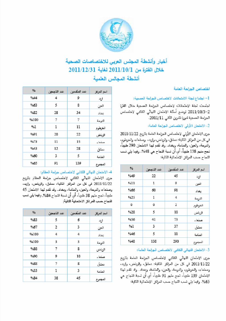
5/17/2018 12-4 _ Act - slidepdf.com
http://slidepdf.com/reader/full/12-4-act 103/110

5/17/2018 12-4 _ Act - slidepdf.com
http://slidepdf.com/reader/full/12-4-act 104/110
JABHS: Journal of the Arab Board of Health Specializations Vol.12, No 4, 2011
2
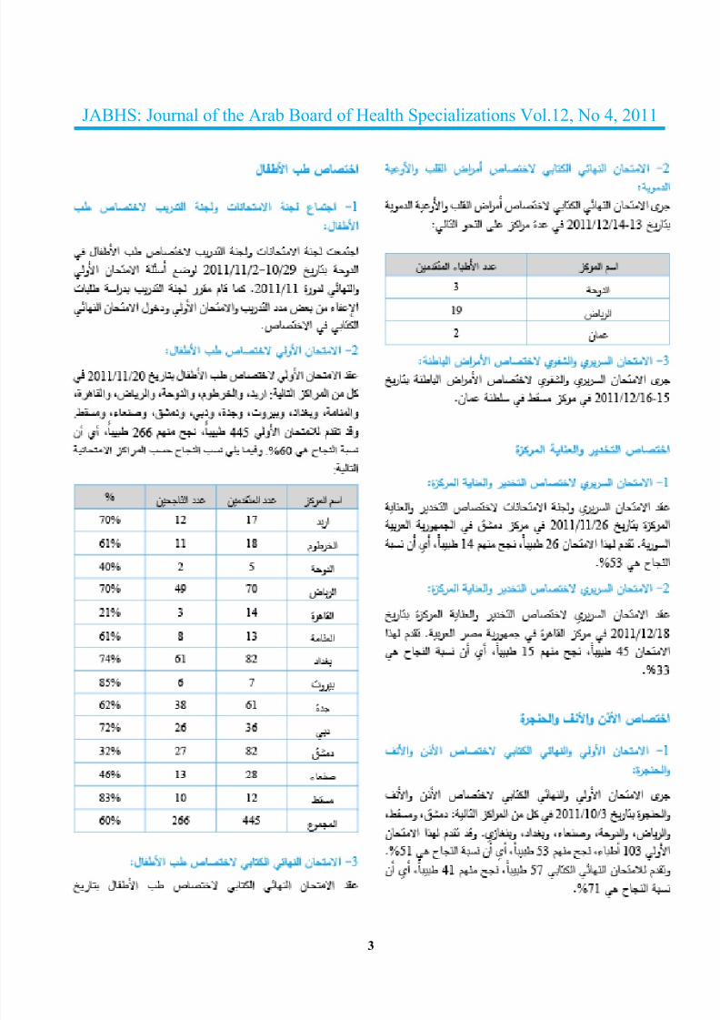
5/17/2018 12-4 _ Act - slidepdf.com
http://slidepdf.com/reader/full/12-4-act 105/110
JABHS: Journal of the Arab Board of Health Specializations Vol.12, No 4, 2011
3

5/17/2018 12-4 _ Act - slidepdf.com
http://slidepdf.com/reader/full/12-4-act 106/110
JABHS: Journal of the Arab Board of Health Specializations Vol.12, No 4, 2011
4
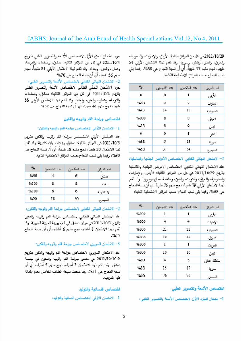
5/17/2018 12-4 _ Act - slidepdf.com
http://slidepdf.com/reader/full/12-4-act 107/110
JABHS: Journal of the Arab Board of Health Specializations Vol.12, No 4, 2011
5

5/17/2018 12-4 _ Act - slidepdf.com
http://slidepdf.com/reader/full/12-4-act 108/110
JABHS: Journal of the Arab Board of Health Specializations Vol.12, No 4, 2011
6

5/17/2018 12-4 _ Act - slidepdf.com
http://slidepdf.com/reader/full/12-4-act 109/110
JABHS: Journal of the Arab Board of Health Specializations Vol.12, No 4, 2011
7
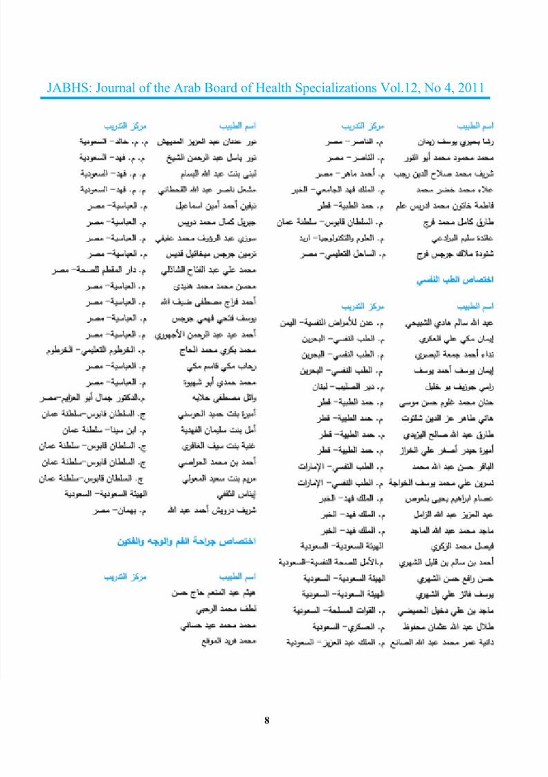
5/17/2018 12-4 _ Act - slidepdf.com
http://slidepdf.com/reader/full/12-4-act 110/110
JABHS: Journal of the Arab Board of Health Specializations Vol.12, No 4, 2011
8