1,2 3 4 eric r. evans4, renate reimschuessel , k
TRANSCRIPT
Pathology and epidemiology of oxalate nephrosis in cheetahs.
Emily P. Mitchell1,2, Molly E. Church3, Sarah M. Nemser4, Betsy Jean Yakes5,
Eric R. Evans4, Renate Reimschuessel4, K. Lemberger6, Peter N. Thompson7,
Karen A. Terio8
1Department of Research and Specialised Services, National Zoological Gardens of
South Africa, Pretoria, South Africa
2Department of Paraclinical Sciences, Faculty of Veterinary Science, University of
Pretoria, Pretoria, South Africa
3Department of Pathology, Microbiology and Immunology, School of Veterinary
Medicine, University of California, Davis, California, USA
4United States Food and Drug Administration, Center for Veterinary Medicine, Office
of Research, Veterinary Laboratory Investigation and Response Network, Laurel,
Maryland, USA
5United States Food and Drug Administration, Center for Food Safety and Applied
Nutrition, Office of Regulatory Science, College Park, Maryland, USA
6Vetdiagnostics, Lyon, France
7Department of Production Animal Studies, Faculty of Veterinary Science, University
of Pretoria, Pretoria, South Africa
8Zoological Pathology Program, University of Illinois, Brookfield, Illinois, USA
Corresponding Author:
Emily. P. Mitchell (neé Lane), Department of Research and Scientific Services,
National Zoological Gardens of South Africa, P O Box 754, Pretoria, South Africa.
Email: [email protected]
1
Abstract
To investigate cases of acute oxalate nephrosis without evidence of ethylene glycol
exposure, archived data and tissues from cheetahs (Acinonyx jubatus) from North
America (n=297), southern Africa (n=257), and France (n=40) were evaluated. Renal
and gastro-intestinal tract lesions were characterized in a subset of animals with
(n=100) and without oxalate crystals (n=165) at death. Crystals were confirmed as
calcium oxalate by Raman spectroscopy in 45 of 47 cheetahs tested. Crystals were
present in cheetahs from 3.7 months to 15.9 years old. Cheetahs younger than 1.5
years were less likely to have oxalates than older cheetahs (p=0.034) but young
cheetahs with oxalates had more oxalate crystals than older cheetahs (p<0.001).
Cheetahs with oxalate crystals were more likely to have renal amyloidosis, interstitial
nephritis or colitis, and less likely to have glomerular loop thickening or gastritis, than
those without oxalates. Crystal number was positively associated with renal tubular
necrosis (p≤0.001), regeneration (p=0.015), and casts (p≤0.001) but inversely
associated with glomerulosclerosis, renal amyloidosis and interstitial nephritis.
Crystal number was unrelated to the presence or absence of colitis and was lower in
southern African than American and European animals (p=0.01). This study found
no evidence that co-existing chronic renal disease (amyloidosis, interstitial nephritis
or glomerulosclerosis), veno-occlusive disease, gastritis or enterocolitis contributed
significantly to oxalate nephrosis. Oxalate-related renal disease should be
considered as a potential cause of acute renal failure especially in young captive
cheetahs. The role of location, diet, stress and genetic predisposition in the
pathogenesis of oxalate nephrosis in cheetahs warrants further study.
2
Key words: Acinonyx jubatus, crystals, cheetahs, intestinal disease, nephrosis,
calcium oxalate, pathology, renal disease
Since the early 1970’s worldwide sporadic cases of oxalate nephrosis have been
documented in captive cheetahs and other large felids.20,36,41,63,72,76,77. Oxalate
nephrosis has also been recorded in other non-domestic mammals including mink67
and koalas.74 While small numbers of oxalate crystals are relatively common in
cheetahs dying of glomerulosclerosis and/or renal amyloidosis, large numbers of
oxalate crystals and associated tubular pathology have been described in cheetah
kidneys, without other renal disease being present (KAT, KL, EPM and Linda
Munson, personal communication). Approximately 10% of captive cheetahs in the
United States and South Africa were estimated to have oxalate crystals in renal
tubules at necropsy (KAT and EPL, unpublished data). This warranted further
investigation of possible risk factors for renal oxalate formation given the vulnerable
conservation status of this species.21
Endogenously produced oxalate is a metabolic by-product of amino acid
(hydroxyproline, glycine and serine) and vitamin C catabolism.5,11,32 Oxalate
nephrosis results from intra-tubular calcium oxalate deposition when calcium oxalate
burdens exceed excretory capacity5,11 either due to genetic defects in glyoxylate
metabolism that result in increased hepatic endogenous production of oxalate, or
secondary or acquired disease.5,10,11,31 Most human forms of primary hyperoxaluria
occur due to mutation of the pyridoxine (vitamin B6) dependent enzyme
alanine:glyoxylate aminotransferase (AGT) or the enzyme
glyoxalate/hydroxypyruvate reductase (GRHPR).5,44,66 Familial tendencies, a point
3
mutation in the GRHPR gene, and associated reduced enzyme function have also
been identified in some domestic cats with oxalate nephrosis.6,15,26,49
Secondary or acquired oxalate nephrosis can be due to severe liver disease46 or
pyridoxine deficiency,5,7,23,65 which result in compromised oxalate metabolism; or
increased absorption of oxalates due to ingestion of oxalates (or their precursors)
including plants,2,3,11,39,58 ethylene glycol and other glycols,1,10,11,30,50 xylitol,13
ascorbic acid,4,47,48,55 and collagen or feathers.18,85 An altered intestinal
microbiome,42,69 excessive bile or long-chain fatty acids10,56 or low intestinal calcium
content due to diet,32 enterocolitis5,10 and intestinal surgical resection9,31 can also
increase oxalate absorption and result in oxalate crystal deposition in the kidney.
Ethylene glycol toxicity was considered, although never proven,72,76, in cheetah
cases with large numbers of crystals, but was thought unlikely in cases from the
United Arab Emirates36 and southern Africa where ethylene glycol (antifreeze) is less
widely utilized. Toxicological evaluation of feed and water supplies in several cases
from the United States, southern Africa and France have failed to find evidence of
contaminating ethylene glycol (DGA Meltzer, M Kinsel, and S Terrell, personal
communications).41 Similarly, ingestion of oxalate-rich plants or sweeteners,
pancreatic insufficiency, ascorbic acid toxicity, high fat or protein diets and
pyridoxidine deficiency were considered unlikely primary causes in cheetahs given
the widely differing environments and management practices in the USA, southern
Africa and France.
However, captive cheetahs suffer from renal diseases such as glomerulosclerosis
and amyloidosis8,51,59 which could result in increased oxalate deposition due to
reduced glomerular filtration and urinary pH, pyridoxine depletion and
hypercalcaemia due to secondary hyperparathyroidism.5,10,11,53,57 Cheetahs also
4
suffer from gastrointestinal tract and liver disease,51,52 which could be risk factors for
oxalate formation. This retrospective pathological study was conducted to describe
the lesions in cheetahs with and without oxalates in their kidneys, and to investigate
the relationship between oxalate crystal deposition and concurrent renal, hepatic or
gastro-intestinal tract disease.
Materials and Methods
Study animals
Cases were selected from pathology databases of cheetahs kept as part of the North
American Association of Zoos and Aquariums’ Species Survival Plan (n=297); and
from cheetah pathology databases in southern Africa (n=257) and France (n=40).
Tissues from cheetahs over one month old that died between 1990 and 2013 were
used as the study population. Histological sections of kidneys from all cheetahs were
screened using polarized light for the presence or absence of crystals. A subset of
cheetahs greater than one month old that died between 1990 and 2013 (n=265) was
selected for detailed histologic examination based on the quality and availability of
relevant histological material. Cases were defined as cheetahs with renal oxalates at
death (n=100). Controls were defined as cheetahs with no renal oxalates at death
(n=165) and similar demographic characteristics to cases including age, sex, the
housing facility at death, and year of death; however, matching was approximate and
not possible in all instances. Geographic distribution of the final dataset of cases and
controls was North America (n=94; 40 cases, 54 controls); southern Africa (n=167;
56 cases, 111 controls) and France (four cases, no controls). All of the North
American and French cheetahs and all but two of the southern African cheetahs
were captive at the time of death.
5
Pathological evaluation
Formalin-fixed tissues were processed routinely and stained with hematoxylin and
eosin, von Kossa and Masson’s trichrome stains.20 Crystal number and the presence
and severity of renal lesions, veno-occlusive disease (VOD), gastritis, enteritis and
colitis were evaluated by two pathologists (KAT and EPM) using a detailed scoring
scheme (Supplemental Table S1) and previously published criteria.8,51,59 A subset of
southern African cases was examined by both KAT and EPM to confirm evaluator
concordance.
Raman Spectroscopic Characterization
Archived, formalin-fixed paraffin embedded kidney sections from cheetahs (n=47; 28
North American, 15 southern African, 4 French) were used for Raman spectroscopic
characterization. Unstained, deparaffinized sections (6-8 µm) on quartz slides
(25×6×1 mm3) (SPI, West Chester, PA, USA) were examined by light microscopy for
crystal presence and location. A case of confirmed canine ethylene glycol toxicity
was used as a control. Raman spectroscopy measurements on the kidney sections
were performed on a Nicolet Almega XR Raman spectrometer equipped with an
Olympus BX51 confocal microscope (Thermo Electron, Madison, WI, USA). The
camera temperature was set at -50 °C through the instrument control software
OMNIC 8 for Dispersive Raman. The complimentary Atlµs program was employed
for imaging the samples and focusing on the birefringent crystals in the tissue.
Spectra were obtained with a 532 nm solid state, diode pumped laser (24 mW) at
100% power, 50 µm pinhole aperture, 2400 lines/mm resolution grating, spectral
range of 2000 to 300 cm-1, and a 100× Olympus objective. Individual spectra were
6

acquired with a 20 sec interrogation time and three exposures after a 30 sec
photobleach. All spectra were baseline corrected using the OMNIC software. Library
comparison for spectra was performed using the OMNIC software and spectral
libraries purchased from Thermo Electron. Specifically, the HR Raman Inorganics
Library (containing a calcium oxalate spectrum) as well as additional spectra
obtained from a known ethylene glycol poisoning in a dog (07N-0544-Ti, U.S. FDA)
were used for confirmation that a spectrum obtained from a crystal was a spectral
match with calcium oxalate. Additional spectral processing, including peak identity
evaluation, was performed with Spectrus Processor 2012 (ACD/Labs, Toronto,
Ontario, Canada).
Statistical analysis
Because matching of cases (n=100) and controls (n=165) was only approximate in
this retrospective study, unmatched analysis was performed. Age of animals at death
was categorized as <1.5, 1.5-<8, 8-<11 and ≥11 years old. Univariate analyses were
conducted to compare lesion prevalence and severity in cases and controls
(Supplemental Table S2), as well as to compare lesion prevalence and severity with
crystal number (Supplemental Table S3). Renal edema, hemorrhage, arteritis,
fibrinoid necrosis, thrombosis, cortical and medullary intra-tubular mineralization, the
presence of other crystals in tubules, intratubular and intracellular pigment in tubules,
tubulitis and VOD were excluded as these lesions were uncommon to rare. The
cellular characteristics of interstitial inflammation were excluded as this was
invariably lymphoplasmacytic. The association between the presence of oxalate
crystals and the presence and severity of each remaining histological lesion
(case/control status) was then assessed using mixed-effects logistic regression
7
Table 1. Lesions significantly associated with the presence of renal oxalate crystals
in cheetahsa
Lesion Odds ratio 95%CI
Interstitial nephritis 5.63 2.10 -15.10
Renal amyloidosis 2.74 1.06 - 7.07
Colitis 2.40 0.99 - 5.83
Glomerular loop thickening 0.31 0.14 - 0.72
Gastritis 0.22 0.07 - 0.73
CI, confidence interval
aAdjusted for age, examiner and region in a mixed-effects logistic regression model
8
(Table 1). Firstly, all potential predictors associated with the outcome with p<0.2 in
the initial analysis were included and sequentially eliminated until all remaining
predictors were significant (p<0.05). Every candidate predictor was then re-tested
one-by-one in the model and retained if significant. The scores for renal amyloidosis,
interstitial inflammation and gastritis were collapsed into dichotomous (0/1) scores
indicating the presence or absence of each lesion, since the original ordinal scores
did not contribute any additional information. Odds ratios were generated describing
the strength of association between the presence and absence of crystals and each
lesion.
Secondly, where oxalate crystals were present (n = 100), the association of each
lesion score with crystal number (average number of cortical tubules containing
crystals in three 100x fields) was assessed using mixed-effects negative binomial
regression, adjusting for age, examiner and institution (random effect). Factors
potentially associated with the severity of oxalate nephrosis were assessed using a
mixed-effects negative binomial regression model, with the same candidate variables
and modelling approach as above. All statistical analyses were done using Stata 14
(StataCorp, College Station, TX, USA). Significance was assessed at p<0.05. The
scores for renal amyloidosis were collapsed into dichotomous (0/1) scores indicating
the presence or absence of amyloidosis in the final multiple negative binomial
regression model of factors associated with the numbers of crystals present (Table
2). Negative binomial regression in this context yields a count ratio (CR), interpreted
as the ratio of the crystal number in a category with the lesion to the crystal number
in the reference category (without the lesion); therefore, a CR < 1 indicates a
negative or inverse association and a CR > 1 indicates a positive association with
crystal number.
9
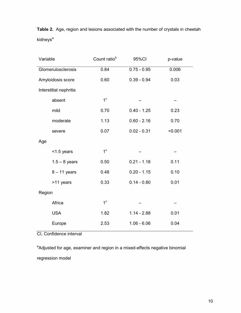
Table 2. Age, region and lesions associated with the number of crystals in cheetah
kidneysa
Variable Count ratiob 95%CI p-value
Glomerulosclerosis 0.84 0.75 - 0.95 0.006
Amyloidosis score 0.60 0.39 - 0.94 0.03
Interstitial nephritis
absent 1c – –
mild 0.70 0.40 - 1.25 0.23
moderate 1.13 0.60 - 2.16 0.70
severe 0.07 0.02 - 0.31 <0.001
Age
<1.5 years 1c – –
1.5 – 8 years 0.50 0.21 - 1.18 0.11
8 – 11 years 0.48 0.20 - 1.15 0.10
>11 years 0.33 0.14 - 0.80 0.01
Region
Africa 1c – –
USA 1.82 1.14 - 2.88 0.01
Europe 2.53 1.06 - 6.06 0.04
CI, Confidence interval
aAdjusted for age, examiner and region in a mixed-effects negative binomial
regression model
10
bThe count ratio is the ratio of the crystal count in animals in that category to the
crystal count in animals in the reference category; or, the fold change in crystal count
for each unit increase in the glomerulosclerosis or amyloidosis score.
cReference category
11
Results
Histopathology
Histologically, kidneys from cheetah cases contained small to very large numbers of
colorless refractive crystals (average 0.3-81 crystals in three 100x fields) in cortical
and medullary tubules forming rosettes, globules and acicular fragments (Fig. 1) that
were birefringent with polarized light (Fig. 2). Crystals were not uniformly distributed
in the renal cortex, with clusters apparently occurring in single or groups of tubules;
crystals were not associated with tracts of inflammation. Affected tubules contained
small amounts of sloughed necrotic cellular debris mixed with variable amounts of
pale amorphous eosinophilic material and, in many cases, were lined by a
discontinuous layer of epithelial cells with variable degrees of necrosis characterized
by shrunken cells with hypereosinophilic cytoplasm and dark basophilic pyknotic
nuclei (Fig. 1). Adjacent tubules were variably dilated and lined by regenerative low
cuboidal epithelial cells with pale basophilic cytoplasm and large crowded oval nuclei
(Fig. 3). Variable degrees of mild interstitial fibrosis and small numbers of intra-
tubular cellular casts were present. Crystals and fine mineral deposits variably
present on tubular basement membranes in the cortex and medulla stained variably
positive with von Kossa which was interpreted as the presence of calcium phosphate
and/or carbonate salts, since melanin pigment was not visible in crystals or on
tubular basement membranes on hematoxylin and eosin stains (Fig. 4). Renal
lesions were present histologically in 46 of 165 (28%) cheetahs without oxalate
crystals and in 87 of 100 (87%) cheetahs with oxalate crystals.
Additional renal lesions included small numbers of deeply basophilic mineralized
crystals in the lumina of both cortical and medullary tubules, rare pale eosinophilic or
12

180x135mm (300 x 300 DPI)
Figures 1–4. Oxalate nephrosis, kidney, cheetah. Figure 1. Oxalate crystals are associated with epithelial necrosis and tubular dilatation; the glomerulus is normal. Hematoxylin and eosin (HE). Figure 2. Polarization highlights moderate numbers of oxalate crystals in tubular lumina (an average of 37 crystals per three 100 fields). HE. Figure 3. Cortical tubular epithelial necrosis, sloughed epithelial cells in the tubular lumina, and regeneration of tubular epithelium. HE. Figure 4. Oxalate crystals and cortical tubular basement membranes are mineralized. von Kossa.
13
yellow pigment in renal tubular epithelial cells or in tubular lumina,; and variable
microvesiculation consistent with lipidosis and or hydropic degeneration in renal
tubular epithelial cell cytoplasm. Tubulitis, renal edema or hemorrhage, arteritis,
vascular fibrinoid necrosis were rarely noted.
Raman spectroscopy
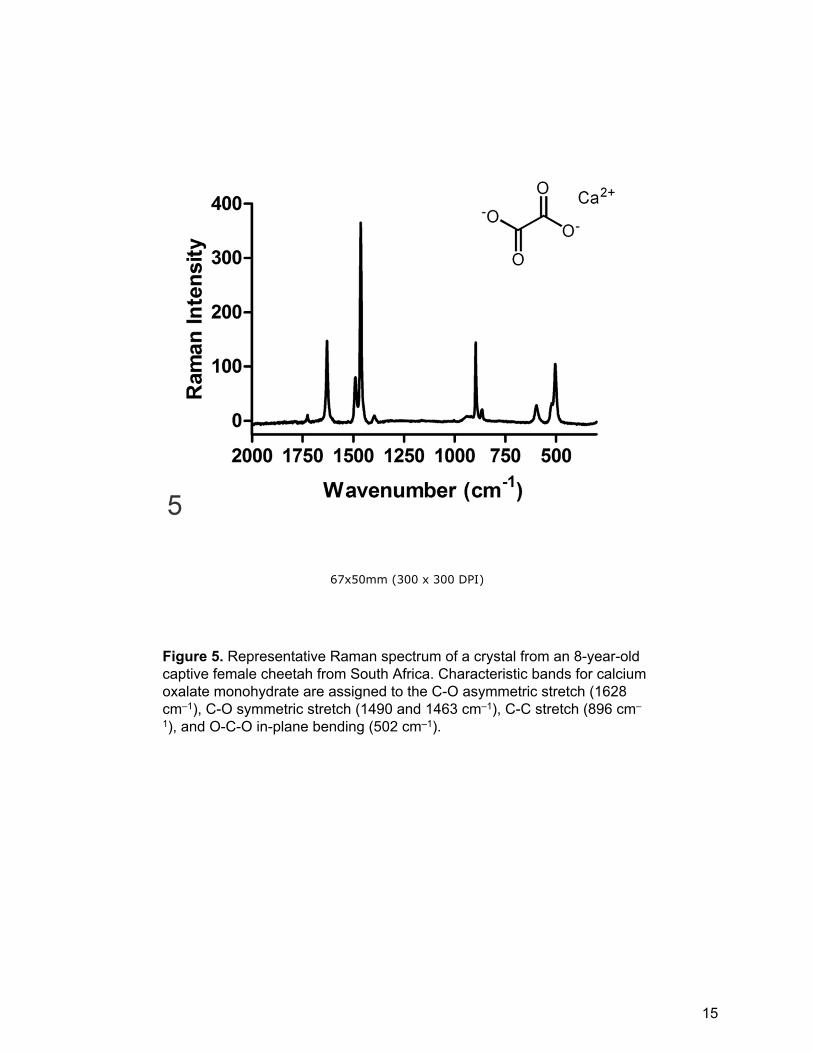
On Raman spectroscopic examination, crystals were easily observed as bright
spheres in the dark tissue background. Crystals in the control case of canine
ethylene glycol poisoning and in 27 (96%) North American, 14 (93%) southern
African, and four (100%) French cases were confirmed as calcium oxalate with no
laser photodegradation of the crystals (Fig. 5). Laser photodamage prevented crystal
identification in two cheetahs (one North American and one southern African). In
addition to calcium oxalate crystals, calcium carbonate, calcium phosphate or
calcium sulphate crystals were present in the kidney tubules of three North American
cheetahs, and one additional North American cheetah sample had a Raman multi-
component spectrum that contained calcium oxalate bands. All of these cheetahs
were >10 years of age and had other underlying renal disease including
glomerulosclerosis and amyloidosis.
Statistical analysis
The presence and numbers of crystals were not significantly different between male
and female cheetahs (p=0.454). The average age of cheetahs with renal oxalate
crystals at death was similar in the North American, French and southern African
populations (3.7 months - 15.9 years old; median = 8.7 years (interquartile range
[IQR]: 6.4-11.3 years). Age distribution was similar in cheetahs without oxalates
14
67x50mm (300 x 300 DPI)
Figure 5. Representative Raman spectrum of a crystal from an 8-year-old captive female cheetah from South Africa. Characteristic bands for calcium oxalate monohydrate are assigned to the C-O asymmetric stretch (1628 cm–1), C-O symmetric stretch (1490 and 1463 cm–1), C-C stretch (896 cm–
1), and O-C-O in-plane bending (502 cm–1).
15
90x65mm (300 x 300 DPI)
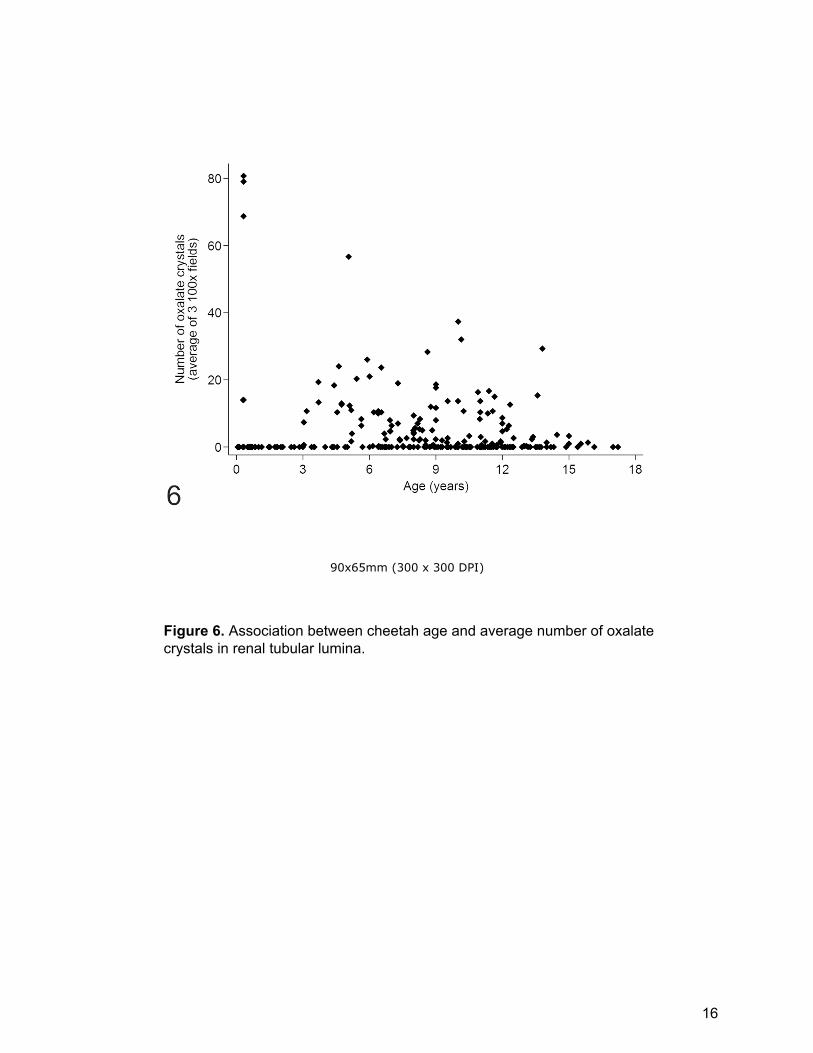
Figure 6. Association between cheetah age and average number of oxalate crystals in renal tubular lumina.
16
(median 8.6 years; IQR: 3.6-11.3 years). Cheetahs older than 1.5 years were much
more likely to have oxalates than younger cheetahs (p=0.034, Supplemental Table
S2) but cheetahs <1.5 years old had significantly more crystals than older cheetahs
(p<0.001, Supplemental Table S3). Although three young cheetahs from one North
American institution had very high numbers of crystals (average of 69-81 (per three
100x fields)) no grouping of crystal number based on age was present (Fig. 6). On
the multivariate mixed effects negative binomial regression analysis cheetahs >11
years old had significantly fewer crystals than those < 1.5 years old (p=0.01, Table
2).
When comparing cheetahs with renal oxalate crystals (cases) with those without
such crystals (controls), tubular changes were significantly associated with the
presence of crystals (Supplemental Table S2). Cases had significantly more renal
tubular necrosis, regeneration, dilatation, protein and cellular casts than controls
(p≤0.001 for each parameter). Compared to controls, cases also had significantly
more lymphoplasmacytic interstitial nephritis (p≤0.001), amyloidosis (p≤0.001),
chronic infarcts (p≤0.001), efferent arteriole arteriolosclerosis (p≤0.001), arcuate
artery arteriosclerosis (p=0.011), cortical (p=0.02) and medullary (p=0.005) tubular
basement membrane mineralization, cortical fibrosis (0.049), hyperplastic collecting
tubule epithelium (p=0.063), enteritis (p=0.001) and colitis (p=0.017). In contrast,
gastritis was significantly more common in controls than cases (p=0.012). Sex
(p=0.899), glomerulosclerosis (p=0.952), glomerular loop thickening (p=0.273),
glomerular hypercellularity (0.743), and medullary fibrosis (p=0.898) had no effect on
the likelihood of having renal oxalate crystals. In the 250 cheetahs for which liver
was available hepatic VOD was present in 22 of 88 (25%) of cases and 32 of 162
(20%) of controls . In addition, 6% of cheetahs without oxalates but only 3% of
17
cheetahs with oxalates had severe VOD. In the multivariable mixed effects logistic
regression analysis comparing cases and controls, the only factors associated with
an increased likelihood of having renal oxalates were interstitial nephritis, and to a
lesser extent, renal medullary amyloidosis and colitis (Table 1). Case cheetahs had
reduced odds of having glomerular loop thickening and gastritis compared to
controls.
When crystal number was evaluated in cheetahs with oxalate crystals in the
kidney (Supplemental Table S3), the number of crystals present was positively
associated with tubular necrosis (p≤0.001), regeneration (p=0.015), cellular casts
(p≤0.001), glomerular hypercellularity (p=0.007) and arteriosclerosis of the efferent
arteriole (p≤0.001). Crystal number was inversely associated with protein casts
(p=0.043), medullary tubular basement membrane mineralization (p=0.002),
collecting duct epithelial hyperplasia (p=0.023), glomerulosclerosis (p=0.009),
glomerular loop thickening (p≤0.001), amyloidosis (p≤0.001), chronic infarcts
(p≤0.001), renal cortical (p≤0.001) and medullary (p=0.002) fibrosis, enteritis
(p=0.001) and colitis (p=0.019). In 31 cheetahs with large numbers of oxalates
(average of 10-81 per three 100x fields), there was no evidence of any underlying
renal disease. Crystal number was unrelated to sex (p=0.455), tubular dilatation
(p=0.409), cortical tubular basement membrane mineralization (p=0.382),
arteriosclerosis of the arcuate artery (p=0.118) or gastritis (p=0.826). The
relationship between crystal number and interstitial nephritis was significant but non-
linear (p≤0.001) since cheetahs with grade two nephritis had more crystals than
those with nephritis grades zero, one and three. In the multivariable mixed effects
negative binomial regression analysis, geographical region was the only factor
positively associated with crystal number (Table 2). On average, cheetahs from the
18
USA and France had significantly more oxalate crystals than those from Africa
(p=0.01). Nine of the 10 cheetahs with highest average number of cortical tubules
containing oxalate crystals (26-81) were from North America (5 different institutions),
and the remaining one was from a South African institution (24). Among the 10
southern African cheetahs with the highest number of crystals (14-24), cheetahs
from one institution were over-represented (50%). The four cheetahs from three
French institutions had an average number of cortical tubules containing crystals of
15 (10-20). One free-ranging Glomerulosclerosis and amyloidosis were negatively
associated with crystal number in the multivariate analysis (Table 2).
Discussion
This study found that the presence of intra-tubular renal oxalate crystals was
associated with renal tubular necrosis, regeneration and the presence of tubular
casts in cheetahs in North America, southern Africa and France; and that increased
numbers of crystals was associated with increased severity of these tubular
changes. This effect of oxalate crystals on tubular epithelium is well documented in
humans and animals.11,28,43,44,49,66,78,83
Raman spectroscopy confirmed that the majority of crystals were calcium oxalate.
Melamine and other renal crystals were ruled out because the Raman spectral
fingerprints did not match these compounds.37,61,68 The two cases in which oxalate
crystals were not found were cheetahs with only a few crystals in the H&E stained
sections, and it is possible that sections prepared for spectroscopy contained no
crystals or that they were damaged by the laser. Calcium carbonate, calcium
phosphate or calcium sulphate crystals, present in the kidney tubules of three North
19
American cheetahs, were not distinguishable from calcium oxalate on routinely
stained sections but were readily identifiable by their Raman spectra. Therefore,
while calcium oxalate monohydrate was the predominant crystal noted other crystal
types can occur in cheetahs. Compound oxalate crystals have also been described
in domestic dogs.64
Oxalate crystals and associated tubular damage occurred in 31 cheetahs without
other renal diseases, all of which were reported to have clinical evidence of renal
failure (inappetance, weakness, vomiting, azotemia, dehydration, polyuria and
depression) and showed varying degrees of parathyroid gland hyperplasia and
metastatic mineralization but no other renal disease. Similar findings have been
described previously in cheetahs36,41,76 and other animals with oxalate
nephrosis.14,20,22,27,28,33,34,40,67,73–75 Oxalate nephrosis, therefore, occurs
independently of other renal diseases in some captive cheetahs and should be
considered in the differential diagnosis of renal disease. Although rare scattered
oxalate crystals may occur secondarily to many types of chronic renal disease11, 11
cheetahs with chronic renal disease also had large numbers of crystals suggesting
that oxalate nephrosis may co-exist with other renal diseases at least in some
cheetahs. Tubular lesions, glomerular hypercellularity and efferent arteriolosclerosis
could contribute to oxalate crystal formation in chronic renal disease as these were
the only renal lesions positively associated with crystal number in these cheetahs.
and While a few oxalate crystals may occur secondarily to concurrent chronic renal
disease in some cases, factors other than concurrent renal disease likely affect renal
oxalate deposition in cheetahs.
No clear evidence was found in our study that oxalate nephrosis occurs
secondary to gastro-intestinal or hepatic disease in cheetahs. Gastritis was more
20
common in controls than cases, and was unrelated to crystal number. On the
univariate analysis, more cheetahs with renal oxalate crystals had enteritis and colitis
than controls, and on the multivariate analysis colitis was associated with an
increased likelihood of having renal oxalates. However, both lesions were inversely
associated with crystal number and therefore likely do not play a key role in the
development of oxalate nephrosis. In humans, fat malabsorption due to enterocolitis
or pancreatic insufficiency may result in reduced intestinal calcium which increases
intestinal oxalate absorption because calcium binds intestinal oxalates 5,10,46,56,81
However, no relationship between enterocolitis and renal oxalate crystal number was
found and pancreatic insufficiency is rare in the parent population of cheetahs.51,52
Veno-occlusive disease was uncommon in animals with and without renal oxalate
crystals. Increased intestinal absorption of oxalates may also result from loss of
intestinal lactic acid bacteria, including Oxalobacter formigenes, that degrade oxalate
into carbon dioxide and formate.42,46,70,82 Research is needed to determine whether
or not cheetahs that receive broad-spectrum antibiotic therapy for Helicobacter-
associated gastritis or other infections have depleted O. formigenes populations,
which may predispose them to oxalate nephrosis.35
Significant geographical and institutional clustering of cheetahs) with abundant
renal oxalate crystals suggests that genetic, dietary or management factors
mayinfluence the prevalence of oxalate nephrosis in captive cheetahs. So far, no
proof of ethylene glycol toxicity has been found in cheetahs, but exposure to
ethylene glycol, xylitol or other oxalate precursors should be thoroughly investigated
in cases of oxalate nephrosis. Identification of dietary factors was beyond the scope
of this retrospective necropsy study. However evaluation of the fluid intake and
protein, fat, collagen, feathers, calcium, magnesium, beet pulp, pyridoxine, ascorbic
21
acid and arachidonic acid levels in captive cheetah diets is needed as these factors
can influence oxalate excretion16–20,31,32,46,60,71,84 Dietary calcium:oxalate ratios affect
O. formigenes colonization of the intestine in rats so this ratio may be important in
cheetah diets.42 Obesity and stress may contribute to oxalate nephrosis in humans,54
and their roles in oxalate nephrosis may warrant further study since obesity may be
present in underactive captive cheetahs and stress has been documented in captive
animals.79
Primary hyperoxaluria (PH) in humans is an autosomal recessive condition
associated with mutations in three genes involved in oxalate metabolism: AGT gene
in PH type 1; GRHPR gene in PH type 2; and 4-hydroxy-2-oxoglutarate aldolase
(HOGA1) in PH type 344. Similarly, both PH type 1 and PH type 2 have been
described in domestic cats. Clear evidence that oxalate nephrosis in cheetahs is a
primary genetic disease was not found in our study, however we did not test genetic
relatedness. In humans and cats with inherited oxalate nephrosis the disease is seen
in juveniles.6,15,26,43,49,78 In this study, although cheetahs <1.5 years old had
significantly more crystals than older cheetahs, which might indicate an inherited
predisposition, relatively large numbers of crystals were seen in cheetahs in all ages.
Lesions which are characteristic of primary disease in humans and cats, including
granulomatous nephritis associated with interstitial oxalates, widespread tissue
oxalate deposition10,12,45,66,80 and neurological disease29,52 were not seen in the
cheetahs in this study.. Manifestation of genetic disease is complex with
environmental influences, multigenetic inheritance, epigenetic effects, incomplete
penetrance and variable expression resulting in differing disease severity and
phenotypes.29 Therefore, whether variations in crystal number are due to differing
manifestations of the same disease or differences in pathogenesis among cheetahs
22
in this study is uncertain. Since cheetahs from the USA, southern African and
French cheetah populations share common founders a detailed genetic analysis,
including sequencing of key genes involved in oxalate metabolism, and resultant
enzyme activity, is needed to determine whether or not cheetahs suffer from a
genetically limited capacity for glyoxylate metabolism.48
Early diagnosis of oxalate nephrosis is important since, this disease is potentially
treated by fluid therapy to reduce oxaluria44,85, pyridoxine to stabilize and enhance
the activity of AGT,5,7,23,65 urine alkalinisation,66,85 and n-3 fatty acid supplementation
to decrease urinary oxalate excretion and free-radical injury.5,24,38,46,57,62,66,70
Urinalysis data was limited to a few cases in this retrospective study, but could be a
valuable diagnostic tool since small numbers of oxalate crystals may be present in
the urine of affected cheetahs.30 However, confirming the presence of oxalate
nephrosis and differentiating it from concurrent glomerulosclerosis or renal medullary
amyloidosis, in azotemic cheetahs is not simple if urinary oxalates are absent. Renal
ultrasonography is non-specific in domestic dogs and cats with oxalate nephrosis.1
Computed tomography scans are used to identify renal crystals in humans,5 but are
not a practical diagnostic modality in cheetahs. At one Southern African institution,
urinary oxalate was elevated in an acutely azotemic cheetah later diagnosed with
oxalate nephrosis without other renal lesions (8000 mg oxalate/gram creatinine,
compared 128 mg /g in a healthy enclosure mate F. Reyers, Golden VetPath,
unpublished data). Since crystal number was higher in young cheetahs and chronic
renal disease in cheetahs is age-related,25,51 oxalate nephrosis should be a primary
consideration in younger cheetahs with renal failure.
In conclusion, this study found that oxalate nephrosis unrelated to ethylene glycol
toxicity occurs in cheetahs. We found no convincing evidence that oxalate nephrosis
23
in captive cheetahs is secondary to renal, hepatic or gastro-intestinal tract disease
although it may occur concurrently with such diseases. Oxalate nephrosis should be
considered as a differential diagnosis in captive cheetahs with renal failure,
especially in young animals that are less likely to be suffering from renal amyloidosis
or glomerulosclerosis. Oxalate nephrosis is of uncertain etiology in captive cheetahs
but a multifactorial pathogenesis, including a primary genetic predisposition, diet,
altered gut microbiome and or stress, is suspected.
The data analyzed in this study are available as Supplemental Materials.
Acknowledgements
This study was made possible by the veterinarians and cheetah holding and
breeding institutions that submitted clinical data and carcasses from captive and
free-ranging cheetahs for examination from the USA: The Association of Zoos and
Aquariums’ Cheetah Species Survival Plan including Anonymous (5 facilities),
Albuquerque Biopark Zoo, Binder Park Zoo, Brevard Zoo, Caldwell Zoo, Chehaw
Wild Animal Park, Cincinnati Zoo and Botanical Garden, Cleveland Metroparks Zoo,
Columbus Zoo and Aquarium, Dickerson Park Zoo, Fort Wayne Children’s Zoo, Fort
Worth Zoo, Fossil Rim Wildlife Center, Honolulu Zoo, Jackson Zoo, Jacksonville Zoo
and Gardens, Kansas City Zoo, The Living Desert, Maryland Zoo in Baltimore,
Mesker Park Zoo and Botanic Gardens, Montgomery Zoo, Nashville Zoo at
Grassmere, Oklahoma City Zoo and Botanical Garden, Phoenix Zoo, San Antonio
Zoo and Aquarium, San Diego Zoo Global, Tulsa Zoo and Living Museum, Utah’s
Hogle Zoo, White Oak, Wildlife Safari, Zoo Knoxville, and Zoo New England); from
southern Africa: AfriCat, Cango Wildlife Ranch, Cheetah Conservation Botswana, Dr.
24
A. Tordiffe, Dr. D. Zimmerman, Dr. K. Good, Dr. P. Buss, Dr. P. Caldwell, Dr. P.
Swartz, Farm Inn, Hoedspruit Endangered Species Centre, Johannesburg Zoo,
Letsatsi La Africa, Lory Park Zoo, Matobo Veterinary Centre, National Zoological
Gardens of South Africa, Rhino and Lion Park, Seaview Predator Park, The Ann van
Dyk Cheetah Centre, Tshwane Nature Conservation; from France: the Safari de
Peaugres, Dr. C. Vitaud, Dr. D. Sarran; Parc des Félins, Mr. G. Breton, Dr. F. Ollivet-
Courtois. Data and cases provided by Drs. Linda Munson and Nadia Robert are also
acknowledged. Pathology laboratory staff at the NZG and Faculty of Veterinary
Science, University of Pretoria, at Anipath, France, and the University of Illinois
Zoological Pathology Program and Veterinary Diagnostic Laboratory provided
excellent technical assistance. The authors thank Dr. Chuck Mohr for his guidance in
developing the renal scoring system. Professors John Lawrence and Leon Prozesky,
Faculty of Veterinary Science, University of Pretoria provided valuable editorial input.
The National Research Foundation, through a core grant to the National Zoological
Gardens of South Africa, provided the funding to conduct pathological examinations
in southern African cheetahs. The views expressed in this publication are those of
the authors and do not necessarily reflect the official policy of the Department of
Health and Human Services, the U.S. Food and Drug Administration, or the U.S.
Government.
25
References
1. Adams WH, Toal RL, Breider MA. Ultrasonographic findings in dogs and cats
with oxalate nephrosis attributed to ethylene-glycol intoxication - 15 Cases
(1984-1988). J Am Vet Med Assoc. 1991;199(4):492-496.
2. Angus KW, Hodgson JC, Hosie BD, et al. Acute nephropathy in young lambs.
Vet Rec. 1989;124(1):9-14.
3. Aslani MR, Movassaghi AR, Najarnezhad V, Pirouz HJ, Bami MH. Acute
oxalate intoxication associated to ingestion of eshnan (Seidlitzia rosmarinus) in
sheep. Trop Anim Health Prod. 2011;43(6):1065-1068.
4. Auer BL, Auer D, Rodgers AL. Relative hyperoxaluria, crystalluria and
haematuria after megadose ingestion of vitamin C. Eur J Clin Invest.
1998;28(9):695-700.
5. Bhasin B, Urekli HM, Atta MG. Primary and secondary hyperoxaluria:
Understanding the enigma. World J Nephrol. 2015;4(2):235-244.
6. Blakemore WF, Heath MF, Bennett MJ, Cromby CH, Pollitt RJ. Primary
Hyperoxaluria and L-glyceric aciduria in the cat. J Inherit Metab Dis.
1988;11:215-217.
7. Blanchard PC, Bai SC, Rogers QR, Morris JG. Pathology associated with
vitamin B-6 deficiency in growing kittens. J Nutr. 1991;121(11 Suppl):77-78.
8. Bolton LA, Munson L. Glomerulosclerosis in captive cheetahs (Acinonyx
jubatus). Vet Pathol. 1999;36(1):14-22.
26
9. Canos HJ, Hogg GA, Jeffery JR. Oxalate nephropathy due to gastrointestinal
disorders. Can Med Assoc J. 1981;124(6):729-733.
10. Chaplin a J. Histopathological occurrence and characterisation of calcium
oxalate: a review. J Clin Pathol. 1977;30(9):800-811.
11. Cianciolo RE, Mohr FC. Urinary System. In: Maxie M, ed. Jubb, Kennedy and
Palmer’s Pathology of Domestic Animals. 6th ed. St Louis, USA: Elsevier Inc.;
2016:425-426.
12. Conger JD, Falk SA, J GS, Burke TJ. A micropuncture study of the early phase
of acute urate nephropathy. J Clin Invest. 1976;58(3):681-689.
13. Conyers R, Rofe A, Bais R, et al. The metabolic production of oxalate from
xylitol. Int J Vitam Nutr Res. 1985;28:9-28.
14. Danpure CJ, Jennings PR, Jansen JH. Enzymological characterization of a
putative canine analog of Primary Hyperoxaluria Type-1. Biochim Biophys
Acta. 1991;1096(2):134-138.
15. Danpure CJ, Jennings PR, Mistry J, et al. Enzymological characterization of a
feline analogue of primary hyperoxaluria type 2: a model for the human
disease. J Inherit Metab Dis. 1989;12(4):403-414.
16. Dijcker JC, Hagen-Plantinga EA, Everts H, Queau Y, Biourge V, Hendriks WH.
Factors contributing to the variation in feline urinary oxalate excretion rate. J
Anim Sci. 2014;92(3):1029-1036.
17. Dijcker JC, Hagen-Plantinga EA, Hendriks WH. Changes in dietary
macronutrient profile do not appear to affect endogenous urinary oxalate
27

excretion in healthy adult cats. Vet J. 2012;194(2):235-239.
18. Dijcker JC, Hagen-Plantinga EA, Thomas DG, Queau Y, Biourge V, Hendriks
WH. The effect of dietary hydroxyproline and dietary oxalate on urinary oxalate
excretion in cats. J Anim Sci. 2014;92(2):577-584..
19. Dijcker JC, Plantinga EA, van Baal J, Hendriks WH. Influence of nutrition on
feline calcium oxalate urolithiasis with emphasis on endogenous oxalate
synthesis. Nutr Res Rev. 2011;24(1):96-110.
20. Douglass EM. Oxalate nephrosis in captive pumas. Mod Vet Pract.
1980;61(9):758-760.
21. Durant SM, Mitchell N, Groom R, et al. The global decline of cheetah Acinonyx
jubatus and what it means for conservation. Proc Natl Acad Sci U S A.
2017;114(3):528-533.
22. Ellis TM, Copland MD, Gaynor WT. Oxalate toxicity in a scaly-tailed possum, a
Patagonian cavy, and a swamp wallaby. J Wildl Dis. 1993;19(3):290-293.
23. Fargue S, Knight J, Holmes RP, Rumsby G, Danpure CJ. Effects of
alanine:glyoxylate aminotransferase variants and pyridoxine sensitivity on
oxalate metabolism in a cell-based cytotoxicity assay. Biochim Biophys Acta-
Molecular Basis Dis. 2016;1862(6):1055-1062.
24. Ferraz RRN, Tiselius HG, Heiberg IP. Fat malabsorption induced by
gastrointestinal lipase inhibitor leads to an increase in urinary oxalate
excretion. Kidney Int. 2004;66(2):676-682.
25. Gillis-Germitsch N, Vybiral P-R, Codron D, Clauss M, Kotzé A, Mitchell E.
28
Intrinsic factors, adrenal gland morphology and disease burden in captive
cheetahs (Acinonyx jubatus) in South Africa. Zoo Biol. 2016;9999:1-10.
26. Goldstein RE, Narala S, Sabet N, Goldstein O, McDonough SP. Primary
Hyperoxaluria in cats is caused by a mutation in the feline GRHPR gene. J
Hered. 2009;100(Supplement 1):S2-S7.
27. Goudas P, Lusis P. Oxalate nephrosis in chinchilla Chinchilla laniger. Can Vet
Journal-Revue Vet Can. 1970;11(12):256-257.
28. Heiene R, Rumsby G, Ziener M, et al. Chronic kidney disease with three cases
of oxalate-like nephrosis in Ragdoll cats. J Feline Med Surg. 2009;11(6):474-
480.
29. Hernandez L, Blazer D, eds. Genes, Behavior, and the Social Environment:
Moving beyond the Nature/nurture Debate. Washington D.C., USA: The
National Academies Press; 2006: 44-67.
30. Hess R, Bartels MJ, Pottenger LH. Ethylene glycol: An estimate of tolerable
levels of exposure based on a review of animal and human data. Arch Toxicol.
2004;78(12):671-680.
31. Hicks K, Evans GB, Rogerson ME, Bass P. Short reports jejuno-ileal bypass,
enteric hyperoxaluria , and oxalate nephrosis : a role for polarised light in the
renal biopsy. J Clin Pathol. 1998;51:700-702.
32. Holmes RP, Goodman HO, Assimos DG. Contribution of dietary oxalate to
urinary oxalate excretion. Kidney Int. 2001;59(1):270-276.
33. Jansen JH, Arnesen K. Oxalate nephropathy in a Tibetan spaniel litter - a
29
probable case of primary hyperoxaluria. J Comp Pathol. 1990;103(1):79-84.
34. Khan SR, Finlayson B, Hackett R. Experimental calcium oxalate
nephrolithiasis in the rat. Am J Pathol. 1982;107(1):59-69.
35. Kharlamb V, Schelker J, Francois F, Jiang J, Holmes RP, Goldfarb DS. Oral
antibiotic treatment of Helicobacter pylori leads to persistently reduced
intestinal colonization rates with Oxalobacter formigenes. J Endourol.
2011;25(11):1781-1785.
36. Kinne J, Wernery U, Veterinary C, Emirates UA. Myelopathy and nephropathy
in big cats in the UAE. In: 3rd Annual Meeting, Wildlife Disease Association
Africa and the Middle East. Cairo, Egypt; 2004:1-17.
37. Kontoyannis CG, Bouropoulos NC, Koutsoukos PG, et al. Use of Raman
spectroscopy for the quantitative analysis of calcium oxalate hydrates:
Application for the analysis of urinary stones. Appl Spectrosc. 1997;51(1):64-
67.
38. Lange JN, Wood KD, Knight J, Assimos DG, Holmes RP. Glyoxal formation
and its role in endogenous oxalate synthesis. Adv Urol. 2012:819202.
39. Last RD, Hill JH, Theron G. An outbreak of perirenal oedema syndrome in
cattle associated with ingestion of pigweed (Amaranthus hybridus L.). J S Afr
Vet Assoc. 2007;78(3):171-174.
40. Lawrence JA, Paton P, Elshove P V. Oxalate nephrosis in dogs in Zimbabwe.
Zimbabwe Vet J. 1989;20(2):41-45.
41. Lemberger K, Sarran D, Robert Terio, K N. Renal oxalate nephrosis in several
30
cheetahs (Acinonyx jubatus) from zoological institutions in France. In:
International Conference on Diseases in Zoo and Wild Animals. Madrid, Spain;
2011.
42. Li X, Ellis ML, Knight J. Oxalobacter formigenes colonization and oxalate
dynamics in a mouse model. Appl Environ Microbiol. 2015;81(15):5048-5054.
43. De Lorenzi D, Bernardini M, Pumarola M. Primary hyperoxaluria (L-glyceric
aciduria) in a cat. J Feline Med Surg. 2005;7(6):357-361.
44. Lorenzo V, Torres A, Salido E. Primary hyperoxaluria. Nefrologia.
2014;34(3):398-412.
45. Maldonado I, Prasad V, Reginato AJ. Oxalate crystal deposition disease. Curr
Rheumatol Rep. 2002;4(3):257-264.
46. Marengo SR, Romani AMP. Oxalate in renal stone disease: the terminal
metabolite that just won’t go away. Nat Clin Pract Nephrol. 2008;4(7):368-377.
47. Mashour S, Turner JFJ, Merrell R. Acute renal failure, oxalosis and vitamin C
supplementation. Chest. 2000;118(2):561-563.
48. Massey LK, Liebman M, Kynast-Gales SA. Ascorbate increases human
oxaluria and kidney stone risk. J Nutr. 2005;135(7):1673-1677.
49. McKerrell RE, Blakemore WF, Heath MF, et al. Primary hyperoxaluria (L-
glyceric aciduria) in the cat - a newly recognized inherited disease. Vet Rec.
1989;125(2):31-34.
50. McMartin K. Are calcium oxalate crystals involved in the mechanism of acute
renal failure in ethylene glycol poisoning? Clin Toxicol. 2009;47(9):859-869.
31
51. Munson L. Diseases of captive cheetahs (Acinonyx jubatus): Results of the
Cheetah Research Council pathology survey, 1989-1992. Zoo Biol.
1993;12:105-124.
52. Munson L, Nesbit JW, Meltzer DGA, Colly LP, Bolton L, Kriek NPJ. Diseases
of captive cheetahs (Acinonyx jubatus jubatus) in South Africa : A 20-year
retrospective survey. J Zoo Wildl Med. 1999;30(3):342-347.
53. Mydlik M, Derzsiova K. Metabolic disorders of vitamin B6 in chronic kidney
disease patients. BANTAO J. 2009;7(2):33-36.
54. Najem GR, Seebode JJ, Samady AJ, Feuerman M, Friedman L. Stressful life
events and risk of symptomatic kidney stones. Int J Epidemiol.
1997;26(5):1017-1023.
55. Nasr SH, Kashtanova Y, Levchuk V, Markowitz GS. Secondary oxalosis due to
excess vitamin C intake. Kidney Int. 2006;70(10):1672.
56. Naya Y, Ito H, Masai M, Yamaguchi K. Association of dietary fatty acids with
urinary oxalate excretion in calcium oxalate stone-formers in their fourth
decade. Br J Urol Int. 2002;89(9):842-846.
57. Palm CA, Westropp JL. Cats and calcium oxalates: strategies for managing
lower and upper tract stone disease. J Feline Med Surg. 2011;13(9):651-660.
58. Panciera RJ, Martin T, Burrows GE, Taylor DS, Rice LE. Acute oxalate
poisoning attributable to ingestion of curly dock (Rumex crispus) in sheep. J
Am Vet Med Assoc. 1990;196(12):1981-1984.
59. Papendick RE, Munson L, O’Brien TD, Johnson KH. Systemic AA amyloidosis
32
in captive cheetahs (Acinonyx jubatus). Vet Pathol. 1997;34(6):549-556.
60. Paßlack N, Burmeier H, Brenten T, et al. Relevance of dietary protein
concentration and quality as risk factors for the formation of calcium oxalate
stones in cats. J Nutr Sci. 2014;3:e51-e51.
61. Pestaner JP, Mullick FG, Johnson FB, Centeno JA. Calcium oxalate crystals in
human pathology. Arch Pathol Lab Med. 1996;120:537-540.
62. Raghavan KG, Lathika KM, Gandhi NM, et al. Biogenesis of L-glyceric
aciduria, oxalosis and renal injury in rats simulating type II primary
hyperoxaluria. Biochim Biophys Acta - Mol Basis Dis. 1997;1362(2-3):97-102.
63. Robert N, Walzer C. Pathological disorders in captive cheetahs. In: Vargas A,
Breitenmoser C, Urs AB, eds. Iberian Lynx Ex Situ Conservation: An
Interdisciplinary Approach. Madrid, Spain: Fundacion Biodiversidad; 2009:272.
64. Ross SJ, Osborne CA, Lulich JP, et al. Canine and Feline Nephrolithiasis. Vet
Clin North Am Small Anim Pract. 1999;29(1):231-250.
65. Runyan T, Gershoff SN. The effect of vitamin B6 deficiency in rats on the
metabolism of oxalic acid precursors. J Biol Chem. 1965;240(5):1889-1892.
66. Salido E, Pey AL, Rodriguez R, Lorenzo V. Primary hyperoxalurias: Disorders
of glyoxylate detoxification. Biochim Biophys Acta-Molecular Basis Dis.
2012;1822(9):1453-1464.
67. Sanford SE. Oxalate nephropathy associated with seizures in mink. Can Vet
Journal-Revue Vet Can. 1988;29(12):1005-1006.
68. Selvaraju R, Raja A, Thiruppathi G. FT-Raman spectral analysis of human
33
urinary stones. Spectrochim Acta Part A-Molecular Biomol Spectrosc.
2012;99:205-210.
69. Siener R, Bangen U, Sidhu H, Hoenow R, von Unruh G, Hesse A. The role of
Oxalobacter formigenes colonization in calcium oxalate stone disease. Kidney
Int. 2013;83(6):1144-1149.
70. Siener R, Jansen B, Watzer B, Hesse A. Effect of n-3 fatty acid
supplementation on urinary risk factors for calcium oxalate stone formation. J
Urol. 2011;185(2):719-724.
71. Siener R, Seidler A, Voss S, Hesse A. The oxalate content of fruit and
vegetable juices, nectars and drinks. J Food Compos Anal. 2016;45:108-112.
72. Silberman MS, Blue J, Mahaffey E. Antifreeze (ethylene glycol) poisoning in a
captive cheetah (Acinonyx jubatus) population. Am Assoc Zoo Vet Annu Proc.
1977:121-122.
73. Skelton-Stroud PN, Glaister JR. Oxalate nephrosis in Macaca fascicularis. Lab
Anim. 1993;28:265-269.
74. Speight KN, Boardman W, Breed WG, Taggart D A., Woolford L, Haynes JI.
Pathological features of oxalate nephrosis in a population of koalas
(Phascolarctos cinereus) in South Australia. Vet Pathol. 2013;50(2):299-307.
75. Speight KN, Haynes JI, Boardman W, et al. Plasma biochemistry and
urinalysis variables of koalas (Phascolarctos cinereus) with and without
oxalate nephrosis. Vet Clin Pathol. 2014;43(2):244-254.
76. Spelman LH, Cambre RC, Pessier AP, et al. Renal oxalosis in a cheetah
34
(Acinonyx jubatus): presumptive ethylene glycol toxicity. In: American
Association of Zoo Veterinarians Annual Conference Proceedings. Omaha,
Nebraska; 1998:185-187.
77. Stoskopf MK, Strandberg JD, Loew FM. Renal oxalosis in large felids
maintained on a commercial diet. In: American Association of Zoo
Veterinarians Annual Conference Proceedings. Knoxville, Tennessee.
1979:154-161.
78. Suzuki T, Uetsuka K, Doi K, Nunoya T. A case of renal oxalosis in a 3-month-
old cat raised under controlled conditions. J Vet Med Sci. 2012;74(3):381-384.
79. Terio KA, Marker L, Munson L. Evidence for chronic stress in captive but not
free-ranging cheetahs (Acinonyx jubatus) based on adrenal morphology and
function. J Wildl Dis. 2004;40(2):259-266.
80. Tomson CR, Channon SM, Parkinson IS, et al. Plasma oxalate concentration
and secondary oxalosis in patients with chronic renal failure. J Clin Pathol.
1988;41:1107-1113.
81. Uzal FA, Plattner BL, Hostetter JM. Alimentary System. In: Maxie MG, ed.
Jubb, Kennedy and Palmer’s Pathology of Domestic Animals. 6th ed. St Louis,
USA: Elsevier Inc; 2016:70.
82. Weese JS, Weese HE, Yuricek L, Rousseau J. Oxalate degradation by
intestinal lactic acid bacteria in dogs and cats. Vet Microbiol. 2004;101(3):161-
166.
83. Yanai T, Wakabayashi S, Masegi T, et al. Subclinical renal oxalosis in wild-
caught Japanese macaques (Macaca fuscata). J Comp Pathol.
35
1995;112(1):127-131.
84. Zemel MB, Schuette SA, Hegsted M, Linkswiler HM. Role of the sulfur-
containing amino-acids in protein-induced hypercalciuria in men. J Nutr.
1981;111(3):545-552.
85. Zentek J, Schulz A. Urinary composition of cats is affected by the source of
dietary protein. J Nutr. 2004;134(8):2162S-2165S.
36