1 neuromuscular fundamentals anatomy and kinesiology 420:024
TRANSCRIPT
1
Neuromuscular Fundamentals
Anatomy and Kinesiology
420:024
2
Outline Introduction Structure and Function Fiber Arrangement Muscle Actions Role of Muscles Neural Control Factors that Affect Muscle Tension

3
Introduction Responsible for movement of body and all of its
joints Muscles also provide:
Over 600 skeletal muscles comprise approximately 40 to 50% of body weight
215 pairs of skeletal muscles usually work in cooperation with each other to perform opposite actions at the joints which they cross
Aggregate muscle action:
4
Muscle Tissue Properties
Irritability or Excitability
Contractility
Extensibility
Elasticity

5
Outline Introduction Structure and Function Fiber Arrangement Muscle Actions Role of Muscles Neural Control Factors that Affect Muscle Tension

6
Structure and Function
Nervous system structure Muscular system structure Neuromuscular function

7
Figure 14.1, Marieb & Mallett (2003). Human Anatomy. Benjamin Cummings.

8
Nervous System Structure
Integration of information from millions of sensory neurons action via motor neurons
Figure 12.1, Marieb & Mallett (2003). Human Anatomy. Benjamin Cummings.

9
Nervous System Structure Organization
Brain Spinal cord
Nerves Fascicles
Neurons
Figure 12.2, Marieb & Mallett (2003). Human Anatomy. Benjamin Cummings.
Figure 12.7, Marieb & Mallett (2003). Human Anatomy. Benjamin Cummings.

10
Nervous System Structure Both sensory and motor neurons in nerves
Figure 12.11, Marieb & Mallett (2003). Human Anatomy. Benjamin Cummings.
11
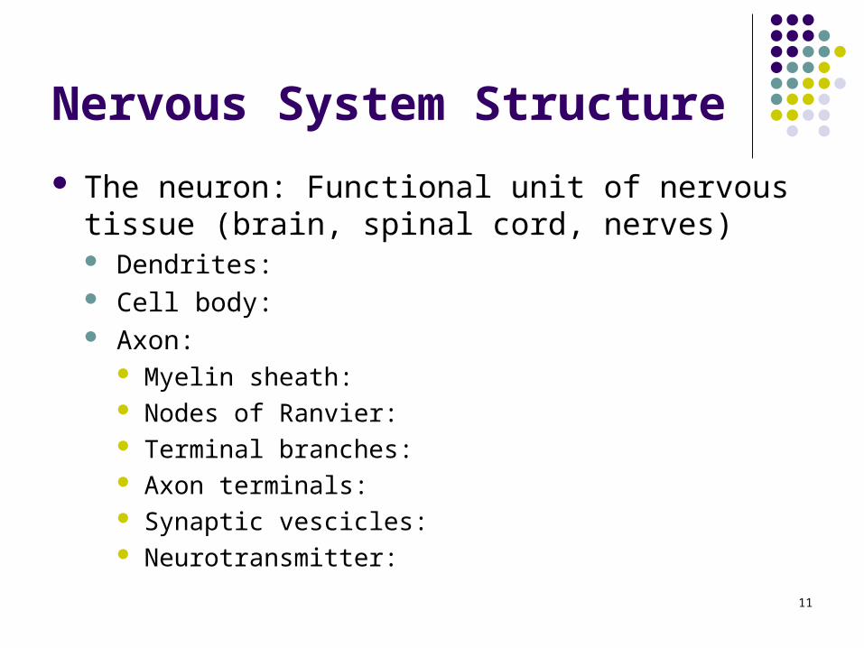
Nervous System Structure
The neuron: Functional unit of nervous tissue (brain, spinal cord, nerves) Dendrites: Cell body: Axon:
Myelin sheath: Nodes of Ranvier: Terminal branches: Axon terminals: Synaptic vescicles: Neurotransmitter:

12
DendritesCell body
Axon
Myelin sheath
Node of Ranvier
Terminal branch
Terminal ending
Figure 12.4, Marieb & Mallett (2003). Human Anatomy. Benjamin Cummings.

13
Figure 12.8, Marieb & Mallett (2003). Human Anatomy. Benjamin Cummings.
Terminal ending
Synaptic vescicle
Neurotransmitter: Acetylcholine (ACh)

14
Figure 12.19, Marieb & Mallett (2003). Human Anatomy. Benjamin Cummings.

15
Structure and Function
Nervous system structure Muscular system structure Neuromuscular function
16
Classification of Muscle Tissue Three types:
1. Smooth muscle
2. Cardiac muscle
3. Skeletal muscle

17
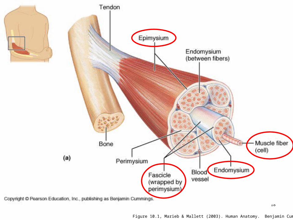
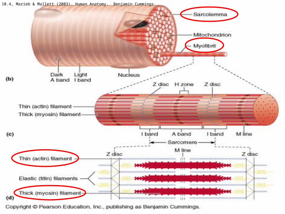
Muscular System Structure Organization:
Muscle (epimyseum) Fascicle (perimyseum)
Muscle fiber (endomyseum) Myofibril Myofilament Actin and myosin
Other Significant Structures: Sarcolemma Transverse tubule Sarcoplasmic reticulum Tropomyosin Troponin
18
Figure 10.1, Marieb & Mallett (2003). Human Anatomy. Benjamin Cummings.
19
Figure 10.4, Marieb & Mallett (2003). Human Anatomy. Benjamin Cummings.

20
http://staff.fcps.net/cverdecc/Adv%20A&P/Notes/Muscle%20Unit/sliding%20filament%20theory/slidin16.jpg

21
Figure 10.8, Marieb & Mallett (2003). Human Anatomy. Benjamin Cummings.

22
Structure and Function
Nervous system structure Muscular system structure Neuromuscular function
23
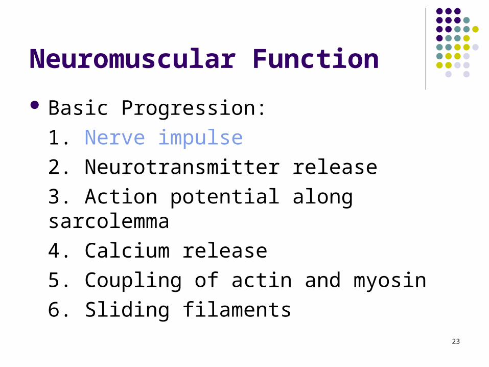
Neuromuscular Function
Basic Progression:
1. Nerve impulse
2. Neurotransmitter release
3. Action potential along sarcolemma
4. Calcium release
5. Coupling of actin and myosin
6. Sliding filaments

24
Nerve Impulse
What is a nerve impulse?
-Transmitted electrical charge
-Excites or inhibits an action
-An impulse that travels along an axon is an ACTION POTENTIAL
25
Nerve Impulse
How does a neuron send an impulse?
-Adequate stimulus from dendrite
-Depolarization of the resting membrane potential
-Repolarization of the resting membrane potential
-Propagation

26
Nerve Impulse
What is the resting membrane potential?-Difference in charge between inside/outside of the neuron
-70 mV
Figure 12.9, Marieb & Mallett (2003). Human Anatomy. Benjamin Cummings.

27
Nerve Impulse
What is depolarization?
-Reversal of the RMP from –70 mV to +30mV
Propagation of the action potential
Figure 12.9, Marieb & Mallett (2003). Human Anatomy. Benjamin Cummings.

28
Nerve Impulse
What is repolarization?
-Return of the RMP to –70 mV
Figure 12.9, Marieb & Mallett (2003). Human Anatomy. Benjamin Cummings.

29
-70 mV
+30 mV
30
Neuromuscular Function
Basic Progression:
1. Nerve impulse
2. Neurotransmitter release
3. Action potential along sarcolemma
4. Calcium release
5. Coupling of actin and myosin
6. Sliding filaments
31
Release of the Neurotransmitter
Action potential axon terminals
1. Calcium uptake
2. Release of synaptic vescicles (ACh)
3. Vescicles release ACh
4. ACh binds sarcolemma

32
Ca2+
ACh
Figure 12.8, Marieb & Mallett (2003). Human Anatomy. Benjamin Cummings.

33Figure 14.5, Marieb & Mallett (2003). Human Anatomy. Benjamin Cummings.
34
Neuromuscular Function
1. Nerve impulse
2. Neurotransmitter release
3. Action potential along sarcolemma
4. Calcium release
5. Coupling of actin and myosin
6. Sliding filaments

35
Ach

36
AP Along the Sarcolemma
Action potential Transverse tubules
1. T-tubules carry AP inside
2. AP activates sarcoplasmic reticulum

37
Figure 14.5, Marieb & Mallett (2003). Human Anatomy. Benjamin Cummings.

38
Neuromuscular Function
1. Nerve impulse
2. Neurotransmitter release
3. Action potential along sarcolemma
4. Calcium release
5. Coupling of actin and myosin
6. Sliding Filaments
39
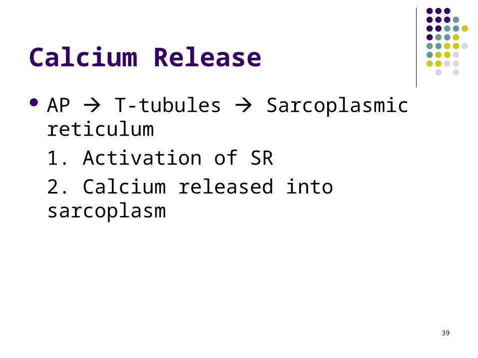
Calcium Release
AP T-tubules Sarcoplasmic reticulum
1. Activation of SR
2. Calcium released into sarcoplasm

40
Sarcolemma
CALCIUM
RELEASE
41
Neuromuscular Function
1. Nerve impulse
2. Neurotransmitter release
3. Action potential along sarcolemma
4. Calcium release
5. Coupling of actin and myosin
6. Sliding filaments

42
Coupling of Actin and Myosin
Tropomyosin Troponin

43
Blocked Coupling of actin and myosin

44
Neuromuscular Function
1. Nerve impulse
2. Neurotransmitter release
3. Action potential along sarcolemma
4. Calcium release
5. Coupling of actin and myosin
6. Sliding filaments

45
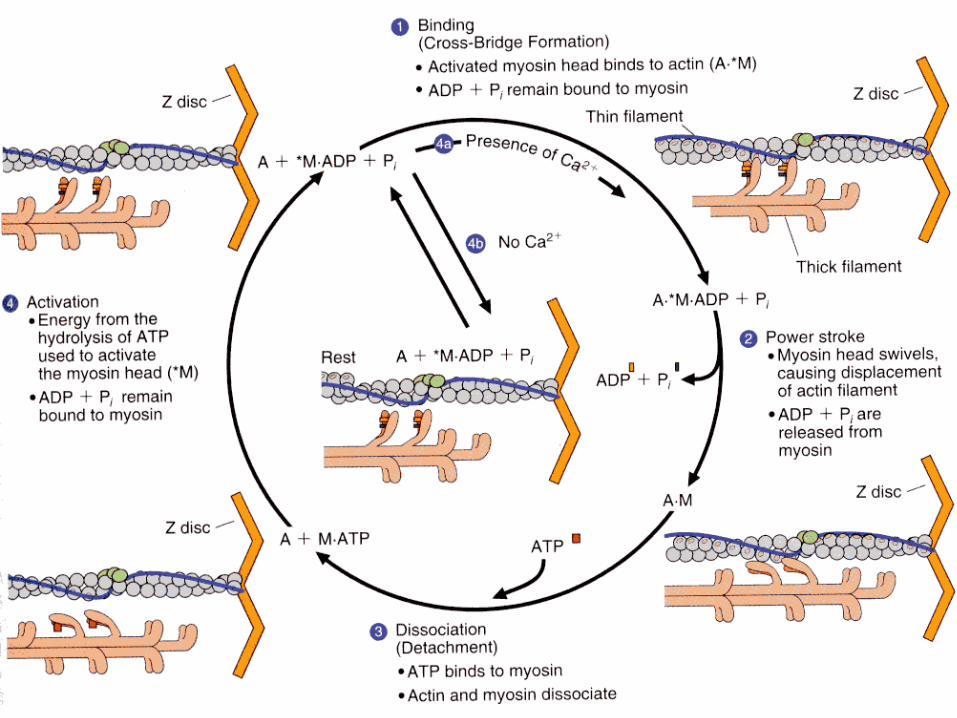
Sliding Filament Theory
Basic Progression of Events
1. Cross-bridge
2. Power stroke
3. Dissociation
4. Reactivation of myosin
46
Cross-Bridge
Activation of myosin via ATP
-ATP ADP + Pi + Energy
-Activation “cocked” position

47
Power Stroke
ADP + Pi are released Configurational change Actin and myosin slide

48
Dissociation
New ATP binds to myosin Dissociation occurs
49
Reactivation of Myosin Head
ATP ADP + Pi + Energy Reactivates the myosin head
Process starts over Process continues until:
-Nerve impulse stops-AP stops-Calcium pumped back into SR-Tropomyosin/troponin back to original position
50
51
Outline Introduction Structure and Function Fiber Arrangement Muscle Actions Role of Muscles Neural Control Factors that Affect Muscle Tension
52
Shape of Muscles & Fiber Arrangement
Muscles have different shapes & fiber arrangements
Shape & fiber arrangement affects
53
Shape of Muscles & Fiber Arrangement
Two major types of fiber arrangements

54
Fiber Arrangement - Parallel
Parallel muscles
Categorized into following shapes: Flat Fusiform Strap Radiate Sphincter or circular

55
Fiber Arrangement - Parallel
Flat muscles
Modified from Van De Graaff KM: Human anatomy, ed 6, Dubuque, IA, 2002, McGraw-Hill.

56
Fiber Arrangement - Parallel
Fusiform muscles
Figure 3.3. Hamilton, Weimar & Luttgens (2005). Kinesiology: Scientific basis for human motion. McGraw-Hill.
57
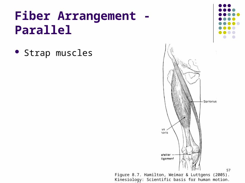
Fiber Arrangement - Parallel
Strap muscles
Figure 8.7. Hamilton, Weimar & Luttgens (2005). Kinesiology: Scientific basis for human motion. McGraw-Hill.
58
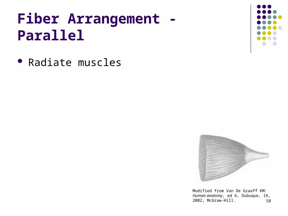
Fiber Arrangement - Parallel
Radiate muscles
Modified from Van De Graaff KM: Human anatomy, ed 6, Dubuque, IA, 2002, McGraw-Hill.
59
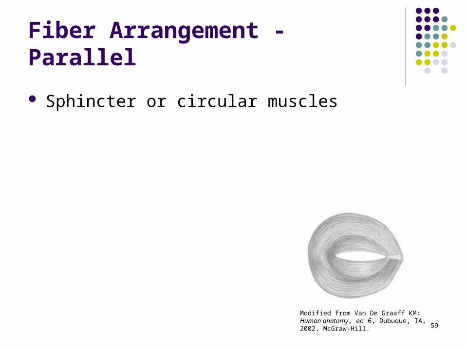
Fiber Arrangement - Parallel
Sphincter or circular muscles
Modified from Van De Graaff KM: Human anatomy, ed 6, Dubuque, IA, 2002, McGraw-Hill.
60
Fiber Arrangement - Pennate
Pennate muscles

61
Fiber Arrangement - Pennate
Categorized based upon the exact arrangement between fibers & tendon
Modified from Van De Graaff KM: Human anatomy, ed 6, Dubuque, IA, 2002, McGraw-Hill.
62
Fiber Arrangement - Pennate
Unipennate muscles

63
Fiber Arrangement - Pennate
Bipennate muscle
64
Fiber Arrangement - Pennate
Multipennate muscles
Bipennate & unipennate produce more force than multipennate
65
Outline Introduction Structure and Function Fiber Arrangement Muscle Actions Role of Muscles Neural Control Factors that Affect Muscle Tension
66
Muscle Actions: Terminology
Origin (Proximal Attachment):

67
Muscle Actions: Terminology
Insertion (Distal Attachment):
68
Muscle Actions: Terminology
When a particular muscle is activated:
Examples: Bicep curl vs. chin-up Hip extension vs. RDL
69
Muscle Actions Action:
Contraction:

70
Muscle Actions Muscle actions can be used to cause,
control, or prevent joint movement or

71
Types of Muscle Actions
IsometricIsometric IsotonicIsotonic
EccentricEccentricConcentricConcentric
MUSCLE ACTION (under tension)MUSCLE ACTION (under tension)
72
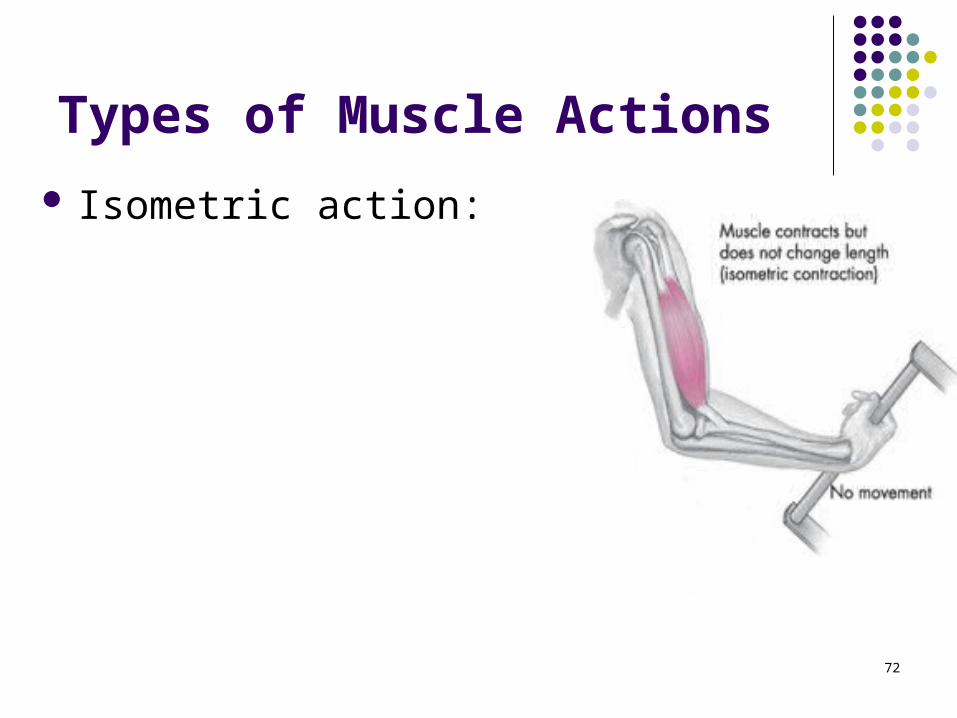
Types of Muscle Actions
Isometric action:

73
Types of Muscle Actions
Isotonic (same tension):
Isotonic contractions are either concentric (shortening) or eccentric (lengthening)

74
Types of Muscle Actions
Concentric contractions involve muscle developing tension as it shortens
Eccentric contractions involve the muscle lengthening under tension

75
Modified from Shier D, Butler J, Lewis R: Hole’s human anatomy & physiology, ed 9, Dubuque, IA, 2002, McGraw-Hill
What is the role of the elbow extensors in each phase?

76
Types of Muscle Actions Isokinetics:

77
Types of Muscle Actions
Movement may occur at any given joint without any muscle contraction whatsoever
78
Outline Introduction Structure and Function Fiber Arrangement Muscle Actions Role of Muscles Neural Control Factors that Affect Muscle Tension

79
Role of Muscles
Agonist muscles

80
Role of Muscles
Antagonist muscles

81

82
Role of Muscles
Stabilizers

83
Role of Muscles
Synergist

84
Role of Muscles
Neutralizers
85
Outline Introduction Structure and Function Fiber Arrangement Muscle Actions Role of Muscles Neural Control Factors that Affect Muscle Tension
86
Factors That Affect Muscle Tension
Number Coding and Rate Coding Length-Tension Relationship Force-Velocity Relationship Angle of Pull Uniarticular vs. Biarticular Muscles Cross-sectional Diameter Muscle Fiber Type Pennation

87
Number Coding & Rate Coding
Difference between lifting a minimal vs. maximal resistance is the number of muscle fibers recruited (crossbridges)
The number of muscle fibers recruited may be increased by

88
Number Coding & Rate Coding
Number of muscle fibers per motor unit varies significantly

89
Number Coding & Rate Coding As stimulus strength increases from threshold,
more motor units (Number Coding) are recruited & overall muscle contraction force increases in a graded fashion
From Seeley RR, Stephens TD, Tate P: Anatomy & physiology, ed 7, New York, 2006, McGraw-Hill.
90
Number Coding & Rate Coding
Greater contraction forces may also be achieved by increasing the frequency or motor unit activation (Rate Coding)
Phases of a single muscle fiber contraction or twitch Stimulus Latent period Contraction phase Relaxation phase

91
Number Coding & Rate Coding
Latent period
Contraction phase
Relaxation phase
From Powers SK, Howley ET: Exercise physiology: theory and application to fitness and performance, ed 4, New York, 2001 , McGraw-Hill.
92
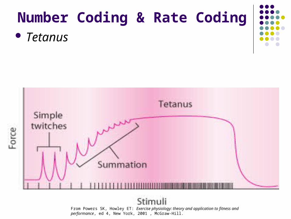
Number Coding & Rate Coding
Summation When successive stimuli are provided before
relaxation phase of first twitch has completed, subsequent twitches combine with the first to produce a sustained contraction
Generates a greater amount of tension than single contraction would produce individually
As frequency of stimuli increase, the resultant summation increases accordingly producing increasingly greater total muscle tension
93
Number Coding & Rate Coding Tetanus
From Powers SK, Howley ET: Exercise physiology: theory and application to fitness and performance, ed 4, New York, 2001 , McGraw-Hill.
94
All or None Principle
Motor unit
Typical muscle contraction
All or None Principle

95
Factors That Affect Muscle Tension
Number Coding and Rate Coding Length-Tension Relationship Force-Velocity Relationship Angle of Pull Uniarticular vs. Biarticular Muscles Cross-sectional Diameter Muscle Fiber Type Pennation

96
Length - Tension Relationship
Maximal ability of a muscle to develop tension & exert force varies depending upon the length of the muscle during contraction
Active Tension
Passive Tension

97
Length - Tension Relationship Generally, depending upon muscle
involved

98
Length - Tension Relationship
Generally, depending upon muscle involved

99Figure 20.2, Plowman and Smith (2002). Exercise Physiology, Benjamin Cummings.

100
Factors That Affect Muscle Tension
Number Coding and Rate Coding Length-Tension Relationship Force-Velocity Relationship Angle of Pull Uniarticular vs. Biarticular Muscles Cross-sectional Diameter Muscle Fiber Type Pennation

101
Force – Velocity Relationship When muscle is contracting (concentrically or
eccentrically) the rate of length change is significantly related to the amount of force potential
102
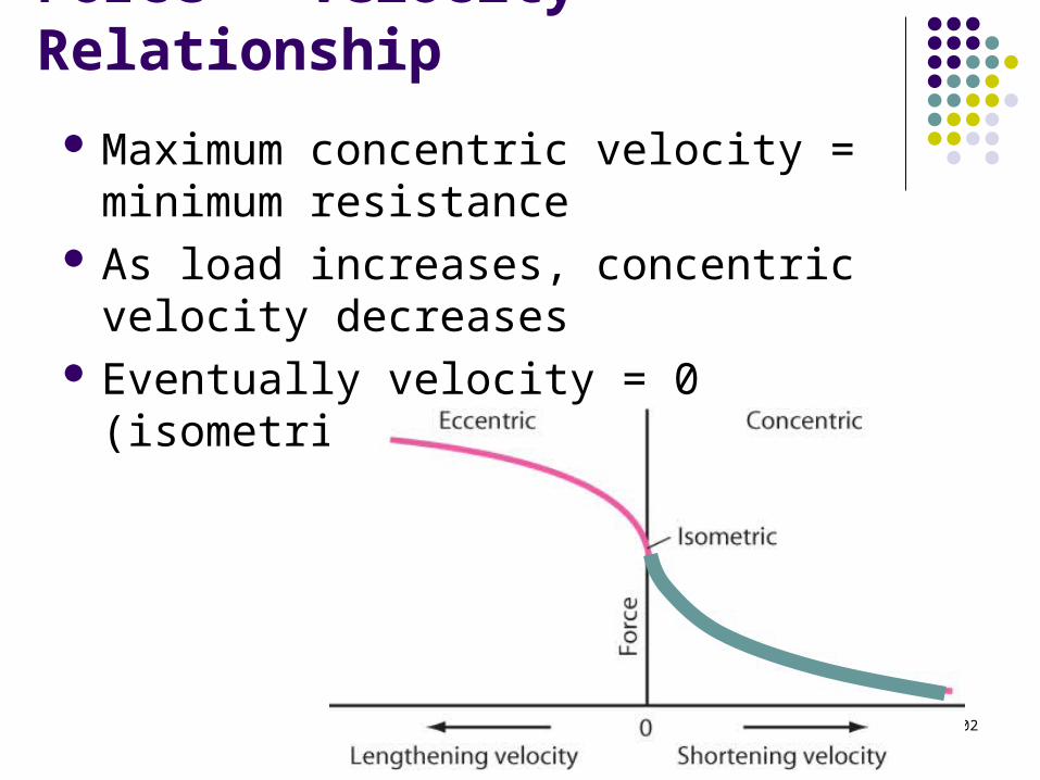
Force – Velocity Relationship
Maximum concentric velocity = minimum resistance
As load increases, concentric velocity decreases
Eventually velocity = 0 (isometric action)
103
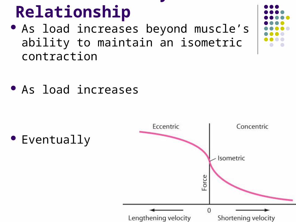
Force – Velocity Relationship As load increases beyond muscle’s ability to
maintain an isometric contraction
As load increases
Eventually
104
Muscle Force – Velocity Relationship
Indirect relationship between force (load) and concentric velocity
Direct relationship between force (load) and eccentric velocity
105
Factors That Affect Muscle Tension
Number Coding and Rate Coding Length-Tension Relationship Force-Velocity Relationship Angle of Pull Uniarticular vs. Biarticular Muscles Cross-sectional Diameter Muscle Fiber Type Pennation

106
Angle of Pull Angle between the line of pull of the muscle & the
bone on which it inserts (angle toward the joint) With every degree of joint motion, the angle of
pull changes Joint movements & insertion angles involve
mostly small angles of pull

107
Angle of Pull Angle of pull changes as joint moves
through ROM Most muscles work at angles of pull less
than 50 degrees Amount of muscular force needed to
cause joint movement is affected by angle of pull – Why?
108
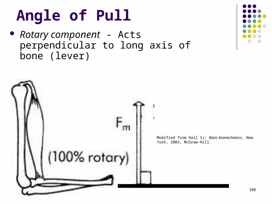
Angle of Pull Rotary component - Acts perpendicular to
long axis of bone (lever)
Modified from Hall SJ: Basic biomechanics, New York, 2003, McGraw-Hill.

109
Angle of Pull If angle < 90 degrees,
the parallel component is a stabilizing force
If angle > 90 degrees, the force is a dislocating force
Modified from Hall SJ: Basic biomechanics, New York, 2003, McGraw-Hill.
What is the effect of >/< 90 deg on ability to rotate the joint forcefully?

110
Factors That Affect Muscle Tension
Number Coding and Rate Coding Length-Tension Relationship Force-Velocity Relationship Angle of Pull Uniarticular vs. Biarticular Muscles Cross-sectional Diameter Muscle Fiber Type Pennation

111
Uni Vs. Biarticular Muscles
Uniarticular muscles
Ex: Brachialis
Ex: Gluteus Maximus
112
Uni Vs. Biarticular Muscles Biarticular muscles
May contract & cause motion at either one or both of its joints
Advantages over uniarticular muscles

113
Advantage #1
Can cause and/or control motion at more than one joint

114
Advantage #2
Can maintain a relatively constant length due to "shortening" at one joint and "lengthening" at another joint (Quasi-isometric)
- Recall the Length-Tension Relationship
115
Advantage #3
Prevention of Reciprocal Inhibition This effect is negated with biarticular
muscles when they move concurrently Concurrent movement:
Countercurrent movement:

116
What if the muscles of the hip/knee were uniarticular?
Hip
Knee
Ankle
Muscles stretched/shortened to extreme lengths!
Implication?

117Figure 20.2, Plowman and Smith (2002). Exercise Physiology, Benjamin Cummings.

118
Hip
Knee
Ankle
Quasi-isometric action? Implication?
119
Active & Passive Insufficiency Countercurrent muscle actions can reduce the
effectiveness of the muscle
As muscle shortens its ability to exert force diminishes
As muscle lengthens its ability to move through ROM or generate tension diminishes
120
Factors That Affect Muscle Tension
Number Coding and Rate Coding Length-Tension Relationship Force-Velocity Relationship Angle of Pull Uniarticular vs. Biarticular Muscles Cross-sectional Diameter Muscle Fiber Type Pennation

121
Cross-Sectional Area
Hypertrophy vs. hyperplasia Increased # of myofilaments
Increased size and # of myofibrils Increased size of muscle fibers
http://estb.msn.com/i/6B/917B20A6BE353420124115B1A511C7.jpg
122
Factors That Affect Muscle Tension
Number Coding and Rate Coding Length-Tension Relationship Force-Velocity Relationship Angle of Pull Uniarticular vs. Biarticular Muscles Cross-sectional Diameter Muscle Fiber Type Reflexes Pennation

123
Muscle Fiber Characteristics
Three basic types:
1. Type I:
-Slow twitch, oxidative, red
2. Type IIb:
-Fast twitch, glycolytic, white
3. Type IIa:
-FOG

124
Factors That Affect Muscle Tension
Number Coding and Rate Coding Length-Tension Relationship Force-Velocity Relationship Angle of Pull Uniarticular vs. Biarticular Muscles Cross-sectional Diameter Muscle Fiber Type Reflexes Pennation
125
Effect of Fiber Arrangement on Force Output
Concept #1: Force directly related to cross-sectional area more fibers
Example: Thick vs. thin longitudinal/fusiform muscle?
Example: Thick fusiform/longitudinal vs. thick bipenniform muscle?
Concept #2: As degree of pennation increases, so does # of fibers per CSA

126