07280 jlggc
TRANSCRIPT
1
The SRA domain of UHRF1 flips 5-methylcytosine out of the DNA helix
Hideharu Hashimoto1, John R. Horton
1, Xing Zhang
1, Magnolia Bostick
2, Steven
Jacobsen2,3
, and Xiaodong Cheng1
1Department of Biochemistry, Emory University School of Medicine, 1510 Clifton Road,
Atlanta, GA 30322, USA
2Department of Molecular Cell and Developmental Biology, University of California,
Los Angeles, 621 Charles E. Young Dr. South, Los Angeles, CA, 90095, USA
3Howard Hughes Medical Institute, University of California, Los Angeles, Los Angeles,
CA, 90095, USA
SUPPLEMENTARY INFORMATION
doi: 10.1038/nature07280
www.nature.com/nature 1

2
Figure S1. Mapping the SRA domain within mouse UHRF1
a, Mouse UHRF1 (mUHRF1) is a 782-residue protein containing four recognizable
domains including a ubiquitin-like domain, a plant homeodomain (PHD) that may be
involved in histone H3 tail binding 1, 2
, a SET and RING associated (SRA) domain that
preferentially binds to hemimethylated CpG sites 3, and a C-terminal Really Interesting
New Gene (RING) domain may confer E3 ubiquitin ligase activity 1. b, Proteolytic
digestion, mass spectrometry, and deletion analyses identified the SRA domain
boundaries of mUHRF1 as residues 419-628. Left panel: SDS-PAGE gel of the purified
recombinant mUHRF1 full length and its trypsin digestion products. Purified
hexahistidine-SUMO-tagged mUHRF1 full length protein (1.7 µg) in 20 mM Hepes 7.0,
400 mM NaCl, 5% glycerol, and 0.1% 2-mercaptoethanol was treated with 0, 0.5, 5, 50
ng of trypsin for 30 min and separated on a 13% SDS gel. Mass spectrometry determined
molecular masses of individual fragments. Each fragment was constructed into a
hexahistidine-SUMO tagged fusion protein and expressed in Escherichia coli. We also
varied the starting and ending amino acids of fragments to reach maximum expression
and solubility. Right panel: SDS-PAGE gel of the purified recombinant SRA domain
(between the molecular weight markers of 20 and 26.6 kDa) used for crystallization.
References:
1. Citterio, E. et al. Np95 is a histone-binding protein endowed with ubiquitin ligase
activity. Mol Cell Biol 24, 2526-35 (2004).
2. Karagianni, P., Amazit, L., Qin, J. & Wong, J. ICBP90, a novel methyl K9 H3
binding protein linking protein ubiquitination with heterochromatin formation.
Mol Cell Biol 28, 705-17 (2008).
3. Bostick, M. et al. UHRF1 plays a role in maintaining DNA methylation in
mammalian cells. Science 317, 1760-4 (2007).
doi: 10.1038/nature07280 SUPPLEMENTARY INFORMATION
www.nature.com/nature 2
3
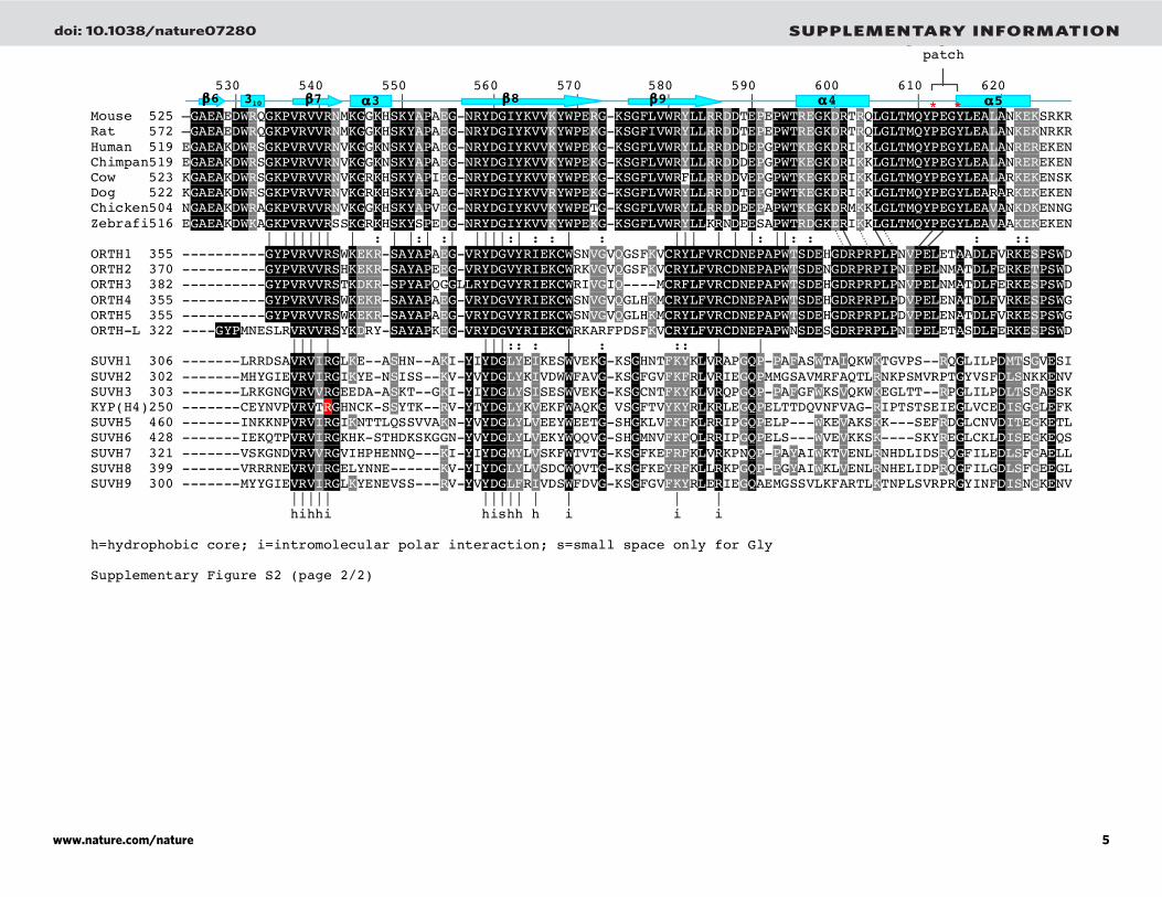
Figure S2. Sequence alignment of mammalian and plant SRA domains
Secondary structural elements are indicated in light blue. Numbering above the sequences
corresponds to the mouse ortholog. White-on-black residues are invariant among the
blocks of sequences examined, while gray-highlighted positions are conserved (with !1
substitution). Positions highlighted in * are responsible for various functions as indicated,
and the red circles with a letter P are amino acids that interact with the DNA phosphate
backbone. Residues from two parts of the polypeptide form the hydrophobic patch.
Mammalian UHRF1 included are Mouse (AAH22167), Rat (NP_001008882), Human
(ABQ59043), Chimpanzee (XP_001139655), Cow (NP_001096568), Dog (XP_868468),
Chicken (XP_418269), and Zebrafish (NP_998242).
Arabidopsis homologs of UHRF1 included are ORTH1 (At5g39550),
ORTH2/VIM1 (At1g57820), ORTH3 (At1g57800), ORTH4 (At1g66040), ORTH5
(At1g66050), and ORTH-like (At4g08590). Also included are the Arabidopsis SRA
containing histone methyltransferase genes (SUVH genes) exemplified by the
KRYPTONITE (KYP) protein involved in DNA methylation control 4. Xn represents an
insertion of variable (n) amino acids in the SUVH proteins.
The large majority of the invariant amino acids are involved in structural and
intramolecular interactions. For example, conserved hydrophobic side chains intercalate
with each other to form the hydrophobic core of the molecule. In addition, many of the
invariant residues are polar or charged and are critically involved in stabilizing a network
of polar interactions involving different parts of molecule. For example, H447 interacts
S464 (Fig. 2e). These two residues are critical for stabilizing a network of polar
interactions involving (1) the main chain carbonyl oxygen of A452, the amino acid next
to the key residue (V451) important for base flipping, (2) R438 interacting with DNA
phosphate, and (3) a water-mediated network involving main chain atom of R448, and
side chains of H455 and R538. R448 and H455 interact with DNA phosphates (see Fig.
1a). R538 interacts with the main chain atoms of H422, G424, and V446 (not shown).
A network of interactions involves D476…R541…D560…R558 (Fig. 2f). D560
is part of the three consecutive invariant residues YDG initially used to name the domain 5. Q504 interacts with the main chain atoms of G485, G487, and W580. N510 interacts
with the main chain atoms of S486 and K505; E571 bridges between W569 and R581.
References
4. Johnson, L. M. et al. The SRA methyl-cytosine-binding domain links DNA and
histone methylation. Curr Biol 17, 379-84 (2007).
5. Baumbusch, L. O. et al. The Arabidopsis thaliana genome contains at least 29
active genes encoding SET domain proteins that can be assigned to four
evolutionarily conserved classes. Nucleic Acids Res 29, 4319-33 (2001).
doi: 10.1038/nature07280 SUPPLEMENTARY INFORMATION
www.nature.com/nature 3

hydrophobic base 5mC CpG patch flipping binding pocket recognition 420 430 440 450 460 470 480 490 500 510 520 | | | | | | | | | | | Mouse 419 PANHFGPIPGVPVGTMWRFRVQVSESGVHRPHVAGIHGRSNDGAYSLVLAGGYEDDVDNGNYFTYTGSGGRDLSGNKRTA--GQSSDQKLTNNNRALALNCHSPINEK Rat 466 PANHFGPIPGVPVGTMWRFRVQVSESGVHRPHVAGIHGRSNDGAYSLVLAGGYEDDVDNGNFFTYTGSGGRDLSGNKRTA--GQSSDQKLTNNNRALALNCHSPINEK Human 414 PSNHYGPIPGIPVGTMWRFRVQVSESGVHRPHVAGIHGRSNDGAYSLVLAGGYEDDVDHGNFFTYTGSGGRDLSGNKRTA--EQSCDQKLTNTNRALALNCFAPINDQ Chimpan414 PSNHYGPIPGIPVGTMWRFRVQVSESGVHRPHVAGIHGRSNDGAYSLVLAGGYEDDVDHGNFFTYTGSGGRDLSGNKRTA--EQSCDQKLTNTNRALALNCFAPINDQ Cow 418 PSNHFGPIPGIPVGTMWRFRVQVSESGVHRPHVAGIHGRSNHGAYSLVLAGGYEDDVDHGNSFTYTGSGGRDLSGNKRTA--EQSCDQKLTNTNRALALNCFAPINDL Dog 417 PSNHYGPIPGIPVGTMWRFRVQVSESGVHRPHVAGIHGRSNDGAYSLVLAGGYEDDVDHGNSFTYTGSGGRDLSGNKRTA--EQSCDQKLTNTNRALALNCSAPINDR Chicken399 PSNHYGPIPGIPVGTMWKFRVQVSESGVHRPHVAGIHGRSNDGAYSLVLAGGYEDDIDHGNSFTYTGSGGRDLSGNKRTA--EQSCDQKLTNMNRALALNCSAPINDK Zebrafi411 PSNHYGPVPGVPVGTLWKFRVQVSESGVHRPHVAGIHGRSNDGAYSLVLAGGYEDDVDDGNEFTYTGSGGRDLSGNKRTA--EQSCDQKLTNMNRALALNCNAAVNDK |: || | | | | || | | || |: : ||| || | | | |||:::: : :::: : || : : :: : | |: || | | | | || | | || |: : ||| || | | | |||:::: : :::: : || : : :: : | |: || | | | | || | | || |: : ||| || | | | |||:::: : :::: : || : : :: : | ORTH1 253 AENDVTRKQGVLVGESWEDRQECRQWGAHFPHIAGIAGQSAVGAQSVALSGGYDDDEDHGEWFLYTGSGGRDLSGNKRINK-KQSSDQAFKN------LSCKM----- ORTH2 268 AENDPVRNQGLLVGESWEDRLECRQWGAHFPHVAGIAGQSTYGAQSVALSGGYKDDEDHGEWFLYTGSGGRDLSGNKRTNK-EQSFDQKFEKSNAALKLSCKL----- ORTH3 280 AENDPVRNQGLLVGESWKGRLACRQWGAHFPHVSGIAGQASYGAQSVVLAGGYDDDEDHGEWFLYTGSGGRILKGNKRTNT-VQAFDQVFLNFNEALRLSCKL----- ORTH4 253 AANDVTRNQGVLVGESWEDRQECRQWGVHFPHVAGIAGQAAVGAQSVALSGGYDDDEDHGEWFLYTGSGGRDLSGNKRVNK-IQSSDQAFKNMNEALRLSCKM----- ORTH5 253 AANDVTRNQGVLVGESWEDRQECRQWGVHFPHVAGIAGQAAVGAQSVALSGGYDDDEDHGEWFLYTGSGGRDLSGNKRVNK-IQSSDQAFKNMNEALRLSCKM----- ORTH-L 229 AEHDPVRNQGVLVGESWENRVECRQWGVHLPHVSCIAGQEDYGAQSVVISGGYKDDEDHGEWFLYTG-RSR---GRHFAN-----EDQEFEDLNEALRVSCEM----- |: :|: : : : | | :| | :: :| | : : |:| :: :| : | :: | SUVH1 206 TKKRPGIVPGVEIGDVFFFRFEMCLVGLHSPSMAGID-X11-PIATSIVSSGYYDNDEGNPDVLIYTGQGGNADK--DK-----QSSDQKLERGNLALEKS-------- SUVH2 202 DKHIVGPVTGVEVGDIFFYRMELCVLGLHGQTQAGID-X11-PIATSIVVSGGYEDDEDTGDVLVYTGHGGQDH--QHK-----QCDNQRLVGGNLGMERS-------- SUVH3 203 MKKRVGTVPGIEVGDIFFSRIEMCLVGLHMQTMAGID-X11-SLATSIVSSGRYEGEAQDPESLIYSGQGGNADK--NR-----QASDQKLERGNLALENS-------- KYP(H4)144 PRKIIGDLPGIDVGHRFFSRAEMCAVGFHNHWLNGID-X15-PLAVSIVMSGQYEDDLDNADTVTYTGQGGHNLTGNKR-----QIKDQLLERGNLALKHC-------- SUVH5 360 GTQIIGTVPGVEVGDEFQYRMELNLLGIHRPSQSGID-X08-LVATSIVSSGGYNDVLDNSDVLIYTGQGG-NV-GKKKNNE--PPKDQQLVTGNLALKNS-------- SUVH6 325 GVHILGEVPGVEVGDEFQYRMELNILGIHKPSQAGID-X07-KVATSIVASGGYDDHLDNSDVLTYTGQGGNVMQVKKKGEELKEPEDQKLITGNLALATS-------- SUVH7 222 TRRRIGAVPGIHVGDIFYYWGEMCLVGLHKSNYGGID-X11-HAAMCVVTAGQYDGETEGLDTLIYSGQGGTDVYGNAR--------DQEMKGGNLALEAS-------- SUVH8 305 MTRRIGPIPGVQVGDIFYYWCEMCLVGLHRNTAGGID-X11-PAATSVVTSGKYDNETEDLETLIYSGHGGKPC-------------DQVLQRGNRALEAS-------- SUVH9 200 DKRIVGSIPGVQVGDIFFFRFELCVMGLHGHPQSGID-X11-PIATSVIVSGGYEDDDDQGDVIMYTGQGGQDRLGR-------QAEHQRLEGGNLAMERS-------- || | | | || | || | | | | | || | | th t t i th h ih t m i m s tt i i t=structural turn; h=hydrophobic core; i=intromolecular polar interaction; m=5mC binding; s=small space only for Gly Supplementary Figure S2 (page 1/2)
* * ** * * * * !1 "1 !2 "2 !3 !4 !5
* *
P P P P P P P P P
doi: 10.1038/nature07280 SUPPLEMENTARY INFORMATION
www.nature.com/nature 4
Hydrophobic patch 530 540 550 560 570 580 590 600 610 620 | | | | | | | | | | Mouse 525 –GAEAEDWRQGKPVRVVRNMKGGKHSKYAPAEG-NRYDGIYKVVKYWPERG-KSGFLVWRYLLRRDDTEPEPWTREGKDRTRQLGLTMQYPEGYLEALANKEKSRKR Rat 572 –GAEAEDWRQGKPVRVVRNMKGGKHSKYAPAEG-NRYDGIYKVVKYWPEKG-KSGFIVWRYLLRRDDTEPEPWTREGKDRTRQLGLTMQYPEGYLEALANKEKNRKR Human 519 EGAEAKDWRSGKPVRVVRNVKGGKNSKYAPAEG-NRYDGIYKVVKYWPEKG-KSGFLVWRYLLRRDDDEPGPWTKEGKDRIKKLGLTMQYPEGYLEALANREREKEN Chimpan519 EGAEAKDWRSGKPVRVVRNVKGGKNSKYAPAEG-NRYDGIYKVVKYWPEKG-KSGFLVWRYLLRRDDDEPGPWTKEGKDRIKKLGLTMQYPEGYLEALANREREKEN Cow 523 KGAEAKDWRSGKPVRVVRNVKGRKHSKYAPIEG-NRYDGIYKVVRYWPEKG-KSGFLVWRFLLRRDDVEPGPWTKEGKDRIKKLGLTMQYPEGYLEALARKEKENSK Dog 522 KGAEAKDWRSGKPVRVVRNVKGRKHSKYAPAEG-NRYDGIYKVVRYWPEKG-KSGFLVWRYLLRRDDTEPGPWTKEGKDRIKKLGLTMQYPEGYLEARARKEKEKEN Chicken504 NGAEAKDWRAGKPVRVVRNVKGGKHSKYAPVEG-NRYDGIYKVVKYWPETG-KSGFLVWRYLLRRDDEEPAPWTKEGKDRMKKLGLTMQYPEGYLEAVANKDKENNG Zebrafi516 EGAEAKDWKAGKPVRVVRSSKGRKHSKYSPEDG-NRYDGIYKVVKYWPEKG-KSGFLVWRYLLKRNDEESAPWTRDGKERIKKLGLTMQYPEGYLEAVAAKEKEKEN | |||||| | : | |:| :| ||||:| : : | : ||| | | |: ||: : : :: ORTH1 355 ----------GYPVRVVRSWKEKR-SAYAPAEG-VRYDGVYRIEKCWSNVGVQGSFKVCRYLFVRCDNEPAPWTSDEHGDRPRPLPNVPELETAADLFVRKESPSWD ORTH2 370 ----------GYPVRVVRSHKEKR-SAYAPEEG-VRYDGVYRIEKCWRKVGVQGSFKVCRYLFVRCDNEPAPWTSDENGDRPRPIPNIPELNMATDLFERKETPSWD ORTH3 382 ----------GYPVRVVRSTKDKR-SPYAPQGGLLRYDGVYRIEKCWRIVGIQ----MCRFLFVRCDNEPAPWTSDEHGDRPRPLPNVPELNMATDLFERKESPSWD ORTH4 355 ----------GYPVRVVRSWKEKR-SAYAPAEG-VRYDGVYRIEKCWSNVGVQGLHKMCRYLFVRCDNEPAPWTSDEHGDRPRPLPDVPELENATDLFVRKESPSWG ORTH5 355 ----------GYPVRVVRSWKEKR-SAYAPAEG-VRYDGVYRIEKCWSNVGVQGLHKMCRYLFVRCDNEPAPWTSDEHGDRPRPLPDVPELENATDLFVRKESPSWG ORTH-L 322 ----GYPMNESLRVRVVRSYKDRY-SAYAPKEG-VRYDGVYRIEKCWRKARFPDSFKVCRYLFVRCDNEPAPWNSDESGDRPRPLPNIPELETASDLFERKESPSWD ||| | |||:: : | : :: | | SUVH1 306 -------LRRDSAVRVIRGLKE--ASHN--AKI-YIYDGLYEIKESWVEKG-KSGHNTFKYKLVRAPGQP-PAFASWTAIQKWKTGVPS--RQGLILPDMTSGVESI SUVH2 302 -------MHYGIEVRVIRGIKYE-NSISS--KV-YVYDGLYKIVDWWFAVG-KSGFGVFKFRLVRIEGQPMMGSAVMRFAQTLRNKPSMVRPTGYVSFDLSNKKENV SUVH3 303 -------LRKGNGVRVVRGEEDA-ASKT--GKI-YIYDGLYSISESWVEKG-KSGCNTFKYKLVRQPGQP-PAFGFWKSVQKWKEGLTT--RPGLILPDLTSGAESK KYP(H4)250 -------CEYNVPVRVTRGHNCK-SSYTK--RV-YTYDGLYKVEKFWAQKG-VSGFTVYKYRLKRLEGQPELTTDQVNFVAG-RIPTSTSEIEGLVCEDISGGLEFK SUVH5 460 -------INKKNPVRVIRGIKNTTLQSSVVAKN-YVYDGLYLVEEYWEETG-SHGKLVFKFKLRRIPGQPELP---WKEVAKSKK---SEFRDGLCNVDITEGKETL SUVH6 428 -------IEKQTPVRVIRGKHK-STHDKSKGGN-YVYDGLYLVEKYWQQVG-SHGMNVFKFQLRRIPGQPELS---WVEVKKSK----SKYREGLCKLDISEGKEQS SUVH7 321 -------VSKGNDVRVVRGVIHPHENNQ---KI-YIYDGMYLVSKFWTVTG-KSGFKEFRFKLVRKPNQP-PAYAIWKTVENLRNHDLIDSRQGFILEDLSFGAELL SUVH8 399 -------VRRRNEVRVIRGELYNNE------KV-YIYDGLYLVSDCWQVTG-KSGFKEYRFKLLRKPGQP-PGYAIWKLVENLRNHELIDPRQGFILGDLSFGEEGL SUVH9 300 -------MYYGIEVRVIRGLKYENEVSS---RV-YVYDGLFRIVDSWFDVG-KSGFGVFKYRLERIEGQAEMGSSVLKFARTLKTNPLSVRPRGYINFDISNGKENV ||||| ||||| | | | | hihhi hishh h i i i h=hydrophobic core; i=intromolecular polar interaction; s=small space only for Gly Supplementary Figure S2 (page 2/2)
!6 !8 !7 310 !9 "3 "5 "4 * *
doi: 10.1038/nature07280 SUPPLEMENTARY INFORMATION
www.nature.com/nature 5

4
Figure S3. mUHRF1 SRA domain binds oligonucleotide with an increased affinity
for hemimethylated CpG site
Increasing amounts of GST-mUHRF1 SRA domain (amino acids 393-621) 3, 6
are
incubated with a radiolabelled oligonucleotide with a single CG site (5'-A GG GA TG
GG GT TT XG TT TT CT CT CT CT C-3' / 5’-G AG AG AG AG AA AA YG AA AC
CC CA TC CC T-3’) that is either unmethylated (X=Y=C; upper panel), hemi-methylated
(X=5mC, Y=C; middle panel), or fully methylated (X=Y=5mC; bottom panel). No
protein is present in lane 1, and the amount of DNA in per reaction was 85 picogram for
unmethylated, 70.4 picogram for hemimethylated, and 83.5 picogram for fully
methylated oligonucleotides. Protein concentration increases in each lane with 0.125 µg
(lane 2), 0.25 µg (lane 3), 0.5 µg (lane 4), 1 µg (lane 5), 2 µg (lane 6), 3 µg (lane 7), 4 µg
(lane 8), 5 µg (lane 9), and 6 µg (lane 10).
References
3. Bostick, M. et al. UHRF1 plays a role in maintaining DNA methylation in
mammalian cells. Science 317, 1760-4 (2007).
6. Sharif, J. et al. The SRA protein Np95 mediates epigenetic inheritance by
recruiting Dnmt1 to methylated DNA. Nature 450, 908-12 (2007).
doi: 10.1038/nature07280 SUPPLEMENTARY INFORMATION
www.nature.com/nature 6
5
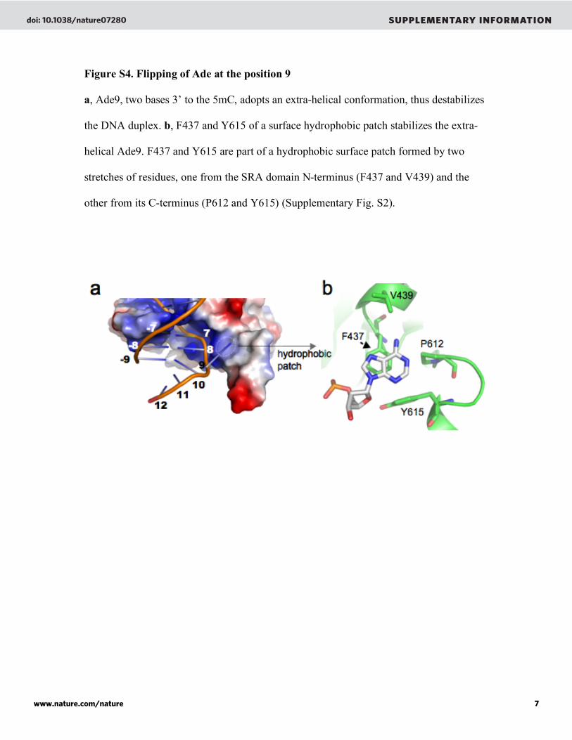
Figure S4. Flipping of Ade at the position 9
a, Ade9, two bases 3’ to the 5mC, adopts an extra-helical conformation, thus destabilizes
the DNA duplex. b, F437 and Y615 of a surface hydrophobic patch stabilizes the extra-
helical Ade9. F437 and Y615 are part of a hydrophobic surface patch formed by two
stretches of residues, one from the SRA domain N-terminus (F437 and V439) and the
other from its C-terminus (P612 and Y615) (Supplementary Fig. S2).
doi: 10.1038/nature07280 SUPPLEMENTARY INFORMATION
www.nature.com/nature 7

6
Figure S5. Schematic SRA-DNA interactions in the space group P212121
The SRA residues that interact with DNA are marked in red; residues in blue belong to
the symmetry-related SRA molecule.
disorder
ed
doi: 10.1038/nature07280 SUPPLEMENTARY INFORMATION
www.nature.com/nature 8
7
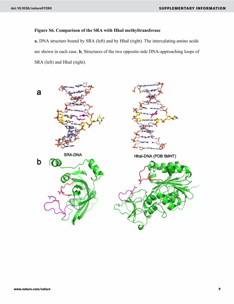
Figure S6. Comparison of the SRA with HhaI methyltransferase
a, DNA structure bound by SRA (left) and by HhaI (right). The intercalating amino acids
are shown in each case. b, Structures of the two opposite-side DNA-approaching loops of
SRA (left) and HhaI (right).
doi: 10.1038/nature07280 SUPPLEMENTARY INFORMATION
www.nature.com/nature 9
8
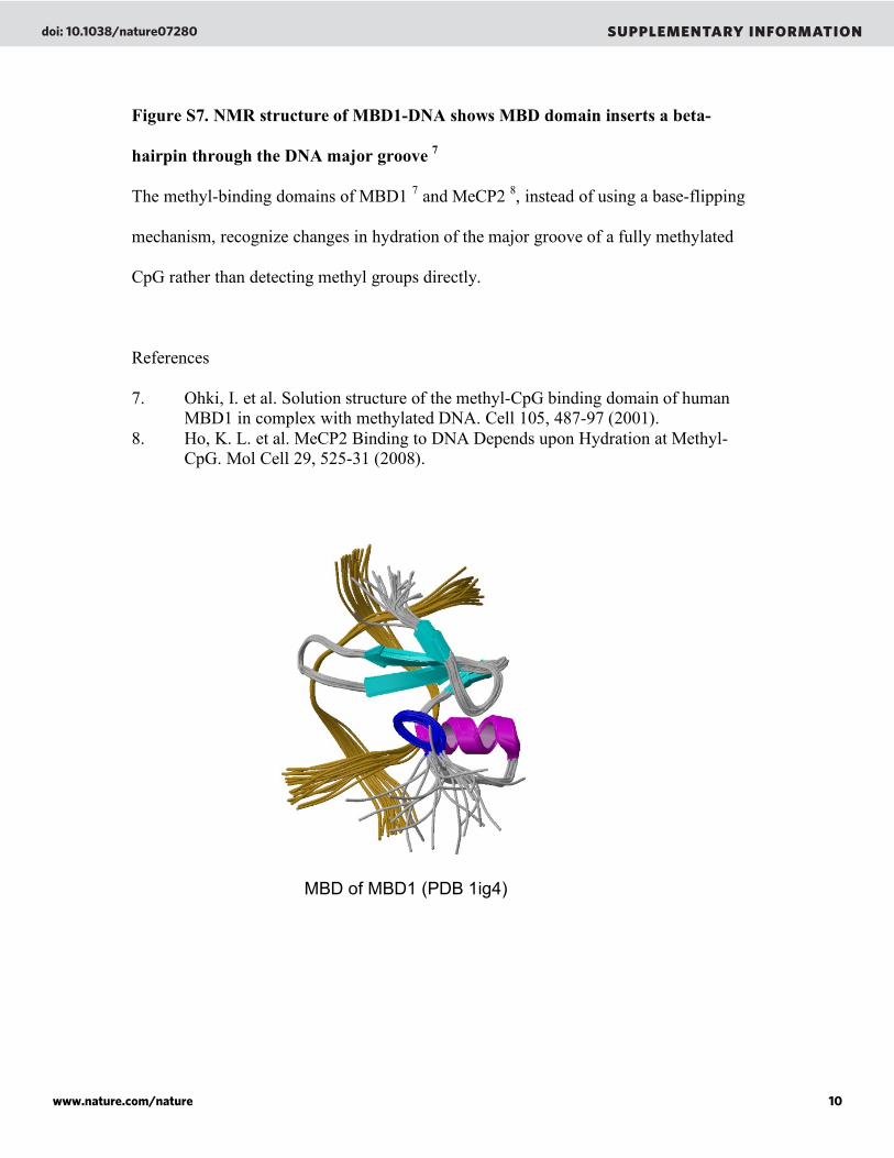
Figure S7. NMR structure of MBD1-DNA shows MBD domain inserts a beta-
hairpin through the DNA major groove 7
The methyl-binding domains of MBD1 7 and MeCP2
8, instead of using a base-flipping
mechanism, recognize changes in hydration of the major groove of a fully methylated
CpG rather than detecting methyl groups directly.
References
7. Ohki, I. et al. Solution structure of the methyl-CpG binding domain of human
MBD1 in complex with methylated DNA. Cell 105, 487-97 (2001).
8. Ho, K. L. et al. MeCP2 Binding to DNA Depends upon Hydration at Methyl-
CpG. Mol Cell 29, 525-31 (2008).
MBD of MBD1 (PDB 1ig4)
doi: 10.1038/nature07280 SUPPLEMENTARY INFORMATION
www.nature.com/nature 10
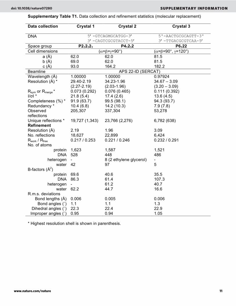
Supplementary Table T1. Data collection and refinement statistics (molecular replacement) Data collection Crystal 1
Crystal 2
Crystal 3
DNA 5’-GTCAGMGCATGG-3’
3’-CAGTCGCGTACCT-5’ 5'-AACTGCGCAGTT-3'
3’-TTGACGCGTCAA-5’
Space group P212121 P41212 P6122
Cell dimensions (!="=#=90°) (!="=90°, #=120°) a (Å) b (Å) c (Å)
62.0 69.0 93.0
62.0 62.0 164.2
81.5 81.5 182.2
Beamline APS 22-ID (SERCAT) Wavelength (Å) 1.00000 1.00000 0.97924 Resolution (Å) * 29.40-2.19
(2.27-2.19) 34.23-1.96 (2.03-1.96)
34.67 – 3.09 (3.20 – 3.09)
Rsym or Rmerge * 0.073 (0.292) 0.076 (0.465) 0.111 (0.392) I/!I * 21.8 (5.4) 17.4 (2.6) 13.6 (4.5) Completeness (%) * 91.9 (63.7) 99.5 (98.1) 94.3 (93.7) Redundancy * 10.4 (6.8) 14.2 (10.3) 7.9 (7.8) Observed reflections
205,307 337,304 53,278
Unique reflections * 19,727 (1,343) 23,766 (2,276) 6,782 (638) Refinement
Resolution (Å) 2.19 1.96 3.09 No. reflections 18,627 22,899 6,424 Rwork / Rfree 0.217 / 0.253 0.221 / 0.246 0.232 / 0.291 No. of atoms
protein 1,623 1,587 1,521 DNA 528 448 486
heterogen - 8 (2 ethylene glycerol) - water 42 97 5
B-factors (Å2) protein 69.6 40.6 35.5
DNA 86.3 61.4 107.3 heterogen - 61.2 40.7
water 62.2 44.7 16.6 R.m.s. deviations
Bond lengths (Å) 0.006 0.005 0.006 Bond angles (˚) 1.1 1.1 1.3
Dihedral angles (˚) 22.3 22.4 22.9 Improper angles (˚) 0.95 0.94 1.05
* Highest resolution shell is shown in parenthesis.
doi: 10.1038/nature07280 SUPPLEMENTARY INFORMATION
www.nature.com/nature 11