ﻢﻴﺣﺮﻟا ﻦﻤﺣﺮﻟا ﷲا...
TRANSCRIPT

بسم اهللا الرحمن الرحيمبسم اهللا الرحمن الرحيمبسم اهللا الرحمن الرحيمبسم اهللا الرحمن الرحيم
اللهم علمنى بما ينفعنى اللهم علمنى بما ينفعنى اللهم علمنى بما ينفعنى اللهم علمنى بما ينفعنى وأنفعنى بما علمتنى وأنفعنى بما علمتنى وأنفعنى بما علمتنى وأنفعنى بما علمتنى
وذدنى علماوذدنى علماوذدنى علماوذدنى علما

BIO-SIGNALS
*The bio-signals potentials are generate at cellular level and the source of these
potentials is ionic in nature.
* cell consists of an ionic conductor separated from the outside environment by
a semi permeable membrane which acts as a selective ionic filter to the ions. This means that some ions can pass through the
membrane freely where as other cannot do so.

*All living matters are composed of cells different types.
* The bio-signals are EMG, EOG, ECG, or
EEG this signals can be analyzed and sensors.
*This signal can monitoring ,rehabilitation ,
feedback functions and control gain .
* EMG is electro mayo graph its detect contraction of the skeletal muscles result in generation of action potentials in the
individual muscle fibers.

* EOG is electro oculo graph its detect the potentials that generated by the movement of the
eye ball.

ELECTRICAL SYSTEM OF HEART


THE ECG WAVEFORM

ECG BASICS
*P wave: represents the depolarization impulse across the atria
*Q, R and S waves: all these three waves
represent the ventricular depolarization
*T wave: represents the repolarization of the ventricles

ECG Basics
*P wave : 0.25 mv
*R wave : 1.60 mv
*Q wave : 25% of R-wave
*T wave : 0.1 to 0.5 mv

ECG Basics
*P-R Interval : 0.12 to 0.20 s
*Q-T Interval : 0.35 to 0.44 s
*S-T Segment : 0.05 to 0.15 s
*P wave : 0.11 s
*QRS Interval : 0.09 s
Electro cardio graph
*ECG is an instrument which records the electrical activity of the heart.
Electrical signals from the heart characteristically precede the normal mechanical function and
monitoring of these signals has great
significance.
*ECG provides valuable information about wide range of cardiac disorders such as the presence
of an inactive part (infarctions) or an
enlargement (cardiac hypertrophy) of the heart muscle.


Block diagram
*The potentials pick up by electrodes are taken to
the lead selector switch. In the lead selector the electrodes are selected two by two according to
the lead program.
*The signal is connected symmetrically to the pair
deferential amplifier. The pre amplifier is usually a three or four stages the output signal pick to
power amplifier.
*Direct writing recorder is usually adequate since
the ESG signal of the interested has limited has bandwidth.

Frequency selective network is an R-C network. Which provides necessary damping of the pen
motor and is preset by the manufacture.
*The auxiliary circuit provide a 1mV calibration
signal and automatic blocking of the amplifier
during a change in the position of the lead switch.
*It may include a speed control circuit for the chart drive motor.

*Isolated preamplifier :
It had traditional for all ECG to have the right leg
(RL) electrode connected to the chassis . And from
there to the ground.
*this provided a ready path for any ground
seeking current through the patient and presented an electrical hazard.

ECG LEADS
*Two electrodes placed over different areas of the
heart and connected to the galvanometer will picket up the electrical current resulting from the
potentials difference between them.
*So that any two sides due to electrical activity of the heart is called (LEAD)


Bipolar lead:
ECG recording by using two electrodes such
that the final trace corresponds to the difference of electrical potentials existing between them. They
are called standard leads
* I lead the electrodes are placed on the
right and the left arm (RA&LA).
* II lead the electrodes are placed on the
right arm and left leg (RA&LL).
* III lead the electrodes are placed on the
left arm and left leg (LA & LL).

Unipolar limb leads:
*Also know as augmented limb leads, examine the
composite potentials from all three limbs simultaneously.
*in all three augmented leads, the signals from
two limbs are summed in resistor network and then applied to the amplifier’s inverting input.
*the signal from the remaining limb electrode is applied to the noninverting input.

*Lead aVR: RA is connected to the no inverting input while LA and LL are summed at the inverting
input
*Lead aVL: LA is connected to the no inverting input, while RA and LL are summed at the
inverting input.
*Lead aVF: LL is connected to the no inverting input while RA and LA are summed at the
inverting input.

Unipolar chest leads:
V1 through V6 are measured with the signals
from certain specified location on the chest applied to the amplifier no inverting input.

Leads Combination
* LEAD I :LA+RA
* LEAD II :LL+RA
* LEAD III :LL+LA
* AVR :RA & LA+LF
* AVF :LF & RA+LA
* AVL :LA & RA+LF


ELECTRODES :
*Bioelectric events have to be picked up from the surface of the body before they
can be put into the amplifier for subsequent record or display.
*This done by using electrodes.
*Electrodes make a transfer from the
ionic conduction in the tissue to the electronic conduction which is necessary for making measurements.

*They are two types of the electrodes are used in practice surface electrodes and the
deep seated electrodes.
*The surface electrodes pick up the potential difference from the tissue surface when placed
over it without damping the live tissue.
*Whereas the deep-seated electrodes indicate the electric potential difference arising inside the live tissue or cell.

Electrode-Tissue Interface:
*used electrodes in patient monitoring and related studies are surface electrodes.
*The notable examples are when they are used for recording ECG, EEG and respiratory activity by impedance pneumography.
*The characteristics of a surface electrodes of a metal electrode and attached to the surface body through electrolyte jell depend on the condition of metal-electrolyte interface .

Metal electrolyte interface :
* They are tendency for each electrodes to discharge ions to the solution and for ions
in the electrolyte to combine with each electrode.
*That creations a charge gradient
(difference- potentials)
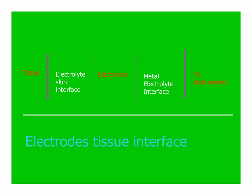
Electrodes tissue interface
Tissue Electrolyte skin interface
Electrolyte Metal Electrolyte Interface
To Instruments

ECG electrodes:
1- Limb electrodes
* rectangular surface
* German silver
* impedance is 2 to 5 KΩ
* using in surgery
2- Floating electrodes:
* used without jell
* contact impedance 50K Ω
3- Pregelled Disposable Electrodes: * stress testing ,long term monitoring
* reducing possibility of artifacts and drift

Electrode Placement

Electrode Placement
* C1 - the fourth intercostals space on the right side of the sternum
* C2 - the fourth intercostals space on the
left edge of the sternum
* C3 half way between C2 and C4
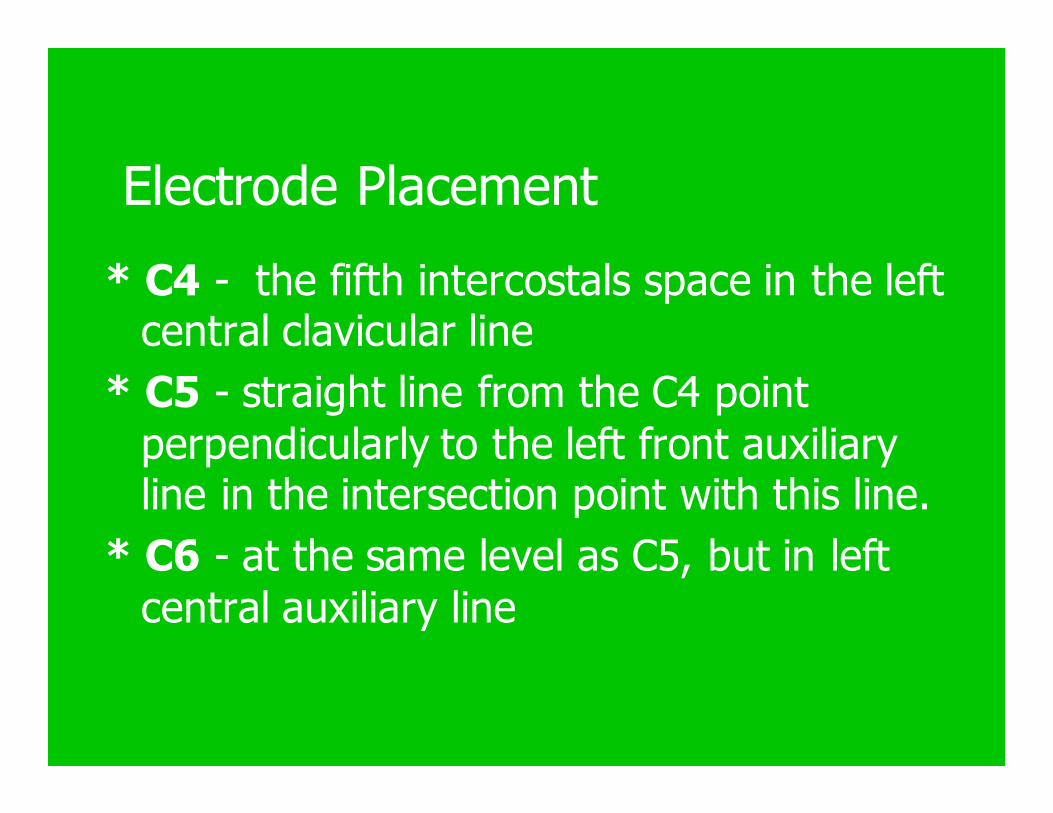
Electrode Placement
* C4 - the fifth intercostals space in the left central clavicular line
* C5 - straight line from the C4 point
perpendicularly to the left front auxiliary line in the intersection point with this line.
* C6 - at the same level as C5, but in left
central auxiliary line

ECG Machine Block Diagram

Preamplifier

Operation Amplifier

Galvanometer: * A galvanometer is a permanent magnet moving coil (PMMC).
* The pen will at rest in the centre of its travel when no current flows in the coil. The movement of the pin is depend on the current supply to the coils.
they are also other types of writing:
* hot-tip (styles) heated by resistor wire.
* the thermal paper its turn black when its heat
* writing knife edge in the thermal recorder.

* Dot matrix printer used with the computer its 27 pin printer make good quality recording waveform
and numerical number like blood pressure,
temperature etc.

Motors :
*The motor is connected to the drive roller through either a drive a chain or a gear train.
*Most machines use ac motors, with tapped
windings to select drive speed.
*Some ac motors provide very accurate drive
speeds that are synchronized to the ac power main’s frequency 60 Hz in the united states.

*Only few models use dc motors, and those regulated speed by using a regulated dc power
supply.
* in some cases an alternator /tachometer on
the motor shaft to provide negative feedback.

Figure 239 small

ECG FAULTS AND TROUBLESHOOTING:-
Most common problem that occur in an ECG are:
PROBLEM 1:-
SYMPTOM: machine runs but the thermal tip stylus does not writes or writes very lightly.
POSSIBLE CAUSE:
(1) Too little heat on the stylus tip .
(2) Insufficient stylus pressure.
TROUBLESHOOTING: Use a screw driver or any isolated tool to gently press the stylus if a dark line appears on the paper the problem is pressure , but if no dark line appears the problem is heat.

ECG FAULTS AND TROUBLESHOOTING:-
SOLUTION:
(1) For no heat check the heater voltage at the stylus
,if voltage is correct change the stylus ,if voltage is
not correct refer the service manual for details on
stylus power drive.
(2) Adjust the stylus pressure , use a pressure gage
and refer to the service manual for the correct
value.on some models pressure must be made at a
specific heater voltage.
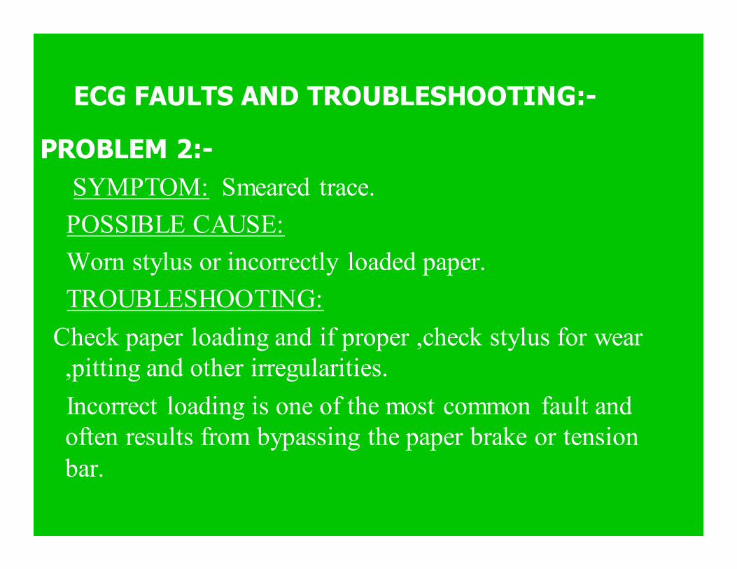
ECG FAULTS AND TROUBLESHOOTING:-
PROBLEM 2:-
SYMPTOM: Smeared trace.
POSSIBLE CAUSE:
Worn stylus or incorrectly loaded paper.
TROUBLESHOOTING:
Check paper loading and if proper ,check stylus for wear
,pitting and other irregularities.
Incorrect loading is one of the most common fault and
often results from bypassing the paper brake or tension
bar.

PROBLEM 3:-
SYMPTOMS: Poor recording.
POSSIBLE CAUSE: Electronic or mechanical problem , bad switch or patient cable.
TROUBLESHOOTING:
(1) Place lead selector switch in STD ,short all electrodes together and press 1- mv cal button.
(2) If normal calibration pulse appears then problem is connection to the patient.
(3) If problem persists then repeat step 1 using known good patient cable .
ECG FAULTS AND TROUBLESHOOTING:-

ECG FAULTS AND TROUBLESHOOTING:-
60 Hz INTERFERENCE:-
CAUSES: 60 Hz interference appears due to power
mains,another cause of 60 Hz interference if a broken
or loose cable or broken power main ground on the
ECG machine, additionally some dc supply also
cause these artifacts.
SOLUTION:
The problem can be isolated by shorting together all
electrodes of the patient cable and checking each
position of the lead selector switch.

ECG FAULTS AND TROUBLESHOOTING:-
(1) If the interference ceases , then the problem is a bad
electrode or no electrolytic gel.
(2) If the interference exists on all positions of the lead
selector switch then the problem is internal of the
machine.
(3) If the problem occurs in certain lead position of the
selector switch then suspect an open wire, use
ohmmeter for continuity.

Stress ECG
*Some potentials dangerous cardiac arrhythmia
and other anomalies shown up only under stress conditions.
*Physicians examine the stress condition by placing
the patient on the treadmill or stair stepper while
monitoring the patient's ECG waveform on both an oscilloscope monitor and a paper chart.
*Modern stress ECG machines are usually equipped with a microcomputer that analyzes the waveform .

*A common method is to perform the stress test, recording the ECG waveform, and then follow the
stress test with a thallium scan.
*The radioactive thallium is taken up by healthy
cardiac cells, so areas of the heart where blood
flow is sufficient appear darker on a gamma camera display then healthy areas.
*These two tests allow the physician to evaluate
the existence, location and extent of cardiac
disease.


Manual Controller
The optional manual controller
Allows personal treadmill operation, while
giving the user control of speed and
elevation functions without interfering
with
medical usage.

Drive Assembly
The low profile design and simplified layout of
drive assembly components provide easy accessibility for maintenance and provides a
high level of performance.

displays Body weight, Time, Distance ,Elevation
Total calories, Calories per minute
Pace (minutes/mile), Heart rate.

ECG PARAMETER
• ECG leads 12 standard • Record speed 5/25/50
• Sensitivity 2.5/5/10/20