Лечение переломов ключицы методом...
TRANSCRIPT
33
Журнал клинической и экспериментальной ортопедии им. Г.А. Илизарова № 2, 2016 г.
Вопросы травматологии
Bari M.M., Islam Shahidul, Shetu N.H., Mahfuzer R.M. Treatment of clavicle fractures by Ilizarov technique // Гений ортопедии. 2016. № 1. С. 33-35.
© Группа авторов, 2016.
УДК 616.717.2-001.5-089.227.84
DOI 10.18019/1028-4427-2016-2-33-35
Лечение переломов ключицы методом ИлизароваM.M. Bari, Shahidul Islam, N.H. Shetu, R.M. Mahfuzer
Ортопедический Центр Бари-Илизарова, Дака, Бангладеш
Treatment of clavicle fractures by Ilizarov techniqueM.M. Bari, Shahidul Islam, N.H. Shetu, R.M. Mahfuzer
Bari-Ilizarov Orthopaedic Centre, Dhaka, Bangladesh
В работе представлено применение мини-аппарата Илизарова при переломах ключицы. Авторы пролечили 28 переломов ключицы мини-фиксатором Илизарова в период с января 1999 года по январь 2015 года в Ортопедическом Центре Бари-Илизарова и NITOR (Дака). Среди 28 пациентов было 20 лиц мужского пола и 8 – женского, средний возраст пациентов – 45 лет (в пределах от 15 до 55 лет). Среди встретившихся осложнений было два случая инфекции путей проведения спиц, которые успешно курированы локальным применением лекарственных препаратов. Мини-аппарат Илизарова и представленная методика – эффективное средство для успешного лечения переломов ключицы. Ключевые слова: ключица, переломы, мини-аппарат Илизарова.
We treated 28 clavicle fractures with a mean age of 45 years (15-55) years with mini Ilizarov fixation from January 1999 to January 2015 at Bari-Ilizarov Orthopaedic Centre and NITOR, Dhaka. Out of 28 patients 20 were male and 8 female. We are presenting here the use of mini Ilizarov apparatus for fracture clavicle. Among the complications 2 wire track infections were observed from which all responded positively to local care. The mini Ilizarov apparatus and technique represents a useful method for the successful healing of fracture clavicle. Keywords: clavicle, fractures, mini Ilizarov apparatus.
INTRODUCTION
Despite being a common fracture, representing 5-10 % of all orthopedic fractures [1], evidence of optional treatment of displaced clavicle fracture is still ambiguous [2]. Primary surgical treatment of displaced midshaft clavicle fractures, with locking plate osteosynthesis as the preferred method, to result in good functionally high union rate and few complications [3-5] – the nonunion rate is reported as high
as 20 % and close to 30 % of all patients are reported to have symptomatic malunion [3]. Structurally the clavicle is a spongy bone since its internal lumen is filled with cellular bone and there is no intramedullary canal. Our research has shown that an axial wire in the spongy bone provides sufficient rigidity for the fixation of splinters, even in cases of minimum axial compression.
MATERIALS AND METHODS
For the last 17 years (1999-2015) 28 cases of fracture clavicle were operated by the Ilizarov technique. The follow up period was 1-15 years. Male predominated with an average age of 45 yrs. as demonstrated in table 1.
Table 1Shows sex incidence
Sex Qty PercentageMale 20 71
Female 08 29
Indications for Ilizarov external fixation are: 1. Fractures of various severity levels; 2. Fracture-dislocation; 3. Dislocation of acromial end of clavicle. The Ilizarov parts are: 1. 2 plates; 2. Posts; 3. Wire fixators; 4. Connecting rods. Surgical procedure The patient is administrated regional anaesthesia placed
under sedation. The operation is performed with the patient supine, with a cushion along (C7-D7) and his or her forearm
behind the back. The patients head is turned in the direction opposite to the injured clavicle but it should not be thrown back, the side of the face must be level with the anterior aspect of the chest. First, the wire insertion points are determined from radiographs in two projections. Radio-opaque markers are placed 2 cm from the joint surfaces of the acromial to the anatomic axis of the bone fragments. Injection needles are also placed to mark the anterior and posterior boundaries of the bone fragments at the insertion levels of the transosseous elements.
Mounting of the device starts from the medial support. The wires are manually fixed in the extreme holes of the connection plate and using wire fixators, so that their guiding ends only slightly protrude over the edge of the plate. The plate is applied onto the skin and the wire directed towards the sternal extremity of the clavicle and manually inserted to the bone. One should again make sure that the wire is located from the upper aspect of the clavicle and in the centre of the bone diameter. A reference point is marked on the wire showing the depth of its insertion and indicating the depth at which it will exit from the lower cortical plate. The bar is then moved away from the skin by 1.5-2 cm. The second wire is inserted until it touches the bone and after making sure that it is located in the centre of the clavicle
34
Гений Ортопедии № 2, 2016 г.
Вопросы травматологии
diameter, the first wire is fixed rigidly to the support. After the second wire is inserted and fixed, the support is similarly mounted on the peripheral fragment [14, 15, 16].
Postoperative protocol In the operating rooms, after the axial compression
wires have been tensioned, the skin around the wires is
examined. A pressure bandage in the form of a sling is applied to the area of the axial compression wire insertion. During first 3-4 days, dressing are changed daily, then as required but not less frequently than every 7-10 days. X-ray can be done after 10 days. In fracture clavicle, the structure is generally dismantled within 5-6 weeks.
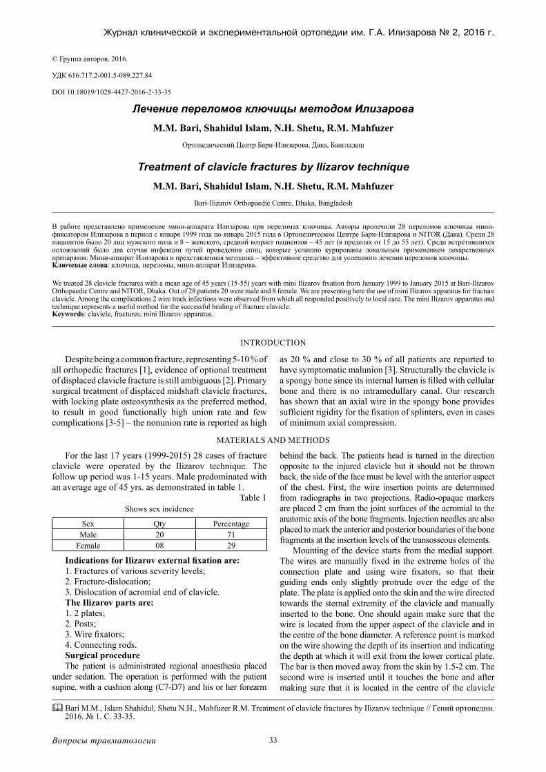
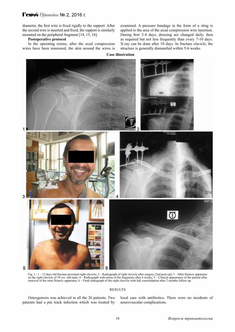
Case illustration
Fig. 1.: 1 – 12 days old fracture proximal right clavicle; 2 – Radiograph of right clavicle after surgery (2nd post-op); 3 – Mini Ilizarov apparatus on the right clavicle of 39 yrs. old male; 4 – Radiograph with union of the fragments after 6 weeks; 5 – Clinical appearance of the patient after removal of the mini Ilizarov apparatus; 6 – Final radiograph of the right clavicle with full consolidation after 2 months follow up
RESULTS
Osteogenesis was achieved in all the 26 patients. Two patients had a pin track infection which was treated by
local care with antibiotics. There were no incidents of neurovascular complications.
35
Журнал клинической и экспериментальной ортопедии им. Г.А. Илизарова № 2, 2016 г.
Вопросы травматологии
DISCUSSION
The mini Ilizarov method for the management of fracture clavicle have many advantages. However, several technical problems can arise if the details of the technique are not allowed precisely. The inexperienced surgeons usually fails to carry out of the whole technique. For successful tissue genesis one must follow the rules and processes of Ilizarov technique. It is evident that regeneration of the bone can be obtained safely. The research and review of existing literature revealed few publications for fracture clavicle treated nonoperatively. The factors that could influence development of clavicular nonunion included, presence of fracture comminution, complete fracture displacement, increased age, and fracture shortening. Fracture displacement following a clavicle fracture was reported to predict pain and dysfunction in
two studies both with follow up of more than two [6, 7] years. In this study fracture displacement was formed to be a predictor for nonunion in all but one publication reporting on this subject [8, 9, 10, 11, 12]. Shortening of the clavicle following a midshaft clavicle fracture has been found to be associated with inferior clinical outcome [13].
A publication by Nowak et al. recognized fracture comminution as a significant risk factor for pain, strength reduction and cosmetic defects after a clavicle fracture but not as a predictor for nonunion(6). We found two cohort studies of large sample size reporting comminution to be a predictor for nonunion in midshaft fracture [8, 9]. All existing randomized studies comparing surgical and nonsurgical treatment of displaced midshaft fractures favour surgical treatment based on slightly better functional outcome.
CONCLUSION
Fracture clavicle with displacement can be easily treated by mini Ilizarov apparatus. Displacement seems to be the most likely factor that is responsible for nonunion. Treating all clavicle fractures with displacement surgically would inevitably lead to over treatment, which is why
future studies need to focus on mini Ilizarov surgery and those who would not. Introducing one K-wire through the fragments and fixing the fragments externally by mini Ilizarov apparatus gives excellent result and better outcome.
REFERENCES
1. Robinson C.M. Fractures of the clavicle in the adult. Epidemiology and classification // J. Bone Joint Surg. Br. 1998. Vol. 80, N 3. P. 479-484.2. Fractures of the clavicle / L.A. Khan, T.J. Bradnock, C. Scott, C.M. Robinson // J. Bone Joint Surg. Am. 2009. Vol. 91, N 2. P. 447-460. 3. Clavicle fractures may be conservatively treated with acceptable results – a systemic review / I. Ban, U. Branner, K. Holck, M. Krasheninnikoff, A. Troelsen
// Dan. Med. J. 2012. Vol. 59, N 7. P. A4457. 4. Operative versus non-operative treatment for clavicle fracture: a meta-analysis / G.D. Liu, S.L. Tong, S. Ou, L.S. Zhou, J. Fei, G.X. Nan, J.W. Gu // Int.
Orthop. 2013. Vol. 37, N 8. P. 1495-1500. 5. Locking plate osteosynthesis of clavicle fractures: complication and reoperation rates in one hundred and five consecutive cases / M. Fridberg, I. Ban, Z.
Issa, M. Krasheninnikoff, A. Troelsen // Int. Orthop. 2013. Vol. 37, N 4. P. 689-692. 6. Nowak J., Holgersson M., Larsson S. Can we predict long-term sequelae after fractures of the clavicle based on initial findings? A prospective study with
nine to ten years of follow-up // J. Shoulder Elbow Surg. 2004. Vol. 13, N 5. P. 479-486. 7. Outcome of clavicular fracture in 89 patients / A. Eskola, S. Vainionpää, P. Myllynen, H. Pätiälä, P. Rokkanen // Arch. Orthop. Trauma Surg. 1986. Vol.
105, N 6. P. 337-338. 8. Risk factors for nonunion after nonoperative treatment of displaced midshaft fractures of the clavicle / I.R. Murray, C.J. Foster, A. Eros, C.M. Robinson //
J. Bone Joint Surg. Am. 2013. Vol. 95, N 13. P. 1153-1158. 9. Estimating the risk of nonunion following nonoperative treatment of a clavicular fracture / C.M. Robinson, C.M. Court-Brown, M.M. McQueen, A.E.
Wakefield // J. Bone Joint Surg. Am. 2004. Vol. 86-A, N 7. P. 1359-1365. 10. Vertical fragment in adult midshaft clavicle fractures: an indicator for surgical intervention / S.J. Kirmani, S.K. Pillai, B.R. Madegowda, S.A. Shahane //
Orthopedics. 2009. Vol. 32, N 10. P. 9-14. doi: 10.3928/01477447-20090818-06. 11. Midshaft fractures of the clavicle with a shortening of more than 2 cm predispose to nonunion / M. Wick, E.J. Müller, E. Kollig, G. Muhr // Arch. Orthop.
Trauma Surg. 2001. Vol. 121, N 4. P. 207-211. 12. Hill J.M., McGuire M.H., Crosby L.A. Closed treatment of displaced middle-third fractures of the clavicle gives poor results // J. Bone Joint Surg. Br. 1997.
Vol. 79, N 4. P. 537-539. 13. Deficits following nonoperative treatment of displaced midshaft clavicular fractures / M.D. McKee, E.M. Pedersen, C. Jones, D.J. Stephen, H.J. Kreder,
E.H. Schemitsch, L.M. Wild, J. Potter // J. Bone Joint Surg. Am. 2006. Vol. 88, N 1. P. 35-40. 14. Ilizarov G.A. Transosseous osteosysthesis. Theoretical and Clinical Aspects of the Regeneration and Growth of Tissue. Springer-Verlage, 1992. P. 450-452. 15. Bari M.M. A color atlas of limb lengthening, surgical reconstruction and deformity correction by Ilizarov technique. 2013. P. 75. 16. Treatment of fracture clavicle by Ilizarov Technique / M.M. Bari, S. Islam, N.H. Shetu, M. Rahman // Orthop. Rheumatol. Open Access J. 2015. Vol. 1,
N 2. ID 555560.
Рукопись поступила 04.05.2016.
Information about the authors:
1. Mofakhkharul Bari – Prof., Ph.D, Chief Consultant, Bari-Ilizarov Orthopaedic Centre, Visiting and Honored Prof., Russian Ilizarov Scientific Centre, Kurgan; e-mail: [email protected].
2. Shahidul Islam – Prof., MD; FCPS, Bari-Ilizarov Orthopaedic Centre. 3. Nazmul Huda Shetu – M.S., Ortho-Consultant, Bari-Ilizarov Orthopaedic Centre. 4. Mahfuzer Rahman – D., Ortho-Consultant, Bari-Ilizarov Orthopaedic Centre.