بسم الله الرحمن الرحيم. connective tissue ( c.t. ) objectives: by the end of this...
TRANSCRIPT

الرحيم الرحمن الله الرحيم بسم الرحمن الله بسم

CONNECTIVE TISSUE( C.T. )
• Objectives:
By the end of this lecture, the student should be able to:
1- Enumerate the general characteristics of C.T.
2- Classify C.T.
3- Classify C.T. proper (C.T.P.)
4- Describe the structure (components) and distribution of different types of C.T.P.
5- Discuss clinical applications related to C.T.P.
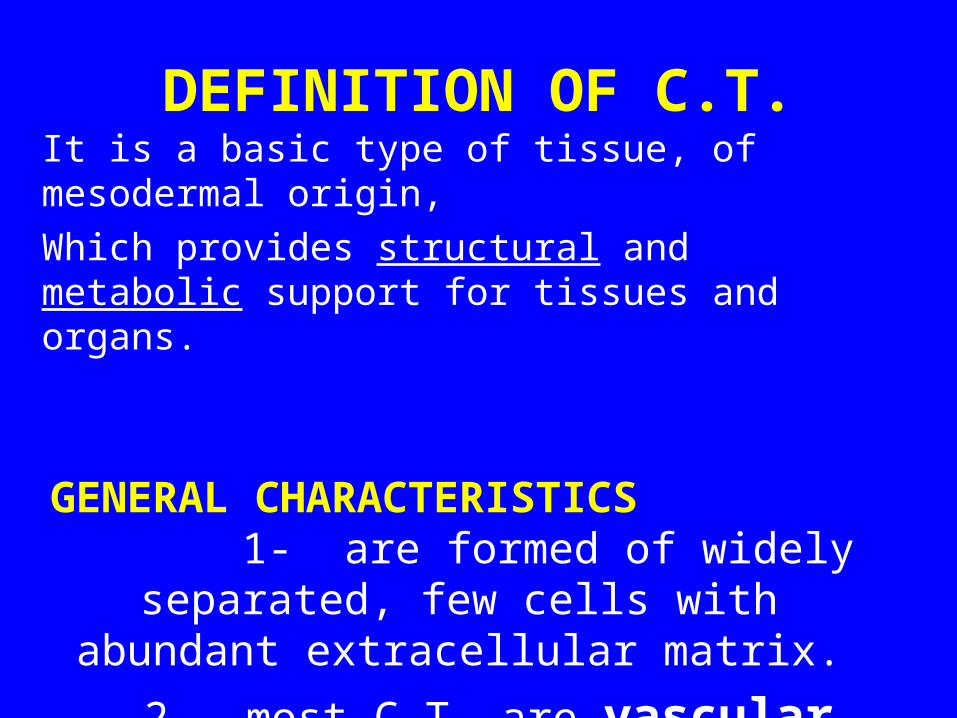
DEFINITION OF C.T.It is a basic type of tissue, of mesodermal origin,
Which provides structural and metabolic support for tissues and organs.
GENERAL CHARACTERISTICS
1- are formed of widely separated, few cells with abundant extracellular matrix.
2- most C.T. are vascular.

TYPES OF C.T.
• 1- C.T. Proper.
• 2- Cartilage.
• 3- Bone.
• 4- Blood.
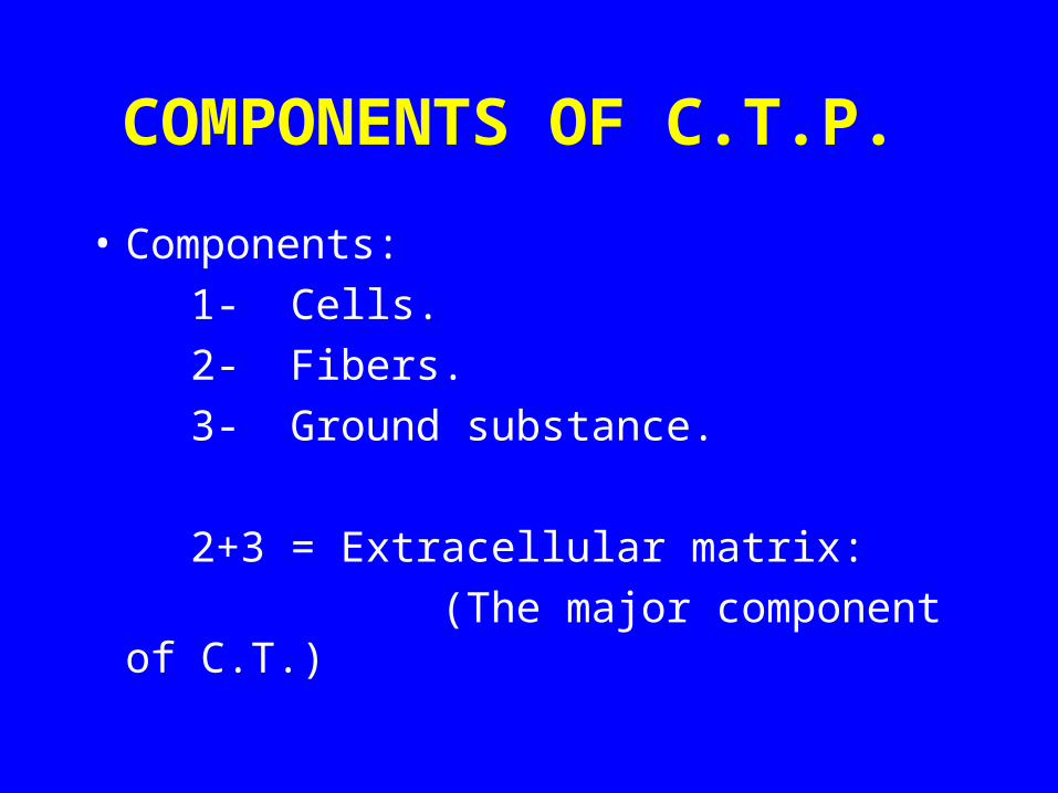
COMPONENTS OF C.T.P.
• Components:
1- Cells.
2- Fibers.
3- Ground substance.
2+3 = Extracellular matrix:
(The major component of C.T.)

TYPES OF C.T. PROPER
(1) Loose (Areolar) C.T.
(2) Dense Collagenous C.T.
(3) Elastic C.T.
(4) Reticular C.T.
(5) Adipose Tissue.

I- LOOSE (AREOLAR) C.T.
L/M:
Contains all the main components of C.T.P.
(no predominant element in loose C.T.)
Sites:
e.g. Subcutaneous tissue.
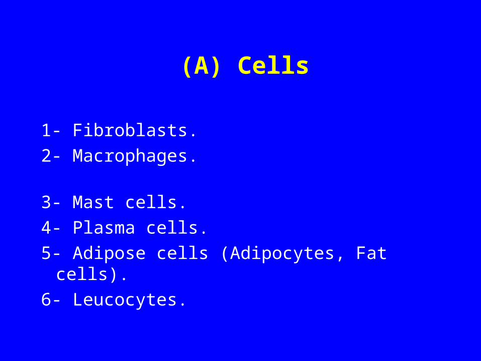
(A) Cells
1- Fibroblasts.
2- Macrophages.
3- Mast cells.
4- Plasma cells.
5- Adipose cells (Adipocytes, Fat cells).
6- Leucocytes.
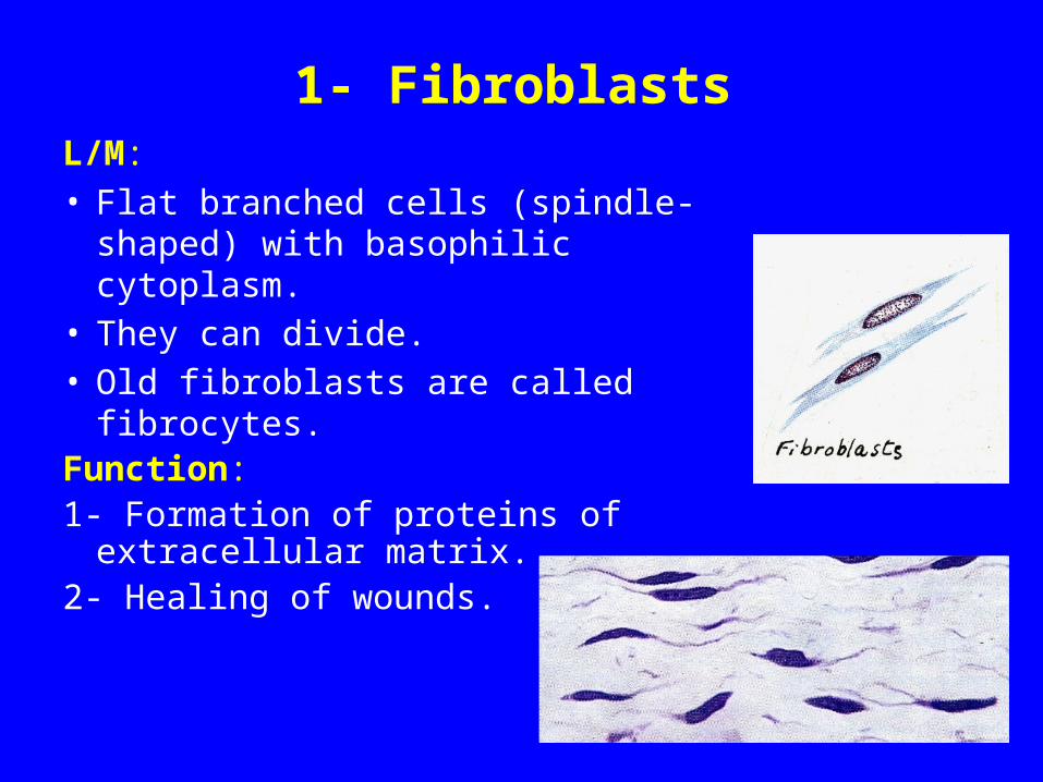
1- FibroblastsL/M:• Flat branched cells (spindle-shaped)
with basophilic cytoplasm.• They can divide. • Old fibroblasts are called fibrocytes.Function:1- Formation of proteins of extracellular
matrix.2- Healing of wounds.

2- Macrophages
L/M:• Basophilic cytoplasm,
rich in lysosomes.• Irregular outlines.• They can divide. • They originate from
monocytes.
Function:
Phagocytosis.
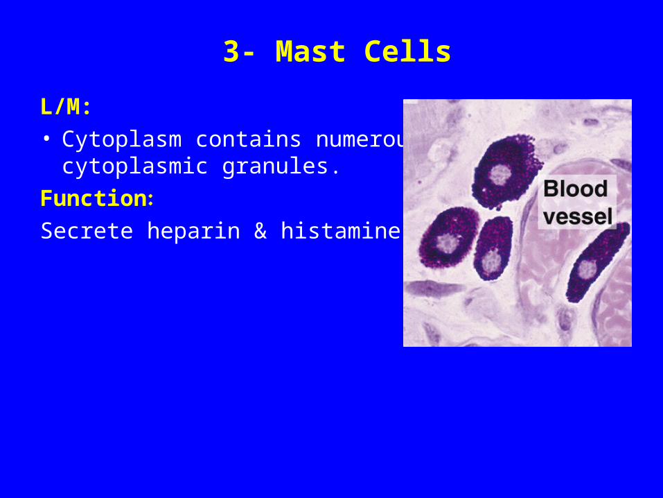
3- Mast Cells
L/M:• Cytoplasm contains numerous
cytoplasmic granules.
Function:
Secrete heparin & histamine.

4- Plasma Cells
L/M: • Basophilic cytoplasm with a negative
Golgi image.• Nucleus: spherical, eccentric with a
clock-face appearance of chromatin.• Derived from B-lymphocytes.
Function:
Secretion of antibodies (immunoglobulins).

5- Adipose Cells(Adipocytes, Fat Cells)
L/M of Unilocular Adipose Cells:• Large spherical, with a single large fat droplet.
• Thin rim of cytoplasm at the periphery.• Nucleus: flattened, peripheral.
Function:
Storage of fat.

(B) Fibers
1- Collagen Fibers (Collagen type I):
• Non-branched fibers, arranged in bundles.
• Acidophilic.
2- Reticular Fibers (collagen type III):
• Form a network.
• Stained black with silver.
3- Elastic Fibers:
• Branched.
• Stained brown with orcein.
N.B. Other important types of collagen include:
type II (in cartilage).type IV (in basement membranes)

II- DENSE COLLAGENOUS C.T.L/M:
Predominance of collagen fibers + Fibroblasts.
Sites of dense collagenous C.T.:
1- Dense irregular: e.g. dermis of the skin, capsule.
2- Dense regular: e.g. tendons, ligaments.
1 2
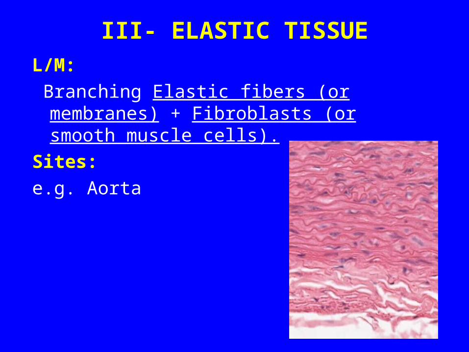
III- ELASTIC TISSUE
L/M:
Branching Elastic fibers (or membranes) + Fibroblasts (or smooth muscle cells).
Sites:
e.g. Aorta
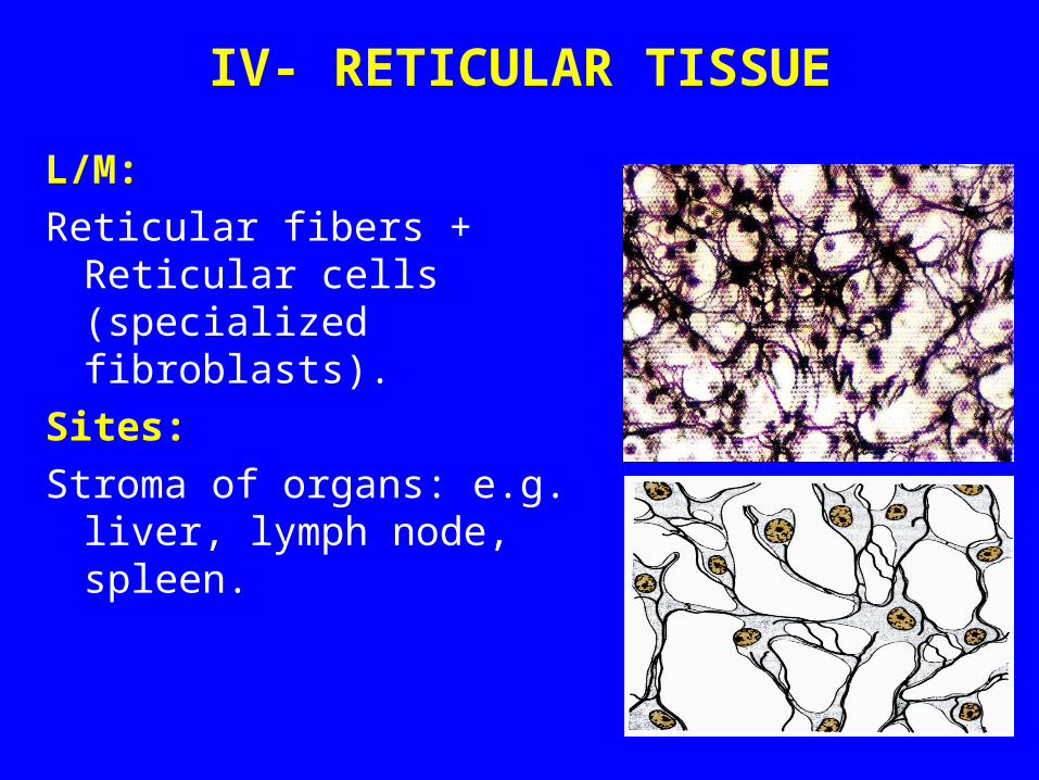
IV- RETICULAR TISSUE
L/M:
Reticular fibers + Reticular cells (specialized fibroblasts).
Sites:
Stroma of organs: e.g. liver, lymph node, spleen.

V- UNILOCULAR ADIPOSE TISSUE(WHITE ADIPOSE TISSUE)
L/M:Is formed of lobules of unilocular
adipose cells.Function:Synthesis, storage & release of fat.Sites:• Subcutaneous layers
especially in buttocks & hips.
• Abdominal wall.• Female breast.• Around the kidney.

Clinical Applications
Bronchial Asthma
Exposure to allergen will stimulate mast cells in the lungs, which leads to release of histamine and other chemicals that lead to contraction of the smooth muscle fibers in the wall of the bronchioles (bronchospasm) leading to dyspnea (difficulty in breathing).

Clinical Applications
Obesity1- Hypertrophic obesity:
It results from the accumulation and storage of fat in the unilocular fat cells (white adipocytes). These cells may increase in size up to four times.
2- Hypercellular obesity:
It results from an increase in number of adipocytes, which may be attributed to increased number of adipocyte precursors in infancy.

Thank youThank you