seltsft.euseltsft.eu/wp-content/uploads/2011/06/mati_pääsuke... · 2013-05-29seltsft.eu
TRANSCRIPT

Biomechanics of the knee joint
Table of contents
• Biomechanical roles of the knee joint complex
• Arthrokinematics of the knee
• Knee joint loading
• Soft tissue mechanics

Knee joint complex
(by Masouros S.D. et al. Orthop Trauma,2010, 24: 84-91)
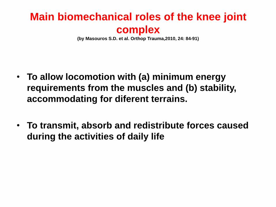
Main biomechanical roles of the knee joint
complex (by Masouros S.D. et al. Orthop Trauma,2010, 24: 84-91)
• To allow locomotion with (a) minimum energy
requirements from the muscles and (b) stability,
accommodating for diferent terrains.
• To transmit, absorb and redistribute forces caused
during the activities of daily life

Arthrokinematics of the knee

Knee joint
• Ginglymus (hinge)
• Arthodial (pivot, gliding)
• 6 degrees of freedom
– 3 rotations
– 3 translations
Frontal axis
(varus-valgus
rotation)
Plokk-ratasliiges
Sagittal axis
(flexion-
Extension)
Transverse axis (interna-external rtation)

Rotation and translation in knee
joint
• Rotation: – flexion-extension: up to 160 deg of flexion
(up to -5 deg flexion – hyperextension)
– varus-valgus: 6-8 deg in extension
– internal-external rotation: 25-30 deg in flexion
• Translation: – anterior-posterior: 5–10 mm
– compression: 2–5 mm
– medio-lateral: 1-2 mm

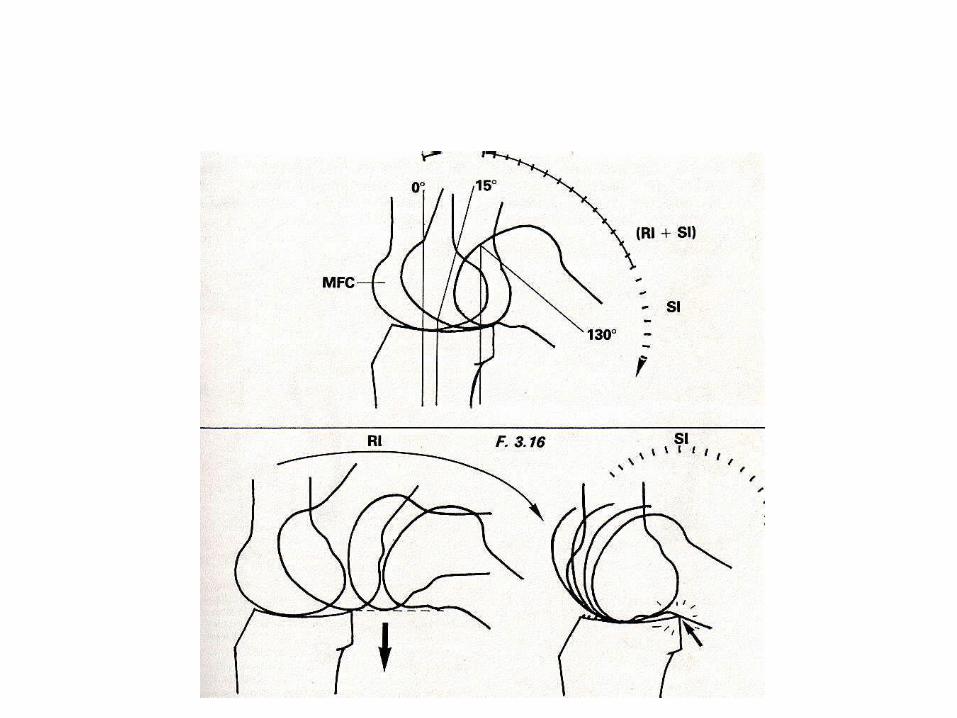
Centre of rotation
for femur motion during flexion-extension (sagittal view)
Rolling Sliding Pure rotation
femur
tibia IC

1st 250 - mainly roll >250 roll and ant glide
Femoral condyles in flexion

Knee joint kinematics in the sagittal plane during gait. a Extension: contact is located centrally. b Early flexion: posterior rolling;
contact continuously moves posteriorly. c Deep flexion: femoral sliding;
contact is located posteriorly; the unlocking of the ACL prevents further femoral roll back.
(Masouros S.D. Et al. Orthop Trauma, 2010, 24: 84-91)
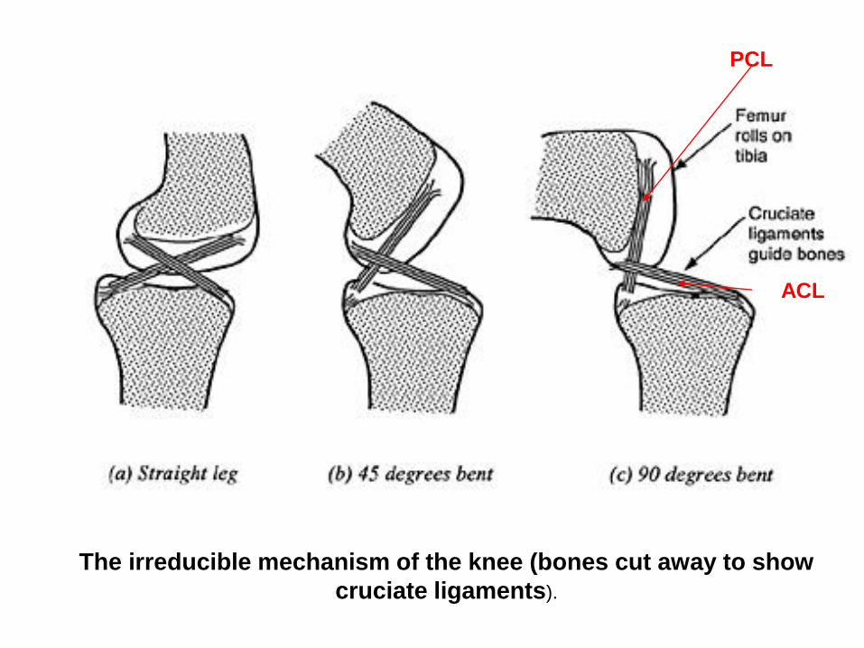
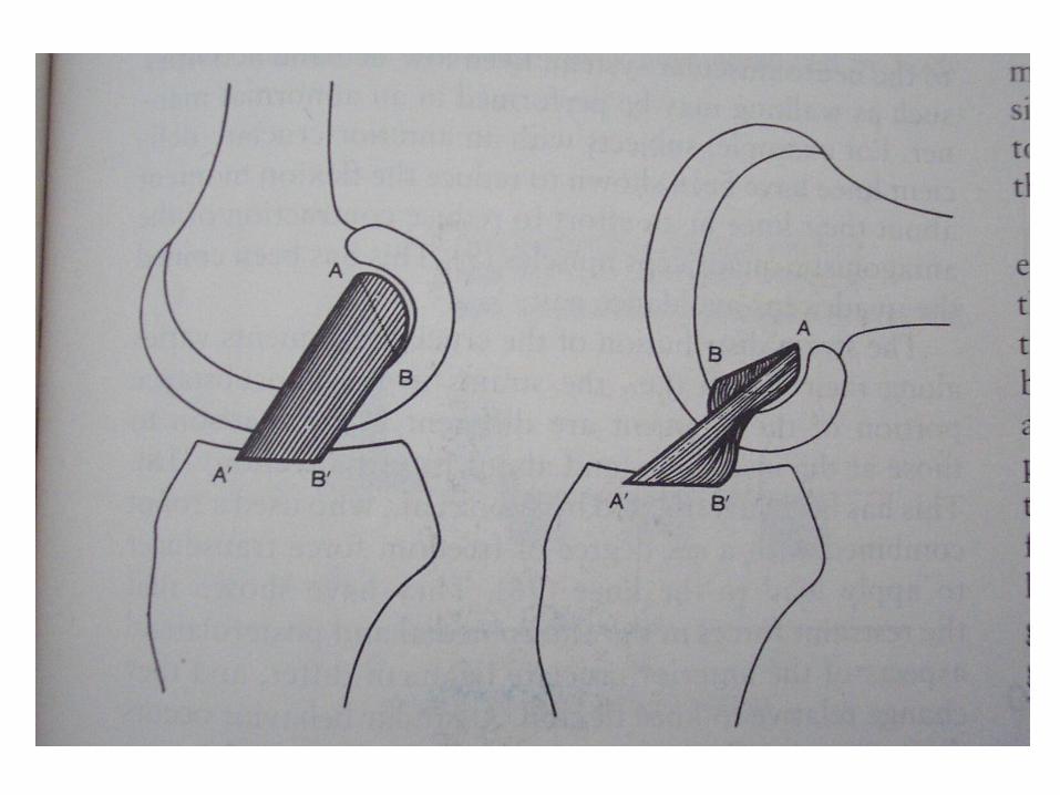
The irreducible mechanism of the knee (bones cut away to show
cruciate ligaments).
ACL
PCL


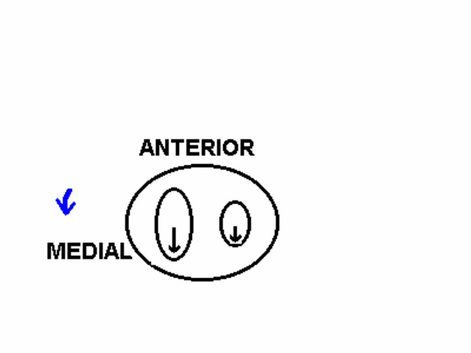
Screw-home during knee extension
During the last 20 degrees of knee extension
anterior tibial glide persists on the
tibia's medial condyle because its articular surface
is longer in that dimension than the lateral condyle's.
Prolonged anterior glide on the medial side produces
external tibial rotation, the "screw-home" mechanism.
LATERAL

Functional range of motion (ROM) at the knee
Activities Knee flexion
Normal gait/level 60°
surfaces
Stair climbing 80°
Sitting/rising from 90°
most chairs
Sitting/rising from 115º toilet seat
Advanced function > 115°

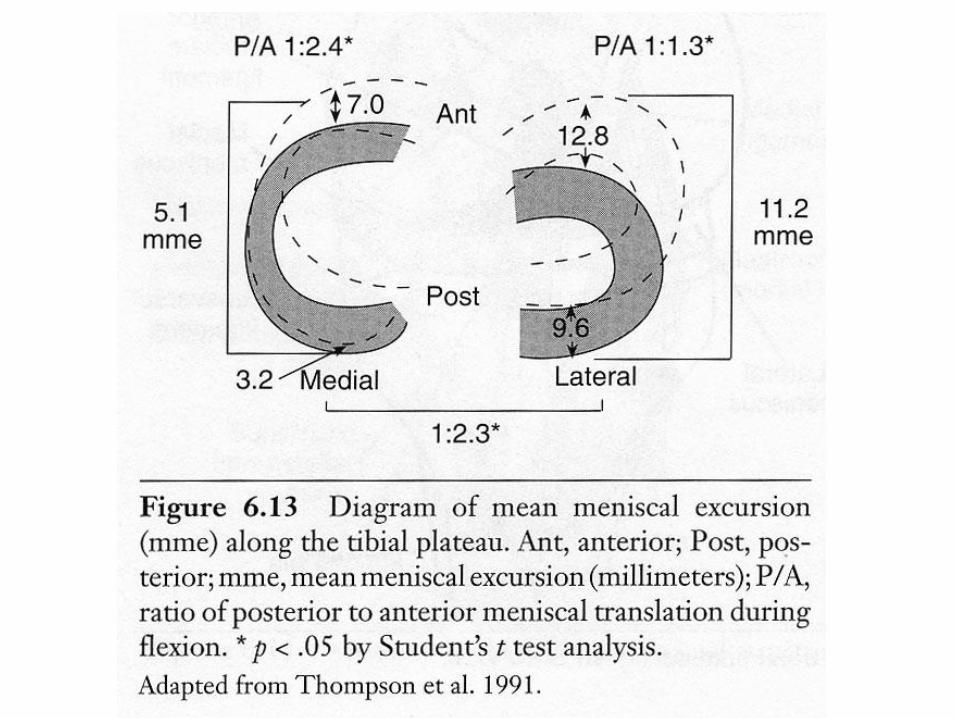
Diagrams showing the mean movement (mm) in each meniscus during flexion (shaded) and extension
(hashed). ANT, anterior; POST, posterior; mme, mean meniscal excursion; P/A, ratio of posterior to
anterior meniscal translation during flexion. (Thompson W.O. Am J Sports Med, 1991, 19: 200-216)
Extension
Flexion
Knee joint loading

Tibiofemoral joint activities:
Flexion & compressive load
• Cycling 60-100 deg 1.2BW
• Walking 15 3.0
• Stairs 60 3.8
• Stairs 45 4.3
• Squat-rise 140 5.0
• Squat-down 140 5.6
[Mow & Hayes, 1991] BW = body weight
Front
VARUS NEUTRAL VALGUS
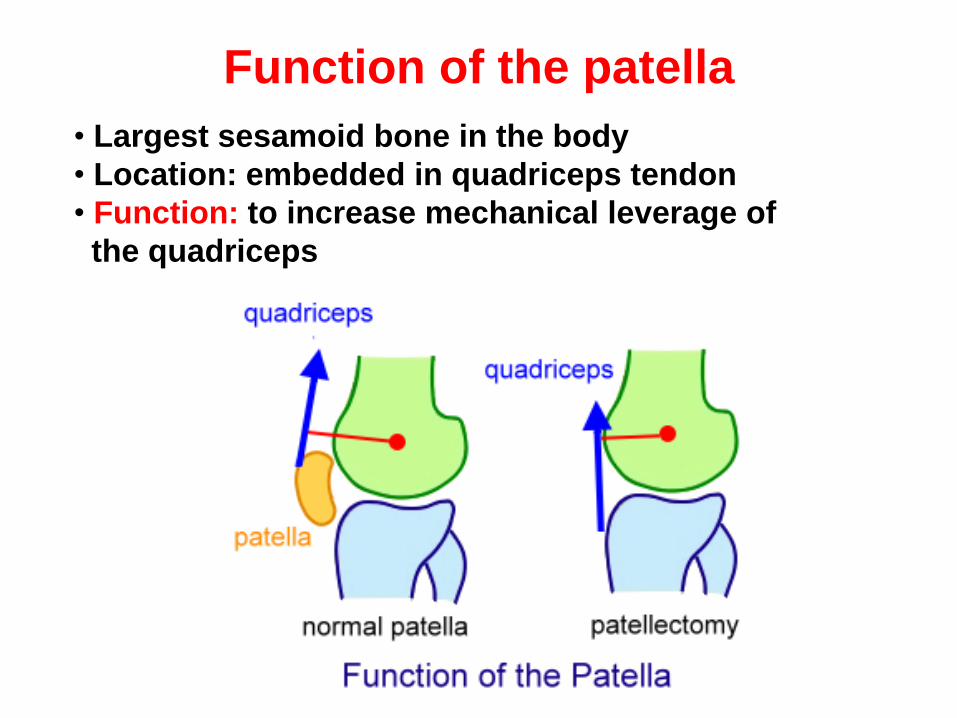
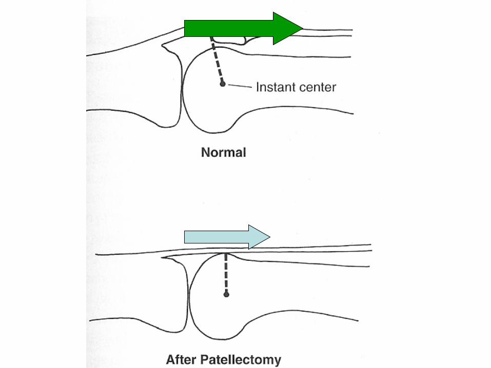
Function of the patella
• Largest sesamoid bone in the body
• Location: embedded in quadriceps tendon
• Function: to increase mechanical leverage of
the quadriceps
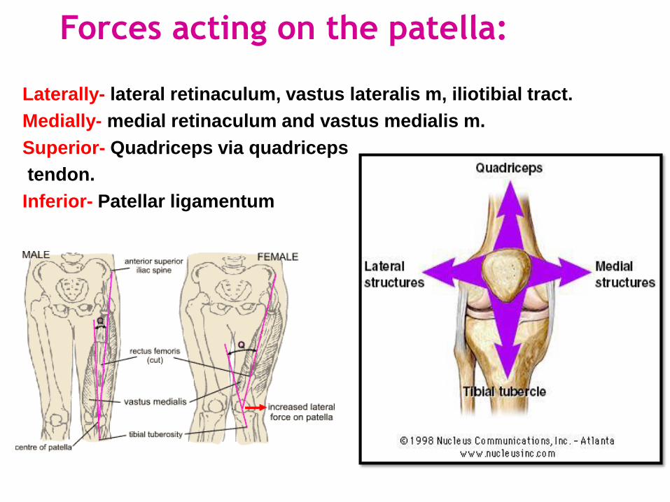
Laterally- lateral retinaculum, vastus lateralis m, iliotibial tract.
Medially- medial retinaculum and vastus medialis m.
Superior- Quadriceps via quadriceps
tendon.
Inferior- Patellar ligamentum
Forces acting on the patella:

Rising up from a chair. Assuming that 0.5 BW is transferred through each leg applied at the foot, then
its line of action is approximately 200 mm posterior to the joint centre; also, the PT line of action is
approximately 35 mm off the joint centre. Moment equilibrium requires PT × 35 mm = 0.5 BW × 200
mm, which results in PT ≈ 3.0 BW. At 90° flexion it has been shown that PT ≈ 70% Q, therefore Q ≈ 4.5
BW. Force equilibrium requires the triangle of forces acting at each joint to be closed; this results in
joint forces of PF ≈ 5.5 BW and TF ≈ 3.5 BW (BW: body weight, PT: patellar tendon, Q: quadriceps
muscle, PF: patellofemoral, TF: tibiofemoral). (Masouros S.D. Et al. Orthop Trauma, 2010, 24: 84-91)
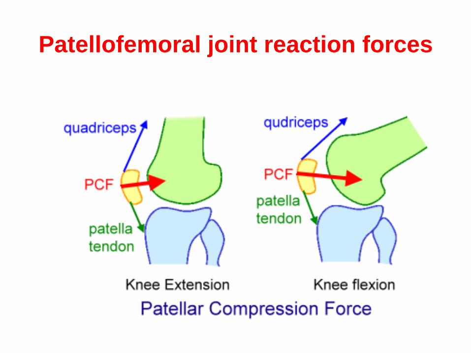
Patellofemoral joint reaction forces
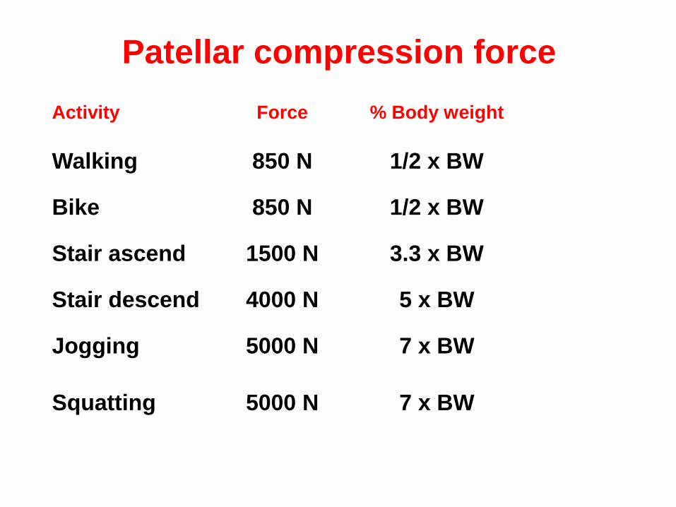
Patellar compression force
Activity Force % Body weight
Walking 850 N 1/2 x BW
Bike 850 N 1/2 x BW
Stair ascend 1500 N 3.3 x BW
Stair descend 4000 N 5 x BW
Jogging 5000 N 7 x BW
Squatting
5000 N
7 x BW
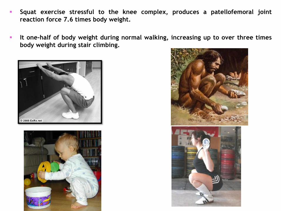
Squat exercise stressful to the knee complex, produces a patellofemoral joint
reaction force 7.6 times body weight.
It one-half of body weight during normal walking, increasing up to over three times
body weight during stair climbing.

Menisci
• Two functions:
– load bearing
– stability
• also, joint lubrication
• prevent capsule, synovial impingement
• shock absorbers

Menisci

Diagram demonstrates the importance of intact meniscal entheses for the load distribution
function of the meniscus. (A) With intact enthesis the load (thick arrows) is transmitted via the
menisci and articular cartilage through a large contact area (left hand picture, small arrows). Part of the
load is transformed to hoop stresses (right hand picture, long arrows). (B) When the insertional
ligaments are transected, the menisci will extrude from the knee joint during loading, and the load is
mainly transmitted via articular cartilage through a reduced contact area (small arrows). (Grood ES:
Adv Orthop Surg 7:193,1984.)

Load-bearing function of the menisci
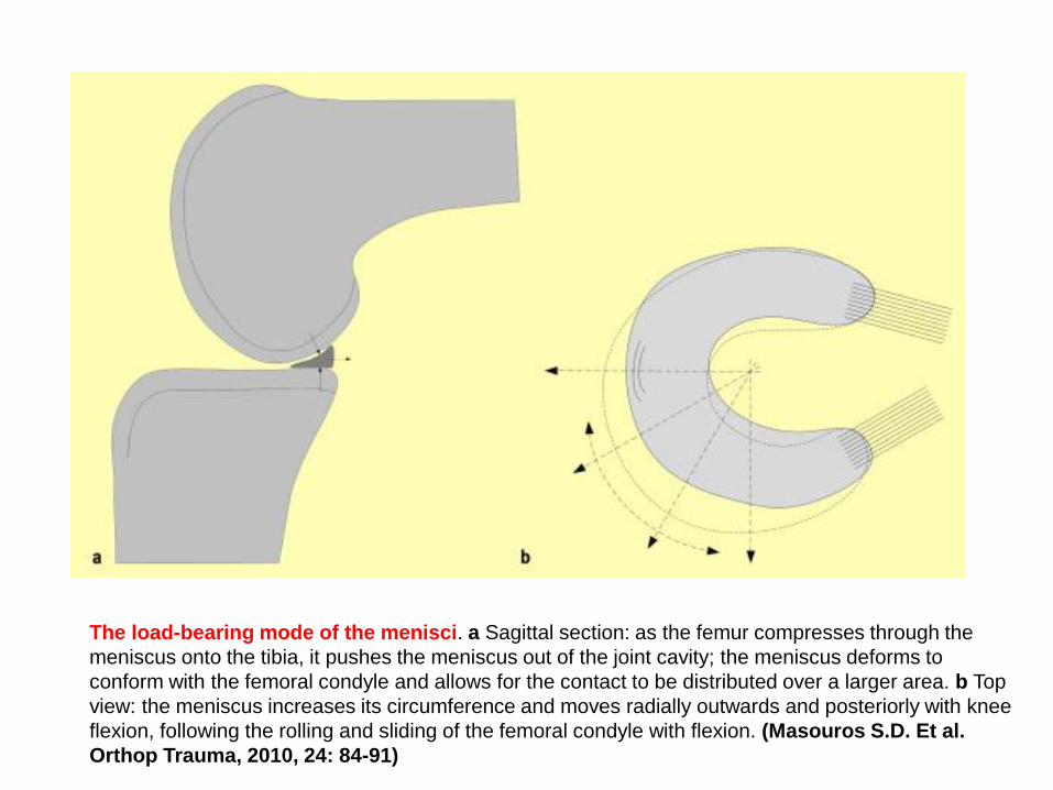
The load-bearing mode of the menisci. a Sagittal section: as the femur compresses through the
meniscus onto the tibia, it pushes the meniscus out of the joint cavity; the meniscus deforms to
conform with the femoral condyle and allows for the contact to be distributed over a larger area. b Top
view: the meniscus increases its circumference and moves radially outwards and posteriorly with knee
flexion, following the rolling and sliding of the femoral condyle with flexion. (Masouros S.D. Et al.
Orthop Trauma, 2010, 24: 84-91)

Free body diagram of forces acting on the meniscus during loading. As the femur presses down
on the meniscus during normal loading, the meniscus deforms radially but is anchored by its anterior
and posterior horns (Fant and Fpost). During loading, tensile, compressive, and shear forces are
generated. A tensile hoop stress (Fcir) results from the radial deformation, while vertical and horizontal
forces (Fv and Fh) result from the femur pressing on the curved superior surface of the tissue. A radial
reaction force (Frad) balances the femoral horizontal force (Fh).
(Athanasiou K.A., Sanchez-Adams J. Engineering of the knee mesiscus. 2009)

Martinez-Villalpado S.M. J Rehab Res Dev 2009, 46: 361-371
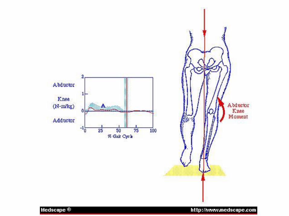
Knee biomechanics during gait
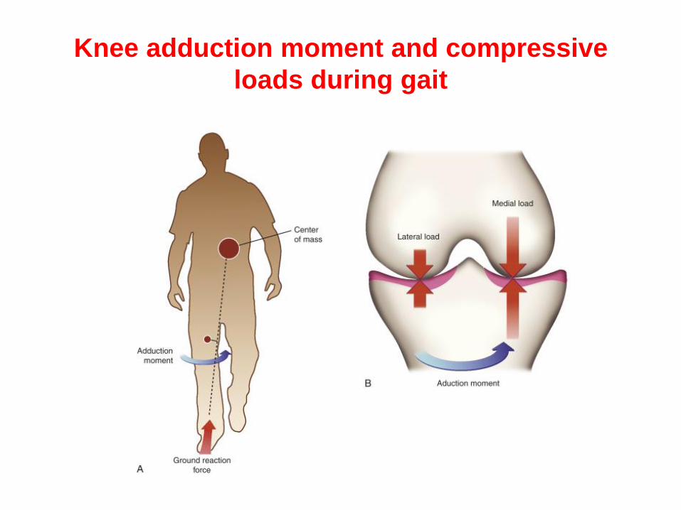
Knee adduction moment and compressive
loads during gait

The knee adduction moment is a result of the magnitude of the
ground reaction force (GRF) times the distance (i.e. moment arm)
from the center of rotation (GRF*LA). The graph of the knee
adduction moment during a gait cycle in a patient with knee OA is
characterized by an increase in the peak and impulse (the area under
the curve) of the moment (Presented by Mali M., 2007).


Zabala M.E. et al.J Biomech 2013, 46: 515-520
All three moments and the total moment at the knee during gait, stair ascent, and stair
descent for ACLR, contralateral, and control knees. (⁎) indicates significant difference
between ACLR and contralateral, (Δ) indicates significant difference between contralateral
and control, and (†) indicates significant difference between ACLR and control knees.

Soft tissue mechanics

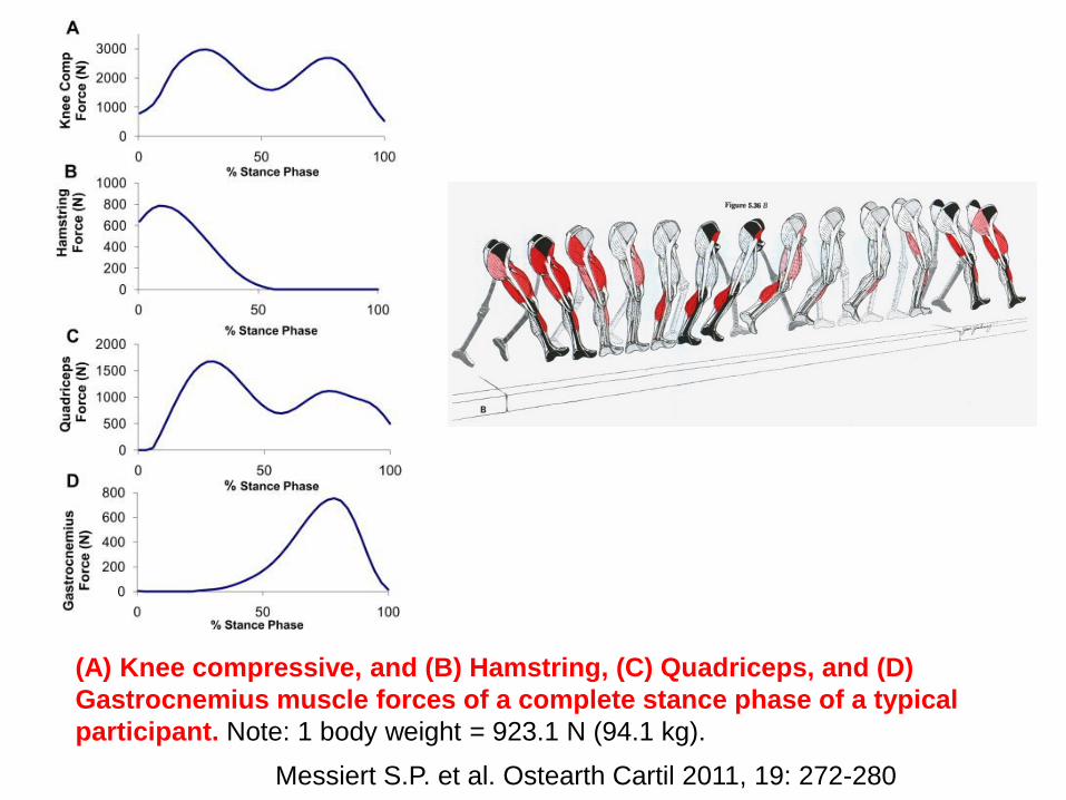
Messiert S.P. et al. Ostearth Cartil 2011, 19: 272-280
Schematic representation of the biomechanical musculoskeletal knee
model used to calculate knee joint loads and muscle forces
Messiert S.P. et al. Ostearth Cartil 2011, 19: 272-280
(A) Knee compressive, and (B) Hamstring, (C) Quadriceps, and (D)
Gastrocnemius muscle forces of a complete stance phase of a typical
participant. Note: 1 body weight = 923.1 N (94.1 kg).

Force-length diagramm of ligaments and
tendons
Patellar Ligament

Questions
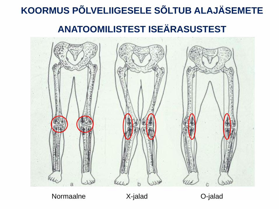
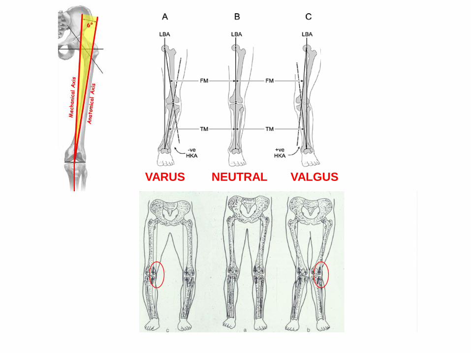
KOORMUS PÕLVELIIGESELE SÕLTUB ALAJÄSEMETE
ANATOOMILISTEST ISEÄRASUSTEST
Normaalne X-jalad O-jalad
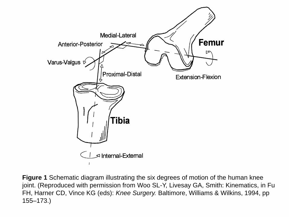
Figure 1 Schematic diagram illustrating the six degrees of motion of the human knee
joint. (Reproduced with permission from Woo SL-Y, Livesay GA, Smith: Kinematics, in Fu
FH, Harner CD, Vince KG (eds): Knee Surgery. Baltimore, Williams & Wilkins, 1994, pp
155–173.)
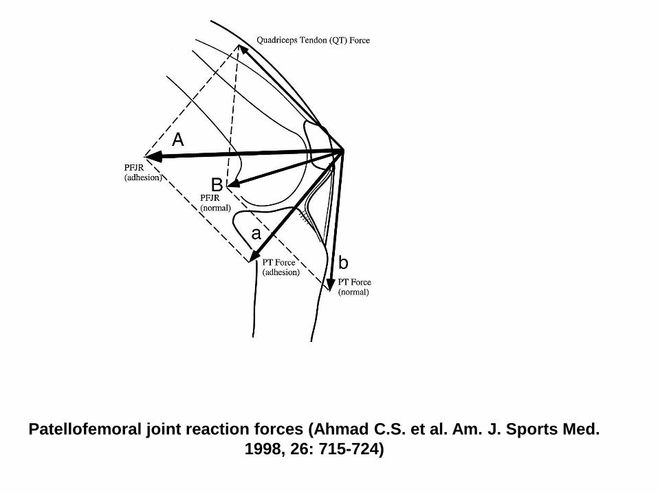
Patellofemoral joint reaction forces (Ahmad C.S. et al. Am. J. Sports Med.
1998, 26: 715-724)
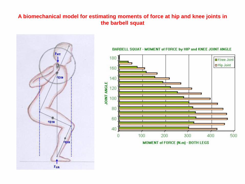
A biomechanical model for estimating moments of force at hip and knee joints in
the barbell squat
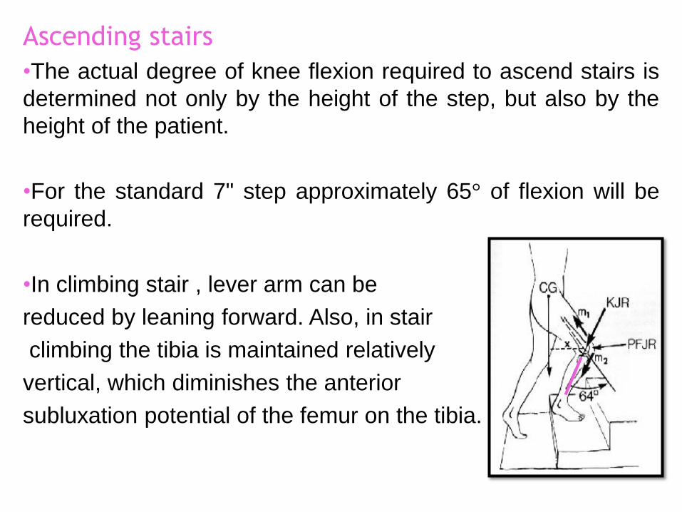
Ascending stairs
•The actual degree of knee flexion required to ascend stairs is
determined not only by the height of the step, but also by the
height of the patient.
•For the standard 7" step approximately 65° of flexion will be
required.
•In climbing stair , lever arm can be
reduced by leaning forward. Also, in stair
climbing the tibia is maintained relatively
vertical, which diminishes the anterior
subluxation potential of the femur on the tibia.

Knee joint
• Knee provides mobility and support
during dynamic and static activities
• Support during weight bearing
• Mobility during non-weight bearing
• Involved with almost any
functional activity of the
lower extremity

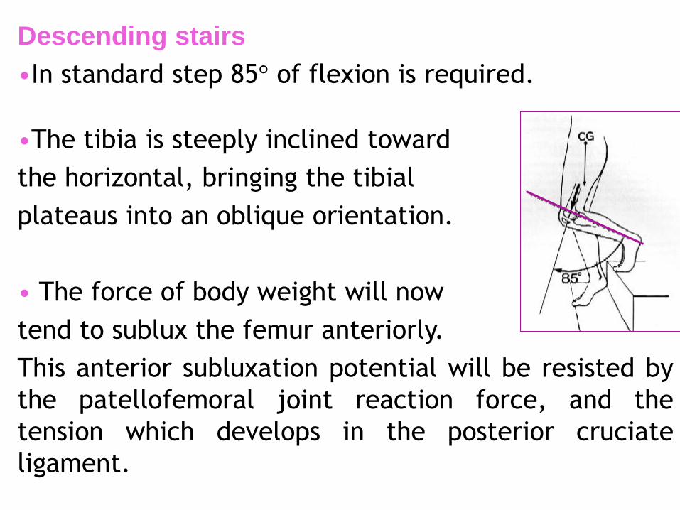
Descending stairs
•In standard step 85° of flexion is required.
•The tibia is steeply inclined toward
the horizontal, bringing the tibial
plateaus into an oblique orientation.
• The force of body weight will now
tend to sublux the femur anteriorly.
This anterior subluxation potential will be resisted by
the patellofemoral joint reaction force, and the
tension which develops in the posterior cruciate
ligament.
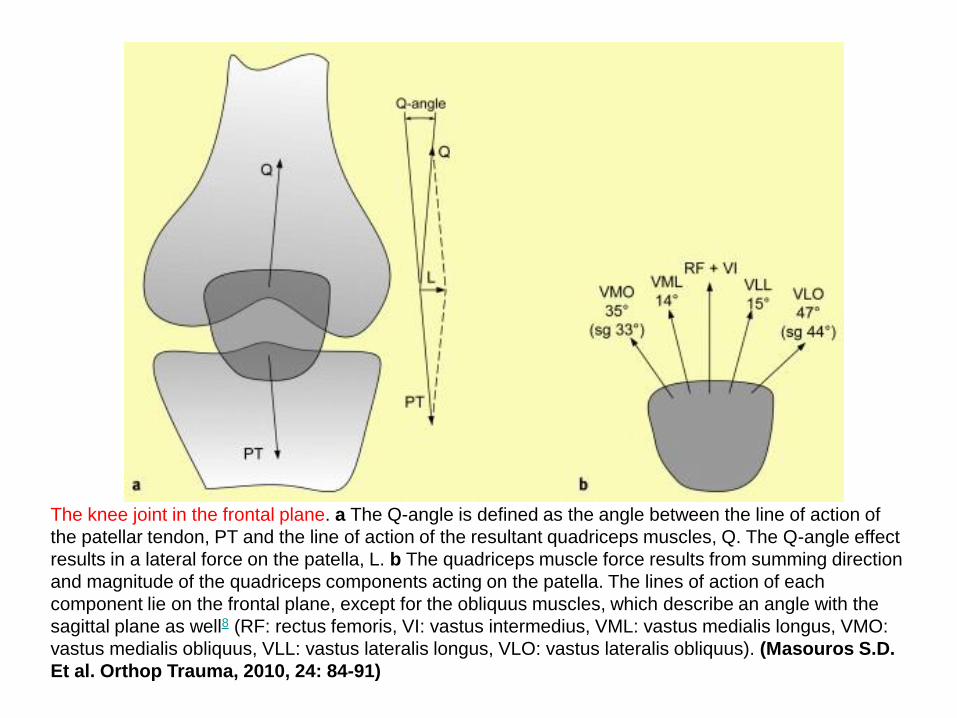
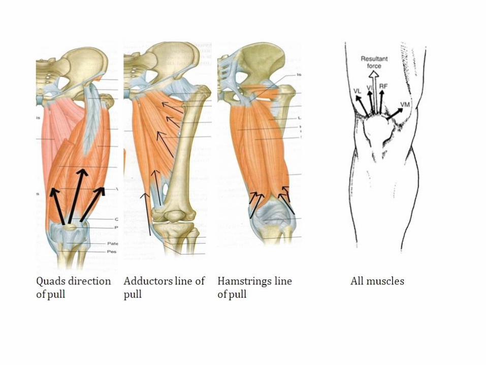
The knee joint in the frontal plane. a The Q-angle is defined as the angle between the line of action of
the patellar tendon, PT and the line of action of the resultant quadriceps muscles, Q. The Q-angle effect
results in a lateral force on the patella, L. b The quadriceps muscle force results from summing direction
and magnitude of the quadriceps components acting on the patella. The lines of action of each
component lie on the frontal plane, except for the obliquus muscles, which describe an angle with the
sagittal plane as well8 (RF: rectus femoris, VI: vastus intermedius, VML: vastus medialis longus, VMO:
vastus medialis obliquus, VLL: vastus lateralis longus, VLO: vastus lateralis obliquus). (Masouros S.D.
Et al. Orthop Trauma, 2010, 24: 84-91)
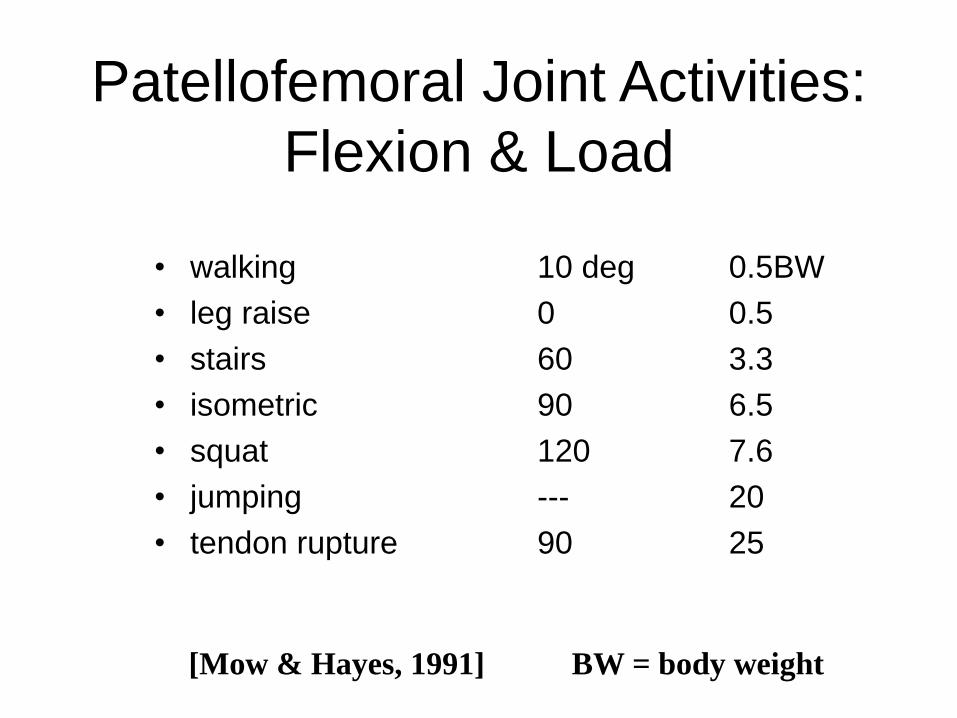
Patellofemoral Joint Activities:
Flexion & Load
• walking 10 deg 0.5BW
• leg raise 0 0.5
• stairs 60 3.3
• isometric 90 6.5
• squat 120 7.6
• jumping --- 20
• tendon rupture 90 25
[Mow & Hayes, 1991] BW = body weight

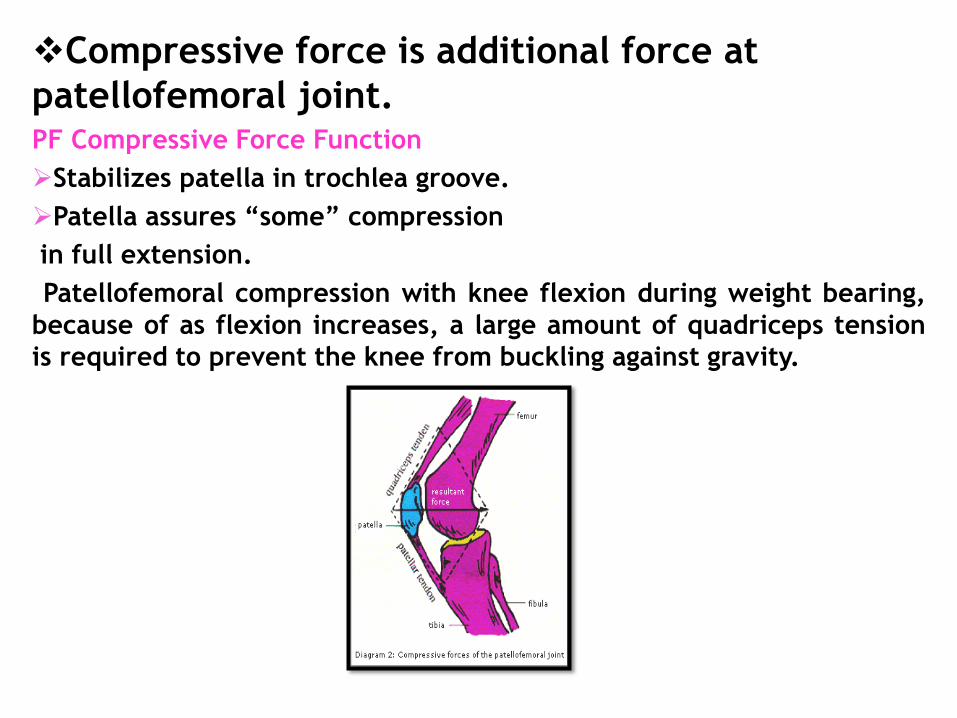
Compressive force is additional force at
patellofemoral joint. PF Compressive Force Function
Stabilizes patella in trochlea groove.
Patella assures “some” compression
in full extension.
Patellofemoral compression with knee flexion during weight bearing,
because of as flexion increases, a large amount of quadriceps tension
is required to prevent the knee from buckling against gravity.
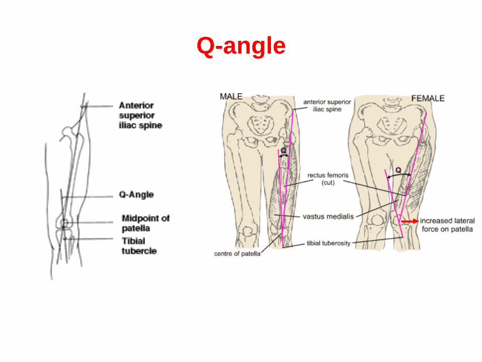
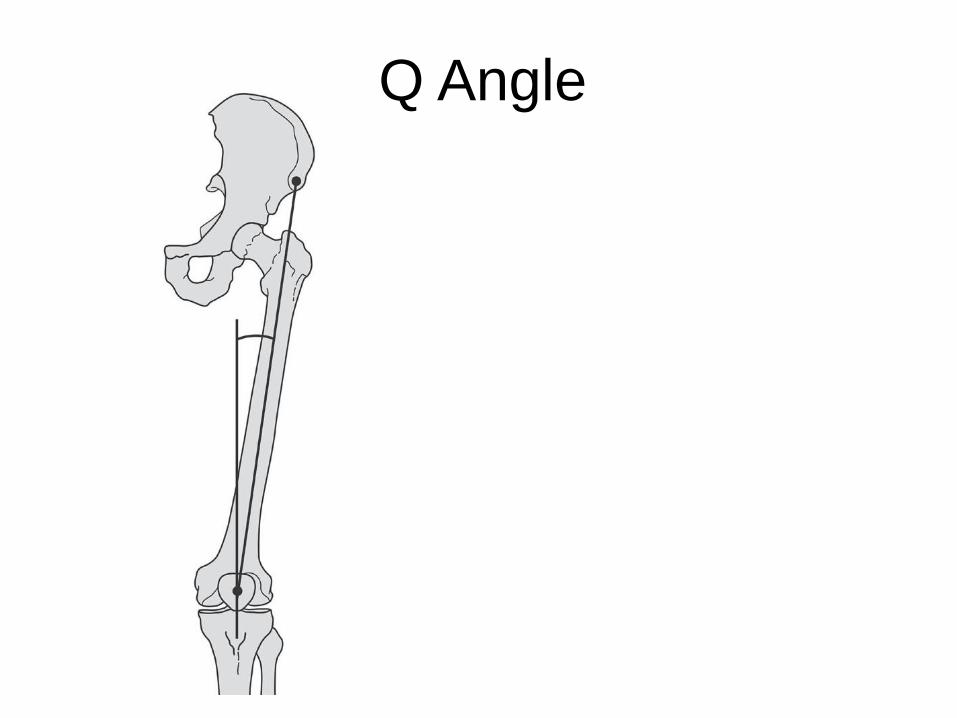
Q-angle

MENISKITE ROLL SURVEJÕUDUDE JAOTAMISEL
SÄÄRELUULE
Meniskiga Menisk on eemaldatud
Reieluu
Sääreluu
Patella Menisk
Surve

Plokkliiges
Ratasliiges


Q-nurk
Eesmine ülemine niudeoga
Q-nurk
Põlvekeder
Sääreluuköprus

The mean movement (mm) in each meniscus during flexion on a weight-bearing
knee. (From Vedi et al. J Bone and Joint Surg Br. 1999;81-B:37-41)
The mean movement (mm) in each meniscus during flexion on a sitting non weight-
bearing knee. (From Vedi et al. J Bone and Joint Surg Br. 1999;81-B:37-41)

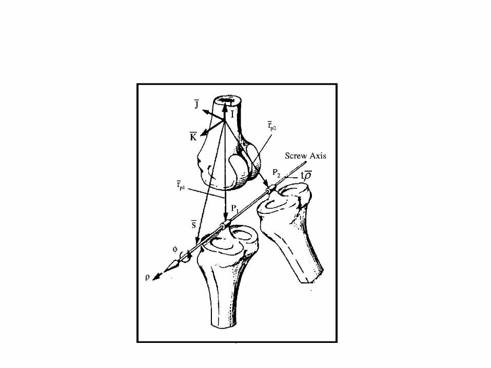
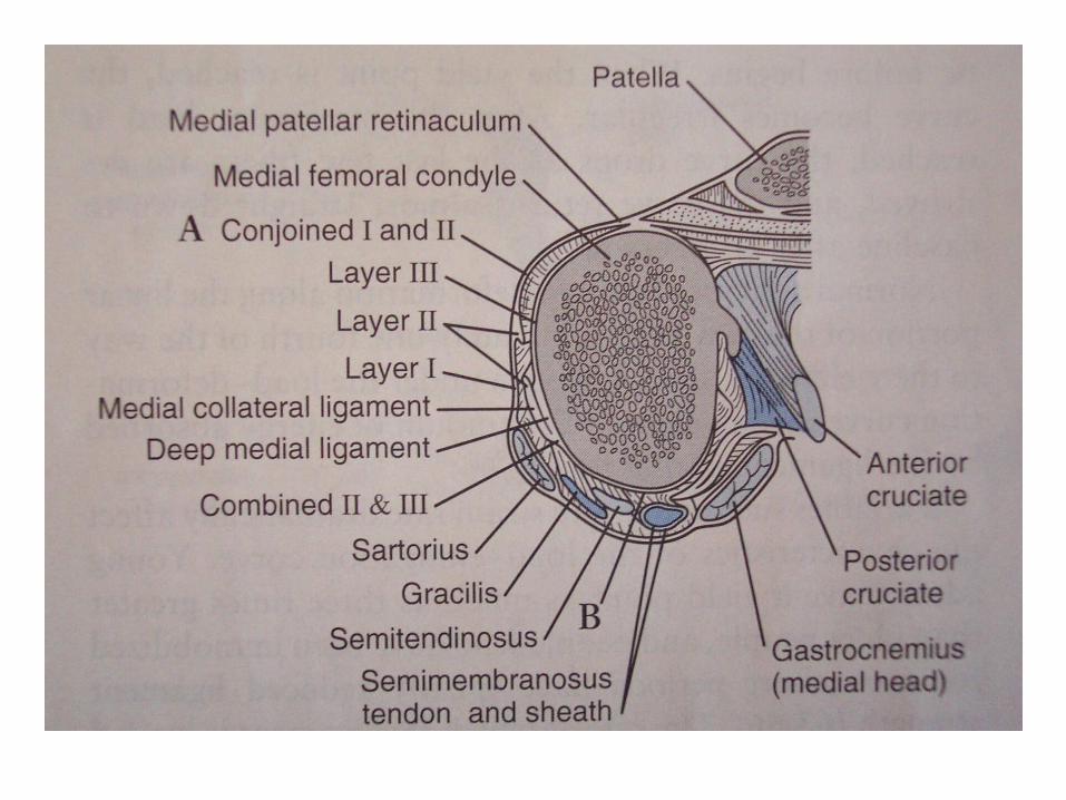
Figure 2. Definition of axes used in the study. The transepicondylar line, as defined in
this study, is the line formed by the insertions of the lateral (LCL) and medial collateral
ligaments (MCL), respectively.

Figure 1. The six degrees of freedom of TF joint motion expressed in a clinical joint coordinate
system. Mediolateral translation (M–L) and flexion–extension (F–E) occur along and about an
epicondylar femoral axis. Joint distraction and internal–external rotation (I–E) occur along and
about a tibial long axis. Anterior–posterior translation (A–P) and varus–valgus (V–V) (or
adduction–abduction) rotation occur along and about a floating axis, which is perpendicular to
both femoral epicondylar and tibial long axes. (Masouros S.D. Et al. Orthop Trauma, 2010, 24:
84-91)

Heel Strike Toe Off Heel Strike
Jo
int
Fo
rce
/Bo
dy W
eig
ht
Stance Swing
Total Knee Joint Force
Force in X direction
Force in Y direction
Force in Z direction
7 x Body Weight
X
Z
Y
KNEE [source
unknown]

Figure 3.19. Free body diagram of a patient ascending a step, m1 = quadriceps force; m2 =
patellar tendon tension; KJR = knee joint reaction; PFJR = patellofemoral joint reaction; CG =
center of gravity; x = flexor lever arm.
(Reproduced with permission from V.T. Inman et al.: Human Walking, Williams & Wilkins,
Baltimore, 1981 (2).)

PF joint force at a extension and b 90° flexion, showing geometrically the increase
of PF joint force with flexion. (PT: patellar tendon, Q: quadriceps muscles, PF:
patellofemoral, TF: tibiofemoral). (Masouros S.D. Et al. Orthop Trauma, 2010, 24:
84-91)
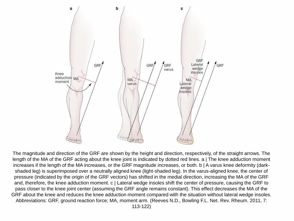
The magnitude and direction of the GRF are shown by the height and direction, respectively, of the straight arrows. The
length of the MA of the GRF acting about the knee joint is indicated by dotted red lines. a | The knee adduction moment
increases if the length of the MA increases, or the GRF magnitude increases, or both. b | A varus knee deformity (dark-
shaded leg) is superimposed over a neutrally aligned knee (light-shaded leg). In the varus-aligned knee, the center of
pressure (indicated by the origin of the GRF vectors) has shifted in the medial direction, increasing the MA of the GRF
and, therefore, the knee adduction moment. c | Lateral wedge insoles shift the center of pressure, causing the GRF to
pass closer to the knee joint center (assuming the GRF angle remains constant). This effect decreases the MA of the
GRF about the knee and reduces the knee adduction moment compared with the situation without lateral wedge insoles.
Abbreviations: GRF, ground reaction force; MA, moment arm. (Reeves N.D., Bowling F.L. Net. Rev. Rheum. 2011, 7:
113-122)

Patellofemoral joint reaction forces
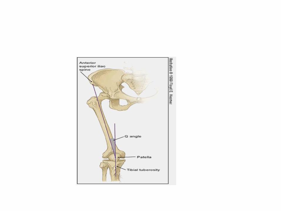

Figure 5. Biomechanical model depicting mean knee joint kinematics during the drop vertical
jump at initial contact and maximal displacement in the ACL-injured and uninjured groups (n
= 9 knees and n = 390 knees, respectively). Left, coronal plane view of knee abduction angle
at initial contact in the ACL-injured and uninjured groups. Center, coronal plane view of
maximum knee abduction angle in the ACL-injured and uninjured groups. Right, sagittal
plane view of maximum knee flexion angle in the ACL-injured and uninjured groups. (Hewett
T.E. Et al. Am J Sports Med 2005, 33: 492-501)
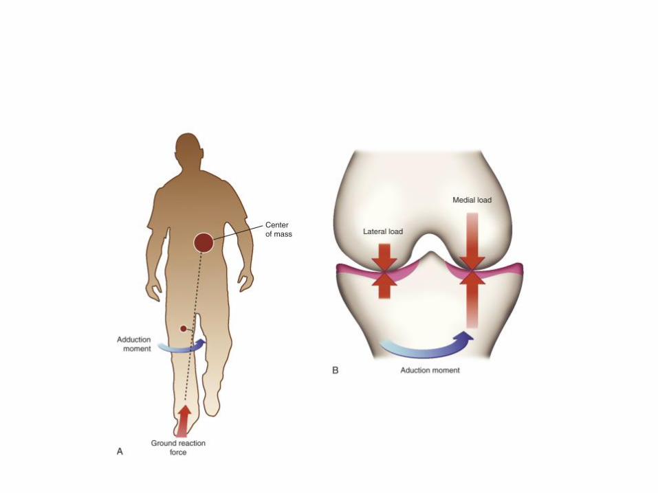
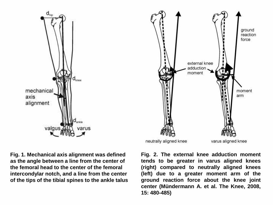
Fig. 2. The external knee adduction moment
tends to be greater in varus aligned knees
(right) compared to neutrally aligned knees
(left) due to a greater moment arm of the
ground reaction force about the knee joint
center (Mündermann A. et al. The Knee, 2008,
15: 480-485)
Fig. 1. Mechanical axis alignment was defined
as the angle between a line from the center of
the femoral head to the center of the femoral
intercondylar notch, and a line from the center
of the tips of the tibial spines to the ankle talus

Fig. 1. The role of the slope and orientation of the tibial plateau on the direction of the
joint compressive force (JCF). (a) At full extension JCF is leaning anteriorly and (b) at
moderate flexion JCF is leaning posteriorly. QPF, quadriceps patellar tendon force; HF,
hamstring force; GRF, ground reaction force. (Hashemi J. et al. J Biomech, 2011, 44:
577-585)

Fig. 2. Ensemble average curves for the TSF and CTRL groups for (a) vertical ground
reaction force; (b) knee flexion angle; (c) sagittal plane knee moment and shank angle
during the stance phase of running: the initial loading period is from footstrike to the
vertical line. Knee flexion angle, internal knee extension moment and distal end of shank
anterior to proximal end are positive. (Milner C.E. et al. Clin Biomech, 2007, 22: 697-703)
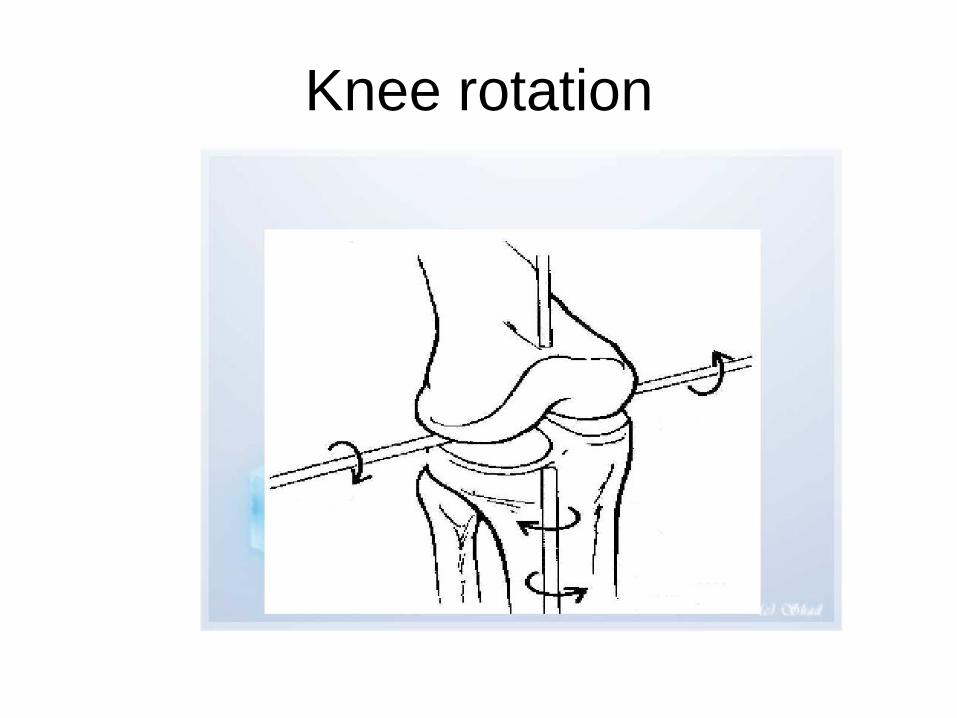
Knee rotation


Resultant for
e has a tenden
c
y to laterally translate the patella

Patellar translation


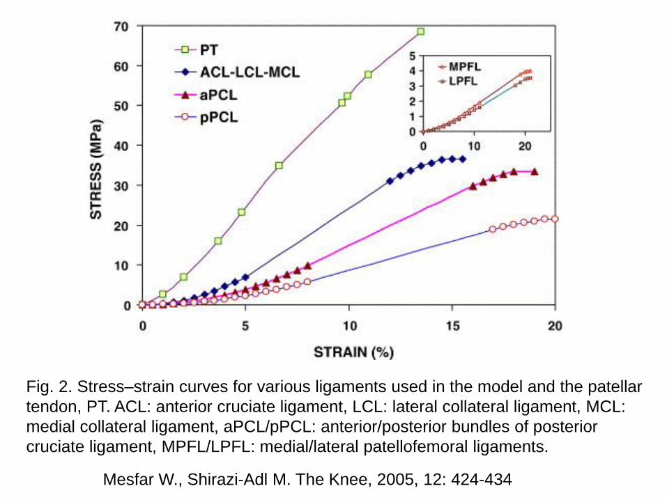
Mesfar W., Shirazi-Adl M. The Knee, 2005, 12: 424-434
Fig. 2. Stress–strain curves for various ligaments used in the model and the patellar
tendon, PT. ACL: anterior cruciate ligament, LCL: lateral collateral ligament, MCL:
medial collateral ligament, aPCL/pPCL: anterior/posterior bundles of posterior
cruciate ligament, MPFL/LPFL: medial/lateral patellofemoral ligaments.

Mesfar W., Shirazi-Adl M. The Knee, 2005, 12: 424-434
Fig. 1. The knee joint finite element models showing cartilage layers, menisci, ligaments, patellar
tendon, and quadriceps muscles. Bony structures are shown only by their primary nodes. Quadriceps
components considered are VMO: vastus medialis obliqus, RF: rectus femoris, VIM: vastus intermidus
medialis, and VL: vastus lateralis (VL). LPFL: lateral patellofemoral ligament, MPFL: medial
patellofemoral ligament.


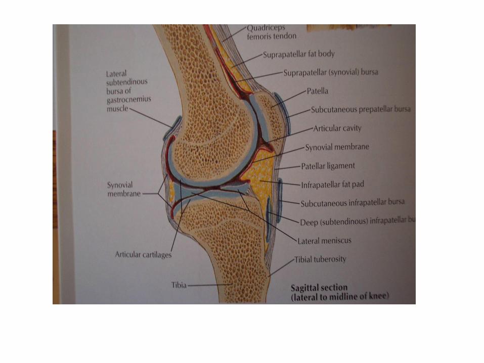
Figure 8.8b
Synovial Joints: Knee –
Other Supporting Structures

Knee Injury

Menisci

Menisci
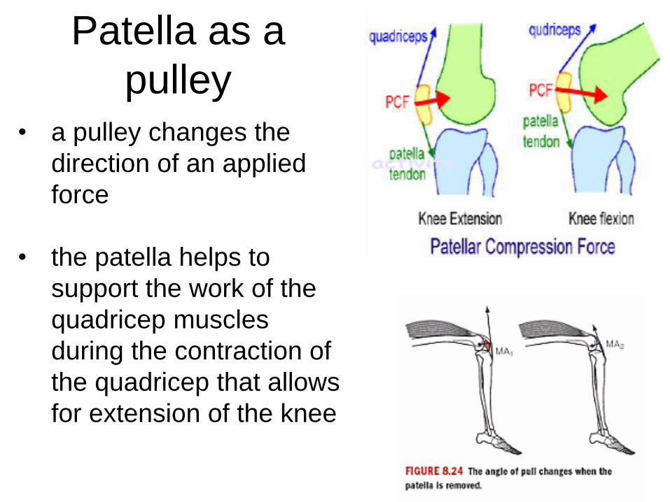
Patella as a
pulley • a pulley changes the
direction of an applied
force
• the patella helps to
support the work of the
quadricep muscles
during the contraction of
the quadricep that allows
for extension of the knee
Q Angle


Problem!
• Givens: Quadriceps tendon is inserted on the tibia 5 cm from the knee joint, and is at a 30deg angle. Weight of the lower leg Is 48 N. Center of gravity of the lower leg is 0.20 m from the knee joint.
1. Determine Fquad
required to hold the lower
leg in static equilibrium
2. Determine the joint
reaction force of the
femur

48 N
T 30°
Fquad
Rx
Ry

Muscles of the Leg

“Q” Angle
• Normal
– about 15°
• Males vs. Females
– wider pelvis


Free Body Diagram
• To obtain muscle and joint
forces:
• That kind of solution will
only provide information on
the hip joint and thigh
muscle forces 350
350
450
X
Y
Z
0
0
M
F

The Thigh
FPat-Fem
FBD-THIGH
WThigh

Knee - Example
A person is wearing a weight boot and doing
lower leg flexion/extension exercises to
strengthen the quadriceps. Determine FM and
FJ for the sitting position shown.
W1=0.06W
W0=0.06W
c=0.28H
b=0.14H
a=0.08H
=450
=150
W – Body Weight,
H – Body Height

Adapted from Fu, Harner, & Vince (eds). Knee Surgery. Baltimore,
MD: Williams & Wilkins, 1994.

Knee Joint Motion
internal
flexion medial
lateral
anterior
posterior
extension
distraction
external
abduction adduction
compression
(Right Knee)
6 degrees of freedom
-3 rotations
-3 translations

Instant Centres (Centre of Rotation)
for Femur Motion during F/E (sagittal view)
rolling sliding pure rotation
femur
tibia
IC at infinity
IC
IC
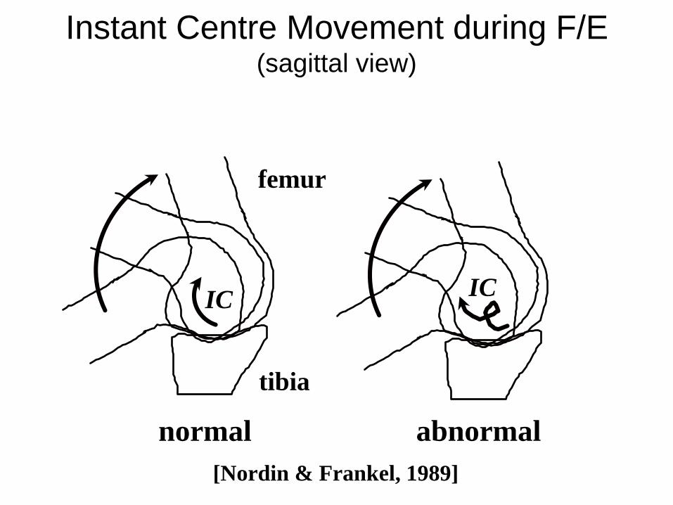
Instant Centre Movement during F/E (sagittal view)
normal abnormal
femur
tibia
IC IC
[Nordin & Frankel, 1989]

Screw-Home Mechanism
femur
tibia
Tibia externally rotates
when knee extends and
screws up into the femur
Tibia internally rotates
and moves distally away
from femur when
knee flexes
flexion
extension
[Nordin & Frankel, 1989]

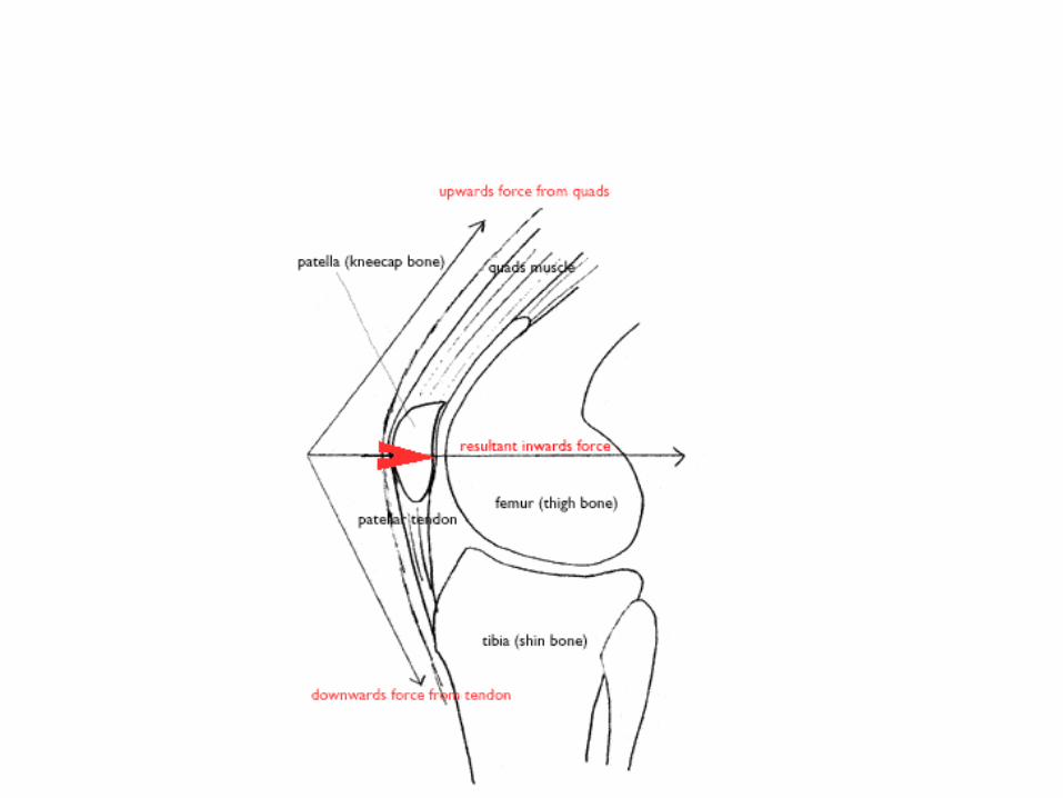
Patella (sagittal view)
femur
tibia
patella
quadricep
muscle
F 1.3F
lever arm

Menisci & Stress Distribution
menisci intact meniscectomy
femur
tibia
patella meniscus
stress
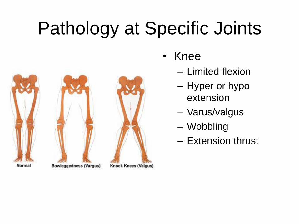
Pathology at Specific Joints
• Knee
– Limited flexion
– Hyper or hypo
extension
– Varus/valgus
– Wobbling
– Extension thrust

Adapted from Fu, Harner, & Vince (eds). Knee Surgery. Baltimore,
MD: Williams & Wilkins, 1994.
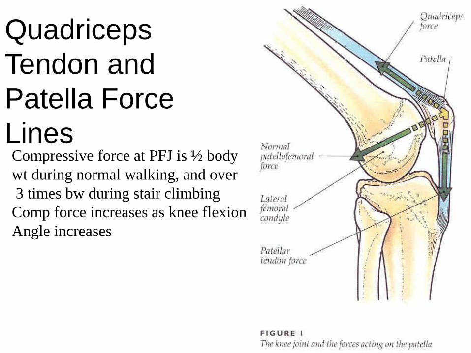
Quadriceps
Tendon and
Patella Force
Lines Compressive force at PFJ is ½ body
wt during normal walking, and over
3 times bw during stair climbing
Comp force increases as knee flexion
Angle increases

Determine forces acting in the joints and
tissue elements during physical activity

A Hinge Joint ?
• Complex Hinge joint
• Ball and Plate model

Instant centre pathway
» Perpendicular to
» Point of contact
of joint surface

Shift of contact point
– Related to joint congruency
– Cruciates and menisci play
important role

Menisci –”The wedge effect”
• Adds to joint
conformity
• Block under a tyre
• Converts tibia onto a
shallow socket
• Weight distribution

Function of patella
• increase moment arm
• protect femur joint surface
• distribute pressure
• adjust joint force

PCL Retention: Disadvantages
See-saw effect
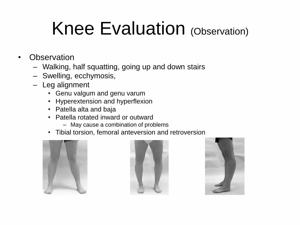
Knee Evaluation (Observation)
• Observation – Walking, half squatting, going up and down stairs
– Swelling, ecchymosis,
– Leg alignment • Genu valgum and genu varum
• Hyperextension and hyperflexion
• Patella alta and baja
• Patella rotated inward or outward – May cause a combination of problems
• Tibial torsion, femoral anteversion and retroversion

Knee Joint Motion
internal
flexion medial
lateral
anterior
posterior
extension
distraction
external
abduction adduction
compression
(Right Knee)
6 degrees of freedom
-3 rotations
-3 translations

Tibiofemoral Joint Activities:
Flexion & Anterior Shear
• cycling 105 deg 0.05BW
• walking 5 0.4
• stairs-down 5 0.6
• squat-rise 140 3.0
• squat-down 140 3.6
[Mow & Hayes, 1991] BW = body weight
Front
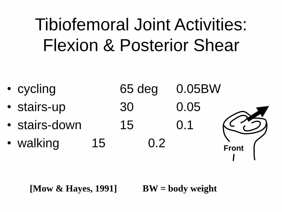
Tibiofemoral Joint Activities:
Flexion & Posterior Shear
• cycling 65 deg 0.05BW
• stairs-up 30 0.05
• stairs-down 15 0.1
• walking 15 0.2
[Mow & Hayes, 1991] BW = body weight
Front

Menisci & Stress Distribution
menisci intact meniscectomy
femur
tibia
patella meniscus
stress
Knee Motion
Knee Flex/Extension
-40
-20
0
20
40
60
80
100
0 20 40 60 80 100
Ext - Flx
Knee Varus/Valgus
-40
-30
-20
-10
0
10
20
30
40
0 20 40 60 80 100
Val - V
ar
Knee Rotation
-50
-40
-30
-20
-10
0
10
20
30
40
0 20 40 60 80 100
Ext - In
t

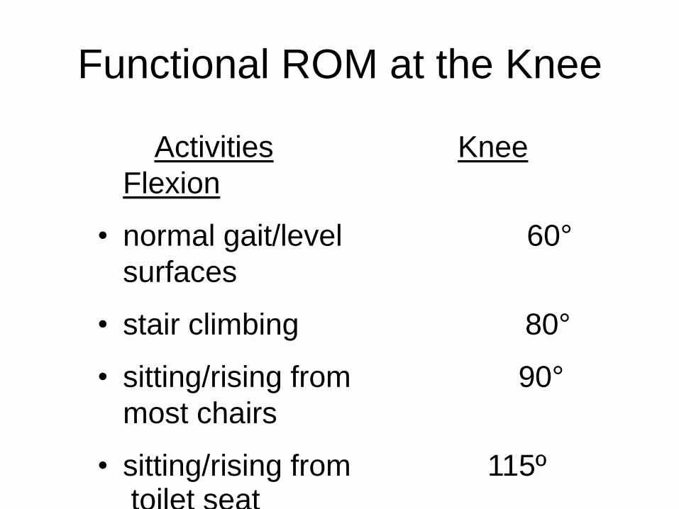
Functional ROM at the Knee
Activities Knee
Flexion
• normal gait/level 60°
surfaces
• stair climbing 80°
• sitting/rising from 90°
most chairs
• sitting/rising from 115º toilet seat
• advanced function > 115°

1st 250 - mainly roll >250 roll and ant glide
Arthrokinematics: Femoral Condyles in Flexion


Menisci
• Fairbank - 1948
• late 60’s - poor results of miniscectomy
• mid 70’s - load transmission confirmed
– 40-60% load is on meniscus
– lateral > medial

• 2 functions
– load bearing
– stability
• also, joint lubrication
• prevent capsule, synovial impingement
• shock absorbers
Load bearing
-composition;

Hoop stress
Forces at the tibiofemoral joint
3 main coplanar forces on the knee joint
In single leg stance, the leg has a valgus orientation
Ground
reaction
force (equal
to body weight)(W)
Patellar
tendon force (P)
Joint
reaction force (J)

Ascending stairs
•The actual degree of knee flexion required to ascend stairs is
determined not only by the height of the step, but also by the
height of the patient.
•For the standard 7" step approximately 65° of flexion will be
required.
•In climbing stair , lever arm can be
reduced by leaning forward. Also, in stair
climbing the tibia is maintained relatively
vertical, which diminishes the anterior
subluxation potential of the femur on the tibia.
Descending stairs
•In standard step 85° of flexion is required.
•The tibia is steeply inclined toward
the horizontal, bringing the tibial
plateaus into an oblique orientation.
• The force of body weight will now
tend to sublux the femur anteriorly.
This anterior subluxation potential will be resisted by
the patellofemoral joint reaction force, and the
tension which develops in the posterior cruciate
ligament.

Kinematics of tibiofemoral joint
Motion(sagittal, transverse and frontal planes).
It is greatest in the sagittal plane (0-140 degree), minimal in the
transverse and frontal planes.
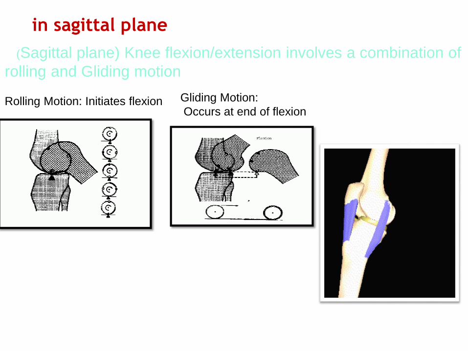
in sagittal plane
(Sagittal plane) Knee flexion/extension involves a combination of
rolling and Gliding motion
Rolling Motion: Initiates flexion Gliding Motion:
Occurs at end of flexion

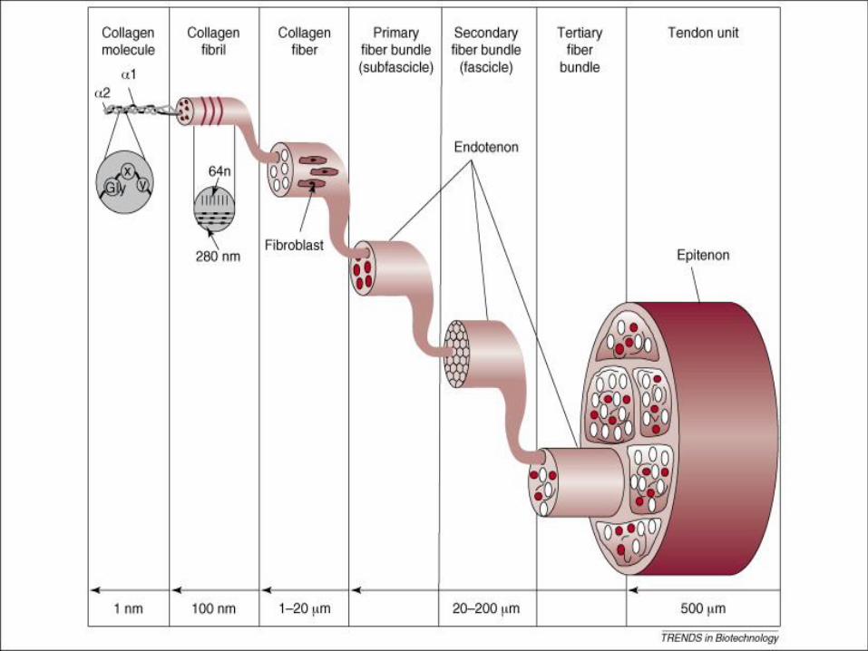
What are Tendons? Tendons are bundles or bands of strong fibers that attach muscles to bones

Rotation between the tibia and femur.
During Knee extension:
“Screw-Home” mechanism:
It is considered a key element to knee stability for standing
upright.
Tibia rolls anteriorly, on the femur, PCL
Elongates.
PCL's pull on tibia causes it to glide
anteriorly.

During the last 20 degrees of knee extension
anterior tibial glide persists on the
tibia's medial condyle because its articular surface
is longer in that dimension than the lateral condyle's.
Prolonged anterior glide on the medial side produces
external tibial rotation, the "screw-home" mechanism.

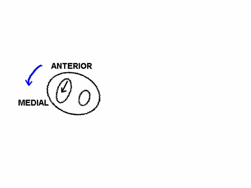
THE SCREW-HOME MECHANISM REVERSES
DURING KNEE FLEXION.
When the knee begins to flex from a position of full
extension .
Tibia rolls posterior, elongating ACL.
ACL's pull on tibia causes it to glide
Posterior.
Glide begins first on the longer medial condyle.

Between 00 extension and 200 flexion
Posterior glide on the medial side produces
Relative tibial internal rotation.
A reversal of the screw - home mechanism.

In transverse plane:
In full extension almost no motion, because of
interlocking of the femoral and tibial condyles.
At 90 degrees of flexion:
• external rotation of the knee ranges (0 -45
)degrees
• internal rotation ranges ( 0 to 30) degrees.
> 90 degrees of knee flexion:
the range of motion ,because of the restriction
function of the soft tissues.

:In frontal plane
In fully extended knee almost no abduction or adduction is possible.
knee is flexed up to 30 degree:
only a few degrees in either passive abduction or passive adduction.
> 30 degrees of flexion:
Motion ,because of the restriction function of the soft tissues.
Maximal knee flexion occurred during lifting, A significant relationship between the length of lower leg and the range of knee motion. The longer leg was, the greater the range of motion.

In the double stance phase of gait
When the body weight is borne equally on both feet
the force which passes through the knee is only a
fraction of body weight.
There is no bending moment around either knee.
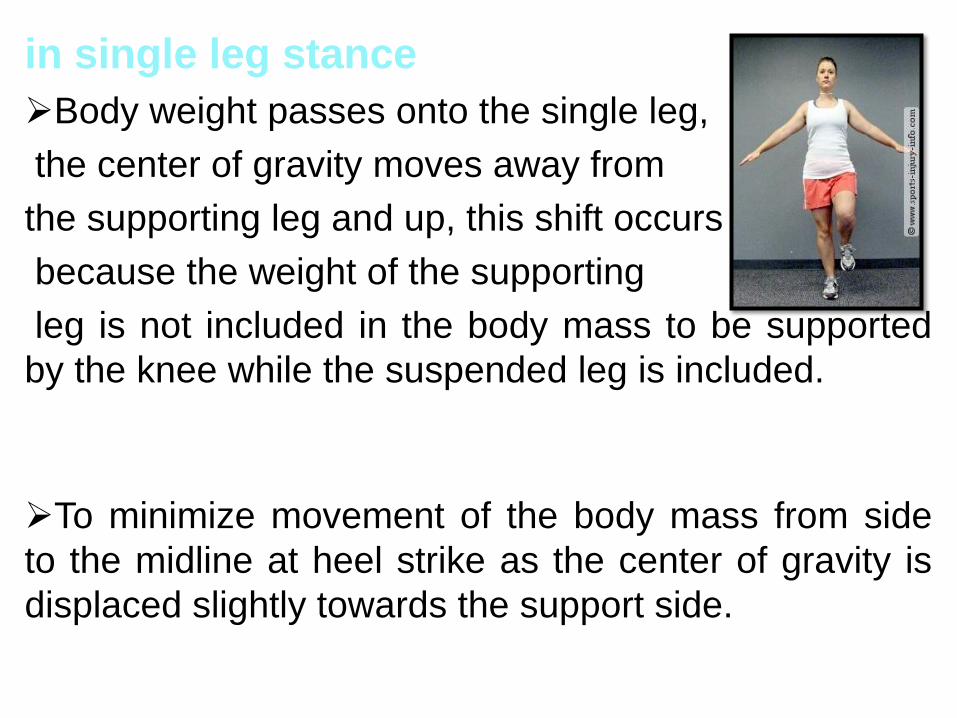
in single leg stance
Body weight passes onto the single leg,
the center of gravity moves away from
the supporting leg and up, this shift occurs
because the weight of the supporting
leg is not included in the body mass to be supported
by the knee while the suspended leg is included.
To minimize movement of the body mass from side
to the midline at heel strike as the center of gravity is
displaced slightly towards the support side.

In man, with upright single leg stance, this
orientation is accomplished by the overall valgus
orientation of the lower extremity which naturally
brings the foot toward the midline.
In single leg stance, therefore, the leg has a valgus
orientation. This situation exerts a bending moment
on the knee which would tend to open the knee
into varus, the ligaments and capsule are tight, in
part because of the "screw-home" mechanism.
These structures resist this bending moment.

During gait
Multiple muscles which cross the joint in the
center or to the lateral side of center combine to
provide a lateral resistance to opening of the lateral
side of the joint. These include the quadriceps-
patellar tendon forces, the lateral gastrocnemius,
popliteus, biceps and iliotibial tract tension.
With increasing knee varus the medial lever arm
increases requiring an increased lateral reaction to
prevent the joint from opening.
In total joint replacement a single cane in the
opposite hand does much to unload the knee and
particularly to reduce the magnitude of the varus
bending moment, a cane in the opposite hand
will reduce knee loading by 46%.
narrow base gait is the norm, and the most energy efficient.
The side to side deviation of the center of gravity is reduced to
approximately 2 cm in each direction toward the support side
or a total of 4 cm through the gait cycle involving both legs.
waddling or broad based gait
lateral displacement will be accentuated requiring greater
energy for walking.
the orientation of the lower extremity to the vertical and to
the center of gravity will be the same during the single leg
support phase of gait.
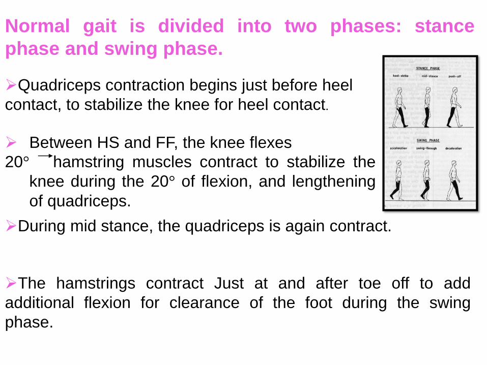
Normal gait is divided into two phases: stance
phase and swing phase.
Quadriceps contraction begins just before heel
contact, to stabilize the knee for heel contact.
Between HS and FF, the knee flexes
20° hamstring muscles contract to stabilize the
knee during the 20° of flexion, and lengthening
of quadriceps.
During mid stance, the quadriceps is again contract.
The hamstrings contract Just at and after toe off to add
additional flexion for clearance of the foot during the swing
phase.

In the absence of a posterior cruciate ligament, only
the collateral ligaments are available to assist the
patellofemoral joint reaction force in providing
anterior-posterior stability.
Many patients with arthritis will report difficulty
descending stairs normally, this will also be true
after total knee replacement. A simple remedy is to
have them descend either sideways or backward,
which is biomechanically the equivalent of
ascending the stairs with its decreased mechanical
and range of motion demands.

Patellofemoral joint:
Patellofemoral joint consist of the articulation of the triangularly shaped patella, encased
in the patellar tendon. The posterior surface of the patella is coverd with articular
cartilage, which reduces friction
between the patella and the femur.
Function of patella
Increase the angle of pull of the quadriceps tendon
Increase the area of contact between the patellar
tendon and the femur, thereby PF joint contact stress.
,

The Q-angle (or "quadriceps angle) is formed in
the frontal plane by two line segments:
Angle formed at the knee joint
By connecting a line from the anterior
iliac crest to the center of the patella.
And a second line from the center of
the patella to the center of the patellar tendon
insertion into the tibial tubercle.
the Q-angle is normally less than 15 degrees in
men and less than 20 degrees in women. An
abnormally large Q-angle usually results in a disorder
called abnormal quadriceps pull

Motion occurs in two planes: Frontal and transverse.
At full extension both medial and lateral
femoral facet articulate with the patella.
> 90degrees of flexion the patella rotate externally, and only
the medial femoral facet articulate with the patella.
At full flexion patella sinks into intercondylar
groove.
Kinematics of patellofemoral joint

Laterally- lateral retinaculum, vastus lateralis m,
iliotibial tract.
Medially- medial retinaculum and vastus medialis m.
Superior- Quadriceps via quadriceps
tendon.
Inferior- Patellar tendon.
Forces acting on the Patella:

Compressive force is additional force at
patellofemoral joint.
PF Compressive Force Function
Stabilizes patella in trochlea groove.
Patella assures “some” compression
in full extension.
Patellofemoral compression with knee flexion
during weight bearing, because of as flexion
increases, a large amount of quadriceps tension is
required to prevent the knee from buckling against
gravity.
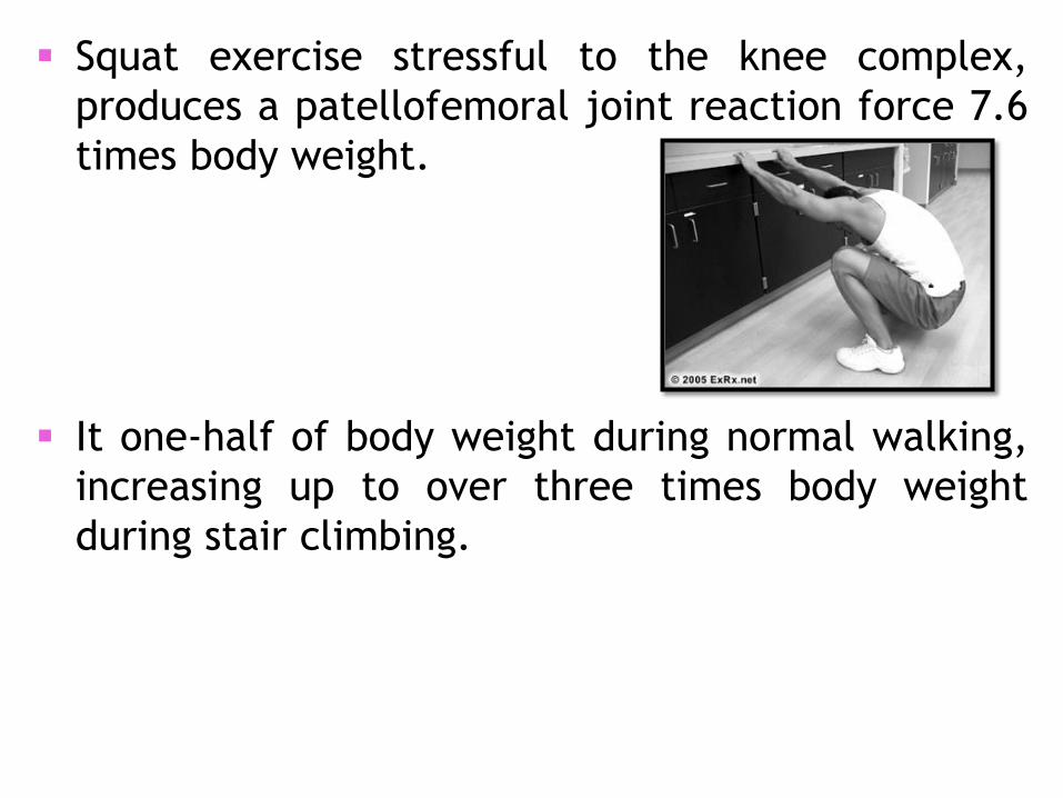
Squat exercise stressful to the knee complex,
produces a patellofemoral joint reaction force 7.6
times body weight.
It one-half of body weight during normal walking,
increasing up to over three times body weight
during stair climbing.
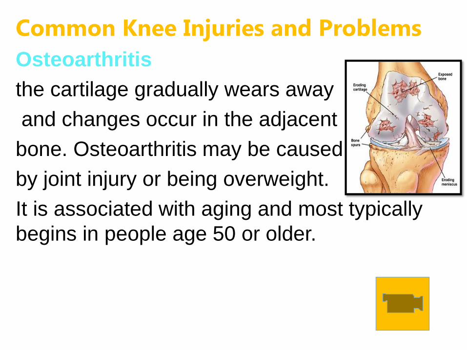
Common Knee Injuries and Problems
Osteoarthritis
the cartilage gradually wears away
and changes occur in the adjacent
bone. Osteoarthritis may be caused
by joint injury or being overweight.
It is associated with aging and most typically
begins in people age 50 or older.

Chondromalacia
Also called chondromalacia patellae, refers to
softening of the articular cartilage of the kneecap.
This disorder occurs most often in young
adults and can be caused by injury,
overuse, misalignment of the patella,
or muscle weakness. Instead of gliding
smoothly across the lower end of the
thigh bone, the kneecap rubs against it,
thereby roughening the cartilage
underneath the kneecap.

Meniscal Injuries
The menisci can be easily injured by the force of
rotating the knee while bearing weight. A partial
or total tear may occur when a person
quickly twists or rotates the upper
leg while the foot stays still. If the
tear is tiny, the meniscus stays
connected to the front and back
of the knee; if the tear is large, the meniscus may
be left hanging by a thread of cartilage. The
seriousness of a tear depends on its location and
extent.
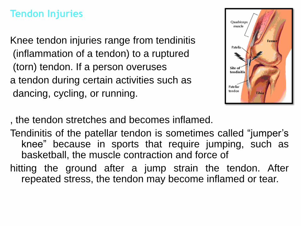
Tendon Injuries
Knee tendon injuries range from tendinitis
(inflammation of a tendon) to a ruptured
(torn) tendon. If a person overuses
a tendon during certain activities such as
dancing, cycling, or running.
, the tendon stretches and becomes inflamed.
Tendinitis of the patellar tendon is sometimes called “jumper’s knee” because in sports that require jumping, such as basketball, the muscle contraction and force of
hitting the ground after a jump strain the tendon. After repeated stress, the tendon may become inflamed or tear.

Medial and Lateral Collateral Ligament Injuries
The medial collateral ligament is more easily injured
than the lateral collateral ligament. The cause of
collateral ligament injuries is most often a blow to
the outer side of the knee that stretches and tears
the ligament on the inner side of the knee. Such
blows frequently occur in contact sports such as
football or hockey.

Knee replacement, or knee arthroplasty
is a common surgical procedure most often
performed to relieve the pain and disability, Knee
replacement surgery can be performed as a partial
or a total knee replacement, the surgery consists
of replacing the diseased or damaged joint
surfaces of the knee with metal and plastic
components shaped to allow continued motion of
the knee.

The knee is a two-joint structure composed of the tibiofemoral joint and the patellofemoral joint.
In the tibiofemoral joint, surface motion occurs in three planes, greatest in sagittal plane. In the patellofemoral joint , surface motion occure in two planes frontal and transverse.
The screw home mechanism of tibiofemoral joint adds stability to the joint in full extension.
Both the tibiofemoral joints and patellofemoral joints are subjected to high forces. The magnitude of the joint reaction force on both joints can reach several times body weight
Although the tibial plateaus are the main load bearing structures in the knee, the cartilage, menisci, and ligaments also bear load.
The patella aids knee extension by lengthening the lever arm of the quadriceps muscle, and allows a better distribution of compressive stress on the femur.


Introduction
• What kind of joint is it?
• Limits of motion
• Normal kinenatics of a step
• Plateau & condyles
• Patello Femoral articulation
• Menisci
• Medial, lateral and anterior stability
• ACL & PCL

Knee joint
• Ginglymus (hinge) ?
• Arthodial (gliding) ?
• 6 degrees of freedom
– 3 rotations
– 3 translations
• Rotations
– flex/ext - -15 to 140 deg
– varus valgus - 6-8 deg in extension
– int/ext rotation - 25 - 30 deg in flexion

• Translations
– AP 5 - 10mm
– comp/dist 2 - 5mm
– medio-lateral 1-2mm
Taking a step
• Just prior to heel strike - max extension &
max external rotation
• heel strike - max valgus
• flat foot - flexion & intrenal rotation
progress
• swing phase - internal rotation continues,
max flexion, max anterior translation.

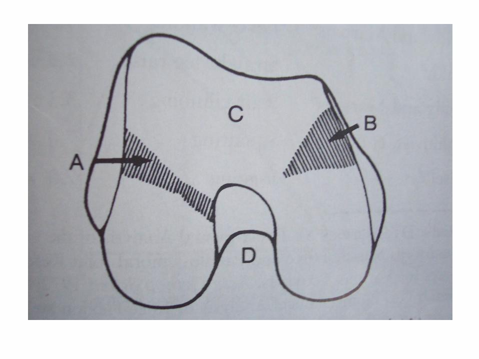
Condyles and plateau

Patellofemoral articulation
• Shape/anatomy of patella
• Anatomy of intercondylar groove
• direction of force
• PFJR vs flexion angle and quads force
• Contact area vs PFJR and stress
• chondromalacia of patella
• Patella functions
– Increases moment arm (increases rotational
torque) 0 - 45 deg
– lever at > 45 deg
• Patellectomy?

• Knee joint stability
– mainly rotational
– miniscectomy +/- ACL and translation
– why differences in lat vs med ? – Structure
– attachments

Attachment sites

Medial & lateral stabilizers
(mostly ligaments) • Ligaments
– most important static stabilizers
– tensile strength - related to composition

Medial side
• Superficial MCL
– Primary valgas restraint -57-78% restraining
moment of knee
– femoral attachment fans out around axis of
rot.
– Lax in flexion
• Semimembranosis (expansion)
– internally rot’s tib on femur
– tenses post/med capsular structures that are
lax in knee flexion
– acts with ACL


Lateral side
• LCL
– Primary varus restraint
– lax in flexion
– Bicepts passes it and blends with insertion
• maintains tension?

• Bicepts
– flexor(with semimembranosis and pes)
– externally rotates tibia
– tenses LCL
– dynamic assistor of PCL

Cruciates
• ACL
– Primary static restraint to anterior
displacement
– tense in extension, ‘lax’ in flexion
• PCL
– Primary restraint to post. Displacement - 90%
– relaxed in extension, tense in flexion
– reinforced by Humphreys or Wrisberg
– restraint to varus/valgus force
– resists rotation, esp.int rot of tibia on femur

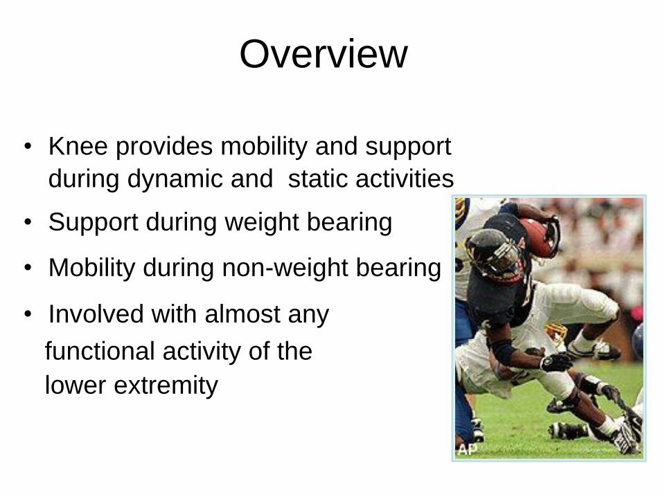
Overview
• Knee provides mobility and support
during dynamic and static activities
• Support during weight bearing
• Mobility during non-weight bearing
• Involved with almost any
functional activity of the
lower extremity
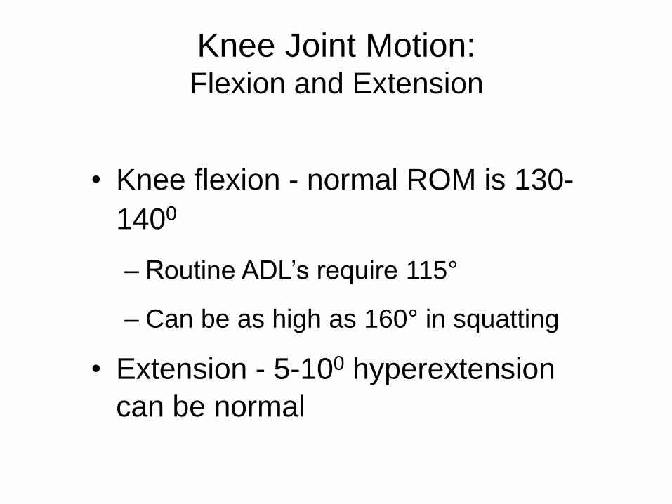
Knee Joint Motion: Flexion and Extension
• Knee flexion - normal ROM is 130-
1400
– Routine ADL’s require 115°
– Can be as high as 160° in squatting
• Extension - 5-100 hyperextension
can be normal

Knee Joint
Function:
Muscle Action

Muscle Action: Flexors
– Semimembranosus,
Semitendinosus, Biceps
femoris, Sartorius, Gracilis,
Popliteus, Gastrocs
– All are 2 jt. muscles
except popliteus & short
head of biceps femoris
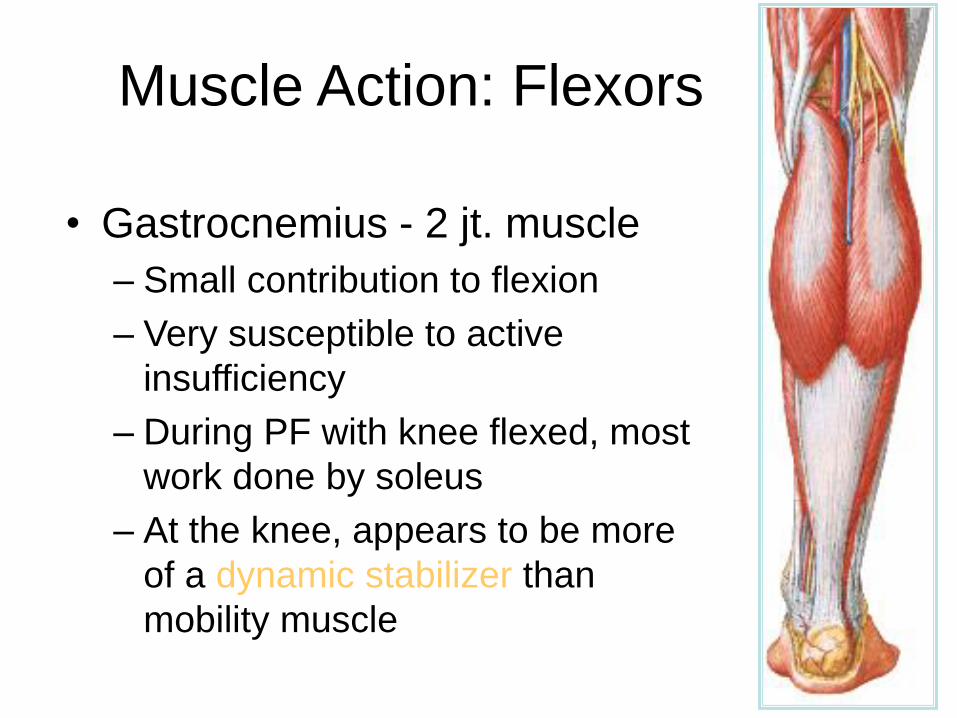
Muscle Action: Flexors
• Gastrocnemius - 2 jt. muscle
– Small contribution to flexion
– Very susceptible to active
insufficiency
– During PF with knee flexed, most
work done by soleus
– At the knee, appears to be more
of a dynamic stabilizer than
mobility muscle

Muscle Action:
Extensors
• Quadriceps
– Rectus femoris - 2 jt.
– Vastus intermedius,
lateralis, medialis
• Resultant pull:
– Lateralis
– Medialis
– Rectus femoris
VL
30-400
RF
5- 70
VML
15-170
VMO
50- 550

Patellofemoral joint

Patellofemoral Joint

Functions of the patella/PFJ
• Increase the mechanical advantage of
the quadriceps muscle group
• Decreases friction - quad tendon &
femoral condyles
• Helps to distribute the compressive
forces that are placed on the femur

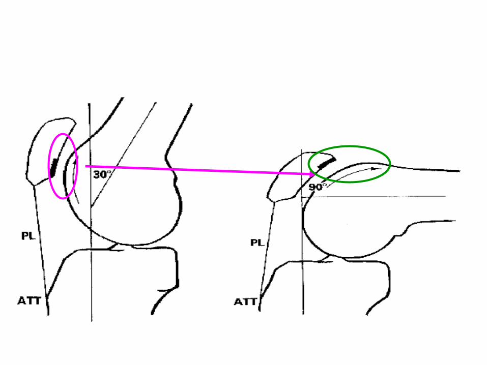
Patellofemoral Joint: Patellofemoral Joint Congruence
• 1st consistent PF contact is between
10-200 flexion with increased contact as
flexion increases
• By 900 all aspects of facets have made
contact except odd which contacts at
>900
• At 1350 - contact is on odd & lat facets

Patellofemoral contact points

Patellofemoral joint stress in
weight-bearing and non-
weightbearing

Biomechanics: clinical
implications
(Grelsamer & Klein, 1998)
• Quad strengthening can be safely performed
in the 0-90 range by varying the mode of
exercise if ROM restrictions are in place.
• Specifically, open chain (NWB) exercises are
most safely carried out from 25-90°, and SLRs
with the knee at 0° of extension are equally
safe.
• Closed chain (NWB) exercises are safest in
the 0-45° range.

DG3: Knee flexion in stance
phase
• As the hip joint
passes over the
foot during the
support phase,
there is some
flexion of the knee.
• This reduces
vertical movements
at the hip, and
therefore of the
trunk and head.

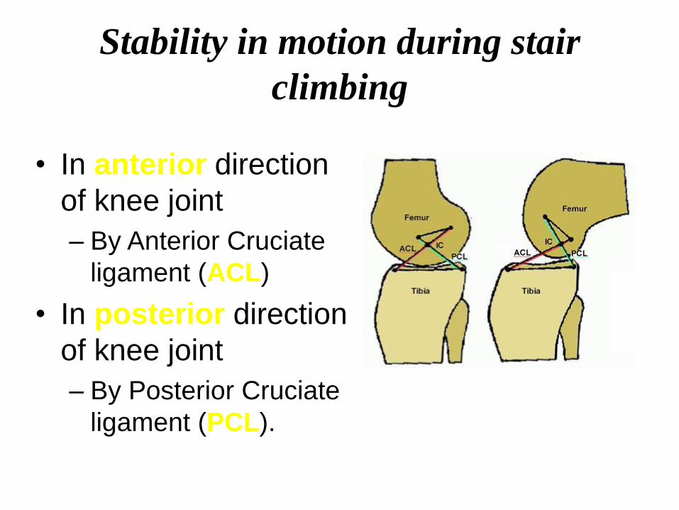
Stability in motion during stair
climbing
• In anterior direction
of knee joint
– By Anterior Cruciate
ligament (ACL)
• In posterior direction
of knee joint
– By Posterior Cruciate
ligament (PCL).

Stability in motion during stair
climbing (cont’d)
• Rotational stability
is due to
– Medial Collateral
Ligament (MCL)
– Lateral Collateral
Ligament (LCL)
• Stability in valgus
– MCL
• Stability in varus
– LCL

Phases in Stair Climbing
• Stance Phase: Weight Acceptance to Foot-off
• Swing Phase: Foot off to foot strike

Joint Moments
• Product of
instantaneous
equivalent muscle
force multiplied by
equivalent lever arm
at a joint
• Instantaneous
estimates of required
strength for doing
motion

Posterior Cruciate Ligament
• Allows femoral condyles to glide and
rotate posteriorly.
Posterior Cruciate Ligament
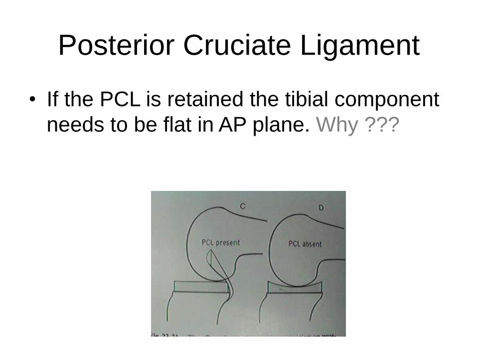
• If the PCL is retained the tibial component
needs to be flat in AP plane. Why ???

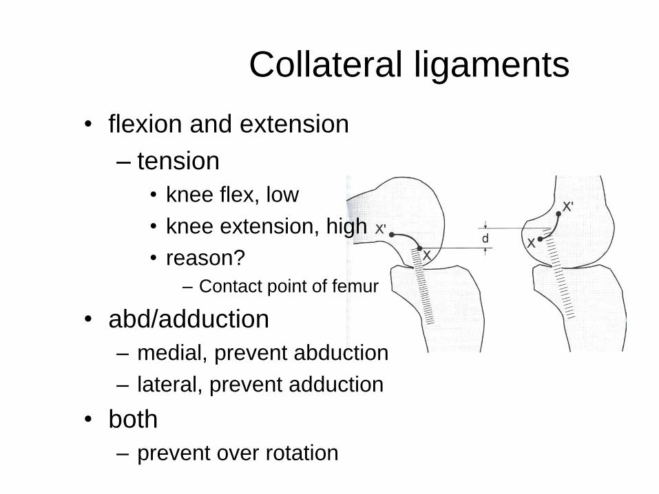
Collateral ligaments
• flexion and extension
– tension
• knee flex, low
• knee extension, high
• reason?
– Contact point of femur
• abd/adduction
– medial, prevent abduction
– lateral, prevent adduction
• both
– prevent over rotation
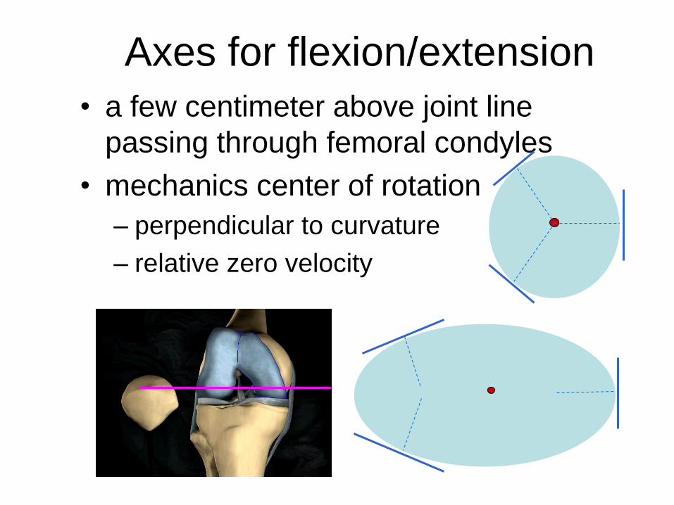
Axes for flexion/extension
• a few centimeter above joint line
passing through femoral condyles
• mechanics center of rotation
– perpendicular to curvature
– relative zero velocity

Methods of defining knee joint axis
– x-rays (perpendicular to curvature)
– instant center of rotation (zero velocity)
– results
• extension 10
• flexion 1
– problems
• devices
– goniometer
– isokinetic dynamometer
• orthotic knee joint

Anatomic basis for
joint motion
• Arthokinematics
– combining rolling and sliding
– flexion to extension
• rolling predominant first
• sliding predominant later
– convex surface rotate on
concave surface
• different direction
– conversely

knee alignment and deformities
• femur-tibial angle
– normal, 170 degrees
– small, genu valgum(knock
knee)
– large, genu varum(bowleg)
• Q-angle
– between femur line and
extended tibial line
– 180 degree - femur-tibial angle
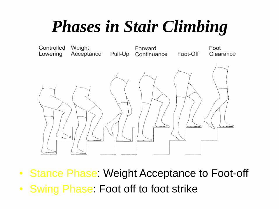
Phases in Stair Climbing
• Stance Phase: Weight Acceptance to Foot-off
• Swing Phase: Foot off to foot strike

Joint Moments
• Product of
instantaneous
equivalent muscle
force multiplied by
equivalent lever arm
at a joint
• Instantaneous
estimates of required
strength for doing
motion

Joint Moments (cont’d)
• Maximum knee flexion angle – During swing phase
• Maximum knee flexion moment – During stance phase
– During descending stairs (49.2° ± 9.5°) was nearly 6 times (external moment) that when ascending stairs (59.5° ± 16.8°)

Crossed four-bar linkage
• Mechanical model
• ACL and PCL as two
crossed bars
• Accounts for
changing roll/glide
ratio with knee flexion
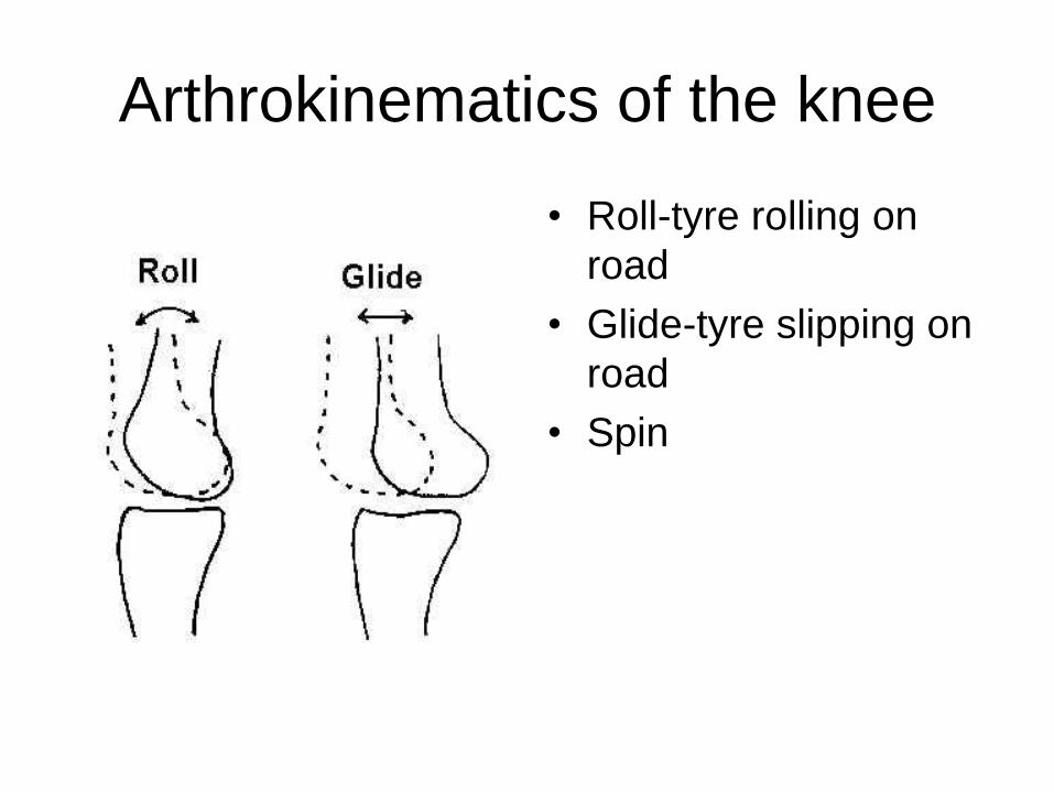
Arthrokinematics of the knee
• Roll-tyre rolling on
road
• Glide-tyre slipping on
road
• Spin

Rule of concavity and convexity
• Roll and glide
simultaneously, in
opposite and same
direction
• Helps prevent
subluxation and
impingement
• Rolling initiates flexion
• Gliding occurs with final
flexion
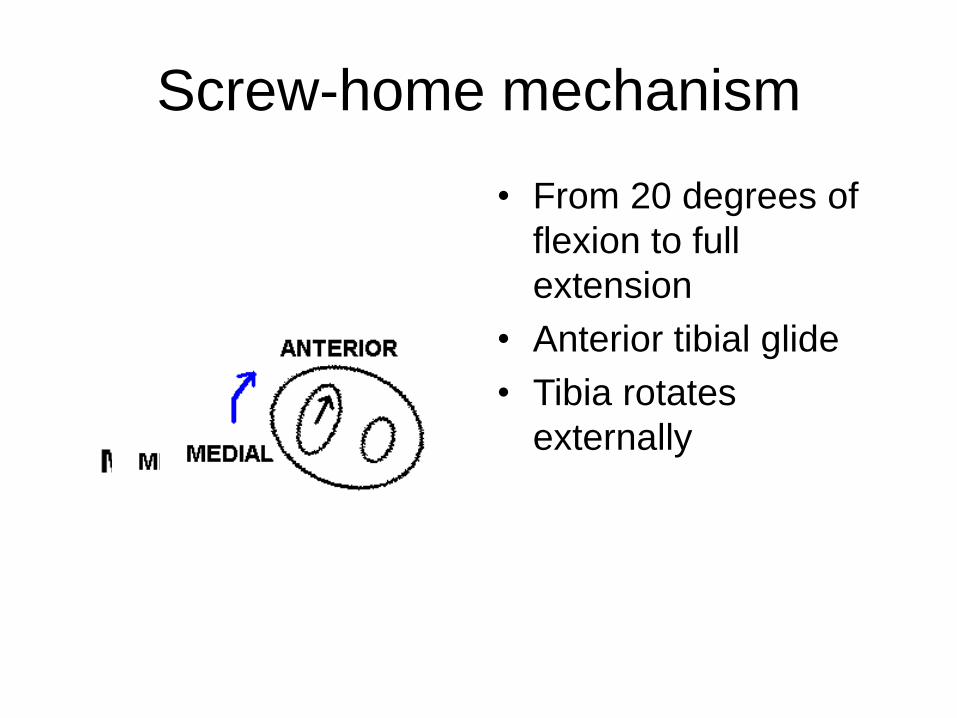
Screw-home mechanism
• From 20 degrees of
flexion to full
extension
• Anterior tibial glide
• Tibia rotates
externally

Screw-home mechanism
• Reverses in knee flexion
• From full extension to 20 degrees of flexion
• Posterior tibial glide
• Internal rotation of tibia

Forces added to the model