zurich open repository and archive year: 2011
TRANSCRIPT
Zurich Open Repository andArchiveUniversity of ZurichMain LibraryStrickhofstrasse 39CH-8057 Zurichwww.zora.uzh.ch
Year: 2011
Chlamydia in canine or feline coronary arteriosclerotic lesions
Sostaric-Zuckermann, I C ; Borel, N ; Kaiser, C ; Grabarevic, Z ; Pospischil, A
Abstract: These findings suggest that there is no obvious correlation between canine and feline coronaryarteriosclerosis and the presence of Chlamydia. In order to draw final conclusions about the correlationbetween Chlamydia and canine atherosclerosis, examination of more samples is required.
DOI: https://doi.org/10.1186/1756-0500-4-350
Posted at the Zurich Open Repository and Archive, University of ZurichZORA URL: https://doi.org/10.5167/uzh-53975Journal ArticlePublished Version
The following work is licensed under a Creative Commons: Attribution 2.0 Generic (CC BY 2.0) License.
Originally published at:Sostaric-Zuckermann, I C; Borel, N; Kaiser, C; Grabarevic, Z; Pospischil, A (2011). Chlamydia in canineor feline coronary arteriosclerotic lesions. BMC Research Notes, 4:350.DOI: https://doi.org/10.1186/1756-0500-4-350
RESEARCH ARTICLE Open Access
Chlamydia in canine or feline coronaryarteriosclerotic lesionsIvan C Sostaric-Zuckermann1, Nicole Borel2*, Carmen Kaiser2, Zeljko Grabarevic1 and Andreas Pospischil2
Abstract
Background: There are numerous reports linking Chlamydia infection to human coronary atherosclerosis. However,there is a lack of data regarding this correlation in dogs and cats, and there are no reports investigating coronaryarteriosclerosis and Chlamydia in these species. The aim of the present study was to examine whether there is acorrelation between canine and feline spontaneous atherosclerosis or arteriosclerosis and the presence ofChlamydia. Archived histopathological samples of dogs (n = 16) and cats (n = 13) with findings of atherosclerosisor arteriosclerosis in heart tissue were examined for the presence of Chlamydiaceae using real-time PCR, ArrayTubeMicroarray and immunohistochemistry. Additionally, arteriosclerotic lesions of all cases were histologically classifiedand graded.
Results: Both canine atherosclerotic cases, and all 14 canine arteriosclerotic cases were negative for Chlamydia.Only one of the 13 arteriosclerotic feline cases was positive for Chlamydia by real-time PCR, revealing C. abortus byArrayTube Microarray. To our knowledge, this is the first description of C. abortus in a cat. Overall, the type andgrade of canine and feline arteriosclerotic lesions revealed similarities, and were predominantly moderate andhyperplastic.
Conclusions: These findings suggest that there is no obvious correlation between canine and feline coronaryarteriosclerosis and the presence of Chlamydia. In order to draw final conclusions about the correlation betweenChlamydia and canine atherosclerosis, examination of more samples is required.
BackgroundTo date, cardiovascular disease [1] is the most prevalent
cause of human mortality. Hardening of the vessels,
generally denominated as “arteriosclerosis”, represents
one of the dominant pathological lesions involved. Most
commonly, the lesion causing this hardening of the ves-
sel walls is a formation of atherosclerotic plaques. This
represents an accumulation of large amounts of lipopro-
teins, predominantly low density lipoproteins (LDLs) in
the arterial wall, commonly called atherosclerosis. The
etiology and pathogenesis of atherosclerosis seems to be
quite complex and is still not completely understood.
However, it was hypothesized that the initial lesion is an
immunologically mediated inflammatory response by the
host, presumably triggered or facilitated by certain infec-
tive agents [2]. The most extensively investigated
infective agents linked with atherosclerosis include: Heli-
cobacter pylori, herpesviruses and Chlamydia (C.) pneu-
moniae [2-5]. Association of C. pneumoniae infection
and atherosclerosis has been established in many reports
using a variety of methods, primarily serology, though
detection of the organism in vascular lesions is also
determined by isolation in culture, polymerase chain
reaction (PCR), electron microscopy, enzyme immu-
noassay, immunohistochemistry, immunocytochemistry,
Tissue Microarray and in situ hybridization [6-13].
Nevertheless, there is lack of concordance when com-
paring methods [14].
Incidence of atherosclerosis in dogs and cats is far less
common than in humans, and is usually a sequela of
other diseases, namely hypothyroidism, diabetes melli-
tus, hypercholesterolemia or hypertriglyceridemia [15].
However, it is still unclear whether atheromatous and/or
any arteriosclerotic lesions in dogs and cats could be
influenced by infectious pathogens, as described in
humans. There is only one report associating chlamydial
* Correspondence: [email protected] for Veterinary Pathology, University of Zurich, Vetsuisse Faculty,Winterthurerstrasse 268, CH-8057 Zurich, SwitzerlandFull list of author information is available at the end of the article
Sostaric-Zuckermann et al. BMC Research Notes 2011, 4:350
http://www.biomedcentral.com/1756-0500/4/350
© 2011 Borel et al; licensee BioMed Central Ltd. This is an open access article distributed under the terms of the Creative CommonsAttribution License (http://creativecommons.org/licenses/by/2.0), which permits unrestricted use, distribution, and reproduction inany medium, provided the original work is properly cited.
infection and atherosclerotic lesions in dogs, but none
for cats [16]. There are no reports linking arteriosclero-
sis as a more widespread and general lesion with chla-
mydial infection. Dogs and cats, as common pets,
usually reach a relatively high age and share the same
living environment with humans. Equally importantly,
they are also potential hosts for chlamydial species.
Therefore it would seem logical to consider the possibi-
lity of chlamydial involvement in these vascular lesions.
The purpose of this study was to try to establish a
correlation between any type of arteriosclerotic changes
in hearts of dogs and cats and chlamydial infection. It is
worth mentioning however, that the initial aim of this
study was to try to establish a correlation between
atherosclerotic lesions and chlamydial infection. Unfor-
tunately, since the number of cases with atherosclerotic
lesions was very low (n = 2), an archive search and
study of cases pertaining to arteriosclerotic lesions was
conducted.
MethodsNumber and selection of cases
A search of the archives of the Institute of Veterinary
Pathology in Zurich was conducted. A computer data-
base was used to find all dog and cat postmortem cases
dating from 1987 until mid-September 2010 having any
description of arteriosclerotic and/or atherosclerotic
lesions in heart tissues. Of the total 7,057 dog and 8,601
cat necropsies, there were 16 canine (0.23%) and 13
(0.15%) feline cases with a previous description of arter-
iosclerosis or atherosclerosis in heart tissue. The mean
age of selected dogs and cats was 10.44 and 9.23 years,
respectively. Of these 16 canine and 13 feline cases, only
paraffin blocks whose hematoxylin and eosin (HE) slides
were described to have some arteriosclerotic or athero-
sclerotic lesion were selected for further investigation.
Preparation of HE slides and microscopic examination
For each selected paraffin block, a corresponding HE
stained slide was prepared according to routine standard
methods. A histopathological examination of all micro-
scopic slides was conducted, and a grade (score) given
of the arteriosclerotic/atherosclerotic lesions for each
heart or aorta. Lesions of coronary arteries in heart tis-
sues were classified into one of the five following grades:
mild, moderate, severe or a combination of contiguous -
e.g. mild to moderate and moderate to severe. Further-
more, using the general categorization and description
of vessel wall hardening lesions, these were described
for each organ as being of atherosclerotic, hyaline (also
described as hypertrophic hyalinization or hyalinosis) or
hyperplastic arteriosclerosis type [17,18]. Finally, the
arterial and/or arteriolar lesions in each heart were
examined for the presence of the following changes:
visible lumen narrowing, vessel wall edema, thrombosis,
and degeneration of leiomyocytes. In the determination
of the previous, only hearts with a minimum of two
arteries and/or arterioles affected with the change were
regarded as positive findings.
DNA extraction
Sections (40 μm) of formalin-fixed and paraffin-
embedded tissue samples were deparaffinized in xylene,
centrifuged at 13,500 × g for 5 min and the xylene was
removed by repeated extraction with ethanol followed
by a second centrifugation and the removal of ethanol.
The pellet was treated overnight with proteinase K on a
thermomixer (55°C, 550 rpm). The DNA was extracted
using the DNeasy Blood and Tissue Kit (Qiagen, Hilden,
Germany) according to the manufacturer’s instructions.
Chlamydiaceae real-time PCR
The extracted DNA of all organ samples (n = 45) was
investigated on an ABI 7500 Fast real-time PCR system
(Applied Biosystems, Foster City, CA, USA) using the
23S-based Chlamydiaceae family-specific real-time PCR
described previously [19]. This method includes primers
Ch23S-F (5’-CTGAAACCAGTAGCTTATAAG CGGT-
3’), Ch23S-R (5’-ACCTCGCCGTTTAACTTAACTCC-
3’), and probe Ch23S-p (FAM-CTCATCA
TGCAAAAGGCACGCCG-TAMRA) and an internal
amplification control consisting of primers EGFP-1-F
(5’-GACCACTACCAGCAGAACAC-3’), EGFP-10-R (3’-
CTTGTACAGCTCGTCCATGC-5’) and probe EGFP-
HEX (HEX-AGCACCCAGTCCGCCCTGAGCA-
BHQ1). A 111-bp product specific for members of the
Chlamydiaceae is also produced as well as a 177-bp
product for the internal amplification control. A final
volume of 25 μl for each tested sample was achieved by
adding 2.5 μl of extracted DNA to 22.5 μl of 2X Taq-
Man® Fast Universal PCR Master Mix (Applied Biosys-
tems) and a final concentration of 500 nM of each
primer and the probe (Microsynth, Balgach, Switzer-
land). The cycling program was started by an initial
denaturation (95°C, 20 sec) followed by 45 cycles of
denaturation and annealing (95°C, 3 sec and 60°C, 30
sec), with an automatically calculated cycle threshold
value. All samples were tested in duplicate. If the dupli-
cates showed a Ct value of < 38, the sample was consid-
ered positive and if the mean Ct value was > 38, it was
considered questionably positive.
ArrayTube (AT) Microarray for species identification of
Chlamydiaceae
Samples that were positive or considered questionably
positive by real-time PCR for Chlamydiaceae were
further examined by the species-specific 23S ArrayTube
(AT) Microarray [20].
Sostaric-Zuckermann et al. BMC Research Notes 2011, 4:350
http://www.biomedcentral.com/1756-0500/4/350
Page 2 of 8
Immunohistochemistry (IHC)
The presence of the chlamydial antigen in paraffin sec-
tions was investigated on cases positive or considered
questionably positive for Chlamydiaceae by real-time
PCR (n = 1) using a Chlamydiaceae family-specific
mouse monoclonal antibody directed against the chla-
mydial lipopolysaccharide (LPS, Clone ACI-P, Progen,
Heidelberg, Germany). A detection kit (Dako, K5003,
Switzerland) was used according to the manufacturer’s
instructions for detection. After deparaffinization in
xylene and rehydration through graded ethanol to water,
the antigen retrieval was performed by 10 min enzyme
digestion (Proteinase K, Dako, S2019, Switzerland).
Endogenous peroxidase activity was inhibited with per-
oxidase-blocking solution for 5 min at room tempera-
ture, then slides were incubated for 60 min with the
primary antibody diluted 1:200 in antibody diluent
(S2022, Dako, Switzerland). The incubation with the
link-antibody and the substrate solution was performed
at RT for 10 min each, after which the slides were
developed in a 3-amino, 9-ethyl-carbazole (AEC) sub-
strate solution for 10 min and counterstained with
hematoxylin. Negative controls of each section were per-
formed using only the antibody diluent instead of the
primary antibody. For the positive control, intestinal tis-
sue from gnotobiotic piglets experimentally infected
with porcine C. suis strain S45 were used [21].
ResultsNumber and selection of cases
Of dogs, 15 animals (93.8%) had recorded lesions only
in the heart (n = 14) or aorta (n = 1), while one animal
(6.2%) showed lesions in the heart and some additional
organs (kidney and lungs). Of cats, seven animals
(53.8%) expressed arteriosclerotic lesions only in the
heart, while the remaining six animals (46.2%) had both
coronary and non-coronary lesions (kidney, eyes, liver,
brain, spleen, stomach).
Microscopic evaluation and grading of the lesions
Dogs
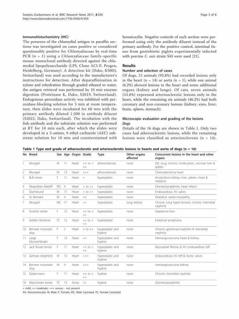
Details of the 16 dogs are shown in Table 1. Only two
cases had atherosclerotic lesions, while the remaining
lesions were classified as arteriosclerosis (n = 14).
Table 1 Type and grade of atherosclerotic and arteriosclerotic lesions in hearts and aorta of dogs (n = 16)
No Breed Sex Age Organ Grade Type Other organsaffected
Concurrent lesions in the heart and otherorgans
1 Mongrel M 11 Heart ++ to +++
atherosclerosis none DIC lung, chronic cholecystitis, necrosis liver &spleen
2 Mongrel M 13 Heart +++ atherosclerosis none Chemodectoma heart
3 Bull terrier F 11 Heart + hyperplastic none Amyloidosis kidney, liver, spleen, heart &intestine
4 Neapolitan Mastiff MC 4 Heart + to ++ hyperplastic none Glomerulonephritis, heart infarct
5 Dachshund M 17 Heart + to ++ hyperplastic none Endocardiosis AV valves
6 St. Bernard M 4 Heart ++ hyperplastic none Dilatative cardio-myopathy
7 Mongrel MC 11 Heart ++ hyperplastic lung, kidney Chronic lung hyper-tension, chronic interstitialnephritis
8 Scottish terrier F 12 Heart ++ to +++
hyperplastic none Hepatoma liver
9 Golden Retriever FC 12 Heart ++ to +++
hyperplastic none Intestinal lymphoma
10 Bernese mountaindog
F 2 Heart + to ++ hyperplastic andhyaline
none Chronic glomerulo-nephritis & interstitialnephritis
11 LargeMünsterländer
F 13 Heart ++ hyperplastic andhyaline
none Hemangiosarcoma heart & kidney
12 Jack Russel terrier F 11 Heart ++ to +++
hyperplastic andhyaline
none Myocardial fibrosis & AV endocardiosis left
13 German shepherd M 13 Heart +++ hyperplastic andhyaline
none endocardiosis AV left & Aortic valves
14 Bernese mountaindog
M 9 Heart +++ hyperplastic andhyaline
none Hemangiosarcoma kidney
15 Dobermann F 11 Heart ++ to +++
hyaline none Chronic interstitial nephritis
16 Manchester terrier FC 13 Aorta ++ hyaline none Glomerulonephritis
+ mild; ++ moderate; +++ severe; - not present
AV, Atrioventricular; M, Male; F, Female; MC, Male Castrated, FC, Female Castrated;
Sostaric-Zuckermann et al. BMC Research Notes 2011, 4:350
http://www.biomedcentral.com/1756-0500/4/350
Page 3 of 8
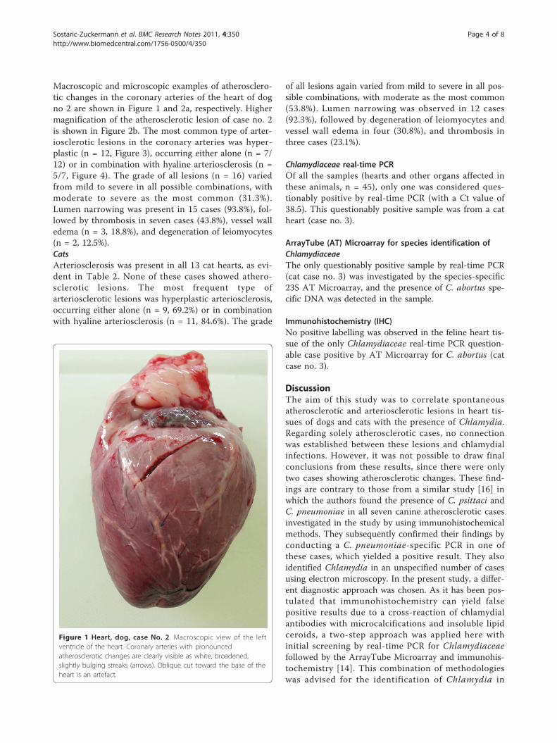
Macroscopic and microscopic examples of atherosclero-
tic changes in the coronary arteries of the heart of dog
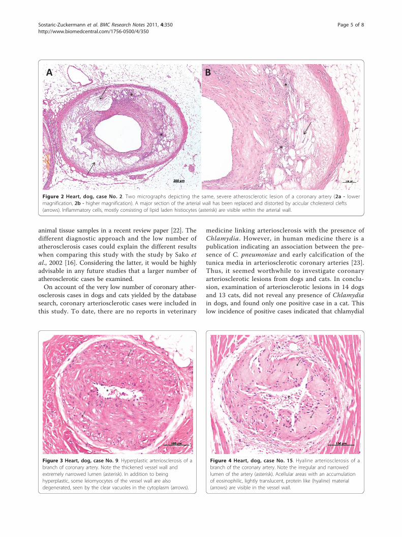
no 2 are shown in Figure 1 and 2a, respectively. Higher
magnification of the atherosclerotic lesion of case no. 2
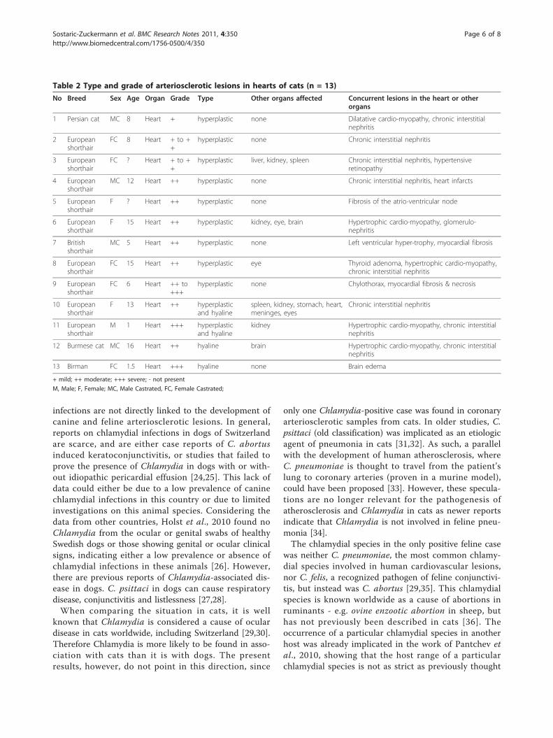
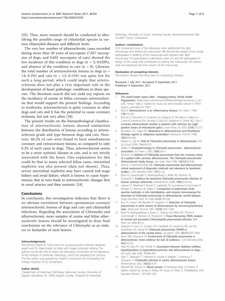
is shown in Figure 2b. The most common type of arter-
iosclerotic lesions in the coronary arteries was hyper-
plastic (n = 12, Figure 3), occurring either alone (n = 7/
12) or in combination with hyaline arteriosclerosis (n =
5/7, Figure 4). The grade of all lesions (n = 16) varied
from mild to severe in all possible combinations, with
moderate to severe as the most common (31.3%).
Lumen narrowing was present in 15 cases (93.8%), fol-
lowed by thrombosis in seven cases (43.8%), vessel wall
edema (n = 3, 18.8%), and degeneration of leiomyocytes
(n = 2, 12.5%).
Cats
Arteriosclerosis was present in all 13 cat hearts, as evi-
dent in Table 2. None of these cases showed athero-
sclerotic lesions. The most frequent type of
arteriosclerotic lesions was hyperplastic arteriosclerosis,
occurring either alone (n = 9, 69.2%) or in combination
with hyaline arteriosclerosis (n = 11, 84.6%). The grade
of all lesions again varied from mild to severe in all pos-
sible combinations, with moderate as the most common
(53.8%). Lumen narrowing was observed in 12 cases
(92.3%), followed by degeneration of leiomyocytes and
vessel wall edema in four (30.8%), and thrombosis in
three cases (23.1%).
Chlamydiaceae real-time PCR
Of all the samples (hearts and other organs affected in
these animals, n = 45), only one was considered ques-
tionably positive by real-time PCR (with a Ct value of
38.5). This questionably positive sample was from a cat
heart (case no. 3).
ArrayTube (AT) Microarray for species identification of
Chlamydiaceae
The only questionably positive sample by real-time PCR
(cat case no. 3) was investigated by the species-specific
23S AT Microarray, and the presence of C. abortus spe-
cific DNA was detected in the sample.
Immunohistochemistry (IHC)
No positive labelling was observed in the feline heart tis-
sue of the only Chlamydiaceae real-time PCR question-
able case positive by AT Microarray for C. abortus (cat
case no. 3).
DiscussionThe aim of this study was to correlate spontaneous
atherosclerotic and arteriosclerotic lesions in heart tis-
sues of dogs and cats with the presence of Chlamydia.
Regarding solely atherosclerotic cases, no connection
was established between these lesions and chlamydial
infections. However, it was not possible to draw final
conclusions from these results, since there were only
two cases showing atherosclerotic changes. These find-
ings are contrary to those from a similar study [16] in
which the authors found the presence of C. psittaci and
C. pneumoniae in all seven canine atherosclerotic cases
investigated in the study by using immunohistochemical
methods. They subsequently confirmed their findings by
conducting a C. pneumoniae-specific PCR in one of
these cases, which yielded a positive result. They also
identified Chlamydia in an unspecified number of cases
using electron microscopy. In the present study, a differ-
ent diagnostic approach was chosen. As it has been pos-
tulated that immunohistochemistry can yield false
positive results due to a cross-reaction of chlamydial
antibodies with microcalcifications and insoluble lipid
ceroids, a two-step approach was applied here with
initial screening by real-time PCR for Chlamydiaceae
followed by the ArrayTube Microarray and immunohis-
tochemistry [14]. This combination of methodologies
was advised for the identification of Chlamydia in
Figure 1 Heart, dog, case No. 2. Macroscopic view of the leftventricle of the heart. Coronary arteries with pronouncedatherosclerotic changes are clearly visible as white, broadened,slightly bulging streaks (arrows). Oblique cut toward the base of theheart is an artefact.
Sostaric-Zuckermann et al. BMC Research Notes 2011, 4:350
http://www.biomedcentral.com/1756-0500/4/350
Page 4 of 8
animal tissue samples in a recent review paper [22]. The
different diagnostic approach and the low number of
atherosclerosis cases could explain the different results
when comparing this study with the study by Sako et
al., 2002 [16]. Considering the latter, it would be highly
advisable in any future studies that a larger number of
atherosclerotic cases be examined.
On account of the very low number of coronary ather-
osclerosis cases in dogs and cats yielded by the database
search, coronary arteriosclerotic cases were included in
this study. To date, there are no reports in veterinary
medicine linking arteriosclerosis with the presence of
Chlamydia. However, in human medicine there is a
publication indicating an association between the pre-
sence of C. pneumoniae and early calcification of the
tunica media in arteriosclerotic coronary arteries [23].
Thus, it seemed worthwhile to investigate coronary
arteriosclerotic lesions from dogs and cats. In conclu-
sion, examination of arteriosclerotic lesions in 14 dogs
and 13 cats, did not reveal any presence of Chlamydia
in dogs, and found only one positive case in a cat. This
low incidence of positive cases indicated that chlamydial
A
*
*
*
*
B
Figure 2 Heart, dog, case No. 2. Two micrographs depicting the same, severe atherosclerotic lesion of a coronary artery (2a - lowermagnification, 2b - higher magnification). A major section of the arterial wall has been replaced and distorted by acicular cholesterol clefts(arrows). Inflammatory cells, mostly consisting of lipid laden histiocytes (asterisk) are visible within the arterial wall.
*
Figure 3 Heart, dog, case No. 9. Hyperplastic arteriosclerosis of abranch of coronary artery. Note the thickened vessel wall andextremely narrowed lumen (asterisk). In addition to beinghyperplastic, some leiomyocytes of the vessel wall are alsodegenerated, seen by the clear vacuoles in the cytoplasm (arrows).
*
Figure 4 Heart, dog, case No. 15. Hyaline arteriosclerosis of abranch of the coronary artery. Note the irregular and narrowedlumen of the artery (asterisk). Acellular areas with an accumulationof eosinophilic, lightly translucent, protein like (hyaline) material(arrows) are visible in the vessel wall.
Sostaric-Zuckermann et al. BMC Research Notes 2011, 4:350
http://www.biomedcentral.com/1756-0500/4/350
Page 5 of 8
infections are not directly linked to the development of
canine and feline arteriosclerotic lesions. In general,
reports on chlamydial infections in dogs of Switzerland
are scarce, and are either case reports of C. abortus
induced keratoconjunctivitis, or studies that failed to
prove the presence of Chlamydia in dogs with or with-
out idiopathic pericardial effusion [24,25]. This lack of
data could either be due to a low prevalence of canine
chlamydial infections in this country or due to limited
investigations on this animal species. Considering the
data from other countries, Holst et al., 2010 found no
Chlamydia from the ocular or genital swabs of healthy
Swedish dogs or those showing genital or ocular clinical
signs, indicating either a low prevalence or absence of
chlamydial infections in these animals [26]. However,
there are previous reports of Chlamydia-associated dis-
ease in dogs. C. psittaci in dogs can cause respiratory
disease, conjunctivitis and listlessness [27,28].
When comparing the situation in cats, it is well
known that Chlamydia is considered a cause of ocular
disease in cats worldwide, including Switzerland [29,30].
Therefore Chlamydia is more likely to be found in asso-
ciation with cats than it is with dogs. The present
results, however, do not point in this direction, since
only one Chlamydia-positive case was found in coronary
arteriosclerotic samples from cats. In older studies, C.
psittaci (old classification) was implicated as an etiologic
agent of pneumonia in cats [31,32]. As such, a parallel
with the development of human atherosclerosis, where
C. pneumoniae is thought to travel from the patient’s
lung to coronary arteries (proven in a murine model),
could have been proposed [33]. However, these specula-
tions are no longer relevant for the pathogenesis of
atherosclerosis and Chlamydia in cats as newer reports
indicate that Chlamydia is not involved in feline pneu-
monia [34].
The chlamydial species in the only positive feline case
was neither C. pneumoniae, the most common chlamy-
dial species involved in human cardiovascular lesions,
nor C. felis, a recognized pathogen of feline conjunctivi-
tis, but instead was C. abortus [29,35]. This chlamydial
species is known worldwide as a cause of abortions in
ruminants - e.g. ovine enzootic abortion in sheep, but
has not previously been described in cats [36]. The
occurrence of a particular chlamydial species in another
host was already implicated in the work of Pantchev et
al., 2010, showing that the host range of a particular
chlamydial species is not as strict as previously thought
Table 2 Type and grade of arteriosclerotic lesions in hearts of cats (n = 13)
No Breed Sex Age Organ Grade Type Other organs affected Concurrent lesions in the heart or otherorgans
1 Persian cat MC 8 Heart + hyperplastic none Dilatative cardio-myopathy, chronic interstitialnephritis
2 Europeanshorthair
FC 8 Heart + to ++
hyperplastic none Chronic interstitial nephritis
3 Europeanshorthair
FC ? Heart + to ++
hyperplastic liver, kidney, spleen Chronic interstitial nephritis, hypertensiveretinopathy
4 Europeanshorthair
MC 12 Heart ++ hyperplastic none Chronic interstitial nephritis, heart infarcts
5 Europeanshorthair
F ? Heart ++ hyperplastic none Fibrosis of the atrio-ventricular node
6 Europeanshorthair
F 15 Heart ++ hyperplastic kidney, eye, brain Hypertrophic cardio-myopathy, glomerulo-nephritis
7 Britishshorthair
MC 5 Heart ++ hyperplastic none Left ventricular hyper-trophy, myocardial fibrosis
8 Europeanshorthair
FC 15 Heart ++ hyperplastic eye Thyroid adenoma, hypertrophic cardio-myopathy,chronic interstitial nephritis
9 Europeanshorthair
FC 6 Heart ++ to+++
hyperplastic none Chylothorax, myocardial fibrosis & necrosis
10 Europeanshorthair
F 13 Heart ++ hyperplasticand hyaline
spleen, kidney, stomach, heart,meninges, eyes
Chronic interstitial nephritis
11 Europeanshorthair
M 1 Heart +++ hyperplasticand hyaline
kidney Hypertrophic cardio-myopathy, chronic interstitialnephritis
12 Burmese cat MC 16 Heart ++ hyaline brain Hypertrophic cardio-myopathy, chronic interstitialnephritis
13 Birman FC 1.5 Heart +++ hyaline none Brain edema
+ mild; ++ moderate; +++ severe; - not present
M, Male; F, Female; MC, Male Castrated, FC, Female Castrated;
Sostaric-Zuckermann et al. BMC Research Notes 2011, 4:350
http://www.biomedcentral.com/1756-0500/4/350
Page 6 of 8
[35]. Thus, more research should be conducted in iden-
tifying the possible range of chlamydial species in var-
ious chlamydial diseases and different hosts.
The very low number of atherosclerotic cases recorded
during more than 20 years of necropsies (7,057 necrop-
sies of dogs, and 8,601 necropsies of cats) showed the
low incidence of this condition in dogs (n = 2; 0.028%),
and absence of the condition in cats (n = 0). Likewise,
the total number of arteriosclerotic lesions in dogs (n =
14; 0.2%) and cats (n = 13; 0.15%) was quite low for
such a long period, which could imply that arterio-
sclerosis does not play a very important role in the
development of heart pathologic conditions in these spe-
cies. The literature search did not yield any reports on
the incidence of canine or feline coronary arteriosclero-
sis that would support the present findings. According
to textbooks, arteriosclerosis is quite common in older
dogs and cats and it has the potential to cause coronary
ischemia, but not very often [18].
The present results on the histopathological classifica-
tion of arteriosclerotic lesions showed similarities
between the distribution of lesions according to arterio-
sclerosis grade and type between dogs and cats. How-
ever, 46.2% of cats were found to have simultaneous
coronary and extracoronary lesions, as compared to only
6.2% of such cases in dogs. Thus, arteriosclerosis seems
to be a more systemic disease in cats, and is not strictly
associated with the heart. One explanation for this
could be that in many selected feline cases, interstitial
nephritis was also present (data not shown). Thus,
severe interstitial nephritis may have caused end stage
kidney and renal failure, which is known to cause hyper-
tension that in turn leads to arteriosclerotic changes first
in renal arteries and then systemic [18].
ConclusionsIn conclusion, this investigation indicates that there is
no obvious correlation between spontaneous coronary
arteriosclerotic lesions of dogs and cats and chlamydial
infections. Regarding the association of Chlamydia and
atherosclerosis, more samples of canine and feline ather-
osclerotic lesions should be investigated to draw final
conclusions on the relevance of Chlamydia as an indu-
cer or bystander of such lesions.
Acknowledgements
The authors thank Dr. Paula Grest for conducting the Institute’s databasesearch and Dr. Maja Rutten for help with image computer editing. Theauthors would also like to thank the laboratory technicians of the laboratoryof the Institute of Veterinary Pathology, Zurich for preparing the sections.The first author was granted by Federal Commission for Scholarships forForeign Students (FCS), Switzerland.
Author details1Department of Veterinary Pathology, Veterinary Faculty, University ofZagreb, Heinzelova 55, 10000 Zagreb, Croatia. 2Institute for Veterinary
Pathology, University of Zurich, Vetsuisse Faculty, Winterthurerstrasse 268,CH-8057 Zurich, Switzerland.
Authors’ contributions
ICSZ conducted most of the laboratory work, performed the lightmicroscopy and drafted the manuscript. NB devised the design of the study,participated in drafting of the manuscript and assisted with lightmicroscopy. CK participated in laboratory work. ZG and AP participated indesign of the study and contributed to writing the manuscript. All authorsread and approved the final version of the manuscript.
Declaration of competing interests
The authors declare that they have no competing interests.
Received: 7 July 2011 Accepted: 9 September 2011
Published: 9 September 2011
References
1. The world health report 2004 - changing history, World Health
Organization. [http://www.who.int/whr/2004/annex/topic/en/annex_2_en.pdf], “Annex Table 2: Deaths by cause, sex and mortality stratum in WHOregions, estimates for 2002”.
2. Ross R: Atherosclerosis is an inflammatory disease. Am Heart J 1999,138:419-420.
3. Niccoli G, Franceschi F, Cosentino N, Giupponi B, De Marco G, Merra G,Conte M, Montone RA, Ferrante G, Bacà M, Gasbarrini A, Silveri NG, Crea F:Coronary atherosclerotic burden in patients with infection by CagA-
positive strains of Helicobacter pylori. Coron Artery Dis 2010, 21(4):217-221.4. Nicholson AC, Hajjar DP: Herpesvirus in atherosclerosis and thrombosis:
etiologic agents or ubiquitous bystanders? Arterioscler Thromb 1998,18(3):339-348.
5. Watson C, Alp NJ: Role of Chlamydia pneumoniae in atherosclerosis. Clin
Sci (Lond) 2008, 114:509-531.6. Saikku P: Seroepidemiology in Chlamydia pneumoniae - atherosclerosis
association. Eur Heart J 2002, 23(4):263-4.7. Ramirez JA: Isolation of Chlamydia pneumoniae from the coronary artery
of a patient with coronary atherosclerosis. The Chlamydia pneumoniae/
Atherosclerosis Study Group. Ann Intern Med 1996, 125(12):979-82.8. Boman J, Hammerschlag MR: Chlamydia pneumoniae and atherosclerosis:
critical assessment of diagnostic methods and relevance to treatment
studies. J Clin Microbiol 2002, 15(1):1-20.9. Borel N, Summersgill JT, Mukhopadhyay S, Miller RD, Ramirez JA,
Pospischil A: Evidence for persistent Chlamydia pneumoniae infection of
human coronary atheromas. Atherosclerosis 2008, 199(1):154-161.10. Lajunen T, Vikatmaa P, Ikonen T, Lepäntalo M, Lounatmaa K, Sormunen R,
Rantala A, Leinonen M, Saikku P: Comparison of polymerase chain
reaction methods, in situ hybridization, and enzyme immunoassay for
detection of Chlamydia pneumoniae in atherosclerotic carotid plaques.
Diagn Microbiol Infect Dis 2008, 61(2):156-164.11. Kuo CC, Gown AM, Benditt EP, Grayston JT: Detection of Chlamydia
pneumoniae in aortic lesions of atherosclerosis by immunocytochemical
stain. Arterioscler Thromb 1993, 13(10):1501-1504.12. Borel N, Mukhopadhyay S, Kaiser C, Sullivan ED, Miller RD, Timms P,
Summersgill JT, Ramirez JA, Pospischil A: Tissue Microarray (TMA) analysis
of normal and persistent Chlamydophila pneumoniae infection. BMC
Infect Dis 2006, 6:152.13. Grayston JT, Kuo CC, Coulson AS, Campbell LA, Lawrence RD, Lee MJ,
Strandness ED, Wang SP: Chlamydia pneumoniae (TWAR) in
atherosclerosis of the carotid artery. Circulation 1995, 92(12):3397-3400.14. Ieven MM, Hoymans VY: Involvement of Chlamydia pneumoniae in
atherosclerosis: more evidence for lack of evidence. J Clin Microbiol 2005,43(1):19-24.
15. Hess RS, Kass PH, Van Winkle TJ: Association between diabetes mellitus,
hypothyroidism or hyperadrenocorticism, and atherosclerosis in dogs. J
Vet Intern Med 2003, 17:489-494.16. Sako T, Takahashi T, Takehana K, Uchida E, Nakade T, Umemura T,
Taniyama H: Chlamydial infection in canine atherosclerotic lesions.
Atherosclerosis 2002, 162(2):253-9.17. Mitchell RN, Schoen FJ: Blood vessels. In Pathologic Basis of Disease.. 8
edition. Edited by: Kumar V, Abbas AK, Fausto N, Aster JC. Philadelphia, USA:Saunders Elsevier; 2010:487-528.
Sostaric-Zuckermann et al. BMC Research Notes 2011, 4:350
http://www.biomedcentral.com/1756-0500/4/350
Page 7 of 8
18. Grant Maxie M, Robinson WF: Cardiovascular system. In Jubb, Kennedy, and
Palmer’s Pathology of Domestic Animals. Volume 3.. 5 edition. Edited by:Grant Maxie M. Philadelphia, USA: Saunders Elsevier; 2007:1-105.
19. Ehricht R, Slickers P, Goellner S, Hotzel H, Sachse K: Optimized DNA
Microarray assay allows detection and genotyping of single PCR-
amplifiable target copies. Mol Cell Probes 2006, 20:60-63.20. Borel N, Kempf E, Hotzel H, Schubert E, Torgerson P, Slickers P, Ehricht R,
Tasara T, Pospischil A, Sachse K: Direct identification of chlamydiae from
clinical samples using a DNA Microarray assay - A validation study. Mol
Cell Probes 2008, 22:55-64.21. Guscetti F, Hoop R, Schiller I, Corboz L, Sydler T, Pospischill A: Experimental
enteric infection of gnotobiotic piglets with Chlamydia psittaci strain of
avian origin. J Vet Med B 2000, 47:561-572.22. Sachse K, Vretou E, Livingstone M, Borel N, Pospischil A, Longbottom D:
Recent developments in the laboratory diagnosis of chlamydial
infections. Vet Microbiol 2009, 135(1-2):2-21.23. Bobryshev YV, Lord RS, Tran D: Chlamydia pneumoniae in foci of “early”
calcification of the tunica media in arteriosclerotic arteries: an incidental
presence? Am J Physiol Heart Circ Physiol 2006, 290(4):1510-1519.24. Hoelzle K, Wittenbrink MM, Corboz L, Hoelzle LE: Chlamydophila abortus-
induced keratoconjunctivitis in a dog. Vet Rec 2005, 157(20):632-633.25. Zini E, Glaus TM, Bussadori C, Borgarelli M, Santilli RA, Tarducci A,
Margiocco ML, Rampazzo A, Meli ML, Maisch B, Pankuweit S: Evaluation of
the presence of selected viral and bacterial nucleic acids in pericardial
samples from dogs with or without idiopathic pericardial effusion. Vet J
2009, 179(2):225-229.26. Holst BS, HanÍs S, Bölske G, Forsberg CL: An investigation on the presence
of Chlamydiaceae in Swedish dogs. Acta Vet Scand 2010, 16:52-63.27. Sprague LD, Schubert E, Hotzel H, Scharf S, Sachse K: The detection of
Chlamydophila psittaci genotype C infection in dogs. Vet J 2009,181(3):274-279.
28. Gresham AC, Dixon CE, Bevan BJ: Domiciliary outbreak of psittacosis in
dogs: potential for zoonotic infection. Vet Rec 1996, 138(25):622-623.29. Sykes JE: Feline chlamydiosis. Clin Tech Small Anim Pract 2005,
20(2):129-34.30. von Bomhard W, Polkinghorne A, Lu ZH, Vaughan L, Vögtlin A,
Zimmermann DR, Spiess B, Pospischil A: Detection of novel chlamydiae in
cats with ocular disease. Am J Vet Res 2003, 64(11):1421-1428.31. Baker JA: A virus causing pneumonia in cats and producing elementary
bodies. J Exp Med 1944, 79:159-72.32. Hoover EA, Kahn DE, Langloss JM: Experimentally induced feline
chlamydial infection (feline pneumonitis). Am J Vet Res 1978, 39:541-547.33. Yang ZP, Kuo CC, Grayston JT: Systemic dissemination of Chlamydia
pneumoniae following intranasal inoculation in mice. J Infect Dis 1995,171(3):736-738.
34. Bart M, Guscetti F, Zurbriggen A, Pospischil A, Schiller I: Feline Infectious
Pneumonia: A Short Literature Review and a Retrospective
Immunohistological Study on the Involvement of Chlamydia spp. and
Distemper Virus. Vet J 2000, 159(3):220-230.35. Pantchev A, Sting R, Bauerfeind R, Tyczka J, Sachse K: Detection of all
Chlamydophila and Chlamydia spp. of veterinary interest using species-
specific real-time PCR assays. Comp Immunol Microbiol Infect Dis 2010,33(6):473-84.
36. Longbottom D, Coulter LJ: Animal chlamydioses and zoonotic
implications. J Comp Pathol 2003, 128(4):217-244.
doi:10.1186/1756-0500-4-350Cite this article as: Sostaric-Zuckermann et al.: Chlamydia in canine orfeline coronary arteriosclerotic lesions. BMC Research Notes 2011 4:350. Submit your next manuscript to BioMed Central
and take full advantage of:
• Convenient online submission
• Thorough peer review
• No space constraints or color figure charges
• Immediate publication on acceptance
• Inclusion in PubMed, CAS, Scopus and Google Scholar
• Research which is freely available for redistribution
Submit your manuscript at www.biomedcentral.com/submit
Sostaric-Zuckermann et al. BMC Research Notes 2011, 4:350
http://www.biomedcentral.com/1756-0500/4/350
Page 8 of 8