zootaxa - fs.fed.us · se presenta una lista de plantas hospederas registradas para la súbfamilia...
TRANSCRIPT

Accepted by J. Brown: 3 Feb. 2010; published: 14 May 2010
ZOOTAXAISSN 1175-5326 (print edition)
ISSN 1175-5334 (online edition)Copyright © 2010 · Magnolia Press
Zootaxa 2466: 1–74 (2010) www.mapress.com/zootaxa/ Monograph
ZOOTAXA
Description of the immature stages and life history of Euselasia (Lepidoptera: Riodinidae) on Miconia (Melastomataceae) in
Costa Rica
KENJI NISHIDAEscuela de Biología, Universidad de Costa Rica, 2060 San José, Costa Rica.
E-mail: [email protected]; [email protected]
Magnolia PressAuckland, New Zealand
2466

NISHIDA2 · Zootaxa 2466 © 2010 Magnolia Press
KENJI NISHIDADescription of the immature stages and life history of Euselasia (Lepidoptera: Riodinidae) on Miconia (Melastomataceae) in Costa Rica(Zootaxa 2466)
74 pp.; 30 cm.
14 May 2010
ISBN 978-1-86977-521-6 (paperback)
ISBN 978-1-86977-522-3 (Online edition)
FIRST PUBLISHED IN 2010 BY
Magnolia Press
P.O. Box 41-383
Auckland 1346
New Zealand
e-mail: [email protected]
http://www.mapress.com/zootaxa/
© 2010 Magnolia Press
All rights reserved.
No part of this publication may be reproduced, stored, transmitted or disseminated, in any form, or by any
means, without prior written permission from the publisher, to whom all requests to reproduce copyright
material should be directed in writing.
This authorization does not extend to any other kind of copying, by any means, in any form, and for any purpose
other than private research use.
ISSN 1175-5326 (Print edition)
ISSN 1175-5334 (Online edition)

Zootaxa 2466 © 2010 Magnolia Press · 3 IMMATURE STAGES OF RIODINID BUTTERFLIES
Table of contents
Abstract ............................................................................................................................................................................... 4Resumen .............................................................................................................................................................................. 4Introduction ......................................................................................................................................................................... 5Materials and methods ........................................................................................................................................................ 6Results ............................................................................................................................................................................... 14Life history of Euselasia chrysippe .................................................................................................................................. 14Life history of Euselasia bettina ...................................................................................................................................... 23Third species of Miconia-feeding Euselasia ..................................................................................................................... 24Diagnosis and description of early stages of Euselasia chrysippe .................................................................................... 26Diagnosis and description of early stages of Euselasia bettina ....................................................................................... 44Diagnosis and description of adult of Euselasia chrysippe .............................................................................................. 55Diagnosis and description of adult of Euselasia bettina ................................................................................................... 56Discussion ......................................................................................................................................................................... 58Acknowledgments ............................................................................................................................................................. 67References ......................................................................................................................................................................... 69

NISHIDA4 · Zootaxa 2466 © 2010 Magnolia Press
Abstract
The immature stages and life histories of Euselasia chrysippe (Bates, 1866) and E. bettina (Hewitson, 1869) aredescribed, providing the first detailed morphological characters for the subfamily Euselasiinae. The larvae of Euselasiachrysippe and E. bettina specialize on several species of Miconia (Melastomataceae). The eggs are stalked (the firstreported case of such in Lepidoptera) and laid in clusters on the underside of leaves. The larvae are gregarious and feed,rest, molt, and pupate ‘synchronously’. Both species have six larval instars which exhibit processionary behaviorthroughout their development. SD2 setae on the prothoracic shield are sensitive to airborne vibrations and are related tothe head-flicking behavior exhibited by larvae while feeding, perhaps as a defensive strategy to deter attacks byparasitoids. Several morphological characters of first instar larvae are unique among Lepidoptera: extra setae, bifurcateddorsal setae on A1–8, and various organs. Sixth instar larvae possess subcircular plates on the dorsolateral surfaces of allsegments of the thorax and abdomen. These smooth plates have a metallic-blue iridescence that is structural in nature.Pupation occurred singly or gregariously under laboratory conditions. The total duration of the life cycle underlaboratory conditions lasted up to eight weeks. New records of parasitoids for Euselasia include Encarsia and Telenomusfrom eggs. A list of host plants recorded for Euselasiinae, a summary of parasitoid records for Euselasia, and summarytables of unique organs and setae of immature stages are provided. Euselasia chrysippe and E. bettina are currentlyconsidered potential biocontrol agents for Miconia calvescens DC in Hawaii.
Key words: auditory mechanoreceptor, biological control of weeds, metalmark butterflies, Calolydella, Corrachia,Encarsia, Euselasia aurantia, E. bettina, E. chrysippe, gregarious, host plants, Miconia calvescens, Nemeobiinae, Neo-tropical, processionary behavior, structural color, Telenomus
Resumen
Se describe en detalle las historias naturales y los estadios inmaduros de Euselasia chrysippe (Bates, 1866) y E. bettina(Hewitson, 1869), brindando por primera vez caracteres morfológicos en detalle para la subfamilia Euselasiinae. Laslarvas se especializan en especies de Miconia (Melastomataceae). Los huevos son sostenidos por un pedúnculo (elprimer caso que se encuentra de esta condición en Lepidóptera) y son puestos en grupos en el lado inferior de las hojas.Las larvas son gregarias y se alimentan, descansan, mudan y pupan ‘sincronizadamente’. Ambas especies pasan por seisestadios larvales y muestran comportamiento procesionario a través de su desarrollo. Las setas SD2 en la placa delprotórax son sensibles a vibraciones del aire, y están relacionadas con el comportamiento de tiritar de cabeza de laslarvas mientras se alimentan, probablemente parte de una estrategia defensiva para detener ataques de parasitoides. Loscaracteres morfológicos de la larva en el primer estadio son únicos entre Lepidoptera por tener un número extra de setas,setas dorsales bifurcadas en A1–8 y varios otros órganos. La larva del sexto estadio posee placas sub-circulares en lasuperficie dorso-lateral de todos los segmentos del tórax y abdomen. Estas placas lizas producen una iridiscencia azulmetálica que es estructural en la naturaleza. La pupación se dio individualmente o de forma gregaria. La duración totaldel ciclo de vida bajo condiciones de laboratorio se extendió hasta ocho semanas. Nuevos registros de parasitoides paraEuselasia incluyen Encarsia y Telenomus a partir de huevos. Se presenta una lista de plantas hospederas registradas parala súbfamilia Euselasiinae, un resumen de parasitoides registrados para Euselasia y cuadros resumidos de órganos y setasde estadios inmaduros. Estos dos riodínidos son actualmente considerados como potenciales agentes de control biológicopara Miconia calvescens DC. en Hawai.
Palabras clave: Calolydella, color estructural, comportamiento de procesión, control biológico de malezas, Encarsia,Euselasia aurantia, E. bettina, E. chrysippe, gregario, mariposas diurnas, plantas hospederas, mecanoreceptor auditorio,Miconia calvescens, neotrópico, Nemeobiinae, Telenomus

Zootaxa 2466 © 2010 Magnolia Press · 5 IMMATURE STAGES OF RIODINID BUTTERFLIES
Introduction
For more than 50 years Miconia calvescens DC. (Melastomataceae) has had a negative impact on the nativeecosystems of several oceanic islands (Vitousek et al. 1997). It is one of the most invasive plants in the Pacificand has been considered one of the world’s most threatening noxious weeds since 1992 (Conant et al. 1997;ISSG 2005; PIER 1999–2006). Loope & Helweg (2004) report that more than $1,000,000 US is spent yearlyto protect native biodiversity on the island of Maui alone. In addition to mechanical and chemical controls,biological control of M. calvescens is important and necessary (Medeiros & Loope 1997). During a search forbiological control agents against M. calvescens in Costa Rica, where the plant is native, immature stages oftwo species of Euselasia Hübner, [1819] (Lepidoptera: Riodinidae: Euselasiinae) were found: E. chrysippe(Bates, 1866) and E. bettina (Hewitson, 1869). They were targeted as possible biocontrol agents for M. calve-scens, and thus their biologies were studied in detail.
The subfamily Euselasiinae Kirby 1871 (1867) is restricted to the Neotropics and contains three tribes:Stygini, Corrachiini, and Euselasiini. Stygini and Corrachiini are each monotypic, consisting of Styx infernalisStaudinger, 1876 and Corrachia leucoplaga Schaus, 1913, respectively (Callaghan & Lamas 2004). The tribeEuselasiini contains three genera: Methone Doubleday, Hades Westwood, and Euselasia. The first two generacontain only one (M. cecillia (Cramer, 1777)) and two species (H. noctula Westwood, 1851; H. hecamodeHewitson, 1870), respectively (Callaghan & Lamas 2004). In contrast, Euselasia contains approximately 170described species (Stichel 1930–1931; Hall & Willmott 1998; Callaghan & Lamas 2004; Hall & Willmott2009) making it the largest genus in the family. The closely related subfamily Nemeobiinae is found in theOld World (Oates & Emmet 1990; Corbet & Pendlebury 1992; Samson et al. 1999; Braby 2000, 2004;Wahlberg 2005).
Despite the large number of species, the biology and immature stages of the genus Euselasia are poorlyknown (D.J. Harvey & J.P.W. Hall, pers. comm. 2006). Although the life history of some pest species onEucalyptus and other myrtaceous species are relatively well studied, most information comes from brief notesin literature published over the past 200 years (see Table 1 for references). Rearing data of ten species ofEuselasia and Hades noctula from northwestern Costa Rica are provided by Janzen & Hallwachs (2009).
All of the records above, including that of Stygini (Lamas 2003) and Corrachiini (K. Nishida, in prep.)indicate that larvae are gregarious and most likely processionary, social (Costa & Fitzgerald 1996, 2005;Fitzgerald 2005a; Costa 2006). The morphology of the early stages of Euselasia has been studied by Harvey(1987a, 1987b, 1989) and eggs of E. hieronymi are illustrated and described by Downey and Allyn (1980).Demography of E. chrysippe on M. calvescens was studied by Allen (2007).
Species of Euselasia occur between southwestern North America (Mexico) and the tropical regions ofSouth America (Northern Argentina) with greatest diversity in the Amazon Basin (DeVries 1997; Funet2005–2007). Many new Euselasia species have been discovered recently (Hall & Willmott 1998; Hall &Lamas 2001; Hall & Willmott 2009; Janzen & Hallwachs 2009; I. Nakamura, pers. comm. 2006), withapproximately 30 species recorded from Costa Rica (DeVries 1997; Janzen & Hallwachs 2009). Most speciesare restricted to specific habitats and hence are rare in collections (DeVries 1997). Host plant familiesrecorded for the genus include Clusiaceae, Euphorbiaceae, Melastomataceae, Myrtaceae, Sapotaceae, andVochysiaceae (Table 1). Previously reported records of parasitoids of Euselasia species include the familiesChalcididae, Ichneumonidae, Trichogrammatidae (Hymenoptera), and Tachinidae (Diptera) (Table 2).
Euselasia chrysippe (Figs. 1–3, 6–8) is recorded from southeastern Mexico to northern Colombia (DeVries1997; NABA 2004; Warren et al. 2005). Currently this species is being reared in quarantine in Hawaii for thebiological control of Miconia calvescens (T. Johnson, pers. comm.). It ranges throughout Costa Rica from sealevel along the Atlantic lowlands (e.g., Cahuita and Tortuguero) to approximately 1500 m along the Northernand Central Volcanic Cordillera and in the Central Pacific Regions (DeVries 1997; INBio 1997–2006; Chacón2001; Janzen & Hallwachs 2009). Flight activities and immature stages are briefly described by DeVries(1997). Previously reported host plants for E. chrysippe include Miconia calvescens, M. elata (Sw.) DC., M.trinervia (Sw.) D. Don ex Loud., Conostegia rufescens Naudin, all Melastomataceae, and one otherundetermined melastome species (Table 1). Adults have been observed visiting sticky buds of Ficus

NISHIDA6 · Zootaxa 2466 © 2010 Magnolia Press
(Moraceae) and extrafloral nectaries of Inga (Fabaceae) and Passiflora (Passifloraceae) (DeVries 1997).Natural enemies recorded for E. chrysippe include parasitoids (Table 2), undetermined ants predating on eggmasses, Reduviidae on recently hatched larvae, Salticidae on mid-instar larvae, and a Polybia sp. (Vespidae)on the last instar larva (Allen 2007).
Little is known about Euselasia bettina (Figs. 4–5, 9–10). It occurs from Nicaragua to Ecuador and hasbeen collected between 400 and 1250 m elevation on both the Atlantic and Pacific slopes in Costa Rica(DeVries 1997; INBio 1997–2006; personal observation). DeVries (1997) described flight activities of theadults. The host plant and early stages have not been reported previously.
Cryptic species are prevalent in Euselasia (DeVries 1997; J.P.W. Hall, pers. comm.). Therefore, carefulobservations with detailed descriptions and illustrations in addition to molecular analyses are suggested forfuture studies of the genus. In this paper, the life histories and morphology of immature stages of E. chrysippeand E. bettina are described, illustrated in detail, and diagnosed, representing the first descriptions of the earlystages of Euselasiinae. General morphological descriptions of the adult and detailed studies on the life historyare provided for field use by biological control workers. A list of host plants recorded for the subfamilyEuselasiinae, a summary of parasitoid records, and unique organs and setae of Euselasia are also supplied.
Materials and methods
Study sites. Other than a brief study in 2007–2008, the field study was mostly conducted from September2000 to December 2003 in primary and secondary forests in Costa Rica, mainly along roadsides at the follow-ing sites: Laguna de Hule (elevation 700–800 m; 1018'15''N, 8412'23''W) (Figs. 11, 51), El Ángel-Cariblanco(750 m; 1015'44.2''N, 8410'19.4''W), and Estación Biológica La Selva of the Organization for Tropical Stud-ies (OTS), Puerto Viejo de Sarapiquí (30 m; 1012'38.6''N, 8340'45.6''W) in Heredia Province; the north side ofLake Arenal (500–550 m; 1028'17''N, 08446'11''W) in Alajuela and Guanacaste provinces; Hitoy Cerere Bio-logical Preserve (100 m; 0940'19''N, 8301'28''W) in Limón Province; and Jicotea, Turrialba (Figs. 12–15)(900 m, 0948'29.3''N, 08331'23.0''W) in Cartago Province. All of these sites are on the Atlantic slope in theTropical Temperate Humid Forest life zone which has no dry season (Herrera S. & Gómez P. 1993). A briefstudy involving the release of adults was conducted in Reserva Ecológica Leonelo Oviedo on the campus ofUniversidad de Costa Rica (UCR) (1160 m; 0956'15''N, 08403'00''W) (Nishida et al. 2009).
Host plant. Miconia calvescens occurs naturally from southern Mexico to Central America, extending intoSouth America to northern Argentina and southern Brazil (Missouri Botanical Garden 2005). The species wasintroduced to the main island of Tahiti from Central America in 1937 as an ornamental in a botanical garden(Meyer & Florence 1996). Today the plant has displaced more than 70% of the Tahitian native forest—vastmonospecific stands of M. calvescens extend across the island’s mountain hillsides (Binggeli 1998; PIER1999–2006). It became established in the Hawaiian Islands after its introduction in 1961 (Medeiros & Loope1997). Recently, M. calvescens became established in Queensland, Australia, and French Polynesia, and italso is reported in Jamaica and Sri Lanka (Csurhes & Edwards 1998; GISP 2003). In Costa Rica, the plantoccurs between 100 and 900 m elevations on the Atlantic slope (Proyecto Miconia UCR, unpublished). Twocolor forms of leaves are found in Costa Rica: in one form they are entirely green and in the other they arepurple on the underside. Mature leaves usually are between 35 and 55 cm long. The underside of the leaf sur-face has loosely scattered minute brown stellate (starlike) trichomes along the veins (Figs. 19, 52–54, 78) andfine, ca. 0.5 mm long translucent or black hairlike trichomes (Fig. 78). In the Neotrops Miconia species arepollinated by various types of bees (Renner 1989). The berrylike fruit contains 100–200 small seeds (Meyer1998; PMIS 2002) and a prolific tree can bear millions of fruits (Hawaii Department of Land & NaturalResources 1996) which are dispersed primarily by birds (Medeiros & Loope 1997, and references therein). Inthe Ecological Preserve on UCR campus there are approximately 15 plants 0.5–2.5 m tall (planted mid-2003)and 10 plants 0.5–1.0 m tall (planted in late 2002) (Allen 2007).
Life history and experiments. Leaves of Miconia calvescens on small saplings (approximately 0.3 m tallwith stem diameter <0.5 cm at the base) and a few cut-off branches of large trees (ca. 6.0 to 8.0 m tall with 10to 15 cm trunk diameter) were examined thoroughly in the field. Small pieces of leaves with eggs or early

Zootaxa 2466 © 2010 Magnolia Press · 7 IMMATURE STAGES OF RIODINID BUTTERFLIES
instar larvae were cut out and placed in plastic vials. Mid- to late-instar larvae were placed in transparent plas-tic bags, along with entire leaves, for transport and rearing in the laboratory at ca. 23.5–24.5 °C daily average.Temperature in the field was measured by a Casio Protrek watch (model PRG-70J) placed near the plants.Under rearing conditions, eggs were checked and exposed to fresh air frequently to minimize fungal growth.When fungal growth was detected on eggs, it was removed with a dry, fine-hair brush. After hatching fromeggs, larvae were transferred onto a fresh leaf using a soft brush, or the cut-out leaves with the larvae were sta-pled onto potted plant leaves. The potted plants were between 30 and 60 cm tall; stem base diameter was lessthan 5.0 mm. The plants usually had 4 to 6 small leaves, 15 to 25 cm long and 8.0 to 13 cm wide. The flowerpots were partially submerged in water to prevent the escape of the larvae. Some larvae were reared in trans-parent plastic bags. Fresh leaves or new potted plants were supplied when all the leaves of a plant had beenused. Pupae were placed and reared in an acrylic chamber approximately 50 cm tall, 80 cm wide, and 60 cmdeep with a clear translucent front in which the humidity was kept high with frequent spraying of distilledwater. Subsets of eggs, larvae of each instar, head capsules of each instar, and pupae were preserved in 75–80% EtOH. Only a few eclosed adults were pinned and spread. The majority of adults were kept alive in thechamber and fed rotten banana (Bauerfeind & Fischer 2005), guava fruit, and distilled water drops. A weaksolution of honey-water (less than 25%) (Braby & Jones 1995; O’Brien et al. 2005) was sprayed on leaves orthe chamber surface until adults were released near planted Miconia calvescens in the Ecological Preserve onthe UCR campus.
One to two day-old females (n = 21) and males (n = 19) consisted of two groups of E. chrysippe rearedfrom eggs collected from the Lake Arenal site which were released at the Albergue La Catarata Butterfly Gar-den in La Fortuna de San Carlos near Lake Arenal in mid-December 2003. The butterfly garden is enclosedwith black-meshed netting. The tallest roof area was ca. 3.5 m and the lowest was ca. 2.0 m from the ground.Butterflies were released at 1700 hours and courtship behavior was observed the next morning between 0630and 0730.
Most of the preliminary observations and experiments of processionary behavior were conducted onflower pot rims using mature last instar larvae of both E. chrysippe and E. bettina searching for pupate sites;they were digitally video-recorded. For a closer examination of the ventral part of the larvae in motion, agroup of last instar E. chrysippe larvae moving in procession were placed in a transparent plastic bag andobserved under a stereo-microscope (32x).
While rearing larvae, an auditory response, possibly representing a defensive behavior, was observed inboth species. After careful examination of the larval morphology, it was discovered that the pair of SD2 setaeon the prothoracic shield are distinct from other setae and appeared to be sensitive to air movement (seeresults). These observations raised several questions: Are the SD2 setae sensitive to airborne vibrations? Dothey have an auditory function? Is the auditory-responding ‘defensive’ behavior triggered by airbornevibrations via the SD2 setae? The questions led to the hypothesis that larvae without SD2 setae will notrespond to airborne vibration and therefore will not show the ‘defensive’ behavior. Experiments conductedbriefly in 2007 are as follows. A group of 50 last instar Euselasia chrysippe larvae was collected at EstaciónBiológica La Selva. An auditory stimulus, violin pizzicato at frequencies between approximately 196 and 700Hz, was given while the larvae fed on leaves. The choice of instrument was determined by convenience; i.e.,the author had available to him a standard four-string violin. The sound frequency (units of cycles perseconds/Hz) followed the fundamental frequency piano table (e.g., in Mathematical Harmonies 2007). After apreliminary experiment, the ‘A’ string (440.00 Hz) was selected as the experimental frequency. SD2 setaewere removed from the prothoracic shield by pulling them out using forceps while the larvae were feeding onleaves. Of the 50 larvae, ten were randomly selected and the SD2 setae were removed. Fourteen untreated lar-vae were randomly selected and placed with the ten treated larvae. The 24 larvae were divided into fourgroups of six larvae and were observed under the stereo-microscope while they fed on leaves. Each group wasexposed to 10 ‘strong’ pizzicato plucks of the ‘A’ string every other second, approximately 15 cm away fromthe larvae, with the upper surface of the violin facing the larval dorsum.
Another experiment was conducted to determine the cause of color differences among pupae. Most of thepupae reared in transparent plastic bags were much paler than those that pupated in darker locations. Thus,

NISHIDA8 · Zootaxa 2466 © 2010 Magnolia Press
two sets of 16 recently prepupated (silk strand-spun) larvae (n = 1 group) in plastic bags were placed in a darkenvironment (in three layers of black plastic bags). One group was removed from the black plastic bags afterca. eight hours and exposed to intense indirect sunlight. The other was kept in the black plastic bags for 32hours.
Immature stages. Most of the observations were made during 2005–2006. More than three groups of livespecimens of each stage of both species were observed and photographed to document general appearance.Preserved specimens of immature stages were examined using a dissecting stereo-microscope (8–64X) and ascanning electron microscope (SEM). Approximate measurements and ratios of measurements of first instarlarvae were obtained using SEM images. For SEM study, specimens were first hydrated, cleaned using a softbrush, and allowed to sit in 100% 409 detergent (Clorox Company of Oakland, California, USA) for 15–20minutes. After preliminary dehydration in a series to 100% EtOH, specimens were further dehydrated in liq-uid CO2 using a Balzers CPD030 critical point dryer. Specimens were coated with gold palladium using aCressington 108A sputter coater. Most of the images were obtained using an Amray 1810 electron microscopewith LaB6 filament at an acceleration voltage of 10 kV. A few images were obtained using a Leica Steroscan440 LaB6. Terminology for structures of the egg follows Downey & Allyn (1980) and DeVries (1997); termi-nology for larval features follows Hinton (1946), Stehr (1987), Albert (1980), Duarte et al. (2005), and Lan-dry et al. (2006); and terminology for pupae follows Mosher (1916) and Patočka & Turčáni (2005). Severalnew terms are used and applied to previously unknown organs and setae.
General. In many instances, images of Euselasia bettina are used to show overlapping data or charactersof E. chrysippe. The term ‘group’ is used herein instead of ‘cohort’ since the larvae of two cohorts or morecan merge together and make a single group. High resolution (3 mega pixels or higher) digital photographswere taken by Nikon Coolpix cameras (models E990 and E4500). Short, low resolution digital videos wererecorded in QuickTime Movie (.MOV) format ( 2006 Apple Computer, Inc.) using the Coolpix cameras. Alldigital images were processed with Adobe Photoshop. Video recordings of processionary behavior, prelimi-nary experiments on larval and pupal behaviors, and other aspects of life history are posted on the Internet(Fitzgerald 2005b; Nishida 2007). Sample sizes indicated by ‘(n = ca.)’ are estimated numbers without preciserecords. Voucher specimens are deposited in the United States National Museum, Washington D.C.; InstitutoNacional de Biodiversidad, Santo Domingo de Heredia, Costa Rica; and the Entomological collections of theMuseo de Zoología, Escuela de Biología, UCR. Nomenclature and scientific names of butterflies followWahlberg et al. (2005) and Callaghan & Lamas (2004), except for Euselasia cheles (Godman & Salvin, 1889)(Janzen & Hallwachs 2009). Bibliographical information of butterfly species was obtained from Lamas et al.(1995) and Lamas (2007). Missouri Botanical Garden (2005) was consulted for the scientific names of plants.
Acronyms, abbreviations, and numerical nomenclature. The following are used in the text and/orfigures: 1 = sensilla basiconica, 2 = sensillum chaetica, 3 = sensillum styloconicum, 4 = sensillum trichodeum(in antennal sensilla); A1, A3 = apical sensilla basiconica, A2 = apical sensillum styloconicum, L1–3 = lateralsensilla basiconica, M1–2 = medial sensillum basiconica, SD = sensillum digitiform (in sensilla of maxillarypalpus); A = abdomen; ACG = área de conservación Guanacaste; AF = adfrontal seta, AFa = adfrontal pore;AHS = arrowhead setae; An = antenna; At = anterior seta; ATP = anterior tentorial pit; BLS = bladelike seta;C = clypeal seta; CBS = clubbed seta; Cl = clypeus; CO = cupola organ; CS = claw-shaped setae; CTO =circular tablet organ; cx = cervix; cxa = coxa; D = dorsal seta; Da = dorsal pore; E = eye piece; F = frontalseta; Fa = frontal pore; FS = flat seta; L = lateral seta; LA = lateral seta on labrum; Li = labium; Lr = labrum;M = medial seta; Ma = mandible; MD = microdorsal; MDa = microdorsal pore; ML = mesothoracic leg; MP =maxillary palpus; Mx = maxilla; P = posteriodorsal seta; PCO = perforated cupola organ; pl = proleg; PL =prothoracic leg; “PP” = “proprioceptor” seta; PPO = perforated plate organ; Pr = proboscis; S = stemmatalseta; SC = silk clipper; SCS = stalk conjunctional structure; SD = subdorsal seta; SO = spherical organ; sp =spiracle; SR = spinneret; SS = substemmatal seta; STS = spatulate-tipped setae; SV = subventral seta; T =thorax; Tsp = thoracic spiracle; tz = subdorsal triangular zone; V = ventral seta; W = wing sclerite; XD =primary seta.

Zootaxa 2466 © 2010 Magnolia Press · 9 IMMATURE STAGES OF RIODINID BUTTERFLIES
TAB
LE 1
.Lis
t of
hos
t pl
ants
rec
orde
d fo
r th
e su
bfam
ily E
usel
asiin
ae.
All
data
of
pers
onal
com
mun
icat
ion
and
pers
onal
obs
erva
tions
are
bas
ed o
n re
arin
g of
im
mat
ure
stag
es. U
ndet
erm
ined
pla
nt sp
ecie
s’ d
ata
from
Janz
en &
Hal
lwac
hs a
s cite
d in
Janz
en &
Hal
lwac
hs (2
009)
.B
utte
rfly
spec
ies [
note
] A
utho
r(s) a
nd y
ear
Hos
t pla
nt fa
mily
H
ost p
lant
spec
ies a
nd a
utho
r [no
te]
Loca
lity
Sour
ce
Styx
St
yx in
fern
alis
Stau
ding
er, 1
876
Myr
sina
ceae
M
yrsi
ne sp
.Pe
ru
Lam
as 2
003
Cor
rach
ia
Cor
rach
ia le
ucop
laga
Scha
us, 1
913
Myr
sina
ceae
M
yrsi
ne c
oria
cea
(Sw
.) R
. Br.
ex R
oem
. & S
chul
t.C
osta
Ric
a K
. Nis
hida
per
s. ob
s. 20
09*
Had
es
H
ades
hec
amed
e
Hew
itson
187
0
Clu
siac
eae
Clu
sia
sp.
Col
ombi
a B
ecca
loni
et a
l. 20
08
Had
es n
octu
la
Wes
twoo
d, 1
851
Ana
card
iace
ae
Anac
ardi
um e
xcel
sum
(Kun
th) S
keel
s C
osta
Ric
a Ja
nzen
& H
allw
achs
200
9 H
ades
noc
tula
W
estw
ood,
185
1 A
naca
rdia
ceae
Rh
us st
riat
a R
uiz
& P
av.
Col
ombi
a B
ecca
loni
et a
l. 20
08
Had
es n
octu
la
Wes
twoo
d, 1
851
Ana
card
iace
ae
Spon
dias
mom
bin
L.C
osta
Ric
a D
eVrie
s 199
7 H
ades
noc
tula
W
estw
ood,
185
1 A
naca
rdia
ceae
Sp
ondi
as m
ombi
n L.
Cos
ta R
ica
Janz
en &
Hal
lwac
hs 2
009
Had
es n
octu
la
Wes
twoo
d, 1
851
Ana
card
iace
ae
Spon
dias
mom
bin
L.C
osta
Ric
a Ja
nzen
& H
allw
achs
200
9 H
ades
noc
tula
W
estw
ood,
185
1 A
naca
rdia
ceae
Sp
ondi
as m
ombi
n L.
Cos
ta R
ica
Har
vey
1987
a H
ades
noc
tula
W
estw
ood,
185
1 A
naca
rdia
ceae
Sp
ondi
as m
ombi
n L.
Cen
tral A
mer
ica
Har
vey
1987
b H
ades
noc
tula
W
estw
ood,
185
1 A
naca
rdia
ceae
Ta
piri
ra m
exic
ana
Mar
chan
d C
osta
Ric
a Ja
nzen
& H
allw
achs
200
9 Eu
sela
sia
Eu
sela
sia
arca
na
Bré
vign
on 1
995
Clu
siac
eae
Clu
sia
sp.
Fren
ch G
uian
a B
révi
gnon
199
5 Eu
sela
sia
amph
idec
ta
(God
man
& S
alvi
n, 1
878)
Eu
phor
biac
eae
H
iero
nym
a ob
long
a (T
ul.)
Mül
l. A
rg.
Cos
ta R
ica
Janz
en &
Hal
lwac
hs 2
009
Euse
lasi
a au
rant
ia(B
utle
r & H
. Dru
ce, 1
872)
M
elas
tom
atac
eae
Mic
onia
app
endi
cula
ta T
riana
C
osta
Ric
a K
. Nis
hida
, per
s. ob
s. 20
06
Euse
lasi
a au
rant
ia(B
utle
r & H
. Dru
ce, 1
872)
M
elas
tom
atac
eae
Mic
onia
cal
vesc
ens S
chra
nk &
Mar
t. ex
DC
.C
osta
Ric
a Pr
oyec
to M
icon
ia U
CR
, unp
ublis
hed
Euse
lasi
a au
rant
ia(B
utle
r & H
. Dru
ce, 1
872)
M
elas
tom
atac
eae
Mic
onia
schl
imii
Tria
naC
osta
Ric
a Ed
uard
o C
hacó
n-M
adrig
al, p
ers.
com
m. 2
007
Euse
lasi
a au
rant
iaca
(Sal
vin
& G
odm
an, 1
868)
C
lusi
acea
e C
lusi
a sp
.C
entra
l Am
eric
a H
arve
y 19
87b
Euse
lasi
a be
ttina
(H
ewits
on, 1
869)
M
elas
tom
atac
ee
Mic
onia
cal
vesc
ens S
chra
nk &
Mar
t. ex
DC
.C
osta
Ric
a Pr
oyec
to M
icon
ia U
CR
, unp
ublis
hed
Euse
lasi
a be
ttina
(H
ewits
on, 1
869)
M
elas
tom
atac
ee
Mic
onia
thea
ezan
s (B
onpl
.) C
ogn.
Cos
ta R
ica
Edua
rdo
Cha
cón-
Mad
rigal
, per
s. co
mm
. 200
5 Eu
sela
sia
nr. c
afus
a -
------
---
Myr
tace
ae
Euge
nia
sp.
Cos
ta R
ica
DeV
ries e
t al.
1994
Eu
sela
sia
chel
es
(God
man
& S
alvi
n, 1
889)
C
lusi
acea
e C
lusi
a cy
lindr
ica
Ham
mel
Cos
ta R
ica
Janz
en &
Hal
lwac
hs 2
009
Euse
lasi
a ch
eles
(G
odm
an &
Sal
vin,
188
9)
Clu
siac
eae
Clu
sia
min
or L
.C
osta
Ric
a Ja
nzen
& H
allw
achs
200
9 Eu
sela
sia
chel
es
(God
man
& S
alvi
n, 1
889)
C
lusi
acea
e C
lusi
a qu
adra
ngul
a B
artle
ttC
osta
Ric
a Ja
nzen
& H
allw
achs
200
9 Eu
sela
sia
chel
es
(God
man
& S
alvi
n, 1
889)
C
lusi
acea
e C
lusi
a sp
.C
osta
Ric
a K
. Nis
hida
per
s. ob
s. 20
02

NISHIDA10 · Zootaxa 2466 © 2010 Magnolia Press
TAB
LE
1. (
cont
inue
d)B
utte
rfly
spec
ies [
note
] A
utho
r(s) a
nd y
ear
Hos
t pla
nt fa
mily
H
ost p
lant
spec
ies a
nd a
utho
r [no
te]
Loca
lity
Sour
ce
Euse
lasi
a ch
rysi
ppe
(H. W
. Bat
es, 1
866)
M
elas
tom
atac
eae
Con
oste
gia
rufe
scen
s Nau
din
Cos
ta R
ica
Janz
en &
Hal
lwac
hs 2
009
Euse
lasi
a ch
rysi
ppe
(H. W
. Bat
es, 1
866)
M
elas
tom
atac
eae
Mic
onia
app
endi
cula
ta T
riana
Cos
ta R
ica
Edua
rdo
Cha
cón-
Mad
rigal
, per
s. co
mm
. 200
7
Euse
lasi
a ch
rysi
ppe
(H. W
. Bat
es, 1
866)
M
elas
tom
atac
eae
Mic
onia
cal
vesc
ens S
chra
nk &
Mar
t. ex
DC
.C
osta
Ric
a D
eVrie
s 199
7
Euse
lasi
a ch
rysi
ppe
(H. W
. Bat
es, 1
866)
M
elas
tom
atac
eae
Mic
onia
cal
vesc
ens S
chra
nk &
Mar
t. ex
DC
.C
osta
Ric
a Pr
oyec
to M
icon
ia U
CR
, unp
ublis
hed
Euse
lasi
a ch
rysi
ppe
(H. W
. Bat
es, 1
866)
M
elas
tom
atac
eae
Mic
onia
don
aean
a N
audi
n C
osta
Ric
a Ed
uard
o C
hacó
n-M
adrig
al, p
ers.
com
m. 2
005
Euse
lasi
a ch
rysi
ppe
(H. W
. Bat
es, 1
866)
M
elas
tom
atac
eae
Mic
onia
ela
ta (S
w.)
DC
.C
osta
Ric
a D
eVrie
s et a
l. 19
94
Euse
lasi
a ch
rysi
ppe
(H. W
. Bat
es, 1
866)
M
elas
tom
atac
eae
Mic
onia
impe
tiola
ris (
Sw.)
D. D
on e
x D
C.
Cos
ta R
ica
K. N
ishi
da, p
ers.
obs.
2007
Euse
lasi
a ch
rysi
ppe
(H. W
. Bat
es, 1
866)
M
elas
tom
atac
eae
Mic
onia
long
ifolia
(Aub
l.) D
C.
Cos
ta R
ica
Edua
rdo
Cha
cón-
Mad
rigal
, per
s. co
mm
. 200
7
Euse
lasi
a ch
rysi
ppe
(H. W
. Bat
es, 1
866)
M
elas
tom
atac
eae
Mic
onia
trin
ervi
a D
. Don
ex
G. D
onC
osta
Ric
a Ja
nzen
& H
allw
achs
200
9
Euse
lasi
a ch
rysi
ppe
(H. W
. Bat
es, 1
866)
M
elas
tom
atac
eae
Janz
en &
Hal
lwac
hs, s
ee d
escr
iptio
n C
osta
Ric
a Ja
nzen
& H
allw
achs
200
9 Eu
sela
sia
eubo
ea
(Hew
itson
, [18
53])
M
yrta
ceae
Eu
geni
a un
iflor
a L.
Bra
zil
Silv
a et
al.
1967
-196
8
Euse
lasi
a eu
boea
(H
ewits
on, [
1853
])
Myr
tace
ae
Euge
nia
pita
nga
(O. B
erg)
Kia
ersk
. B
razi
l Si
lva
et a
l. 19
67-1
968
Eu
sela
sia
eubo
ea
(Hew
itson
, [18
53])
M
yrta
ceae
M
yrci
aria
cha
rtac
ea O
. Ber
gB
razi
l Si
lva
et a
l. 19
67-1
968
Eu
sela
sia
eubo
ea
(Hew
itson
, [18
53])
M
yrta
ceae
M
yrci
aria
cha
rtac
ea O
. Ber
gB
razi
l M
onte
193
4 Eu
sela
sia
eubo
ea
(Hew
itson
, [18
53])
M
yrta
ceae
un
dete
rmin
ed sp
. B
razi
l Li
ma
1950
Eu
sela
sia
eubu
le
(R. F
elde
r, 18
69)
Myr
tace
ae
Euge
nia
cost
aric
ensi
s O. B
erg
Cos
ta R
ica
Janz
en &
Hal
lwac
hs 2
009
Euse
lasi
a eu
bule
(R
. Fel
der,
1869
) M
yrta
ceae
Eu
geni
a va
leri
oi S
tand
l. [c
ited
as E
. val
erii]
Cos
ta R
ica
Mill
er e
t al.
2006
Eu
sela
sia
eubu
le
(R. F
elde
r, 18
69)
Myr
tace
ae
Psid
ium
gua
java
L. [
intro
duce
d]C
osta
Ric
a Ja
nzen
& H
allw
achs
200
9 Eu
sela
sia
euce
rus
(H
ewits
on, 1
872)
M
yrta
ceae
Eu
caly
ptus
pan
icul
ata
Sm. [
intro
duce
d]B
razi
l B
run
et a
l. 19
77
Euse
lasi
a eu
ceru
s
(Hew
itson
, 187
2)
Myr
tace
ae
Euca
lypt
us p
anic
ulat
a Sm
. [in
trodu
ced]
Bra
zil
Nag
araj
a 19
83
Euse
lasi
a eu
ceru
s
(Hew
itson
, 187
2)
Myr
tace
ae
Euca
lypt
us sp
. [in
trodu
ced]
Bra
zil
Mac
edo
1976
Eu
sela
sia
euce
rus
(H
ewits
on, 1
872)
M
yrta
ceae
Eu
caly
ptus
sp. [
intro
duce
d]B
razi
l Si
lva
et a
l. 19
67-1
968
Eu
sela
sia
euce
rus
(H
ewits
on, 1
872)
M
yrta
ceae
Eu
geni
a pi
tang
a (O
. Ber
g) K
iaer
sk.
Bra
zil
Silv
a et
al.
1967
-196
8
Euse
lasi
a eu
ceru
s
(Hew
itson
, 187
2)
Myr
tace
ae
Euge
nia
pita
nga
(O. B
erg)
Kia
ersk
. B
razi
l R
onna
193
4 Eu
sela
sia
euce
rus
(H
ewits
on, 1
872)
M
yrta
ceae
Eu
geni
a pi
tang
a (O
. Ber
g) K
iaer
sk.
Bra
zil
Lim
a 19
28
Euse
lasi
a eu
ceru
s
(Hew
itson
, 187
2)
Myr
tace
ae
Euge
nia
unifl
ora
L.B
razi
l Si
lva
et a
l. 19
67-1
968
Eu
sela
sia
euce
rus
(H
ewits
on, 1
872)
M
yrta
ceae
Eu
geni
a un
iflor
a L.
Uru
guay
B
ieza
nko
et a
l. 19
57
Euse
lasi
a eu
ceru
s
(Hew
itson
, 187
2)
Myr
tace
ae
Euge
nia
unifl
ora
L.U
rugu
ay
Ruf
finel
li 19
67

Zootaxa 2466 © 2010 Magnolia Press · 11 IMMATURE STAGES OF RIODINID BUTTERFLIES
TAB
LE
1. (
cont
inue
d)B
utte
rfly
spec
ies [
note
] A
utho
r(s) a
nd y
ear
Hos
t pla
nt fa
mily
H
ost p
lant
spec
ies a
nd a
utho
r [no
te]
Loca
lity
Sour
ce
Euse
lasi
a eu
ceru
s
(Hew
itson
, 187
2)
Myr
tace
ae
Euge
nia
sp.
Bra
zil
Lim
a 19
28
Euse
lasi
a eu
ceru
s
(Hew
itson
, 187
2)
Myr
tace
ae
Psid
ium
cat
tleia
num
Sab
ine
Bra
zil
Bec
calo
ni e
t al.
2008
Eu
sela
sia
euce
rus
(H
ewits
on, 1
872)
M
yrta
ceae
Ps
idiu
m c
attle
ianu
m S
abin
e U
rugu
ay
Ruf
finel
li 19
67
Euse
lasi
a eu
ceru
s
(Hew
itson
, 187
2)
Myr
tace
ae
Psid
ium
cat
tleia
num
Sab
ine
U
rugu
ay
Bie
zank
o et
al.
1957
Eu
sela
sia
euce
rus
(H
ewits
on, 1
872)
M
yrta
ceae
Ps
idiu
m g
uaja
va L
. [in
trodu
ced]
Bra
zil
Lim
a 19
47
Euse
lasi
a eu
ceru
s
(Hew
itson
, 187
2)
Myr
tace
ae
Psid
ium
gua
java
L. [
intro
duce
d]B
razi
l Si
lva
et a
l. 19
67-1
968
Eu
sela
sia
euce
rus
(H
ewits
on, 1
872)
M
yrta
ceae
Ps
idiu
m c
attle
ianu
m S
abin
e B
razi
l Si
lva
et a
l. 19
67-1
968
Eu
sela
sia
euce
rus
(H
ewits
on, 1
872)
M
yrta
ceae
Ps
idiu
m sp
. [ci
ted
as G
uaya
benb
aum
es]
Bra
zil
Hof
fman
n 19
31
Euse
lasi
a eu
geon
(Hew
itson
, 185
6)
Sapo
tace
ae
Chr
ysop
hyllu
m c
unei
foliu
m (R
udge
) A. D
C.
[c
ited
as C
. cuc
umifo
lium
]Pa
ragu
ay-A
rgen
tina
Jörg
ense
n 19
32
Euse
lasi
a eu
geon
(Hew
itson
, 185
6)
Sapo
tace
ae
Chr
ysop
hyllu
m g
onoc
arpu
m
(Mar
t. &
Eic
hler
ex
Miq
.) En
gl.
Arg
entin
a H
ayw
ard
1969
Euse
lasi
a eu
geon
(Hew
itson
, 185
6)
Sapo
tace
ae
Chr
ysop
hyllu
m g
onoc
arpu
m
(Mar
t. &
Eic
hler
ex
Miq
.) En
gl.
Para
guay
Jö
rgen
sen
1924
Euse
lasi
a eu
lione
(H
ewits
on, 1
856)
M
yrta
ceae
Ps
idiu
m g
uaja
va L
. [in
trodu
ced]
Ecua
dor
DeV
ries e
t al.
1994
Eu
sela
sia
nr. e
ulio
ne
----
------
- M
yrta
ceae
Ps
idiu
m sp
.Ec
uado
r D
eVrie
s et a
l. 19
94 /
Bec
calo
ni e
t al.
2008
Eu
sela
sia
eury
one
(H
ewits
on, 1
856)
C
lusi
acea
e M
ahur
ea p
alus
tris
Aub
l.Fr
ench
Gui
ana
Bré
vign
on 1
997
Euse
lasi
a hi
eron
ymi
(God
man
& S
alvi
n, 1
868)
M
yrta
ceae
Eu
geni
a ca
puli
(Sch
ltdl.
& C
ham
.) H
ook.
& A
rn.
Mex
ico
Ken
dall
1976
/ D
eVrie
s 199
7 Eu
sela
sia
hyge
nius
(Sto
ll, 1
787)
M
yrta
ceae
Eu
caly
ptus
gra
ndis
W. H
ill e
x M
aide
n [in
trodu
ced]
Bra
zil
Nag
araj
a 19
83
Euse
lasi
a hy
geni
us(S
toll,
178
7)
Myr
tace
ae
Psid
ium
cat
tleia
num
Sab
ine
Bra
zil
Bec
calo
ni e
t al.
2008
Eu
sela
sia
hyge
nius
(Sto
ll, 1
787)
M
yrta
ceae
Ps
idiu
m sp
.?B
razi
l B
ecca
loni
et a
l. 20
08
Euse
lasi
a hy
geni
us(S
toll,
178
7)
Myr
tace
ae
Euca
lypt
us u
roph
ylla
S. T
. Bla
ke [i
ntro
duce
d]B
razi
l Za
nunc
io e
t al.
1990
, 199
5 Eu
sela
sia
'hyg
eniu
s'DH
J01
(Sto
ll, 1
787)
C
lusi
acea
e M
arila
laxi
flora
Rus
byC
osta
Ric
a Ja
nzen
& H
allw
achs
200
9 Eu
sela
sia
labd
acus
(Sto
ll, 1
780)
C
lusi
acea
e M
amm
ea a
mer
ican
a L.
Surin
ame
Sepp
182
8-48
, see
also
Mös
chle
r 187
8
Euse
lasi
a la
bdac
us(S
toll,
178
0)
Clu
siac
eae
Mam
mea
am
eric
ana
L.Su
rinam
e Se
itz 1
924
(pro
babl
y fro
m S
epp
1828
-48)
/ D
eVrie
s 199
7 Eu
sela
sia
mel
apha
ea[c
ited
as E
. api
saon
](H
übne
r, 18
23)
Myr
tace
ae
Euca
lypt
us sp
p. [i
ntro
duce
d]B
razi
l Za
nunc
io e
t al.
1990
, 199
5

NISHIDA12 · Zootaxa 2466 © 2010 Magnolia Press
TAB
LE
1. (
cont
inue
d)B
utte
rfly
spec
ies [
note
] A
utho
r(s) a
nd y
ear
Hos
t pla
nt fa
mily
H
ost p
lant
spec
ies a
nd a
utho
r [no
te]
Loca
lity
Sour
ce
Euse
lasi
a m
elap
haea
(H
übne
r, 18
23)
Myr
tace
ae
Euca
lypt
us sp
. [in
trodu
ced]
Bra
zil
Anj
os e
t al.
1986
Eu
sela
sia
mel
apha
ea
(Hüb
ner,
1823
) M
yrta
ceae
Eu
caly
ptus
clo
ezia
na F
. Mue
ll. [i
ntro
duce
d]B
razi
l Za
nunc
io e
t al.
1990
Eu
sela
sia
mel
apha
ea
(Hüb
ner,
1823
) M
yrta
ceae
Eu
caly
ptus
pan
icul
ata
Sm. [
intro
duce
d]B
razi
l Za
nunc
io e
t al.
1995
Eu
sela
sia
mel
apha
ea
(Hüb
ner,
1823
) M
yrta
ceae
Eu
geni
a un
iflor
a L.
Uru
guay
B
ieza
nko
et a
l. 19
74
Euse
lasi
a m
elap
haea
(H
übne
r, 18
23)
Myr
tace
ae
Gua
java
cat
tleya
na (S
abin
e) K
untz
e
[cite
d as
Psi
dium
cat
tleia
num
]U
rugu
ay
Bie
zank
o et
al.
1974
Euse
lasi
a m
elap
haea
[c
ited
as E
. api
saon
](H
übne
r, 18
23)
Myr
tace
ae
Euge
nia
unifl
ora
L.B
razi
l B
ieza
nko
et a
l. 19
78
Euse
lasi
a m
elap
haea
[c
ited
as E
. api
saon
](H
übne
r, 18
23)
Myr
tace
ae
Gua
java
cat
tleya
na (S
abin
e) K
untz
e
[cite
d as
Psi
dium
cat
tleia
num
]B
razi
l B
ieza
nko
et a
l. 19
78
Euse
lasi
a m
idas
(F
abric
ius,
1775
) C
lusi
acea
e To
vom
ita sp
.Fr
ench
Gui
ana
Bré
vign
on 1
995
Euse
lasi
a m
ystic
a
(Sch
aus,
1913
) M
yrta
ceae
C
alyp
tran
thes
chy
trac
ulia
(L.)
Sw.
Cos
ta R
ica
Janz
en &
Hal
lwac
hs 2
009
Euse
lasi
a m
ystic
a
(Sch
aus,
1913
) M
yrta
ceae
Eu
geni
a ac
apul
cens
is S
teud
.C
osta
Ric
a Ja
nzen
& H
allw
achs
200
9 Eu
sela
sia
mys
tica
(S
chau
s, 19
13)
Myr
tace
ae
Euge
nia
cost
aric
ensi
s O. B
erg
Cos
ta R
ica
Janz
en &
Hal
lwac
hs 2
009
Euse
lasi
a m
ystic
a
(Sch
aus,
1913
) M
yrta
ceae
Eu
geni
a hy
parg
yrea
Sta
ndl.
Cos
ta R
ica
Janz
en &
Hal
lwac
hs 2
009
Euse
lasi
a m
ystic
a
(Sch
aus,
1913
) M
yrta
ceae
Eu
geni
a sa
lam
ensi
s Don
n. S
m.
Cos
ta R
ica
Janz
en &
Hal
lwac
hs 2
009
Euse
lasi
a m
ystic
a
(Sch
aus,
1913
) M
yrta
ceae
Eu
geni
a m
ontic
ola
(Sw
.) D
C.
Cos
ta R
ica
Bec
calo
ni e
t al.
2008
Eu
sela
sia
mys
tica
(S
chau
s, 19
13)
Myr
tace
ae
Psid
ium
sp.
Cos
ta R
ica
Har
vey
1987
b Eu
sela
sia
mys
tica
(S
chau
s, 19
13)
Myr
tace
ae
Psid
ium
frie
dric
hsth
alia
num
(O. B
erg)
Nie
d.C
osta
Ric
a Ja
nzen
& H
allw
achs
200
9 Eu
sela
sia
mys
tica
(S
chau
s, 19
13)
Myr
tace
ae
Psid
ium
gua
java
L. [
intro
duce
d]C
osta
Ric
a Ja
nzen
& H
allw
achs
200
9 Eu
sela
sia
mys
tica
(S
chau
s, 19
13)
Myr
tace
ae
Psid
ium
spp.
Cos
ta R
ica
DeV
ries e
t al.
1994
Eu
sela
sia
pello
nia
St
iche
l, 19
19
Voc
hysi
acea
e Vo
chys
ia g
uate
mal
ensi
s Don
n. S
m.
Cos
ta R
ica
Janz
en &
Hal
lwac
hs 2
009
Euse
lasi
a pr
ocul
a
(God
man
& S
alvi
n, 1
885)
M
elas
tom
atac
eae
Oss
aea
mic
rant
ha (S
w.)
Mac
fad.
ex
Cog
n.C
osta
Ric
a Ja
nzen
& H
allw
achs
200
9 Eu
sela
sia
proc
ula
(God
man
& S
alvi
n, 1
885)
M
yrta
ceae
Eu
geni
a sp
.C
osta
Ric
a D
eVrie
s 199
7 Eu
sela
sia
regi
penn
is
(But
ler &
Dru
ce, 1
872)
M
yrta
ceae
Eu
geni
a tr
unca
ta O
. Ber
g C
osta
Ric
a Ja
nzen
& H
allw
achs
200
9 Eu
sela
sia
rhod
ogyn
e
(God
man
, 190
3)
Clu
siac
eae
Clu
sia
odor
ata
Seem
.Pa
nam
a D
eVrie
s et a
l. 19
94
Euse
lasi
a rh
odog
yne
(G
odm
an, 1
903)
C
lusi
acea
e C
lusi
a qu
adra
ngul
a B
artle
ttC
osta
Ric
a Ja
nzen
& H
allw
achs
200
9 Eu
sela
sia
thus
neld
a
Mös
chle
r, 18
83
Clu
siac
eae
Car
aipa
sp.
Fren
ch G
uian
a B
révi
gnon
199
7
*K. N
ishi
da, i
n pr
epar
atio
n.
Not
e: T
he h
ost p
lant
, Mar
ila sp
. (C
lusi
acea
e) fo
r E. c
hrys
ippe
list
ed in
Bec
calo
ni e
t al.
2008
, is a
n er
ror (
D. H
. Jan
zen,
pe
rs. c
omm
. 200
4).

Zootaxa 2466 © 2010 Magnolia Press · 13 IMMATURE STAGES OF RIODINID BUTTERFLIES
TABLE 2. Summary of parasitoid records for Euselasia. Voucher numbers, etc. from rearing data of Janzen &Hallwachs (2009) and other sources as indicated. Temporary voucher names in [ ]; Koino = koinobiont, Endo= endo parasitoid.
Host Euselasia species: Voucher code / source Parasitoid family Biological information
Parasitoid names
Euselasia bettina:
Encarsia cf. porteri (Mercet) this study Ahelinidae egg parasitoid
Telenomus sp. this study Scelionidae egg parasitoid
Euselasia cheles:
Calolydella [Wood01DHJ10] 07-SRNP-42208 Tachinidae koino-endo, solitary, possibly attacks late instar, larva come out from host prepupa.
Euselasia chrysippe:
Encarsia cf. porteri (Mercet) this study Ahelinidae egg parasitoid
Calolydella [Janzen01] series of 04-SRNP-55192, etc.
Tachinidae koino-endo, solitary, possibly attacks late instar, larva come out from host prepupa.
Campylochaeta [Janzen01DHJ05] series of 04-SRNP-55451
Tachinidae koino-endo, solitary, possibly attacks late instar, larva come out from host prepupa.
tachijanzen01 Janzen41 04-SRNP-55476 Tachinidae koino-endo, solitary, possibly attacks late instar, larva come out from host prepupa.
Calolydella sp. this study Tachinidae koino-endo, solitary, possibly attacks late instar, larva come out from host prepupa.
Euselasia 'hygenius'DHJ01:
Houghia [Wood03b] series of 99-SRNP-5220 Tachinidae koino-endo, solitary, possibly attacks late instar, larva come out from host pupa.
Houghia [Wood03bDHJ04] series of 99-SRNP-5215 Tachinidae koino-endo, solitary, possibly attacks late instar, larva come out from host pupa.
Houghia [Wood27] series of 04-SRNP-2924 Tachinidae koino-endo, solitary, possibly attacks late instar, larva come out from host pupa.
Euselasia melaphaea:
Trichogramma maxacalii Voegele and Pointel
Oliveira et al. 2000 Trichogrammatidae egg parasitoid
Euselasia mystica:
chalJanzen01 janzen01 seirs of 06-SRNP-56653 Chalcididae koino-endo, solitary, possibly attacks late instar, pupates in host pupa.
see voucher 02-SRNP-4037.20 Chalcididae koino-endo, solitary, possibly attacks late instar, pupates in host pupa.
see voucher 04-SRNP-41146 Tachinidae koino-endo, solitary, possibly attacks late instar, larva come out from host pupa.
Podogaster guissellea Gauld [DHJ01] series of 03-SRNP-3527 Ichneumonidae koino-endo, solitary, possibly attacks late instar, larva come out from host pupa.
Podogaster guissellea Gauld 06-SRNP-20118 Ichneumonidae koino-endo, solitary, possibly attacks late instar, pupates in host pupa.
Houghia [Houghia Wood03bDHJ04] 02-SRNP-33526, etc. Tachinidae koino-endo, solitary, possibly attacks late instar, larva come out from host pupa.
Houghia [Wood27] 04-SRNP-2924, etc. Tachinidae koino-endo, solitary, possibly attacks late instar, larva come out from host pupa.
Hyphantrophaga blanda (OstenSacken) [DHJ06]
04-SRNP-48758, etc. Tachinidae koino-endo, solitary, possibly attacks late instar, larva come out from host pupa.
Leptostylum [ Janzen42] series of 05-SRNP-49495
Tachinidae koino-endo, solitary, possibly attacks late instar, larva come out from host pupa.
Zizyphomyia [Wood06] series of 06-SRNP-40252, etc.
Tachinidae koino-endo, solitary, possibly attacks late instar, larva come out from host pupa.

NISHIDA14 · Zootaxa 2466 © 2010 Magnolia Press
Results
Life history of Euselasia chrysippe (Figs. 1–50)
The life history of Euselasia chrysippe is similar to that of E. bettina.
FIGURES 1–10. Adults of E. chrysippe and E. bettina. 1) Male E. chrysippe upper surface, Turrialba, reared; 2) FemaleE. chrysippe upper surface; 3) Male E. chrysippe in pale orange color form, upper surface, Turrialba, reared; 4) Male E.bettina upper surface, Laguna de Hule, reared; 5) Female E. bettina upper surface; 6) Recently eclosed male E. chry-sippe, Lake Arenal; 7) Male E. chrysippe feeding on rotten banana peel under captive conditions in acrylic chamber,Lake Arenal; 8) Female E. chrysippe resting on M. calvescens leaf, Lake Arenal; 9) Male E. bettina, Laguna de Hule; 10)Female E. bettina feeding on rain water on a leaf.
Habitat. All life stages usually were found in primary to secondary wet forests in light gaps, along trails,streams, rivers, gorges, valleys, and lakes. This includes the habitat surrounding Laguna de Hule (Fig. 11)(eggs and larvae found in December 2003), El Ángel-Cariblanco (eggs and larvae found in September 2002,December 2003), OTS La Selva Station (adults, eggs, and larvae found in January 2007, June 2008), the north

Zootaxa 2466 © 2010 Magnolia Press · 15 IMMATURE STAGES OF RIODINID BUTTERFLIES
side of Lake Arenal (eggs, larvae, pupae found in August–November 2003), Hitoy Cerere Biological Preserve(larvae found in April 2004), and Jicotea, Turrialba (Figs. 12–15) (eggs, larvae, and pupae found in October2000, September 2001, January, August, October–December 2002). Except for the adults (a few individualmales resting on the underside of leaves of Miconia impetiolaris (Sw.) D. Don.) observed at La Selva Station,no adults were observed in the field.
FIGURES 11–20. Life history of E. chrysippe and E. bettina, in part. 11) Habitat of E. chrysippe and E. bettina, and M.calvescens at Laguna de Hule, Alajuela province, Costa Rica; 12) Habitat of E. chrysippe and M. calvescens at a gorge inJicotea-Turrialba, Alajuela province; 13) Young trees M. calvescens growing on a steep hill among sugarcane at Jicoteasite, arrow indicates young tree with larvae; 14) Young trees of M. calvescens, approximately 2.5 m high, Jicotea; 15)Close view of the young tree in figure 13, arrow indicates position of resting larvae; 16) Female ovipositing at the Eco-logical Preserve, UCR; 17) Egg mass, general dorsal view; 18) White colored eggs; 19) Unknown damage frequentlyfound on clusters of eggs, dorso-lateral view; 20) Close-up dorso-lateral view of eggs (note eggs raised from leafsurface).

NISHIDA16 · Zootaxa 2466 © 2010 Magnolia Press
Oviposition and eggs. Oviposition by some released females was observed in an open area on the westside of the river slope of the Leonelo Oviedo Preserve during September, 2003. Oviposition began about aweek after eclosion and the release of laboratory-reared adults and lasted for approximately a week; Allen(2007) reported up to nearly four weeks. Females flew near planted Miconia calvescens, usually within 50 cmof the plant. The flight was fluttery, followed by perching on the underside of a leaf. The female beforecommencing oviposition rubbed her abdominal tip side to side against the leaf surface (n = 1). This female laid 45eggs (Figs. 16, 18) in 30 minutes from the time of rubbing her abdomen until she left the oviposition site. Sev-eral bouts of oviposition were observed between 1100 and 1500 hours under conditions that varied fromsunny to drizzly with an air temperature of approximately 26 °C.
Eggs (Figs. 16–21; 52–56 E. bettina) were laid in tight clusters, usually between primary veins on theunderside of mature leaves. Each egg was elevated (Figs. 20 and 53; E. bettina) from the plant surface by ashort stalk (Fig. 54; E. bettina). No overlapping of eggs (one on top of another), as noted by DeVries (1997),was observed during this study (n = 50 egg masses). The mean number of eggs in each cluster was 70 (SD =20; range = 44–113; n = 29). At least some of the eggs in more than 33% of the egg masses collected at LakeArenal (n = 30) were damaged, with conspicuous scattered remains of egg shells or displaced eggs (Fig. 19)and a few masses with dead eggs covered by fungal growth. Usually only one cluster of eggs was observedper leaf, although in some cases up to three clusters were present. Eggs were found mostly on leaves locatedca. 30 cm from the tip of a branch on large (7.0–8.0-m tall) trees. In a few cases at Lake Arenal, eggs werefound in the mid-portion of ca. 1.5-m tall saplings. At UCR Preserve where there were no large trees, severalovipositions were observed on 50-cm tall saplings. On large trees (n = 3 trees) at Lake Arenal (25–27 July2003), approximately 30% of the mature leaves contained either eggs or larvae. No noticeable color changeoccurred in eggs prior to larval hatching.
Larvae and feeding. All of the laboratory-reared larvae molted five times, i.e., there are six-instars (n =30 groups). Larvae were gregarious and processionary throughout their development. They were synchronousin their feeding, movements between sites, resting, and molting, as described by DeVries (1997). The firstinstar larvae (Figs. 21–24, 57–59) hatched ‘synchronously’ with each larva chewing out a circular hole alongthe egg rim (Fig. 21). During the first few hours after hatching, the group remained motionless around the eggmass (Fig. 21). After the entire group hatched, the larvae began to feed on the egg shells along the peripheryof the egg mass. Most egg shells in the central part of the egg mass, except for the central stalked portion ofeach empty egg, were not fed upon (Figs. 22, 57). Larvae later formed a tight group and moved away, formingseveral lines (Fig. 57); they then formed a single line composed of the entire group. On the underside of theleaf on which eggs were laid, the larvae lined up side by side in a more or less circular pattern (Fig. 22) andfed on the lower epidermis and mesoderm of the plant, leaving the upper epidermis intact. Kendall (1976)described a similar behavior in Euselasia hieronymi (Godman & Salvin, 1868). This feeding resulted indamage ca. 7 x 20 mm to 20 x 40 mm of pale-brown to brown scar patches (Figs. 23, 60). After feeding, thelarvae traveled in a single line, often in tail-to-head contact, to another feeding site on the same leaf (Fig. 23).Larvae usually fed continuously day and night until the pre-molting period. Otherwise, larvae were seldomobserved resting between feeding; resting occurred in clusters on the underside of leaves (e.g., Fig. 33) andoccasionally on leaf petioles or stems in mid- to late-instars (Figs. 29, 37, 69). Clumping behavior wasdescribed by Hoffmann (1931) in the larvae of Euselasia eucerus (Hewitson, 1872). Larvae molted on theunderside of leaves, except in one case in which a group of fourth instars molted on a stem. The entire bodycolor of the larvae became less translucent and pale yellowish-white in the pre-molting period in each earlyinstar (Figs. 24, 59, 63). During mid- to late-instars, the pale color was more conspicuous on the head,thoracic segments, and last few abdominal segments. Before molting, larvae clustered in a tight circle withheads pointed inwards, usually between the primary central vein and primary side veins (Fig. 24). The larvaemolted ‘synchronously’ within a short period of time to the second instar, and within a few hours moved inprocession to a different feeding site on the same leaf. They did not feed on their molted skins and headcapsules which sometimes remained attached to the leaf (also documented by Brévignon (1997) in Euselasiaeuryone) before moving to a new feeding site.

Zootaxa 2466 © 2010 Magnolia Press · 17 IMMATURE STAGES OF RIODINID BUTTERFLIES
FIGURES 21–29. Life history of E. chrysippe. 21) First instar hatching and lining up around the egg mass, dorsal view;22) First instar in rasp-feeding formation, dorsal view (note remaining of egg shells on top); 23) Late stage first instar inprocession, dorsal view, arrows showing direction of movement (note feeding damage on left); 24) Pale-colored pre-molting stage first instar, dorsal view; 25) Late stage second instar and its feeding damage, dorsal view; 26) Mid-stagethird instar and its feeding damage, dorsal view; 27) Close-up of third instar, dorsal view; 28) Mid-stage fourth instarfeeding in parallel position on upper- and underside of leaf, dorsal view; 29) Fourth instar larvae clustered and resting onhost trunk near base.
Second instar larvae (Figs. 25, 61) continued to feed in the same manner as first instars. During the secondinstar prior to molting or the third instar after molting, the larvae (Figs. 26–27, 62–63) moved to another leaf.

NISHIDA18 · Zootaxa 2466 © 2010 Magnolia Press
Some groups of early third instar larvae fed on the leaf either by grazing the inferior surface, skeletonizing, oron the entire leaf tissue including thin veins (Fig. 26) (Feeding mode and mandibular morphology are detailedin the discussion.) Usually from the third instar onward (Figs. 26–37, 42–44, 62–73) the larvae chewedthrough the entire thickness of the leaf except for the primary veins. The remnants of primary veins werechewed off by the larvae in pieces of 10–15 mm long and dropped to the ground. The larvae usually lined upparallel to each other on the underside of a leaf apex as they fed on the entire thickness of the leaf (Fig. 39)and on occasion on both the upper and underside of the leaf (Figs. 28, 68, 72). Late instar larvae also fed onleaf petioles when the food source was limited. None of the 30 groups reared on potted plants fed on the pur-plish young leaves of the apical shoot (Fig. 41). Most of the feeding was on the peripheral areas of leaves andless on the area of the central primary vein. Thus, feeding damage left a more or less triangular central portionof the leaf intact (Fig. 39). This type of damage was also observed in the field.
Processionary behavior. After feeding, mature last instar larvae in captivity traveled in procession,searching for places to pupate (Figs. 43–44, 74). The procession lasted for a few hours (n = 2 groups), mostlyalong the rim of the flower pot in a circle (Fig. 74). Eventually, the procession broke, and the larvae split intoseveral groups and clustered loosely, reaching the outer side of the flower pot. Each larva spun a looseplatform of silk on the pot substrate and prepupated (Figs. 45, 75). Pupation occurred in clusters of a few to asmany as 60 individuals (these 60 pupae occupied an area of ca. 11 x 6.0 cm flat surface), but some pupatedsingly (Figs. 46–47, 76–77). During field work, only three single pupae and one cluster of two pupae werefound on undersides of host leaves. Despite careful searches of host tree trunks, tree bark of other trees in thevicinity, broad leaves of small plants, etc., no other pupae were observed in the field. Thus the ‘usual’ pupa-tion site is unknown.
Processionary behavior (Figs. 23, 43–44, 57) was observed in all larval instars, from recently hatched firstinstar larvae (Fig. 57) to pre-prepupal stage larvae (Figs. 43–44, 74). The minimum number of larvae in aprocession was two; a single larvae traveling along an apparent ‘chemical trail’ left by other larvae was alsoobserved. When in a procession, the larvae were usually in tail-to-head contact in a directed single line, i.e.,the posterior end of the larva in front frequently was in contact with the head of the larva behind. Whilemarching, the contact was accomplished mostly by the long setae projecting posteriorly from the anal plate ofthe larva in front with the long setae projected forwards from the prothoracic shield of the larva behind (Figs.43–44). However, it was not uncommon to see a break in the contact between adjacent larvae, with the spacebetween two larvae frequently exceeding the total length of the setae. Even with the loss of tactile contact,larvae maintained the procession. Each larva also swung its head from side to side and laid silk on themarching substrate (Figs. 40, 43). When the procession halted, the head of each larva was pressed against thelast abdominal segment of the larva in front of it (Fig. 43). Even in this situation the larva continued to swingthe head from side to side. When viewed closely from the side, marching larvae were observed to drag theA10 prolegs and the lower tip of the last abdominal segment on the substrate.
One group of last instar larvae was observed ventrally through a plastic bag revealing that each time thelarvae moved forward, they retracted their A10 prolegs and convexly expanded the middle part of the cuticlewhere claw-shaped setae (Figs. 206–207) are located. On the center of the convexly expanded cuticle, a semi-translucent concave ‘spot’ was present. In contrast, when not marching, the cuticle between the prolegs wasretracted and concave; thus the claw-shaped setae were not in contact with the substrate. Additionally, severallarvae in procession were observed drinking water droplets that adhered to the surface of the plastic bag.
During observations of behavior, I slightly manipulated the procession. When the ‘leading’ larva in theprocession was removed, the following larva then led the procession (n = 4 removals). Each larva, includingthe ‘leading’ larva, stopped marching when it lost its setal contact with the larva behind (n = 10 stops). Whenthe contact was reestablished, the larva in front began to march again (n = 10 reestablishment). When one ofthese halted larvae was pushed gently by a soft brush from behind (n = 4 pushes), the larva started marching.Marching larvae which were not in setal contact with each other ‘preferred’ following a more recently made,less-silkened trail instead of a slightly older trail which was heavily covered with silk (n >50 larvae, 3 groups).

Zootaxa 2466 © 2010 Magnolia Press · 19 IMMATURE STAGES OF RIODINID BUTTERFLIES
FIGURES 30–41. Life history of E. chrysippe. 30) Pre-molt third instar (arrows) intermixed with mid-fourth instar,dorsal view; 31) Brown head mature fifth instar, dorsal view; 32) Bright orange head mature fifth instar, dorsal view; 33)Mid-stage sixth instar larvae on underside of leaf, dorsal view; 34) Heads and caudal area of mid-stage sixth instar,fronto-lateral view (note head color variations); 35) Head of sixth instar, frontal view; 36) metallic-blue iridescence ofsixth instar, lateral view of left A3–6; 37) Mid-stage sixth instar clustered on stem and petiole area of potted host; 38)Remains of dead sixth instar parasitized by Calolydella tachinid fly larva; 39) Sixth instar feeding (note triangular formdamage); 40) Accumulated silk from procession of mature sixth instar on basal area of potted host; 41) Feeding damagemade by fifth and sixth instar on potted host (note remains of young leaves at apex).

NISHIDA20 · Zootaxa 2466 © 2010 Magnolia Press
FIGURES 42–47. Life history of E. chrysippe. 42) A10, caudal view, showing general chaetotaxy and metallic-blueiridescence; 43) Sixth instar tail-to-head contact, lateral view; 44) Sixth instar larvae in procession, dorso-lateral view;45) Prepupae on side of flower pot surface; 46) Pupae, dorsal view; 47) Pupa, lateral view (note silken girdle on T3–A1).
Some larvae marching parallel in the same direction to another processionary line merged into one line. Inorder to merge, the line with the leading larva encountering another marching line usually waited on the sideof the marching line. Then each larva on the waiting line either forcefully stuck its head in between twomarching larvae or moved into the line when there was an available space between two larvae. When twooppositely marching lines encountered each other, i.e., lateral setae of larvae of each line were in contact, thelarvae of the slower-moving line made a U-turn by breaking its line and merged into the faster-moving line, asdescribed previously.

Zootaxa 2466 © 2010 Magnolia Press · 21 IMMATURE STAGES OF RIODINID BUTTERFLIES
First instar larvae on host leaves traveled approximately 1.0 mm per second in a procession (n = 1 group,ca. 24°C air temperature); last instar larvae moved ca. 2.0 cm per second at their fastest pace (n = 1 group, ca.30°C substrate surface temperature). In direct sunlight, the larvae on top of the leaf traveled rapidly to theunderside where they made a large processionary mass with up to four parallel lines.
Auditory sensitive defensive behavior and other behaviors. Late instar larvae showed ‘pulsed-headflicking’ behavior only once against voice trials during the study (n = 30 groups and 30 voices total). Thisbehavior (reacting to voice) was commonly observed in Euselasia bettina; these results are detailed in the sec-tion on E. bettina. The flicks appeared less vigorous and were repeated faster than those of E. bettina. Thefour groups of last instars feeding on leaves exposed to the sounds of violin pizzicato responded each timeeither with a single and rapid synchronized head retraction per exposure (and remaining motionless for awhile) or less frequently, with ‘weak’ two to five head flicks after retracting the head. The motionless perioddiminished after each exposure to the sound. After about 10 exposures, the larvae simply responded to thesounds by retracting the head. Larvae in which SD2 setae on T1 shield had been removed responded signifi-cantly less to the sounds (2.43+1.27 SD) than the untreated larvae (9.30+1.10 SD) (χ2
Yates = 22, P <0.001). Inother words, larvae without setae usually continued to feed, showing no reaction to the sounds.
Late instar larvae frequently flicked the last abdominal segments either individually or simultaneously as agroup while feeding, especially when they were disturbed, apparently by leaf vibration or contact from behindby other larvae that were wandering around. When pressed gently on the dorsal abdominal segments of fifthand sixth instars by a soft brush, the larva reacted by curling its body and/or with regurgitation of green fluid.
Intermixtures of different larval groups occurred both in the field and under laboratory conditions. Whentwo or more groups of larvae in different stages and/or instars were placed on a single potted plant, theymerged together as a single group and were reared successfully. For example, thirteen young fifth instars and32 mature fourth instars that were preparing to molt were gathered into a single larval group (Fig. 30). In thiscondition, most of the young fifth instar larvae stayed still with the molting fourth instars without feeding.Furthermore, a group of E. chrysippe fifth instars and E. bettina sixth and fifth instars was feeding together ona leaf under rearing conditions (Fig. 68). In the field, a group of eleven mature second instars, 43 maturefourth, and one young fifth instar formed a group.
Pupation and pupae. Under lab conditions pupae usually were oriented vertically with their headspointing downwards (n = 150) (Figs. 46–47; 76–77) although in a few the heads were oriented sideways. Mostpupae on the flower pots were found on the shaded side (n = 142). Pupae were attached to the surface via acremaster and densely-spun silk and secured by a silken girdle that passed over T3–A1 (Figs. 46–47; 76–77).The silk girdle was held by a silk-clipping structure (silk-clipper) on the dorsum of A1. When molested, thepupae jerked the anterior part of the body from side to side by laterally wiggling segments A5–10. Flexibleintersegments were found between the A4 and 5, A5 and 6, and A6 and 7. When setae projecting laterallycame into contact with the setae of another pupa, a chain reaction was triggered, and the entire group of pupaereacted similarly and continued to jerk for more than five minutes (n = 1 cluster of 9 pupae). When the setaewere not in contact, the movement of a pupa did not trigger other pupae to start jerking. No audible sounds(e.g., creaking or clicking by ‘stridulation’) were noticed while the pupae reacted.
General coloration of the pupae varied between pale yellowish-white (Figs. 48–50) to brown with black(Figs. 46–47; 76–77). Differences in color appeared to be affected by the brightness (light intensity) of thesurrounding microenvironment during the early part of the prepupal stage (see result of the experimentbelow). Under natural day-light conditions, pupae found on black flower pots in shaded areas were alwaysdarker than those in less shaded areas (n = ca. 300; 6 groups); larvae in transparent plastic bags, whichpupated on the plastic bag in the shaded area (dorsally covered with leaves) were darker than those placed inareas without shade (n = 48; 2 groups). Larvae that pupated on host leaves also varied from pale-brown tobrown with black according to the light intensity of the microenvironment (n = 23; 2 groups). Pupae on theplastic bag surface without shade were the palest (Figs. 48–50) (n = 13 pupae; 2 groups). About 24 hours priorto adult eclosion the eyes turned reddish-brown and the wings turned orange with black margins. About 12hours prior to adult eclosion the entire body of the pupae became darker (Fig. 6).

NISHIDA22 · Zootaxa 2466 © 2010 Magnolia Press
All of the 32 prepupae on transparent plastic bags treated in dark environment in two different conditionsturned brown with black pupae, i.e., there were no differences in coloration between the two treatments. Thisobservation suggests that determination of color probably occurs in the early part of the prepupal stage, i.e.,probably within a few hours after they fix themselves on the substrate.
FIGURES 48–50. Pale form E. chrysippe pupa. 48) Lateral view; 49) Dorsal view; 50) Frontal view.
Adult eclosion. Adult eclosion (n = 7 groups) (Fig. 6) occurred somewhat synchronously (eclosed withinless than an hour) but differed between the sexes. It occurred during the morning usually between 0800 and1000 hrs with females eclosing approximately 24 hours prior to males. The ratios of emerging males tofemales reared from eggs were: 15:13, 15:29, 15:33, 40:66, the total being 85:141 (1:1.7) (χ2
Yates = 13, P<0.001). The adults initiated weak short flights from approximately 1 hour after eclosion and longer flights 2–3 hours after eclosion. At night, adults rested on the side of the incubation chamber, with their heads orienteddownwards. There were no observations of adults moving their hind wings back and forth against each otherwhile at rest as suggested by Robbins (1985).
Eggs in females. Dissections of laboratory-reared females revealed that the numbers of eggs in the abdo-men are as follows. In a female that was a few hours-old there were approximately 20 undeveloped (small andwhite) eggs and no developed eggs. When food was provided after eclosion, 25–30 more-or-less developedeggs (i.e., normal frustum shape, but not pigmented around the rim area) were observed in a 2–3 day-oldfemale. Approximately 160 developed eggs and fifteen undeveloped eggs were found in the abdomen of three10–15 day-old females. All these females were unmated.
Territorial, courtship, and mating behaviors. Territorial, courtship, and mating behaviors wereobserved under sunny conditions in the early morning between 0630 and 0730 hrs at the butterfly garden atLake Arenal. In an open area of the sunny side of the garden, where a 2.5 m tall M. calvescens and severalother plants with broad leaves were planted, four male E. chrysippe chased rapidly after a female at around0640 hr. The flights occurred between 2.0–3.0 m above the ground. Other males were perched singly on top ofbroad leaves >2.0 m above the ground dashing out and chasing away ‘intruders’ (conspecific males or anyother butterfly) that flew nearby. One pair was first seen in chase: the female landed on top of a leaf, and thenthe male landed right behind her. The male immediately opened and closed his wings 4–5 times per secondand touched the caudal part of the female. Several seconds later the female flew away, and the male remainedon the leaf. Between 0700 and 0710 hrs two mating couples were observed on the underside of leaves (10 x4.0 cm in size) located in the south corner of the open area, approximately 2.5 m above the ground. After 0715hrs, no E. chrysippe males chased females, although males perching on tops of leaves persisted. At 0740 hrs,the two mating couples were gone, so copulation probably lasted less than 30 minutes. Under captiveconditions in the acrylic chamber, males and females that fed on the given food survived for more than threeweeks. However, no courtship, mating, or oviposition occurred in the chamber.
Duration of each life stage. The average duration of each life stage reared under lab conditions follows(n = 12 groups). The egg stage lasted up to 28 days from the moment of oviposition. First instar lasted 4 days,

Zootaxa 2466 © 2010 Magnolia Press · 23 IMMATURE STAGES OF RIODINID BUTTERFLIES
second instar 5 days, third instar 4 days, fourth instar 4 days, fifth instar 4 days, sixth instar 5–6 days (for atotal of 26–27 days, with each instar having a pre-molting period of 1–2 days), prepupae 1 day, pupa 7 daysfor females and 8 days for males. Total duration from egg to adult eclosion was approximately 8 weeks.Observations of both male and female adults released in Reserva Ecológica Leonelo Oviedo suggest thatadults live longer than a month (also see Allen 2007).
Parasitoids. Egg and larval parasitoids were reared during this study. A species of Aphelinidae, probablyEncarsia porteri (Mercet) (all males, n = 280), was reared from eggs collected at Lake Arenal and El Ángel-Cariblanco. Parasitized eggs had a bluish-gray tint (Figs. 55–56) and then turned black. Adult aphelinidsemerged from the upper surface of the eggs by chewing out a circular hole, ca. four-fifths of the top of the egg.The edge of the exit hole was ridged less regularly than holes made by E. chrysippe larvae. The rate of parasit-ism of egg masses collected was relatively low, approximately 18% of more than 50 egg masses collected;conversely, the rate of parasitism within each parasitized egg mass was high, reaching between 92 and 100%of eggs (n = 9).
A species of Calolydella (Tachinidae) was reared from each individual in a group of five recently moltedlast instar larvae collected at Jicotea, Turrialba (Figs. 13, 15). Prior to emergence of the Calolydella larvae, theE. chrysippe larvae dispersed along the host plant and remained motionless for approximately a day. Afteremergence of the Calolydella larva, the empty skin of the E. chrysippe larva was brownish and deflated andremained attached to the plant surface (Fig. 38).
Other host plants. Other host plants recorded for Euselasia chrysippe are Miconia donaeana Naudin(Melastomataceae) from Vereh, Turrialba, Cartago (E. Chacón, pers. comm. 2005) and M. impetiolaris (Sw.)D. Don. (pers. obs. 2007), M. appendiculata Triana and M. longifolia (Aubl.) DC. (E. Chacón, pers. comm.2007) from OTS La Selva station near Puerto Viejo de Sarapiquí, Heredia. Additionally, leaves of Clusia flavaJacq. (Clusiaceae), two Eucalyptus spp. (Myrtaceae), Eugenia truncata O. Berg (Myrtaceae), and Psidiumguajava L. (Myrtaceae) were also given to fourth to last instar E. chrysippe (collected on Miconia impetiolarisat OTS La Selva Station) and the larvae failed to feed on the leaves (no choice test, n = 2 groups); however,the larvae fed on leaves of M. calvescens on the UCR campus and completed the life cycle. These larvae alsolined up parallel to each other and fed briefly (about 30 minutes) on the leaves of Conostegia xalapensis DC.These larvae continued to feed on M. calvescens and successfully completed the life cycle.
Life history of Euselasia bettina (Figs. 11, 51–77)
In general, the life history of E. bettina is similar to of E. chrysippe. A few differences are detailed below.The early stages were found at Laguna de Hule in September–December 2003 and at El Ángel-Cariblanco
in December 2003. Adults were encountered more frequently in the field than E. chrysippe. E. bettina hasbeen observed flying near the ranger station at Tapant National Park in Orosi (1250 m; 946'00''N, 8347'30''W)in late April and early July 2005 (pers. obs.), at Reserva Biológica Alberto Manuel Brenes (1013'12''N,8436'6''W–1013'01''N, 8435'55''W; 850–1000 m) in San Ramón in late September 2004 (I. Nakamura, pers.comm. 2006) and mid-July 2005 (M. Hoshi, pers. comm. 2005), and in El Ángel-Cariblanco Region in July2005 (E. Chacón, pers. comm. 2005).
Several groups of males chasing after females were seen mainly between 0730 and 0900 hrs in open areasalong a road side under sunny conditions in all three sites mentioned above. Their flight was rather fast, andthe animals flew in the same area between about 0.5 and 3.0 m above the ground. Males habitually perched onleaves in the same area where they were flying as mentioned by DeVries (1997).
The mean number of eggs (Figs. 52–56) in three clusters was 89 (SD = 9; range = 82–99). A recentlyeclosed female contained approximately 20 small undeveloped eggs and no developed eggs.
The pupation site is unknown under natural conditions. Male and female adults eclosed in the eveningbetween 1800 and 2030 hrs under laboratory fluorescent light conditions in the acrylic chamber. It tookapproximately 30 minutes for newly eclosed adults to dry and expand their wings, and adults flew vigorously
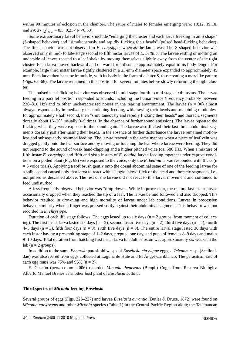
NISHIDA24 · Zootaxa 2466 © 2010 Magnolia Press
within 90 minutes of eclosion in the chamber. The ratios of males to females emerging were: 18:12, 19:18,and 29: 27 (χ2
Yates = 0.5, 0.25< P <0.50).Some extraordinary larval behaviors include “enlarging the cluster and each larva freezing in an S shape”
(S-shaped behavior) and “simultaneously and rapidly flicking their heads” (pulsed head-flicking behavior).The first behavior was not observed in E. chrysippe, whereas the latter was. The S-shaped behavior wasobserved only in mid- to late-stage second to fifth instar larvae of E. bettina. The larvae resting or molting onunderside of leaves reacted to a leaf shake by moving themselves slightly away from the center of the tightcluster. Each larva moved backward and outward for a distance approximately equal to its body length. Forexample, large third instar larvae tightly clustered in a 23-mm diameter space expanded to approximately 45mm. Each larva then became immobile, with its body in the form of a letter S, thus creating a mazelike pattern(Figs. 65–66). The larvae remained in this position for several minutes before slowly reforming the tight clus-ter.
The pulsed head-flicking behavior was observed in mid-stage fourth to mid-stage sixth instars. The larvaefeeding in a parallel position responded to sounds, including the human voice (frequency probably between230–310 Hz) and to other uncharacterized noises in the rearing environment. The larvae (n = 30) almostalways responded by immediately discontinuing feeding, withdrawing their heads and remaining motionlessfor approximately a half second, then “simultaneously and rapidly flicking their heads” and thoracic segmentsdorsally about 15–20°, usually 3–5 times (in the absence of further sound emission). The larvae repeated theflicking when they were exposed to the sound again. The larvae also flicked their last three abdominal seg-ments dorsally just after raising their heads. In the absence of further disturbance the larvae remained motion-less and subsequently resumed feeding. The larvae reacted in the same manner when a piece of leaf vein wasdragged gently onto the leaf surface and by moving or touching the leaf where larvae were feeding. They didnot respond to the sound of weak hand-clapping and a higher pitched voice (ca. 580 Hz). When a mixture offifth instar E. chrysippe and fifth and sixth instars of E. bettina larvae feeding together under captive condi-tions on a potted plant (Fig. 68) were exposed to the voice, only the E. bettina larvae responded with flicks (n= 5 voice trials). Applying a soft brush gently onto the dorsal abdominal setae of one of the feeding larvae fora split second caused only that larva to react with a single ‘slow’ flick of the head and thoracic segments, i.e.,not pulsed as described above. The rest of the larvae did not react to this larval movement and continued tofeed undisturbed.
A less frequently observed behavior was “drop down”. While in procession, the mature last instar larvaeoccasionally dropped when they reached the tip of a leaf. The larvae behind followed and also dropped. Thisbehavior resulted in drowning and high mortality of larvae under lab conditions. Larvae in processionbehaved similarly when a finger was pressed softly against their abdominal segments. This behavior was notrecorded in E. chrysippe.
Duration of each life stage follows. The eggs lasted up to six days (n = 2 groups, from moment of collect-ing). The first instar larva lasted six days (n = 2), second instar five days (n = 2), third five days (n = 2), fourth4–5 days (n = 3), fifth four days (n = 3), sixth five days (n = 3). The entire larval stage lasted 30 days witheach instar having a pre-molting stage of 1–2 days, prepupa one day, and pupa of females 8–9 days and males9–10 days. Total duration from hatching first instar larva to adult eclosion was approximately six weeks in thelab (n = 2 groups).
In addition to the same Encarsia parasitoid wasps of Euselasia chrysippe eggs, a Telenomus sp. (Scelioni-dae) was also reared from eggs collected at Laguna de Hule and El Ángel-Cariblanco. The parasitism rate ofeach egg mass was 75% and 96% (n = 2).
E. Chacón (pers. comm. 2006) recorded Miconia theaezans (Bonpl.) Cogn. from Reserva BiológicaAlberto Manuel Brenes as another host plant of Euselasia bettina.
Third species of Miconia-feeding Euselasia
Several groups of eggs (Figs. 226–227) and larvae Euselasia aurantia (Butler & Druce, 1872) were found onMiconia calvescens and other Miconia species (Table 1) in the Central-Pacific Region along the Talamancan
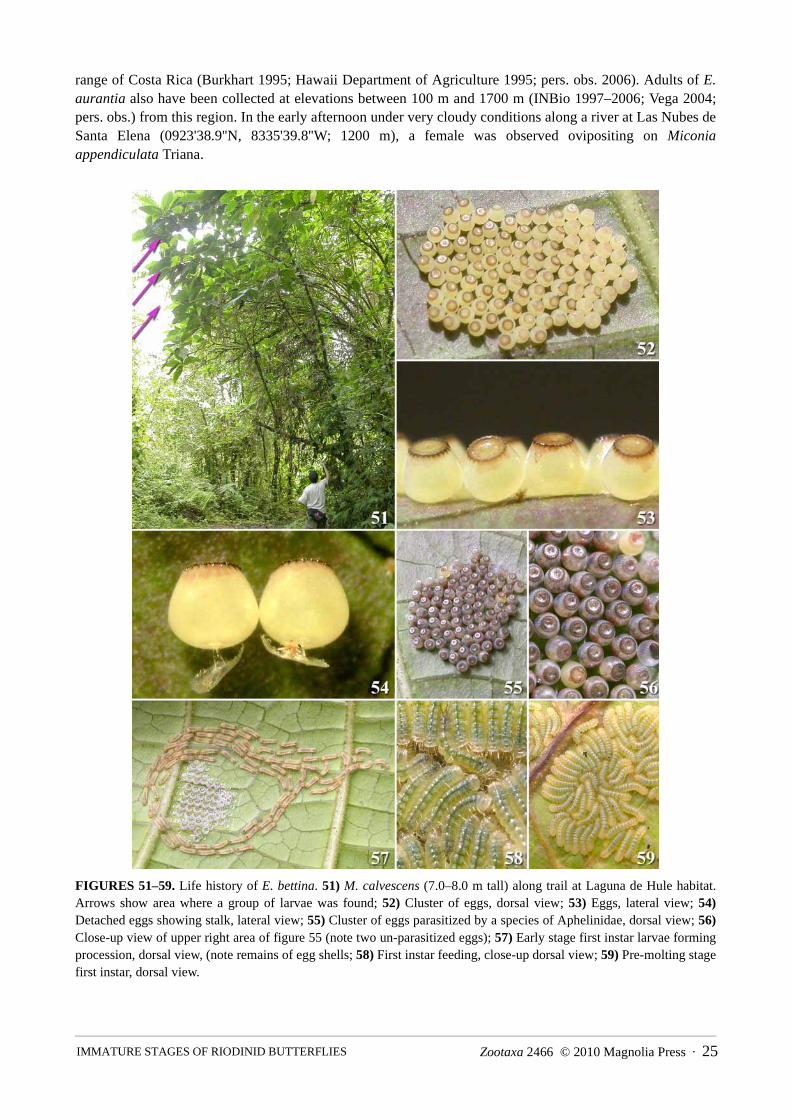
Zootaxa 2466 © 2010 Magnolia Press · 25 IMMATURE STAGES OF RIODINID BUTTERFLIES
range of Costa Rica (Burkhart 1995; Hawaii Department of Agriculture 1995; pers. obs. 2006). Adults of E.aurantia also have been collected at elevations between 100 m and 1700 m (INBio 1997–2006; Vega 2004;pers. obs.) from this region. In the early afternoon under very cloudy conditions along a river at Las Nubes deSanta Elena (0923'38.9''N, 8335'39.8''W; 1200 m), a female was observed ovipositing on Miconiaappendiculata Triana.
FIGURES 51–59. Life history of E. bettina. 51) M. calvescens (7.0–8.0 m tall) along trail at Laguna de Hule habitat.Arrows show area where a group of larvae was found; 52) Cluster of eggs, dorsal view; 53) Eggs, lateral view; 54)Detached eggs showing stalk, lateral view; 55) Cluster of eggs parasitized by a species of Aphelinidae, dorsal view; 56)Close-up view of upper right area of figure 55 (note two un-parasitized eggs); 57) Early stage first instar larvae formingprocession, dorsal view, (note remains of egg shells; 58) First instar feeding, close-up dorsal view; 59) Pre-molting stagefirst instar, dorsal view.

NISHIDA26 · Zootaxa 2466 © 2010 Magnolia Press
FIGURES 60–67. Life history of E. bettina. 60) Old feeding damages made by early instars; 61) Late stage second instarfeeding, dorsal view; 62) Mid-stage third instar resting, dorsal view; 63) Pre-molting stage third instar and severalrecently molted fourth instar (darker individuals), arrows pointing at adhered molted larval skins; 64) Late stage fourthinstar, dorsal view; 65) Late stage fourth instar showing S-shaped behavior, dorsal view; 66) early stage fifth instarshowing S-shaped behavior, dorsal view; 67) Late stage fifth instar resting on young leaves, dorsal view.
Diagnosis and description of early stages of Euselasia chrysippe
EGGDiagnosis: The egg of Euselasia chrysippe is semi-translucent pale-brown and ca. 0.42 mm in diameter.
Pigmentation in the lateral wall extends beyond the apical rim up to ca. one-third of the egg height. The widthof the crenulated rim area is ca. one-fifth of the dorsal surface diameter. The apical rim is purplish dark brown.

Zootaxa 2466 © 2010 Magnolia Press · 27 IMMATURE STAGES OF RIODINID BUTTERFLIES
The rosette of the third row lacks closed cells. The egg of E. bettina is less translucent and yellowish palebrown (creamy-white), slightly larger (0.45 mm diameter), with narrower pigmentation, extending beyond theapical rim to ca. one-sixth of the egg height, and the width of the crenulated rim area is narrower, ca. one-sev-enth of dorsal surface diameter. The apical rim is purplish brown. The rosette of the third row has severalclosed cells.
FIGURES 68–77. Life history of E. bettina. 68) Late stage sixth instar (dashed arrows) and mid-stage fifth instar (solidarrows) with mid-stage E. chrysippe fifth instar (no arrows) feeding; 69) Mid- to late stage sixth instar resting on pottedhost stem, dorsal view; 70) Mid- to late stage sixth instar feeding, lateral view; 71) close-up of larva showing iridescence(A2–6) of figure70; 72) Cephalic view of sixth instar feeding; 73) Caudal view of sixth instar (cephalic view on upperright larva); 74) Mature sixth instar showing processional behavior on rim of flower pot (arrows indicating direction ofmovement); 75) Prepupa, dorsal view, head pointing downwards; 76) Pupa, dorsal view; 77) Pupa, lateral view (notesilken girdle spun around T3–A1 and silken substrate).

NISHIDA28 · Zootaxa 2466 © 2010 Magnolia Press
Description: (Figs. 17–20, 78–89). Upright, frustum-shape, broadly rounded vetrnally (Figs. 20, 78–79,84, 53–54), with basally attached stalk (Figs. 78–79, 87, 54), ca. 0.42 mm in diameter (widest at one-thirdfrom lower bottom), ca. 0.4 mm tall, generally semi-translucent pale-brown with apical rim area purplish-dark-brown; coloration may vary within a cluster from entirely white to dark purple near micropyle and/orapical rim and beyond (Figs. 18–19); chorion smooth up to ca. 64X magnification, but weakly sculptured (seebelow), semi-translucent to opaque; micropyle area (Figs. 80–81) slightly concave with a rosette sculpturingwith 3–5 micropyles more or less evenly distributed along inner hub; rosette composed of three rows of veryshallow ridged polygons. The first row consisting of 9–13 closed cells, second row with 14–19 (mostly)closed cells, third row without closed cells (n = 4); dorsal surface including and surrounding rosette withslightly raised repetitive concentric circles (Fig. 81); dorsal surface brown to purplish brown from micropyle,beyond a darkly pigmented, shallowly crenulated apical rim, up to ca. one-third of egg height on lateral wall,i.e., edge (height) of the lateral wall pigmentation uneven (Fig. 20); crenulate rim ca. one-fifth of dorsal sur-face diameter, porous, and outer portion confluent with vertically arranged and evenly distributed short lateralridges (Fig. 80); lateral ridges with 2–3 crests, each crest with an apical aeropyle (Fig. 80); latero-ventralaspect broadly rounded (Figs. 20, 78–79, 84, 53–54); chorion with shallow hexagonally arranged wartlikestructures, each structure connected with shallow ridges, more conspicuous near ventral aspect (Figs. 84, 87);venter slightly concave, with a raised domelike stalk connecting structure in the center (Figs. 82–84); stalkconnecting structure ca. 80–100 µm wide (n = 5), irregularly ridged and porous (Fig. 83), with a wide basalstalk (Figs. 79, 87), absent on eggs extracted from female abdomen (probably a product of secretion fromaccessory gland); stalk translucent with brownish tint (Fig. 54), flexible, slightly elastic, ca. 0.09–0.12 mmlong and 0.04 mm diameter in middle portion (narrowest), base width up to 0.25 mm (n=12); a thin,inconspicuous, membranous part of stalk extended beyond connecting structure up to ca. 100 µm radius, i.e.,ca. one-fourth of egg width; wide stalk base with fiberlike extensions (Figs. 79, 87), fiberlike structure(probably originated from remnant of accessory gland fluid running through grooves on plant surface beforehardening); center portion of inner surface of ventral chorion (opposite surface of stalk conjunctionalstructure) slightly raised and concave (Figs. 85–86); concave area with more or less hexagonally shapedgrooves (Fig. 86).
Materials examined: Lake Arenal, 2003 (SEM, n = 4 complete specimens and 3 shells).
FIRST INSTAR LARVA Diagnosis: The head capsule width of Euselasia chrysippe is ca. 0.20 mm; the head is translucent pale-
brown; the body is semi-translucent white to pale-brown; and the prolegs on A10 have 7 crochets in alateroseries. In E. bettina the head capsule is wider (ca. 0.25 mm); the head is slightly darker; the body is lesstranslucent (creamy); and the prologs on A10 have 8–9 crochets in a lateroseries.
Description (Figs. 21–24, 90–135, 146–147): Body length 0.8–2.1 mm, initially semi-translucent white topale-brown, with trace of brown internal organs (Fig. 21) which gradually change to pale-green upon com-mencement of feeding on plant tissue (Figs. 22–23), more white (less translucent) during pre-molting period(Fig. 24); integument covered with microtrichia, but less dense or absent on T1 shield, anal plate, andpinacula. Head (Figs. 92–97): Hypognathous, circular, slightly compressed laterally, ca. 0.20 mm wide, semi-translucent pale-brown; stemmata (Figs. 21, 93–96) dark-brown, 1–5 arranged in crescent and equally spaced,except 5 slightly distant from 4, 6 distantly positioned and equidistant from 1–5; oral area brown; setal groupsA, C, F, L, P, SS, and S1–2 with setae nearly equal in length, long, ca. same length as epicranium suture, withmicro-projections along mid-portion; MD-group microscopic; AF and S3 short, ca. one-fourth to one-fifthlength of A, C, F, L, P, SS, and S1–2 setae, without micro-projections (Figs. 93–96); AF1 contiguous withmid-portion of adfrontal suture; AFa near dorsal angle of frons; AF2 lateral to dorsal angle of frons and dorso-laterally mesal to AFa; A1 at mid-distance between stemma 3 and C2; A2 laterally between AF1 and stemma3; A3 between stemma 1 and P1; MD1–3 microscopic; MDa contiguously dorso-mesal to MD3; C1 and C2above clypeus near lower angle of frons, C1 distal to adfrontal suture and C2 proximal to adfrontal suture; F1near lower angle of frons dorsal to clypeus; Fa contiguously dorso-mesal to F1; L1 lateral to A3 and dorso-lateral to stemma 1; P1 lateral to intersection of epicranial suture and ecdysial line; P2 dorso-lateral and

Zootaxa 2466 © 2010 Magnolia Press · 29 IMMATURE STAGES OF RIODINID BUTTERFLIES
slightly mesal to P1, more or less equidistant from MD1 and P1; S1 posterior to stemma 3; S2 dorso-posteriorto stemma 6; S3 posterior to and slightly ventral to stemma 6; SS1 between antennal socket and galea; SS2posterior to stemma 5; SS3 posterior to antennal socket in ventral aspect. Labrum (Fig. 97) with M1 nearmeson, shorter than M2; M2 between M1 and LA2; M3 ventral to M2, same length as M1; LA1 projectingfrom inner surface, dorsal to LA2 and shorter than LA2; LA2 lateral to M2 and as long as M2; LA3 ventral toLA2 and lateral to M3. Mandible (Fig. 146) with six large palmate teeth; inner surface weakly concave;mandibular setae subequal, longer seta with micro-projections near middle (Fig. 147); sensilla of antenna asfigured (Fig. 98); maxillary palpus and sensilla as figured (Figs. 99–100). Thorax: T1 (Figs. 90, 92, 101–114): Shield (Figs. 90, 92, 101–104, 106) with six pairs of setae, XD1 (XD1a, b) and XD2 (XD2a, b), semi-translucent white to pale-brown, situated along anterior margin, more or less evenly distributed, projectingforward; XD1b slightly posterior to anterior margin; XD1b, D1–2, and XD2b thicker and longer than othersetae; D1–2 dark-brown, projecting upward; all setae except SD2 with micro-projections along entire length;circular tablet organs (= flat cylindrical to circular tabletlike sensillum or lenticle) (Figs. 90, 101–105, Table3) on anterior margin near middle, ca. 2.0 times as wide as but shorter than XD1a setal socket base, poresabsent under 1530X magnification, flanked by XD1a and D1; cupola organ (= sensillum campaniformium,G.T. Baker, pers. comm. 2006) in form of slightly raised ring base with slightly raised papilla in center (Figs.90, 102, 104, Table 3) posterior to XD1b, base ca. one-half of XD1b socket base, papilla one-third of base,perforations absent; D1 equidistant and diagonally behind circular tablet organs and cupola organ; D2 poste-rior to XD2a; pore (Da) between D1–2; two spherical organs (= sensillum in form of sphere on concave plate)(Figs. 90, 102–103, 106–107, Table 3) along latero-frontal margin, with shape varying from flat to papilloid,concave base plate ca. one-half of D2 setal socket, sphere one-half of base; subdorsal porton of shield alonginner lateral margin slightly concave (= subdorsal groove) (Figs. 101–103, 106); SD1 absent; SD2 (= sensoryseta, D.J. Harvey, pers. comm. 2006) (Figs. 90, 92, 101–103, 106, 108–109) featherlike, on anterior portion ofsubdorsal groove, projecting dorso-posteriorly, semi-translucent white to pale-brown, ca. three-fourths lengthof XD1 or D-group on shield, thinner than other setae on shield, narrower at base, loosely socketed in dome-shape pinaculum (Fig. 108); L1 semi-translucent white to pale-brown, on pinaculum in middle of segment, aslong and spinose as XD1b, D1–2, and XD2b, but slightly thinner (Figs. 90, 92, 101–103, 110); L2–3 absent;spiracle circular (Figs. 90, 92, 103, 110); SV-group setae semi-translucent white to pale-brown, bisetose (Figs.90, 92, 110); SV1 projecting laterally and SV2 slightly forward and downward; spherical organ diagonallybelow and posterior to SV1 on pinaculum (Figs. 9, 110); V1 short, simple (Figs. 90, 111); V2 absent; bladelikeseta (Figs. 90, 92–97, 111–114, Table 3), nearly as long as coax plus femur, on pinaculum of basi-anteriorportion of coxa, semi-translucent white, large and broadly flattened, ca. 3 times as long as wide, withlongitudinal ridges, occasionally with a few dentations on apical part; leg setae with spatulate-tip, locatedmesally (Harvey 1987a, 1987b; somewhat similar to structure in microlepidoptera, e.g., Davis et al. 1992 andDavis 1999) (Figs. 93–94, 96, 111, 115, Tables 3–4), two spatulate-tipped setae on tarsus, as long as tarsalsegment, projected downward from above claw, robust, dorso-ventrally flattened with stout longitudinalridges dorsally reaching spatulate area, laterally widened, length of spatulate-tip approximately half-length ofentire seta, 1.5 x as wide as basal area, spatulate-tip weakly curved upward (dorsally) and apex sharply tosmoothly pointed, ventral surface more or less flattened and rough; absent on tibia and femur. T2–T3 (Figs.90, 92, 101–103, 111, 115, 119): A pair of pinacula on dorsum, transversely narrow, with D-group setae; D1dark brown, D2 semi-translucent white to pale-brown, nearly one-half length of D-group setae on T1, bothwith micro-projections, projecting upward (Figs. 90, 92, 101–103); SD1 (Figs. 90, 103, 119) in mid-lateralportion without pinaculum, short, simple; L-group (Figs. 90, 92, 95, 103, 111) trisetose, grouped together onpinaculum projecting laterally, L1 slightly backward, L2 slightly forward, and L3 slightly downward, semi-translucent white to pale-brown with micro-projections, slightly shorter than L1 on T1; spherical organanteriorly between SD1 and L-group (Figs. 90, 95, 103); SV-group as in T1, but spherical organ absent (Figs.90, 95, 111); two spatulate-tipped setae on tarsus, about as long as tarsus, two on tibia, similar to and about 2times as long as spatulate-tipped setae on tarsus, but basal part of seta more cylindrical; spatulate-tipped setaeabsent on femur (Figs. 111, 115, Tables 3–4); bladelike seta and V1 as in T1 (Figs. 90, 111). Abdomen: A1(Figs. 90, 92, 118–119, 122–123): Pinacula on dorsum wider than those on T2–3; D1–2 brown to pale-brown

NISHIDA30 · Zootaxa 2466 © 2010 Magnolia Press
with micro-projections, ca. 1.5 times longer than D-group setae on T2–3, bifurcate near base, ca. 0.15–0.17total length of seta, thicker branch projecting outward, thinner branch projecting inward (Figs. 90, 92, 117–118, 120, Table 3; Samson et al. (1999)); spherical organ (Figs. 90, 118, Table 3) contiguous with inner-poste-rior portion of D1 on dorsal pinaculum, same size as in T1 shield; SD1 and subdorsal spherical organ nearspiracle as in T2–3 (Figs. 90, 119, Table 3); spiracles as in T1; L-group with five setae projecting laterally,three in middle on same pinaculum, similar to those on T2–3 (Figs. 90, 92, 119); SV-group absent; flat seta(small bladelike seta) on subventral area (Figs. 90, 122–123, Table 3), ca. two-thirds length of bladelike setaon T1–3, anterior-caudally flattened, somewhat similar to bladelike seta but smaller, projecting ventrally; SV-group absent; V1 longer than those of T1–3, V2 absent (Figs. 90, 122–123). A2 (Figs. 90, 92, 116, 119, 122–123): As in A1 except color of D1–2 semi-translucent white to pale-brown, V-group bisetose, V2 mesallycontiguous with flat seta (Figs. 90, 122–123). A3 (Figs. 90, 92, 122, 124): As in A1–2 except ventrally flatseta absent and proleg present (Fig. 124); SV-group with seven setae, SV1–4 grouped together laterally onpinaculum of proleg, SV5–7 on anterior part of proleg (Figs. 90, 92, 124); V-group trisetose (Figs. 90, 124);proleg with 5–6 uniordinal crochets in mesoseries, interrupted in middle, with a short 5–6 lateroseries, i.e.,three separate rows of 5–6 crochets arranged in a subtriangular pattern on planta in center (Figs. 124–126).A4–6 (Figs. 90, 92, 125–126): As in A3 except spherical organ on inner-posterior of D1 absent, replaced byperforated plate organ (Figs. 90, 120–121, Table 3), larger than spherical organ, diameter similar to D1 socketbase, concave and platelike with pores in center. A7 (Figs. 90, 92, 120–121): Same as in A4–6 except ventersame as in A1, flat seta somewhat more slender than those of A1–2. A8–9 (Figs. 90, 92, 127–131): Similar toA7 but with segments apparently fused laterally (A8+9), separated dorsally to subdorsally and subventrally toventrally (A8–9). Dorsal and subdorsal portion of A8 with organ (either spherical organ or perforated plateorgan) absent on pinaculum posterior to D1; D1 bifurcate, D2 (not bifurcate) slightly longer than D1 (Figs. 90,92, 128); SD-group absent; subdorsal spherical organ anterior to spiracle absent; spherical organ posterior tospiracle (= spiracle spherical organ) near A9 (Figs. 90, 128–129, Table 3). Dorsal and subdorsal portion of A9with D- and SD-group absent; spherical organ posterior to spiracle spherical organ of A8 (Figs. 128–129,Table 3). Lateral portion of A8+9 (Figs. 90, 127) with L-group with eight setae, six in middle on pinacula(Figs. 90, 127). Subventral and ventral portion of A8–9 (Figs. 90, 130) as in A7, but V1 slightly shorter (Figs.90, 130). A10 (Figs. 90–92, 127–135): Anal plate with eight pairs of long, slender and stout setae with micro-projections, a pair of short microscopic setae, and two pairs of cupola organs (Figs. 90–91 128–129, 132–133,Table 3); chaetotaxy as in Figs. 90–91, 132 and as follows: two pairs of long, stout setae similar to those onXD- and D-group on T1 shield, evenly distributed dorsally along anterior margin of plate; a pair of shortmicroscopic setae similar to SD1 seta on T2–A7 slightly posterior between two stout setae of anterior margin;lateral edge with a pair of long, stout setae similar to XD- and D-group on T1 shield projecting posterio-later-ally; middle portion of plate with two pairs of long, stout setae similar to XD- and D-group on T1 shieldevenly distributed and projecting caudally; two pairs of less stout setae similar to L-group on A8+9 projectingcaudally from between posterior of three pairs of setae in middle portion and lateral edges; a pair of slendersetae similar to L-group on A8+9 projecting dorso-laterally from anterior to each lateral edge setae; cupolaorgan anterior to slender seta on lateral edge (Figs. 90, 128–129, Table 3), another cupola organ slightly poste-rior to setae in middle portion and anterior to inner pair of slender setae (Figs. 90–91, 132–133); SV-groupwith four short hairlike setae (Figs. 90, 130–131), spherical or papilla-shaped spherical organ above SV-grouppinaculum (Figs. 90, 127, 130); “PP” (=paraproctal seta) on each side of posterior end of prolegs (Figs. 90,130, 132); V1 located anterior to proleg (Figs. 90, 130, 134); eight claw-shaped setae (apparently paired)(Figs. 90, 130, 134–135, Table 3) between prolegs, stout, cylindrical, curved apically, 4 times as long as wide,shorter than V1 and SV setae; proleg with seven uniordinal crochets in lateroseries forming semicircle andwith 4–5 uniordinal crochets in mesoseries arranged in a semicircle with a median gap.
Materials examined: Lake Arenal, 2003 (SEM, n = 3; dissecting stereo-microscope, n = 2).

Zootaxa 2466 © 2010 Magnolia Press · 31 IMMATURE STAGES OF RIODINID BUTTERFLIES
Table 3. Summarized table of unique organs and setae in each larval instar of Euselasia. Numbers in parenthesis
refer to numbers of characters presence on one side. ---------- = data unrecorded.
......continued
Characters 1st instar 2nd instar 3rd instar 4th instar 5th instar 6th instarhead: AHS absent present present present present presentbody: AHS absent present present present present presentT1 shield: CTO on anterior-mesal present (1) absent absent absent absent absentT1 shield: CTO on posterior-lateral absent present (2–3) present (3) present (9–
10)present (17–20)
present (30–40)
T1 shield: SO on anterior-lateral present (2) absent present (1) - - -T1 shield: CO on anterior-middle present (1) present (1) present (1) - - -T1 shield: D2 present present present present present presentT1–3 legs: BLS present present (smaller) present (smaller) present
(smaller)present
(smaller)present
(smaller)T1–3 legs: STS present present present present present presentA1–8: bifurcated D group yes no no no no noA1–2 and A7–9: FS on subventer present present present - - absentA10: CS between prolegs present (8) present (8) present (12) - - present (ca. 40)A10 shield: CTO in anterior-lateral absent present (3+1seta or 4) present (4+1seta
or 5)present (5–7) present
(14–15)present (33–36)
Organs on T1
SO near spiracle absent absent absent - - -spiracle PCO absent absent absent absent absent absentSO on SV pinaculum present (1) present (1) present (2) - - present (10)organ on mid-anterior portion of shield absent SO (1) SO (1) or 1 seta - - -
Organs on T2
SO bt/ SD- and L-group present (1) present (1) absent - - present (5)SO on SV pinaculum absent absent or present (2) absent - - present (3)organ behind D1 seta (bt/ D1–2 ) absent absent absent - - SO (3)organ on mid-anterior of pinaculum on dorsum
absent SOl (1) or absent SO (2) or AHS (1)
- - -
Organs on T3
SO bt/ SD- and L-group present (1) present (1) absent - - present (5)SO on SV pinaculum absent absent or present (1–
2)absent - - present (3)
organ behind D1 seta (bt/ D1–2 ) absent absent absent - - SOl (10)organ on mid-anterior of pinaculum on dorsum
absent SOl (1) SOl (1) - - -
Organs on A1
SO near spiracle bt/ SD- and L-group present (1) present (1) present (4) - - -spiracle PCO absent absent absent - - -organ behind D1 seta (bt/ D1–2 ) SO (1) (papilla
shaped)SO (1) SO (1) - - SO (3)
Organs on A2
SO near spiracle present (1) absent or present (1) present (1) - - -spiracle PCO absent raised (1) absent - - -organ behind D1 seta (bt/ D1–2 ) SO (1) (papilla
shaped)SO (1) SO (1) - - -
Organs on A3
SO near spiracle bt/ SD- and L-group present (1) absent or present (1) present (2–3) - - present (3–5)spiracle PCO absent raised (1) raised (1) - - raised, flat, or
sunken (24–33)organ behind D1 seta (bt/ D1–2 ) SO (1) (papilla
shaped)SOl (1) SOl (1) - - -
Organs on A4
SO near spiracle bt/ SD- and L-group present (1) present (1) present (1–3) - - present (1)spiracle PCO absent flat (1) flat, raised, or
sunken (4) - - raised or
sunken (25–17)organ behind D1 seta (bt/ D1–2 ) PPO (1) SOl (1) SO (1) - - -

NISHIDA32 · Zootaxa 2466 © 2010 Magnolia Press
Table 3. (continued)
SECOND INSTAR Diagnosis: Head capsule width of Euselasia chrysippe is ca. 0.33 mm; the body is pale grayish-green
without any dark spots on dorsum; and the proleg on A10 has 9–10 uniordinal crochets in a lateroseries andfive crochets in mesoseries. In E. bettina the head capsule is wider (ca. 0.35 mm); the body color is darker,more grayish, with dorsal pinacula dark-gray to black on T2–A8; and the proleg on A10 has 11–12 uni- tobiordinal crochets in a lateroseries and six uniordinal crochets in mesoseries.
Description (Figs. 25, 136–145): Differs from first instar as described and figured below (also see Table3). Remaining instars essentially same (i.e., presence and position of setae) as second instar. Body length 2.0–2.9 mm, pale grayish-green with dull green lines dorso-longitudinally and subdorsally along segmental folds.Head (Figs. 136–137): Width ca. 0.33 mm, semi-translucent pale orange-brown, 1–4 extra setae in some setalgroups; arrowhead setae (= more or less triangular), with short, flat, apex pointed, ridged medio-longitudi-nally, with cylindrical base (Figs. 136–139, Table 3), scattered on head, but more concentrated on frons, ca. aslong as stemma diameter and twice as long as wide, projecting forward, apparently in pairs, i.e., in similarpositions in left and right half of the head; mandible as in first instar, but with seven distinct teeth. Thorax: T1(Figs. 137–139): Shield pale-orange, with eleven pairs of relatively long setae with micro-projections alonganterior and lateral margins, three pairs of short slender needlelike setae with micro-projections in middle ofshield, two pairs of very short setae on near lateral margin of shield, SD2 as in first instar, twelve pairs ofarrowhead setae along anterior and posterior margin, and 2–3 pairs of circular tablet organs on latero-posteriormargin (when circular tablet organ absent on one side, usually replaced with a seta or arrowhead setae); D1–2
Characters 1st instar 2nd instar 3rd instar 4th instar 5th instar 6th instarOrgans on A5
SO near spiracle bt/ SD- and L-group present (1) present (1) present (1) - - present (2–3)spiracle PCO absent flat (1) sunken (3) - - sunken (12) and
raised (22)Organ behind D1 seta (bt/ D1–2 ) PPO (1) spherical (1) spherical (1) - - -
Organs on A6
SO near spiracle bt/ SD- and L-group present (1) absent absent - - present (2)spiracle PCO absent flat (1) sunken (2–3) - - sunken (19–21)SO bt/ SD- and L-group absent absent or present (1) absent - - present (1)Organ behind D1 seta (bt/ D1–2 ) PPO (1) SO (1) SO (1) - - present (4)
Organs on A7
SO near spiracle bt/ SD- and L-group present (1) present (1) present (1) - - present (3–6)spiracle PCO absent flat (2–3) flat to sunken (5–
6) - - sunken (28–38)
organ behind D1 seta (bt/ D1–2 ) PPO (1) spherical (1) spherical (1–2) - - -Organs on A8–9
spiracle SO on A8 present (1) present (2–3) present (5–7) - - present (58) (n =1)
spiracle PCO on A8 absent absent absent - - absentorgan behind D1 seta (bt/ D1–2 ) on A8 absent SO (1) SO (2) - - -SO on A9 behind A8 spiracle SO present (1) absent absent - - absent
Organs on A10
CTO on anterior-lateral of shield absent present (2+1 seta or 3) present (5–6) - - present (25–38)CO on shield posterior corner (triangle zone)
present (1) present (1) absent - - -
Organ on lateral of proleg SO (1) CO (1) CO (1) - - -SO on posterior of proleg absent present (1) present (3–4) - - -CO on lateral of shield present (1) or 1 seta absent absent or (1) - - -Species of specimens examined E. chrysippe, E.
bettinaE. chrysippe E. bettina E. chrysippe E.
chrysippeE. chrysippe, E.
bettinaNumber of specimens examined under SEM
n=3, n=3 n=2 n=2 (n=1) stereo-microscope
(n=1) stereo-
microscope
n=2, n=2

Zootaxa 2466 © 2010 Magnolia Press · 33 IMMATURE STAGES OF RIODINID BUTTERFLIES
thick, brown, projecting upward; 1–2 spherical organs posterior to D1–2 (position differs from first instar andsometimes replaced by a fine short seta); six pairs of semi-translucent white to pale-brown setae (XD-groupand other setae) projecting forward from anterior margin, shorter and thinner than D1–2; four pairs of arrow-head setae along posterior margin (arrowhead setae sometimes replaced by fine short setae), some setaeslightly spiraled apically; L-group bisetose; SV-group trisetose, spherical organ as in first instar; bladelike setanearly as long as femur, similar to arrowhead setae but slightly larger (Fig. 139); arrowhead setae dense,projecting from ventro-subventral portion along thoracic legs (Fig. 139); spatulate-tipped setae on tarsus andtibia (Fig. 139, Tables 3–4) as in first instar. T2–T3 (Figs. 139–140): As in T1 except pale-gray dorsally;dorsum with pinacula with 6–7 setae, D1–2 as in first instar with four other semi-translucent white to pale-brown setae, thinner and shorter than D1–2 (Fig. 140), L-group with six semi-translucent white to pale-brown,somewhat featherlike setae, projecting laterally (sometimes replaced with spherical organ, e.g., Fig. 141); fourspatulate-tipped setae on tibia, two on femur (Fig. 139, Tables 3–4). Abdomen. A1 (Fig. 140): As in T2–T3except D-group with seven setae; D1–2 not bifurcate (Fig. 140); spiracles with a spherical organ (= spiraclespherical organ); SV area with some arrowhead setae; V-group bisetose; flat seta as in first instar, but difficultto distinguish from arrowhead setae. A2: As in A1 except a raised perforated cupola organ (PCO) similar tothat described by Harvey (1987b, 1989) (i.e., lenticle dorsal to spiracle = spiracular PCO) (Fig. 141, Table 3);V-group trisetose. A3–6 (Fig. 141): As in A2 except flat spiracular PCO on A4–6; proleg with 5–7 uniordinalcrochets in latero- and mesoseries, as in first instar. A7 (Fig. 141): As in A2 except 2–3 flat spiracular PCOsclustered together; SV seta, short spinelike laterally contiguous to flat seta. A8–9 (Figs. 142, 145): Dorsal andsubdorsal portions of A8 as in A7 except spiracular PCOs absent; 2–3 spiracular spherical organs posterior tospiracle; SD-group absent. Dorsal and subdorsal portions of A9 with D- and SD-group absent as in first instar;spherical organ posterior to A8 spiracular spherical organ absent (Table 3). Lateral portions of A8+9 with L-group of eleven setae. Subventral and ventral portions of A8–9 as in A2. A10 (Figs. 142–145): anal plate (Fig.143) with six brown thick setae projecting dorso-caudally along with other slender, long or short setae, 2–4circular tablet organs on each side of anterior margin (circular tablet organs sometimes absent on one side andreplaced with seta) (Fig. 143, Table 3), cupola organ on each latero-posterior margins (Fig. 144, Table 3);eight claw-shaped setae as in first instar (Fig. 145, Table 3), a few thick, setae posterior and anterior to claw-shaped setae; proleg with two spherical organs, on lateral pinaculum and posterior end respectively; 9–10uniordinal crochets in semicircular lateroseries and five uniordinal crochets in mesoseries.
Material examined: Lake Arenal, 2003 (SEM, n = 2; dissecting stereo-microscope, n = 1).
TABLE 4. Number of spatulate-tipped setae in mesal portion of each thoracic leg segment of larval Euselasia. (n =3 each).
Specimens Tarsas Tibia Femur Coxa
E. chrysippe / E. bettina first instar T1 2 0 0 0
E. chrysippe / E. bettina first instar T2 2 2 0 0
E. chrysippe / E. bettina first instar T3 2 2 0 0
E. chrysippe second instar T1 2 2 0 0
E. chrysippe second instar T2 2 4 2 0
E. chrysippe second instar T3 2 4 2 0
E. bettina third instar T1 2 2 0 0
E. bettina third instar T2 2 4–6 3 0
E. bettina third instar T3 2 5 4–5 0
E. chrysippe sixth instar T1 2 3 0 0
E. chrysippe sixth instar T2 2 9–11 11–12 0
E. chrysippe sixth instar T3 2 10–14 11–13 0

NISHIDA34 · Zootaxa 2466 © 2010 Magnolia Press
THIRD INSTAR Diagnosis: The head capsule width of Euselasia chrysippe is ca. 0.53 mm; the overall body color is
greenish-pale-gray; the T1 shield is pale-orange; the color of the dorsal pinacula on T2–A8 is gray becomingfaint in posterior segments; the mesal line and intersegmental membrane are dull green; the anal plate is pale-gray; the proleg on A10 has 6–7 crochets in mesoseries. Head capsule width of E. bettina is slightly wider, ca.0.55 mm; the overall body color is darker (gray); the T1 shield has a pair of black transverse lines; the dorsalpinacula of T2–A8 are prominent black; the mesal line, intersegmental membrane, and anal plate are black;and the proleg on A10 has more crochets (8–9) than E. chrysippe.
Description (Figs. 26–27, 30, 148): Similar to previous instar except for the following. Body length 2.8–5.0 mm, overall color greenishi-pale-gray. Head: Width ca. 0.53 mm, orange; more secondary setae in eachsetal group and more arrowhead setae compared to previous instar (this tendency continues in remaininginstars); mandible (Fig. 148) somewhat palmate, with seven small, distinctly dentate teeth, inner surfaceconcave.
FIGURES 78–83. SEM of egg of E. chrysippe. 78) Lateral view, in situ (note stellate trichomes of host leaf); 79) Latero-ventral view, showing stalk; 80) Apical rim and micropyle area, dorso-lateral view; 81) Micropyles and rosette, dorso-lateral view; 82) Stalk conjunctional structure in the center, ventral view, stalk removed; 83) Stalk conjunctionalstructure, ventral view, (remnants of stalk seen as white).
Thorax and abdomen. T1: whitish-pale-orange. T2–A8: Pinacula on dorsum gray and faint in posteriorsegments (presence and level of gray varied between larvae within a group); lateral lobes semi-translucent

Zootaxa 2466 © 2010 Magnolia Press · 35 IMMATURE STAGES OF RIODINID BUTTERFLIES
pale-brown; a faint whitish-pale-gray longitudinal line subdorsally along lateral lobes; prolegs with 7–12uniordinal crochets in lateroseries and 18–21 weakly biordinal crochets in mesoseries. A9: Dorsally andsubdorsally as in second instar except a short, slender SD seta in mid-subdorsum. A10: Anal plate pale-gray(opaque); proleg with crochets weakly biordinal, 13–14 in semicircular lateroseries and 6–7 uniordinal cro-chets in mesoseries.
Material examined: Lake Arenal, 2003 (dissecting stereo-microscope, n = 1); La Selva, 2007 (dissectingstereo-microscope, n = 1).
FIGURES 84–89. SEM of egg of Euselasia. 84) Stalk conjunctional structure (stalk removed) and hexagonally arrangedsculpturing of chorion, ventro-lateral view (E. bettina); 85) Concave structure at center part of ventral inner surface(opposite surface of stalk conjunctional structure), dorso-lateral view (E. bettina); 86) Concave structure (E. bettina)(white narrow lines = silk); 87) Stalk, lateral view (E. bettina); 88) Apical rim and micropyle area (E. bettina), dorso-lateral view; 89) Micropyles and rosette (E. bettina), dorso-lateral view.
FOURTH INSTARDiagnosis: Overall body color of Euselasia chrysippe is grayish-green; the head capsule width is ca. 0.72mm; the color of the T1 shield is mostly orange; the pinacula on dorsum are dull black; the mesal line andintersegmental membrane are dull green; the A10 proleg has 20–22 biordinal crochets in lateroseries. Overallbody color of E. bettina is grayish-yellow; the head capsule is slightly wider (ca. 0.80 mm); the T1 shield ismostly black; the pinacula on dorsum are darker, in prominent black; mesal line and intersegmental membraneare dark-green; and the proleg on A10 has more crochets (25–26).

NISHIDA36 · Zootaxa 2466 © 2010 Magnolia Press
Description (Figs. 28–30, 149): Similar to previous instar except as described below. Body length 5.2–8.6mm; overall color slightly more grayish and greenish dorsally compared to third instar; mesal line andintersegmental membrane dull green. Head: Width ca. 0.72 mm, bright orange; mandible (Fig. 149) withdistal area laterally widened and straight, with five indistinct teeth, inner surface more concave and steeplyangled than previous instars. Thorax and abdomen. T1: Shield orange, posterior ca. one-fourth with a pair ofnarrow, dull black, transverse lines, with three pairs of dark-brown setae projecting dorsally and several semi-translucent white to pale-brown, slender setae projecting over head from anterior margin; 9–10 circular tabletorgan in posterior-lateral margin (Table 3); L-group with 8–10 long setae, SV-group with 8–10 long setae. T2–A8: Dorsum with dull black pinacula with a dark-brown to semi-translucent brown seta and several semi-translucent pale-brown to white setae projecting dorsally; proleg with 13–16 biordinal crochets in each row.A9: Dorsally and subdorsally as in previous instar. A10: Anal plate with a pair of dark-gray to black trans-verse arch-shaped lines, with four pairs of thick, dark-brown setae and several semi-translucent pale-brown towhite thinner setae projecting dorsally to caudally, with semi-translucent white, slender, featherlike setaealong latero-posterior margins; circular tablet organ (Table 3); space between arch (medio-posterior portion)whitish; proleg with 20–22 biordinal crochets in lateroseries in semicircle and 11–12 biordinal crochets inmesoseries.
Material examined: Lake Arenal, 2003 (dissecting stereo-microscope, n = 3).
FIGURE 90. Chaetotaxy of first instar larva of Euselasia chrysippe and E. bettina (lateral view). CO = cupola organ, CS= claw-shaped setae, CTO = circular tablet organ, cxa = coax, pl = proleg, “PP” = “proprioceptor” seta, PPO = perforatedplate organ, SO = spherical organ.
FIGURE 91. Chaetotaxy of anal plate of first instar larva of Euselasia chrysippe and E. bettina (caudal view). CO =cupola organ.

Zootaxa 2466 © 2010 Magnolia Press · 37 IMMATURE STAGES OF RIODINID BUTTERFLIES
FIFTH INSTARDiagnosis: The fifth instar Euselasia chrysippe is gray to yellowish-gray; the head capsule width is ca. 1.23mm; the T1 shield is mostly orange; and the proleg on A10 has 11–13 crochets in a mesoseries. E. bettina isgreenish-black to greenish-dull yellow; the head capsule width is slightly wider (ca. 1.30 mm); the T1 shieldis entirely black or mostly black with little orange; and.A10 has more crochets (15–16).
FIGURES 92–97. SEM of Euselasia first instar larva. 92) Lateral view (E. bettina); 93) Head, frontal view, (E. chry-sippe); 94) Head and cervical area, dorso- lateral view (E. chrysippe); 95) Head and T1–2, lateral view (E. bettina); 96)Head and T1, ventral view (E. chrysippe); 97) Mouthparts, frontal view (E. chrysippe).
Description (Figs. 31–32, 68): Similar to previous instar except as described below. Body length 9.0–13.2mm, overall color gray to yellowish-gray, lateral lobes slightly more protuberent; minute semi-translucentwhite to pale-brown, short, hairlike setae sparsely throughout body surface (at 32X). Head: Wdith ca. 1.23mm, orange to dark-brown (Fig. 31) or bright orange (Fig. 32). Thorax and abdomen. T1: Shield bright todull orange, with a pair of dark-brown patches in posterior one-fourth to one-half, 3–4 pairs of prominentdark-brown setae projected dorsally and subdorsally along with a few semi-translucent white slender setaeprojecting over head; circular tablet organ lenslike, i.e., translucent or colorless (Table 3); L-group with 10–12long setae; SV-group with 10–12 long setae. T2–A8: Pinacula on dorsum black with two dark-brown setaealong with several semi-translucent pale-brown to white slender setae projecting dorsally; proleg with 18–21biordinal crochets in each row. A9: Dorsally and subdorsally as in previous instar except SD bisetose. A10:

NISHIDA38 · Zootaxa 2466 © 2010 Magnolia Press
Anal plate arched, with 8–10 dark-brown setae and with several slender semi-translucent white to pale-brownsetae projecting latero-caudally; circular tablet organ (see Table 3); space between arch (posterior middle part)white; proleg with 28–33 biordinal crochets in semicircular lateroseries and 11–13 biordinal crochets inmesoseries.
Material examined: Lake Arenal, 2003 (dissecting stereo-microscope, n = 3).
FIGURES 98–103. SEM of Euselasia first instar (E. chrysippe). 98) Left antenna showing arrangement of sensilla, fron-tal view; 99) Left galea showing lobe of galea and maxillary palpus, dorso-frontal view; 100) Maxillary palpus and sen-silla arrangement, dorso-frontal view; 101) Head and T1–3, dorso-frontal view; 102) T1 shield, dorsal view; 103) T1–3,dorso-lateral view.
SIXTH INSTARDiagnosis: The sixth instar Euselasia chrysippe is greenish-dark-gray to greenish-dull black; the head capsulewidth is ca. 1.65 mm; the color of the head is bright orange, black, or a mixture of these two; arrowhead setaeare cone-shaped (not flattened), ridged, and spiraled apically; the curvature of the ventral margin of thelabrum is narrowly angled (ca. 110°); the mandible is small (0.38 mm wide), with the dentation less distinctthan in E. bettina, and the extension of the fifth tooth is somewhat widened at edge; the T1 shield is orange tobright orange and without iridescence; the pinacula on the dorsum have a pale-gray oval line; the iridescenceon structural color plates is faint metallic-blue; a proleg on A10 has 11–13 crochets in mesoseries. E. bettinasixth instar is brownish-amber with reddish or greenish tint; and head is slightly wider (ca. 1.75 mm) and

Zootaxa 2466 © 2010 Magnolia Press · 39 IMMATURE STAGES OF RIODINID BUTTERFLIES
black with a pair of brownish-orange to pale-orange patches running parallel to epicranial suture; thearrowhead setae are flattened’ the curvature of the ventral margin of labrum is more widely angled (ca. 120°);the mandible is larger (ca. 0.45 mm wide) and dentation is slightly more distinct; the T1 shield is brightorange with reddish tint, and black transverse lines have faint bluish-gray iridescence; the pinacula on thedorsum are black with a white oval line; the iridescence of the structural color plates is brighter and metallicsky-blue; and the proleg on A10 has more crochets in mesoseries (15–16) than in E. chrysippe.
FIGURES 104–109. SEM of Euselasia first instar (E. chrysippe). 104) Paired circular tablet organs on medio-anteriorpart of T1 shield, dorso-frontal view; 105) Circular tablet organ, lateral view; 106) T1 shield showing spherical organs(SOs) and SD2, dorso-lateral view; 107) Spherical organs; 108) Pinaculum of SD2; 109) Middle to apical area of SD2,lateral view.
Description (Figs. 33–39, 42–44, 150, 152–156, 159, 161, 163, 168–194, 196–207): Similar to previousinstar except as described below. Body length 12.8–19.0 mm, overall color greenish-dark-gray to greenish-dull black (Figs. 33–34, 36–37, 43–44), distinct from of other instars; spherical organs scattered sparsely onbody, more concentrated near spiracles, SV pinacula, and on anal plate near circular tablet organ (Figs. 183,186, 188, 204). Head (Figs. 34–35, 43, 152–154): Width ca. 1.65 mm, bright orange (Figs. 34–35, 37), black(Figs. 39, 43), or mixture of these two (Figs. 34, 44); cuticle of head capsule semi-translucent pale-brown todark-brown; arrowhead setae semi-translucent with slight brownish tint (Figs. 34–35), most cone-shaped (notflattened), ridged, and spiraled apically (i.e., shape differs from those of second instar) (Figs. 161, 163),

NISHIDA40 · Zootaxa 2466 © 2010 Magnolia Press
densely scattered over head, more so on frons, stemmatal area, and oral area (Figs. 152–156, 183); moresparsely scattered on other parts of body; each setal group with numerous setae; labrum as figured (Figs. 155,159), curvature of ventral margin widely angled (ca. 120°); mandible (Fig. 150) 0.38 mm wide, distally morewidened and straight, with five indistinct teeth, edge of tooth farthest from condyle straight and continuous (n= 5); sensilla of antenna and maxillary palpus as figured (Figs. 169–170). Thorax. T1 (Figs. 33–35, 37, 43–44, 171–176): Shield orange to bright orange on anterior half, posterior half with a pair of black transverselines and posterior-lateral margins with a whitish line on each side (Figs. 33–35); six pairs of stout, dark-brown to black setae with microprojections dorsally and dorso-laterally (Figs. 34–35, 43–44, 171), three setaeon orange anterior portion, one on orange lateral portion, and two on white posterior portion; six pairs ofsemi-translucent pale-brown to white, long, slender setae projecting over head from anterior portion tosubdorsal portion of shield with several other short, semi-translucent white to pale-brown slender setae; SD2(Figs. 43, 171–175) semi-translucent white to pale brown, length ca. one-half that of stout setae, withmicroprojections on shield, loosely socketed; circular tablet organ (Fig. 172, Table 3) ca. 22 µm in diameter,translucent or with brownish tint, porelike structure in inner center; L-group with ca. 15 setae projectedlaterally, long setae about as long as body width, short setae ca. one-third length of long setae, semi-translucent white to pale-brown setae with micro-projections; SV-group (Fig. 183) with >10 semi-translucentpale-brown slender setae of various lengths (<0.4 mm) projecting laterally; bladelike seta (Fig. 182, Table 3)on legs, straight, basal half cylindrical, apical half flat with pointed tip; spatulate-tipped setae (Figs. 176–181,Tables 3–4) slightly longer than each leg segment, colorless semi-translucent (in both live and preservedspecimens), spatulate-tip ventrally with irregular narrow grooves (Figs. 177–179) and some with small oval“windows” (Fig. 178), dorsally with longitudinal ridges more or less along entire seta (Fig. 180); spatulate-tipped setae on femur commonly with sharply pointed apex (Fig. 181). T2–T3 (Figs. 33–34, 43–44, 196):Pinaculum on dorsum (Fig. 196) blackish and slightly greenish, with a pale-gray semi-oval area in middle; D-group with three dark-brown to black stout setae with microprojections with approximately 8–10 semi-trans-lucent white to pale-brown setae projecting dorsally; structural color plate on anterior subdorsum of blackpinaculum, with faint metallic-blue iridescence (Figs. 36, 43), also present on abdominal segments (Figs.195–197, 199–201); anterior subdorsum of color plate with two slender semi-translucent white to pale-brownsetae; black-colored area densely covered by microtrichia (Figs. 196–197); structural color plate concave andcontiguous to tonofibrillary platelets (Fig. 199), with sparsely scattered microtrichia; anterior part of platewith two slender semi-translucent white to pale-brown setae projected forward; L-group projecting in twolayers with two types of semi-translucent white to pale-brown setae (Figs. 192–194), one above longer (ca.two-thirds of body width) with micro-projections (ca. 10 setae), and the other shorter and featherlike (ca. 30),projected along lateral lobes; SV-group with ca. 10 long semi-translucent pale-brown setae and other shortsetae. Abdomen. A1–2 (Figs. 33, 44): Structural color plate larger than on T2; dorsal pinaculum with 3–4dark-brown to black stout setae with microprojections along with 6–7 semi-translucent pale-brown to whitesetae; SV-group with one long, slender seta with micro-projections, V-group with 5–8 setae (A2 with moresetae), one mesally located longer than others, flat seta absent. A3–6 (Figs. 33, 36, 44, 184–185, 191, 197,206): Similar to A1–2 except dorsal pinaculum with four dark-brown to black stout setae withmicroprojections (Fig. 191) with seven semi-translucent white to pale-brown setae; structural color plate withtwo setae on anterior part, one on middle, and two on posterior part; longitudinal white line subdorsally alongspiracles between dorsal pinacula and lateral lobes (through A3–9); lateral lobes semi-translucent dull white;spiracular PCOs (Figs. 184–187, Table 3) surrounding spiracles, more cephalad and dorsally to spiracle;PCOs translucent (in live and preserved specimens), raised, flat, sunken; proleg with 18–21 biordinal crochetsin each series. A7 (Figs. 33, 36, 206): D-group with 5–6 dark-brown to black stout setae withmicroprojections and approximately 6–7 semi-translucent white to pale-brown slender setae projectingdorsally; subdorsally as in A3–6; ventrally as in A1 except V-group with 2–3 setae, one located mesally aslong as proleg on A6. A8-9: Dorsal and subdorsal portion of A8 (Figs. 33, 44) similar to A7 except subdorsumof anterior part of color plate with 1–2 semi-translucent white to pale-brown slender seta; longitudinal whiteline prominently white and broader than in other segments, with spiracle in center; spiracular spherical organsas figured (Figs. 188–190, Table 3). Dorsal and subdorsal portions of A9 (Figs. 33–34, 43–44, 202–203)

Zootaxa 2466 © 2010 Magnolia Press · 41 IMMATURE STAGES OF RIODINID BUTTERFLIES
similar to that of previous instar (unable to distinguish SD setae from minute setae in D and SD area).Subventral and ventral portions of A8 (Fig. 206) as in A7 except V-group with two setae. Subventral andventral portions of A9 (Fig. 206) with SV absent. A10 (Figs. 33–34, 42–44, 202– 207): Anal plate andsurrounding area as in previous instar except anal plate with faint metallic-blue iridescence, 11-12 dark-brownto black stout setae with microprojections and 5–7 semi-translucent white to pale-brown long setae projectingdorsally to caudally; ca. 10 brown to dark-brown long, slender setae numerous shorter featherlike semi-translucent white to pale-brown setae projecting posterio-laterally from lateral to posterior margins of plate(Figs. 42, 44, 202–203); circular tablet organ (Figs. 203–205, Table 3) same shape as in T1; claw-shaped setae(Figs. 206–207) as short as other slender setae on A10 venter, 13.5 µm thick, i.e., more slender than inprevious instars, semi-translucent white to pale-brown, 33–36 on posterior middle portion, surrounded byslightly more slender setae; when expanded in procession, convex with a small semi-translucent circular spotin middle between prolegs (Figs. 206–207); proleg with 28–33 biordinal crochets in semicircular lateroseriesand 11–13 biordinal crochets in mesoseries.
Material examined: Lake Arenal, 2003 (SEM, n = 2; dissecting stereo-microscope, n = 5).
FIGURES 110–115. SEM of Euselasia first instar. 110) T1, lateral view, (E. chrysippe); 111) Left thoracic legs, blade-like seta (BLS) and spatulate-tipped setae (STS), ventral view (E. chrysippe); 112) Bladelike seta on left T1 leg, ventralview (E. chrysippe); 113) Bladelike seta on left T1 leg, frontal view (E. bettina); 114) Bladelike seta on left T1 leg, ven-tro-caudal view (E. chrysippe); 115) Spatulate-tipped setae on tarsus and tibia of left T2 leg, posterior view (E. chry-sippe).

NISHIDA42 · Zootaxa 2466 © 2010 Magnolia Press
FIGURES 116–121. SEM of Euselasia first instar (E. chrysippe). 116) L-group setae (arrows) on right side of A2–4,dorsal view; 117) Left side of abdominal segments showing bifurcate D1–2 (arrows), fronto-lateral view; 118) BifurcateD2 on left A1 and spherical organ (SO) behind D1, dorsal view; 119) SD and L-group and spherical organ (SO) on T3–A2, lateral view; 120) Bifurcate D1–2 and perforated plate organ (PPO) behind D1 on right side A7, dorso-lateral view;121) Perforated plate organ on left side A7, dorso-lateral view.
PREPUPA Diagnosis: The prepupa of Euselasia chrysippe is dark brownish-gray with pale-brown. E. bettina is
amber with black.Description (Fig. 45): Length 11.5–12.8 mm; dorsum dark brownish-gray (slightly greenish) with pale-
brown lines mesally and on pinacula, pale-brown laterally, faint metallic-blue iridescence on structural colorplates observed during prepupal stage.
Material examined: Lake Arenal, 2003 (n = 30).
PUPADiagnosis: The pupa of Euselasia chrysippe is 11.5–12.0 mm long and 5.0–5.5 mm wide. That of E.
bettina is usually longer and wider, 11.4–13.5 mm long, 5.7–6.4 mm wide.Description (Figs. 6, 46–50, 208–225, Table 5): Width 5.0–5.5 mm, length 11.5–12.0 mm, and dorso-
ventrally ca. 3.0 mm tall; dorsally convex, middle widened, caudally tapered, ventrally flattened; pale-creamy

Zootaxa 2466 © 2010 Magnolia Press · 43 IMMATURE STAGES OF RIODINID BUTTERFLIES
yellow to brown with brown to dark-brown patches; (approximately 12 hours prior to adult eclosion the pupabecomes reddish dark-brown); cuticle with narrowly crenulate sculpturing (e.g., Figs. 208, 214) on dorsal andlateral surfaces except in following locations: subdorsal triangular zone (Fig. 208) on T3 next to A1, middlearea of wing sclerites below triangular zone; cuticle more roughly sculptured on darkly pigmented areas; cuti-cle ventrally smooth except: outer one-third of eye piece, basal portion of antenna, pro- and T2 legs,proboscis, middle to apical area of wing sclerite; mouth parts as figured (Fig. 210), with wide clypeus andbroad mandibles (some specimens with narrower clypeus and mandibles as in Fig. 211); eye piece with threedistinct longitudinal linear parts (Fig. 209); inner part of center portion convex, middle part and inner partforming convex curve together; in some specimens middle part with a notch at posterior end (Fig. 211); outerpart concave with narrowly crenulate sculpturing; antenna extending slightly beyond wing sclerite, extendingto posterior margin of A4; proboscis extending ca. 1.5 times as long as adjacent T1 leg, slightly narrowed atmiddle portion of prothoracic leg sclerite widened at border of T2 leg sclerite; T2 leg adjacent to eye pieceextending to wide posterior portion of proboscis; anterior end of area between head and T1 with a smoothlycurved notch; thorax broadly rounded, T2 tallest; posterior end of A1–A7 dorsally flattened, gradually taperedcaudally, A3 widest; thoracic spiracle (Figs. 208, 212–213) on dorsum of lateral edge between T1–2; thoracicspiracle sculpturing on anterior dorsum of T1 pale-brown with platelike bases with spines projected forwards,inner side of T1 to posterior part of T2 black with cone-shaped spines of varying size (Fig. 213); abdominalspiracles white to pale-brown, oval, slightly protuberant spiracles on A2–3 located subdorsally, spiracles onA4–7 lateral, spiracles absent on A8+9 (six in total); clubbed setae (Figs. 46–50, 76–77, 208–209, 214–219,222–224) on dorsum 1.0–1.5 mm long, nearly as long as A4, brown, on subdorsum to lateral area thinner andpaler; slender pale-brown setae, 1.0–2.0 mm long, on lateral and subventral margins of T1, T3, and A2–8+9,and dorsum to subdorsum of A10, of posterior end of T1 through of A4; number, location, and type of setaeindicated in Table 5; brown to pale-brown fine short hairlike setae (Figs. 214–215, 219, 222–224) scatteredmore or less throughout body; clubbed setae and slender setae with longitudinal shallow ridges, with fine,short, needlelike ridged and spiraled spine projections throughout seta; spines more dense near apex (Figs.216–218), with cuticular ring base raised (short broad cone = chalazae), apparently not socketed, i.e., continu-ous (Figs. 214–215, 219, 224); surface of broad cone near cuticular ring rough (Figs. 215, 219); basal area ofclubbed setae wider than apical area immediately below clubbed apex; apex of clubbed setae composed ofdensely grouped, ridged and spiraled setae on slightly swollen setal tip (Figs. 217–218); silk-clipper (astructure which sustain silk girdle) on mid-dorsum of A1 (Fig. 214), mid-dorsum of A1 with a narrowtransverse groove, a pair of narrow ‘clips’ projecting from anterior end of groove, a broad “clip” projectingfrom posterior of groove between narrow “clips” (see Dias Filho 1980); intersegmental membrane betweenA4 and A5, A5 and A6, and A6 and A7 (Figs. 208–209, 219) widely exposed from subdorsal to lateral; lateralportion of outer surface intersegmental membrane in anterior A5–7 with spinose sculpturing (Fig. 220) andlateral portion of inner surface intersegmental membrane in posterior A4–A6 with transversely ridgedsculpturing (Fig. 221) (similar to ‘tooth-cast’ system sensu Downey & Allyn 1973); notch between head andT1 dark-brown; posterior edge of T1 with a brown to dark-brown transverse line; posterior region of thoracicspiracle dark-brown to black; latero-anterior margin of tallest ovoid area of T2 with a dark-brown transverseline; dorsum of middle posterior portion of T2 dark-brown; cuticular ring base of eight clubbed setae on mid-dorsum of T2 dark-brown; subdorsal triangular zone on T3 next to A1 dark-brown; lateral side of wingsclerite margin dark-brown; wing sclerite with 2–3 longitudinal dark-brown stripes laterally; between dorsalsetae on A2–3 with a pair of dark-brown areas and pale whitish-brown area; A4 mostly dark-brown; A5–7dorsally dark-brown, subdorsally pale whitish-brown; raised cuticular ring bases on dorsum of A5–7 black;dorsum of A8+9 black; A10 dorsally black, subdorsally pale whitish-brown; cremaster consisting of semi-translucent pale-brown, double-hooked spines (Fig. 225), lined along ventral folds of A10 (forming V-shape)and continued to surround posterior end of A10 (Figs. 222–224); pupal shell semi-translucent pale brown toreddish pale brown (Fig. 6).
Material examined: Lake Arenal, 2003 (SEM, n = 2 complete specimens and 2 exuviae; dissecting ste-reo-microscope, n = 3 complete specimens and 10 exuviae).

NISHIDA44 · Zootaxa 2466 © 2010 Magnolia Press
FIGURES 122–127. SEM of Euselasia first instar. 122) Flat setae (FS) and prolegs, ventro-lateral view (E. chrysippe);123) Flat seta (FS) and V-group on T3–A2, ventral view (E. bettina); 124) Prolegs and SV-group on A3, ventro-lateralview (E. chrysippe); 125) Arrangement of crochets on right side of A5 proleg, ventral view, lateroseries on top (E. chry-sippe); 126) Crochets on left side of A5 proleg, ventro-lateral view, lateroseries on top (E. chrysippe); 127) L-group, flatsetae, and spherical organ (SO) on left side A8+9, and spherical organ (SO) on A10 proleg, ventral view (E. chrysippe).
Diagnosis and description of early stages of Euselasia bettina
EGGDiagnosis: The egg of Euselasia bettina is opaque (creamy-white), 0.45 mm in diameter. Pigmentation of
the lateral wall extends beyond the apical rim, up to ca. one-sixth egg height. The width of the crenulated rimis ca. one-seventh of the dorsal surface diameter. The apical rim is purplish- brown. The rosette of the thirdrow has a few closed cells. The egg of E. chrysippe is semi-translucent pale-brown and slightly smaller (0.42mm diameter), with wider pigmentation, extending beyond apical rim to ca. one-third of egg height, and thewidth of the crenulated rim area is wider, ca. one-fifth of dorsal surface diameter. The apical rim is purplish-dark-brown. The rosette of the third row has no closed cells.

Zootaxa 2466 © 2010 Magnolia Press · 45 IMMATURE STAGES OF RIODINID BUTTERFLIES
Description (Figs. 52–56, 78–89): Similar to that of E. chrysippe, except as described below. Approxi-mately 0.45 mm diameter; opaque yellowish pale brown to creamy-white with apical rim area purplish-brown; edge of pigmentation beyond apical rim uneven up to ca. one-sixth of egg height (Figs. 53–54); widthof crenulated rim narrow, ca. one-seventh of dorsal surface diameter; three micropyles on innermost circle;first and second rows complete (11 on first and 15 on second row), third incomplete with a few closed cells.
Material examined: Laguna de Hule, 2003 (n = 3 live egg masses; SEM, n = 2 eggs).
TABLE 5. Number, location, and type of setae on pupa of Euselasia chrysippe and E. bettina. (numbers on oneside); CBS = clubbed seta; * = including setae in frontal area, and short slender setae were not counted here.
FIRST INSTAR LARVADiagnosis: The head capsule width of Euselasia bettina is ca. 0.25 mm; the head is semi-translucent pale-
brown; the body color is slightly darker and less translucent than E. chrysippe; and the proleg on A10 has 8–9crochets in lateroseries. In E. chrysippe the head capsule width is narrower (ca. 0.20 mm) and translucentpale-brown; the body color is more translucent; and the proleg on A10 has less crochets (7).
Description (Figs. 57–59, 90–135): Similar to E. chrysippe, except as described below. Body length 0.8–2.2 mm. Head: Width ca. 0.25 mm, semi-translucent pale-brown, slightly darker than E. chrysippe; proleg onA10 with 8–9 uniordinal crochets in lateroseries in semicircle and 4–5 uniordinal crochets in mesoseries.
Material examined: Laguna de Hule, 2003 (SEM, n = 3; dissecting stereo-microscope, n = 1).
SECOND INSTAR Diagnosis: The head capsule width of Euselasia bettina is ca. 0.35 mm; the body is gray with dark-gray to
black dorsal pinacula; the proleg on A10 has 11–12 uni- to biordinal crochets in lateroseries and six uniordinalcrochets in mesoseries. In E. chrysippe the head capsule width is narrower (ca. 0.33 mm); the body is palegrayish-green without dark dorsal pinacula; and the proleg on A10 has less crochets (9–10 uniordinal crochetsin lateroseries and five crochets in mesoseries).
Description (Fig. 61): Similar to E. chrysippe, except as described below. Body length 2.1–3.8 mm, pale-gray with dark-gray to black pinacula dorsally, mesal line and intersegmental membrane on dorsum dark-green, semi-translucent pale-brown laterally. Head: Width ca. 0.35 mm, pale orange-brown. T1: Shield pale-orange with a pair of thin, brown to dark-brown transverse lines in posterior portion. T2–3: With a pair ofdark-brown to black pinacula. A1–8: With a pair of black pinacula on dorsum on each segment as in T2–3, but
E. chrysippe (n = 4) E. bettina (n = 4)
Segment CBS on dor-sum
CBS on sub-dorsum to lat-eral
long slender seta on lateral to subventer
CBS on dor-sum
CBS on sub-dorsum to lat-eral
long slender seta on lateral to subventer
T1 1 9* 8–10* 1 8–10* 8–9*
T2 5 1 0 5 1 0
T3 2 1 0–2 1–2 1 2
A1 1 0 0 1 0 0
A2 2 3 2 2 3–4 1–2
A3 2 3 2 2 3 2
A4 2 2–3 2–4 2 2 3
A5 2 1 1–3 2 1 2–5
A6 2 1 2–3 2 1 1–4
A7 2 2 0–2 2 2 2–3
A8+9 1 0 1–2 1 0 1–2
A10 0 0 3 (on dorsum to subdorsum)
0 0 3 (on dorsum to sub-dorsum)

NISHIDA46 · Zootaxa 2466 © 2010 Magnolia Press
pinacula wider; proleg with 6–8 uniordinal crochets in each row. A10: Anal plate with a pair of pale-brown todark-brown transverse lines; proleg with 11–12 uni- to biordinal crochets in semicircular lateroseries and sixuniordinal crochets in mesoseries.
Material examined: Laguna de Hule, 2003 (dissecting stereo-microscope, n = 1).
THIRD INSTAR Diagnosis: The head capsule width of Euselasia bettina is ca. 0.55 mm; the body is gray with a pair of
transverse black lines on T1 shield and prominent black pinacula on dorsum of T2–A8; the dorsal mesal line,intersegmental membrane, and anal plate are black; and the proleg on A10 has 8–9 uniordinal crochets inmesoseries. In E. chrysippe the head capsule is slightly narrower (ca. 0.53 mm); the body is greenish-pale-gray; the T1 shield is pale-orange; the dorsum of T2–A8 has a pair of gray pinacula, becoming faint posteri-orly; the dorsal mesal line and intersegmental membrane are dull green; anal plate is pale-gray; and the prolegon A10 has slightly less crochets (6–7) in mesoseries.
FIGURES 128–133. SEM of Euselasia first instar. 128) Spherical organs (SO) and cupola organ (CO) on A8–10, lateralview (E. chrysippe); 129) Spherical organs (SO) and cupola organ (CO) on left side of A8–10, lateral view (E. chry-sippe); 130) Flat seta (FS), SV- and V-group, spherical organ (SO), and claw-shaped setae (CS) on A8–10, ventro-lateralview (E. chrysippe); 131) Flat seta (FS) on right side of A9 (E. chrysippe); 132) A9–10 and arrangement of setae on analplate, caudal view (E. chrysippe); 133) Cupola organ (CO) in D2 triangular setal group on A10, caudo-lateral view (E.bettina).
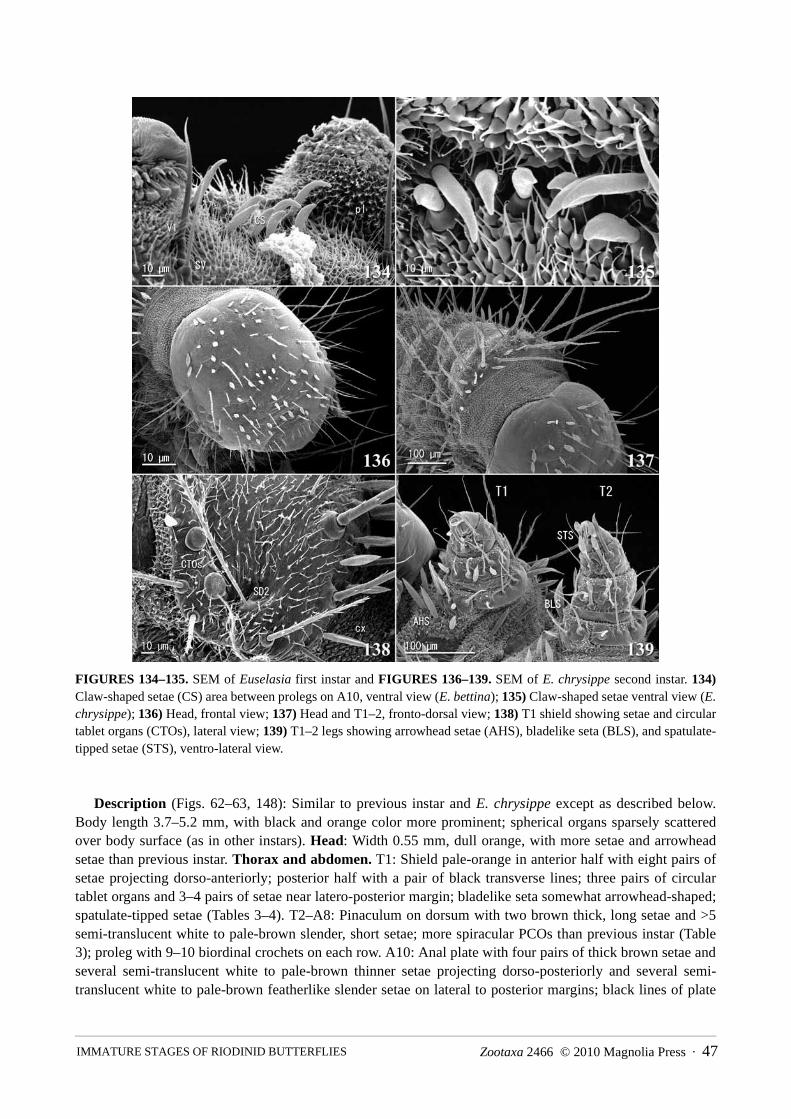
Zootaxa 2466 © 2010 Magnolia Press · 47 IMMATURE STAGES OF RIODINID BUTTERFLIES
FIGURES 134–135. SEM of Euselasia first instar and FIGURES 136–139. SEM of E. chrysippe second instar. 134)Claw-shaped setae (CS) area between prolegs on A10, ventral view (E. bettina); 135) Claw-shaped setae ventral view (E.chrysippe); 136) Head, frontal view; 137) Head and T1–2, fronto-dorsal view; 138) T1 shield showing setae and circulartablet organs (CTOs), lateral view; 139) T1–2 legs showing arrowhead setae (AHS), bladelike seta (BLS), and spatulate-tipped setae (STS), ventro-lateral view.
Description (Figs. 62–63, 148): Similar to previous instar and E. chrysippe except as described below.Body length 3.7–5.2 mm, with black and orange color more prominent; spherical organs sparsely scatteredover body surface (as in other instars). Head: Width 0.55 mm, dull orange, with more setae and arrowheadsetae than previous instar. Thorax and abdomen. T1: Shield pale-orange in anterior half with eight pairs ofsetae projecting dorso-anteriorly; posterior half with a pair of black transverse lines; three pairs of circulartablet organs and 3–4 pairs of setae near latero-posterior margin; bladelike seta somewhat arrowhead-shaped;spatulate-tipped setae (Tables 3–4). T2–A8: Pinaculum on dorsum with two brown thick, long setae and >5semi-translucent white to pale-brown slender, short setae; more spiracular PCOs than previous instar (Table3); proleg with 9–10 biordinal crochets on each row. A10: Anal plate with four pairs of thick brown setae andseveral semi-translucent white to pale-brown thinner setae projecting dorso-posteriorly and several semi-translucent white to pale-brown featherlike slender setae on lateral to posterior margins; black lines of plate

NISHIDA48 · Zootaxa 2466 © 2010 Magnolia Press
connected to each other and arch-shaped; posterior middle portion of plate whitish; twelve claw-shaped setaebetween prolegs (Table 3); proleg with 14 biordinal crochets in lateroseries and 8–9 biordinal crochets inmesoseries.
Material examined: Laguna de Hule, 2003 (SEM, n = 2; dissecting stereo-microscope, n = 2).
FIGURES 140–145. SEM of E. chrysippe second instar. 140) Arrangement of D-group on T3–A2, dorsal view; 141)PCOs contiguous to spiracles on A6–7, lateral view; 142) spherical organs (SO) and circular tablet organs (CTOs) on
right side of A8+9–A10, lateral view; 143) Anal plate showing setal arrangement and circular tablet organs (CTOs)
(circular tablet organ replaced with a seta = arrow, and left end circular tablet organ missing), dorso-caudal view; 144)Cupola organ (CO) on anal plate and spherical organs (SO) on proleg on right side A10, dorso-lateral view; 145) Flat seta
(FS) on A8–9 and claw-shaped setae (CS) between A10 prolegs, ventral view.
FOURTH INSTAR Diagnosis: The head capsule width of Euselasia bettina is ca. 0.80 mm; the overall body color is grayish-
yellow; the T1 shield is mostly black; the pinacula on dorsum are prominently blackl the mesal line andintersegmental membrane are dark-green; the proleg on A10 has 25–26 biordinal crochets in lateroseries. InE. chrysippe the head capsule width is slightly narrower (ca. 0.72 mm); the overall body color is grayish-green; the T1 shield is mostly orange; the pinacula on dorsum are dull black; the mesal line andintersegmental membrane are dull green; and the proleg on A10 has 20–22 crochets in lateroseries.

Zootaxa 2466 © 2010 Magnolia Press · 49 IMMATURE STAGES OF RIODINID BUTTERFLIES
Description (Figs. 63–65): Similar to of E. chrysippe and previous instar, except as described below. Bodylength 7.6–9.8 mm, recently molted larvae dark-gray with prominent black pinacula (Fig. 63), mid- to late-stage larvae grayish-yellow. Mesal line and intersegmental membrane dark-green. Head: Width 0.80 mm,dark-brown to black. Thorax and abdomen. T1: Shield dark-brown to black, slightly orange to yellowishfrontally. T2–8: With black pinaculum on dorsum; a faint white longitudinal line subdorsally along laterallobes; proleg with 14–17 biordinal crochets in each row. A10: Posterior middle portion of anal plate promi-nently white; proleg with 25–26 biordinal crochets in lateroseries and 12–13 biordinal crochets in mesoseries.
Material examined: Laguna de Hule, 2003 (dissecting stereo-microscope, n = 3).
FIGURES 146–151. SEM of mandibles of Euselasia (inner surface). 146) First instar, left (E. chrysippe); 147) Firstinstar right mandibular setae, inner view (E. chrysippe); 148) Third instar, left (E. bettina); 149) Fourth instar, left (E.chrysippe); 150) Sixth instar, left (E. chrysippe); 151) Sixth instar, left (E. bettina).
FIFTH INSTAR Diagnosis: The fifth instar of Euselasia bettina is greenish-black to greenish-dull yellow with prominent
black pinacula on dorsum; the head capsule is ca. 1.30 mm wide; the T1 shield is entirely or mostly black witha little orange; and the proleg on A10 has 15–16 crochets in a mesoseries. The fifth instar of E. chrysippe isgray to yellowish-gray with less promiment black pinacula; the head capsule is slightly narrower (ca. 1.23mm); the T1 shield is mostly orange; and the proleg on A10 has 11–13 crochets in mesoseries.

NISHIDA50 · Zootaxa 2466 © 2010 Magnolia Press
Description: (Figs. 66–68). Similar to E. chrysippe and previous instar, except as described below. Bodylength 10.0–14.5 mm; overall color slightly darker and with larger and prominent black pinacula on dorsumcompared to E. chrysippe fifth instar (Fig. 68); recently molted and early stage dark as in fourth instar, butslightly greenish (Fig. 66) and with whitish-pale color around black pinacula on dorsum; overall colorbecoming paler as larva matures (Figs. 67–68). Head: Width 1.30 mm, black. Thorax and abdomen. T1:Shield dark-brown to black or dark-brown with pale-yellow to orange. A3–6: Proleg with 21–24 biordinalcrochets on each row. A10: Proleg with 32–36 biordinal crochets in lateroseries and 15–16 biordinal crochetsin mesoseries.
Material examined: Laguna de Hule, 2003 (dissecting stereo-microscope, n = 3)
FIGURES 152–158. SEM of Euselasia sixth instar. 152) Head, frontal view (E. chrysippe); 153) Head, latero-frontalview (E. chrysippe); 154) Head, frontal-ventral view (E. chrysippe); 155) Mouthparts, frontal view (E. chrysippe); 156)Areas between labrum and antenna, frontal view (E. chrysippe); 157) Areas between labrum and antenna, frontal view(E. bettina); 158) Mouthparts, frontal view (E. bettina).
SIXTH INSTAR Diagnosis: The sixth instar of Euselasia bettina is brownish-amber with reddish or greenish tint; the head
capsule is ca. 1.75 mm wide; the color of the head is black with a pair of brownish-orange to pale-orangepatches running parallel to the epicranial suture; the arrowhead setae are flattened as in the first instar; the

Zootaxa 2466 © 2010 Magnolia Press · 51 IMMATURE STAGES OF RIODINID BUTTERFLIES
curvature of ventral margin of labrum is widely angled (ca. 120°); the mandible is large (ca. 0.45 mm wide)with more distinct dentition than in E. chrysippe; the T1 shield is bright orange with reddish tint, with blacktransverse lines with a faint bluish-gray iridescence; the pinacula on the dorsum is black with an oval whiteline; the iridescence of the structural color plates is bright and metallic sky-blue; and the proleg on A10 hasbiordinal 15–16 crochets in mesoseries. The sixth instart of E. chrysippe is greenish-dark-gray to greenish-dull black; the head is slightly narrower (ca. 1.65 mm) and bright orange or black, or intermixture of these twocolors; the arrowhead setae are cone-shaped (not flattened), ridged and spiraled apically; the curvature of theventral margin of the labrum is more narrowly angled (ca. 110°); the mandible is smaller (0.38 mm wide) withless distinct dentation; the T1 shield is orange to bright orange, without iridescence; the oval line on the dorsalpinacula is pale-gray; the iridescence of the structural color plate is less bright, faint metallic-blue; and theproleg on A10 has 11–13 crochets in mesoseries.
FIGURES 159–164. SEM of Euselasia sixth instar. 159) Labrum, inner surface (E. chrysippe); 160) Labrum, inner sur-face (E. bettina); 161) Frons showing arrowhead setae, cupola organs, and other setae, fronto-ventral view (E. chry-sippe); 162) Left side frons showing adfrontal suture area and arrowhead setae, cupola organ, and other setae, fronto-lateral view; 163) Arrowhead setae of figure 161 (E. chrysippe); 164) Arrowhead setae of figure 162 (E. bettina).

NISHIDA52 · Zootaxa 2466 © 2010 Magnolia Press
Description: (Figs. 68–73, 157–158, 160, 162, 164–207). Similar to E. chrysippe and previous instarexcept as described below. Body length 15.0–22.0 mm, coloration most distinct from all other instars and E.chrysippe; brownish-amber with reddish or greenish tint, mesal line on dorsum and intersegmental membranedull black to dark-green. Head (Figs. 70, 72–73, 157–158, 160, 162, 164, 165–170): Width ca. 1.75 mm,black with a pair of brownish-orange to pale-orange patches running parallel to epicranial suture; arrowheadsetae (Figs. 72, 157–158, 162, 164) flat, variable in size; labrum as figured (Figs. 158, 160), curvature ofventral margin narrowly angled (ca. 110°); mandible (Fig. 151) similar to E. chrysippe but larger (ca. 0.45 mmwide), dentation slightly more distinct (n = 5). Thorax and abdomen. T1: Shield reddish-orange to orangewith black and inner portion black with faint bluish-gray iridescence. T2–A8: Pinacula on dorsum penelipticalblack ring surrounding an oval white line dorsally and structural color plate subdorso-anteriorly; structuralcolor plate with metallic sky-blue iridescence with center concave and black (Figs. 69–71, 195); proleg with21–26 biordinal crochets on each row. A10: Anal plate with metallic sky-blue iridescence as in othersegments; proleg with 31–33 biordinal crochets in lateroseries in semicircle and 15–16 biordinal crochets inmesoseries.
Material examined: Laguna de Hule, 2003 (SEM, n = 2; dissecting stereo-microscope, n = 3).
FIGURES 165–170. SEM of Euselasia sixth instar. 165) Head, frontal-ventral view (E. bettina); 166) Head, latero-fron-tal view (E. bettina); 167) Head, ventral view (E. bettina); 168) Oral area showing spinneret and maxillary palpus (E.bettina); 169) Left antenna showing sensillar arrangement, frontal view (E. bettina); 170) Left maxillary palpus showingsensillar arrangement (E. chrysippe).

Zootaxa 2466 © 2010 Magnolia Press · 53 IMMATURE STAGES OF RIODINID BUTTERFLIES
FIGURES 171–176. SEM of Euselasia sixth instar (E. chrysippe). 171) T1 shield, dorsal view; 172) Right side latero-posterior margin of T1 shield showing SD2, other setae, and circular tablet organs (CTOs), dorsal view; 173) SD2 setanear apex, lateral view; 174) Basal area of SD2 seta, dorso-lateral view; 175) Socketed area of SD2, dorso-lateral view;176) Bladelike seta on right T1 leg, ventro-lateral view.
PREPUPADiagnosis: The prepupa of Euselasia bettina is amber with black. That of E. chrysippe is dark brownish-
gray with pale-brown.Description (Fig. 75): Length 13.2–13.7 mm (n = 3); overall color amber with black.Material examined: Laguna de Hule, 2003 (n = 15).
PUPA Diagnosis: The pupa of Euselasia bettina is 11.4–13.5 mm long and 5.7–6.4 mm wide. That of E.
chrysippe is usually smaller, 11.5–12.0 mm long and 5.0–5.5 mm wide.Description (Figs. 76–77, 208–225): Similar to E. chrysippe except as described below. Length 11.4–13.5
mm, width 5.7–6.4 mm, depth 2.5–3.5 mm; dark-brown to black areas more prominent; pupal shell colorlesssemi-translucent in thorax, head, and wing sclerite, intersegmental membrane A4–5 and A5–6 orange-brown,

NISHIDA54 · Zootaxa 2466 © 2010 Magnolia Press
occasionally with brown spots on center of each eye; weak sculturing on outer one-third of eye, basal portionsof antenna, prothoracic and mesothoracic legs, and proboscis, and mid- to apical area of wing sclerites; mouthparts as figured (Figs. 210–211), most specimens examined (8 of 10) with widened clypeus and broadenedmandibles as in Fig. 210; some specimens more like Fig. 211 with narrower clypeus and mandibles, i.e., fronsand clypeus-mandible area forming straight parallel lines; middle portion of eye with a notch at posterior end;notch absent in some specimens.
Material examined: Laguna de Hule, 2003 (SEM, n = 2 complete specimens and 2 shells; dissecting ste-reo-microscope, n = 3 complete specimens and 10 shells).
FIGURES 177–182. SEM of Euselasia sixth instar. 177) Spatulate-tipped setae on right T2 leg, ventral view (E. chry-sippe); 178) Spatulate-tipped setae on tibia of right T1 leg, ventral view (E. chrysippe); 179) Spatulate-tipped setae ontibia of right T1 leg, ventro-lateral view (E. chrysippe); 180) Ventral surface of spatulate-tipped setae on tibia of left T2leg (E. chrysippe); 181) Dorsal surface of spatulate-tipped setae on tibia of T1 leg (E. bettina); 182) Spatulate-tippedsetae on tibia and femur of right T3 leg, ventral view (note spatulate-tipped setae on femur having sharply pointed apex)(E. bettina).

Zootaxa 2466 © 2010 Magnolia Press · 55 IMMATURE STAGES OF RIODINID BUTTERFLIES
Diagnosis and description of adult of Euselasia chrysippe
Diagnosis: The discal areas of the upper wing surface of males of E. chrysippe are reddish-orange; femalesare yellowish-orange. Both sexes have 5–7 submarginal black spots on the underside of the hindwings. Theupper wing surface of both males and females of E. bettina are black to dark-gray. The submarginal blackspots along outer margin are faint or absent, except for the apical spot.
Description: Sexually dimorphism conspicuous (Figs. 1–3, 6–7, 8, 16); six to seven small black spots(third one from apex most conspicuous) along outer submargin of hind wings in both sexes. Underside ofwings more orange in male, outer margin of wings more rounded in female. For additional details of dimor-phism see DeVries (1997). Rearing of a group of larvae collected in Refugio Vida Silvestre La Marta inPejibaye de Turrialba produced several males having the upper wing surface yellowish-orange (i.e., similar tothat of females) (Fig. 3).
Material examined: Lake Arenal, 2003 (n = 2 pinned males and 2 pinned females); Turrialba, 2000 (n =22 pinned males and females).
FIGURES 183–188. SEM of Euselasia sixth instar. 183) SV-group and spherical organs on right side T1, lateral view(E. chrysippe); 184) Raised PCOs surrounding spiracle of left A5, lateral view (right = caudad) (E. chrysippe); 185)Raised PCO of figure 184; 186) Sunken PCOs surrounding and spherical organs near left A7 spiracle, lateral view (bot-tom = caudad, right = dorsal) (E. chrysippe); 187) Sunken PCO (E. bettina); 188) Spherical organ near A7 spiracle (E.chrysippe).

NISHIDA56 · Zootaxa 2466 © 2010 Magnolia Press
FIGURES 189–194. SEM of Euselasia sixth instar. 189) Left A8+9 spiracle and spiracular spherical organs surroundingspiracle (top = caudad, right = ventral) (E. chrysippe); 190) Spiracular spherical organs, posterior lower corner of spira-cle in figure 189; 191) dark-brown dorsal seta of left A5, lateral view (E. chrysippe); 192) L-group on lateral lobe; 193)L-group white non-plumose seta; 194) L-group white plumose seta (E. bettina).
Diagnosis and description of adult of Euselasia bettina
Diagnosis: The upper wing surface of males and females of E. bettina is dark-gray to black. The submarginalblack spots along the outer margin of the hindwings are inconspicuous or very faint except the one near theapex. Faint black spots are absent near the apex. Discal areas of the upperside of the wings of E. chrysippe arereddish-orange in the male, yellowish-orange in female, and the submarginal black spots are more conspicu-ous (5–7 spots).
Description: Sexual dimorphism slightly inconspicuous; male (Figs. 4, 9) with upperside of wings velvetblack, female (Figs. 5, 10) dark-brown with grayish tint (DeVries 1997). Underside slightly more orange inmale, outer margin of wings more rounded in female. Four to five small to faint black spots along outersubmargin of hind wing of both sexes (that closest to apex larger).
Material examined: Laguna de Hule, 2003 (n = 4 pinned males and 4 pinned females).

Zootaxa 2466 © 2010 Magnolia Press · 57 IMMATURE STAGES OF RIODINID BUTTERFLIES
FIGURE 195. Metallic-blue iridescence of structural color plate of an EtOH preserved E. bettina sixth instar larva, left
side A6, lateral view, left = cephalad.
FIGURES 196–201. SEM of Euselasia sixth instar. 196) Pinaculum on lateral part of dorsum of abdominal segment,dorso-lateral view (right = cephalad) (E. chrysippe); 197) Anterior edge of structural color plate on right A5, right side ofthickly haired area = dark-pigmented area, dorso-lateral view (right = cephalad) (E. chrysippe); 198) Dark-pigmentedarea contiguously with structural color plate (right = cephalad) (E. bettina); 199) Tonofibrillary platelets in dark-pigmented area of structural color plate, dorso-lateral view (right = caudad) (E. chrysippe); 200) Structural color plate,lateral view (E. bettina); 201) Structural color plate surface (E. chrysippe).

NISHIDA58 · Zootaxa 2466 © 2010 Magnolia Press
FIGURES 202–207. SEM of Euselasia sixth instar. 202) Anal plate, dorso-caudal view (E. chrysippe); 203) Anal plateshowing circular tablet organs, dorso-caudal view (E. chrysippe); 204) Anal plate showing circular tablet organs andspherical organs, dorsal view (E. chrysippe); 205) Circular tablet organ on A10 (note pores on surface in linear form),dorsal view (E. chrysippe); 206) Venter of A6–A10 showing L-group and claw-shaped setae (CS), ventral view (E.bettina); 207) claw-shaped setae between A10 prolegs, ventral view (note more or less circular area in the center part) (E.bettina).
Discussion
Host plants and host specificity of EuselasiaAlthough members of Euselasia feed on a wide array of plant species, every species whose biology is
known is stenophagous, i.e., has a very narrow host range that includes a small number of species within oneto a few related genera. For example, in the field, searching for eggs and larvae on Miconia affinis DC. and M.paleacea Cogn. that were growing contiguously with M. calvescens did not reveal the presence of E.chrysippe. Also, under rearing conditions late instar larvae of E. chrysippe collected at Arenal did not feed onthe leaves of M. affinis (no-choice test, n = 1 group).

Zootaxa 2466 © 2010 Magnolia Press · 59 IMMATURE STAGES OF RIODINID BUTTERFLIES
FIGURES 208–211. SEM of Euselasia pupa. 208) Dorsal view, (E. chrysippe); SC = silk clipper, sp = spiracle, Tsp =thoracic spiracle, tz = subdorsal triangular zone; 209) Ventral view, (E. chrysippe); An = antenna, E = eye piece (a: innerpart, b: middle, c: outer), ML = mesothoracic leg, Mx = maxilla, PL = prothoracic leg, Pr = proboscis, W = wing sclerite;210) Mouth parts, ventral view (E. chrysippe); ATP = anterior tentorial pit, Cl = clypeus, Li = labium, Lr = labrum, Ma =mandible; 211) Mouth parts, ventral view (note notch on outer lower portion of eye piece) (E. bettina), ATP? = probablyanterior tentorial pit.
In regards to the use of Conostegia rufescens as a larval host by Euselasia chrysippe, the genus Conostegiais a member of Miconieae and thus closely related to Miconia (Michelangeli et al. 2004). The preliminaryresults of molecular work by Michelangeli et al. (2004) revealed that Miconia is polyphyletic. The similarityof these genera is also suggested by the observation that most gall-inducing species of Mompha (Lepidoptera:Gelechioidea: Momphidae) in Costa Rica are found on both Conostegia and Miconia (Nishida 2005).
Although it is a preliminary assessment, Euselasia species can be separated into six groups on the basis oftheir host plant families (Table 1): a Clusiaceae group (Malpighiales), a Melastomataceae group (Myrtales), aMyrtaceae group (Myrtales), a Sapotaceae group (Ericales), a Vochysiaceae group (Myrtales), and a Euphor-biaceae group (Malpighiales) (Stevens 2009).
DeVries (1997) and J.P.W. Hall (pers. comm. 2006) mention that cryptic species are common in Euselasia.For example, the wing pattern of the adults of Euselasia ‘hygenius’ DHJ01 in northwestern Costa Rica (Jan-

NISHIDA60 · Zootaxa 2466 © 2010 Magnolia Press
zen & Hallwachs 2009) and E. hygenius in Brazil are extremely similar, but based on the mitochondrial geneCOI, the two are different species. Larvae of the former were found on Clusiaceae (Janzen & Hallwachs 2009)and those of the latter on Myrtaceae (Beccaloni et al. 2008, etc.).
Surprisingly, host records of E. procula are from two different plant families, both in Costa Rica. Onerecord is from a southwestern rainforest feeding on Myrtaceae (DeVries 1997) and the other from a northwest-ern rainforest feeding on Melastomataceae (Janzen & Hallwachs 2009).
Miconia-feeding Euselasia species as possible biological control agents of Miconia calvescensThe use of biological control agents for the management of invasive plant pests on tropical islands and
other tropical habitats provides a viable alternative to traditional approaches of weed control and mayrepresent one the least invasive and ecologically safe methods, especially when compared to the application ofherbicides and other chemical treatmentss (Johnson & Denslow 2005; Denslow & Johnson 2006).
FIGURES 212–217. SEM of Euselasia pupa. 212) Thoracic spiracle between T1 (right) and T2 (left), dorsal view (E.chrysippe); 213) Surface sculpture of thoracic spiracle (E. chrysippe); 214) Mesal portion of dorsum of T3–A1 showingsilk-clipper structure (silken girdle in situ) on A1 (E. bettina); 215) Cone-shaped raised base of clubbed setae; 216) Stemof D-seta on left A6, dorso-lateral view (E. chrysippe); 217) Apical area of D-seta on left A6, lateral view (E. bettina).
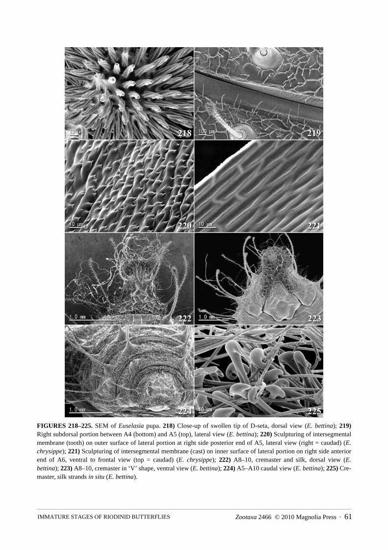
Zootaxa 2466 © 2010 Magnolia Press · 61 IMMATURE STAGES OF RIODINID BUTTERFLIES
FIGURES 218–225. SEM of Euselasia pupa. 218) Close-up of swollen tip of D-seta, dorsal view (E. bettina); 219)Right subdorsal portion between A4 (bottom) and A5 (top), lateral view (E. bettina); 220) Sculpturing of intersegmentalmembrane (tooth) on outer surface of lateral portion at right side posterior end of A5, lateral view (right = caudad) (E.chrysippe); 221) Sculpturing of intersegmental membrane (cast) on inner surface of lateral portion on right side anteriorend of A6, ventral to frontal view (top = caudad) (E. chrysippe); 222) A8–10, cremaster and silk, dorsal view (E.bettina); 223) A8–10, cremaster in ‘V’ shape, ventral view (E. bettina); 224) A5–A10 caudal view (E. bettina); 225) Cre-master, silk strands in situ (E. bettina).

NISHIDA62 · Zootaxa 2466 © 2010 Magnolia Press
FIGURES 226. Egg cluster of Euselasia aurantia, dorso-lateral view. Note the differences in color of eggs comparedwith E. chrysippe and E. bettina.
FIGURE 227. Egg cluster of E. aurantia, lateral view. Note space between eggs and plant surface.
Miconia-feeding Euselasia can be considered potential biological control agents for Miconia calvescensfor the following reasons: 1) larvae are highly specific to their host plants as mentioned above (see also Table1) and no native or economically important cultivated Melastomataceae are found in Hawaii. However, giventhe presence of commercial guava orchards (Hawaiian Alien Plant Studies 1998) and Eucalyptus plantations(Whitesell et al. 1992), host specificity tests using cultivated guava leaves (Psidium guajava L.), Eucalyptus,and other endemic myrtaceous plants (e.g. Eugenia spp. and Metrosideros spp.) is strongly recommended.This is because both Myrtaceae and Melastomataceae are in the order Myrtales; some species of Euselasia areknown to feed on Eugenia, Eucalyptus, and Psidium (Table 1) and E. procula is reported to feed on bothMelastomataceae and Myrtaceae. Moreover, the immature stages of E. chrysippe have been found only onmelastomes through the massive caterpillar rearing project led by D. H. Janzen and W. Hallwachs over nearly30 years (Janzen & Hallwachs 2009). 2) The larvae are gregarious feeders that devour leaf tissue in largequantities in relatively short periods of time, and their life cycle turnover is relatively short, approximatelytwo months (this study; Allen 2007). However, the egg stage lasts relatively long compared to the larval andpupal stages and to the egg stage of other butterflies in general. According to Allen (2007), the egg stagelasted up to 6 weeks under field conditions at Reserva Ecológica Leonelo Oviedo. 3) They inhabit a widerange of elevations: E. chrysippe from sea level to 1500 m, E. bettina from 400 to 1250 m, and E. aurantiafrom 100 to 1700 m. 4) Adults feed on rotten fruit (under laboratory conditions; personal observation),extrafloral nectarines, and other non-floral sources (under natural conditions) (DeVries 1997). Also, the factthat females contain more than 160 eggs plus undeveloped immature eggs (this study) suggests that adultsprobably live for more than a month and that females oviposit more than once (Allen 2007). Another aspect offruit-feeding is that it may function to keep adults alive and reproducing under laboratory conditions and inthe field after release, i.e., they are not dependent on certain flowers for feeding.

Zootaxa 2466 © 2010 Magnolia Press · 63 IMMATURE STAGES OF RIODINID BUTTERFLIES
Egg and egg stalkThe frustum-shaped eggs of the three Miconia-feeders documented in this study (E. chrysippe, E. bettina,
and E. aurantia) are very similar in shape to those of the non-Miconia-feeders E. mystica and E. hieronymi,which feed on myrtaceous plants (Downey & Allyn 1980; DeVries 1997). In contrast, Hoffmann (1931)extracted 56 eggs from a reared female of E. eucerus and stated that the eggs are round with lines on thechorion.
The general coloration and pigmentation of eggs described appear to be relatively reliable for distinguish-ing species within the three Miconia-feeders. The eggs of E. aurantia (Figs. 226–227) can be distinguishedfrom the other species studied herein by the following general characters: translucent pale-brown (slightlydull compared to E. chrysippe) with pigmentation of the apical rim area being purplish-faint black, extendingclose to one-half the egg height (becoming paler farther from the rim) along with dark lines extending fromthe lateral ridges. Several eggs lacked pigmentation except on the rim and lateral ridges, giving the impressionthat they are pale. Variation in the pigmentation within an egg cluster may not be uncommon, as observed inE. chrysippe and E. aurantia.
Stalked eggs were previously unreported in Lepidoptera. Thus far I have observed stalks on eggs of threespecies of Euselasia (i.e., E. aurantia, E. bettina, and E. chrysippe) but not on the eggs of Corrachialeucoplaga (Euselasiinae). Although the eggs of E. hieronymi (Downey & Allyn 1980) and E. mystica(DeVries 1997) were illustrated, stalks on the eggs were not documented. The function of the stalk isunknown apart from the fact that it keeps the eggs raised above the surface of the leaves.
The eggs of Corrachia leucoplaga are lozenge-shaped (i.e., short cylindrical), with a smooth surface; how-ever, they resemble the eggs of Euselasia in the following: purplish pigmentation along the rim; lower bottomslightly wide, i.e. very slightly frustum shaped with round latero-ventral area; and concave micropyle area (n= 170 eggs; K. Nishida, in prep.). The stalk is absent (n = 167 eggs/cluster) and the stalk conjunctionalstructure apparently is absent (n = 3 eggs extracted from a female); i.e., it was not observed using the same64X stereo-microscope that revealed the presence of the stalk and stalk conjunctional structure on the eggs ofEuselasia species.
Harvey (1987b) and DeVries (1997) noted that the smooth barrel- to frustum-shaped eggs of Euselasiinaeare unique within Riodinidae, and Lepidoptera in general, and that this character state potentially suggests aclose relationship between Styx, Corrachia, and Euselasia. Hence, it is likely that stalked egg and stalkconjunctional structure is derived either within the genus Euselasia or the tribe Euselasiini. Furthermore, eggshape and pigmentation, and the biology of Praetaxila segecia punctaria (Fruhstorfer, 1904) in northern Aus-tralia and Hamearis lucina (Linnaeus, 1758) (Riodinidae: Nemeobiinae) in Europe are somewhat similar tothose of Euselasia, although the eggs of the former species are laid in a loose cluster on the substrate (i.e.,without stalks) (Harvey 1987b; Oates & Emmet 1990; Samson et al. 1999; Braby 2004; Kettner 2009).
General morphology of the larvae and pupaeThe general appearance of the larvae and pupae of the two Miconia-feeding species, E. chrysippe and E.
bettina, is similar, especially when compared to other Euselasia species that feed on other plant families(Hoffmann 1931; Brévignon 1997; DeVries 1997; Janzen & Hallwachs 2009). In fact, it is difficult to distin-guish the larvae of E. chrysippe and E. bettina until they reach the last instar. Except for coloration and minordifferences in body size, the two can be distinguished only by the form of the arrowhead setae. According toP.E. Allen (pers. comm. 2007), the mature last instar larva of E. aurantia is quite similar to that of E. bettina,and the overall appearance of the pupa is virtually identical to that of E. chrysippe and E. bettina. There are nosignificant differences in morphology and coloration that clearly separate the pupae of E. chrysippe and E.bettina.
Larval morphologyA detailed examination of the larvae of Euselasia chrysippe and E. bettina revealed many morphological
features previously unrecorded in lepidopteran larvae, particularly in the first instar. In many Lepidoptera thefirst instar shows specialized behaviors which often are related to establishment on the host plant (Zalucki et

NISHIDA64 · Zootaxa 2466 © 2010 Magnolia Press
al. 2002). Hence, further studies are required to determine the possible functions of the circular tablet organs,the bladelike setae, the flat setae, the large number of setae in most of the setal groups (in contrast to theabsence or reduction in the number of setae in some groups), and the bifurcate D-group setae, which occuronly in first instar larvae.
Bifurcate dorsal setae are reported in exceedingly few Lepidoptera. They were observed in the first instarof Praetaxila segecia punctaria (Samson et al. 1999), potentially representing a synapomorphy for Praetaxilaand Euselasia. However, because the first instars of so few other riodinids have been reported, the taxonomicdistribution of this character state is poorly known. Extraordinarily, the dorsal setae of first instar larvae ofsome species of Catasticta group (Pieridae: Pierinae) are bifurcated at the apex (Braby and Nishida, in press).
Differences in the shapes of PCOs (sunken, flat, or raised) and spherical organs (flat to papillalike) may beartifacts of larval preservation or in preparation for SEMs. It is also uncertain whether the sphere part of thespherical organ and/or the raised part of the PCO are part of the secretion or part of the organ itself.
Other major differences between the first instar and other instars of these two Euselasia species are thelocation of the circular tablet organ and spherical organ on the T1 shield, as well as the large, stout bladelikeseta on each T1 leg. The functions and homologies of these features among different instars and among otherspecies are unknown. The conclusion that lateral fusion has occurred in A8 and A9 was determined byexternal appearance, not by dissections to observe internal muscle attachment.
The complement of setae on the anal plate is unusual, exceeding the numbers seen in other setal groups onother segments (N. Kristensen and I. Hasenfuss, pers. comm. 2006). However, the naming of the setae wasavoided because their homologies with other setae remain unknown.
Feeding mode and mandibular morphologyThe mandibles of first and second instars possess sharply pointed, distinct teeth, the tips of which overlap
slightly, allowing shearing of leaf tissue. Larvae of these instars feed by grazing on the surface tissue of theleaf. Considerable change in mandible morphology occurs between the second and third instars, and there is aconcomitant change in the feeding mode from grazing (in the second instar) to grazing, skeletonizing, andfeeding on entire leaf tissue, including the thin veins (in the third instar). The mandibles in the third instar pos-sess less distinct teeth with a concave inner surface, possibly allowing them to feed both by grazing the sur-face (as in earlier instars) and clipping through entire leaf tissue (as in later instars). The single sharp ridgeliketeeth with a more deeply concave inner surface of later instars likely allow larvae to bite through the entireleaf tissue. Changes in feeding modes and mandibular morphology also have been observed in the larvae ofthe leaf-tying moth Psilocorsis cryptolechiella (Chamber, 1872) (Oecophoridae) on Fagus grandifolia Ehrh.(Fagaceae) (Embree 1958 [cited as P. gaginella]) and in external-feeding Heterocampa obliqua (Packard,1864) (Notodontidae) on Quercus macrocarpa Michx. (Fagaceae) (Godfrey et al. 1989). Godfrey et al. (1989)hypothesized that loss of the sharply pointed mandibular teeth in these two species is related to the change infeeding mode from grazing on leaf surface tissue to clipping the entire leaf tissue. This observation in Eusela-sia may represent the first documented case of changing mandibular morphology associated with feedingmode in butterflies.
Structural color platesHigh magnification SEM images (up to 41,000X) of cuticle surface of the structural color plates did not
reveal any micro-structures that might produce iridescence. Even under this magnification, the surface wassmooth. It is possible that such structures were modified or destroyed in the process of SEM specimen prepa-ration. The structural color plate of larvae boiled in water and preserved in 75% EtOH did not show a metal-lic-blue iridescence as is obvious in live specimens (Fig. 195). None of the larvae treated with 100% EtOHand critical point dried showed any of the metallic color. However, dry-preserved molted skins of both specieshad dark purplish shiny iridescence. When the skins were soaked in water, the iridescence on the structuralcolor plate became pale metallic-purple to pale-blue. It is likely is that the structure is internal, i.e., locatedbelow the cuticular surface in multiple semi-translucent layers. Thus, it may be necessary to dissect the struc-tural color plate and use SEM or use transmission electron microscope to reveal its structure (M. Hoshi, pers.comm. 2006).

Zootaxa 2466 © 2010 Magnolia Press · 65 IMMATURE STAGES OF RIODINID BUTTERFLIES
Structural colors are uncommon in larvae of Lepidoptera, and presumably they function in aposematism.There are several examples of aposematism in gregarious larvae of other Lepidoptera (e.g., S.-Tullberg &Hunter 1996), but bright coloration obviously is not restricted to gregarious species. Solitary last instar larvaeof some Melese and Bertholdia species (Arctiidae) have rows of bright metallic-blue spots on the dorsum(pers. obs. 2007). Last instar larvae of Doleschallia bisaltide (Cramer, 1777), D. dascylus Godman & Salvin,1880, D. noorna Grose-Smith & Kirby, 1893, Yoma sabina (Cramer, 1780) (Nymphalidae: Nymphalinae)exhibit metallic-blue on the basal area of their dorsal to lateral setae (Igarashi & Fukuda 1997, 2000), and sev-eral species of saturniids have silver markings on their dorsum: e.g., Actias dubernardi (Oberth.) (Ylla et al.2005); Sphingicampa hubbardi (Dyar) (Sawby 2005); Syssphinx species (Janzen & Hallwachs 2009); andsome African saturniids (R. Peigler, pers. comm. 2006).
Number of larval instarsIn the family Riodinidae, it is not unusual for larvae to have more than five instars (D.J. Harvey, pers.
comm. 2006). For example, Lemonias caliginea (Butler, 1867) [cited as Anatole rossi] has six (Ross 1964),and Calephelis borealis (Grote & Robinson, 1866) (dos Passos 1936) and C. muticum McAlpine, 1937(McAlpine 1938) have eight to nine instars; all are members of Riodininae. Zanuncio et al. (1990, 1995)reported that the larvae of Euselasia hygenius on Eucalyptus urophylla S. T. Blake has six instars and thatEuselasia melaphaea [cited as E. apisaon], E. eucerus, and E. hygenius occulta have five instars. It should benoted that E. hygenius occulta, or E. occulta Stichel, 1919 is a synonym of E. hygenius, according toCallaghan & Lamas (2004). According to Chacón (2001), E. chrysippe has five larval instars. Although thereappear to be differences in the number of instars, the presence of six instars in Euselasia is apparentlycommon. The number of larval instar in related genera and tribes is not available. Furthermore, Hamearislucina and Praetaxila segecia punctaria (Nemeobiinae) have four (Oates & Emmet 1990) and five (Samson etal. 1999) instars, respectively. It is noteworthy that mis-counting of instars frequently occurs during the rear-ing of middle instars owing to their extreme similarity and rapid development (pers. obs.; P.E. Allen, pers.comm. 2007).
Processionary behaviorThe processionary behavior of the larvae is facilitated, at least in part, by tactile stimuli (contacts made by
setae) and an extra-silk chemical substance(s) which functions as a trail pheromone. The tactile stimuli appearto take priority over the chemical marker and silk as shown in other processionary lepidopteran species(Fitzgerald & Pescador-Rubio 2002; Fitzgerald 2003). However, no experiments were conducted to determineif silk laid during the procession serves any function beyond allowing larvae to grip the substrate duringlocomotion. Halting and reinitiating the procession are most likely controlled by setal contact of the larva withthose in front and behind. Although the origin of the trail pheromone is unknown, the following two organsare possible sources or disseminators of it: 1) the claw-shaped setae on the venter of A10 between the prolegs(Figs. 130, 134–135, 145, 206–207), and 2) mesally located spatulate-tipped setae on the thoracic legs (Figs.111, 115, 176–181). These are the only organs that are present in all larval instars and appear to make consis-tent contact with the substrate. Larvae dragged the tip of the last abdominal segment on the substrate whilemoving in procession (Fig. 43; Nishida 2007), suggesting that setae here, perhaps including the cuticle, mightbe the pheromone source. The same type of dragging behavior has been observed in several other procession-ary lepidopteran caterpillars, for example Hylesia lineata Druce (Saturniidae), Gloveria, and Malacosoma(Lasiocampidae) (Fitzgerald 2003, and references therein). In addition, Fitzgerald & Pescador-Rubio (2002)observed ‘specialized’ setae on the ventral surface of the tip of the last abdominal segment (i.e., located in aposition similar to that in Euselasia) on Hylesia lineata larvae that are morphologically unique and suggestedthat these setae are the most likely source of the trail pheromone.
With regard to the second possible source, the spatula-shaped setal tips are appropriate for spreading a sub-stance and they make contact with the substrate during locomotion. In Riodinidae, these setae are known onlyin Euselasiinae and are considered to be a synapomorphy for the subfamily. This character state has not beenreported in any other butterfly species (Harvey 1987b; D.J. Harvey, pers. comm. 2006). Furthermore, all

NISHIDA66 · Zootaxa 2466 © 2010 Magnolia Press
known euselasiine larvae are gregarious, and possibly all exhibit processionary behavior (see references inTable 1). It should also be noted that the larvae of Praetaxila segecia punctaria, and Abisara kausambi C. &R. Felder, 1860 (Nemeobiinae) in the Old World, exhibit gregarious habit (Samson et al. 1999, Igarashi &Fukuda 2000) and most likely are processionary.
Laboratory mixtures of larvae in different instars and of E. chrysippe and E. bettina suggest that the trailpheromone is probably similar in composition between instars and between these two species. The observa-tion that larvae react to touch with a fine brush suggests that the tactile stimuli are not species specific.
Jörgensen (1932) reported that small groups of larvae of Euselasia eugeon (Hewitson, 1856) were foundinside a silk structure on Chrysophyllum cuneifolium (Rudge) A. DC. (Sapotaceae). If the silk structure wasconstructed by the larvae, it will be worth investigating because this behavior appears to be absent in E.chrysippe and E. bettina.
Possible function the SD2 sensitive setae and retracting and flicking of the headThe results of the setal removal experiment suggest that the receipt of air-borne vibrations is related to the
retracting of the head and ‘pulsed-head flicking’ behavior. The SD2 seta on the T1 shield is the only setawhich is socketed loosely on the pinaculum; thus it is likely to be the most sensitive to air movement (D.J.Harvey, pers. comm. 2006). With slight air movement, only the SD2 seta waved like a feather from the basalsocket in a maximum angle of 15 to 20° (personal observation). The basal part of the seta and the movementappear to be similar to that of Barathra brassicae L. (Noctuidae) in which the seta is located on the dorsum ofT1 (Markl & Tautz 1975; Tautz 1977). Tautz & Markl (1978) observed that B. brassicae reacted to airbornevibrations of an approaching wasp, Dolichovespula media (Retzius) (Vespidae), by contraction of the thoracicsegments and squirming and dropping from the leaf. However, after removal of the ‘sensitive setae’ (referredto as sensory hairs), the larvae no longer reacted to the vibrations. In their experiment, ‘hairless’ larvae had asignificantly higher rate of attack by the vespid wasp than larvae with an intact seta (P <0.001), and they con-cluded that the ‘hearing’ capacity of the larva can function as a defense against predatory or parasitoid waspsor flies. The retracting of the head and ‘pulsed-head flicking’ in the larvae of E. chrysippe and E. bettina prob-ably also has a defensive function against predators or parasitoids.
Loosely socketed setae located in a similar position on the T1 shield have been documented by Ballmer &Pratt (1988) (referred to as sensory setae) in 23 species of Lycaenidae in five subfamilies and in two species ofRiodinidae, Zemeros flegyas (Guerin, 1843), and Melanis pixe (Boisduval, 1836). Similar setae also have beenfound in other lycaenid genera, e.g., Calospila, Erora, Stymon (D. J. Harvey, unpublished), and Calycopsis(Duarte et al. 2005, labeled as SD1); and in other riodinid genera, e.g., Calephiles, Eurybia (D. J. Harvey,unpublished), and Juditha (Hall & Harvey 2001). Setae homologous with the loosely socketed setae also werereported from all segments in Nymphalidae (Harvey 1991). This type of seta has not been observed inendophagous larvae (D.R. Davis & D. Adamski, pers. comm. 2007).
Miller et al. (2006) noted that the penultimate instar larvae of Hylesia lineata raise their head and the fronthalf of the body in response to human vocalization, which may be similar to their response to tachinid wingbuzzing. Braby & Nishida (2010) observed that the rapid and frequent raising of the head and anterior body inlate instar Pereute charops (Boisduval, 1836) and P. cheops (Staudinger, 1884) (Pieridae: Pierinae) droveaway some chalcidid wasps and a tachinid fly. It is also noteworthy that some lepidopteran larvae are knownto detect substrate-borne vibrations (Yack et al. 2001) as may be the situation in E. chrysippe and E. bettina.
Pupation site and ‘tooth-cast’ system of pupal abdominal segmentsThe pupation site in the field was away from the host plants but still remains unknown. Under captive con-
ditions, the larvae pupated gregariously or singly on the underside of surrounding objects, so in nature theymay occur on underside of leaves, branches, or on trunks, etc. where they are protected from direct sunlightand rainfall. Larvae of Euselasia melaphaea pupate on the underside of leaves and on trunks of host trees(Zanuncio et al. 1990 [cited as E. apisaon]). A solitary pupa of E. procula was found on the adaxial surface ofa leaf of Eugenia sp. (DeVries 1997; P. DeVries, pers. comm. 2004). Miller et al. (2006) note that the larvae ofE. eubule pupate singly on leaves near the host plant. Hoffmann (1931) briefly notes that the larvae of E.eucerus pupate together.

Zootaxa 2466 © 2010 Magnolia Press · 67 IMMATURE STAGES OF RIODINID BUTTERFLIES
The two Euselasia species in this study both possessed a ‘tooth-cast’ system of spinelike sculpturing in theintersegmental membrane on the outer surface of the lateral portion at the anterior edge of segments A5–7(Fig. 220) and a complementary transversely ridged sculpturing on the inner surface of the lateral portion ofthe intersegmental membrane at the posterior edge of segments A4–A6 (Fig. 221). The ‘tooth-cast’ system ofthe two Euselasia species does not appear to be involved in sound production. However, no precisemeasurements were taken to evaluate very high or very low frequencies or substrate borne sounds potentiallyproduced by Eusleasia. In lycaenids Downey & Allyn (1973) also concluded that these structures are notinvolved in producing stridulatory sounds that function for protection or defense.
It is interesting to note that Downey & Allyn (1973) observed the ‘tooth-cast’ sysetm between abdominalsegments 4 and 5 and between 5 and 6 in lycaenids but did not find similar structures in the riodinid speciesthey studied (all in the subfamily Riodininae). Hence, it is possible that these structures are restricted to Euse-lasiinae in Riodinidae; however, the taxonomic distribution of the feature is too poorly known to draw suchconclusions.
Adult eclosion and flight hoursFemales of E. chrysippe and E. bettina always eclosed before males. This is consistent with observations
by Brévignon (1997) for E. thusnelda and E. euryone. The later eclosion of males in the two Euselasia speciesin this study, combined with the findings of Brévignon (1997), suggest that inbreeding in Euselasia mostlikely occurs in very low frequency. However, according to Miller et al. (2006) males typically eclose one daybefore females in Riodinidae; they indicate that males of E. eucerus (Hoffmann 1931) eclosed prior to thefemales, but males and females of E. eubule eclosed on the same day.
In the field, adults of E. bettina were observed more commonly than those of E. chrysippe, contrary toobservations of the immature stages. The flight activity of E. chrysippe was very early (starting between 0630and 0730 hr) and short compared to other butterflies in general. This early and short period of daily activitycould possibly explain why they were observed less frequently than E. bettina which has a daily activity pat-tern slightly later (between 0730 and 0900 hr). DeVries (1997) described flight activities of both E. chrysippeand E. bettina; he recorded flight hours for E. bettina similar to those reported above, but for E. chrysippe dif-ferent from those reported above.
ParasitoidsEggs of both Euselasia chrysippe and E. bettina were parasitized by Encarsia cf. porteri. E. porteri is a
common species in the Neotropics and is known to have an interesting biology in which females develop incompletely different sets of hosts than males (Polaszek & Hanson 2006). Males develop principally in eggs ofLepidoptera and the females in whiteflies (Hemiptera: Aleyrodidae). All Encarsia reared in the present studywere males. Unfortunately, there have been introductions of Encarsia species in Hawaii to control whiteflies(e.g., Heu 2002–2004). It seems that E. porteri has not been introduced to Hawaii since the species is notlisted among the 17 species of Encarsia recorded in Hawaii (Bishop Museum 2002).
Another egg parasitoid reported from Euselasia is Trichogramma maxacalii Voegele & Pointel(Trichogrammatidae) in eggs of Euselasia apisaon feeding on Eucalyptus in Sao Paulo and Minas Gerais,Brazil (Oliveria et al. 2000). This parasitoid also is absent from the list of the Bishop Museum (2002). Janzen& Hallwachs (2009) provide data on parasitoids reared from some Euselasia species which is summarized inTable 2. With regard to Calolydella sp. (Tachinidae) reared from Euselasia chrysippe in this study, it probablyis not host-specific, as several species of Calolydella have been reared from various families of Macrolepi-doptera and from an argid sawfly (Janzen & Hallwachs 2009).
Acknowledgments
I express my deepest gratitude to the following: Gerardo Lamas, Museo de Historia Natural and UniversidadNacional Mayor de San Marcos, Lima, Peru, who provided accurate taxonomic information on Euselasia andStyx; Philip J. DeVries, Department of Biological Sciences, University of New Orleans, New Orleans, Louisi-

NISHIDA68 · Zootaxa 2466 © 2010 Magnolia Press
ana, who shared unpublished host plant information; Frank Almeda, Department of Botany, California Acad-emy of Sciences, San Francisco, California, who gave valuable information and literature onMelastomataceae; Orlando Vargas, La Selva Biological Station, Organization for Tropical Studies, PuertoViejo de Sarapiquí, Heredia, who identified Miconia impetiolaris; Paul E. Hanson, Escuela de Biología, UCRand Andrew Polaszek, Department of Entomology, British Natural History Museum, London, who identifiedthe parasitoid wasps; Monty Wood, Invertebrate Biodiversity, ECORC, Agriculture and Agri-Food Canada,Ottawa, who identified the tachinid fly and provided useful information; Proyecto Miconia, UCR, team mem-bers, especially Eduardo Chacón and Pablo Allen, who helped obtain information on Miconia and Euselasia;Miconia team in Brazil, especially Robert W. Barreto, Departamento Fitopatologia, and Marcelo andElisângela, Departamento Biologia Animal, Universidade Federal de Viçosa, Minas Gerais, who provided lit-erature and host plant information; Ichiro Nakamura, Department of Pathology, School of Medicine and Bio-medical Sciences, State University of New York at Buffalo, Buffalo, New York, who provided GPS data ofHitoy Cerere and his collecting information of Euselasia; Motoki Hoshi, College of AgrobiologicalResources, University of Tsukuba, Ibaraki, Japan, who assisted with the capturing of adult Euselasia andshared general information on structural color; Jimbo Utsugi, Department of General Systems Studies,Graduate School of Arts and Sciences, The University of Tokyo, Tokyo; Ric S. Peigler, Department ofBiology, University of the Incarnate Word, San Antonio, Texas and Akito Y. Kawahara, Maryland Center forSystematic Entomology, University of Maryland, Maryland, who provided information on metallic-spottedsaturniid moth larvae; Julie Leialoha, Big Island Invasive Species Committee, Hilo, Hawaii, who sharedinformation on the elevation of Miconia distribution in Big Hawaii Island; Don J. Harvey, Robert K. Robbins,Jason P.W. Hall, Donald R. Davis, Patricia and Gentili-Poole, Entomology Department, National Museum ofNatural Histroy, Smithsonian Institution, Washington, DC, and Thomas J. Henry and David Adamski, USDA,Systematic Entomology Laboratory, National Museum of Natural History, who supplied valuable unpublishedinformation and literature; D.J. Harvey who provided an unlimited supply of literature and unpublished dataon Euselasia and other butterflies, together with his thoughtful advice; Niels P. Kristensen, EntomologyDepartment, Zoological Museum University of Copenhagen, Copenhagen, Denmark, Ivar Hasenfuss,Baiersdorf, Germany, and Gerald T. Baker, Department of Entomology and Plant Pathology, Mississippi StateUniversity, Starkville, Mississippi, who gave helpful suggestions on larval structures on some of the SEMimages; Furumi Komai, Department of General Education, Osaka University of Arts, Osaka, Japan, who kind-heartedly made available of his lab use; Terrence D. Fitzgerald, Department of Biological Sciences, State Uni-versity of New York at Cortland, Cortland, New York, James T. Costa, Western Carolina University,Cullowhee, and Highlands Biological Station of University of North Carolina, Highlands, North Carolina,Sjaak Koster, Department of Entomology, National Museum of Natural History (Naturalis), Leiden, TheNetherlands, Michael F. Braby, Biodiversity Conservation Division-Department of Natural Resources, Envi-ronment and the Arts, Palmerston, Australia, and Satoshi Kakishima, Graduate School of Science,Department of Biological Science and Akira Yoshioka, Graduate School of Agricultural and Life Sciences,The University of Tokyo, who provided literature; and Daniel H. Janzen and Winnie Hallwachs, Departmentof Biology, University of Pennsylvania, Philadelphia, Pennsylvania, who made valuable comments on thehost plant table and gave numerous host data through http://janzen.sas.upenn.edu/caterpillars/database.lassowhich is supported by the NSF.
Special environment, materials, and use of the SEM were provided by Scott Whittaker, SEM LaboratoryManager, Laboratories of Analytical Biology, Smithsonian Institution, Washington DC and D. Adamski,Systematic Entomology Laboratory, USDA, National Museum of Natural History, Washinton, DC. ElizabethHeffington, Beaman Library, Lipscomb University, Nashville, Tennessee, reviewed a draft of the manuscript.Ricardo Alvarado Quiroz, UCR, reviewed the Spanish abstract. Tracy Johnson, Institute of Pacific IslandsForestry, USDA Forest Service, Albany, California, William Eberhard and Daniel Briceño, Escuela deBiología, UCR, P. E. Hanson, M.F. Braby, J.P.W. Hall, and D. Adamski provided helpful suggestions on themanuscript. The following people assisted with the translation of some of the literature: R. W. Barreto,Monika Springer, Escuela de Biología, UCR, Menno Van Zuijen, Natuurmonumenten, 'S-Graveland, and S.Koster.

Zootaxa 2466 © 2010 Magnolia Press · 69 IMMATURE STAGES OF RIODINID BUTTERFLIES
José Di Stefano, Escuela de Biología, UCR, kindly made available access to the Reserva EcológicaLeonelo Oviedo. Albergue La Catarata (Catarata Lodge) (http://www.cataratalodge.com/) at Lake Arenalallowed me to use the butterfly garden. The Castro Rodriguez family provided hospitality during investiga-tions in the Lake Arenal region. Special thanks are due to Russell Boyd, Department of Defense Security,North Dakota, for posting the short vides of Euselasia online. The field and lab work was partially supportedby the state of Hawaii, Department of Land and Natural Resources, USGS Biological Resources Division andthe National Park Service, via the University of Hawaii Cooperative Studies Unit, and from USDA Forest Ser-vice International Programs. Javier Guevara and directors of various National Parks (MINAE/SINAC) andother biological preserves kindly granted permission and access to work in the field. Finally, but mostimportantly, many thanks are due to D. Adamski for his critical comments, guidance, and support on thisstudy.
References
Albert, P.J. (1980) Morphology and innervation of mouthpart sensilla in larvae of the spruce budworm, Choristoneurafumiferana (Clem.) (Lepidoptera: Tortricidae). Canadian Journal of Zoology, 58, 842–851.
Allen M., P.E. (2007) Demografía, patrón de supervivencia y efectos del tamaño de grupo en larvas gregarias de Eusela-sia chrysippe (Lepidoptera: Riodinidae), un potencial agente de control biológico de Miconia calvescens (Melasto-mataceae) en Hawai. Escuela de Biología, Universidad de Costa Rica. [Master’s thesis].
Anjos, N., Santos, G.P. & Zanuncio, J.C. (1986) Pragas do eucalipto e seu controle. Informe agropecuario (Belo Hori-zonte), 12(141), 50–58.
Ballmer, G.R. & Pratt, G.F. (1988) A survey of the last instar larvae of the Lycaenidae of California. Journal of Researchon the Lepidoptera, 27, 1–81.
Bates, H.W. (1866) New species of butterflies from Guatemala and Panama, collected by Osbert Salvin and F. du CaneGodman, Esqs. Entomologist’s Monthly Magazine, 3(27), 49–52, (28), 85–88, (30), 133–136, (31), 152–157.
Bauerfeind, S.S. & Fischer, K. (2005) Effects of adult-derived carbohydrates, amino acids and micronutrients on femalereproduction in a fruit-feeding butterfly. Journal of Insect Physiology, 51, 545–554.
Beccaloni, G.W., Viloria, Á.L., Hall, S.K. & Robinson, G.S. (2008) Catalogue of the hostplants of the Neotropical butter-flies / Catálogo de las plantas huésped de las mariposas neotropicales. Monografías Tercer Milenio Vol. 8., S.E.A.,Zaragoza, The Natural History Museum, London. 536 pp.
Biezanko, C.M., Ruffinelli, A. & Carbonell, C.S. (1957) Lepidoptera del Uruguay. Lista anotada de especies. Revista dela Facultad de Agronomía. Universidad de la República (Montevideo), 46, 1–152.
Biezanko, C.M., Ruffinelli, A. & Link, D. (1974) Plantas y otras sustancias alimenticias de las orugas de los lepidópterosuruguayos. Revista do Centro de Ciências Rurais (Santa Maria, Rio Grande do Sul), 4(2), 107–147.
Biezanko, C.M., Mielke, O.H.H. & Wedderhoff, A. (1978) Contribution to the faunistic study of the Riodinidae of RioGrande do Sul, Brazil (Lepidoptera). Acta Biológica Paranaense (Curitiba), 7, 7–22.
Binggeli, P. (1998) Miconia calvescens DC. (Melastomataceae). University of Wales, Bangor. Available from http://www.bangor.ac.uk/~afs101/iwpt/web-sp10.htm (accessed 31 July 2005).
Bishop Museum (2002) Hawaiian arthropod checklist. The State Museum of Natural and Cultural History Honolulu,Hawaii. Available from http://www2.bishopmuseum.org/HBS/checklist/query.asp?grp=Arthropod (accessed 31 July2005).
Braby, M.F. (2000) The butterflies of Australia: their identification, biology, and distribution (two volumes). CSIROPublishing, Collingwood, Autralia. Vol. 1. xx + 458 pp; Vol. 2. vii + 976 pp.
Braby, M.F. (2004) The complete field guide to butterflies of Australia. CSIRO Publishing, Collingwood, Australia. x +340 pp.
Braby, M.F. & Jones, R.E. (1995) Reproductive patterns and resource allocation in tropical butterflies: influence of adultdiet and seasonal phenotype on fecundity, longevity and egg size. Oikos, 72, 189–204.
Braby, M.F. & Nishida, K.N. (2010) The immature stages, larval food plants and biology of Neotropical mistletoe butter-flies (Lepidoptera: Pieridae). II. The Catasticta group (Pierini: Aporiina). Journal of Natural History, in press.
Brévignon, C. (1995) Description de nouveaux Riodinidae de Guyane Française (Lepidoptera). Lambillionea, 95(4),553–560.
Brévignon, C. (1997) Notes sur les Nemeobiinae de Guyane Française, II–le groupe de Euselasia euryone (Hewitson,1856), (Lepidoptera: Riodinidae). Lambillionea, 97(1)(2), 116–120.
Brun, Ph., Carnevalli, N., Pedersolli, J.L., Ribeiro, C.M. & G. de Moraes, G.W. (1977) Importante de Trichogramma sp.(Hym.: Trichogrammatidae) dans la biocoenosed’Euselasia eucerus (Lep.: Riodinidae) ravageur de l’eucalyptus.Entomophaga, 22(2), 193–198.

NISHIDA70 · Zootaxa 2466 © 2010 Magnolia Press
Burkhart, R. (1995) Natural enemies of Miconia calvescens. Hawaii Department of Agriculture. [unpublished report].Butler, A.G. & Druce, H. (1872) Description of new genera and species of Lepidoptera from Costa Rica. Cistula Entomo-
logica, 1(5), 95–118.Callaghan, C.J. & Lamas, G. (2004) Riodinidae, pp. 141–170. In Checklist part 4A. Hesperioidea–Papilionoidea. Lamas,
G. (ed.), Atlas of Neotropical Lepidoptera. volume 5A. Heppner, J.B. (ed.), Association for Tropical Lepidoptera/Sci-entific Publishers, Gainesville. xxxvi + 439 pp.
Chacón, I.A. (2001) INBio. Species of Costa Rica. Euselasia chrysippe Bates, 1872. Available from: http://darnis.inbio.ac.cr/ubisen/ (accessed 8 August 2005).
Corbet, A.S. & Pendlebury, H.M. (1992) The butterflies of the Malay Peninsula. 4th edn. (Revised by J.N. Eliot),Malayan Nature Society, Kuala Lumpur. x + 595 pp, 69 pls.
Conant, P., Medeiros, A.C. & Loope, L.L. (1997) A multiagency containment program for Miconia (Miconia calves-cens), an invasive tree in Hawaiian rain forests. pp. 249–254. In: Assessment and management of plant invasions.Luken, J.O. & Thieret, J.W. (eds.). Springer, XIV + 324 pp., 59 illust.
Costa, J.T. (2006) The other insect societies. Harvard University Press, Cambridge and London. 602 pp. + 24 color & 45line illust.
Costa, J.T. & Fitzgerald, T.D. (1996) Developments in social terminology: semantic battles in a conceptual war. Trendsin Ecology & Evolution, 11(7), 285–289.
Costa, J.T. & Fitzgerald, T.D. (2005) Social terminology revisited: Where are we ten years later? Annales ZoologiciFennici, 42, 559–564.
Cramer, P. (1775–1782) De uitlandische Kapellen voorkomende in de drie Waereld-Deelen Asia, Africa en America.[Papillons exotiques des trios parties du monde l’Amérique]. Vol. 1–3. S.J. Baalde; B. Wild and J. Van Schoonhoven& Comp., Utrecht, Amsterdam. xxx + 400 pp (pls.).
Csurhes, S. & Edwards, R. (1998) Potential environmental weeds in Australia: Candidate species for preventative con-trol. The Director of the National Parks and Wildlife, Biodiversity Group, Environment Australia. Canberra. 208 pp.Available from http://www.deh.gov.au/biodiversity/invasive/publications/weeds-potential/index.html#download(accessed 27 November 2006).
Davis, D.R., Pellmyr, O. & Thompson, J.N. (1992) Biology and systematics of Greya Busck and Tetragma, new genus(Lepidoptera: Prodoxidae). Smithsonian Contributions to Zoology, No. 524. Smithsonian Institution Press, Washing-ton, D.C., iii + 88 pp.
Davis, D.R. (1999) The monotrysian Heteroneura. pp. 65–90. In: Handbook of zoology, Lepidoptera, moths and butter-flies volume 1: Evolution, systematics, and biogeography. Kristensen, N.P. (ed.). Walter de Gruyter. Berlin and NewYork. x + 491 pp.
Denslow, J.S. & Johnson, M.T. (2006) Biological control of tropical weeds: research opportunities in plant-herbivoreinteractions. Biotropica, 38(2), 139–142.
DeVries, P.J., Chacón, I.A. & Murray, D. (1994) Toward a better understanding of host use and biodiversity in riodinidbutterflies (Lepidoptera). Journal of Research on the Lepidoptera [1992], 31, 103–126.
DeVries, P.J. (1997) The butterflies of Costa Rica and their natural history: volume II: Riodinidae. Princeton UniversityPress, New Jersey. xxvi + 288 pp, 25 pls.
Dias Filho, M.M. (1980) Morfologia da pupa de alguns Riodinidae brasileiros. Revista Brasileira de Entomologia, 24(3/4), 181–191.
Dos Passos, C.F. (1936) The life history of Calephelis borealis. Canadian Entomologist, 68, 167–170.Downey, J.C. & Allyn, A.C. (1973) Butterfly ultrastructure, 1. Sound production and associated abdominal structures in
pupae of Lycaenidae and Riodinidae. Bulletin of the Allyn Museum, 14, 1–48.Downey, J.C. & Allyn, A.C. (1980) Eggs of Riodinidae. Journal of the Lpidopterists’ Society, 34(2), 133–145.Duarte, M., Robbins, R.K. & Mielke, O.H.H. (2005) Immature stages of Calycopis caulonia (Hewitson, 1877) (Lepi-
doptera, Lycaenidae, Theclinae, Eumaeini), with notes on rearing detritivorous hairstreaks on artificial diet. Zoot-axa, 1063, 1–31.
Embree, D.G. (1958) The external morphology of the immature stages of the beech leaf tire, Psilocorsis faginella(Chamb.) (Lepidoptera: Oecophoridae), with notes on its biology in Nova Scotia. The Canadian Entomologist, 90,166–174.
Felder, R. (1869) Diagnosen neuer von dem k.k. Oberlieutenant H. v. Hedemann in Mexico in den Jahren 1865–1867gesammelter Lepidopteren. Verhandlungen der kaiserlich-königlichen zoologisch-botanischen Gesellschaft in Wien,19, 465–580.
Fitzgerald, T.D. (2003) Role of trail pheromone in foraging and processionary behavior of pine processionary caterpillarsThaumetopoea pityocampa. Journal of Chemical Ecology, 29, 513–532.
Fitzgerald, T.D. (2005a) Social caterpillars. State University of New York at Cortland. Available from http://web.cortland.edu/fitzgerald/ (accessed 31 July 2005).
Fitzgerald, T.D. (2005b) Other species of social caterpillars. In: Social caterpillars. State University of New York at Cor-tland. Available from http://web.cortland.edu/ fitzgerald/otherspecies.html (accessed 3 March 2006).

Zootaxa 2466 © 2010 Magnolia Press · 71 IMMATURE STAGES OF RIODINID BUTTERFLIES
Fitzgerald, T.D. & Pescador-Rubio, A. (2002) The role of tactile and chemical stimuli in the formation and maintenanceof the processions of the social caterpillar Hylesia lineata (Lepidoptera: Saturniidae). Journal of Insect Behavior,15(5), 659–674.
Funet. (2005–2007) Available at http://www.funet.com/pub/sci/bio/life///insecta/lepidoptera/ditrysia/papilionoidea/lycaenidae/riodininae/euselasia/index.html (accessed 27 March 2007).
GISP (2003) Global Invasive Species Programme, CAB International. Available from http://www.cabi-bioscience.ch/wwwgisp/gtc4cs6.htm (accessed 5 January 2006).
Godfrey, G.L., Miller, J.S. & Carter, D.J. (1989) Tow mouthpart modifications in larval Notodontidae (Lepidoptera):their taxonomic distributions and putative functions. Journal of New York Entomological Society, 97(4), 455–470.
Godman, F.D. & Salvin, O. (1868) In: Salvin, O. & Godman, F.D. On some new species of diurnal Lepidoptera fromSouth America. Annals and Magazine of Natural History, (4)2(8), 141–152.
Godman, F.D. & Salvin, O. (1878) Descriptions of new species of Central-American butterflies of the family Erycinidae.Proceedings of the Zoological Society of London, 1872(2), 360–369.
Godman, F.D. & Salvin, O. (1885–1888) Biologia Centrali-Americana. Insecta. Lepidoptera-Rhopalocera. London,Dulau & Co., Bernard Quaritch. 1–2.
Godman, F.D. & Salvin, O. (1889) Descriptions of new species of Rhopalocera from Mexico and Central America.Annals and Magazine of Natural History, 3(6), 351–358.
Godman, F.D. (1903) Notes on some Central and South American Erycinidae, with descriptions of new species. Transac-tions of the Entomological Society of London, 1903(4), 529–550 (20–23 pls.).
Hall, J.P.W. & Harvey, D.J. (2001) A phylogenetic analysis of the Neotropical riodinid butterfly genera Juditha, Lem-onias, Thisbe, and Uraneis, with a revision of Juditha (Lepidoptera: Riodinidae: Nymphidiini). Systematic Entomol-ogy, 26, 453–490.
Hall, J.P.W. & Lamas, G. (2001) Five new riodinid species from northwestern dry forest and northeastern Andean cloudforest habitats in Peru (Lepidoptera: Riodinidae). Revista Peruana de Entomología, 42, 9–19.
Hall, J.P.W. & Willmott, K.R. (1998) Nine new species and one new subspecies of Euselasia from Ecuador (Lepidoptera:Riodinidae). Tropical Lepidoptera, 9 (suppl. 1), 27–35.
Hall, J.P.W. & Willmott, K.R. (2009) Two new species of Euselasia (Riodinidae: Euselasiinae) from western Ecuador.Tropical Lepidoptera Research, 19(1), 52–55.
Harvey, D.J. (1987a) Riodinidae (Papilionoidea). pp. 446–447. In Stehr, F.W. (ed.), Immature insects vol. 1. Kendall/Hunt Publishing Company. Dubuque, Iowa. xiv + 754 pp.
Harvey, D.J. (1987b) The higher classification of the Riodinidae (Lepidoptera). University of Texas, Austin. Ph.D disser-tation, vii + 216 pp.
Harvey, D.J. (1989) Perforated cupola organs on larvae of Euselasiinae (Riodinidae). Journal of the Lepidopterists’ Soci-ety, 43(3), 247–249.
Harvey, D.J. (1991) Higher classification of the Nymphalidae, Appendix B. pp. 255–273. In Nijhout, H.F. (ed.), Thedevelopment and evolution of butterfly wing patterns. Smithsonian Institution Press, Washington D.C. 318 pp.
Hawaii Department of Land & Natural Resources (1996) Wanted Miconia dead or alive. [leaflet].Hawaii Department of Agriculture (1995) The search for biological control of Miconia calvescens. Biological Control
Section, Plant Pest Control Branch, Hawaii Department of Agriculture. Available from http://www.botany.hawaii.edu/faculty/ cw_smith/mc_control.htm (accessed 31 July 2005).
Hawaiian Alien Plant Studies. (1998) Botany Department, University of Hawaii. Available from http://www.bot-any.hawaii.edu/faculty/cw_smith/aliens.htm (accessed 12 February 2007).
Hayward, K.J. (1969) Datos para el estudio de la ontogenia de lepidópteros argentinos. Miscelánea. Instituto MiguelLillo. Universidad Nacional de Tucumán, 31, 1–142.
Herrera S., W. & Gómez P., L.D. (1993) Mapa de unidades bióticas de Costa Rica. Escala 1:685.000. US Fish & WildlifeService – TNC – INCAFO – CBCCR – INBio – Fundación Gómez – Dueñas. San José, Costa Rica. [map].
Heu, R.A., Nagamine, W.T., Kumashiro, B.R. & Watanabe, T.M. (2002–2004) Giant whitefly Aleurodicus dugesiiCockerell (Homoptera: Aleyrodidae). Plant Pest Control Branch, Division of Plant Industry, Hawaii Department ofAgriculture (June 2004 No. 02–04). Available from http://www.hawaiiag.org/hdoa/npa/ npa02-04-giantwf.pdf(accessed 24 August 2006).
Hewitson, W.C. (1852–1854) Illustrations of new species of exotic butterflies, selected chiefly from the collections of W.Wilson Saunders and William C. Hewitson, vol. 1, 2–10. John Van Voorst, London.
Hewitson, W.C. (1854–1856) Illustrations of new species of exotic butterflies, selected chiefly from the collections of W.Wilson Saunders and William C. Hewitson, vol. 1, 11–20. John Van Voorst, London.
Hewitson, W.C. (1869) Remarks on and descriptions of new species of butterflies collected by Mr. Buckely in Ecuador.London, John Van Voorst. 1, ii + 16 pp., 2, 17–32, 3, 33–48.
Hewitson, W.C. (1867–1871) Illustrations of new species of exotic butterflies, selected chiefly from the collections of W.Wilson Saunders and William C. Hewitson, vol. 4. John Van Voorst, London. 114 pp + 60 pls.
Hewitson, W.C. (1872) Illustrations of new species of exotic butterflies, selected chiefly from the collections of W. Wilson

NISHIDA72 · Zootaxa 2466 © 2010 Magnolia Press
Saunders and William C. Hewitson, vol. 5(82–83). John Van Voorst, London.Hinton, H.E. (1946) On the homology and nomenclature of the setae of lepidopterous larvae, with some notes on the
phylogeny of the Lepidoptera. Transactions of the Royal Entomological Society of London, 97, 1–37.Hoffmann, F. (1931) Euselasia eucerus Hew. (Erycinidae). Entomologische Rundschau, 48, 55–56.Hübner, J. (1823) Zuträge zur Sammlung exotischer Schmettlinge [sic], bestehend in Bekundigung einzelner Fliegmuster
neuer oder rarer nichteuropäischer Gattungen, 2, 1–14. Jacob Hübner, Augsburg.Igarashi, S. & Fukuda, H. (1997) The life histories of Asian butterflies. Vol. 1. Tokai University Press, Tokyo. xix + 549
pp.Igarashi, S. & Fukuda, H. (2000) The life histories of Asian butterflies. Vol. 2. Tokai University Press, Tokyo. xxviii +
712 pp.INBio (1997–2006) Instituto Nacional de Biodiversidad. Atta. Available from http://www.inbio.ac.cr/ bims/BIMS.html
(accessed 31 July 2005).ISSG (2005) Invasive Species Specialist Group. Global invasive species database. Available from http://www.issg.org/
database/species/references.asp?si=2&fr=1&sts= (accessed 31 July 2005).Janzen, D.H. & Hallwachs, W. (2009) Database homepage. Caterpillars, pupae, butterflies and moths of the ACG.
Available from http://janzen.sas.upenn.edu/caterpillars/database.lasso (accessed 10 March 2009).Johnson, M.T. & Denslow, J.S. (2005) Biological control of weeds in Hawaii: History and prospects. USDA, Forest
Service, Pacific Southwest Research Station, Institute of Pacific Islands Forestry, Research Update, June 2005.Available from http://www.fs.fed.us/psw/topics/ecosystem_processes/tropical/invasive/ipif_biocontrol03.pdf(accessed 10 October 2007). [PDF].
Jörgensen, P. (1924) Observaciones biológicas de lepidópteros sudamericanos. Revista de la Sociedad Científica del Par-aguay, 1(6), 84–89.
Jörgensen, P. (1932) Lepidopterologisches aus Sudamérica. Deutsche Entomologische Zeitschrift Iris, Dresden, 46, 37–66.
Kendall, R.O. (1976) Larval foodplants and life history notes for some metalmarks (Lepidoptera: Riodinidae) from Mex-ico and Texas. Bulletin of the Allyn Museum, 32, 1–12.
Kettner, M. (2009) Bestimmungshilfe des Lepiforums: Hamearis Lucina. Available from http://www.lepiforum.de/cgi-bin/lepiwiki.pl?Hamearis_Lucina.
Lamas, G., Robbins, R.G. & Field, W.D. (1995) Bibliography of butterflies, volume 124: An annotated bibliography ofthe Neotropical butterflies and skippers (Lepidoptera: Papilionoidea and Hesperioidea). In: Atlas of NeotropicalLepidoptera. Heppner, J.B. (ed.). Association for Tropical Lepidoptera and Scientific Publishers, Gainesville. xiv +463 pp.
Lamas, G. (2003) Las mariposas de Machu Picchu. Guía ilustrada de las mariposas del Santurario Histórico de MachuPicchu, Cuzco, Perú. PROFONANPE, Lima, vi + 221 pp., 34 pls.
Lamas, G. (2007) Bibliography of butterflies, an annotated bibliography of the Neotropical butterflies and skippers (Lep-idoptera: Papilionoidea and Hesperioidea), revised electronic edition. Available from: http://museohn.unmsm.edu.pe/divisiones/zoologia/entomologia/annotated_bibliography_of_the_neotropical_butterflies.pdf (accessed 12 November 2007). [PDF].
Landry, B., Adamski, D. Schmitz, P., Parent, C.E. & Roque-Albelo, L. (2006) Taygete sphecophila (Meyrick) (Lepi-doptera; Autostichidae): Redescription of the adult, description of the larva and pupa, and impact on Polistes wasps(Hymenoptera; Vespidae) nests in the Galapagos Islands. Revue Suisse de Zoologie, 113(2), 307–323.
Lima, A.C. (1928) Segundo catalogo systematico dos insectos que vivem nas plantas do Brasil e ensaio de bibliographiaentomologica brasileira. Archivos da Escola Superior de Agricultura e Medicina Veterinaria (Niterói), 8(1/2), 69–301.
Lima, A.C. (1950) Insetos do Brasil. 6o Tomo, Capítulo 28, Lepidópteros, 2a Parte. Série Didática Num. 8. Brasil, EscolaNacional de Agronomia, Rio de Janeiro. 420 pp.
Lima, A.D.F. (1947) Insetos fitófagos de Santa Catarina. Boletim fitosanitario (Rio de Janeiro), 2(3/4), 233–251.Loope, L. & Helweg, D.A. (2004) Invasive species prevention for oceanic islands. In: Special issue on island biodiver-
sity. The international journal of island affairs (INSULA, February), 67–72.Macedo, N. (1976) Aspectos principais de estudo sobre pragas de Eucalipto. Comunicação Técnica. Projecto de
Desenvolvimento e Pesquisa florestal (Brasília), 4, 1–11.Markl, H. & Tautz, J. (1975) The sensitivity of hair receptors in caterpillars of Barathra brassicae L. (Lepidoptera, Noc-
tuidae) to particle movement in a sound field. Journal of Comparative Physiology, 99, 79–87.Mathematical Harmonies. (2007) Available from: http://amath.colorado.edu/outreach/demos/music/MathMusicS-
lides.pdf (accessed 12 February 2007). [PDF].McAlpine, W.S. (1938) Life history of Calephelis muticum (McAlpine); Lepidoptera. Bulletin of the Brooklyn Entomo-
logical Society, 33, 111–121.Medeiros, A.C. & Loope, L.L. (1997) Status, ecology, and management of the invasive plant, Miconia calvescens DC.
(Melastomataceae) in the Hawaiian Islands. Museum Occasional Papers, 48, 23–36.

Zootaxa 2466 © 2010 Magnolia Press · 73 IMMATURE STAGES OF RIODINID BUTTERFLIES
Meyer, J.-Y. & Florence, J. (1996) Tahiti’s native flora endangered by the invasion of Miconia calvescens DC. (Melasto-mataceae). Journal of Biogeography, 23, 775–781.
Meyer, J.-Y. (1998) Observations on the reproductive biology of Miconia calvescens DC. (Melastomataceae), an alieninvasive tree on the Island of Tahiti (South Pacific Ocean). Biotropica, 30, 609–624.
Michelangeli, F.A., Penneys, D.S., Giza, J., Soltis, D., Hils, M.H. & Skean, J.D. (2004) A preliminary phylogeny of thetribe Miconieae (Melastomataceae) based on nrITS sequence data and its implications on inflorescence position.Taxon, 53(2), 279–290.
Miller, J.C., Janzen, D.H. & Hallwachs, W. (2006) 100 caterpillars: Portraits from the tropical forests of Costa Rica. TheBelknap Press of Harvard University Press, Cambridge and London. 264 pp.
Missouri Botanical Garden (2005) W3TROPICOS Specimen database. Available from http://mobot.mobot.org/W3T/Search/vast.html (accessed 11 May 2005).
Monte, O. (1934) Borboletas que vivem em plantas cultivadas. Secretaria de Agricultura de Minas Gerais, Série Agríco-la, 21, VIII + 221 pp.
Mosher, E. (1916) A classification of the Lepidoptera based on characters of the pupa. Bulletin of the Illinois State Labo-ratory of Natural History, 12(2), 1–165.
Möschler, H.B. (1878) Exotisches. Surinamsche Vlinders von J.C. Sepp en Zoon. Amsterdam 1848–1852. Entomologis-che Zeitung 39, 424–443.
Möschler, H.B. (1883) Beiträge zur Schmetterlings-Fauna von Surinam. V. Verhandlungen der kaiserlich-königlichenzoologisch-botanischen Gesellschaft in Wien, 32(2), 303–362 (17–18 pls.). [supplement].
NABA (2004) North American Butterfly Association. Checklist and English names of Mexican butterflies. Availablefrom http://www.naba.org/chapters/nabast/mexnames.htm (accessed 24 February 2005).
Nagaraja, H. (1983) Descriptions of Trichogrammatidae (Hymenoptera) from Brazil. Revista Brasileira de Biologia,43(1), 37–44.
Nishida, K. (2005) Diversity and biology of gall-inducing Lepidoptera in Costa Rica. Abstract booklet. p.78./PDF p.80.Biodiversity of galling arthropods and their associates—The 4th international symposium of gall forming insectsand symposium of the IUFRO working group 7.03.02 gall-forming insects. September 5–9, 2005, Kyoto, Japan.[Booklet / PDF].
Nishida, K. (2007) Life histories of Euselasia chrysippe and E. bettina. Available from http://www.kenjinishida.net/vid-eos.html (accessed 5 February 2009). [Videos: in QuickTime Movie (.MOV) ( 2006 Apple Computer, Inc.)].
Nishida, K., Nakamura, I. & Morales, C.O. (2009) Plants and butterflies of a small urban preserve in the Central Valleyof Costa Rica. Revista Biología Tropical, 57 (supplement 1), 31–67.
Oates, M. & Emmet, A.M. (1990) Hamearis lucina (Linnaeus). pp. 177–179. In The butterflies of Great Britain and Ire-land. The moths and butterflies of Great Britain and Ireland Vol. 7 Part 1 (Hesperiidae to Nymphalidae), Emmet,A.M. & Heath, J. et al. (eds.), Harley Books, Colechester, UK. 370 pp.
O'Brien, D.M., Boggs, C.L. & Fogel, M.L. (2005) The amino acids used in reproduction by butterflies: a comparativestudy of dietary sources using compound-specific stable isotope analysis. Physiological and Biochemical Zoology,78(5), 819–827.
Oliveira, H.N., Zanuncio, J.C., Pratissoli, D. & Cruz, I. (2000) Parasitism rate and viability of Trichogramma maxacalii(Hym.: Trichogrammatidae) parasitoid of the Eucalyptus defoliator Euselasia apisaon (Lep.: Riodinidae), on eggs ofAnagasta kuehniella (Lep.: Pyralidae). Forest Ecology and Management, 130, 1–6.
Patočka, J. & Turčáni, M. (2005) Lepidoptera pupae—Central European species. Apollo Books, Stenstrup, Denmark.1542 pp., 277 plates, 1–321 pp.
PIER (1999–2006) Pacific island ecosystems at risk. Institute of Pacific Islands Forestry. Available from http://www.hear.org/pier/species/miconia_calvescens.htm (accessed 11 January 2007).
PMIS (2002) Noxious and nuisance plant management information system. Available form http://el.erdc.usace.army.mil/pmis/plants/html/miconia_.html (accessed 31 July 2005).
Polaszek, A. & Hanson, P.E. (2006) Capítulo 11.4 Aphelinidae, pp. 322–333 In: Hymenoptera de la región Neotropical.Hanson, P.E. & Gauld, I.D. (eds.). Memoirs of the American Entomological Institute, 77, 1–997.
Renner, S.S. (1989) A survey of reproductive biology in Neotropical Melastomataceae and Memecylaceae. Annals of theMissouri Botanical Garden, 76(2), 496–518.
Robbins, R.K. (1985) Independent evolution of “false head” behavior in Riodinidae. Journal of the Lepidopterists’ Soci-ety, 39, 224–225.
Ronna, E. (1934) Pragas e moléstias das plantas herbáceas. Boletim. Escola de Agronomia e Veterinaria “Eliseu Maciel”(Pelotas), 14, 1–8.
Ross, G.N. (1964) Life history studies on Mexican butterflies. II. Early stages of Anatole rossi a new myrmecophilousmetalmark. Journal of Research on the Lepidoptera. 3(2), 81–94.
Ruffinelli, A. (1967) Insectos y otros invertebrados de interés forestal. Silvicultura (Maldonado), 25, 5–79.Samson, P.R., Johson, S.J. & Wilson, P.R. (1999) The life history of Praetaxila segecia punctaria (Fruhstorfer) (Lepi-
doptera: Lycaenidae: Riodininae). Australian Entomologist 26, 57–63.

NISHIDA74 · Zootaxa 2466 © 2010 Magnolia Press
Sawby, R. (2005) Arizona biodiversity image gallery. Available from http://www.gc. maricopa.edu/biology/aznature/index.html (accessed 12 January 2006).
Schaus, W. (1913) New species of Rhopalocera from Costa Rica. Proceedings of the Zoological Society of London,1913(3), 339–367 (50–54 pls.).
Seitz, A. (1924) Erycinidae: Hades. p. 634. In: The Macrolepidoptera of the world, vol. 5, the American Rhopalocera. A.Seitz (ed.), Stuttgart. 2 volumes (text & atlas), viii + 1139 pp; vi + 203 pls.
Silva, A.G.d’A e, Gonçalves, C.R., Galvão, D.M., Gonçalves, A.J.L., Gomes, J., Silva, M. do N. & Simoni, L. (1967–1968) Quarto catálogo dos insetos que vivem nas plantas do Brasil, sues parsitos e predadores. Parte I (2 vols.) &parte II (2 vols). Ministerio da Agricultura. Rio de Janeiro, GB, Brasil. , Parte I 622 pp. + Parte II 265 pp.
Staudinger, O. (1876) Neue Lepidopteren des südamerikanischen Faunengebiets. Verhandlungen der kaiserlich-königlichen zoologisch-botanischen Gesellschaft in Wien, 25, 89–124.
Stehr, F.W. (1987) Order Lepidoptera 26, pp. 288–596. In Stehr, F. (ed.), Immature insects vol. 1. Kendall/Hunt Publish-ing Company. Dubuque, Iowa. xiv + 754 pp.
Stevens, P.F. (2009) Angiosperm phylogeny website, version 9, June 2008. Available from http://www.mobot.org/MOBOT/research/APweb/ (accessed 14 March 2009).
Stichel, H. (1919) Vorarbeiten zu einer revision der Riodinidae Grote (Erycinidae Swains.) (Lep. Rhop.). IV. DeutscheEntomologische Zeitschrift, 1919(1–2), 161–171.
Stichel, H. (1930–1931) Riodinidae. Lepidopterorum catalogus. vol. 26. Pars. 38, 40, 41, 44. W. Junk, Berlin. [2] + 796pp.
Stoll, C. (1780–1782) De Uitlandsche Kapellen voorkomende in de drie Waereld-deelen Asia, Africa en America.[Papillons exotique des trois parties de Monde l'Asie, l'Afrique et l'Amerique]. Vol. 4. S.J. Baalde; B. Wild and J.Van Schoonhoven & Comp., Utrecht, Amsterdam. 252 pp (pls.)
Stoll, C. (1787–1791) Aanhangsel van het Werk, de uitlandische Kapellen, voorkomende in de drie Vaereld-Deelen Asia,Africa en keurige afbeeldingen van surinaamsche rupsen en poppen; als mede van veele zeldzaame en nieuwe ont-dekte uitlandische dag- en nagt-kapellen. Nic. Th. Gravius, Amsterdam. viii + 184 pp (pls.).
S.-Tullberg, B. & Hunter, A.F. (1996) Evolution of larval gregariousness in relation to repellent defenses and warningcoloration in tree-feeding Macrolepidoptera: a phylogenetic analysis based on independent contrasts. BiologicalJournal of the Linnean Society, 57, 253–276 + 7 figs.
Tautz, J. (1977) Reception of medium vibration by thoracical hairs of caterpillars of Baratura brassicae L. (Lepidoptera,Noctuidae). I. Mechanical properties of the receptor hairs. Journal of Comparative Physiology, 118, 13–31.
Tautz, J. & Markl, H. (1978) Caterpillars detect flying wasps by hairs sensitive to airborne vibration. Behavioral Ecologyand Sociobiology, 4, 101–110.
Vega, G. (2004) Fauna de mariposas (Lepidoptera: Rhopalocera) de la cuenca del Río Savegre, Costa Rica. Brenesia, 61,109–124.
Vitousek, P.M., D'Antonio, C.M., Loope, L.L., Rejmanek, M. & Westerbrooks, R. (1997) Introduced species: a signifi-cant component of human-caused global change. New Zealand Journal of Ecology, 21(1), 1–16.
Wahlberg, N., Braby, M.F., Brower, A.V.Z., de Jong, R., Lee, M.-M., Nylin, S., Pierce, N.E., Sperling, F.A.H., Vila, R.,Warren, A.D. & Zakharov, E. (2005) Synergistic effects of combining morphological and molecular data in resolv-ing the phylogeny of butterflies and skippers. Proceedings of Royal Society B, 272, 1577–1586.
Warren, A.D., Llorente–Bousquets, J.E., Luis–Martínez, A. & Vargas–Fernández, I. (2005) The interactive listing ofMexican butterflies. Available from http://www.mariposasmexicanas.com/ (accessed 2 February 2005).
Westwood, J.O. (1850–1852) In: Doubleday, E. & Westwood, J.O., The genera of diurnal Lepidoptera: comprising theirgeneric characters, a notice of their habits and transformations, and a catalogue of the species of each genus. Long-man, Brown, Green and Longmans, London. xiii + 534 pp (pls.).
Whitesell, C.D., DeBell, D.S., Thomas, H., Strand, R.F. & Crabb, T.B. (1992) Short-rotation management of Eucalyptus:guidelines for plantations in Hawaii. General Technical Report, PSW-GTR-137. Pacific Southwest Research Station,Forest Service, USDA. Albany, California. 30 pp. Available from http://www.fs.fed.us/psw/publications/documents/psw_gtr137/psw_gtr137a.pdf (accessed 5 October 2007). [PDF].
Yack, J.E., Smith, M.L. & Weatherhead, P.J. (2001) Caterpillar talk: Acoustically mediated territoriality in larval Lepi-doptera. Proceedings of the National Academy of Sciences of the United States of America (PNAS), 98(20), 11371–11375.
Ylla, J., Peigler, R.S. & Kawahara, A.Y. (2005) Cladistic analysis of moon moths�using morphology, molecules, andbehaviour: Actias Leach, 1815; Argema�Wallengren, 1858; Graellsia Grote, 1896 (Lepidoptera: Saturniidae).Revista de Lepidopterología, Madrid, 33(131), 299–317.
Zanuncio, J.C., Garcia, J. F., Santos, G.P., Zanuncio, T.V. & do Nascimento, E.C. (1990) Biologia e consumo foliar delagartas de Euselasia apisaon (Dalman, 1823) (Lepidoptera: Riodinidae) em Eucalyptus spp. Revista Árbore, 14(1),45–54.
Zanuncio, T.V., Zanuncio, J.C., Torres, J.B. & Laranjeiro, A.J. (1995) Biologia de Euselasia hygenius (Lepidoptera, Rio-dinidae) e seu consumofoliar em Eucalyptus urophyla. Revista Brasileira de Entomologia, 39(3), 487–492.