zika and flavivirus shell disorder: virulence and fetal
TRANSCRIPT

biomolecules
Article
Zika and Flavivirus Shell Disorder: Virulence andFetal Morbidity
Gerard Kian-Meng Goh 1,* , A. Keith Dunker 2 , James A. Foster 3,4
and Vladimir N. Uversky 5,6
1 Goh’s BioComputing, Singapore 548957, Singapore2 Center for Computational Biology, Indiana and Bioinformatics, Indiana University School of Medicine,
Indianapolis, IN 46202, USA; [email protected] Department of Biological Sciences, University of Idaho, Moscow, ID 83844, USA; [email protected] Institute for Bioinformatics and Evolutionary Studies, University of Idaho, Moscow, ID 83844, USA5 Department of Molecular Medicine, Morsani College of Medicine, University of South Florida, Tampa,
FL 33612, USA; [email protected] Institute for Biological Instrumentation, Russian Academy of Sciences, Pushchino,
142290 Moscow Region, Russia* Correspondence: [email protected]; Tel.: +65-8648-5440
Received: 5 October 2019; Accepted: 4 November 2019; Published: 6 November 2019�����������������
Abstract: Zika virus (ZIKV) was first discovered in 1947 in Africa. Since then, sporadic ZIKV infectionsof humans have been reported in Africa and Asia. For a long time, this virus was mostly unnoticeddue to its mild symptoms and low fatality rates. However, during the 2015–2016 epidemic in Centraland South America, when millions of people were infected, it was discovered that ZIKV causesmicrocephaly in the babies of mothers infected during pregnancy. An examination of the M and Cproteins of the ZIKV shell using the disorder predictor PONDR VLXT revealed that the M proteincontains relatively high disorder levels comparable only to those of the yellow fever virus (YFV).On the other hand, the disorder levels in the C protein are relatively low, which can account for thelow case fatality rate (CFR) of this virus in contrast to the more virulent YFV, which is characterizedby high disorder in its C protein. A larger variation was found in the percentage of intrinsic disorder(PID) in the C protein of various ZIKV strains. Strains of African lineage are characterized by higherPIDs. Using both in vivo and in vitro experiments, laboratories have also previously shown thatstrains of African origin have a greater potential to inflict higher fetal morbidity than do strains ofAsian lineage, with dengue-2 virus (DENV-2) having the least potential. Strong correlations werefound between the potential to inflict fetal morbidity and shell disorder in ZIKV (r2 = 0.9) and DENV-2(DENV-2 + ZIKV, r2 = 0.8). A strong correlation between CFR and PID was also observed whenZIKV was included in an analysis of sets of shell proteins from a variety of flaviviruses (r2 = 0.8).These observations have potential implications for antiviral vaccine development and for the designof cancer therapeutics in terms of developing therapeutic viruses that penetrate hard-to-reach organs.
Keywords: Zika; protein intrinsic disorder; shell disorder; dengue; virulence; microcephaly; vaccine;yellow fever; morbidity; fetal
1. Introduction
The Zika virus (ZIKV) was first isolated in 1947 when three owl monkeys from the Zika Forest,Uganda, were found to have a fever [1,2]. Even though the first human case was reported in 1952 andalthough this virus has been circulating in Asia and Africa, very few ZIKV cases were noticed andrecorded before the 2015/2016 epidemic, since the symptoms were generally mild [1,3] and fatality was
Biomolecules 2019, 9, 710; doi:10.3390/biom9110710 www.mdpi.com/journal/biomolecules
Biomolecules 2019, 9, 710 2 of 18
rare. In 2014, the first sign of an oncoming ZIKV epidemic appeared in French Polynesia with around35,000 known cases of infection. This was followed by a full-blown epidemic in Latin and CentralAmerica in 2015/2016, with approximately 1.5 million estimated cases in Brazil alone [3,4]. With such alarge number of infections, physicians were able to notice exceptionally large numbers of microcephalycases among the babies of women who were infected by ZIKV during their pregnancies [1,3,5,6].Microcephaly refers to a condition in which a baby is born with a smaller than usual brain and head.Further studies of ZIKV in laboratories using animal models confirmed the ability of ZIKV to penetrateboth the placenta and the brain [7–13].
ZIKV is a member of the genus Flavivirus, which encompasses a large variety of arthropod-borneenveloped RNA viruses, including dengue virus (DENV), West Nile virus (WNV), and the notoriouslypathogenic yellow fever virus (YFV). Similarly to YFV and DENV, ZIKV is also spread by the mosquito,Aedes aegypti [2,6,14]. A flavivirus genome encodes a polyprotein that includes 10 proteins, threestructural proteins (capsid (C), precursor membrane (PrM), and envelope (E) proteins), and sevennonstructural (NS) proteins (NS1, NS2a, NS2b, NS3, NS4a, NS4b, and NS5) [14,15]. The immaturevirion contains an abundance of PrM protein, which is cleaved during maturation, and only M remainsin a mature virion [16,17]. In a mature virion, the outermost shell contains E protein, which is followedby a shell containing M and C proteins. In an immature virion, however, both E and PrM are containedin the outer shell [16,17].
The results presented in this study pertain to the concept of protein intrinsic disorder, which isrelated to proteins or protein segments that have no ordered structure, but this lack of structure can berelated to the function of these proteins [18–21]. Such proteins are often known by other names, such as“natively unfolded” and “intrinsically unstructured” proteins [20,22]. The penetrance of disorder in theproteins from flaviviruses and ZIKV has already been analyzed [23–25]. The abundance and specificfunctionality of intrinsically disordered proteins in viral proteomes in general have been consideredin several large-scale computational studies [26,27] and have been specifically analyzed for humanpapillomaviruses [28,29], different strains of the influenza virus [30], human immunodeficiency virustype -1 (HIV-1) [31], human hepatitis C virus [32,33], Dengue virus [34], respiratory syncytial virus [35],Chikungunya virus [36], and Alkhurma virus [37]. Furthermore, previous studies have found correlationsbetween virulence and the shell disorder of DENV and flaviviruses, although the sets used in thosestudies did not include ZIKV proteins [2,14]. The shell proteins examined were M and C proteins,and the highly pathogenic YFV was found to contain the highest levels of shell disorder among thefairly large variety of flaviviruses looked at. Although ZIKV shell disorder has also been inspectedin other studies, a comparative analysis between flaviviruses and ZIKV strains with respect to shelldisorder and virulence has not been conducted as of yet. Intriguingly, while ZIKV is not known for itsvirulence [5], there are distinct disparities in the levels of potential to cause fetal morbidity betweenvarious ZIKV strains of different lineages [38,39]. However, no attempt has been made to correlatefetal morbidity and shell disorder until now. The research conducted in this paper demonstrates notonly that ZIKV fits well within the previously observed correlations between flavivirus virulence andshell disorder, but that the uniqueness of the ZIKV shell disorder pattern is consistent with the varyinglevels of fetal morbidity caused by strains of different lineages.
2. Materials and Methods
The appropriate proteins and their sequences were carefully searched and checked uponretrieval from UniProt (http://www.uniprot.org) or NCBI (http://www.ncbi.nlm.nih.gov/structure/).After downloading the sequences and respective protein data bank (PDB) files, the necessary informationwas imported into the database using tools implemented in JAVA [40]. The corresponding proteinsequences in FASTA format were fed into the PONDR VLXT neural network (http://www.pondr.com).The results were given as scores from 0 to 1. Residues with scores of 0.5 and over represent disorderedresidues [41,42]. An important ratio used in this and previous studies is the percentage of intrinsicdisorder (PID), which is defined as the number of residues predicted to be disordered in a protein

Biomolecules 2019, 9, 710 3 of 18
chain divided by the total number of residues in the chain. The PID can range from 0% (perfectlyordered) to 100% (totally disordered).
The predicted intrinsic disorder-related information was retrieved and fed into a MYSQL databasethat has been mentioned in previous papers [40]. Another JAVA-DBI program then automaticallycreated codes that were readable in Jmol (www.jmol.com), which generated a 3D representation ofprotein crystal structures with disorder annotations that could be visualized from the colors providedby the codes. The Jmol software also required the respective PDB (protein data bank) files, which couldbe obtained from NCBI, as mentioned above.
A PONDR VLXT predictor was used in this study, since it is one of the most informative toolsfor structural proteins, namely for viral shells, where protein–protein interactions play vital roles inorder–disorder transitions [41–44]. It has successfully been utilized to study the shell disorder of a largevariety of viruses, including HIV, smallpox, and polio viruses [2,14,25,30,45–52]. PONDR represents asuite of computational tools for sequence-only predictions of the intrinsic disorder predisposition ofquery in proteins. PONDR VLXT was the earliest predictor created at the end of the last century [41,42].It involves the use of neural networks that are trained to recognize protein sequences that are orderedand those that are not [53]. Statistical methods, including regression analysis and analysis of variance(ANOVA), were used to analyze the PONDR VLXT results along with data on virulence and morbidity.The statistical package R version 3.6.0. [54] was used to obtain the results. Before obtaining thenecessary results, the respective codes had to be written in R.
3. Results
3.1. The Correlation between Shell Disorder in Flaviviruses and Zika and Virulence and Fetal Morbidity
While ZIKV is not known for its virulence, fetal morbidity, as already mentioned, is a threat fromZIKV infection [1–3,6]. ZIKV strains are generally categorized as of African or Asian lineage, and theepidemic that occurred in 2015–2016 involved Asian strains that had crossed the Pacific Ocean toreach the Americas. At least two laboratories have found that ZIKV strains of African lineage aremore dangerous to fetuses than those of Asian lineage [38,55]. The classical isolate for the strains ofAfrican lineage is MR-766, which was discovered in Uganda in 1947 [1,56]. A PID-based wide selectionof ZIKV strains and isolates allowed us to see that the strains can be divided into four groups thatare consistent with their lineages but that are independent of the dates of the isolates (Figure 1A andTable 1). Interestingly, our results show that there are at least two MR-766 isolates from Uganda-1947with distinct shell disorder features (Table 1). Furthermore, DENV-2 has also been shown to causemicrocephaly in fetuses and newborns, but to a lesser extent when compared to ZIKV [38,39,55].This has been observed in several laboratories using in vivo and in vitro methods. Chicken liver cells,human nerve cells, and mouse models were used, and the levels of destruction by ZIKV and DENV-2were observed according to the amount of cells destroyed [38,39,55]. The PIDs of DENV-2 and ZIKVare compared in Figure 1 and Table 1.
Various statistical methods were used to reaffirm not just the statistical significance of the groupingby shell PID (especially the C protein), but also the statistical significance of a strong correlationbetween the PID of the C protein and fetal morbidity, given the evidence of greater fetal morbidity ininfections by MR-766. Regression analyses and an ANOVA conducted on the data shown in Table 1provided evidence that the shell PID differences between strains of the African and Asian lineages arestatistically significant (Table 1; regression: r2 = 0.9, F = 15, p < 0.01; one-way ANOVA with C PID asthe independent variable: F = 53, p < 0.01). When data related to DENV-2 were added to the analysis,a strong correlation that was statistically significant was found (r2 = 0.8, F = 16, p < 0.01).
Biomolecules 2019, 9, 710 4 of 18
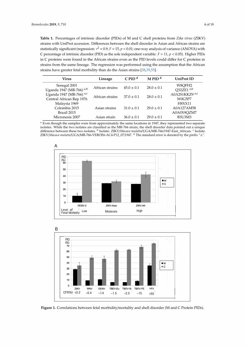
Table 1. Percentages of intrinsic disorder (PIDs) of M and C shell proteins from Zika virus (ZIKV)strains with UniProt accession. Differences between the shell disorder in Asian and African strains arestatistically significant (regression: r2 = 0.9, F = 15, p < 0.01; one-way analysis of variance (ANOVA) withC percentage of intrinsic disorder (PID) as the sole independent variable: F = 11, p < 0.05). Higher PIDsin C proteins were found in the African strains even as the PID levels could differ for C proteins instrains from the same lineage. The regression was performed using the assumption that the Africanstrains have greater fetal morbidity than do the Asian strains [38,39,55].
Virus Lineage C PID d M PID d UniProt ID
Senegal 2001Uganda 1947 (MR-766) a,b African strains 45.0 ± 0.1 28.0 ± 0.1 W8QFH2
Q32ZE1 a,b
Uganda 1947 (MR-766) a,c
Central African Rep 1976 African strains 37.0 ± 0.1 28.0 ± 0.1 A0A2S1KKZ9 a,c
W0G5P7Malaysia 1969Colombia 2015
Brazil 2015Asian strains 31.0 ± 0.1 29.0 ± 0.1
H8XX11A0A127AM58A0A0X9QZM7
Micronesia 2007 Asian strain 36.0 ± 0.1 29.0 ± 0.1 B3U3M3a Even though the samples were from approximately the same locations in 1947, they represented two separateisolates. While the two isolates are classified as the MR-766 strain, the shell disorder data pointed out a uniquedifference between these two isolates. b Isolate: ZIKV/Macaca mulatta/UGA/MR-766/1947-East_African. c Isolate:ZIKV/Macaca mulatta/UGA/MR-766-VEROE6-AC4-P12_07/1947. d The standard error is denoted by the prefix “±”.
Figure 1. Correlations between fetal morbidity/mortality and shell disorder (M and C Protein PIDs).
Biomolecules 2019, 9, 710 5 of 18
(A) Correlation between fetal morbidity and ZIKV–Dengue virus 2 (DENV-2) shell disorder (regression:r2 = 0.8, F = 16, p < 0.01; independent variables: C and M PIDs). See Figure S1 for more details on thedata points; (B) correlation between flavivirus case fatality ratio (CFR) and shell disorder (regression:r2 = 0.8, F = 29, p < 0.01; independent variables: C and M PIDs). With the exception of the ZIKV data,much of the data can be found in a previous publication [25].
While Figure 1A and Table 1 involve the determination of the correlation between the fetalmorbidity associated with ZIKV infection and ZIKV shell disorder (PID), Figure 1B and Table 2 representthe results of the analysis of the correlation between the case fatality ratio ((CFR), i.e., virulence) and theshell disorder of various flaviviruses, including ZIKV. Information on the CFRs of various flavivirusesis publicly available [14,57–60]. Because the symptoms of ZIKV are usually very mild and fatalities areextremely rare, for a long time, the virus was shrouded in mystery [2,3].
It is therefore likely that the CFR is very close to zero, even if most of the currently available dataare those from infections arising in the Americas, which involved strains of Asian lineage, not Africanlineage [1,4–6]. A strong correlation was found between flavivirus CFR and shell disorder, as seen inFigure 1B and Table 2. While a correlation between flavivirus CFR and shell disorder has already beenreported [2,14], the data presented here include results for ZIKV, which was not previously considered.
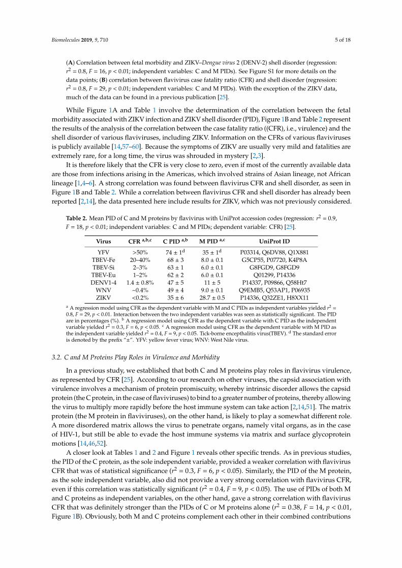
Table 2. Mean PID of C and M proteins by flavivirus with UniProt accession codes (regression: r2 = 0.9,F = 18, p < 0.01; independent variables: C and M PIDs; dependent variable: CFR) [25].
Virus CFR a,b,c C PID a,b M PID a,c UniProt ID
YFV >50% 74 ± 1d 35 ± 1d P03314, Q6DV88, Q1X881TBEV-Fe 20–40% 68 ± 3 8.0 ± 0.1 G5CP55, P07720, K4P8ATBEV-Si 2–3% 63 ± 1 6.0 ± 0.1 G8FGD9, G8FGD9TBEV-Eu 1–2% 62 ± 2 6.0 ± 0.1 Q01299, P14336DENV1-4 1.4 ± 0.8% 47 ± 5 11 ± 5 P14337, P09866, Q58Ht7
WNV ~0.4% 49 ± 4 9.0 ± 0.1 Q9EMB5, Q53AP1, P06935ZIKV <0.2% 35 ± 6 28.7 ± 0.5 P14336, Q32ZE1, H8XX11
a A regression model using CFR as the dependent variable with M and C PIDs as independent variables yielded r2 =0.8, F = 29, p < 0.01. Interaction between the two independent variables was seen as statistically significant. The PIDare in percentages (%). b A regression model using CFR as the dependent variable with C PID as the independentvariable yielded r2 = 0.3, F = 6, p < 0.05. c A regression model using CFR as the dependent variable with M PID asthe independent variable yielded r2 = 0.4, F = 9, p < 0.05. Tick-borne encepthalitis virus(TBEV). d The standard erroris denoted by the prefix “±”. YFV: yellow fever virus; WNV: West Nile virus.
3.2. C and M Proteins Play Roles in Virulence and Morbidity
In a previous study, we established that both C and M proteins play roles in flavivirus virulence,as represented by CFR [25]. According to our research on other viruses, the capsid association withvirulence involves a mechanism of protein promiscuity, whereby intrinsic disorder allows the capsidprotein (the C protein, in the case of flaviviruses) to bind to a greater number of proteins, thereby allowingthe virus to multiply more rapidly before the host immune system can take action [2,14,51]. The matrixprotein (the M protein in flaviviruses), on the other hand, is likely to play a somewhat different role.A more disordered matrix allows the virus to penetrate organs, namely vital organs, as in the caseof HIV-1, but still be able to evade the host immune systems via matrix and surface glycoproteinmotions [14,46,52].
A closer look at Tables 1 and 2 and Figure 1 reveals other specific trends. As in previous studies,the PID of the C protein, as the sole independent variable, provided a weaker correlation with flavivirusCFR that was of statistical significance (r2 = 0.3, F = 6, p < 0.05). Similarly, the PID of the M protein,as the sole independent variable, also did not provide a very strong correlation with flavivirus CFR,even if this correlation was statistically significant (r2 = 0.4, F = 9, p < 0.05). The use of PIDs of both Mand C proteins as independent variables, on the other hand, gave a strong correlation with flavivirusCFR that was definitely stronger than the PIDs of C or M proteins alone (r2 = 0.38, F = 14, p < 0.01,Figure 1B). Obviously, both M and C proteins complement each other in their combined contributions

Biomolecules 2019, 9, 710 6 of 18
to virulence, which can be seen in both Figure 1B and Table 2. There was also a statistically significantinteraction between the two factors (p < 0.01).
3.3. Regions of Disorder in the M Protein: ZIKV versus YFV and DENV-2
Figure 1 and Tables 1 and 2 demonstrate that ZIKV is a peculiar virus, since it has a relativelydisordered M protein but also a relatively ordered C protein. This accounts for the ZIKV characteristicsof low mortality rates and yet high fetal morbidity rates. However, the use of PID values is inadequatein providing a closer look at the differences in the disordered regions among the various viruses.Figures 2 and 3 provide us with the opportunity for a detailed look at the disorder differences by regionof the proteins among several flaviviruses.
Figure 2A shows that disordered regions of the M proteins of ZIKV and YFV are relatively largeand overlap. With the disordered regions being found mostly around the N-termini of the M proteinsof the three flaviviruses shown, a much shorter disordered region is found in the DENV-2 M protein.These observations could be used as a template for designing a ZIKV vaccine, especially when lookingat the PONDR VLXT plot of the ZIKV M protein. The asterisk (*) in Figure 2 shows areas that could bepotential targets in the development of a ZIKV vaccine.
Figure 2. Cont.

Biomolecules 2019, 9, 710 7 of 18
Figure 2. Comparative PONDR VLXT plots of the M and C proteins from various flaviviruses,including ZIKV. (A) M proteins of ZIKV (UniProt: A0A127AM58), DENV-2 (UniProt: P29990), and YFV(UniProt: Q1X881). (B) C proteins of ZIKV (UniProt: H8XX11, Msia69, see Table 1), WNV (UniProt:Q9Q694), and DENV-2 (UniProt: P29990). Regions with scores of 0.5 or above represent disorder.Disorder differences between C proteins can be traced mainly to mutations and disorder near theN- and C-termini. The regions with an asterisk (*) denote potential targets for the development of aZIKV vaccine. With the exception of Msia69 (Malaysia 1969) ZIKV, the strains were randomly chosen.Msia69 was deliberately chosen as a representative Asian strain that has a low C PID. The YFV Mprotein was chosen for (A), since it has one of the highest PIDs among flaviviruses. WNV was notnecessary for (A), as specific strains of DENV-2 have an even lower M PID (PID: 6%) than does the ZIKV(PID: 9%). On the other hand, WNV was chosen for (B), as the low WNV C PID (among flaviviruses)allows us to suggest strategies for ZIKV vaccine development.
3.4. PONDR VLXT Plot for C Proteins from ZIKV and Other Flaviviruses
The PONDR VLXT plot for the C proteins of three flaviviruses, ZIKV, DENV-2, and WNV, can befound in Figure 2B. As mentioned, studies have shown that disorder in the inner shells of viruses ingeneral tend to be associated with greater virulence because of the ability of proteins to bind morepromiscuously, thus providing the viruses with greater ability to replicate more rapidly. Similarly,Figure 1B and Table 2 suggest that the reason that the ZIKV’s CFR is extremely low has to do withthe relatively low mean PID of its C protein (35 ± 6%, CFR ~0). While these results tell us about thedifferences with regard to the mean C PIDs among the various flaviviruses, they do pinpoint thedisorder differences among the various flaviviruses along the entire stretch of the various flavivirus Cproteins, especially with respect to ZIKV. Figure 2B attempts to address this problem by showing thatthe disorder differences between C proteins from ZIKV and other flaviviruses lie in two areas near theN- and C-termini, respectively.
3.5. PONDR VLXT Plot for C Proteins from Different ZIKV Strains
While Figure 2B, Figure 1B, and Table 2 provide evidence that disorder in the C proteincontributes to the virulence of flaviviruses, Figure 1A, Figure 2A, Figure 3, and Table 1point to the contribution of ZIKV C protein disorder to fetal morbidity. More specifically,a rather large disordered region is missing near the N-terminus of the strains of Asian lineage,which, as already mentioned, inflict morbidity to fetuses at a lower rate than do their African
Biomolecules 2019, 9, 710 8 of 18
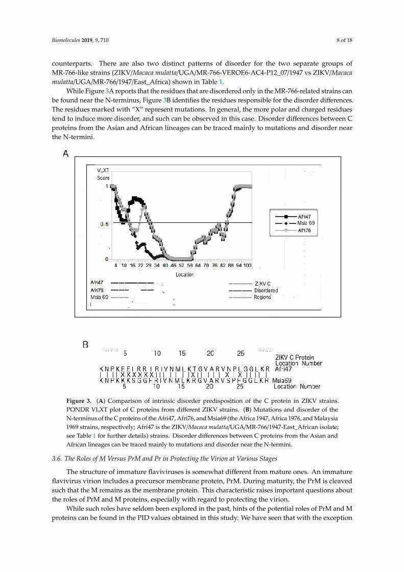
counterparts. There are also two distinct patterns of disorder for the two separate groups ofMR-766-like strains (ZIKV/Macaca mulatta/UGA/MR-766-VEROE6-AC4-P12_07/1947 vs ZIKV/Macacamulatta/UGA/MR-766/1947/East_Africa) shown in Table 1.
While Figure 3A reports that the residues that are disordered only in the MR-766-related strains canbe found near the N-terminus, Figure 3B identifies the residues responsible for the disorder differences.The residues marked with “X” represent mutations. In general, the more polar and charged residuestend to induce more disorder, and such can be observed in this case. Disorder differences between Cproteins from the Asian and African lineages can be traced mainly to mutations and disorder nearthe N-termini.
Figure 3. (A) Comparison of intrinsic disorder predisposition of the C protein in ZIKV strains.PONDR VLXT plot of C proteins from different ZIKV strains. (B) Mutations and disorder of theN-terminus of the C proteins of the Afri47, Afri76, and Msia69 (the Africa 1947, Africa 1976, and Malaysia1969 strains, respectively; Afri47 is the ZIKV/Macaca mulatta/UGA/MR-766/1947-East_African isolate;see Table 1 for further details) strains. Disorder differences between C proteins from the Asian andAfrican lineages can be traced mainly to mutations and disorder near the N-termini.
3.6. The Roles of M Versus PrM and Pr in Protecting the Virion at Various Stages
The structure of immature flaviviruses is somewhat different from mature ones. An immatureflavivirus virion includes a precursor membrane protein, PrM. During maturity, the PrM is cleavedsuch that the M remains as the membrane protein. This characteristic raises important questions aboutthe roles of PrM and M proteins, especially with regard to protecting the virion.
While such roles have seldom been explored in the past, hints of the potential roles of PrM and Mproteins can be found in the PID values obtained in this study. We have seen that with the exception

Biomolecules 2019, 9, 710 9 of 18
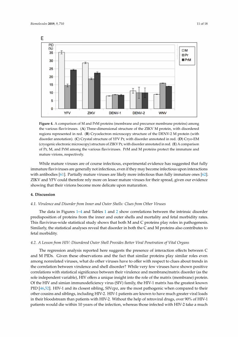
of YFV and ZIKV, most other flaviviruses have highly ordered M proteins to protect their respectivemature virions (Figure 1B and Table 2). Figure 4A shows that the M protein is relatively more disordered(in red) than in DENV-1 (Figure 4B). As with the PONDR VLXT plots in Figure 2, the disorderedregions of both ZIKV and DENV-1 are restricted to segments near the N-termini. While we have seenthat the M proteins of ZIKV and YFV are relatively disordered, the same cannot be said about Pr.The Pr proteins for YFV and ZIKV are quite ordered, as seen in Figure 4C–E. The Pr disorder levels arerelatively low for most flaviviruses [2,25], as seen in the cases of ZIKV, DENV, and WNV. With Pr beingmostly ordered for most flaviviruses and M being the same for most flaviviruses (with the exception ofYFV and ZIKV), one can conclude that both Pr and M contribute to the order of the PrM proteins formost flaviviruses, and this is especially important with respect to protecting the immature virion.
The following question then arose: How do ZIKV and YFV protect their immature virion giventheir abnormally high M disorder? A hint to the answer to this question lies in the PID of their Prproteins. As we can see in Figure 4C–E, the Pr proteins of both ZIKV and YFV are quite ordered, wellwithin the range of most flaviviruses. As a result, fairly ordered PrM proteins are seen in both ZIKVand YFV. The study therefore shows that the immature virions of ZIKV and YFV are more protectedagainst environmental insults than are their corresponding mature virions, which could suggest aslightly different life cycle for YFV and ZIKV.
Figure 4. Cont.

Biomolecules 2019, 9, 710 10 of 18
Figure 4. Cont.
Biomolecules 2019, 9, 710 11 of 18
Figure 4. A comparison of M and PrM proteins (membrane and precursor membrane proteins) amongthe various flaviviruses. (A) Three-dimensional structure of the ZIKV M protein, with disorderedregions represented in red. (B) Cryoelectron microscopy structure of the DENV-2 M protein (withdisorder annotation). (C) Crystal structure of YFV Pr, with disorder annotated in red. (D) Cryo-EM(cryogenic electronic microscopy) structure of ZIKV Pr, with disorder annotated in red. (E) A comparisonof Pr, M, and PrM among the various flaviviruses. PrM and M proteins protect the immature andmature virions, respectively.
While mature viruses are of course infectious, experimental evidence has suggested that fullyimmature flaviviruses are generally not infectious, even if they may become infectious upon interactionswith antibodies [61]. Partially mature viruses are likely more infectious than fully immature ones [62].ZIKV and YFV could therefore rely more on lesser mature viruses for their spread, given our evidenceshowing that their virions become more delicate upon maturation.
4. Discussion
4.1. Virulence and Disorder from Inner and Outer Shells: Clues from Other Viruses
The data in Figures 1–4 and Tables 1 and 2 show correlations between the intrinsic disorderpredisposition of proteins from the inner and outer shells and mortality and fetal morbidity rates.This flavivirus-wide statistical study shows that both M and C proteins play roles in pathogenesis.Similarly, the statistical analyses reveal that disorder in both the C and M proteins also contributes tofetal morbidity.
4.2. A Lesson from HIV: Disordered Outer Shell Provides Better Viral Penetration of Vital Organs
The regression analysis reported here suggests the presence of interaction effects between Cand M PIDs. Given these observations and the fact that similar proteins play similar roles evenamong nonrelated viruses, what do other viruses have to offer with respect to clues about trends inthe correlation between virulence and shell disorder? While very few viruses have shown positivecorrelations with statistical significance between their virulence and membrane/matrix disorder (as thesole independent variable), HIV offers a unique insight into the role of the matrix (membrane) protein.Of the HIV and simian immunodeficiency virus (SIV) family, the HIV-1 matrix has the greatest knownPID [46,52]. HIV-1 and its closest sibling, SIVcpz, are the most pathogenic when compared to theirother cousins and siblings, including HIV-2. HIV-1 patients are known to have much greater viral loadsin their bloodstream than patients with HIV-2. Without the help of retroviral drugs, over 90% of HIV-1patients would die within 10 years of the infection, whereas those infected with HIV-2 take a much

Biomolecules 2019, 9, 710 12 of 18
longer time to succumb to the disease, if at all [46,63]. The roles of the matrix protein in virulence haveto do with viral ability to evade the host immune system and also the ability to bind promiscuouslyto more host proteins [18,19,21,22]. An important characteristic of HIV is its ability to penetrate vitalorgans, including the brain, with ease [14]. Apparently, the promiscuous binding potential arisingfrom the disordered matrix shell allows HIV to have easier access to specialized cells. We shall use thisargument again in the cases of ZIKV and YFV, which have high PIDs for their M proteins (28.6 ± 0.5%and 35 + 1%, respectively).
4.3. Virulence and Inner Shell Disorder Involve a Different “Trojan Horse” Strategy in Immune Evasion
While disorder in the matrix shell is associated with virulence and the immune evasion foundin HIV, disorder in the inner shell (capsid or nucleocapsid) of other viruses, such as Ebola virus andNipah virus), is often also strongly associated with virulence [45,51]. Virulence arising from disorderin the inner shell involves a different strategy of immune evasion, which can be referred to as Trojanhorse, since the virus attempts to replicate rapidly before the host immune system is able to recognizeits presence [2,14]. It does so by using the disordered inner shell proteins that play important roles inthe replication of the virus, where disorder assists the process by allowing for greater promiscuous(and therefore more efficient) binding of host proteins.
4.4. Taking Turns to Protect the Virion from Environmental Insults
Curiously, a conflicting trend has also been observed in HIV: when the outer shell is disordered,then the inner shells tend to be more ordered [14,52]. Conversely, if the outer shell is more ordered,the inner shells tend to be more disordered. These conflicting trends can be seen in a variety of viruses,including Nipah, corona, and Ebola viruses, not just HIV [2,45,49–51]. The reason for this has to dowith the fact that both shell layers play their roles in protecting the virion from environmental insults.If the outer layer is more disordered, the inner layer compensates by being more ordered. If, on theother hand, the outer layer is more ordered, the inner layer, depending on the type of environment thevirus is exposed to, has the luxury of being less ordered.
4.5. Conflicting Trends Can Be Found among Flaviviruses: Protecting the Virion Versus Immune Evasion
The two aforementioned seemingly conflicting trends that are seen in other viruses can also beobserved in flaviviruses and ZIKV. It is for this reason that we observed statistical significance in theinteraction effects between the PIDs of C and M proteins as independent variables. Furthermore, strongflavivirus CFR correlations were not attainable until both C and M PIDs were used as independentvariables (r2 = 0.8, F = 29, p < 0.01). A closer look at the data in Figure 1B allows us to conclude thatsome flaviviruses do indeed have more ordered M proteins when their C proteins are disorderedand vice versa. For example, tick-bone encephalitis virus (TBEV) viruses have higher PIDs in theirC proteins than do DENV viruses, but they have lower PIDs for M proteins. Similarly, ZIKV has alow PID value for C but a high PID for M. There is also evidence of this trend within ZIKV strainsthemselves: we can notice a slightly lower M disorder in MR-766 strains (PID: 28 vs 29%, Table 1)despite their higher C PID values even as M PIDs remain relatively high for all ZIKV strains.
This trend is, however, not the case with every flavivirus. A striking exception is YFV,which provides some evidence for the other trend that involves strategies of immune evasionusing disorder in either or both M and C shells. More specifically, disorder in M and C proteins is likelyto play different but complimentary roles in maintaining higher viral load, especially in vital organsnecessary to evade the immune system, and YFV is somewhat unique in using both C and M disorderto a much greater extent than other flaviviruses do. This will be further described in the next section.

Biomolecules 2019, 9, 710 13 of 18
4.6. YFV Uses both Inner and Outer Shell Disorder to Evade Immune Systems, Resulting inHigh Pathogenicity
Relative to other flaviviruses, YFV is characterized by highly disordered C and M proteins.Two modes of action are at work here. A highly disordered YFV has an overwhelmingly high PID inits C protein (~75%). Interestingly, the mean PID of its M protein (~35%) was also far higher than thecorresponding values for all other flavivirus species found in our sample. It is likely that the highlydisordered C protein allows for more rapid replication via its promiscuity in protein binding. As aresult of the rapid replication, greater viral loads are more attainable, leading to greater virulence.A higher disorder in the M protein, however, allows the virus to more easily penetrate different organs,including vital organs such as the liver [2,15]. Apparently, YFV’s extremely high pathogenicity, whichreaches above a 50% CFR, arises from its unique ability to utilize disorder from both its M and Cproteins to maintain a viral load as a means of evading the immune system.
4.7. The Shell Disorder Model Predicts Correctly that ZIKV Is Able to Penetrate Vital Organs withGreater Efficiency
While YFV has greater disorder in both its M and C proteins, ZIKV offers us a unique opportunityto study a flavivirus with a high PID in its M protein (35 ± 6%) but a low PID in its C protein (28.6± 1%). If we use the aforementioned model based on our knowledge of shell disorder, the modelwill predict ZIKV as a virus that is of low pathogenicity but has the ability to penetrate vital organs.This prediction is extremely accurate in the case of ZIKV. We are able to see that ZIKV infection isusually nonfatal, with CFR being close to 0%, but ZIKV also has an uncanny ability to penetrate thebrain and placenta, as seen in its ability to cause microcephaly [12].
4.8. ZIKV C PID Variation Correlates with Fetal Morbidity: A “Trojan Horse”
We have seen that flaviviruses use disorder in M and C proteins to evade host immune systems.YFV uses both M and C to penetrate vital organs and proliferate rapidly. The two modes becomea deadly mix. The ZIKV uses its high M disorder to penetrate the placenta and brain, but we havealso noticed that, while ZIKV C PID values are generally low, there is noticeable variation in the CPIDs seen in the different lineages and strains. This PID variation in the C protein correlates withfetal morbidity that is dependent on the lineage of ZIKV. The results fit perfectly with what has beenobserved in other viruses. The “Trojan horse” theory tells us that the higher C disorder allows for moreefficient proliferation of the virus in the larger variety of organs that it is able to penetrate [2,49].
4.9. Evolution of ZIKV Variation in Fetal Morbidity Is Dependent on the Optimal Viral Load Necessary forMore Efficient Transmission Among Its Primary Primate Hosts
While variation in ZIKV M PID is only 1%, ZIKV C PID can be as low as 31% and as high as 45%.Inspection of the PONDR VLXT plot shows that the disordered regions lie in both the N- and C-terminieven if the ZIKV C mutations arise entirely within the N-terminus (Figure 3). This provides clues to theevolution of ZIKV with respect to its ability to cause fetal morbidity in its hosts. It is likely that ZIKVhad to evolve with the primate species that it most commonly infects. For certain primate hosts, it findsgreater fitness when a greater viral load is necessary for it to infect a larger number of hosts because ofthe peculiarities found in the hosts’ immune system: having a more disordered C protein does the trick.It is therefore likely that the different PIDs of C proteins from the various ZIKV strains and lineagesare the result of the types of primates that the peculiar ZIKV strain primarily infects and its ability tofine-tune its C disorder to meet the optimal viral load necessary for more efficient transmission of thevirus in a given host, as seen in the case of other viruses and other flaviviruses [2,14,25,39,49,50].

Biomolecules 2019, 9, 710 14 of 18
4.10. Experimental Evidence for the Role of C and M Proteins in Replication and Immune Evasion: Rolesfor Disorder
Experiments do provide evidence pointing to the important roles of C and M proteins in replicationand immune evasion. Flavivirus protein C, along with other viral proteins, assists in the assembly andpackaging of viral particles during replication, in addition to assisting the protein M to move to theendoplasmic reticulum (ER). Additionally, the C protein is trafficked to the nucleus through bindingwith importin [64]. Upon doing so, it interacts with histones to force the cell to stay longer in the G1stage of the cell cycle so as to buy more time for viral replication [65]. In the cytoplasm, protein C bindsto the Sec3 protein in order to inhibit Sec3 proteasomal antiviral activity [66].
As for the M protein, there is still much to be uncovered about its functions. It is, however, knownthat PrM binds to the ER for two reasons. It protects the E proteins from damage by host enzymes viathe formation of a beta barrel [67]. The other reason is that PrM prevents E from being so embeddedin the host membrane that it cannot be removed in the later stages of the virion replication process.While the Pr segment is ordered to protect E from the proteases, the greater flexibility of M in manyflaviviruses is likely needed to ensure greater efficiency in its binding to the ER.
Similarly, greater disorder in the C protein offers plenty of opportunity for greater bindingefficiency, given its aforementioned experimentally proven functions. For instance, the greater disorderin the C protein could help its binding to histones with greater efficiency. Protein disorder not onlyprovides greater efficiency in protein–protein binding but also promotes the binding of proteins tonucleic and fatty acids as well.
4.11. Clues for Developing a ZIKV Vaccine
Figure 3 reiterates the minimum requirements necessary in the development of a ZIKV vaccine:the PID of the C protein has to be as low as possible. Figure 2B, on the other hand, suggests ways thata ZIKV can be used for a ZIKV vaccine developed by comparing the PONDR VLXT plots of the ZIKVC protein to those of other flaviviruses. More specifically, it shows a region near the C-terminus that isordered in WNV but disordered in ZIKV. This region could be a good target for the development of anattenuated ZIKV, as it indicates a region that is not necessarily disordered but is still tolerated by acousin, WNV.
Just as relatively high disorder levels in the M protein (PID: 28%, 29%) are a hallmark of the ZIKV,an effective vaccine needs to have an M protein with a lower PID so that an attenuated virus will notpenetrate the placenta of pregnant women. Figures 2A and 4 show that the disordered regions of the Mprotein are near the N-terminus, and mutations to attenuate the virus must be in that region. Since theM protein of DENV has a much shorter disordered region (Figures 2B and 4A,B), Figure 2A identifiesan area that is ordered in DENV and would be a good target of mutations during the developmentof an attenuated ZIKV. Again, since that region is ordered in DENV-2, order there could be toleratedby ZIKV.
5. Conclusions
There were two main ZIKV oddities discovered in this research. The first has to do with therelatively high disorder in the ZIKV M protein, which is at a level that is seldom found among otherflaviviruses, with the exception of YFV. The other is the somewhat large variation in the disorder ofZIKV depending on the strain and lineage of the virus. These oddities are consistent with what wenow know as characteristics of ZIKV. Just as other viruses (such as WNV and HIV-1) that have highouter shell (matrix/membrane) PIDs are able to penetrate vital organs, ZIKV can also penetrate theplacenta and brain [12]. Furthermore, scientists have discovered that strains of the African lineagepossess a greater potential to cause fetal morbidity. This fact correlates with the variations in thedisorder levels of the ZIKV C protein. Disorder in the inner shell has already been associated withvirulence and higher viral loads in other flaviviruses and other unrelated viruses, such as in the Ebolaand Nipah viruses.
Biomolecules 2019, 9, 710 15 of 18
There are important implications for these findings. The results presented here could lead tothe development of a new vaccine through the creation of attenuated strains resulting from moreordered C and M proteins. The regions for potential vaccine targets are disordered regions of the ZIKVM and C proteins that are ordered in other flavivirus species. Exploration of a ZIKV vaccine couldinclude mutations in these areas. Other applications could include building a more comprehensivemodel that could anticipate the behaviors of new or little-known viruses. Lastly, the ability of ZIKVand YFV to penetrate vital organs as a result of shell disorder adds to a growing body of knowledgethat encompasses a variety of viruses, such as HIV, that use such strategies to enter or to hide inhard-to-reach organs. An important potential application of this is in the field of cancer oncolysis,which involves the use of viruses to “attack” tumors, since both share the same pathways [68,69].Viruses could be engineered to evade host immune systems that impede the effectiveness of therapeuticviruses. Moreover, since cancers in hard-to-reach organs such as the brain are often problematic forvirotherapies and chemotherapies [14,70], a better understanding of the roles of shell disorder in theviral penetration of such organs will lead to improved therapies.
Supplementary Materials: The following are available online at http://www.mdpi.com/2218-273X/9/11/710/s1,Figure S1: Line graph showing correlations between fetal morbidity and shell disorder (M and C Protein PIDs).
Author Contributions: Conceptualization, G.K.-M.G.; data curation, G.K.-M.G. and V.N.U.; formal analysis,G.K.-M.G.; investigation, G.K.-M.G.; methodology, A.K.D.; resources, A.K.D. and J.A.F.; software, G.K.-M.G.;validation, G.K.-M.G.; visualization, A.K.D. and J.A.F.; writing—original draft, G.K.-M.G. and V.N.U.;writing—review and editing, V.N.U.
Funding: J.A.F. was funded in part by NSF DBI0939454 and the BEACON Center for Evolution in Action.
Conflicts of Interest: G.K.-M.G. is an independent researcher and the owner of Goh’s BioComputing, Singapore.G.K.-M.G. has also written a book (“Viral Shapeshifters: Strange Behaviors of HIV and Other Viruses”) on arelated subject. The authors have no other potential conflict of interests. The funders had no role in the design ofthe study; in the collection, analyses, or interpretation of data; in the writing of the manuscript; or in the decisionto publish the results.
References
1. Song, B.-H.; Yun, S.-I.; Woolley, M.; Lee, Y.-M. Zika virus: History, epidemiology, transmission, and clinicalpresentation. J. Neuroimmunol. 2017, 308, 50–64. [CrossRef] [PubMed]
2. Goh, G.K. Viral Shapeshifters: Strange Behaviors of HIV and Other Viruses; Simplicity Research Institute:Singapore, 2017.
3. Petersen, L.R.; Jamieson, D.J.; Powers, A.M.; Honein, M.A. Zika Virus. N. Engl. J. Med. 2016, 374, 1552–1563.[CrossRef] [PubMed]
4. Heukelbach, J.; Alencar, C.H.; Kelvin, A.A.; De Oliveira, W.K.; Cavalcanti, L.P.D.G. Zika virus outbreak inBrazil. J. Infect. Dev. Ctries. 2016, 10, 116–120. [CrossRef] [PubMed]
5. Chen, H.-L.; Tang, R.-B. Why Zika virus infection has become a public health concern? J. Chin. Med. Assoc.2016, 79, 174–178. [CrossRef]
6. Hafiz, M.Y.; Mahmood, S.U.; Shoaib, M.; Yusuf, F.H. Concern over Zika virus outbreak: Another alarmingglobal threat. Infect. Drug Resist. 2016, 9, 149–151. [CrossRef]
7. Udenze, D.; Trus, I.; Berube, N.; Gerdts, V.; Karniychuk, U. The African strain of Zika virus causes moresevere in utero infection than Asian strain in a porcine fetal transmission model. Emerg. Microbes Infect. 2019,8, 1098–1107. [CrossRef]
8. Gurung, S.; Reuter, N.; Preno, A.; Dubaut, J.; Nadeau, H.; Hyatt, K.; Singleton, K.; Martin, A.; Parks, W.T.;Papin, J.F.; et al. Zika virus infection at mid-gestation results in fetal cerebral cortical injury and fetal death inthe olive baboon. PLoS Pathog. 2019, 15, e1007507. [CrossRef]
9. Pena, L.J.; Guarines, K.M.; Silva, A.J.D.; Leal, L.R.S.; Félix, D.M.; Silva, A.; De Oliveira, S.A.; Ayres, C.F.J.;Júnior, A.S.; De Freitas, A.C. In vitro and in vivo models for studying Zika virus biology. J. Gen. Virol. 2018,99, 1529–1550. [CrossRef]
10. Gorman, M.J.; Caine, E.A.; Zaitsev, K.; Begley, M.C.; Weger-Lucarelli, J.; Uccellini, M.B.; Tripathi, S.;Morrison, J.; Yount, B.L.; Dinnon, K.H., 3rd; et al. An Immunocompetent Mouse Model of Zika VirusInfection. Cell Host Microbe 2018, 23, 672–685.e6. [CrossRef]

Biomolecules 2019, 9, 710 16 of 18
11. Schreur, P.J.W.; Van Keulen, L.; Anjema, D.; Kant, J.; Kortekaas, J. Microencephaly in fetal piglets following inutero inoculation of Zika virus. Emerg. Microbes Infect. 2018, 7, 1–11. [CrossRef]
12. Hirsch, A.J.; Roberts, V.H.J.; Grigsby, P.L.; Haese, N.; Schabel, M.C.; Wang, X.; Lo, J.O.; Liu, Z.; Kroenke, C.D.;Smith, J.L.; et al. Zika virus infection in pregnant rhesus macaques causes placental dysfunction andimmunopathology. Nat. Commun. 2018, 9, 263. [CrossRef] [PubMed]
13. Winkler, C.W.; Peterson, K.E. Using immunocompromised mice to identify mechanisms of Zika virustransmission and pathogenesis. Immunology 2018, 153, 443–454. [CrossRef] [PubMed]
14. Goh, G.K.-M.; Dunker, A.K.; Foster, J.A.; Uversky, V.N. HIV Vaccine Mystery and Viral Shell Disorder.Biomolecules 2019, 9, 178. [CrossRef] [PubMed]
15. Acheson, N.H. Fundamentals of Molecular Virology; Wiley: Hoboken, NJ, USA, 2007.16. Sirohi, D.; Kuhn, R.J. Zika Virus Structure, Maturation, and Receptors. J. Infect. Dis. 2017, 216, S935–S944.
[CrossRef]17. Prasad, V.M.; Miller, A.S.; Klose, T.; Sirohi, D.; Buda, G.; Jiang, W.; Kuhn, R.J.; Rossmann, M.G. Structure of
the immature Zika virus at 9 Å resolution. Nat. Struct. Mol. Boil. 2017, 24, 184–186. [CrossRef]18. Dunker, A.K.; Brown, C.J.; Lawson, J.D.; Iakoucheva, L.M.; Obradovic, Z. Intrinsic Disorder and Protein
Function†. Biochemistry 2002, 41, 6573–6582. [CrossRef]19. Wright, P.E.; Dyson, H.J. Intrinsically unstructured proteins: Re-assessing the protein structure-function
paradigm. J. Mol. Boil. 1999, 293, 321–331. [CrossRef]20. Uversky, V.N.; Gillespie, J.R.; Fink, A.L. Why are “natively unfolded” proteins unstructured under physiologic
conditions? Proteins: Struct. Funct. Bioinform. 2000, 41, 415–427. [CrossRef]21. Dunker, A.K.; Lawson, J.D.; Brown, C.J.; Williams, R.M.; Romero, P.; Oh, J.S.; Oldfield, C.J.; Campen, A.M.;
Ratliff, C.M.; Hipps, K.W.; et al. Intrinsically disordered protein. J. Mol. Graph. Model. 2001, 19, 26–59.[CrossRef]
22. Tompa, P. Intrinsically unstructured proteins. Trends Biochem. Sci. 2002, 27, 527–533. [CrossRef]23. Giri, R.; Kumar, D.; Sharma, N.; Uversky, V.N. Intrinsically Disordered Side of the Zika Virus Proteome.
Front. Microbiol. 2016, 6, 144. [CrossRef] [PubMed]24. Mishra, P.M.; Uversky, V.N.; Giri, R. Molecular Recognition Features in Zika Virus Proteome. J. Mol. Boil.
2018, 430, 2372–2388. [CrossRef] [PubMed]25. Goh, G.K.-M.; Dunker, A.K.; Uversky, V.N. Correlating Flavivirus virulence and levels of intrinsic disorder in
shell proteins: Protective roles vs. immune evasion. Mol. BioSyst. 2016, 12, 1881–1891. [CrossRef] [PubMed]26. Xue, B.; Blocquel, D.; Habchi, J.; Uversky, A.V.; Kurgan, L.; Uversky, V.N.; Longhi, S. Structural Disorder in
Viral Proteins. Chem. Rev. 2014, 114, 6880–6911. [CrossRef] [PubMed]27. Xue, B.; Dunker, A.K.; Uversky, V.N. Orderly order in protein intrinsic disorder distribution: Disorder in 3500
proteomes from viruses and the three domains of life. J. Biomol. Struct. Dyn. 2012, 30, 137–149. [CrossRef][PubMed]
28. Xue, B.; Ganti, K.; Rabionet, A.; Banks, L.; Uversky, V.N. Disordered interactome of human papillomavirus.Curr. Pharm. Des. 2014, 20, 1274–1292. [CrossRef] [PubMed]
29. Uversky, V.N.; Roman, A.; Oldfield, C.J.; Dunker, A.K. Protein Intrinsic Disorder and Human Papillomaviruses:Increased Amount of Disorder in E6 and E7 Oncoproteins from High Risk HPVs. J. Proteome Res. 2006, 5,1829–1842. [CrossRef]
30. Goh, G.K.-M.; Dunker, A.K.; Uversky, V.N. Protein intrinsic disorder and influenza virulence: The 1918H1N1 and H5N1 viruses. Virol. J. 2009, 6, 69. [CrossRef]
31. Xue, B.; Mizianty, M.J.; Kurgan, L.; Uversky, V.N. Protein intrinsic disorder as a flexible armor and a weaponof HIV-1. Cell Mol. Life Sci. 2012, 69, 1211–1259. [CrossRef]
32. Dolan, P.T.; Roth, A.P.; Xue, B.; Sun, R.; Dunker, A.K.; Uversky, V.N.; LaCount, D.J. Intrinsic disorder mediateshepatitis C virus core-host cell protein interactions. Protein Sci. 2015, 24, 221–235. [CrossRef]
33. Fan, X.; Xue, B.; Dolan, P.T.; LaCount, D.J.; Kurgan, L.; Uversky, V.N. The intrinsic disorder status of thehuman hepatitis C virus proteome. Mol. BioSyst. 2014, 10, 1345–1363. [CrossRef] [PubMed]
34. Meng, F.; Badierah, R.A.; Almehdar, H.A.; Redwan, E.M.; Kurgan, L.; Uversky, V.N. Unstructural biology ofthe dengue virus proteins. FEBS J. 2015, 282, 3368–3394. [CrossRef] [PubMed]
35. Whelan, J.N.; Reddy, K.D.; Uversky, V.N.; Teng, M.N. Functional correlations of respiratory syncytial virusproteins to intrinsic disorder. Mol. BioSyst. 2016, 12, 1507–1526. [CrossRef] [PubMed]

Biomolecules 2019, 9, 710 17 of 18
36. Singh, A.; Kumar, A.; Yadav, R.; Uversky, V.N.; Giri, R. Deciphering the dark proteome of Chikungunyavirus. Sci. Rep. 2018, 8, 5822. [CrossRef]
37. Redwan, E.M.; AlJaddawi, A.A.; Uversky, V.N. Structural disorder in the proteome and interactome ofAlkhurma virus (ALKV). Cell Mol. Life Sci. 2019, 76, 577–608. [CrossRef]
38. Shao, Q.; Herrlinger, S.; Zhu, Y.-N.; Yang, M.; Goodfellow, F.; Stice, S.L.; Qi, X.-P.; Brindley, M.A.; Chen, J.-F.The African Zika virus MR-766 is more virulent and causes more severe brain damage than current Asianlineage and dengue virus. Development 2017, 144, 4114–4124. [CrossRef]
39. Beaver, J.T.; Lelutiu, N.; Habib, R.; Skountzou, I. Evolution of Two Major Zika Virus Lineages: Implicationsfor Pathology, Immune Response, and Vaccine Development. Front. Immunol. 2018, 9, 9. [CrossRef]
40. Goh, G.K.-M.; Dunker, A.K.; Uversky, V.N. Protein intrinsic disorder toolbox for comparative analysis ofviral proteins. BMC Genom. 2008, 9, S4. [CrossRef]
41. Garner, E.; Romero, P.; Dunker, A.K.; Brown, C.; Obradovic, Z. Predicting Binding Regions within DisorderedProteins. Genome Inform Ser Workshop Genome Inform. 1999, 10, 41–50.
42. Romero, P.; Obradovic, Z.; Kissinger, C.; Villafranca, J.E.; Dunker, A.K. Identifying disordered regions inproteins from amino acid sequence. In Proceedings of the IEEE International Conference on Neural Networks,Houston, TX, USA, 12 June 1997; pp. 90–95.
43. Cheng, Y.; Oldfield, C.J.; Meng, J.; Romero, P.; Uversky, V.N.; Dunker, A.K. Mining α-Helix-FormingMolecular Recognition Features with Cross Species Sequence Alignments†. Biochemistry 2007, 46, 13468–13477.[CrossRef]
44. Oldfield, C.J.; Cheng, Y.; Cortese, M.S.; Romero, P.; Uversky, V.N.; Dunker, A.K. Coupled Folding and Bindingwith α-Helix-Forming Molecular Recognition Elements†. Biochemistry 2005, 44, 12454–12470. [CrossRef][PubMed]
45. Goh, G.K.; Foster, J.A.; Dunker, A.K.; Uversky, V.N. Nipah virulence and v shell disorder. In SCON Summiton Vaccine and HIVAIDS Conferences, Las Vegas, NVUSA; 12 November 2019; in press.
46. Goh, G.K.-M.; Dunker, A.K.; Uversky, V.N. A comparative analysis of viral matrix proteins using disorderpredictors. Virol. J. 2008, 5, 126. [CrossRef] [PubMed]
47. Fonseca, A.A.; Heinemann, M.B.; Leite, R.C.; Reis, J.K.P. A comparative analysis of envelope and tegumentproteins of suid herpesvirus 1, bovine herpesvirus 1 and bovine herpesvirus 5. Arch. Virol. 2010, 155,1687–1692. [CrossRef] [PubMed]
48. Xue, B.; Williams, R.W.; Oldfield, C.J.; Goh, G.K.-M.; Dunker, A.K.; Uversky, V.N. Viral disorder or disorderedviruses: Do viral proteins possess unique features? Protein Pept. Lett. 2010, 17, 932–951. [CrossRef] [PubMed]
49. Goh, G.K.-M.; Dunker, A.K.; Uversky, V.N. Understanding Viral Transmission Behavior via Protein IntrinsicDisorder Prediction: Coronaviruses. J. Pathog. 2012, 2012, 1–13. [CrossRef]
50. Goh, G.K.-M.; Dunker, A.K.; Uversky, V. Prediction of Intrinsic Disorder in MERS-CoV/HCoV-EMC Supportsa High Oral-Fecal Transmission. PLoS Curr. 2013, 5, 5. [CrossRef]
51. Goh, G.K.-M.; Dunker, A.K.; Uversky, V.N. Detection of links between Ebola nucleocapsid and virulenceusing disorder analysis. Mol. BioSyst. 2015, 11, 2337–2344. [CrossRef]
52. Goh, G.K.-M.; Dunker, A.K.; Uversky, V.N. Shell disorder, immune evasion and transmission behaviorsamong human and animal retroviruses. Mol. BioSyst. 2015, 11, 2312–2323. [CrossRef]
53. Romero, P.; Obradovic, Z.; Li, X.; Garner, E.C.; Brown, C.J.; Dunker, A.K. Sequence complexity of disorderedprotein. Proteins Struct. Funct. Bioinform. 2001, 42, 38–48. [CrossRef]
54. R Core Team. R: A Language and Environment for Statistical Computing; R Foundation for Statistical Computing:Vienna, Austria, 2019; ISBN 3-900051-07-0.
55. Zhang, Z.; Sun, M.; Deng, J.; Yu, J.; Yang, X.; Zhao, W.; Chen, G.; Wang, P. Zika Virus Induced More SevereInflammatory Response Than Dengue Virus in Chicken Embryonic Livers. Front. Microbiol. 2019, 10, 1127.[CrossRef]
56. Gong, Z.; Xu, X.; Han, G.-Z. The Diversification of Zika Virus: Are There Two Distinct Lineages?Genome Boil. Evol. 2017, 9, 2940–2945. [CrossRef] [PubMed]
57. Diaz-Quijano, F.A.; Waldman, E.A. Factors Associated with Dengue Mortality in Latin America and theCaribbean, 1995–2009: An Ecological Study. Am. J. Trop. Med. Hyg. 2012, 86, 328–334. [CrossRef] [PubMed]
58. Halstead, S.B. Flavivirus vaccine. In Molecular Virology and Control of Flavivirus; Shi, P., Ed.; Caister:Wymanham, UK, 2012; p. 185.

Biomolecules 2019, 9, 710 18 of 18
59. Mickiene, A.; Lindquist, L.; Laiskonis, A. Tick-borne encephalitis—Clinical course and outcome. In Progressin Encephalitis Research; Ebert, R.A., Ed.; Nova Science Publishers: Hauppauge, NY, USA, 2005; pp. 1–30.
60. McKenzie, J.; Pinger, R.R.; Kotecki, J. Epidemiology: The study of disease, injury and death in the community.In An Introduction to Community Health, 8th ed.; Jones and Bartlett Publishers: Sudbury MA, USA, 2008; p. 70.
61. Rodenhuis-Zybert, I.A.; Van Der Schaar, H.M.; Voorham, J.M.D.S.; Van Der Ende-Metselaar, H.; Lei, H.-Y.;Wilschut, J.; Smit, J.M. Immature Dengue Virus: A Veiled Pathogen? PLoS Pathog. 2010, 6, e1000718.[CrossRef] [PubMed]
62. Zybert, I.A.; Van Der Ende-Metselaar, H.; Wilschut, J.; Smit, J.M. Functional importance of dengue virusmaturation: Infectious properties of immature virions. J. Gen. Virol. 2008, 89, 3047–3051. [CrossRef][PubMed]
63. Goudsmit, J. Viral Sex: The Nature of AIDS; Oxford University Press: New York, NY, USA, 1997.64. Bhuvanakantham, R.; Chong, M.-K.; Ng, M.-L. Specific interaction of capsid protein and importin-α/β
influences West Nile virus production. Biochem. Biophys. Res. Commun. 2009, 389, 63–69. [CrossRef][PubMed]
65. Colpitts, T.M.; Barthel, S.; Wang, P.; Fikrig, E. Dengue Virus Capsid Protein Binds Core Histones and InhibitsNucleosome Formation in Human Liver Cells. PLoS ONE 2011, 6, e24365. [CrossRef]
66. Bhuvanakantham, R.; Ng, M.-L. West Nile virus and dengue virus capsid protein negates the antiviral activityof human Sec3 protein through the proteasome pathway. Cell. Microbiol. 2013, 15, 1688–1706. [CrossRef]
67. Li, L.; Lok, S.-M.; Yu, I.-M.; Zhang, Y.; Kuhn, R.J.; Chen, J.; Rossmann, M.G. The Flavivirus PrecursorMembrane-Envelope Protein Complex: Structure and Maturation. Science 2008, 319, 1830–1834. [CrossRef]
68. Wei, D.; Xu, J.; Liu, X.X.-Y.; Chen, Z.-N.; Bian, H. Fighting Cancer with Viruses: Oncolytic Virus Therapy inChina. Hum. Gene Ther. 2018, 29, 151–159. [CrossRef]
69. Chernajovsky, Y.; Layward, L.; Lemoine, N. Fighting cancer with oncolytic viruses. BMJ 2006, 332, 170–172.[CrossRef]
70. Chaurasiya, S.; Chen, N.G.; Warner, S.G. Oncolytic Virotherapy versus Cancer Stem Cells: A Review ofApproaches and Mechanisms. Cancers 2018, 10, 124. [CrossRef] [PubMed]
© 2019 by the authors. Licensee MDPI, Basel, Switzerland. This article is an open accessarticle distributed under the terms and conditions of the Creative Commons Attribution(CC BY) license (http://creativecommons.org/licenses/by/4.0/).