zeb2 regulates cell fate at the exit from epiblast state ... · in human embryonic stem cells ......
TRANSCRIPT

Zeb2 Regulates Cell Fate at the Exit fromEpiblast State in Mouse Embryonic Stem Cells
AGATA STRYJEWSKA,a RUBEN DRIES,a,b TIM PIETERS,c,d,e,f GRIET VERSTAPPEN,a ANDREA CONIDI,b
KATHLEEN CODDENS,a ANNICK FRANCIS,a LIEVE UMANS,a WILFRED F. J. VAN IJCKEN,b,g
GEERT BERX,d,e LEO A. VAN GRUNSVEN,h FRANK G. GROSVELD,b STEVEN GOOSSENS,c,d,e,i
JODY J. HAIGH,c,d,i DANNY HUYLEBROECKa,b
Key Words. Cell differentiation • DNA-methylation • Embryonic stem cells • Pluripotent stem cells• Repressors • RNA-sequencing • Transcription factors • Transcriptom
ABSTRACT
In human embryonic stem cells (ESCs) the transcription factor Zeb2 regulates neuroectoderm ver-sus mesendoderm formation, but it is unclear how Zeb2 affects the global transcriptional regulato-ry network in these cell-fate decisions. We generated Zeb2 knockout (KO) mouse ESCs, subjectedthem as embryoid bodies (EBs) to neural and general differentiation and carried out temporalRNA-sequencing (RNA-seq) and reduced representation bisulfite sequencing (RRBS) analysis in neu-ral differentiation. This shows that Zeb2 acts preferentially as a transcriptional repressor associatedwith developmental progression and that Zeb2 KO ESCs can exit from their na€ıve state. However,most cells in these EBs stall in an early epiblast-like state and are impaired in both neural and mes-endodermal differentiation. Genes involved in pluripotency, epithelial-to-mesenchymal transition(EMT), and DNA-(de)methylation, including Tet1, are deregulated in the absence of Zeb2. Theobserved elevated Tet1 levels in the mutant cells and the knowledge of previously mapped Tet1-binding sites correlate with loss-of-methylation in neural-stimulating conditions, however, afterthe cells initially acquired the correct DNA-methyl marks. Interestingly, cells from such Zeb2 KO EBsmaintain the ability to re-adapt to 2i 1 LIF conditions even after prolonged differentiation, whileknockdown of Tet1 partially rescues their impaired differentiation. Hence, in addition to its role inEMT, Zeb2 is critical in ESCs for exit from the epiblast state, and links the pluripotency network andDNA-methylation with irreversible commitment to differentiation. STEM CELLS 2016;35:611–625
SIGNIFICANCE STATEMENT
The transcription factor Zeb2 is critical for exit from the epiblast state in mouse ESCs andfor neural and general differentiation. In addition to its role in EMT it links the pluripotencynetwork and DNA-methylation with irreversible commitment to differentiation.
INTRODUCTION
Na€ıve mouse embryonic stem cells (mESCs),primed epiblast stem cells (EpiSCs), andembryonic germ cells are pluripotent cells thatcan be used as cell culture models to studypluripotent cell states and fate decisions thatoccur during embryogenesis [1–6], transitionsthat require changes of the transcriptome andmethylome. The ground state of self-renewingmESCs can be achieved by simultaneous addi-tion of chemical inhibitors (of MAPK and GSK3signaling) and LIF (referred to as 2i1 LIF) [7].When compared to a population of na€ıveembryonic stem cells (ESCs), ground-state ESCsdisplay higher and more homogeneous expres-sion of key pluripotency genes, lower levels ofdifferentiation markers and reduced DNA-methylation [8, 9].
DNA-methylation status has profoundeffects on embryonic gene expression. It is
controlled by DNA (cytosine-5)-methyltransfer-
ases (Dnmt3a/3b/3l) that are highly active in
ESCs and early embryos and establish newmethylation patterns and by Dnmt1 that cop-
ies the patterns onto daughter cells [10, 11].
Active demethylation is orchestrated by Ten-
eleven translocation methylcytosine dioxyge-
nases (Tet) [12, 13]. Tet1 levels are high inESCs and decrease upon differentiation, corre-
lating with exit from pluripotency, and Tet1
steers mesendoderm versus trophectoderm
decisions in preimplantation embryos [14, 15].Tet1 is also important during somatic reprog-
ramming for genome demethylation as well as
activation/maintenance of Oct4 and Nanog
[16–18].
aDepartment of Developmentand Regeneration, KULeuven, Leuven 3000,Belgium; bDepartment of CellBiology, gCenter for Biomics,Erasmus University MedicalCenter, Rotterdam 3015 CN,The Netherlands; cVIBInflammation ResearchCenter (IRC), Unit VascularCell Biology, dDepartment ofBiomedical MolecularBiology, eVIB-IRC, UnitMolecular and CellularOncology, Ghent University,Ghent 9052, Belgium; fCenterfor Medical Genetics, GhentUniversity Hospital, Ghent9000, Belgium; hDepartmentof Cell Biology, Liver CellBiology Lab, Vrije UniversiteitBrussel, Jette 1090, Belgium;iACBD - Blood Cancers andStem Cells, GroupMammalian FunctionalGenetics, Monash University,Melbourne, VIC, 3004,Australia
A.S. and R.D. are co-firstauthors.
Correspondence: DannyHuylebroeck, Ph.D., Departmentof Cell Biology, Erasmus MCEe-1040b, Wytemaweg 80, Rotter-dam 3015 CN, The Netherlands.Telephone: 131-10-7043169;Fax: 131-10-7044743; e-mail:[email protected]
Received March 3, 2016;accepted for publicationSeptember 12, 2016; firstpublished online in STEM CELLS
EXPRESS October 14, 2016.
VC AlphaMed Press1066-5099/2016/$30.00/0
http://dx.doi.org/10.1002/stem.2521
STEM CELLS 2017;35:611–625 www.StemCells.com VC 2016 The Authors STEM CELLS published byWiley Periodicals, Inc. on behalf of AlphaMed Press
EMBRYONIC STEM CELLS/INDUCED
PLURIPOTENT STEM CELLS
This is an open access articleunder the terms of the CreativeCommons Attribution License,which permits use, distributionand reproduction in anymedium, provided the originalwork is properly cited.

Zeb2 (Sip1, Zfhx1b) downregulates E-cadherin (Cdh1) andthereby steers epithelial-to-mesenchymal transition (EMT)[19], which is relevant to stem cell fate, but also tumorigene-sis [20, 21]. Mutations in ZEB2 cause Mowat-Wilson syndrome(MOWS; OMIM#235730), including defects in the central andperipheral nervous system (CNS, PNS) [22–24]. Many in vivostudies confirm the critical roles of Zeb2 in embryogenesisand neurodevelopment in particular. Zeb2 KO mice die shortlyafter E8.5 and have multiple defects, including in somitogene-sis [25], the neural plate and neural crest cells [26]. Cell-typespecific Zeb2 KO mice develop defects in, for example, theCNS [27–29] and PNS [30–32]. Such studies in embryonicbrain revealed cell autonomous, but also non-autonomousZeb2 actions. In human (h) ESCs, Zeb2 regulates cell fate:upon Zeb2 knockdown (KD) they commit toward mesendo-derm, while Zeb2 overproduction enhances neurogenesis [33].ZEB2 is controlled by Nanog, Oct4, and Sox2 in hESCs, but keygenes downstream of Zeb2 in ESCs, and during early neuraldevelopment, remain to be determined, and Zeb2 KO hESCshave not been reported. In order to enter lineage commit-ment, the pluripotency network in ESCs and EpiSCs needs tobe distinguished [34, 35]. The list of factors promoting exitfrom na€ıve or ground state is growing, yet more key playersremain to be identified [36–38]. Exit from pluripotencybeyond the primed epiblast state requires efficient, irrevers-ible silencing of the transcriptional pluripotency network(including Oct4 and Nanog silencing, which persist in EpiSCs),acquisition and maintenance of DNA-methyl marks, and initia-tion of differentiation.
Using Zeb2 KO ESCs, we identified Zeb2 as a critical playerfor initiating and executing the differentiation programs. Uponwithdrawal of 2i1 LIF from Zeb2 KO ESC populations, somecells only sometimes commit to differentiation, but insteadthe gross population usually stalls as pluripotent, epiblast-likecells that maintain the ability to re-adapt to 2i1 LIF evenafter prolonged exposure to differentiation protocols. Thedefective silencing of the pluripotency program prevents theseZeb2 KO cells from undergoing neural and general (includingmesendodermal) differentiation. RNA-seq revealed that Dnmtand Tet family mRNA levels are deregulated in Zeb2 KO cells.Such cells correctly acquire methyl marks early during neuraldifferentiation (ND), but do not maintain these and revert toa more na€ıve methylome state. Tet1 levels depend on thepresence of Zeb2 and in Zeb2 KO cells (displaying elevatedTet1) Tet1 KD rescues their ability to exit from their pluripo-tent state and re-enter lineage commitment.
MATERIALS AND METHODS
ESC Lines
All experiments on live mice used for deriving embryos forestablishing the ESCs were performed in the Leuven labaccording to institutional (KU Leuven P153/2012), national(lab license LA1210584, Belgian government) and international(2010/63/EU) guidelines and regulations. KU Leuven approvedthe experiments and confirmed that all experiments weredone conform to the regulatory standards.
Two independent ESC derivations were performed. First,control lines were derived by interbreeding Zeb2flox/flox CD1mice [39]. Blastocysts were plated on mitomycin-C inactivated
mouse embryonic fibroblasts (mitC-MEFs) in ESC derivationmedium1 LIF, and allowed to attach, and were re-fed daily.After 5–6 days, the inner cell mass was separated from thetrophectodermal layer, trypsinized and replated on mitC-MEFs.They were further grown until subconfluency and expanded.From these ESCs, Zeb2 KO lines were derived by nucleofectionof linearized, blasticidin-selectable (48 hours) pcDNA6-His-eGFP:Cre vector to low-passage ESCs using Amaxa A-23(Lonza, Braine-l’Alleud, BE, www.lonza.com). Five control ESClines and two KO lines, confirmed as such by genotyping(details available on request), were established. Second,Zeb21/- mice were crossed with R26-iPSC mice that contain aRMCE cassette in the ROSA26 (R26) locus [40]. The secondR26 allele contained the LacZ reporter [41]. New control andRMCE-compatible Zeb2 KO ESC lines (three clones; mixed129/Bl6 background) were derived using a protocol [42] inwhich pluripotin was replaced with 1 mM PD0325901 and 3mM CHIR99021. To obtain R26_Zeb2 lines, RMCE technology[43] was used to insert N-terminally Flag epitope (Flag)tagged, wild-type Zeb2 cDNA into R26 of Zeb2 KO ESCs.
ESC Cultures and Sorting
ESC maintenance: ESCs were maintained feeder-cell free in2i1 LIF medium. N2B27 was prepared as described [44]. For2i1 LIF medium, 1 lM PD0325901 (Axon, 1408, Axon Med-chem, Groningen, NL, www.axonmedchem.com), 3 lMCHIR99021 (Axon, 1386) [7], and 1,000 U LIF/ml (Millipore,ESG1107, Merck Millipore, Zwijndrecht, BE, www.merckmilli-pore.com) were added. Directed neural differentiation: On d0,3 3 106 ESCs were plated in a 10-cm bacterial petri dish inembryoid body (EB) medium (KO DMEM (Invitrogen,10829018, Thermo Fisher Scientific, Merelbeke, BE, www.ther-mofisher.com), 15% fetal bovine serum (FBS, Life Technolo-gies, 10270106, Thermo Scientific, Aalst, BE, www.fishersci.be/be), 0.1 mM nonessential amino acids (NEAA), 1 mM sodiumpyruvate, 0.1 mM 2-mercaptoethanol, 50 U/ml of penicilline/streptomycine, P/S). On d2 the EB medium was refreshed; ond4 it was changed to N2B271 retinoic acid (Sigma-Aldrich,R2625, Overijse, BE, www.sigmaaldrich.com; 500 nM) andrefreshed on d6. Between d8 and d15 EBs were cultured inN2B27, which was refreshed every other day. General EB dif-
ferentiation: On d0, 3 3 106 ESCs were plated in a 10-cmdish in EB medium (KO DMEM Invitrogen, 10829018), 10%FBS, 0.1 mM NEAA, 1 mM sodium pyruvate, 0.1 mM 2-mercaptoethanol, 50 U/ml of P/S and changed every otherday till d15. EBs on d15 were dissociated using Liberase(Roche, 05401020001, Roche Biochem Reagents, Overijse, BE,www.sigmaaldrich.com). Living cells were stained with propi-dium iodide (Sigma-Aldrich, P4864) shortly before sorting.ESC-to-EpiLC conversion: ESCs were differentiated according toHayashi et al. [45]. Briefly 105 ESCs were plated per well of a 12-well plate coated with fibronectin (16.7 lg/ml, Millipore,FC010) in N2B27 containing Activin A (20 ng/ml, Peprotech,120-14E, Peprotech via Bio-Connect, Huissen, NL, www.pepro-tech.com), bFGF (12 ng/ml, Peprotech, 100-18C), and KSR (1%,Gibco, 10828010, Gibco, Merelbeke, BE, www.thermofisher.com). Medium was changed after 24 hours. The EpiLCs were col-lected after 48 hours.
612 Balancing Fate Decisions in Embryonic Stem Cells
VC 2016 The Authors STEM CELLS published by Wiley Periodicals, Inc. on behalf of AlphaMed Press STEM CELLS

shRNA-Mediated Knockdown
Control shRNA was used by combining MISSION Target shRNAin control vector SHC002 (Sigma-Aldrich). The Tet1 shRNA(shTet: 50-tcatctacttctcacctagtg-30) was cloned into pLKO1.Control and Tet1 lentiviruses were produced by standardmethods (see www.addgene.org/tools/protocols/pLKO).
Chromatin Immunoprecipitation
107 ESCs (from the R26_Zeb2 line) were used per experiment.Cells were cross-linked for 10 minutes with ice-cold 1% formal-dehyde, sonicated using a Branson Digital Sonifier (10 pulses, 30seconds ON; 60 seconds OFF, amplitude 10). 10 mg of anti-Flag(Sigma-Aldrich, F3165) and 10 mg of control mouse IgG (SantaCruz, sc-2025, Santa Cruz Biotechnology via Bio-Connect,Huissen, NL, www.scbt.com) were used. Chromatin isolationand chromatin immunoprecipitation (ChIP) were done asdescribed [46]. Phenol-chloroform purified DNA was used astemplate for qPCR to amplify the proximal promoters of Nanog
and Cdh1. For primers, see Supporting Information Table SII.
Immunohistochemistry and IndirectImmunofluorescence
EBs were fixed overnight with 4% paraformaldehyde followedby progressive alcohol-assisted dehydration and paraffinembedding. 6-lm sections were used for Immunohistochemis-try (IHC) and Immunofluorescence (IF), which were carriedout on Ventana Ultra Discovery (Roche, Vilvoorde, BE, www.ventana.com). The following antibodies were used: Zeb2 (cus-tom antibody; Seuntjens et al., 2009), bIIITubulin (Abcam,ab78078), Oct4 (Abcam, ab19857, Cambridge, UK, www.abcam.com), Nanog (Abcam, 80892), Cdh1 (BD TransductionLabs, 610182, Erembodegem, BE, www.bd.com), Tet1 (Milli-pore, 09-872), Desmin (Abcam, ab8592-500), Hnf4a (Abcam,ab41898), Sox17 (R&D Systems, AF1924, Abingdon, UK, www.rndsystems.com), and Alexa Fluor tagged secondary antibodies(Jackson Immunoresearch; 1:1,000, via Bio-Connect, Huissen,NL, www.jacksonimmuno.com). For Figure 1A, ESCs were fixedfor 10 minutes with ice-cold paraformaldehyde and blockedfor 30 minutes at 248C with 0.1% Triton X100-1% BSA in PBS.Anti-Oct4 (Abcam, ab19857) and anti-Nanog (Abcam, 80892)(both 1:1,000) were used as antibodies, with DAPI as nuclearcounterstain (Life Technologies, D1306).
For the quantifications presented in Supporting Informa-tion Figure S7 (for Oct4, Hnf4a, Sox17, Tet1, and bIIITubulin),we manually quantified (using Fiji software) the number ofpositive cells as well as total numbers of DAPI1 cells andshow the results as percentage (of 1cells/total) instead ofshowing absolute cell numbers, because embryoid bodieshave varying sizes. For the two non-nuclear markers, E-cadherin and Desmin, we used Fiji software to calculate thetotal area of staining and we normalized it to the total areaof DAPI staining. We made the graphs and did the statisticalanalysis using Prism7 software.
High-Throughput Real-Time PCR
In a first step non or unreliably expressed genes wereremoved based on quality information and a minimumthreshold of 50% detection in all samples. Next, low qualitysamples were removed based on outlier detection of aggrega-tion scores of all assay expression probabilities, calculated in
all samples. Subsequently Ct values of the samples werequantile normalized and possible missing values were imputedusing expression information of biological replicates. An over-all limit-of-detection (LOD) was determined as the sum of the75% quantile of normalized Ct values and a constant, that is,10. To compare between assay levels and display on thegraphs we retrieved log2 expression values by subtracting theLOD score with normalized Ct values and obtained roughabsolute expression estimations by raising 2 to the power ofthe log2 score.
RNA-Seq Analysis
Total RNA was isolated using a Qiagen RNeasy (Qiagen, 74104,Antwerp, BE, www.qiagen.com). cDNAs were generated withTruseq RNA kit and sequenced (Illumina TruSeq v3 protocol onHiSeq2000, with a single read 36 bp and 7 bp index).Sequenced fragments were mapped to the mouse genomeGRCm38 (Ensembl) using Tophat2 (v2.0.13). A count table forannotated genes was generated with featureCounts (v1.4.6);genes were further classified in different biotypes based on Vegagene and transcript annotation (vega.sanger.ac.uk/info/about/gene_and_transcript_types.html). RNA-seq expression data: tocompare counts between samples we converted them to Tran-script Per Million (TPM) values. To retain only informative geneswe filtered based on biotype, expression and standard variabilityusing the aforementioned TPM values. First, we removed allgenes belonging to short noncoding categories, in the next stepwe selected only these genes that have at least five transcripts/million in at least three samples and, finally, we removed the20% lowest variable genes. The raw counts were imported inthe R-Package DESeq2 [47] to test for differential expressionbetween pairwise time-points of KO and Ctrl samples or to per-form time-series analysis, therefore we created a design matrixthat controls for differences at d0 and allows to assess the effectof factor time on gene expression between KO and Ctrl samples.RNA-seq clustering: we used Principal Component Analysis (PCA)or unsupervised hierarchical clustering based on 1—Spearmancorrelation distance scores with average linkage. RNA-seq gene
ontology: to identify biological processes that are negativelyenriched in Zeb2 KO, we sorted genes according to their pi-value(2log 10[q value] * log FC) based on DESeq2 time-series analy-sis. The obtained ranked list was input for the GseaPrerankedtool with only –nperm 3,000, -set_max 500 -set_min 10 deviat-ing from the default parameters. RNA-seq motif sequence analy-
sis: for imple motif analysis between KO and Ctrl at d6 wedefined promoter regions as6 2 Kb from the transcription startsite (TSS) and counted the occurrences for putative binding siteof Zeb2 (double YACCTG sequences with maximum gap of 40bp) for all (up and down) differentially expressed genes (DEGs)(p < 0.01 and absolute log 2 FC > 1) and, as background, thepromoter regions of all genes. One-sided Fisher’s exact test wasused to determine significant over or under representation ofthis motif in promoter regions of DEGs relative to the genome-wide promoter regions.
Data deposition: the RNA-seq data have been depositedas data set GSE75618 and are available at www.ncbi.nlm.nih.gov/geo/query/acc.cgi?acc5GSE75618.
DNA-Methylome Analysis by RRBS
Total DNA was isolated by digestion with proteinase K and precipi-tation with isopropanol. RRBS was performed by NXT-Dx (www.
Stryjewska, Dries, Pieters et al. 613
www.StemCells.com VC 2016 The Authors STEM CELLS published by Wiley Periodicals, Inc. on behalf of AlphaMed Press

nxt-dx.com) using the premium RRBS kit (Diagenode). RRBS proc-
essing: the quality of sequencing reads was assessed by FastQC(v0.11.3_devel) and Trim Galore (v0.3.7) in –rrbs mode. Thesereads were then mapped to genome GRCm38 (Ensembl) using
bismark (v0.14.1) with parameters –bowtie2 –maxins 1,000, allow-ing a maximum insert size of 1,000 bp for paired-end sequences.To extract methylation information in a CpG context from bothstrands we used bismark_methylation_extractor with parameters
Figure 1. Knockout of Zeb2 impairs embryonic stem cells (ESC) neural differentiation (ND) (for general differentiation [GD], see Sup-porting Information Fig. S1). (A): Scheme of the 15-day ND protocol; RA: retinoic acid. (B): RT-qPCR of Zeb2 in Ctrl ESCs during ND. SDof two technical replicates is shown. (C): Immunohistochemistry for Zeb2 (brown) in Ctrl embryoid bodies (EBs) (Ctrl) on d6 and d15 ofND. (D-F): Ctrl and Zeb2 KO (KO) ND-EBs stained for Nestin (red, panel D) on d12, bIIITubulin (green, panel E) on d15 and costainedfor MAP2 (green) and GFAP (red) on d15 (panel F). Scale bars: 50 mm. Results shown are from one experiment and are representativefor three experiments. (G) Heatmap for samples collected in pluripotency and during ND and GD with clustering based on Spearmancorrelation distances of quantile-normalized RT-qPCR values. Abbreviation: ND, neural differentiation.
614 Balancing Fate Decisions in Embryonic Stem Cells
VC 2016 The Authors STEM CELLS published by Wiley Periodicals, Inc. on behalf of AlphaMed Press STEM CELLS

–paired-end –no_overlap –comprehensive. We used the R-packagemethylKit [48] and custom R-scripts to further analyze the data. Inbrief, we considered only CpGs with a minimum sequencing depthof 5x and removed the top 0.1% with highest coverage. To visualizeglobal percentage methylation, histograms were created with 5%-methylation bins. For all further analyses, we only retained CpGsthat were present in all samples. RRBS genomic regions: genomiccoordinates for genes were retrieved from GRCm38 and only coor-dinates for protein-coding genes were used.We downloaded mm9enhancer coordinates provided at http://chromosome.sdsc.edu/mouse/download.html, converted them to mm10 coordinatesusing CrossMap (v0.1.8), and extended them in both directionswith 1 kb. CpG islands (CGI) and transposable elements (TE) wereretrieved via the UCSC table browser for GRCm38/mm10, with theCGI and RepeatMasker tracks, respectively. The genomic coordi-nates for Canyons were retrieved from [49]. We used a CpGobserved/expected ratio of 0.29 to distinguish low and high-CpGdensity promoters [50]. Regions that do not belong to any of theaforementioned regions (e.g., intergenic regions) are described as“other.” RRBS data analysis: to identify differentially methylatedregions (DMRs) and analyze global methylation dynamics/differ-ences we averaged methylation in 400bp-tiles containing at leastthree CpGs. Tiles with more than 20% difference in methylationand a q value <0.05 were assigned as significant DMRs, or simplyDMRs.
Data deposition: the methylome analysis data have beendeposited as a data subset of GSE75618 and are available atwww.ncbi.nlm.nih.gov/geo/query/acc.cgi?acc5GSE75618.
Analysis of Published Tet1-Binding Peaks in ESCs
Data for Tet1 ChIP-seq for mouse ESCs was downloaded fromGEO (GSM659803, GSM659799). Reads were aligned toGRCm38 using bowtie with parameters –e 70 –k 1 –m 1 –n 2
–concise. Peaks were indicated with MACS software usingdefault parameters. To study enrichment of Tet1 at demethyl-ated regions, peaks were assigned to the closest demethyl-ated region.
RESULTS
ESC Differentiation is Impaired in Absence of Zeb2
We generated Zeb2 KO [26, 39] along with Zeb2flox/flox controlmESCs (Ctrl). In 2i1 LIF, these ESC lines as population main-tain high Nanog and Oct4 (Supporting Information Fig. S1A),proliferate comparably (Supporting Information Fig. S1B) andhave a high and similar clonogenic capacity (670%, not
shown), showing that Zeb2 is dispensable for pluripotencyand self-renewal in ground-state conditions.
Because of the documented role of Zeb2 in neural devel-opment [22–24, 27–29] we investigated ND of Zeb2 KO ESCs,subjecting them as EBs to ND using retinoic acid [modifiedfrom Ref. 51] (Fig. 1A). In Ctrl EBs, the very low Zeb2 mRNAlevels increased between day d0 and d4 after withdrawal of2i1 LIF as well as during the acquisition of neural fate(between d4 and d6) and remained high till the end of our15-day ND protocol (Fig. 1B). The first Zeb2-positive (Zeb21)cells are detected by IHC on d6, being intense from d8 (not
shown) till the end of the experiment (Fig. 1C). Absence ofneural progenitor (Nestin1), neuronal (bIIITubulin1, Map21),and astroglial (GFAP1) markers (IF; Fig. 1D-1F; for
quantifications of neural conversion for the ESC lines dis-cussed here and for other lines, see Supporting InformationFig. S7, here panel C) showed that ND was abolished in Zeb2
KO EBs. Thus, Zeb2 is crucial for mESCs to acquire neural fate,in line with observations that Zeb2 KD in hESCs makes thesecells favor mesendoderm over neuroectoderm fate [33].
To validate whether Zeb2 genetic inactivation of in mESCswould also yield increase in mesendoderm, we subjectedZeb2 KO ESC to general differentiation (GD; Supporting Infor-mation Fig. S1C) allowing commitment to all cell fates for15d, and monitored Zeb2 mRNA/protein in Ctrl cells (Support-ing Information Fig. S1D, 1E) and stained for mesoderm,endoderm, and neural markers, respectively (Supporting Infor-mation Figs. S1F-1H, S7E-7G). This showed that Zeb2 KO mESChave impaired early differentiation not restricted to ND, butwhich affects all three germ layers.
Gene expression changes in Zeb2 KO mESCs after expo-sure to differentiating cues were also analyzed via 40 markermRNAs for neuroectoderm, mesoderm, endoderm and pluri-potency, respectively, using reverse transcription quantitativepolymerase chain reaction (RT-PCR) on d0, 4, 6, and 15 in Ctrland Zeb2 KO cells, in ND and GD. Importantly, Zeb2 “rescue”ESC lines were included in this RT-qPCR analysis (d0 and d15;Supporting Information Fig. S2F, 2G) by introducing Zeb2 (N-tagged with Flag3/Strep-tag) as cDNA in R26 (see SupportingInformation Experimental Procedures) of Zeb2 KO cells (here-after named R26_Zeb2). This restored the differentiation ofthese ESCs (IHC/IF, RT-qPCR, see Supporting Information Figs.S2A-2E, S7C, 7E-7G). The expression heatmap (Fig. 1G) withsamples clustered based on quantile-normalized expressionvalues showed clear separations between d15 Ctrl andR26_Zeb2 cells both in GD and ND, the d6 Ctrl in ND, and therest of the samples including d15 Zeb2 KO cells, further sup-porting our observation that Zeb2 KO ESCs stay largelyuncommitted and display overall reduced differentiationcapacity.
Zeb2 Acts Preferentially as a Transcriptional RepressorAssociated with Developmental Progression
Temporal RNA-seq of Ctrl and Zeb2 KO ESCs would show inmore detail Zeb2-dependent effects on early cell-state/fatedecisions and identified potential mediators of the impaireddifferentiation phenotype downstream of Zeb2. Here wechose ND wherein we can distinguish three stages that corre-spond in Ctrl cells to (i) ground-state ESCs (d0, very low Zeb2mRNA/protein), (ii) multipotent progenitors (d4, low Zeb2,cultured in presence of serum, induction of markers of threelineages are observed) (for details, see Supporting InformationFig. S3A-3C; trophectoderm markers are documented in Sup-porting Information Fig. S3D), and (iii) early neural progenitors(d6, high Zeb2). For each stage we performed RNA-seq forthree independent experiments. PCA illustrated that both Ctrland Zeb2 KO on d0 are situated close together, but on d4they already follow different trajectories (Fig. 2A). This coin-cides with the first induction of Zeb2 (between d0 and d4 inCtrl; Fig. 1B), indicating that Zeb2 influences cell-fate decisionsvery early-on when cells normally exit from their ground-stateand undergo lineage priming.
To gain insight into what processes might be involved inthe establishment of the early differences between Ctrl andZeb2 KO cells, we identified the top positive and negative
Stryjewska, Dries, Pieters et al. 615
www.StemCells.com VC 2016 The Authors STEM CELLS published by Wiley Periodicals, Inc. on behalf of AlphaMed Press
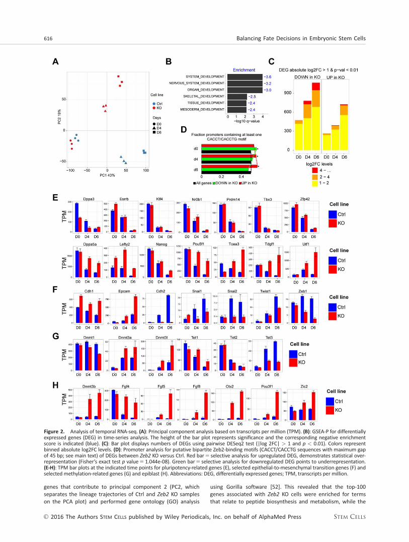
genes that contribute to principal component 2 (PC2, whichseparates the lineage trajectories of Ctrl and Zeb2 KO sampleson the PCA plot) and performed gene ontology (GO) analysis
using Gorilla software [52]. This revealed that the top-100genes associated with Zeb2 KO cells were enriched for termsthat relate to peptide biosynthesis and metabolism, while the
Figure 2. Analysis of temporal RNA-seq. (A): Principal component analysis based on transcripts per million (TPM). (B): GSEA-P for differentiallyexpressed genes (DEG) in time-series analysis. The height of the bar plot represents significance and the corresponding negative enrichmentscore is indicated (blue). (C): Bar plot displays numbers of DEGs using pairwise DESeq2 test (|log 2FC| > 1 and p < 0.01). Colors representbinned absolute log2FC levels. (D): Promoter analysis for putative bipartite Zeb2-binding motifs (CACCT/CACCTG sequences with maximum gapof 45 bp; see main text) of DEGs between Zeb2 KO versus Ctrl. Red bar5 selective analysis for upregulated DEG, demonstrates statistical over-representation (Fisher’s exact test p value5 1.044e-08). Green bar5 selective analysis for downregulated DEG points to underrepresentation.(E-H): TPM bar plots at the indicated time points for pluripotency-related genes (E), selected epithelial-to-mesenchymal transition genes (F) andselected methylation-related genes (G) and epiblast (H). Abbreviations: DEG, differentially expressed genes; TPM, transcripts per million.
616 Balancing Fate Decisions in Embryonic Stem Cells
VC 2016 The Authors STEM CELLS published by Wiley Periodicals, Inc. on behalf of AlphaMed Press STEM CELLS

top-100 genes associated with Ctrl cells were mainly enrichedfor epigenome-related terms, such as histone modificationand chromatin organization (for the gene lists, see SupportingInformation Table SIII).
We next applied a time-series analysis (see Materials andMethods) on our RNA-seq data set to assess the effect of thefactor “time” and identify genes that have a different dynamicexpression profile in KO versus Ctrl cells (Supporting Informa-tion Table SIV, RNASeq_Time_Series). Gene set enrichment(GSEA) analysis [53] of genes displaying this different dynamicbehavior showed strong negative enrichment for various dif-ferentiation/developmental categories within the top-10 hits(Fig. 2B) and further confirmed that at least the vast majorityof Zeb2 KO cells in EBs indeed remain uncommitted. We also(re-)confirmed that Zeb2 KO cells do not acquire neural fate(using Pax6, Zfp521, and Neurog1; Supporting InformationFig. S3A). The early-neuroectoderm markers Gbx2 and Hoxa1
previously shown to be correctly induced upon differentiationin Zeb2 KD hESCs [33], were not induced in Zeb2 KO mESCs. Thisindicates that genetic inactivation of Zeb2 results in a moresevere neural acquisition phenotype than the KD (SupportingInformation Fig. S3A). We examined the expression of other celllineage markers in our RNA-seq data to exclude that Zeb2 KOcells would preferentially induce non-neural fates (SupportingInformation Fig. S3B-3D). Although a small increase in thosemarkers was observed in Ctrl EBs, they were either almostabsent (for mesoderm, Supporting Information Fig. S3C) ormarkedly lower (trophectoderm and endoderm; SupportingInformation Fig. S3B, 3D) in Zeb2-deficient EBs.
While this RNA-seq data analysis significantly expands ourprevious characterization of the cells and confirms that Zeb2
inactivation globally affects ESC differentiation, it also provid-ed the possibility to discover potential Zeb2-dependent candi-date genes responsible for the impaired differentiation ofZeb2 KO ESCs. Therefore, we performed pairwise RNA-seqanalysis at all three time-points and identified DEGs (p value<0.01 and log 2 Fold Change (FC) >1; Supporting InformationTable SV, RNAseq_Pairwise). Consistent with the divergentPCA trajectories we observed an increase in both number ofDEGs and their FC over time (Fig. 2C).
Upon neural induction the majority of genes that wereeither up or down on d4 (multipotent progenitor stage) main-tained this status on d6 (early neural progenitor), 69% and72%, respectively. Numbers of DEGs increased between d4and d6. To further filter for direct transcriptional regulation byZeb2 we performed binding motif analysis within promoters(2 kb up and downstream of the transcription start site, TSS)of DEGs. We searched for two motifs, the E-box sequence 50-CACCTG-30 and 50-CACCT-30, interspaced by 45 bp max [54].The genes upregulated during differentiation in Zeb2 KO cellsshowed an increase in enrichment for the selected Zeb2-binding motifs (Fig. 2D, red bars and line), while the oppositetrend was observed for downregulated genes (Fig. 2D, greenbars and line). This suggests that Zeb2 functions preferentiallyas a transcriptional repressor during differentiation.
Zeb2 KO ESCs Stall in an Epiblast-like State
Zeb2-deficiency leads to impaired differentiation of ESCs andZeb2 preferentially acts as repressor. We, therefore, investigat-ed whether the pluripotency network was properly silenced inZeb2 KO ESCs, in particular the genes associated with the
na€ıve state and known as rapidly downregulated upon with-drawal of 2i1 LIF [38]. Klf4, Tbx3, Zfp42, Prdm14, Essrb,
Nr0b1, and Dppa3 were all properly downregulated in bothZeb2 KO and Ctrl ESCs (Fig. 2E, upper panel). However, a sig-nificant set of factors that are part of a larger pluripotencynetwork or involved in initiation of differentiation were not atall or only partially downregulated, such as Lefty2, Tcea3,Dppa5a, Utf1, and Tdgf1 [44, 45] (Fig. 2E, lower panel). Thisgroup also included Pou5f1 and Nanog, key players in theacquisition of pluripotency and early development [55, 56].All genes in the latter group contain putative binding sites forZeb2 within 2 kb from their TSS, suggesting that Zeb2 is acandidate direct repressor of (at least some) genes involvedin pluripotency maintenance. In line with the role of Zeb2 inEMT [19] we observed that in Zeb2 KO cells Cdh1expressionremains high, Epcam is strongly induced and Cdh2, Snai-1/2,Twist1, and Zeb1 were not induced to the same extent in dif-ferentiation conditions (Fig. 2F). This confirms that these ESCshave defective EMT consistent with previously documentedroles of Zeb2, including downregulation of Cdh1, in other celltypes.
Both Dnmt3b (Fig. 2H) and Dmt3l (Fig. 2G) have putativeZeb2-binding sites and were upregulated in Zeb2 KO duringdifferentiation. Together with other genes they determineDNA-methylation at this stage, hence we monitored Dnmt1,Dnmt3a, Tet1, Tet2, and Tet3 expression [10, 12, 14, 15]. Inaddition to high expression of all three Dnmt3 genes, theTet1/2 to Tet3 expression switch is only partially achieved; itnormally occurs during transition from pluripotent stem cellsto differentiated cells [15], but in our case Tet3 induction islimited and Tet1 expression is higher (Fig. 2G).
Although EBs are inherently heterogeneous, all our afore-mentioned results indicate that at least part of the cells arestalled in an epiblast-like cell state in which early epiblastmarkers are induced whereas a number of pluripotency genesgets downregulated [45, 56, 57]. We observed a strongincrease of expression of the established postimplantationepiblast genes Otx2, Pou3f1 (Oct6), Dnmt3b, Zic2, and Fgf5
[53, 58] (Fig. 2H). This data suggests that at least a fraction ofZeb2 KO cells undergoes lineage priming and acquiresepiblast-like cell (EpiLC) features.
Zeb2 KO ESCs Display Less EfficientESC-to-EpiLC Conversion
To test whether Zeb2 KO cells could acquire to epiblast-likefate, we subjected them (in parallel with the Ctrl line) to a 48hour-long ESC-to-EpiLC conversion [45] and examined thetranscriptional changes, using high-throughput qPCR (seeMaterials and Methods), of a set of markers shown to becomparably and differentially expressed, respectively, betweenESCs and EpiLCs [based on Ref. 55]. To evaluate whether EpiLCacquisition depends on the presence of Fgf21ActivinA, weincluded also samples of 48 hour-long differentiation in pureN2B27. There were no obvious morphological differencesbetween Ctrl and Zeb2 KO cells in 2i1 LIF or after 48 hoursESC-to-EpiLC conversion and both lines acquired more flatmorphology (Fig. 3A).
First, we analyzed expression of Nanog, Oct4 and Sox2,which according to Buecker and coworkers are comparablebetween ESCs and EpiLCs [57]. Nanog was downregulated inboth Ctrl and Zeb2 KO lines after 48 hours, but in the Zeb2
Stryjewska, Dries, Pieters et al. 617
www.StemCells.com VC 2016 The Authors STEM CELLS published by Wiley Periodicals, Inc. on behalf of AlphaMed Press

KO cells it was still expressed at higher levels (for Nanog
downregulation, see [45]). Oct4 was retained in Ctrl cells andslightly increased in the Zeb2 KO cells after 48 hours, whileSox2 was downregulated and retained, respectively (Fig. 3B).Next, we looked at Nr0b1, Prdm14, Zfp42, Esrrb, Klf2, andKlf4, which should be expressed at higher level in ESCs ascompared to EpiLCs. All these markers were found downregu-lated in both lines, but in the Zeb2 KO line Nr0b1, Zfp42, andKlf2 continued to display higher mRNA levels as compared toCtrl cells, whereas Prdm14, Esrrb, and Klf4 mRNA reachedafter 48 hours similar levels in both lines (Fig. 3C). Last, weanalyzed Dnmt3a, Dnmt3b, Fgf15, Fgf5, Otx2, and Pou3f1, aset of markers expected to be expressed at lower levels inESCs as compared to EpiLCs. With the exception of Dnmt3a inthe Zeb2 KO line, all markers were induced in both Ctrl andKO cell lines. In particular, Dnmt3b and Otx2 are induced inCtrl and Zeb2 KO lines to the same extent, while Fgf5 andPou3f1 show higher mRNA levels in Ctrl versus Zeb2 KO cells.Fgf15 in EpiLC conversion is expressed at higher levels in the
Zeb2 KO line as compared to Ctrl and after 48 hours inN2B27 its expression is comparable in both lines (Fig. 3D).Taken together, the results obtained in ESC-to-EpiLC conver-sion and 48 hours of N2B27 are comparable, meaning thatthe transcriptional changes of genes analyzed are not influ-enced by the presence of Fgf21ActivinA in the medium.
We conclude that, at population level, Zeb2 KO cells pre-sent with a less efficient conversion to EpiLC phenotype,likely resulting from a combination of both na€ıve and primedstates. Since epiblast fate requires more efficient pluripo-tency gene silencing than observed in the Zeb2 KO cells anda significant induction of markers such as Fgf5 and Pou3f1,we suggest that—in our EBs—a fraction of Zeb2 KO cellsremains in na€ıve ESC state, while the remaining cells maystill undergo EpiLC conversion. Single-cell mRNA analysis isneeded in future experiments to provide insight into theproportions of cells that stay in the na€ıve versus primedstate as well as revealing the presence of (few) cells pro-gressing toward differentiation.
Figure 3. Zeb2 KO embryonic stem cells (ESCs) display less efficient ESC-to-EpiLC conversion. (A): Phase-contrast images of Ctrl andZeb2 KO cells in 2i1 LIF and after 48 hours of EpiLC conversion. Scale bar: 50 lM. (B-D): RT-qPCR analysis of Ctrl and Zeb2 KO sampleson d0, after 48 hours in EpiLC medium and after 48 hours N2B27 in medium. (B): Transcripts comparable between ESC and EpiLC(Nanog, Pou5f1, Sox2). (C): Transcripts higher in ESC versus EpiLC (Nr0b1, Prdm14, Zfp42, Esrrb, Klf2, Klf4). (D): Transcripts lower in ESCsversus EpiLCs (Dnmt3a, Dnmt3b, Fgf15, Fgf5, Otx2, Pou3f1). These three categories (shown in panels B, C, D) are based on [66]. Resultsshown are from one experiment; error bars are from three biological samples. Abbreviation: ESCs, embryonic stem cells.
618 Balancing Fate Decisions in Embryonic Stem Cells
VC 2016 The Authors STEM CELLS published by Wiley Periodicals, Inc. on behalf of AlphaMed Press STEM CELLS

Pluripotent Potential Is Retained in Zeb2-DeficientEmbryoid Bodies
Pou5f1 (Oct4) and Nanog, two crucial pluripotency-supportingfactors maintained in epiblast cells, remained high (as seen bywestern blotting and RT-qPCR, Supporting Information Fig.S5A, 5B) and were present in a large fraction of cells in Zeb2
KO ND-EBs till d15 (Fig. 4A, 4B). Again, this observation couldbe extended to GD-EBs (Supporting Information Fig. S5C, 5D;d15). In addition, high numbers of Cdh11 cells were observedin Zeb2 KO EBs (Fig. 4B) and this was also seen at proteinand mRNA levels on d15 (Supporting Information Fig. S5A,5B). R26_Zeb2 rescue partially restored downregulation ofOct4 (and Cdh1) mRNA/protein (Supporting Information Fig.S2C). To confirm that this is the direct result of Zeb2 bindinga ChIP-qPCR was carried out over the Zeb2-binding motif (Fig.4C, 4D). This showed enrichment of Flag-tagged Zeb2 (usingR26_Zeb2 ESCs) Cdh1 promoter [19, 26] and its new candi-date target Nanog.
To document the persistence of the pluripotent state upondifferentiation in Zeb2 KO cells, we dissociated Ctrl and Zeb2 KOEBs on d15 (in ND or GD), sorted the living cells and platedthese at 500 cells/well as single cells in 2i1 LIF. Alkalinephosphatase (AP)1 ESC colonies derived from EBs subjected to
differentiation (Fig. 4E, 4F) were quantified on d9 (Fig. 4G). In atypical experiment, Ctrl cells subjected to ND did not give rise toAP1 cells, whereas Zeb2 KO cells in 2i1 LIF yielded on average8 colonies/well. In GD, Ctrl cells gave rise to less than 1 (calculat-ed 0.2) AP1 colony/well, whereas for Zeb2 KO cells this was 4colonies/well on average. Based on AP read-out, this showsthese latter cells have the remarkable ability to re-adapt to2i1 LIF, like ESCs and EpiSCs, and that they form AP1 colonieseven up to d15 of differentiation treatment. Without assess-ment at single-cell level, we cannot discriminate whether theseAP1 colonies arose exclusively from epiblast-like or more na€ıveZeb2 KO cells since both cell types can adapt to 2i1 LIF. Terato-ma formation, using EBs subjected to ND for 12 days showedthat Ctrl EBs failed to form teratomas, while Zeb2 KO EBs gaverise to teratomas in 4 weeks (Supporting Information Fig. S5E).These data show, therefore, that Zeb2 genetic inactivation leads tomaintenance of pluripotency even after prolonged differentiation.
The Zeb2 KO Embryoid Bodies, Subjected to ND, Failto Maintain the Initially Acquired DNA-Methylation
Zeb2 KO EBs show impaired cell differentiation and deregulatedexpression of the core methylation machinery genes. Thisprompted us to examine the acquisition and maintenance of
Figure 4. Pluripotency genes are not efficiently downregulated during differentiation in Zeb2 knockout (KO) embryonic stem cells (ESCs).(A): Control (Ctrl) and Zeb2 KO (KO) embryoid bodies (EBs) stained for Nanog (brown) on d4, d6, and d15 of neural differentiation (ND). (B):Ctrl and KO EBs costained for Oct4 (green) and Cdh1 (red) on d4, 6, and 15 of ND. Panels A, B show results from one experiment that is repre-sentative for three experiments. Scale bar: 50 mm. (C, D): Zeb2 chromatin immunoprecipitation (using anti-Flag antibody) on Cdh1 (panel C)and Nanog promoter (panel D). Results shown are from one experiment and representative for three experiments. SD of two technicalrepeats is shown. (E, F): Ctrl and KO ESCs subjected to ND (panel E) and general differentiation (panel F) for 15 days, dissociated and platedat 500 cells/well in 2i. The resulting ESC colonies (indicated by arrows) were visualized by staining for AP, and panel (G) represents theaverage number of AP1 colonies obtained after plating the cells. Abbreviations: ND, neural differentiation; GD, general differentiation.
Stryjewska, Dries, Pieters et al. 619
www.StemCells.com VC 2016 The Authors STEM CELLS published by Wiley Periodicals, Inc. on behalf of AlphaMed Press

CpG-methylation (meCpG) that accompanies the decision of irre-versible ESC differentiation. Retaining the same time/samplesetups as for RNA-seq and again using ND, single-base profileswere generated of methylation by RRBS in both Ctrl and Zeb2
KO on d0, d4, and d6. The genome of ground-state (d0) ESCswas globally hypomethylated [59]. On d4 both cell populationsgained methylation in agreement with our observation thatthey are in an epiblast-like (for KO) or multipotent (Ctrl) state.However a significant drop of meCpG was observed in the d6Zeb2 KO cell population, suggesting that part of the CpG methyl-ation is lost (Fig. 5A). The progressive accumulation of meCpG inour EBs has a striking resemblance with that observed in vivo[60], that is, our d0 population profile is similar to blastocyst-stage embryos (between E3.5 and E4.5), while d4 and d6Ctrl populations have a similar distribution profile as epiblastembryos (E6.5). In contrast, KO d6 resembles early-epiblastembryos (E5.5) with a reduction in meCpG at both gene bodiesand 10kb-flanking regions (Fig. 5B) [60].
We further investigated changes in the (de)methylation pro-cess by considering CpGs covered in all samples and averagingmethylation in 400bp-tiles, with a total of 1,84,564 tiles. Thisidentified differentially methylated regions (DMRs) (absolutemethylation change >20% and q value <0.05) in both a timeand pairwise-dependent manner (Supporting Information TableSVI, RRBS_Pairwise). Both Ctrl and KO cells significantly gainedmethylation in respectively 33.8% and 33.5% of all tiles betweend0 and d4 (Fig. 5C, left panels; Supporting Information Fig. S6A).During this period, no single significant loss-of-methylation wasobserved (Supporting Information Fig. S6B). Next, between d4and d6 Ctrl cells maintained a very stable level of methylationwith only little gain or loss-of-methylation, that is, 0.1% of alltiles (Fig. 5C, right top panel; Supporting Information Fig. S6A,6B). In agreement with the observed overall lower methylationat d6 (Fig. 5A, 5B), KO cells had 10 times more tiles (1806 or 1%of all tiles) with significant loss-of-methylation and only 90 tiles(0.05% of all tiles) with gain-of-methylation (Fig. 5C, right
Figure 5. Analysis of temporal RRBS during neural differentiation. (A): Distribution histogram for individual meCpGs on d0, d4, and d6in Ctrl and Zeb2 KO populations. (B): meCpG distribution at gene bodies and 10kb-flanking regions of protein-coding genes. (C): Densityplots for pairwise comparisons of meCpG (in 400bp-tiles) between d0 and d4 in Ctrl (top) and d4 and d6 in KO (bottom) cells. (D): Densi-ty plot for pairwise comparison of meCpG (in 400bp-tiles) on d6 between Ctrl and KO. In C, D the density points increase from purple todark red. (E): Enrichment plot of Tet1-binding peaks centered around demethylated regions on d6 in a pairwise comparison betweenZeb2 KO versus Ctrl. (F): Violin plots showing gain and loss-of-methylation over time in identified genomic regions, that is, enhancers,CpG islands, canyons, transposable elements, high-CpG content promoters (HCP), low-CpG content promoter, exons, introns, other non-defined genomic regions, and globally at the whole-genome (genome) in Ctrl and Zeb2 KO cells. Abbreviations: CGI, CpG islands; TE,transposable elements; HCP, high-CpG content promoters; LCP, low-CpG content promoter.
620 Balancing Fate Decisions in Embryonic Stem Cells
VC 2016 The Authors STEM CELLS published by Wiley Periodicals, Inc. on behalf of AlphaMed Press STEM CELLS
bottom panel; Supporting Information Fig. S6A, 6B; see alsoSupporting Information Table SVII, RRBS_Temporal). Further-more, analysis of these aforementioned DMR in both Ctrl andKO cells revealed that these regions initially acquired methyla-tion in both Ctrl and KO cells at d4, but this methylation wasonly maintained in Ctrl cells (Supporting Information Fig. S6C).
To investigate whether demethylation was selective for spe-cific genomic regions, we profiled the methylation dynamics ofenhancers, CGI, canyons, TE, high-CpG content (HCP) and low-CpG content promoters (LCP), exons and introns. As reportedbefore [49], resistance to gain-of-methylation occurs for canyonsand high-CpG regions (CGI and HCP), while all other regions(enhancers, TE, LCP, exons, and introns) were susceptible togain-of-methylation. In contrast, the Zeb2 KO population isunable to maintain this methylation initially acquired in allaforementioned genomic regions (Fig. 5F).
Failure to Maintain Acquired DNA-Methylation DuringND Is Associated with Tet1-Binding; Tet1 Knockdownin Zeb2 KO ESCs Facilitates Silencing of Nanog, Oct4,
and Cdh1 and Partially Rescues the LineageDifferentiation Phenotypes
Regions that lose methylation in d6 Zeb2 KO populationsinitially acquired methylation comparable to Ctrl (SupportingInformation Fig. S6C). We also compared d6 of both Ctrl andZeb2 KO populations and as expected observed a similar num-ber of tiles with loss-of-methylation (1938, or 1% of all tiles)(Fig. 5D) and we observed also an increased level of Tet1 (Fig.2). We, therefore, asked whether the regions that lose methyl-ation correlate with Tet1-binding. Figure 5E shows thatregions that lose methylation in Zeb2 KO cells are enrichedfor Tet1-binding in normal ESCs: we could do this by combin-ing analysis of published ChIP-seq data for Tet1 in mESCs [61]with our region-specific loss of methylation data on d6 (com-pared between Ctrl and Zeb2 KO). This strongly suggests thatthe observed demethylation in the Zeb2 KO cells is an activeprocess mediated by elevated Tet1 levels in agreement withDNA-demethylation being initiated at Tet1-binding sites [62].
Tet1 remains high in the Zeb2 KO EBs even on d15 of differ-entiation in contrast to its normal downregulation during NDand GD (Fig. 6A, 6B). To test whether high Tet1 levels lead toinefficient silencing of Nanog, Oct4, and Cdh1 and hence a blockin differentiation of these cells, we transduced control and Zeb2
KO ESC lines with a lentivirus expressing shRNA directed againstTet1 (called Ctrl_Tet1shRNA, Zeb2KO_Tet1shRNA, respectively).Tet1 was almost undetectable in Ctrl and Zeb2 KO lines targetedwith Tet1 shRNA (Fig. 6C, 6D; for quantifications of Tet1 staining,see Supporting Information Fig. S7A). In 2i1 LIF, the Tet1 KDlines maintained their undifferentiated characteristics (not
shown). We subjected these Tet1shRNA lines to ND and GD,respectively, along with the same lines receiving control non-targeting shRNA (Ctrl_CtrlshRNA, Zeb2KO_CtrlshRNA). Thesecontrol shRNA lines behaved as expected in differentiation (Fig.6E-6J), and Zeb2 was indeed absent from Zeb2KO_CtrlshRNAand Zeb2KO_Tet1shRNA EBs at the end of GD (Fig. 6H) and ND(data not shown). After 15 days, Zeb2KO_Tet1shRNA cells sub-jected to either ND or GD efficiently decreased Nanog, Oct4,and Cdh1 mRNA to low levels at the end of GD (Fig. 6E, 6F; NDdata not shown; for quantifications of Oct4 and Cdh1, see Sup-porting Information Fig. S7B, 7D). In Zeb2KO_Tet1shRNA linessubjected to GD, partial rescue of differentiation to mesoderm
(Fig. 6I; for quantifications of Desmin, see Supporting Informa-tion Fig. S7G) and endoderm (Fig. 6G; for quantifications ofHnf4a and Sox17, see Supporting Information Fig. S7E, 7F) wasobserved, but not to neuroectoderm, (data not shown). Partialrescue of ND was observed only when Zeb2KO_Tet1shRNA cellswere subjected to ND (Fig. 6J; for quantification of bIIITubulin,see Supporting Information Fig. S7C). Thus, Tet1 remains high inZeb2 KO cells during differentiation, and forced downregulationof Tet1 in these cells in such conditions enables decreasingNanog, Oct4, and Cdh1 transcription and partially rescuesdifferentiation. We conclude that Zeb2-deficiency duringdifferentiation leads to higher Tet1, which is associated withimproper reduction of Nanog and Oct4, resulting in impaireddifferentiation.
DISCUSSION
Using Zeb2 genetic inactivation in ESCs for the first time aswell as rescue in such Zeb2 KO cells via reintroduction of R26-driven Zeb2-cDNA, Zeb2 is shown critical for these cells toundergo three-lineage differentiation. We propose that Zeb2drives lineage commitment and specification by acting onmultiple sets of Zeb2-dependent genes. First, Zeb2 is animportant EMT-inducer [19]. Zeb2 KO ESCs retain epithelialcharacteristics when subjected to differentiation. Their pheno-type appears even more severe than the recently describedKO in ESCs of another known EMT-regulator, Snai1, which stilldifferentiate [63]. Second, the downregulation of importantpluripotency network regulators depends on Zeb2. In contrastto Ctrl, Zeb2 KO ESCs retain high Tet1, Oct4, and Nanog dur-ing differentiation. In ESCs, Tet1 is involved in a positive regu-latory loop with Nanog and Oct4. Tet1 co-operates withNanog, while the KD of Nanog weakens Tet1-binding to itstargets (including Oct4, Esrrb). Tet1 was also shown to actdownstream of Oct4, and downregulation of Oct4 leads todecreased Tet1 [14, 15, 17, 18]. Tet1 acts downstream of his-tone deacetylase Sirt6 to control ESC fate in differentiatingconditions [50]. Also, like in our system, the Tet1 KD allowedsilencing of Oct4 and Nanog and rescued the Sirt6 KO differ-entiation defect. Thus, Tet1 has a global inhibitory role in reg-ulating key pluripotency genes during ESC differentiation, andthis work identifies Zeb2 as an (indirect) upstream factorimportant for achieving correct Tet1 levels.
We describe a link between Zeb2 and regulation of DNA-methylation status. Acquisition of DNA-methyl marks duringembryogenesis is thought to be unidirectional [60], but stud-ies in ground-state na€ıve ESCs and EpiSC, respectively, showthat the methylomes are interconvertible in vitro when differ-ent conditions are applied [59, 64]. Our RRBS showed thatcorrect DNA-methylation patterns are initially acquired byZeb2 KO cells, but that this pattern cannot be sustained: Zeb2
KO cells revert the methylome to a more na€ıve state, whichagrees with the maintenance of their undifferentiated pheno-type associated with persistence of Nanog and Oct4. Remark-ably, this reversion in Zeb2 KO cells is facilitated in absence ofadditional cues or signals, like LIF and/or 2i. We hypothesizethat Tet1 levels are maintained by the key pluripotency genesin Zeb2 KO cells. Steady-state high-Tet1 would then activelydemethylate the genome and contribute to preserving high-Nanog and high-Oct4 in the mutant cells. Enrichment of Tet1-
Stryjewska, Dries, Pieters et al. 621
www.StemCells.com VC 2016 The Authors STEM CELLS published by Wiley Periodicals, Inc. on behalf of AlphaMed Press

binding at regions that lost methylation in Zeb2 KO cells fur-ther supports this hypothesis. Tet1 KD in these Zeb2 KO cellsfacilitated downregulation of Nanog and Oct4 as well asCdh1, but their differentiation phenotype was only partiallyrescued.
The aforementioned discussed results raise the questionon how it is possible that Zeb2 KO ESCs, in which Tet1 mRNAis not downregulated, can still undergo DNA-methylation.
First, the main function of Tet1 may be to actively catalyzedemethylation rather than prevent methylation per se [12,13]. We hypothesize that between days 0 and 4 the gain-of-methylation in both Ctrl and Zeb2 KO cells is driven by thevery early events linked to the withdrawal of 2i1 LIF andentering the primed state of pluripotency. We have, however,not documented that what seems like an equal total gain inCtrl and KO cells, also occurs with the same dynamics
Figure 6. Tet1 knockdown in Zeb2 knockout embryonic stem cells facilitates their definitive pluripotency exit and partially restores theirneural (neural differentiation [ND]) and general differentiation (GD) defect. (A): RT-qPCR of Tet1 mRNA in Ctrl (blue) and Zeb2 KO (red) lineson d0, d4, d6, and d15 of ND. SD of two technical replicates is shown. (B): Ctrl and Zeb2 KO embryoid bodies (EBs) stained for Tet1 (brown)on d4, d6, and d15 of GD. (C): RT-qPCR of Tet1 mRNA on d0 (violet) and d15 (green) in Ctrl_CtrlshRNA, Zeb2KO_CtrlshRNA, Ctrl_Tet1shRNAand Zeb2KO_Tet1shRNA lines. SD of two technical replicates is shown. (D): Ctrl_Tet1shRNA and Zeb2KO_Tet1shRNA EBs costained for Tet1(brown) on d15 of GD. Scale bar B, D: 50 mm. (E-J): Ctrl_CtrlshRNA, Zeb2KO_CtrlshRNA, Ctrl_Tet1shRNA and Zeb2KO_Tet1shRNA EBs stainedfor the indicated markers. (E): Nanog (brown) on 15 of GD. F. Oct4 (green) and Cdh1 (red) on d15 of GD. (G): Hnf4a (brown) on d15 of GD.(H): Zeb2 (brown) on d15 of GD. (I): Desmin (brown) on d15 of GD. (J): bIIITubulin (red) on d15 of ND. Scale bar: 75 mm. (E-I): Scale bar: 50mm. In all panels, results shown are from one experiment and representative for three experiments.
622 Balancing Fate Decisions in Embryonic Stem Cells
VC 2016 The Authors STEM CELLS published by Wiley Periodicals, Inc. on behalf of AlphaMed Press STEM CELLS
(because we analyzed in detail only days 0 and 4 of differenti-ation) and altogether reflects precisely that these cells under-go the same changes. In other words, Ctrl cells could beentering three-lineage differentiation program(s) (which wewere able to confirm by analyzing their transcriptional profile)whereas Zeb2 KO cells could be stalled in the epiblast-likestate and both changes would manifest by the same grossmethylation pattern. It has been previously published thatthere is a large gain of methyl marks in ESCs when they tran-sit from ground (2i1 LIF) to serum1 LIF conditions, both ofwhich maintain functional pluripotency [62]. Hence, theobserved acquisition of methyl marks can be partially a reflec-tion of entering the metastable state by the Zeb2 KO cells.
It has previously been shown that Dnmt1 KO ESCs showdecreased total DNA-methylation levels, whereas DNA of Dnmt 1/2
mutant ESCs is still highly methylated [65]. It could be that theobserved loss of DNA-methylation in our Zeb2 KO ESCs on d6 is par-tially caused by decreased Dnmt1 levels that cannot sustain theacquired methylation pattern in the presence of high Tet1, whichcontinuously catalyzes DNA-demethylation. Interestingly, theexpression of Dnmt3a/3b/3l was higher in Zeb2 KO as compared toCtrl ESCs. We hypothesize that the observed increase in de novomethyltransferase gene expression could be a counter-acting mech-anism to sustain the balance between DNA-methylation anddemethylation. The end result, loss of DNA-methylation, couldhence be due to high constant levels of Tet1 that on itself is sus-tained by the pluripotency genes.
Using GO analysis (done on PC2), we also noted that aber-rant chromatin changes and histone modifications could con-tribute to the differentiation phenotype in Zeb2 KO ESCs.ESCs have a unique, open chromatin that changes rapidlyupon cell differentiation, thereby influencing transcriptionalregulation and cell identity [4, 62]. Given the differences inthe transcriptional profile of Ctrl versus Zeb2 KO ESCs, wehypothesize that, in addition to the DNA-methylation relatedphenotype followed-up here, Zeb2 KO ESCs might also have adifferent chromatin (more ESC-like) structure, which contrib-utes to their undifferentiated phenotype.
It is also likely that Zeb2 controls other important cell fateregulators at multiple stages of differentiation in addition tothe pluripotency genes and Tet1 investigated here. For exam-ple, similar to described in vivo functions of Zeb2 in myelino-genesis in embryonic CNS [32], Zeb2 may also counteractgenes that are inhibitory for neural conversion during ESC dif-ferentiation; it may also act as an activator of other targetgenes depending on its cofactors [66, 67] which altogetherwould then promote neurogenesis. Subsequent work will haveto encompass the mapping of the genome-wide binding sitesof Zeb2 in mESCs. As the current anti-Zeb2 antibodies failin such studies (not shown), these studies will require anendogenous tagging approach within Zeb2 to identify Zeb2DNA-binding sites and also stage-relevant protein partners ofZeb2.
CONCLUSION
The transcription factor Zeb2 is critical for exit from the epi-blast state in mouse ESCs and links the pluripotency network
and DNA-methylation with irreversible commitment to differ-entiation. Zeb2 KO ESCs display impaired differentiation bystalling in an epiblast-like state. Using RNA-seq, we concludethat in differentiating conditions EMT, pluripotency, lineagecommitment and DNA-(de)methylation genes are deregulatedin Zeb2 KO embryoid bodies. Using RRBS, we demonstratethat these cells cannot maintain their initially acquired DNA-methylation marks in neural-stimulating condition and do noteffectively downregulate Oct4, Nanog, and Tet1 in differentia-tion conditions. Tet1 KD partially rescues the impaired differ-entiation of the KO cells.
ACKNOWLEDGMENTS
We thank Rita Khoueiry, Kian Koh, Abishek Sohni, An Zwijsen,Raymond Poot for many suggestions, Derk ten Berge also forcritical advice on the ESC-to-EpiLC conversion experimentsand Ton Verkerk for help with image digital processing. DHwas supported by KU Leuven Research Council (GOA-11/012),FWO-V (G.0954.11, G.0782.14), Belspo IUAP-VII/07 (DH andJH), InfraMouse Hercules Foundation (ZW09-03), and ErasmusMC funds. FG was supported by the EC project BluePrint. JHwas supported by FWO-V (G.0568.13), the Belgian Federationfor the Study Against Cancer (BFAC), NH and MRC (1047995,1051485). SG is FWO-V post-doc.
AUTHOR CONTRIBUTIONS
A.S.: Conception and design, collection and/or assembly of data,data analysis and interpretation, manuscript writing, finalapproval of manuscript; R.D.: Conception and design, collectionand/or assembly of data, data analysis and interpretation, man-uscript writing, final approval of manuscript; T.P.: Conceptionand design, provision of study material or patients, collectionand/or assembly of data, data analysis and interpretation, man-uscript writing, final approval of manuscript; G.V.: Conceptionand design, provision of study material or patients; AC.: Concep-tion and design, manuscript writing; K.C.: Collection and/orassembly of data; A.F.: Collection and/or assembly of data; L.U.:Collection and/or assembly of data; W.V.I.: Collection and/orassembly of data, final approval of manuscript; G.B.: Provisionof study material or patients; L.v.G.: Conception and design, pro-vision of study material or patients, final approval of manu-script; F.G.: Conception and design, manuscript writing, finalapproval of manuscript; S.G.: Conception and design, provisionof study material or patients, collection and/or assembly ofdata, manuscript writing, final approval of manuscript; J.H.: Con-ception and design, financial support4. Provision of study mate-rial or patients, collection and/or assembly of data, manuscriptwriting, final approval of manuscript; D.H.: Conception anddesign, financial support, data analysis and interpretation, man-uscript writing, final approval of manuscript.
DISCLOSURE OF POTENTIAL CONFLICTS OF INTEREST
The authors indicate no potential conflicts of interest.
Stryjewska, Dries, Pieters et al. 623
www.StemCells.com VC 2016 The Authors STEM CELLS published by Wiley Periodicals, Inc. on behalf of AlphaMed Press
REFERENCES
1 Martin GR. Isolation of a pluripotent cellline from early mouse embryos cultured inmedium conditioned by teratocarcinomastem cells. Dev Biol 1981;78:7634–7638.
2 Evans MJ, Kaufman MH. Establishmentin culture of pluripotential cells from mouseembryos. Nature 1981;292:154–156.
3 Brons IGM, Smithers LE, Trotter MWB et al.Derivation of pluripotent epiblast stem cells frommammalian embryos. Nature 2007;448:191–195.
4 Tesar PJ, Chenoweth JG, Brook FA et al.New cell lines from mouse epiblast sharedefining features with human embryonicstem cells. Nature 2007;448:196–199.
5 Matsui Y, Zsebo K, Hogan BLM. Deriva-tion of pluripotential embryonic stem cellsfrom murine primordial germ cells in culture.Cell 1992;70:841–847.
6 Leitch HG, Blair K, Mansfield W et al.Embryonic germ cells from mice and ratsexhibit properties consistent with a genericpluripotent ground state. Development 2010;137:2279–2287.
7 Ying Q-L, Wray J, Nichols J et al. Theground state of embryonic stem cell self-renewal. Nature 2008;453:519–523.
8 Marks H, Kalkan T, Menafra R et al. Thetranscriptional and epigenomic foundationsof ground state pluripotency. Cell 2014;149:590–604.
9 Leitch HG, McEwen KR, Turp A et al.Naive pluripotency is associated with globalDNA hypomethylation. Nat Struct Mol Biol2013;20:311–316.10 Okano M, Bell DW, Haber DA et al. DNAmethyltransferases Dnmt3a and Dnmt3b areessential for de novo methylation and mam-malian development. Cell 1999;99:247–257.11 Goll MG, Bestor T. Eukaryotic Cytosinemethyltransferases. Annu Rev Biochem 2005;74:481–514.12 Tahiliani M, Koh KP, Shen Y et al. Con-version of 5-methylcytosine to 5-hydroxymethylcytosine in mammalian DNAby MLL partner TET1. Science 2009;324:930–935.13 Kohli RM, Zhang Y. TET enzymes, TDGand the dynamics of DNA demethylation.Nature 2013;502:472–479.14 Ito S, D’alessio AC, Taranova OV et al.Role of Tet proteins in 5mC to 5hmC conver-sion, ES-cell self-renewal and inner cell massspecification. Nature 2010;466:1129–1133.15 Koh KP, Yabuuchi A, Rao S et al. Tet1and Tet2 regulate 5-hydroxymethylcytosineproduction and cell lineage specification inmouse embryonic stem cells. Cell Stem Cell2011;8:200–213.16 Takahashi K, Yamanaka S. Induction ofpluripotent stem cells from mouse embryonicand adult fibroblast cultures by defined fac-tors. Cell 2006;126:663–676.17 Costa Y, Ding J, Theunissen TW et al.NANOG-dependent function of TET1 andTET2 in establishment of pluripotency.Nature 2013;495:370–374.18 Gao Y, Chen JJ, Li K et al. Replacementof Oct4 by Tet1 during iPSC induction revealsan important role of DNA methylation andhydroxymethylation in reprogramming. CellStem Cell 2014;12:453–469.
19 Comijn J, Berx G, Vermassen P et al. Thetwo-handed E-box binding zinc finger proteinSIP1 downregulates E-cadherin and inducesinvasion. Mol Cell 2001;7:1267–1278.20 Kim Y-S, Yi B-R, Kim N-H et al. Role ofthe epithelial-mesenchymal transition and itseffects on embryonic stem cells. Exp MolMed 2014;46:e108.21 Pieters T, van RF. Role of cell–cell adhe-sion complexes in embryonic stem cell biolo-gy. J Cell Sci 2014;127:2603–2613.22 Mowat DR, Croaker GD, Cass DT et al.Hirschsprung disease, microcephaly, mentalretardation, and characteristic facial features:Delineation of a new syndrome and identifi-cation of a locus at chromosome 2q22-q23.J Med Genet 1998;35:617–623.23 Cacheux V, Dastot-Le Moal F, K€a€ari€ainenH et al. Loss-of-function mutations in SIP1Smad interacting protein 1 result in a syn-dromic Hirschsprung disease. Hum MolGenet 2001;10:1503–1510.24 Wakamatsu N, Yamada Y, Yamada Ket al. n-1, cause a form of Hirschsprung dis-ease. Nat Genet 2001;27:369–370.25 Maruhashi M, Van De PT, Huylebroeck Det al. Involvement of SIP1 in positioning ofsomite boundaries in the mouse embryo.Dev Dyn 2005;234:332–338.26 Van de Putte T, Maruhashi M, Francis Aet al. Mice lacking ZFHX1B, the gene thatcodes for Smad-interacting protein-1, reveala role for multiple neural crest cell defects inthe etiology of Hirschsprung disease-mentalretardation syndrome. Am J Hum Genet2003;72:465–470.27 Seuntjens E, Nityanandam A,Miquelajauregui A et al. Sip1 regulatessequential fate decisions by feedback signal-ing from postmitotic neurons to progenitors.Nat Neurosci 2009;12:1373–1380.28 McKinsey GLL, Lindtner S, Trzcinski Bet al. Dlx1&2-dependent expression of Zfhx1b(Sip1, Zeb2) regulates the fate switchbetween cortical and striatal interneurons.Neuron 2013;77:83–98.29 van den Berghe V, Stappers E, VandesandeB et al. Directed migration of cortical interneur-ons depends on the cell-autonomous action ofSip1. Neuron 2013;77:70–82.30 Van de Putte T, Francis A, Nelles L et al.Neural crest-specific removal of Zfhx1b inmouse leads to a wide range of neurocristo-pathies reminiscent of Mowat-Wilson syn-drome. Hum Mol Genet 2007;16:1423–1436.31 Jeub M, Emrich M, Pradier B et al. Thetranscription factor Smad-interacting protein1 controls pain sensitivity via modulation ofDRG neuron excitability. Pain 2011;152:2384–2398.32 Weng Q, Chen Y, Wang H et al. Dual-mode modulation of Smad signaling bySmad-interacting protein Sip1 is required formyelination in the CNS. Neuron 2012;73:713–728.33 Chng Z, Teo A, Pedersen RA et al. SIP1mediates cell-fate decisions between neuro-ectoderm and mesendoderm in human plu-ripotent stem cells. Cell Stem Cell 2010;6:59–70.34 Trott J, Martinez Arias A. Single cell line-age analysis of mouse embryonic stem cellsat the exit from pluripotency. Biol Open2013;2:1049–1056.
35 Festuccia N, Osorno R, Wilson V et al.The role of pluripotency gene regulatory net-work components in mediating transitionsbetween pluripotent cell states. Curr OpinGenet Dev 2013;23:504–511.36 Kaji K, Caballero IMM, MacLeod R et al.The NuRD component Mbd3 is required forpluripotency of embryonic stem cells. NatCell Biol 2006;8:285–292.37 Betschinger J, Nichols J, Dietmann Set al. Exit from pluripotency is gated byintracellular redistribution of the bHLH tran-scription factor Tfe3. Cell 2013;153:335–347.38 Leeb M, Dietmann S, Paramor M et al.Genetic exploration of the exit from self-renewal using haploid embryonic stem cells.Cell Stem Cell 2014;14:385–393.39 Higashi Y, Maruhashi M, Nelles L et al.Generation of the floxed allele of the SIP1(Smad-interacting protein-1) gene for Cre-mediated conditional knockout in the mouse.Genesis 2002;84:82–84.40 Haenebalcke L, Goossens S, Naessens Met al. Efficient ROSA26-based conditionaland/or inducible transgenesis using RMCE-compatible F1 hybrid mouse embryonic stemcells. Stem Cell Rev Rep 2013;9:774–785.41 Soriano P. Generalized lacZ expressionwith the ROSA26 Cre reporter strain. NatGenet 1999;21:70–71.42 Pieters T, Haenebalcke L, Hochepied Tet al. Efficient and user-friendly pluripotin-based derivation of mouse embryonic stemcells. Stem Cell Rev Rep 2012;8:768–778.43 Haenebalcke L, Goossens S, Dierickx Pet al. The ROSA26-iPSC mouse: A conditional,inducible, and exchangeable resource forstudying cellular (de)differentiation. Cell Rep2013;3:335–341.44 Gaspard N, Bouschet T, Herpoel A et al.Generation of cortical neurons from mouseembryonic stem cells. Nat Protoc 2009;4:1454–1463.45 Hayashi K, Ohta H, Kurimoto K et al.Reconstitution of the mouse germ cell speci-fication pathway in culture by pluripotentstem cells. Cell 2011;146:519–532.46 Lee TI, Johnstone SE, Young RA. Chroma-tin immunoprecipitation and microarray-based analysis of protein location. Nat Protoc2006;1:729–748.47 Love MI, Huber W, Anders S. Moderatedestimation of fold change and dispersion forRNA-seq data with DESeq2. Genome Biol2014;15:550.48 Akalin A, Kormaksson M, Li S et al.methylKit: A comprehensive R package forthe analysis of genome-wide DNA methyla-tion profiles. Genome Biol 2012;13:R87.49 Jeong M, Sun D, Luo M et al. Large con-served domains of low DNA methylationmaintained by Dnmt3a. Nat Genet 2014;46:17–23.50 Etchegaray J-P, Chavez L, Huang Y et al.The histone deacetylase SIRT6 controlsembryonic stem cell fate via TET-mediatedproduction of 5-hydroxymethylcytosine. NatCell Biol 2015;17:545–557.51 Bain G, Ray WJ, Yao M et al. Retinoicacid promotes neural and represses mesoder-mal gene expression in mouse embryonicstem cells in culture. Biochem Biophys ResCommun 1996;223:691–694.
624 Balancing Fate Decisions in Embryonic Stem Cells
VC 2016 The Authors STEM CELLS published by Wiley Periodicals, Inc. on behalf of AlphaMed Press STEM CELLS

52 Eden E, Navon R, Steinfeld I et al.GOrilla: A tool for discovery andvisualization of enriched GO terms inranked gene lists. BMC Bioinformatics 2009;10:1–7.53 Subramanian A, Tamayo P, Mootha VKet al. Gene set enrichment analysis: Aknowledge-based approach for interpretinggenome-wide expression profiles. Proc NatlAcad Sci USA 2005;102:15545–15550.54 Remacle JE, Kraft H, Lerchner W et al.New mode of DNA binding of multi-zinc fin-ger transcription factors: deltaef1 familymembers bind with two hands to two targetsites. EMBO J 1999;18:5073–5084.55 Nichols J, Zevnik B, Anastassiadis K et al.Formation of pluripotent stem cells in themammalian embryo depends on the POUtranscription factor Oct4. Cell 1998;95:379–391.56 Chambers I, Colby D, Robertson M et al.Functional expression cloning of Nanog, apluripotency sustaining factor in embryonicstem cells. Cell 2003;113:643–655.
57 Buecker C, Srinivasan R, Wu Z et al.Reorganization of enhancer patterns in tran-sition from na€ıve to primed pluripotency. CellStem Cell 2014;14:838–853.58 Folmes CDL, Dzeja PP, Nelson TJ et al.Metabolic plasticity in stem cell homeostasisand differentiation. Cell Stem Cell 2016;11:596–606.59 Qian X, Kim JK, Tong W et al. DPPA5supports pluripotency and reprogramming byregulating NANOG turnover. STEM CELLS 2016;34:588–600.60 Auclair G, Guibert S, Bender A et al.Ontogeny of CpG island methylation and spe-cificity of DNMT3 methyltransferases duringembryonic development in the mouse.Genome Biol 2014;15:545.61 Wu H, D’Alessio AC, Ito S et al. Dualfunctions of Tet1 in transcriptional regulationin mouse embryonic stem cells. Nature 2011;473:389–393.62 Habibi E, Brinkman AB, Arand J et al.Whole-genome bisulfite sequencing of twodistinct interconvertible DNA methylomes of
mouse embryonic stem cells. Cell Stem Cell2013;13:360–369.63 Lin Y, Li X-Y, Willis AL et al. Snail1-depen-dent control of embryonic stem cell pluripo-tency and lineage commitment. Nat Commun2014;5:3070.64 Chambery A, Vissers JPC, Langridge JIet al. Qualitative and quantitative proteomicprofiling of Cripto2/2 embryonic stem cellsby means of accurate mass LC2MS analysis.J Proteome Res 2009;8:1047–1058.65 Lei H, Oh SP, Okano M et al. De novoDNA cytosine methyltransferase activities inmouse embryonic stem cells. Development1996;122:3195–3205.66 Conidi A, Cazzola S, Beets K et al. FewSmad proteins and many Smad-interactingproteins yield multiple functions and actionmodes in TGFb/BMP signaling in vivo. Cyto-kine Growth Factor Rev 2011;22:287–300.67 Hegarty SV, Sullivan AM, O’keeffe GW.Zeb2: A multifunctional regulator of nervoussystem development. Prog Neurobiol 2015;132:81–95.
See www.StemCells.com for supporting information available online.