you tube: steph curry ankle breaker - bonepit.combonepit.com/lectures/hind foot trauma syed...
TRANSCRIPT
You Tube: Steph Curry Ankle Breaker

Hind Foot Trauma
Syed Ali MD


Calcaneus Articulations
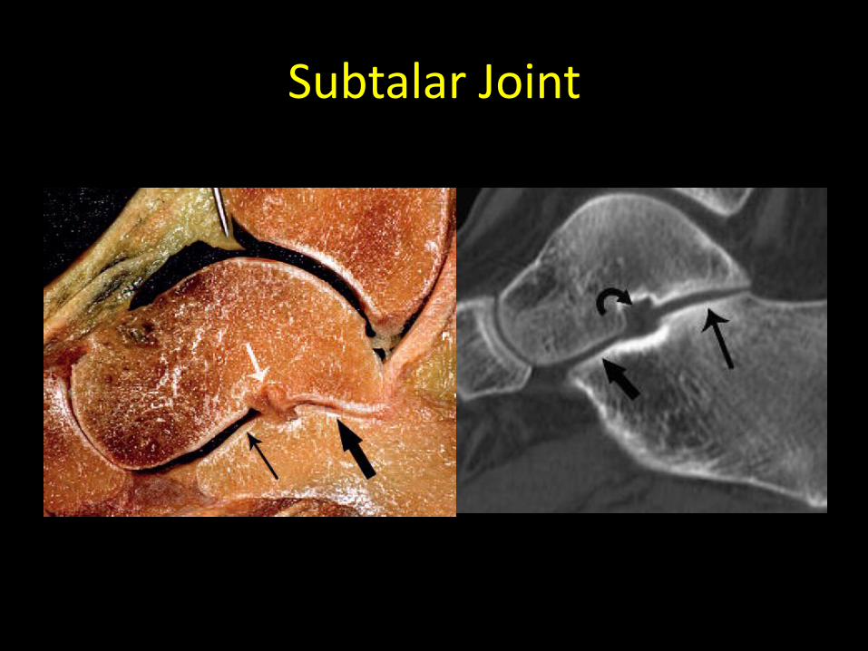
• Subtalar Joint: Superiorly, the calcaneus articulates with the talus at the talocalcaneal joint making contact at anterior, middle and posterior facets.
• Chopart Joint: Anteriorly, the calcaneus articulates with the navicular (talocalcaneonavicular joint) and the cuboid (calcaneocuboid joint) bones.

SUBTALAR JOINT

Superior Surface
• 4 articulating surfaces • Posterior, middle, and anterior
facets • Calcaneal sulcus • Sinus tarsi: calcaneal sulcus and
talus • Middle facet supported by
sustentaculum Tali • Anterior facet supported by the calcaneal beak. • Triangular anterior surface of the
calcaneus articulates with the cuboid
• k

Subtalar Joint
Subtalar Joint

Subtalar Joint

Subtalar Joint (Harris View)

Subtalar Ligaments
• Tarsal sinus ligaments, including:
a) Cervical ligament b) Talocalcaneal
interosseous ligament c) Medial and Lateral
Talocalcaneal ligaments 1) Lateral, 2) Intermediate, and 3) Medial roots of the
inferior extensor retinaculum

Medial and Lateral Talocalcaneal ligaments

Interosseous Talocalcaneal Ligament
• thick quadrilateral ligament
• originates from the sulcus calcanei, near the capsule of the posterior subtalar joint.
• .

Cervical Ligament
• u

Subtalar Dislocation (peritalar or hindfoot dislocation)
• ; • l

Subtalar Dislocation
• 65% to 80% are medial dislocations
• Remaining are lateral dislocations
• Case reports of anterior or posterior dislocations
• Osteochondral Fractures: up to 100% of lateral dislocations and 12%–38% medial dislocations
• Routine postreduction CT has been recommended to detect these fractures more accurately
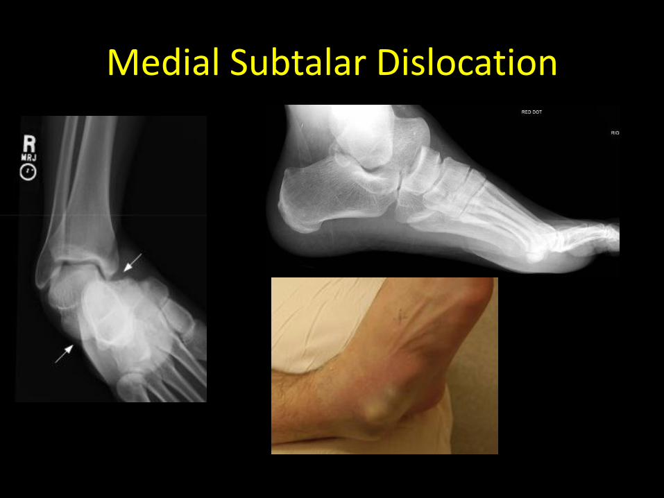
Medial Subtalar Dislocation

Lateral Subtalar Dislocation
Lateral Subtalar dislocation

Total Dislocation(pan-talar and luxatio tali totalis)
• Dislocation of the talus from all its articulations,
• Uncommon
• Devastating injury resulting from high-energy trauma.
• Almost all are open injuries.
• Avascular necrosis and osteomyelitis
• p
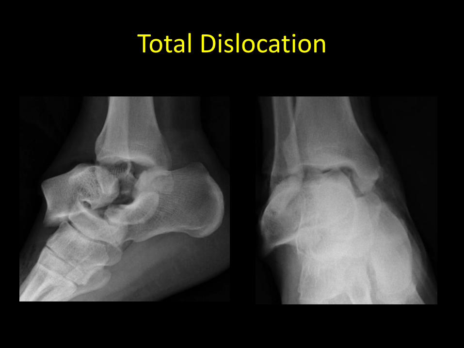
Total Dislocation

You Tube: Steph Curry Chris Paul

Calcaneus

Tensile trabeculae and Compression trabeculae
k

“Pseudocyst”
• “Pseudocyst” of the calcaneus
• gg

Lateral surface
• Flat and subcutaneous
• Central peroneal tubercle (PB and PL)
• Retrotrochlear Eminence
• Calcaneofibular ligament posteriorly
• Lateral talocalcaneal ligament attaches anterosuperiorly
• j

Medial Surface
• Sustentaculum tali is at the anterior aspect of the medial surface.
• Groove inferior to it transmits the flexor hallucis longus tendon.
• Neurovascular bundle runs adjacent to the medial border of the
calcaneus

• Normal Boehler angle: 20°- 40
• BA < less than 20° Posterior facet collapse from an underlying fracture

• Normal Critical angle of Gissane: 95-105
• - CAG > 130°: Posterior facet collapse from an underlying fracture

Tendons

Radiographic Views
• (a) anteroposterior and oblique views assessment of the calcaneocuboid joint
• (b) axial view of the heel (Harris view), and
• (c) Brodens View: dorsiflexion and internal rotation of the foot to better visualize the subtalar joint and posterior facet.
• (d) spine, contralateral foot and knee
• l

Harris Beath View
• Difficult in acute setting
• Demonstrates body of the calcaneus, middle facet of the subtalar joint and sustenaculum tali
• Heel width
• (Harris RI, JBJS 1948:30Br:624)
• d

Broden’s View
• Better visualize subtalar joint • Supine w/ foot rests on the
film cassette with neutral dorsiflexion;
• Entire lower leg and foot is internally rotated 45 deg;
• Central beam directed toward the lateral malleolus;
• Films are obtained at 10, 20, 30, and 40 deg. of cephalic tilt
• (Bruden Acta Radiol 31:85;1949).
• h

CT scan
• For fracture classification, particularly with the Sanders classification
• Reformat images parallel and perpendicular to the posterior facet off the sagittal reformatted images.
• s

Calcaneal Fractures
• Most common tarsal bone to be fractured
• 60% of fractures involving the tarsal bones
• 1%–2% of all fractures
• 17% open fractures
• Axial loading in men 30–60 years old
• Poor outcomes.
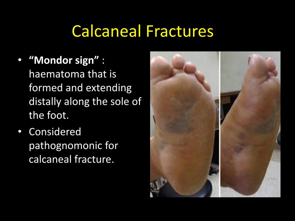
Calcaneal Fractures
• “Mondor sign” : haematoma that is formed and extending distally along the sole of the foot.
• Considered pathognomonic for calcaneal fracture.


Essex-Lopresti classification system
tongue type fracture depression type fracture

Essex-Lopresti classification system
• gg
tongue type fracture
y
depression type fracture
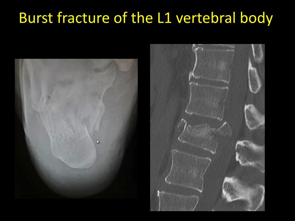
Burst fracture of the L1 vertebral body

Sagittal shear fracture
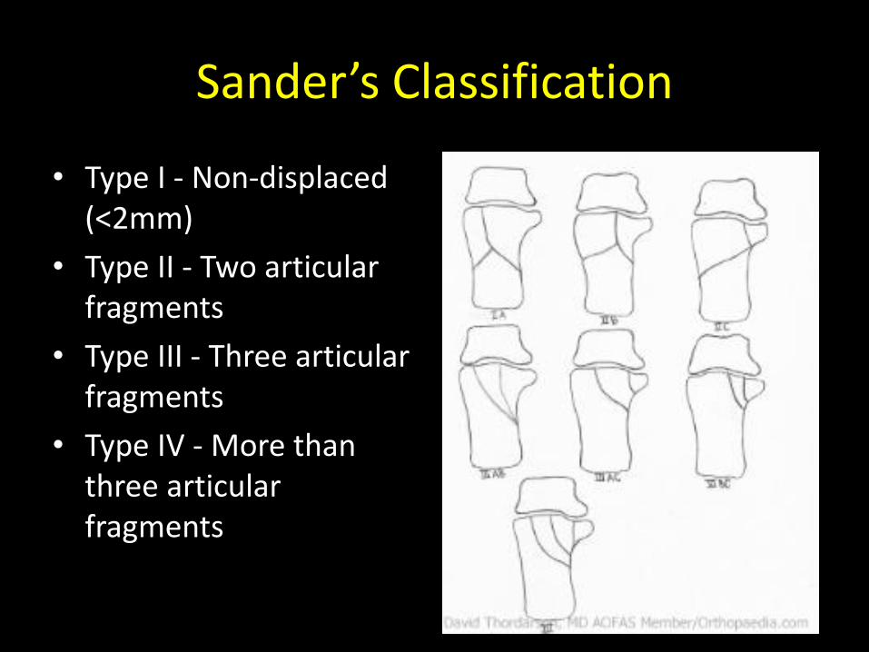
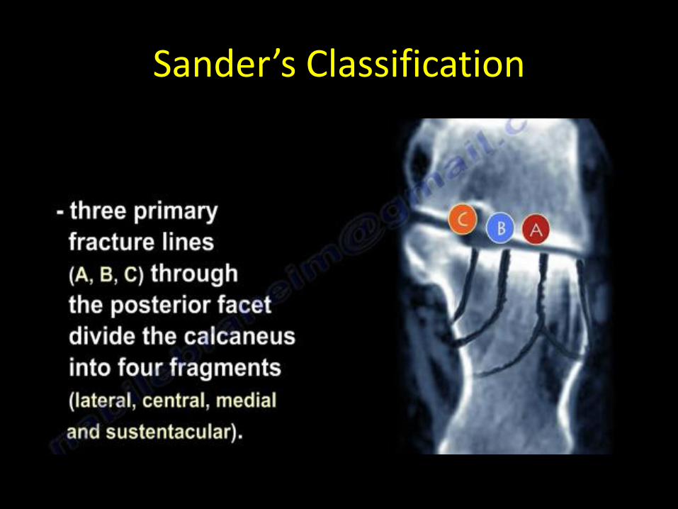
Sander’s Classification
• Type I - Non-displaced (<2mm)
• Type II - Two articular fragments
• Type III - Three articular fragments
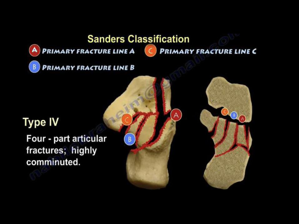
• Type IV - More than three articular fragments
Sander’s Classification


Calcaneus Fractures
• l


Sanders et al
• Type I fracture were treated without surgery
• Type II and type III fractures who underwent surgery experienced excellent or good clinical results in 73% and 70% of cases, respectively
• Only 9% of patients with type IV fractures had excellent or good clinical results after surgical treatment.
• Sanders et al have shown that although anatomic reduction is necessary for a good clinical outcome
• Success is not guaranteed, possibly related to cartilage necrosis at the time of injury.

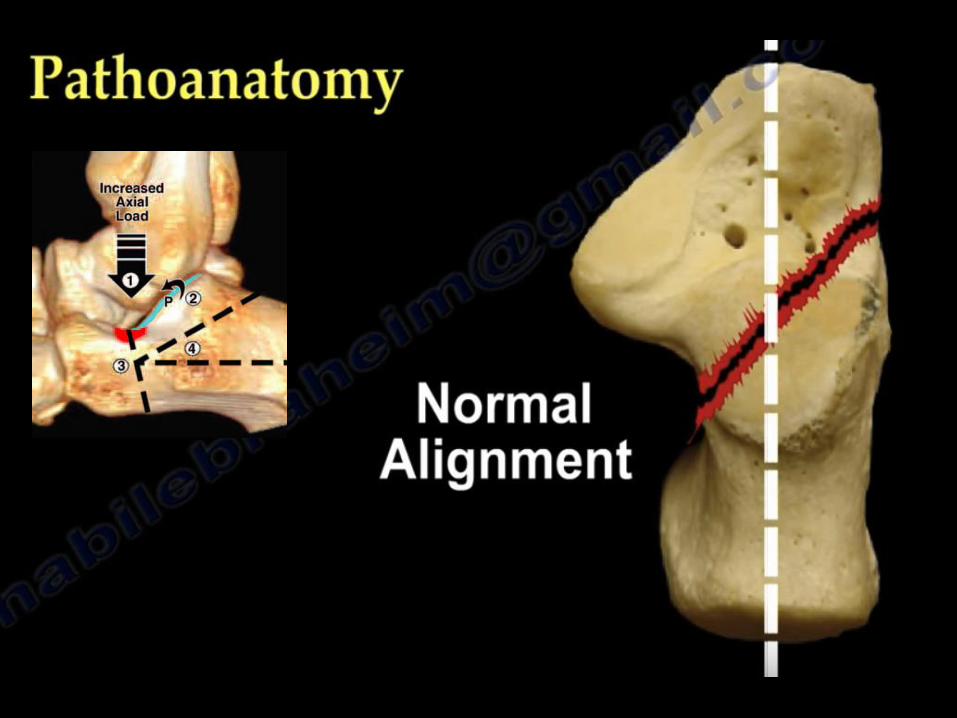
Typical Features
• (a) Loss of height due to impaction and rotation of the tuberosity fragment,
• (b) increase in width due to lateral displacement of the tuberosity fragment, and
• (c) disruption of the posterior facet of the subtalar joint
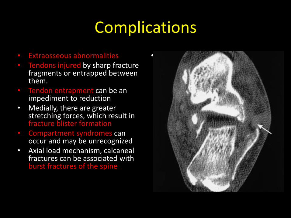
Complications
• Extraosseous abnormalities • Tendons injured by sharp fracture
fragments or entrapped between them.
• Tendon entrapment can be an impediment to reduction
• Medially, there are greater stretching forces, which result in fracture blister formation
• Compartment syndromes can occur and may be unrecognized
• Axial load mechanism, calcaneal fractures can be associated with burst fractures of the spine
• p
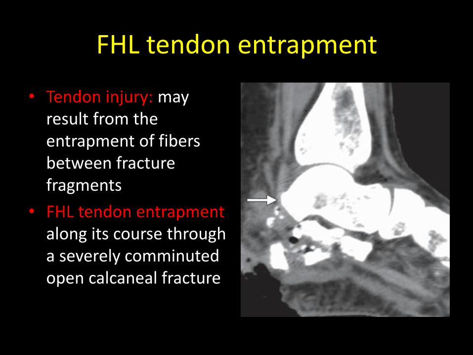
FHL tendon entrapment
• Tendon injury: may result from the entrapment of fibers between fracture fragments
• FHL tendon entrapment along its course through a severely comminuted open calcaneal fracture

Peroneus longus tendon entrapment
• Peroneus longus tendon entrapment of the peroneus longus tendon caused by “blowout” of the lateral aspect of the calcaneus. A Sanders type IIA intraarticular calcaneal fracture is also noted.

Fracture Blister

Extra-articular Fractures
• Account for about 25% of calcaneal fractures
• All fractures that do not involve the posterior facet of the subtalar joint
• Displaced fractures involving more than 25% of the calcaneocuboid articular surface are usually treated with ORIF
• Nonunion is the most common complication

Extensor digitorum brevis Avulsion

Achilles tendon avulsion type fracture

Achilles tendon avulsion type fracture
• Achilles tendon: avulsion type fracture of the calcaneal tuberosity posteriorly, there is a high risk of tissue necrosis and possible infection if management is delayed
• l

Bifurcate ligament
• The bifurcate ligament, also designated the “ligament of Chopart,” is formed by two ligaments: the medial calcaneocuboid ligament and lateral calcaneonavicular ligament

Bifurcate Ligament
l

Treatment (Conservative)
• Nonsurgical treatment is indicated for
• (a) nondisplaced or minimally displaced (Sanders type I) closed fractures;
• (b) extraarticular fractures; or
• (c) patients who have peripheral vascular disease, who smoke, or who have other surgical contraindications.
• Early range of motion without reduction or weight bearing

Treatment (surgical intervention )
• For displaced intraarticular (Sanders 2-4) and open fractures, surgical intervention is recommended within the first 3 weeks
• Before early consolidation of the fracture but not until swelling has decreased (skin wrinkles)
• Intervention consists of ORIF and arthrodesis if fracture is comminuted

You Tube Steph Curry George Hill

Talus

Talus Fractures
• less than 1% of all fractures
• 3% and 6% of fractures in the foot.
• Result of high-energy trauma
• Severity of talar injuries
• Complications and long-term disability
• Timely diagnosis and appropriate categorization are important for treatment planning

Talus Superior Projection

Talus Inferior Projection

Vascular Anatomy of Talus

Vascular Anatomy of Talus

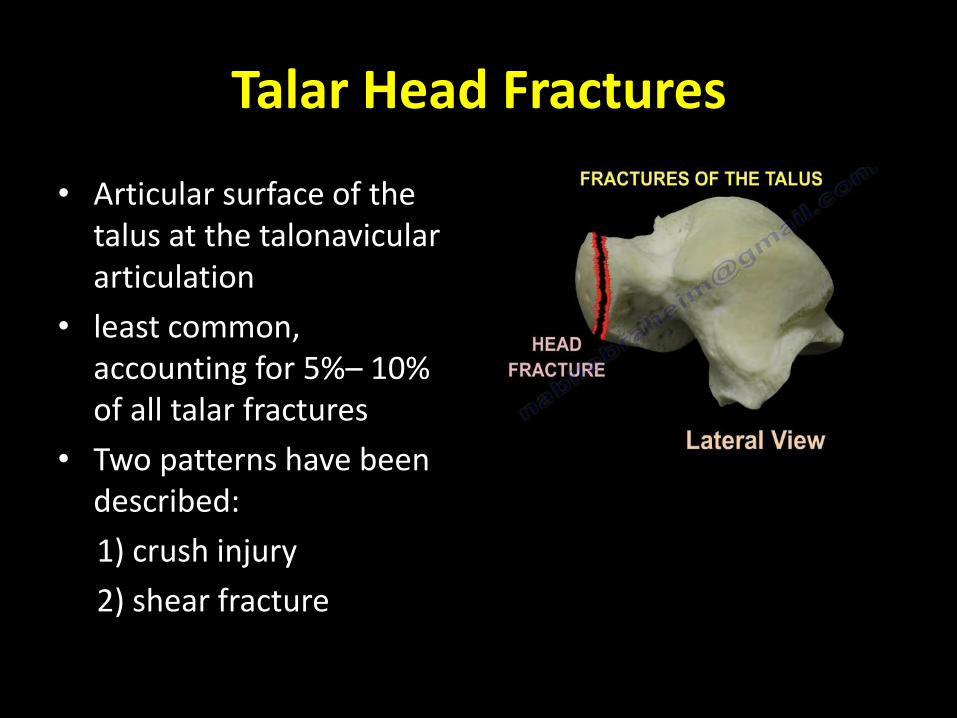
Talar Head Fractures
• Articular surface of the talus at the talonavicular articulation
• least common, accounting for 5%– 10% of all talar fractures
• Two patterns have been described:
1) crush injury
2) shear fracture
• ll

Talar Head Fractures

Talus Neck Fractures
• High incidence of talar neck fractures explained by its small cross-sectional area and vascular ingrowth, which increases the neck’s porosity
• Mechanism of this injury is forced dorsiflexion of the talus against the anterior aspect of the tibia, preceded by rupture of the posterior subtalar ligaments.
• Motor vehicle or motorcycle collisions and high-level falls

Hawkins-Canale Type I


Hawkins-Canale Type I

Hawkins-Canale Type 2


Hawkins-Canale Type 2

Hawkins-Canale Type 3


Hawkins-Canale Type 3

Hawkins-Canale Type 4


Hawkins-Canale Type 4

Surgery
• Conservative for nondisplaced talar neck fractures.
• Therefore, even subtle displacement of the talar neck fracture must be detected.
• Most type II fractures are treated with surgical reduction.
• In type III and IV fractures, closed reduction may be initially attempted in the emergency department to relieve skin tension and minimize soft-tissue injury followed with ORIF

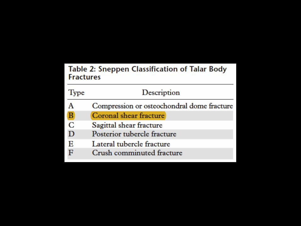
Talar Body Fractures
• Intra-articular
• Incidence ranges widely from 13% to 61%
• Sneppen classification
• ,

Sneppen Classification


Medial talar dome osteochondral fracture

Lateral talar dome osteochondral fracture


Sagittal and Coronal Shear Type
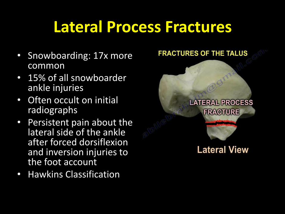
Lateral Process Fractures
• Snowboarding: 17x more common
• 15% of all snowboarder ankle injuries
• Often occult on initial radiographs
• Persistent pain about the lateral side of the ankle after forced dorsiflexion and inversion injuries to the foot account
• Hawkins Classification
• f

“Hawkins Classification”
McCrory and Bladin

Type I (simple) fracture

Type II (cortical avulsion) fracture
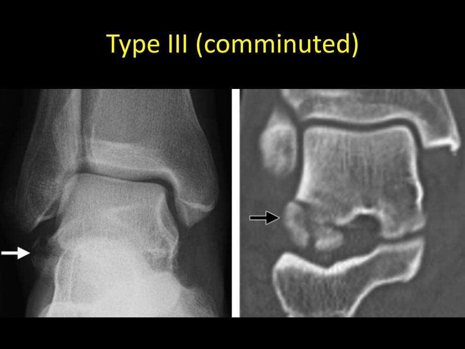
Type III (comminuted)
Lateral Process Fractures

Lateral Process Fractures
von Knoch et al(2007)'s V-sign

Lateral Process Fractures
Immediately after injury 5 month Post surgery


Posterior Process Fracture
• Involve the lateral tubercle more than the medial tubercle
• Forced plantar flexion, leading to compression of the posterior process between the tibia and calcaneus.
• Also direct trauma to the posterior ankle.
• p

Posterior medial tubercle fracture (Cedell Fracture) (Uncommon)

Posterior Process Fracture

Posterior Process Fracture

References
• Daftary A, Haims AH, Baumgaertner MR. Fractures of the calcaneus: a review with emphasis on CT. RadioGraphics 2005;25(5):1215–1226.
• Furey A, Stone C, Squire D, Harnett J. Os calcis fractures: analysis of interobserver variability in using Sanders classification. J Foot Ankle Surg 2003;42(1):21–23.7.
• Gray H. Anatomy of the human body. Philadelphia,Pa: Lea & Febiger, 1918; Bartelby.com; 2009. • Soeur R, Remy R. Fractures of the calcaneus with displacement of the thalamic portion. J Bone Joint
Surg Br 1975;57(4):413–421. • Germann CA, Perron AD, Miller MD, Powell SM, Brady WJ. Orthopedic pitfalls in the ED: calcaneal • fractures. Am J Emerg Med 2004;22(7):607–611. • Berry GK, Stevens DG, Kreder HJ, McKee M, Schemitsch E, Stephen DJ. Open fractures of the
calcaneus: a review of treatment and outcome. J Orthop Trauma 2004;18(4):202–206. • Heier KA, Infante AF, Walling AK, Sanders RW. Open fractures of the calcaneus: soft-tissue injury
determines outcome. J Bone Joint Surg Am 2003; 85-A(12):2276–2282. • Nicklebur S, Dixon T, Probe R. Calcaneus fractures: eMedicine orthopedic surgery. http://emedicine
medscape.com/article/1232246-overview. Published October 1, 2009. Accessed May 2010. • Richman JD, Barre PS. The plantar ecchymosis signin fractures of the calcaneus. Clin Orthop Relat
Res 1986;(207):122–125. • Benson E, Conroy C, Hoyt DB, et al. Calcaneal fractures in occupants involved in severe frontal • motor vehicle crashes. Accid Anal Prev 2007;39(4):794–799. • Melinevski et al
