york, amherst, ny14260, usa author manuscript nih public
TRANSCRIPT

DELIVERY OF THERAPEUTIC PROTEINS
Dipak S. Pisal*, Matthew P. Kosloski*, and Sathy V. Balu-Iyer*,ϕ* Department of Pharmaceutical Sciences, University at Buffalo, The State University of NewYork, Amherst, NY14260, USA
AbstractThe safety and efficacy of protein therapeutics are limited by three interrelated pharmaceuticalissues, in vitro and in vivo instability, immunogenicity and shorter half-lives. Novel drugmodifications for overcoming these issues are under investigation and include covalent attachmentof poly(ethylene glycol) (PEG), polysialic acid, or glycolic acid, as well as developing newformulations containing nanoparticulate or colloidal systems (e.g. liposomes, polymericmicrospheres, polymeric nanoparticles). Such strategies have the potential to develop as nextgeneration protein therapeutics. This review includes a general discussion on these deliveryapproaches.
KeywordsProtein delivery; PEGylation; Liposomes; hyperglycosylation; Poly(lactic/glycolic) acid
1.0 INTRODUCTIONSince the late 20th century numerous therapeutic proteins and peptides have emerged in themarket. PHARMA 2010 reported that biotech products accounted for more than 35% of the37 new active substances launched in 2001.1 In 2007, global biotech drug sales grew attwice the rate of traditional small molecule drugs (12.5% vs 6.4%) with total revenues of$75 billion US. Biotech drugs accounted for one fifth of all blockbuster drugs in the marketas of 2008.2 From a therapeutic perspective, proteins offer the distinct advantage of specificmechanisms of action and are highly potent. Despite these advantages, biotech productsmust overcome the hurdles posed by high molecular weight, short half-lives, instability, andimmunogenicity. Several strategies have been evaluated in an effort to improve the currentlimitations of therapeutic peptides and proteins in the creation of so called “secondgeneration” protein therapeutics. Most efforts center around one of two approaches—eithera change in the agent itself (e.g. mutations in protein structure or covalent attachment ofmoieties) or by a change in formulation.3 In contrast to modifying the protein structure,covalent chemical attachment of compounds such as poly(ethylene glycol) (PEG) orpolysialic acid (PSA) to therapeutic protein represent a relatively new approach. Drugformulation systems, such as liposomes, polymeric microspheres, and polymericnanoparticles, are another means to help overcome the current limitations of proteintherapeutics.4,5
The intent of this review is to provide a general discussion of approaches being applied toimprove safety and efficacy of protein therapeutics. This includes the areas of PEGylation,
ϕCorresponding Author: Formerly Sathyamangalam V. Balasubramanian, Ph.D., Department of Pharmaceutical Sciences, 521Hochstetter Hall, University at Buffalo, State University of New York Amherst, NY 14260, Telephone: (716) 645-2842 (x245), Fax:(716) 645-3693, [email protected].
NIH Public AccessAuthor ManuscriptJ Pharm Sci. Author manuscript; available in PMC 2011 June 1.
Published in final edited form as:J Pharm Sci. 2010 June ; 99(6): 2557–2575. doi:10.1002/jps.22054.
NIH
-PA Author Manuscript
NIH
-PA Author Manuscript
NIH
-PA Author Manuscript

lipid based vehicles, hyper glycosylation, and polymeric micro/nanospheres, specificallyPLGA microspheres.
2.0 PEGYLATIONThe conjugation of polymers to proteins had been in practice since the 1950s, but it was thedevelopment of PEGylation that provided the real breakthrough in enhancing thepharmaceutical properties of proteins and peptides in a viable manner 6 PEGylation, thecovalent attachment of PEG moieties to a therapeutic agent, was first reported in the 1970s.7,8 Experiments attempting to improve delivery aspects via PEGylation found not only theintended benefits, but overall enhancement of stability, pharmacokinetics, and therapeuticutility of molecules.9–12 PEGylation has been extensively discussed in the literature.3,9,12,13
This section primarily focuses on summarizing the various aspects of PEGylation, the keyproblems with first generation of PEGylation, and details recent discoveries from in vivostudies.
2.1 PEGylation: Principles and ProcessAs the name suggests, PEG moieties are repeating units of ethylene glycol that are both inertand amphiphilic in nature. PEG is synthesized by anionic ring opening polymerization ofethylene oxide initiated by nucleophilic attack of a hydroxide ion on the epoxide ring.Several derivatives of PEG molecules are available that vary in molecular weight andstructure, such as linear or branched.14 Figure 1 depicts the structural units of commonlyused PEGs.10
The process of PEGylation involves formation of a stable covalent bond between activatedPEG polymers and the polypeptide drug of interest. PEG is first activated by preparingderivatives with functional groups at one or both of the terminal ends. Functionalizationthrough the use of cyanuric chloride, PEG-succinimidyl succinate, and imidazolyl formateare some of the more common approaches and are displayed in Figure 2.9 This covalentattachment is generally made at the ∞ or ε amino groups of lysine, N-terminal amino groupof cysteine, histidine, arginine, aspartic acid, glutamic acid, serine, threonine, tyrosine, or C-terminal carboxylic acid.15 Among these amino acids, the most common choices forPEGylation are lysine and proteins N-terminal amino group.
Proteins and peptides are very labile molecules so the coupling reactions should utilize onlymild chemical conditions. The first generation of PEG derivatives were produced byattaching PEG activated via different chemistries to ε amino groups of lysine and included:(a) PEG succinimidyl carbonate, (b) PEG benzotriazole carbonate, (c) PEG dichlorotriazine,(d) PEG tresylate, (e) PEG p-nitrophenyl carbonate, (f) PEG trichlorophenyl carbonate, (g)PEG carbonylimidazole and (h) PEG succinimidyl succinate.16 Among these techniques,cyanuric chloride, imidazoyl formate, succinimidyl carbonates of PEG and succinimidylsuccinate methods were more commonly studied and are shown in figure 2. This techniqueresulted in modification of multiple lysine groups and produced mixtures of PEG isomerswith variable molecular masses. Most of the first generation reactions were conducted usingalkylating agents that nonspecifically modify multiple amino acids to form secondary aminelinkages with other protein molecules. Owing to its simple and straightforward nature, suchfirst generation PEGylation strategies were filled with numerous complications owinglargely to its hydroxyl group. End products were often contaminated with impurities,conjugation was restricted to low molecular weight PEG, linkages could prove relativelyunstable, modification lacked specificity, and immune response directed towards thechemical linker could result.16,17 Methoxy-PEG (mPEG) in particular had a highsusceptibility to form irreversible aggregates with protein due to the inherent 10–15% diolcontamination in the procedure.9,18
Pisal et al. Page 2
J Pharm Sci. Author manuscript; available in PMC 2011 June 1.
NIH
-PA Author Manuscript
NIH
-PA Author Manuscript
NIH
-PA Author Manuscript

To overcome these limitations, second generation PEGylation evolved with an array ofchemistries to improve PEG derivatives and their conjugation to therapeutics. Utilization ofcarboxylic acid intermediates of PEG allowed removal of almost 97% of diol impurities.19Since cysteine residues are far less prolific than lysine, conjugation to the thiol groups ofcysteine allowed more precise attachment.20 The incorporation of degradable linkages (e.g.para- or ortho-disulfide of benzyl urethane) provided a means to release drugs within theendosomes of cell.21 Heterobifunctional and branched PEG derivatives also allow morecontrol of the spatial arrangement of PEG.19 Site specific pegylation of protein drugs can beperformed by reductive alkylation with PEG-aldehyde.22–24 Pegylation of hemoglobin(HbA) can be achieved by site specific modification of its 4 α-amino groups ( two Vall(α)and two Val (β) residues) by glyceraldehyde in the presence of sodium cyanoborohydride.25
Reductive alkylation of HbA with methoxy polyethylene glycol 5000 propionaldehydegenerated a PEGylated Hb carrying an average of six PEG chains that exhibited an increasedO2 affinity.26
Site specific attachment approaches by enzymatic catalysis or reversible protection havebeen developed that utilize several naturally occurring enzymes, such as specific ornonspecific transglutaminase (Tgase) which is capable of recognizing glutamine as asubstrate. Tgase catalyses the transglutamination between PEG’s amino group and theglutamine’s amide, this site specific technology is more useful for yielding selectivePEGylation over thiol-selective and amino-terminus selective procedures and also occurs atmild reaction conditions.27 Other enzymes may also be employed like tyrosinase forconjugation to tyrosine and sialyltransferase for attaching a cytidine monophosphate (CMP)derivative of PEGylated sialic acid to the O- acetylgalactosamine residue of glycosylatedproteins.28,29
2.2 Rationale of PEGylationPEGylation of a molecule can result in alterations of various physicochemical propertiesincluding: i) increased size and molecular weight of a molecule, ii) changes in conformation,iii) steric hinderence of intermolecular interactions, iv) increased hydrophilhcity, and v)changes in electrostatic binding properties—all of which may affect the pharmacologicalbehavior of the conjugates.9 The most prominent effect of PEGylation is a prolongedcirculation time of conjugated therapeutics owing to a decreased rate of clearance by thekidney and/or a reduction of proteolysis and opsonization.30 PEG molecules are highlyhydrated which increases the hydrodynamic radius of the conjugate approximately 5–10 foldhigher than would be predicted from molecular weight alone 31–33. This increased radiusimproves solubility and decreases the rate of glomerular filtration. It has been reported thatthe total PEG mass required to retard the glomerular filtration of small molecules isapproximately 40–50 kDa whereas the molecular weight cut off for globular proteins is ~60kDa. PEGylated proteins show a drastic reduction in urinary clearance above the 20–30 kDarange.32 Chronic intravenous administration of PEGylated proteins have shown someunintended consequences such as vacuolation of renal cortical tubular epithelium inlaboratory animals34. However, most of these incidents of tubular vacuolation have occuredin animals exposed to the toxic doses of PEG during toxicologic evaluation.35 Theprotective shell formed by PEGylation also prevents uptake and clearance byreticuloendothelial cells, decreases the formation of neutralizing antibodies against theprotein by masking antigenic sites, and offers protection from proteolytic enzymes such astrypsin, chymotrypsin, and proteases.19,36,37 In addition, it has been reported thatPEGylation increases the absorption half-life of subcutaneously administered agents and isassociated with a decreased volume of distribution.32 PEGylation, in combination withtargeted drug delivery, may be efficiently used to decrease the clearance, alter thedistribution, or even enhance the delivery of therapeutic molecules. Increases in protein
Pisal et al. Page 3
J Pharm Sci. Author manuscript; available in PMC 2011 June 1.
NIH
-PA Author Manuscript
NIH
-PA Author Manuscript
NIH
-PA Author Manuscript
stability have been reported as a result of PEG masking hydrophobic sites on the proteins’surface involved in non-covalent interactions that lead to subsequent aggregation, loss ofactivity, or increased immunogenicity.38 Branched chain PEG moieties can increase the sizeof the total conjugate without resultant increase in number of attachment sites and have beenshown to improve stability in response to changes in pH, proteolytic digestion, andtemperature change as compared to linear PEGs.19
PEGylation is not without its drawbacks. Due to steric interference of excessive PEGylation,as often results with larger proteins, a loss of activity and binding affinity with the intendeddrug-target for a therapeutic molecule frequently results. Even with smaller proteins, somereduction in activity is almost a given. However, this reduction in binding may be offset byincreased systemic exposure. In case of growth hormone a high binding analog was preparedby site directed mutagenesis of parent growth hormone by a single point glycine to lysinemutation in the BS2 site (G120K)39. This G120K-GH antagonist exhibited a very shortplasma half-life of approximately 30 min, similar to the 15 min half-life reported for theparent GH.24. Pegylation with 5 kDa PEG to the antagonist using a random conjugationapproach yielded a protein with a dramatically longer half-life at more than 100 h, but whichlost 186-fold potency in receptor binding studies compared with parent GH. Thus, byfinding the pharmacokinetic and pharmacodynamic balance pegylated growth hormonereceived the US FDA approval in 2003 as second line therapy in treatment of acromegaly.40
Incidents of increased aggregation after PEG conjugation have been reported as well.41 Theother complication of PEGylation is polydispersity of the attached moities. PEG, being asynthetic polymer, is polydispered and exhibits a range of molecular weights. Thepolydispersity index (PDI), a measure of the distribution of molecular mass and can becalculated as the average molecular weight (Mw) divided by number average molecularweight (Mn) [Mw/Mn]. The PDI ranges from approximately 1.01 for low molecular weightPEG oligomer (3–5 kDa) to 1.2 for high molecular weight oligomers (20 kDa). Thispolydispersity is transferred to the PEG conjugated proteins and can make the formulationmore prone aggregation.17 Although infrequent, it may also be possible for anti-PEGantibodies to form, usually when repeated chronic doses of PEGylated proteins are given.These antiPEG antibodies may potentially increase the clearance of protein or drug deliveryvehicles attached to PEG.42
2.3 Preclinical and Clinical Development of PEGylation for Protein DeliveryPEGylation has made tremendous advances since its discovery. The FDA approved the firstPEGylated therapeutic, pegadamase (Adagen), in 1990. Since then numerous PEGylatedpolypeptides and macromolecules have reached the market with many more currentlyundergoing various stages of clinical development.43 PEGylated proteins currently in themarket are summarized in Table 1. There are numerous reports demonstrating benefit ofPEGylation to various therapeutic molecules and Table 2 indicates relative changes inpharmacokinetic properties of various macromolecular drugs and their PEGylatedconjugates in a preclinical setting. Some representative examples from these molecules arediscussed here and detailed information can be found elsewhere in the literature.9,14,40,44
PEG-anti-TNF-Fab was just approved for rheumatoid arthritis and Crohn’s disease in 2008.A PEGylated diFab antibody (PEG-CDP791) targeting vascular endothelial growth factorreceptor-2 has been studied for application in solid tumors by UCB-ImClone systems and isin phase II clinical trial.45 PEG-arginine deaminase, formed by the attachment of 10–12chains of 20 kDa PEG is in phase II clinical trial for hepatocellular carcinoma and anotherform with 12kDa PEG is in the preclinical stage.46,47 PEG-arginase, a pegylated arginasewith 5kDa PEG is also in phase II clinical trial. PEG-glutaminase is in phase II clinical trialto enhance activity of 6-Diazo-5-Oxo-L-Norleucine in colorectal and lung cancer.48
Pisal et al. Page 4
J Pharm Sci. Author manuscript; available in PMC 2011 June 1.
NIH
-PA Author Manuscript
NIH
-PA Author Manuscript
NIH
-PA Author Manuscript

There are various companies involved in the Pegylation of small molecules and largemolecules such as proteins and antibody fragments. Nektar therapeutic is abiopharmaceutical company that is heavily involved in developing new drugs based on itsPEG conjugation technology. There are 9 approved products on the pegyaltion technologywhich Nektar shares with other pharmaceutical companies and the recent pipeline can befound on its product pipeline. PolyTherics is involved in using TheraPEG™ technology, aninnovative, patent protected approach to the PEGylation of therapeutic proteins and antibodyfragments. Beyond targeted PEGylation, PolyTherics has also developed proprietarypolymer-based technologies for application to large proteins. Since its inception Enzonpharmaceuticals is involved in pegylation of various drugs and so far have successfulmarketed products such as ONCASPAR and ADAGEN and have various drugs includingsmall and large molecules in their pipeline.
PEGylation of the enzyme asparginase, used clinically for the treatment of acute leukemia,resulted in an increase of half-life from 20 hours for the unPEGylated parent enzyme to 357hours for pegaspargenase. It also protected asparginase from proteolytic degradation bytrypsin and reduced immune response.19,49 PEGylation of interleukin-6 not only producedmore than a 100 fold increase in half-life, resulting in a 500 fold increase in thrombopoieticpotency, but also decreased plasma IgGl production and adverse effects.50 Clinical studieswith Peginterferon α2b (PegIntron) have shown that it is superior to unPEGylated IFN-α2bin the initial treatment of chronic hepatitis C.51 The PEGylated form showed a significantdecrease in serum hepatitis c virus RNA and exhibited several fold lower clearancecompared to free IFN-α2b.52
PEGylation of IFN-α2a with first generation chemistry did not yield any clinical advantagedue to decrease in the activity but use of second generation 40 kDa branched PEG whichalso increased the size of total parent-PEG conjugate and reduced renal clearance by 100fold and increased half-life from 9 to 77 hours relative to native IFN-α2a.43
Nonspecific pegylation usually results in the heterogeneously PEGylated conjugates,whereas site specific PEGylation of specific functional groups (free cystines,oligosaccharides, alcohols, etc…) offers more precise control. Nonspefic pegylation oftumor necrosis factor (TNF)-α resulted in heterogeneous conjugates with decreasedbioactivity where as site specific pegylation of mono-pegylated TNF-α showed superiormolecular uniformity, demonstrated higher bioactivity in vitro, and greater antitumortherapeutic potency than randomly mono-PEGylated TNF-α.53
N-terminal site-specific PEGylation of rhG-CSF with a 30 kDa PEG resulted in longer invivo circulation half-life and 60% higher drug bioavailability than mono-PEG20-GCSF,made with 20 kDa PEG.54
It can be surmised that PEG improves the in vivo efficacy of protein drugs by altering thebalance between pharmacokinetic (PK) and pharmacodynamic (PD) effects. The decrease inbinding affinity is compensated with increased overall exposure of drug. PEG conjugationhas evolved over the last two decades to emerge as a viable pharmaceutical tool.Combination of the PEGylation approach with novel drug delivery technologies such asconjugating with hydrogels, small molecules, antibody fragments, lipids, sachharides, andbiomaterials is currently under investigation.29,55
3.0 HYPERGLYCOSILATIONGlycosylation has been the most heavily studied post-translational peptide modification. Thenature of the carbohydates attached to a protein plays an important role in dictatingstructure, function, activity, immunogenicity, and pharmacokinetics.56 The patterns of
Pisal et al. Page 5
J Pharm Sci. Author manuscript; available in PMC 2011 June 1.
NIH
-PA Author Manuscript
NIH
-PA Author Manuscript
NIH
-PA Author Manuscript

protein glycosylation are heterogenous, with the same protein often capable of displayingany one of numerous carbohydrates at a single glycosylation site.57,58 For recombinantproteins, glycosylation is highly dependent on the cell, while the machinery necessary forglycosylation is absent from bacterial expression systems.59 Amongst the eukaryotes,mammalian cells (e.g. Chinese hamster ovary cells) are preferred for recombinant proteinsdue to the similarity in glycosylation patterns to human proteins. Beyond attempting tomimic the glycosylation patterns of endogenous proteins, attempts to hyperglycosylatetherapeutic proteins have been utilized in a strategy that closely resembles that ofPEGylation. Attachment of additional carbohydrates to the protein serves to reduceinteractions with clearance mechanisms and antigen presenting cells (APCs) in an effort toprolong circulation and reduce immunogenicity.
3.1 Hyperglycosylation: Principles and ProcessAlterations in endogenous glycosylation patterns can have a drastic impact on thebiophysical properties of proteins. Removal of N-linked polysaccharides from Factor VIII(FVIII), a blood coagulation factor used as a first line treatment for Hemophilia A, led to aloss of over 30% of the proteins activity and significantly increased the propensity foraggregation.60 At the same time, a preparation of FVIII with the B-domain (the regioncontaining the vast majority of the protein’s glycosylation sites) removed is commerciallyavailable that retains full activity.61,62 Even monoclonal antibodies, possessing only oneglycosylation site at Asn297 in the CH2 region of each heavy chain, part of the Fc domain,exhibit biological activity that is highly dependent on the glycosylation pattern.63Antibodies that require interaction between the Fc domain and either Fcγ or C1qcomplement receptors to induce cytotoxicity show vastly reduced signaling fordeglycosylated forms. It has also been proposed that different glycoforms may activate thesereceptors to varying extent. However, predicting the effects of altered glycoforms is acomplex task that must be dealt with on a case by case basis. Fortunately, for proteintherapeutics in general attaching additional carbohydrate groups seems to exhibit a morepredictable impact than altering the native glycoforms.
In many ways hyperglycosylation can be seen as the natural evolution of PEGylation.Whereas PEG is an exogenous and not readily degraded moiety, many of the glyco groupsare ubiquitously expressed throughout the body and easily broken down. Additional sugarsmay be attached either by specific chemical reactions in situ or via site directed mutagenesisto introduce additional glycosylation sites into the primary structure of the protein.Polysialic acid (PSA) is an endogenous oligosaccharide that, like PEG, is available invarying sizes regulated by the number of repeating subunits, strongly hydrophilic, andcovalently linkable to the ε amino group of lysine via reductive amination.64 PSA is foundthroughout mammalian tissues and can be broken down into sialic acid upon endocytosis. Itis comprised of several monomeric subunits with the same basic nine-carbon backbone andheterogenous substitutions at several carbons.65 The most common subunit in humans is 5-N-acetylneuraminic acid (Neu5Ac) polymerized by α 2,8-glycosidic bonds.66 Certain typesof bacteria will utilize PSA attached to their cell walls to inhibit detection by the immunesystem. Bacterial PSA is identical to the human counterpart but fails to react with lowaffinity antibodies circulating in the body.67 These characteristics make it an ideal candidatefor protein conjugation.
With respect to site directed mutagenesis, protein glycosylation may come as either N-linkedor O-linked oligosaccharides. N-linked oligosaccharides, including PSA added by theproducing cell line, are attached to the consensus sequence of Asn-Xxx-Ser/Thr where Xxxis anything but proline.68 O-linked oligosaccharides do not require a specific sequence butare generally found attached to either serine or threonine.69 From a design perspective it ismuch easier to control additional N-linked glycosylation than O-linked. Many genetic tools
Pisal et al. Page 6
J Pharm Sci. Author manuscript; available in PMC 2011 June 1.
NIH
-PA Author Manuscript
NIH
-PA Author Manuscript
NIH
-PA Author Manuscript

exist for altering the DNA sequence of recombinant proteins to add or change amino acids,but care must be taken to ensure the biological properties of the protein are not alteredbeyond the addition of oligosaccharides. Substitutions of amino acids should be limited tothose with similar physio-chemical properties to minimize the impact on the global structureof the protein.70 It should be mentioned that the presence of a consensus sequence does notguarantee successful glycosylation.57,71 Location should be carefully considered whenchoosing a sequence to alter. The site must have a local conformation that renders itaccessible within the endoplasmic reticulum (ER) for post translational modification.Introduction of a glycosylation site into a region of the protein responsible for bindingnecessary for receptor or substrate binding would not be desired due to inhibition of proteinactivity while placement within an immunodominant epitope would be highly likely to resultin favorable changes in immunogenicity.
3.2 Rationale of HyperglycosylationOwing to the similarity between the principle of hyperglycosylation and PEGylation, manyof the same advantages apply to both. Hyperglycosylation can extend biological half-life,reduce immunogenicity, and improve solubility. Apparent increases in therapeutic efficacywill likely stem from prolonged residence in the body; hyperglycosylated forms areexpected to have reduced or equivalent activity to their native counterparts but not greater.The distinct advantage over PEG stems from the native and biodegradable nature of theoligosaccharides. There is some concern that PEG may build up over time in tissues as aresult of chronic administration of agents with extensive distribution. By contrast,endogenous carbohydrates such as PSA are readily digested. Although PEG is regarded asnon-immunogenic, there have been a few examples where IgG antibodies directed againstPEG or the PEG linker have been reported.72 Current beliefs claim that due to the stealthproperties of PSA employed by bacterial proteins, no clinically significant immune responsetowards the PSA of conjugated therapeutics is likely.64,67 The main limitations ofattachment of PEG and PSA will likely stem from inhibition of activity or unfavorableconformational changes induced by the covalent modification of the protein.
3.3 Preclinical and Clinical Development of Hyperglycosylation for Protein DeliveryIt has been proposed that shorter PSA derivatives such as colominic acids may serve greaterutility for attachment to protein therapeutics.64 Investigation into polysialyation ofasparaginase by reductive amination found that the half-life was increased in mice by 3–4fold and immunogenicity reduced while activity remained within 82–86% of the native formfollowing attachment of colominic acid.73,74 Similar improvements were found in bothnaive mice and those that had been immunized with the native form of the enzyme.Polysialylated forms of insulin have also been produced using colominic acid derivatizationand shown to prolong glucose suppression in mice by 2–3 fold versus the native form.75 Fabantibody fragments may also be ideal targets for polysialylation as they lack the extensivelymphatic recycling conferred by the Fc portion of the antibody and thus exhibit asubstantially shorter half-life compared to full length IgG monocloncal antibodies.76 For afab against the placental-like alkaline phosphatase antigen expressed on germ cellcarcinomas, chemical polysialylation with colominic acid increased both half-life and tumordeposition by 3 fold while a fab against anticarcinoembryonic antigen exhibited a 5 foldlonger half-life and 20 fold lower immunoreactivity following polysialylation.77,78 Severalpharmaceutical companies have polysialylated therapeutics in various stages ofdevelopment.
Darbepoetin alfa, a hyperglycosylated form of EPO, is already approved for sale in the U.S.and is marketed by Amgen. Site directed mutagenesis was used to alter five individualamino acids, introducing two additional N-glycosylation sites into the primary structure, for
Pisal et al. Page 7
J Pharm Sci. Author manuscript; available in PMC 2011 June 1.
NIH
-PA Author Manuscript
NIH
-PA Author Manuscript
NIH
-PA Author Manuscript

a total of five sites.79 Clonal selection allowed isolation of a form in which all five of theconsensus sequences were successfully glycosylated. This analog exhibited a three foldlonger half-life, creating the potential for once a week dosing compared to the three weeklyinjections required for traditional EPO therapy. During clinical trials, only 9 of 2660 patientsdeveloped antibodies capable of binding darbepoetin alfa where none existed before, none ofwhich were neutralizing antibodies.80 Advanced therapeutic agents like darbepoetin alfa, incombination with restricted routes of administration and more stringent regulatory oversight,have helped vastly limit incidences of life threatening pure red cell aplasia induced by EPOreplacement therapy.81 Lipoxen, a company invested in the science of hyperglycosylation,has polysialylated forms EPO, GCSF, interferon-alpha-2b, and insulin in various stages ofclinical and preclinical development.82
4.0 PLGA MICROSPHERES & NANOPARTICULATE DRUG DELIVERYPolymeric nanoparticulates have been explored as drug delivery vehicles for decades.83,84
Table 3 shows the lists of currently marketed drug formulations that utilize biodegradablemicroparticles.85 This broad heading encompasses, among others, polymeric micelles,hydrogels, microparticles, self-diffusion systems, biodegradable polymers, cellulosederivatives, porous membranes, and dendrimers.86,87 Here, focus will be placed onPoly(D,L-lactic-coglycolic-acid) (PLGA) microspheres and nano-particulate deliveryapproaches due to their wide acceptance as a biocompatible material approved for use inhumans by the US FDA.85
PLGA is polymer of lactic and glycolic acid joined by ester bonds. PLGA and its derivativeshave received much attention from drug delivery groups and have been widely studied forapplication in nano/microparticles encapsulating therapeutic drugs.88 Polymeric particlesare classified based on their size with the diameter of microparticles ranging from 1 to 250μm, while nanoparticles fall in the range of 10 to 1000 nm. PLGA microparticles aregenerated by polymerization of lactic acid and glycolic acid residues through ester linkage.The ratio of lactic acid and glycolic acid controls numerous aspects of the formulation.89
The most widely used PLGA composition of 50:50 has demonstrated the fastestbiodegradation rate of the polymers with near complete degradation in 50–60 days. Variousother combinations (e.g. 65:35, 70:30, 75:25) have been studied in the preclinical settingwith progressively increasing half lives correlating to the percentage of lactic acid.90 Onceadministered to the body, the ester bond is hydrolyzed by esterases in the blood, releasingthe drug from the polymer and allowing the polymer acids to become metabolized andcleared from the body.91
4.1 PLGA Microspheres: Principles and ProcessThe PLGA microspheres for protein delivery can be prepared by several methods with themost widely used techniques for proteins conjugation being: (i) spray drying, (ii) doubleemulsion, and (iii) phase separation-coacervation.92
Spray drying involves dissolution of the polymer in organic solvents to which the drug issubsequently added during high speed homogenization. This dispersed phase mixture isfurther atomized in a stream of heated air leading to the formation of microspheres that arecollected from the airstream by a cyclone separator. Remaining solvent is removed byvacuum drying in a nitrogen environment. Although this method produces a well definedparticle size and is highly reproducible, the harsh conditions can lead to the aggregation anddenaturation of proteins and antigens.93 To account for the sensitive nature of thesetherapeutics, spray freeze drying can be applied in which the dissolved polymers are mixedwith lyophilized protein to form a suspension. This suspension is only briefly homogenized,then sprayed over frozen ethanol. Frozen microspheres are formed instantaneously and can
Pisal et al. Page 8
J Pharm Sci. Author manuscript; available in PMC 2011 June 1.
NIH
-PA Author Manuscript
NIH
-PA Author Manuscript
NIH
-PA Author Manuscript

be stored in −70 °C to allow extraction of the solvents by the frozen ethanol that is laterseparated by the process of ultrafiltration. These microspheres are subsequently dried withnitrogen gas and can be dry sieved with the desired size steel mesh.92
The double emulsion method involves dissolution of protein in an aqueous medium made ofpolymer dissolved in an organic solvent and either homogenized or sonicated to form awater/oil emulsion. This primary emulsion may be used directly, but preference is to create asecondary emulsion by rapidly transferring the primary emulsion into an aqueous mediumcontaining a stabilizer such as polyvinyl alcohol. This mixture is further homogenized/sonicated forming the secondary emulsion. Residual organic solvent can be removed byseparating the phases with heat, vaccum, or in the case of sensitive protein, manualextraction. This process generally produces higher yields and more efficient encapsulationover other methods, but is dependent on the properties of the polymer—modification ofrelease profiles of drug from the microspheres after formation has proven relatively difficult.There may also be some additional stability concerns. Regardless, this method has beenapplied for the encapsulation of lysozyme, recombinant human epidermal growth factor, andleuteinizing hormone-releasing hormone agonist.94,95
Phase separation, like spray freeze drying, comprises mixing solid lyophilized protein withpolymeric organic solution. Silicone oil is slowly added to the mixture to extract thepolymer from its solvent. The polymer rich liquid phase encapsulates the dispersed proteindrug, leading to the formation of embryonic microspheres that are subsequently washed withheptane and subjected to hardening. Diphtheria toxoid and decapeptyl depot have both beenencapsulated through this method.96,97
Both phase separation and emulsion particle formation occur in the liquid phase, as opposedto spray drying that occurs in a stream of hot air or on the solid ethanol. To reduce the harshstress of the process, spray drying method with carbon dioxide can be performed at roomtemperature and is known as supercritical fluid (SCF) extraction.98 Briefly, the processinvolves concentration of protein solution as water is extracted by the carbon dioxide. Theprotein and other constituents are precipitated as a result of the increasing concentration andthe dissolution of the SCF in the solution. The particles formed are dried by extraction of theremaining solvent with SCF.99
Generally, proteins encapsulated by the emulsion or phase separation method are vulnerableto the aggregation, denaturation, oxidation, and cleavage, particularly at aqueous-organicsolvent interfaces, which may lead to a loss of activity for the encapsulated protein. Newermodifications involving the addition of excipients including stabilizers (e.g. albumin),surfactants, or bulking agents (e.g. trehalose and mannitol) to the protein phase are underinvestigation to help maintain protein stability.97 Modification of the PLGA polymer itselfcan also help to improve the stability, release profile, and potentially facilitate drugtargeting. It has been demonstrated that increases in the hydrophihcity of PLGA polymersby addition of hydrophilic moieties such as PEG led to an increase in the stability of protein.Various thermosensitive and triblock copolymers of PLGA may provide alternative meansof achieving better formulation characteristics.100
4.2 Rationale of PLGA MicrospheresPLGA derivatives are biocompatible, biodegradable, and can provide release rates on themagnitude of days, weeks, or months. Their high tensile strength, thermoplasticity, andproven non-toxicity in conjunction with the possibility of regulable release profiles,crystallinity, degradation rates, and hydrophihcity make PLGA microspheres a viableprotein delivery vehicle. They are also easily administered via injection. Severalmacromolecular drugs such as protein and peptide therapeutics, vaccines, and antigens have
Pisal et al. Page 9
J Pharm Sci. Author manuscript; available in PMC 2011 June 1.
NIH
-PA Author Manuscript
NIH
-PA Author Manuscript
NIH
-PA Author Manuscript
been successfully incorporated into PLGA microspheres.91,101,102 The favorablecharacteristics of the vehicle itself must be balanced against the negative effects on proteinstability during preparation and storage. Degradation of PLGA leads to accumulation oflactic and glycolic acids within the drug delivery device, thereby causing a significantreduction in pH of the surrounding microenvironment which can in turn facilitatedenaturation of encapsulated protein, aggregation, and loss of activity.103 True zero orderrelease from the PLGA devices can also be difficult as release is proportional to surfacearea, a property that will fluctuate with erosion of the vehicle.104
Several new polymers derived from PLGA have been developed in attempts to resolveissues with protein stability, degradation, and loading. Harmful reactions that may occur inthe process of PLGA formulation, such as deamidation and thiol-catalyzed disulfideexchange, can be addressed by adding assorted excipients to act as glass formers, alteringthe protein structure, or by protecting the native protein conformation.105 Co-encapsulationof surfactant poloxamer 188 and pluronic F127 are being explored for their modulatingeffect on the erosion rate of the PLGA polymer.100
4.3 Preclinical and Clinical Development of PLGA microspheres for Protein DeliveryEncapsulation of proteins into PLGA particles can improve the pharmacokinetic profiles ofthe drugs and subsequently reduce the required frequency of administration. Generallymicroparticles are given by i.m. or s.c. routes where as nanoparticles are given via the i.v.route. Primary successes of PLGA have come from smaller peptides and protein molecules.106
Jeong and coworkers developed a thermo-gelling biodegradable formulation made of PLGAand PEG moieties. A single injection of PLGA/PEG graft copolymers containing insulincontrolled blood glucose levels from 5 to 16 days in diabetic rats with the duration afunction of polymer compositions. This same thermogelling formulation, when used forchondrocyte cell delivery, showed remarkable improvement of cartilidge defects ascompared to the control formulation.107 In other reports, triblock copolymers includinghydrophilic PEG incorporated into PLGA have been used for encapsulation of severalsproteins including cytochrome C, ovalbumin, and tetanous toxoid (TT) producing between72–92 % encapsulation.
Insulin like growth factor I (IGF-1) is currently under investigation for use inneurodegenerative illness. IGF-1 and its binding protein IGFBP-3 are both induced byhuman growth hormone (hGH). When hGH encapsulated in PLGA microspheres wasadministered to rhesus monkeys by a single s.c. injection it elevated hGH, IGF-1, andIGFBP-3 serum concentrations for more than 1 month. This enhanced effect was superior tothat observed in animals receiving daily injection of un-encapsulated hGH.108 Sustaineddelivery is important for improving patient compliance as well as in maintaining the hGHlevels in deficient children who require daily injection of recombinant hGH for severalyears.
PLGA-PEG encapsulated IGF-1 has also seen utility in the de novo generation of adiposetissue in vivo. Encapsulated IGF-1 provided long term release of protein and was capable ofinducing adipogenic differentiation to mature lipid containing adipocytes from nonadipocytecell pools in vivo.109 In other studies with recombinant Human epidermal growth factor(rHEGF), PLA coated microspheres showed significantly improved healing of gastric ulcers.When administered as single s.c. injections in male Sprague dawely rats, the gastric ulcerhealing effect was increased by 1.44 fold compared to twice daily s.c. injections of rhEGFsaline solution and was observed for almost 11 days after administration.95
Pisal et al. Page 10
J Pharm Sci. Author manuscript; available in PMC 2011 June 1.
NIH
-PA Author Manuscript
NIH
-PA Author Manuscript
NIH
-PA Author Manuscript

Even though all these different strategies led to the development of different PLGApolymeric derivatives which includes; diblock, triblock, multi-block and star shaped blockwith PEG, there is still need for exhaustive studies. Success in preclinical settings hasdemonstrated the versatility of PLGA systems, but before translation to the clinical settingcan occur un-answered issues with regards to toxicological, proof of concept, anddegradation mechanisms need to be resolved.
5.0 OTHER NOVEL POLYMERIC DELIVERY APPROACHESBeyond PEGylation and glycosylation, other compounds are being explored for polymericconjugation and delivery of protein therapeutics such as polyamino acid polymers (e.g.polyglutamic acid (PGA), N-(2-hydroxypropyl)methacrylamide copolymer (HPMA), andhybrid modified PEG polymers) and are well covered in the literature elsewhere 45,110,111.Polymers of amino acids have shown great promise for protein delivery. Falmel’s Medusa®polymer, made of glutamic acid and vitamin E, is one such vehicle that is amphiphilic innature and forms nanoparticle in water.112 Currently various protein-polymer compoundsare at various stages of clinical trials and have demonstrated long acting sustained release ofthese protein drugs. These include insulin (FT-105), interferon α-2b (IFN alpha-2b XL) andinterleukin (IL-2 XL).113 The preclinical studies at Flamel has also been demonstrated thatadministration of growth hormone and interferon beta with Medusa polymer yield a longeracting therapeutic than just free protein drug alone.113 Similarly, hybrid PEG polymers,such as protein grafted copolymers (PGC™), have been explored for their applicability forprotein drug delivery. Currently, PharmaIN is conducting preclinical studies with glucagonlike peptide-1 using this PGC technology.114 Hydroxyethyl starch (HES) is a modifiedpolymer made from natural maize starch. Natural starch is comprised of amylopectin fibers.This amylopectin after acid hydrolysis followed by hydroxyethylation of glucose residuesforms HES polymers. This polymeric delivery technology offers prolongation of circulationhalf-life by increasing the stability of the molecule and decreasing renal clearance, resultingin increased biological activity. Fresenius Kabi, a Europian Pharma company, is applyingHESylation technology to various protein drugs including EPO and G-CSF, and has shownimproved pharmacokinetic and pharmacodynamic properties115.
6.0 LIPID DRUG DELIVERYLipid drug delivery is another area that has shown great promise for use with therapeuticproteins.116–118 As a whole, lipid delivery encompasses liposomes, solid lipid nanoparticles,oily suspensions, submicron lipid emulsions, lipid implants, lipid microbubbles, inverselipid micelles, cochliar liposomes, and lipid microtubules, and lipid microcylinders (Figure3A).119 The most remarkable advantage of lipid drug delivery is the flexibility to formdifferent types of lipid drug delivery vehicles based upon the molecular structure of thelipids used in the composition. Natural or synthetic phospholipids tend to form bilayervesicle called liposomes, whereas high melting point fats of natural or synthetic origin formsa solid hydrophobic core having a monolayer of phospholipid coating dubbed a solid lipidnanoparticle. Lipid cochleates can be formed by the combination of liposomes made fromnegatively charged lipids such as phosphatidylserine (PS) with divalent counter ion (e.g.Ca2+) and are packed spiral shaped lipidic structures with no aqueous space.120 Other lipidproducts such as oily suspensions can be produced for sustained release of proteins orpeptides by dispersing proteins in natural or synthetic oils and lipid micelles ormicrotubules/micrcylinders can be produced by crystallizing surfactants into the natural orsynthetic lipids.119
Among these delivery systems, liposomes, triglyceride emulsions, and micellar systemshave been used for parenteral administration of small molecule therapeutics for decades and
Pisal et al. Page 11
J Pharm Sci. Author manuscript; available in PMC 2011 June 1.
NIH
-PA Author Manuscript
NIH
-PA Author Manuscript
NIH
-PA Author Manuscript

discussed extensively in the literature, including dedicated theme issues.118,121 Since thepioneering observation over 40 years ago by Alec Bangham that phospholipids in aqueoussystems forms closed vesicles, liposomes have been at the forefront of lipid drug delivery.Subsequent discussion in this section will primarily focus on liposomes and lipidic particles,but the principles remain widely applicable. Clearly it is impossible to address all pertinentissues, so focus will be placed on some key issues, including achievements, challenges andlimitations of liposomes/lipid particles in protein and peptide delivery.
6.1 Liposomal Formulation: Principles and ProcessLiposomes are spherical, self-closed structures formed by one or more concentric lipidbilayers with an encapsulated aqueous phase in the center and between the bilayers.Liposomes can be prepared through a number of methodologies, but most of them focus onthe hydration of dried lipid films in an aqueous environment. After rehydration, size may beregulated by extruding the liposomes through membranes of a given pore size. Liposomesare characterized in terms of size, number of bilayers, and surface charge. Primaryclassifications are; i) small unilamellar vesicles around 100 nm in size formed by a singlebilayer. ii) Large unilamellar vesicles range in size from 200 to 800 nm. iii) Multilamellarvesicles which range around 500 to 5000 nm and consists of several concentric bilayers(Figure 4).52 Surface charge is dictated by the individual phospholipids that comprise theliposome.
Generally therapeutic proteins of interest can be either encapsulated within the liposome orchemically conjugated to the surface groups. Figure 3B depicts the structure of a unilamellarliposome with potential protein association and additional modifications. Table 4 shows theassociation efficiency of few proteins in liposomes and in solid lipid nanoparticles.122
Passive encapsulation can be achieved by incubating protein or peptide with the liposomesat or slightly below the transition temperature of lipids used in the preparation. Activeloading of protein or peptide drug, also called triggered loading, may be achieved bysubjecting liposomes to increased temperature in the presence of protein in ethanolic bufferand gentle swirling for a specific duration. This method is easy to perform, fast, andnormally results in higher encapsulation efficiency.123 Generally proteins are expected toreside in the aqueous core, but exposed hydrophobic regions may interact with the lipidmembrane. Such protein lipid interactions generally retain bioactivity of the protein.124
Conjugation of proteins directly to the surface of liposomes was first investigated usingglutaraldehyde or 1-ethyl-3-(3-dimethylaminopropyl) carbodiimide (EDC), but sophisticatedchemistries have since been developed that include selective bi-functional coupling agentsand have been widely discussed elsewhere in the literature.125,126 Mostly theseconjugation reactions are based on three reactions that are quite efficient and selective: i)formation of amide bonds between amino groups and activated carboxyl groups, ii)formation of disulphide bonds between pyridyldithiols and thiols, and iii) formation of thioether bonds between maleimide derivatives and thiols.127,128 These conjugation reactionsprompted the evolution of traditional liposome into more advanced forms that include: i)immunoliposomes that are conjugated to antibodies or antibody fragments129,130, ii)stealth liposomes associated with PEG that form a protective layer preventing recognition byopsonins and slowing clearance52,131,132, iii) long circulating immunoliposomes coatedwith both a protective polymer as well as antibody52,132, and iv) next generation liposomesthat allow modification of surface by a number of compounds, either alone or in concert,including stimuli sensitive lipids, polymers, cell penetrating peptides, diagnostic agents, etc.52,133
Release rate of the drug, in vivo stability, and biodistribution can all be regulated bycontrolling the size, surface charge, surface hydrophobicity, and membrane fluidity of the
Pisal et al. Page 12
J Pharm Sci. Author manuscript; available in PMC 2011 June 1.
NIH
-PA Author Manuscript
NIH
-PA Author Manuscript
NIH
-PA Author Manuscript

vesicles.124 Formulation of peptides and proteins with liposomes alters the pharmacokineticprofile of these therapeutic molecules and also improve their therapeutic efficacy (in a broadsense) by overcoming the various limitations challenges that limit the use of the parentagents.
6.2 Rationale of Liposomes/Lipidic ParticleAs we have discussed, proteins and peptides, owing to their surface charge distribution andlarge size, have a limited ability to cross the cell membranes.134 Repeated injections ofprotein drugs over the long therapeutic periods are one factor that contributes toimmunogenicity. Incorporation of proteins and peptides into the liposomal formulation helpsto circumvent these limitations. Most of the phospholipids used in the formation ofliposomes are biocompatible and protect encapsulated peptide/proteins from the inactivatingeffects of aggregation during storage or any other physical inactivation during producthandling till the time it gets in patients body without any added adverse effects.5,135 Theampiphillic nature of phospholipids, more specifically the subsequent formation of anaqueous core and hydrophobic bilayer, make them suitable for use with protein therapeuticsdisplaying a wide variety of biophysical characteristics.133 The primary advantage offeredby lipid particles or liposomes is to avoid the need for covalent attachment or modificationof protein drug, thus preventing the subsequent loss of activity that poses the majorlimitation for PEG and PSA attachment.
Liposomes, either alone or with additional targeting groups, offer the opportunity to deliverproteins directly into cells or even inside individual cellular compartment.52 Although lessof an issue with stealth liposomes designed to avoid opsonization, many liposomes arerapidly phagocytosed by the reticulo-endothelial system (RES). If not taken into account,this can result in rapid clearance of the liposomes from blood. Liposomal size was found tobe the most important factor influencing lymphatic uptake and lymph node localization of scadministered liposomes.136 For a long time, adjusting the size of liposomes was the primarymeans of regulating their in vivo characteristics. Further advances in experimenting withdifferent phospholipid compositions and preparation methods have made it easier toaccommodate different needs by controlling size, charge, and surface properties of theliposomes.137
The combination of lipid vehicles with additional excipients offers numerous advantages butuse of these additives in parenteral formulations has posed considerable challenges to bothpharmaceutical and regulatory scientists.138 More mechanistic studies investigatingpharmaceutical characteristics of lipids, their interactions with protein/peptide drugs, andtheir behavior in the physiological environments is in progress and will help in addressingregulatory issues. Over the years, United States Food and Drug Administration (FDA) hasworked diligently on the development of standards and regulations for liposomal deliveryand in 2001 published a draft guidance document for industry.139
6.3 Preclinical and Clinical Development of Lipidic Delivery for ProteinsThe beneficial effects of liposomes have been applied to a wide variety of proteins. Super-oxide dismutase (SOD) is an enzyme that protects from the effects of superoxide anion, acytotoxic agent used during phagocytosis. Encapsulation of SOD into liposomes has beendemonstrated to increase activity, prolong circulation, and decrease membrane peroxidationin various regions of the brain.140 Liposomes were shown to improve the anti-inflammatoryactivity of superoxide dismutase (SOD) in comparative studies between different liposomalformulations (PEG-liposomes, stearylamine (SA)-liposomes) and free SOD in an arthritic ratmodel. Both PEG-liposomes and SA-liposomes showed a greater therapeutic activitycompared to free SOD.141
Pisal et al. Page 13
J Pharm Sci. Author manuscript; available in PMC 2011 June 1.
NIH
-PA Author Manuscript
NIH
-PA Author Manuscript
NIH
-PA Author Manuscript

Liposome encapsulated asparginase improved the survival of animals with asparginasedependent PI534 tumors and reduced generation of neutralizing antibodies against theenzyme when compared with free asparginase.142,143 Liposomal formulation of tissueplasminogen activating factor demonstrated the same degree of lysis compared to a nearly 4fold higher dose of native enzyme when injected in rabbits with jugular vein thrombosis.144Incorporation of recombinant IL-2 into liposomes increased the plasma circulation time by 8fold as compared to the native IL-2 when injected subcutaneously in mice.145 Furtherstudies with the liposomal preparation of IL-2 found it to be significantly more effectivethan free IL-2 alone at inhibiting the experimental metastases of M5076 in mice.146
Muramyl dipeptide (MDP), an immunomodulator, significantly reduced the number ofmetastatic colonies in the liver when incorporated in mannosylated liposomes compared tothe free MDP treatment, which showed only modest inhibition of metastasis.147 Liposomaldelivery has also been used for targeted delivery of insulin to the liver. Orally deliveredinsulin containing phosphatidylethanol liposomes in rats showed highly variable butsustained suppression of glucose levels.148 When PEGylated insulin was administered iv torats, insulin was slowly released and steady state concentrations were sustained for extendedperiods of time.149
Phospholipids such as phosphatidylserine (PS) or phosphatidylinositol (PI) can play animportant role in improving the PK of the protein drug and also reduce antibody response.PS has been reported to induce secretion of transforming growth factor beta (TGF-β), ananti-inflammatory cytokine that could inhibit antigen specific CD4+ T-cells and B-cells,thereby reducing the immunogenicity of the encapsulated protein.150 O-Phospho-1-serine(OPLS), a phospholipid with a PS head group, interacts with recombinant human FVIII(rFVIII) via the protein’s lipid binding domain to form a FVIII-lipid complex that exhibitsimproved stability and reduced aggregation.151 Further studies in hemophilia A miceshowed that this FVIII-OPLS lipid complex also decreased immunogenicity compared to thefree form.151 Binding of rFVIII to PS liposomes showed increased stability and reducedimmunogenicity when administered subcutaneously. Such formulations may serve particularutility in sc administration by protecting the protein from degradation at the site of injection.152
PI containing lipidic nano particles offers a multifunctional delivery strategy to improvesafety and efficacy of protein therapeutics. Since immunogenicity is due to several factorssuch as aggregation of the protein, frequent administrations and processing by antigenpresenting cells, multifunctional approaches are very effective in reducing immunogenicity.PI improves stability of FVIII, reduces immunogenic response against protein in HemophiliaA mice and prolongs the circulation time (unpublished results). Inclusion of PI resulted inlower uptake by the RES system.153 It has been also demonstrated that PI affect the steps ofT-cell activation and thus serves as immunomodulator.154
Liposome can be PEGylated to prolong the circulation time.155 Incorporation of PEG in tothe lipid component of liposome provides an advantage that as PEG is not covalentlyattached to the protein any loss in activity or binding affinity of protein to the receptor isavoided.156
The plasma circulation time and clotting efficacy of rFVIII formulated in a PEGylatedliposome rFVIII (PEGLip-FVIII) was significantly enhanced as compared to free FVIII asobserved in hemophilic and non-hemophilic mice. Hemophilic mice displayed plasmacirculating half-life increase of 1.5 fold (6.5 hr to 10 hr) versus free FVIII while non-hemophilic mice possessed plasma concentrations of (PEGLip-FVIII) significantly higherthan free FVIII even after 24 and 30 hours post injection.157 A PEGylated PS containingliposomal formulation of rFVIII exhibited much lower inhibitory anti-rFVIII antibody titers
Pisal et al. Page 14
J Pharm Sci. Author manuscript; available in PMC 2011 June 1.
NIH
-PA Author Manuscript
NIH
-PA Author Manuscript
NIH
-PA Author Manuscript

and increased AUC by 1.5 fold (36 to 59 IU/mL-hr) compared to rFVIII alone.158 BayerBiological Products Division has recently investigated a PEGylated liposome for creating alonger acting FVIII (BAY 79–4980). The results from preclinical models confirm earlierreports of an increase in half-life from 6.5 hr for free FVIII to 10 hrs for BAY 79–4980 andearly clinical trials have shown a prolonged time to the next bleeding episode by nearlydouble that of patients receiving free FVIII.159,160 In vitro and in vivo studies with a FVIII-cochleate complex have also demonstrated that the molecular interaction between FVIII andPS may provide a basis for the design of novel FVIII lipidic structures for deliveryapplications.120,161
New applications for liposomes continue to emerge, such as the antibody directed enzymeprodrug therapy (ADEPT) based on the site specific activation of chemically modifiedinactive phospholipid derivatives of various anticancer and antiviral agents. The benefits ofusing phospholipid prodrugs are covered elsewhere in the literature.162 Liposomes are alsowidely used in gene delivery, siRNA delivery, aerosol delivery, and in various diagnosticimaging applications.163 Numerous phase I and phase II studies have been conducted forsafety and efficacy of liposomes in the management of tumors and data from all theseclinical trails demonstrated that liposomes are safe and efficacious in a wide variety oftumors and patient population164.
7.0 Guidance for the Selection of the TechnologyAt this stage there are no simple predictors that will guarantee the best delivery strategy suchas which polymer to conjugate a given protein or whether liposomal delivery will bewarranted. Here we propose some rationale to help narrow the possibilities, but it is theopinion of authors that each protein should be handled on a case by case basis withdedicated studies. Various factors such as molecular weight, size, and availability of surfacegroups to link number of chains per molecule play an important role for the choice ofcovalent modification or the use of drug delivery vehicle to be used for the respectiveprotein. PEGylation may be preferred if the protein is small in size and rapidly removed byrenal clearance. The molecular weight cutoff for glomerular filtration of globular proteins isaround 60 kDa but closer to 30 kDa for PEGylated proteins, so low molecular weightproteins stand to benefit the most from the reduced renal clearance and improved half-lifeconferred by PEGylation. If the protein is large in size, labile and/or require extensivemacromolecular interaction for its biological activity, PEGylation should be attempted withcaution as it can lead to substantial loss of activity. The cost to benefit ratio cannot beoverlooked if PEGylation leads to drastic decrease in the activity, as the increasedconcentration of proteins required may not be cost effective. Further, for protein containingmore conjugation sites on the surface, care should be taken as PEGylation can lead toheterogeneity in the conjugation and establishing lot-lot consistency will be problematic.Molecular modeling and three dimensional structure of the protein could be used to engineerconjugation sites. PEGylated are largely regarded as safe and are already in the market.However, chronic administration of PEGylated proteins may lead to an accumulation ofPEG within cells and form kidney vacuoles unintended consequences of therapy. In someinstances anti PEG antibody have been observed. Thus, the choice depends on proteinmolecule under consideration and intended therapy also play critical role in the choice oftherapy.
If the protein is intended for the antigen delivery or for sustained release and a stable proteinthat can handle harsh preparation procedure such as exposure to organic solvents, thenPLGA microsphere will be useful. The intrinsic stability and conformational stability inorganic solvents can be investigated for a protein using biophysical and biochemical studiesin simulated conditions to help in decision making process.
Pisal et al. Page 15
J Pharm Sci. Author manuscript; available in PMC 2011 June 1.
NIH
-PA Author Manuscript
NIH
-PA Author Manuscript
NIH
-PA Author Manuscript

For labile, larger proteins and for targeted delivery, lipid particles offer advantages. Theparticle preparation and loading procedures are mild that can retain biological activity ofprotein. Lipid particle are generally considered safe based on safety profile of lipid productthat are already in the market as they are biodegradable. Further, lipid particles made of PSand PI offer multifunctional delivery strategies for reduction in immunogenicity and canprolong circulation half-life of protein therapeutics. Further use of PS containing lipidparticles offer advantage of sc delivery of proteins. The robust loading procedures offers anadvantage of bed site loading option that require no modification or changes to activeingredient. Lipid particle are amenable for freeze-drying that can provide required shelf-life.Liposome formulations can be easily manufactured and scale up of the technology isfeasible. Bulk lipids are produced according to GMPs are available from various vendorssuch as Avanti polar and Avastin.
8.0 CONCLUSIONSWhen compared to the small molecule drugs, therapeutic proteins and peptides need specialformulation strategies to become viable therapeutic protein products. As discussed above,various forms of micro/nanoparticulate drug delivery systems have been explored for thesecond generation protein and peptide delivery. These systems come in different sizes,shapes, and compositions. The more promising candidates are being extensively investigatedfor utility in drug delivery which includes but not limited to pegylated protein products,liposomes, pegylated liposomes, microspheres, nanocapsules, macromolecular aggregates,etc. In the future we can expect an increasing number of second generation protein productsto incorporate these formulation approaches. In addition to drug delivery, liposomes,PEGylation, hyperglycosylation, and PLGA microspheres are being explored for variousother applications such as diagnostic imaging, targeted delivery, gene delivery, and siRNAdelivery. As discussed in this review, each of these formulation strategies has their ownadvantages and disadvantages. Some of these technologies have successfully made tomarket, but delivery of required therapeutic concentration of protein or peptide product to itssite of action, specifically when the target is outside of the systemic circulation, without anysubstantial adverse reactions is still a challenge to overcome. Progress can be achieved byfurther scientific research in these technologies, which will lead to the much improvedsecond generation of protein therapeutics with added advantages of more safe andefficacious drug, favorable clinical outcomes, site-specific delivery, and improved patientcompliance.
AcknowledgmentsThe authors thank NIH, NHLBI grant number HL-70227 to SVB for the financial support. Mr. Dipak Pisal issupported by a student fellowship from Pfizer.
References1. Tang L, Persky AM, Hochhaus G, Meibohm B. Pharmacokinetic aspects of biotechnology products.
J Pharm Sci 2004;93(9):2184–2204. [PubMed: 15295780]2. Malik NN. Drug discovery: past, present and future. Drug Discov Today 2008;13(21–22):909–912.
[PubMed: 18852066]3. Soderquist RG, Milligan ED, Sloane EM, Harrison JA, Douvas KK, Potter JM, Hughes TS, Chavez
RA, Johnson K, Watkins LR, Mahoney MJ. PEGylation of brain-derived neurotrophic factor forpreserved biological activity and enhanced spinal cord distribution. J Biomed Mater Res A. 2008
4. Almeida AJ, Souto E. Solid lipid nanoparticles as a drug delivery system for peptides and proteins.Adv Drug Deliv Rev 2007;59(6):478–490. [PubMed: 17543416]
5. Banakar UV. Advances and opportunities in delivery of therapeutic proteins and peptides. JBiomater Appl 1997;11 (4):377–429. [PubMed: 9178093]
Pisal et al. Page 16
J Pharm Sci. Author manuscript; available in PMC 2011 June 1.
NIH
-PA Author Manuscript
NIH
-PA Author Manuscript
NIH
-PA Author Manuscript

6. Veronese FM, Harris JM. Introduction and overview of peptide and protein pegylation. Adv DrugDeliv Rev 2002;54(4):453–456. [PubMed: 12052707]
7. Abuchowski A, McCoy JR, Palczuk NC, van Es T, Davis FF. Effect of covalent attachment ofpolyethylene glycol on immunogenicity and circulating life of bovine liver catalase. J Biol Chem1977;252(11 ):3582–3586. [PubMed: 16907]
8. Abuchowski A, van Es T, Palczuk NC, Davis FF. Alteration of immunological properties of bovineserum albumin by covalent attachment of polyethylene glycol. J Biol Chem 1977;252(11):3578–3581. [PubMed: 405385]
9. Harris JM, Chess RB. Effect of pegylation on pharmaceuticals. Nat Rev Drug Discov 2003;2(3):214–221. [PubMed: 12612647]
10. Harris JM, Martin NE, Modi M. Pegylation: a novel process for modifying pharmacokinetics. ClinPharmacokinet 2001;40(7):539–551. [PubMed: 11510630]
11. Kozlowski A, Harris JM. Improvements in protein PEGylation: pegylated interferons for treatmentof hepatitis C. J Control Release 2001;72(1–3):217–224. [PubMed: 11390000]
12. MacEwen EG, Rosenthal R, Matus R, Viau AT, Abuchowski A. A preliminary study on theevaluation of asparaginase. Polyethylene glycol conjugate against canine malignant lymphoma.Cancer 1987;59(12):2011–2015. [PubMed: 3567863]
13. Pyatak PS, Abuchowski A, Davis FF. Preparation of a polyethylene glycol: superoxide dismutaseadduct, and an examination of its blood circulation life and anti-inflammatory activity. ResCommun Chem Pathol Pharmacol 1980;29(1):113–127. [PubMed: 7403670]
14. Veronese FM, Pasut G. PEGylation: Posttranslational bioengineering of protein biotherapeutics.Drug Discov Today: Technol. 2009 (in press).
15. Roberts MJ, Harris JM. Attachment of degradable poly(ethylene glycol) to proteins has thepotential to increase therapeutic efficacy. J Pharm Sci 1998;87(11):1440–1445. [PubMed:9811503]
16. Roberts MJ, Bentley MD, Harris JM. Chemistry for peptide and protein PEGylation. Adv DrugDeliv Rev 2002;54(4):459–476. [PubMed: 12052709]
17. Veronese FM. Peptide and protein PEGylation: a review of problems and solutions. Biomaterials2001;22(5):405–417. [PubMed: 11214751]
18. Jain A, Jain SK. PEGylation: an approach for drug delivery. A review. Crit Rev Ther Drug CarrierSyst 2008;25(5):403–447. [PubMed: 19062633]
19. Monfardini C, Schiavon O, Caliceti P, Morpurgo M, Harris JM, Veronese FM. A branchedmonomethoxypoly(ethylene glycol) for protein modification. Bioconjug Chem 1995;6(1):62–69.[PubMed: 7711105]
20. Goodson RJ, Katre NV. Site-directed pegylation of recombinant interleukin-2 at its glycosylationsite. Biotechnology (N Y) 1990;8(4):343–346. [PubMed: 1366535]
21. Yokoyama M, Okano T, Sakurai Y, Kikuchi A, Ohsako N, Nagasaki Y, Kataoka K. Synthesis ofpoly(ethylene oxide) with heterobifunctional reactive groups at its terminals by an anionicinitiator. Bioconjug Chem 1992;3(4):275–276. [PubMed: 1390981]
22. Kinstler, OBGNE.; Farrar, CE.; DePrince, RB. N-terminally chemically modified proteincompositions and methods. United States Patent. 5985265. 1999.
23. Kinstler O, Molineux G, Treuheit M, Ladd D, Gegg C. Mono-N-terminal poly(ethylene glycol)-protein conjugates. Advanced Drug Delivery Reviews 2002;54(4):477–485. [PubMed: 12052710]
24. Kinstler OBD, Lauren S, Paige A, Hamburger J, Treuheit M. Characterization and stability of N-terminally pegylated Rhg-CSF. Pharm Res 1995;13:996–1002. [PubMed: 8842035]
25. Acharya A, Sussman L, Manning J. Schiff base adducts of glyceraldehyde with hemoglobin.Differences in the Amadori rearrangement at the alpha-amino groups. J Biol Chem 1983;258(4):2296–2302. [PubMed: 6822561]
26. Hu T, Prabhakaran M, Acharya SA, Manjula BN. Influence of the chemistry of conjugation ofpoly(ethylene glycol) to Hb on the oxygen-binding and solution properties of the PEG-Hbconjugate. Biochem J 2005;392(Pt 3):555–564. [PubMed: 16111474]
27. Sato H. Enzymatic procedure for site-specific pegylation of proteins. Advanced Drug DeliveryReviews 2002;54(4):487–504. [PubMed: 12052711]
Pisal et al. Page 17
J Pharm Sci. Author manuscript; available in PMC 2011 June 1.
NIH
-PA Author Manuscript
NIH
-PA Author Manuscript
NIH
-PA Author Manuscript

28. Chen T, Small DA, Wu L-Q, Rubloff GW, Ghodssi R, Vazquez-Duhalt R, Bentley WE, Payne GF.Nature-Inspired Creation of Proteina’ Polysaccharide Conjugate and Its Subsequent Assemblyonto a Patterned Surface. Langmuir 2003;19(22):9382–9386.
29. Veronese FM, Pasut G. PEGylation, successful approach to drug delivery. Drug Discov Today2005;10(21):1451–1458. [PubMed: 16243265]
30. Bailon P, Berthold W. Polyethylene glycol-conjugated pharmaceutical proteins. Pharm SciTechnol Today 1998;1:352–356.
31. Knauf M, Bell D, Hirtzer P, Luo Z, Young J, Katre N. Relationship of effective molecular size tosystemic clearance in rats of recombinant interleukin-2 chemically modified with water-solublepolymers. J Biol Chem 1988;263(29):15064–15070. [PubMed: 3049599]
32. Caliceti P, Veronese FM. Pharmacokinetic and biodistribution properties of poly(ethylene glycol)-protein conjugates. Advanced Drug Delivery Reviews 2003;55(10):1261–1277. [PubMed:14499706]
33. Bhat R, Timasheff SN. Steric exclusion is the principal source of the preferential hydration ofproteins in the presence of polyethylene glycols. Protein Science 1992;1(9):1133–1143. [PubMed:1304392]
34. Bendele A, Seely J, Richey C, Sennello G, Shopp G. Short Communication: Renal TubularVacuolation in Animals Treated with Polyethylene-Glycol-Conjugated Proteins. Toxicol Sci1998;42(2):152–157. [PubMed: 9579027]
35. Young MA, Malavalli A, Winslow N, Vandegriff KD, Winslow RM. Toxicity and hemodynamiceffects after single dose administration of MalPEGhemoglobin (MP4) in rhesus monkeys.Translational Research 2007;149(6):333–342. [PubMed: 17543852]
36. Delgado C, Francis GE, Fisher D. The uses and properties of PEGlinked proteins. Crit Rev TherDrug Carrier Syst 1992;9(3–4):249–304. [PubMed: 1458545]
37. Nucci ML, Shorr R, Abuchowski A. The therapeutic value of poly(ethylene glycol)-modifiedproteins. Adv Drug Deliv Rev 1991;6:133–151.
38. Rajan RS, Li T, Aras M, Sloey C, Sutherland W, Arai H, Briddell R, Kinstler O, Lueras AM,Zhang Y, Yeghnazar H, Treuheit M, Brems DN. Modulation of protein aggregation bypolyethylene glycol conjugation: GCSF as a case study. Protein Sci 2006;15(5):1063–1075.[PubMed: 16597829]
39. Muller AF, Kopchick JJ, Flyvbjerg A, van der Lely AJ. Growth Hormone Receptor Antagonists. JClin Endocrinol Metab 2004;89(4):1503–1511. [PubMed: 15070905]
40. Fishburn CS. The pharmacology of PEGylation: balancing PD with PK to generate noveltherapeutics. J Pharm Sci 2008;97(10):4167–4183. [PubMed: 18200508]
41. Veronese FM, Mero A, Caboi F, Sergi M, Marongiu C, Pasut G. Site-specific pegylation of G-CSFby reversible denaturation. Bioconjug Chem 2007;18(6):1824–1830. [PubMed: 17941684]
42. Wang X, Ishida T, Kiwada H. Anti-PEG IgM elicited by injection of liposomes is involved in theenhanced blood clearance of a subsequent dose of PEGylated liposomes. Journal of ControlledRelease 2007;119(2):236–244. [PubMed: 17399838]
43. Hershfield MS. PEG-ADA replacement therapy for adenosine deaminase deficiency: an updateafter 8.5 years. Clin Immunol Immunopathol 1995;76(3 Pt 2):S228–232. [PubMed: 7554473]
44. Fontana A, Spolaore B, Mero A, Veronese FM. Site-specific modification and PEGylation ofpharmaceutical proteins mediated by transglutaminase. Adv Drug Deliv Rev 2008;60(1):13–28.[PubMed: 17916398]
45. Pasut G, Veronese FM. Polymer-drug conjugation, recent achievements and general strategies.Progress in Polymer Science 32(8–9):933–961.
46. Izzo F, Marra P, Beneduce G, Castello G, Vallone P, De Rosa V, Cremona F, Ensor CM,Holtsberg FW, Bomalaski JS, Clark MA, Ng C, Curley SA. Pegylated Arginine DeiminaseTreatment of Patients With Unresectable Hepatocellular Carcinoma: Results From Phase I/IIStudies. J Clin Oncol 2004;22(10):1815–1822. [PubMed: 15143074]
47. Ascierto PA, Scala S, Castello G, Daponte A, Simeone E, Ottaiano A, Beneduce G, De Rosa V,Izzo F, Melucci MT, Ensor CM, Prestayko AW, Holtsberg FW, Bomalaski JS, Clark MA, SavarajN, Feun LG, Logan TF. Pegylated Arginine Deiminase Treatment of Patients With Metastatic
Pisal et al. Page 18
J Pharm Sci. Author manuscript; available in PMC 2011 June 1.
NIH
-PA Author Manuscript
NIH
-PA Author Manuscript
NIH
-PA Author Manuscript

Melanoma: Results From Phase I and II Studies. J Clin Oncol 2005;23(30):7660–7668. [PubMed:16234528]
48. Pasut G, Veronese FM. PEG conjugates in clinical development or use as anticancer agents: Anoverview. Advanced Drug Delivery Reviews. In Press, Corrected Proof.
49. Liang TJ, Rehermann B, Seeff LB, Hoofnagle JH. Pathogenesis, natural history, treatment, andprevention of hepatitis C. Ann Intern Med 2000;132(4):296–305. [PubMed: 10681285]
50. Tsutsumi Y, Tsunoda S, Kamada H, Kihira T, Kaneda Y, Ohsugi Y, Mayumi T. PEGylation ofinterleukin-6 effectively increases its thrombopoietic potency. Thromb Haemost 1997;77(1):168–173. [PubMed: 9031469]
51. Lindsay, Karen L.; Trepo, Christian; Heintges, Tobias; Shiffman, Mitchell L.; Gordon, Stuart C.;Hoefs, John C.; Schiff, Eugene R.; Goodman, Zachary D.; Laughlin, Mark; Yao, Ruji; Albrecht,Janice K. Hepatitis Interventional Therapy Group. A randomized, double-blind trial comparingpegylated interferon alfa-2b to interferon alfa-2b as initial treatment for chronic hepatitis C.Hepatology 2001;34(2):395–403. [PubMed: 11481625]
52. Torchilin VP. Recent advances with liposomes as pharmaceutical carriers. Nat Rev Drug Discov2005;4(2):145–160. [PubMed: 15688077]
53. Yamamoto Y, Tsutsumi Y, Yoshioka Y, Nishibata T, Kobayashi K, Okamoto T, Mukai Y,Shimizu T, Nakagawa S, Nagata S, Mayumi T. Site-specific PEGylation of a lysine-deficientTNF-alpha with full bioactivity. Nat Biotechnol 2003;21(5):546–552. [PubMed: 12665803]
54. Zhai Y, Zhao Y, Lei J, Su Z, Ma G. Enhanced circulation half-life of site-specific PEGylated rhG-CSF: Optimization of PEG molecular weight. Journal of Biotechnology 2009;142(3–4):259–266.[PubMed: 19497340]
55. Morpurgo M, Veronese FM, Kachensky D, Harris JM. Preparation of characterization ofpoly(ethylene glycol) vinyl sulfone. Bioconjug Chem 1996;7(3):363–368. [PubMed: 8816961]
56. Sinclair AM, Elliott S. Glycoengineering: the effect of glycosylation on the properties oftherapeutic proteins. J Pharm Sci 2005;94(8):1626–1635. [PubMed: 15959882]
57. Medzihradszky KF, Besman MJ, Burlingame AL. Structural characterization of site-specific N-glycosylation of recombinant human factor VIII by reversed-phase high-performance liquidchromatography-electrospray ionization mass spectrometry. Anal Chem 1997;69(19):3986–3994.[PubMed: 9322435]
58. Narhi LO, Arakawa T, Aoki KH, Elmore R, Rohde MF, Boone T, Strickland TW. The effect ofcarbohydrate on the structure and stability of erythropoietin. J Biol Chem 1991;266(34):23022–23026. [PubMed: 1744097]
59. Schmidt FR. Recombinant expression systems in the pharmaceutical industry. Appl MicrobiolBiotechnol 2004;65(4):363–372. [PubMed: 15480623]
60. Kosloski MP, Miclea RD, Balu-Iyer SV. Role of Glycosylation in Conformational Stability,Activity, Macromolecular Interaction and Immunogenicity of Recombinant Human Factor VIII.AAPS J. 2009
61. Lusher JM, Lee CA, Kessler CM, Bedrosian CL. The safety and efficacy of B-domain deletedrecombinant factor VIII concentrate in patients with severe haemophilia A. Haemophilia2003;9(1):38–49. [PubMed: 12558777]
62. Pittman DD, Alderman EM, Tomkinson KN, Wang JH, Giles AR, Kaufman RJ. Biochemical,immunological, and in vivo functional characterization of B-domain-deleted factor VIII. Blood1993;81(11):2925–2935. [PubMed: 8499631]
63. Jefferis R. Recombinant antibody therapeutics: the impact of glycosylation on mechanisms ofaction. Trends in Pharmacological Sciences 2009;30(7):356–362. [PubMed: 19552968]
64. Gregoriadis G, Jain S, Papaioannou I, Laing P. Improving the therapeutic efficacy of peptides andproteins: a role for polysialic acids. Int J Pharm 2005;300(1–2):125–130. [PubMed: 16046256]
65. Varki NM, Varki A. Diversity in cell surface sialic acid presentations: implications for biology anddisease. Lab Invest 2007;87(9):851–857. [PubMed: 17632542]
66. Muhlenhoff M, Eckhardt M, Gerardy-Schahn R. Polysialic acid: three-dimensional structure,biosynthesis and function. Curr Opin Struct Biol 1998;8(5):558–564. [PubMed: 9818258]
Pisal et al. Page 19
J Pharm Sci. Author manuscript; available in PMC 2011 June 1.
NIH
-PA Author Manuscript
NIH
-PA Author Manuscript
NIH
-PA Author Manuscript

67. Mazmanian SK, Liu CH, Tzianabos AO, Kasper DL. An immunomodulatory molecule ofsymbiotic bacteria directs maturation of the host immune system. Cell 2005;122(1):107–118.[PubMed: 16009137]
68. Pless DD, Lennarz WJ. Enzymatic conversion of proteins to glycoproteins. Proceedings of theNational Academy of Sciences of the United States of America 1977;74(1):134–138. [PubMed:264667]
69. Bause E, Lehle L. Enzymatic N-glycosylation and O-glycosylation of synthetic peptide acceptorsby dolichol-linked sugar derivatives in yeast. Eur J Biochem 1979;101(2):531–540. [PubMed:391559]
70. Bordo D, Argos P. Suggestions for “safe” residue substitutions in site-directed mutagenesis. J MolBiol 1991;217(4):721–729. [PubMed: 2005621]
71. Egrie JC, Browne JK. Development and characterization of novel erythropoiesis stimulatingprotein (NESP). Nephrol Dial Transplant 2001;16(Suppl 3):3–13. [PubMed: 11402085]
72. Ganson NJ, Kelly SJ, Scarlett E, Sundy JS, Hershfield MS. Control of hyperuricemia in subjectswith refractory gout, and induction of antibody against poly(ethylene glycol) (PEG), in a phase Itrial of subcutaneous PEGylated urate oxidase. Arthritis Res Ther 2006;8(1):R12. [PubMed:16356199]
73. Fernandes AI, Gregoriadis G. Polysialylated asparaginase: preparation, activity andpharmacokinetics. Biochim Biophys Acta 1997;1341(1):26–34. [PubMed: 9300806]
74. Fernandes AI, Gregoriadis G. The effect of polysialylation on the immunogenicity and antigenicityof asparaginase: implication in its pharmacokinetics. Int J Pharm 2001;217(1–2):215–224.[PubMed: 11292557]
75. Jain S, Hreczuk-Hirst DH, McCormack B, Mital M, Epenetos A, Laing P, Gregoriadis G.Polysialylated insulin: synthesis, characterization and biological activity in vivo. Biochimica etBiophysica Acta (BBA) - General Subjects 2003;1622(1):42–49.
76. Evelyn DL, Ryan JH, Joseph PB. Antibody pharmacokinetics and pharmacodynamics. Journal ofPharmaceutical Sciences 2004;93(11):2645–2668. [PubMed: 15389672]
77. Constantinou A, Epenetos AA, Hreczuk-Hirst D, Jain S, Deonarain MP. Modulation of AntibodyPharmacokinetics by Chemical Polysialylation. Bioconjugate Chemistry 2008;19(3):643–650.[PubMed: 18307285]
78. Constantinou A, Epenetos AA, Hreczuk-Hirst D, Jain S, Wright M, Chester KA, Deonarain MP.Site-Specific Polysialylation of an Antitumor Single-Chain Fv Fragment. Bioconjugate Chemistry2009;20(5):924–931.
79. Egrie JC, Browne JK. Development and characterization of darbepoetin alfa. Oncology (WillistonPark) 2002;16(10 Suppl 11):13–22. [PubMed: 12435169]
80. Physicians’ Desk Reference. 63. Thomson PDR; Montvale, New Jersey: 2009 . www.PDR.net81. McKoy JM, Stonecash RE, Cournoyer D, Rossert J, Nissenson AR, Raisch DW, Casadevall N,
Bennett CL. Epoetin-associated pure red cell aplasia: past, present, and future considerations.Transfusion 2008;48(8):1754–1762. [PubMed: 18482185]
82. Lipoxen. 2009 [accessed in July 2009].http://www.lipoxen.com/pipeline/polyxen-product-pipeline.aspx
83. Gref R, Minamitake Y, Peracchia MT, Trubetskoy V, Torchilin V, Langer R. Biodegradable long-circulating polymeric nanospheres. Science 1994;263(5153):1600–1603. [PubMed: 8128245]
84. Richardson TP, Murphy WL, Mooney DJ. Polymeric delivery of proteins and plasmid DNA fortissue engineering and gene therapy. Crit Rev Eukaryot Gene Expr 2001;11(1–3):47–58.[PubMed: 11693965]
85. Mundargi RC, Babu VR, Rangaswamy V, Patel P, Aminabhavi TM. Nano/micro technologies fordelivering macromolecular therapeutics using poly(D, L-lactide-co-glycolide) and its derivatives. JControl Release 2008;125(3):193–209. [PubMed: 18083265]
86. Nishiyama N, Kataoka K. Current state, achievements, and future prospects of polymeric micellesas nanocarriers for drug and gene delivery. Pharmacology & Therapeutics 2006;112(3):630–648.[PubMed: 16815554]
87. Kost J, Langer R. Responsive polymeric delivery systems. Advanced Drug Delivery Reviews2001;46(1–3):125–148. [PubMed: 11259837]
Pisal et al. Page 20
J Pharm Sci. Author manuscript; available in PMC 2011 June 1.
NIH
-PA Author Manuscript
NIH
-PA Author Manuscript
NIH
-PA Author Manuscript

88. Cheng J, Teply BA, Sherifi I, Sung J, Luther G, Gu FX, Levy-Nissenbaum E, Radovic-MorenoAF, Langer R, Farokhzad OC. Formulation of functionalized PLGA-PEG nanoparticles for in vivotargeted drug delivery. Biomaterials 2007;28(5):869–876. [PubMed: 17055572]
89. Esmaeili F, Ghahremani MH, Esmaeili B, Khoshayand MR, Atyabi F, Dinarvand R. PLGAnanoparticles of different surface properties: Preparation and evaluation of their body distribution.International Journal of Pharmaceutics 2008;349(1–2):249–255. [PubMed: 17875373]
90. Soppimath KS, Aminabhavi TM, Kulkarni AR, Rudzinski WE. Biodegradable polymericnanoparticles as drug delivery devices. Journal of Controlled Release 2001;70(1–2):1–20.[PubMed: 11166403]
91. Cai Q, Shi G, Bei J, Wang S. Enzymatic degradation behavior and mechanism of Poly(lactide-co-glycolide) foams by trypsin. Biomaterials 2003;24(4):629–638. [PubMed: 12437957]
92. Freitas S, Merkle HP, Gander B. Microencapsulation by solvent extraction/evaporation: reviewingthe state of the art of microsphere preparation process technology. Journal of Controlled Release2005;102(2):313–332. [PubMed: 15653154]
93. Bittner B, Morlock M, Koll H, Winter G, Kissel T. Recombinant human erythropoietin (rhEPO)loaded poly(lactide-co-glycolide) microspheres: influence of the encapsulation technique andpolymer purity on microsphere characteristics. European Journal of Pharmaceutics andBiopharmaceutics 1998;45(3):295–305. [PubMed: 9653634]
94. Li Y-P, Pei Y-Y, Zhang X-Y, Gu Z-H, Zhou Z-H, Yuan W-F, Zhou J-J, Zhu J-H, Gao X-J.PEGylated PLGA nanoparticles as protein carriers: synthesis, preparation and biodistribution inrats. Journal of Controlled Release 2001;71(2):203–211. [PubMed: 11274752]
95. Han K, Lee K-D, Gao Z-G, Park J-S. Preparation and evaluation of poly(-lactic acid) microspherescontaining rhEGF for chronic gastric ulcer healing. Journal of Controlled Release 2001;75(3):259–269. [PubMed: 11489314]
96. Johansen P, Moon L, Tamber H, Merkle HP, Gander B, Sesardic D. Immunogenicity of single-dose diphtheria vaccines based on PLA/PLGA microspheres in guinea pigs. Vaccine 1999;18(3–4):209–215. [PubMed: 10506644]
97. Sinha VR, Trehan A. Biodegradable microspheres for protein delivery. Journal of ControlledRelease 2003;90(3):261–280. [PubMed: 12880694]
98. Bouchard A, Jovanovic N, de Boer AH, Martín Á, Jiskoot W, Crommelin DJA, Hofland GW,Witkamp G-J. Effect of the spraying conditions and nozzle design on the shape and sizedistribution of particles obtained with supercritical fluid drying. European Journal ofPharmaceutics and Biopharmaceutics 2008;70(1):389–401. [PubMed: 18534833]
99. Jovanović N, Bouchard A, Hofland GW, Witkamp G-J, Crommelin DJA, Jiskoot W. Stabilizationof Proteins in Dry Powder Formulations Using Supercritical Fluid Technology. PharmaceuticalResearch 2004;21(11):1955–1969. [PubMed: 15587916]
100. Choi S-W, Kim J-H. Design of surface-modified poly(d, l-lactide-coglycolide) nanoparticles fortargeted drug delivery to bone. Journal of Controlled Release 2007;122(1):24–30. [PubMed:17628158]
101. Singh S, Webster DC, Singh J. Thermosensitive polymers: synthesis, characterization, anddelivery of proteins. Int J Pharm 2007;341(1–2):68–77. [PubMed: 17513075]
102. Mundargi RC, Babu VR, Rangaswamy V, Patel P, Aminabhavi TM. Nano/micro technologies fordelivering macromolecular therapeutics using poly(d, l-lactide-co-glycolide) and its derivatives.Journal of Controlled Release 2008;125(3):193–209. [PubMed: 18083265]
103. Carrasquillo KG, Stanley AM, Aponte-Carro JC, De Jésus P, Costantino HR, Bosques CJ,Griebenow K. Non-aqueous encapsulation of excipientstabilized spray-freeze dried BSA intopoly(lactide-co-glycolide) microspheres results in release of native protein. Journal of ControlledRelease 2001;76(3):199–208. [PubMed: 11578736]
104. Ravi Kumar MNV, Bakowsky U, Lehr CM. Preparation and characterization of cationic PLGAnanospheres as DNA carriers. Biomaterials 2004;25(10):1771–1777. [PubMed: 14738840]
105. Cleland JL. Development of stable formulations for PLGA/PLA microsphere vaccines. Researchin Immunology 1998;149(1):45–47.
Pisal et al. Page 21
J Pharm Sci. Author manuscript; available in PMC 2011 June 1.
NIH
-PA Author Manuscript
NIH
-PA Author Manuscript
NIH
-PA Author Manuscript

106. Chan JM, Zhang L, Yuet KP, Liao G, Rhee J-W, Langer R, Farokhzad OC. PLGA-lecithin-PEGcore-shell nanoparticles for controlled drug delivery. Biomaterials 2009;30(8):1627–1634.[PubMed: 19111339]
107. Jeong B, Lee KM, Gutowska A, An YH. Thermogelling biodegradable copolymer aqueoussolutions for injectable protein delivery and tissue engineering. Biomacromolecules 2002;3(4):865–868. [PubMed: 12099835]
108. Johnson OL, Cleland JL, Lee HJ, Charnis M, Duenas E, Jaworowicz W, Shepard D, ShahzamaniA, Jones AJ, Putney SD. A month-long effect from a single injection of microencapsulatedhuman growth hormone. Nat Med 1996;2(7):795–799. [PubMed: 8673926]
109. Yuksel E, Weinfeld AB, Cleek R, Waugh JM, Jensen J, Boutros S, Shenaq SM, Spira M. De novoadipose tissue generation through long-term, local delivery of insulin and insulin-like growthfactor-1 by PLGA/PEG microspheres in an in vivo rat model: a novel concept and capability.Plast Reconstr Surg 2000;105(5):1721–1729. [PubMed: 10809103]
110. Duncan R. The dawning era of polymer therapeutics. Nat Rev Drug Discov 2003;2(5):347–360.[PubMed: 12750738]
111. Heller J. Polymers for controlled parenteral delivery of peptides and proteins. Advanced DrugDelivery Reviews 10(2–3):163–204.
112. Chan YP, Meyrueix R, Kravtzoff R, Nicolas F, Lundstrom K. Review on Medusa:a polymer-based sustained release technology for protein and peptide drugs. Expert Opin Drug Deliv2007;4(4):441–451. [PubMed: 17683256]
113. Flamel. 2009. http://www.flamel.com/techAndProd/medusa.shtml114. PharmaIN. 2009. http://www.pharmain.com/PrdcPplnPGCGLP1.html115. GMBH FKD. Hesylation Technology; AAPS National Biotechnology Conference. Toronto.
2008. Canada abstract116. Chonn A, Cullis PR. Recent advances in liposomal drug-delivery systems. Current Opinion in
Biotechnology 1995;6(6):698–708. [PubMed: 8527843]117. Porter CJH, Wasan KM, Constantinides P. Lipid-based systems for the enhanced delivery of
poorly water soluble drugs. Advanced Drug Delivery Reviews 2008;60(6):615–616. [PubMed:18160174]
118. Constantinides PP, Wasan KM. Advances in lipid-based drug solubilization and targeting.Advanced Drug Delivery Reviews 2004;56(9):1239–1240.
119. Rawat M, Singh D, Saraf S, Saraf S. Lipid Carriers: A Versatile Delivery Vehicle for Proteins andPeptides. Yakugaku Zasshi 2008;128(2):269–280. [PubMed: 18239375]
120. Miclea RD, Varma PR, Peng A, Balu-Iyer SV. Development and characterization of lipidiccochleate containing recombinant factor VIII. Biochim Biophys Acta 2007;1768(11):2890–2898.[PubMed: 17936245]
121. Davis SS. Coming of age of lipid-based drug delivery systems. Advanced Drug Delivery Reviews2004;56(9):1241–1242. [PubMed: 15109766]
122. Martins S, Sarmento B, Ferreira DC, Souto EB. Lipid-based colloidal carriers for peptide andprotein delivery — liposomes versus lipid nanoparticles. Int J Nanomedicine 2007;2(4):595–607. [PubMed: 18203427]
123. Ramani, Karthik; Miclea, Razvan D.; Purohit, Vivek S.; Mager, Donald E.; Straubinger, RobertM.; Balu-Iyer, Sathy V. Phosphatidylserine containing liposomes reduce immunogenicity ofrecombinant human factor VIII (rFVIII) in a murine model of hemophilia A. Journal ofPharmaceutical Sciences 2008;97(4):1386–1398. [PubMed: 17705286]
124. Drummond DC, Meyer O, Hong K, Kirpotin DB, Papahadjopoulos D. Optimizing Liposomes forDelivery of Chemotherapeutic Agents to Solid Tumors. Pharmacol Rev 1999;51(4):691–744.[PubMed: 10581328]
125. Torchilin VP, Trubetskoy VS. Which polymers can make nanoparticulate drug carriers long-circulating? Advanced Drug Delivery Reviews 1995;16(2–3):141–155.
126. Maeda H, Sawa T, Konno T. Mechanism of tumor-targeted delivery of macromolecular drugs,including the EPR effect in solid tumor and clinical overview of the prototype polymeric drugSMANCS. Journal of Controlled Release 2001;74(1–3):47–61. [PubMed: 11489482]
Pisal et al. Page 22
J Pharm Sci. Author manuscript; available in PMC 2011 June 1.
NIH
-PA Author Manuscript
NIH
-PA Author Manuscript
NIH
-PA Author Manuscript

127. Reulen SWA, Brusselaars WWT, Langereis S, Mulder WJM, Breurken M, Merkx M. Proteina’Liposome Conjugates Using Cysteine-Lipids And Native Chemical Ligation. BioconjugateChemistry 2007;18(2):590–596. [PubMed: 17315942]
128. Szoka FC. Review of Liposomes: Edited by Vladimir P. Torchilin, Volkmar Weissig, OxfordUniversity Press, Oxford, 2003, 424 pp. Journal of Controlled Release 2004;96(2):353–353.
129. Mastrobattista E, Koning GA, van Bloois L, Filipe AC, Jiskoot W, Storm G. Functionalcharacterization of an endosome-disruptive peptide and its application in cytosolic delivery ofimmunoliposome-entrapped proteins. J Biol Chem 2002;277(30):27135–27143. [PubMed:12021269]
130. Bendas G, Krause A, Schmidt R, Vogel J, Ulrich R. Selectins as new targets forimmunoliposome-mediated drug delivery: A potential way of anti-inflammatory therapy.Pharmaceutica Acta Helvetiae 1998;73(1):19–26. [PubMed: 9708035]
131. Moghimi SM, Szebeni J. Stealth liposomes and long circulating nanoparticles: critical issues inpharmacokinetics, opsonization and protein-binding properties. Progress in Lipid Research2003;42(6):463–478. [PubMed: 14559067]
132. Maruyama K, Takizawa T, Takahashi N, Tagawa T, Nagaike K, Iwatsuru M. Targeting efficiencyof PEG-immunoliposome-conjugated antibodies at PEG terminals. Advanced Drug DeliveryReviews 1997;24(2–3):235–242.
133. Gabizon AA. Liposome circulation time and tumor targeting: implications for cancerchemotherapy. Advanced Drug Delivery Reviews 1995;16(2–3):285–294.
134. Benson HA, Namjoshi S. Proteins and peptides: strategies for delivery to and across the skin. JPharm Sci 2008;97(9):3591–3610. [PubMed: 18200531]
135. Ramani K, Purohit VS, Miclea RD, Middaugh CR, Balasubramanian SV. Lipid binding region(2303–2332) is involved in aggregation of recombinant human FVIII (rFVIII). Journal ofPharmaceutical Sciences 2005;94(6):1288–1299. [PubMed: 15858858]
136. Oussoren C, Zuidema J, Crommelin DJA, Storm G. Lymphatic uptake and biodistribution ofliposomes after subcutaneous injection.: II. Influence of liposomal size, lipid composition andlipid dose. Biochimica et Biophysica Acta (BBA) - Biomembranes 1997;1328(2):261–272.
137. Doktorovova S, Souto EB. Nanostructured lipid carrier-based hydrogel formulations for drugdelivery: A comprehensive review. Expert Opinion on Drug Delivery 2009;6(2):165–176.[PubMed: 19239388]
138. Chen M-L. Lipid excipients and delivery systems for pharmaceutical development: A regulatoryperspective. Advanced Drug Delivery Reviews 2008;60(6):768–777. [PubMed: 18077051]
139. U.S. Department of Health and Human Services FaDA, Center for Drug Evaluation and Research.Draft Guidance for Industry: Liposome Drug Products - Chemistry, Manufacturing and Controls;Human Pharmacokinetics and Bioavailability; and Labeling Documentation. Office of Trainingand Communications, Division of Drug Information, HFD-240; Rockville, MD: (Internet)http://www.fda.gov/downloads/Drugs/GuidanceComplianceRegulatoryInformation/Guidances/ucm070570.pdf
140. Stanimirovic DB, Markovic M, Micic DV, Spatz M, Mrsulja BB. Liposome-entrapped superoxidedismutase reduces ischemia/reperfusion ‘oxidative stress’ in gerbil brain. Neurochem Res1994;19(12):1473–1478. [PubMed: 7877716]
141. Luisa Corvo M, Jorge JCS, van’t Hof R, Cruz MEM, Crommelin DJA, Storm G. Superoxidedismutase entrapped in long-circulating liposomes: formulation design and therapeutic activity inrat adjuvant arthritis. Biochimica et Biophysica Acta (BBA) - Biomembranes 2002;1564(1):227–236.
142. Gaspar MM, Perez-Soler R, Cruz ME. Biological characterization of L-asparaginase liposomalformulations. Cancer Chemother Pharmacol 1996;38(4):373–377. [PubMed: 8674161]
143. Jorge JC, Perez-Soler R, Morais JG, Cruz ME. Liposomal palmitoyl-L-asparaginase:characterization and biological activity. Cancer Chemother Pharmacol 1994;34(3):230–234.[PubMed: 8004756]
144. Heeremans JL, Prevost R, Bekkers ME, Los P, Emeis JJ, Kluft C, Crommelin DJ. Thrombolytictreatment with tissue-type plasminogen activator (t-PA) containing liposomes in rabbits: acomparison with free t-PA. Thromb Haemost 1995;73(3):488–494. [PubMed: 7667833]
Pisal et al. Page 23
J Pharm Sci. Author manuscript; available in PMC 2011 June 1.
NIH
-PA Author Manuscript
NIH
-PA Author Manuscript
NIH
-PA Author Manuscript
145. Kanaoka E, Takahashi K, Yoshikawa T, Jizomoto H, Nishihara Y, Hirano K. A novel and simpletype of liposome carrier for recombinant interleukin-2. J Pharm Pharmacol 2001;53(3):295–302.[PubMed: 11291744]
146. Kanaoka E, Takahashi K, Yoshikawa T, Jizomoto H, Nishihara Y, Uchida N, Maekawa R, HiranoK. A significant enhancement of therapeutic effect against hepatic metastases of M5076 in miceby a liposomal interleukin-2 (mixture). J Control Release 2002;82(2–3):183–187. [PubMed:12175736]
147. Opanasopit P, Sakai M, Nishikawa M, Kawakami S, Yamashita F, Hashida M. Inhibition of livermetastasis by targeting of immunomodulators using mannosylated liposome carriers. J ControlRelease 2002;80(1–3):283–294. [PubMed: 11943405]
148. Kisel MA, Kulik LN, Tsybovsky IS, Vlasov AP, Vorob’yov MS, Kholodova EA, ZabarovskayaZV. Liposomes with phosphatidylethanol as a carrier for oral delivery of insulin: studies in therat. Int J Pharm 2001;216(1–2):105–114. [PubMed: 11274812]
149. Kim A, Yun MO, Oh YK, Ahn WS, Kim CK. Pharmacodynamics of insulin in polyethyleneglycol-coated liposomes. Int J Pharm 1999;180(1):75–81. [PubMed: 10089294]
150. Hoffmann PR, Kench JA, Vondracek A, Kruk E, Daleke DL, Jordan M, Marrack P, Henson PM,Fadok VA. Interaction between Phosphatidylserine and the Phosphatidylserine Receptor InhibitsImmune Responses In Vivo. J Immunol 2005;174(3):1393–1404. [PubMed: 15661897]
151. Purohit VS, Ramani K, Sarkar R, Kazazian HH Jr, Balasubramanian SV. Lower inhibitordevelopment in Hemophilia A mice following administration of recombinant factor VIII -O-phospho-L-serine complex. J Biol Chem. 2005 M500163200.
152. Purohit VS, Balasubramanian SV. Interaction of Dicaproyl Phosphatidylserine With RecombinantFactor VIII and Its Impact on Immunogenicity. AAPS J 8(2):E362–370. [PubMed: 16796387]
153. Gabizon A, Papahadjopoulos D. Liposome formulations with prolonged circulation time in bloodand enhanced uptake by tumors. Proc Natl Acad Sci U S A 1988;85(18):6949–6953. [PubMed:3413128]
154. Caselli E, Baricordi OR, Melchiorri L, Bellini F, Ponzin D, Bruni A. Inhibition of DNA synthesisin peripheral blood mononuclear cells treated with phosphatidylserines containing unsaturatedacyl chains. Immunopharmacology 1992;23(3):205–213. [PubMed: 1500286]
155. Allen TM, Cullis PR. Drug Delivery Systems: Entering the Mainstream. Science 2004;303(5665):1818–1822. [PubMed: 15031496]
156. Stuart DD, Allen TM. A new liposomal formulation for antisense oligodeoxynucleotides withsmall size, high incorporation efficiency and good stability. Biochimica et Biophysica Acta(BBA) - Biomembranes 2000;1463(2):219–229.
157. Baru M, Carmel-Goren L, Barenholz Y, Dayan I, Ostropolets S, Slepoy I, Gvirtzer N, Fukson V,Spira J. Factor VIII efficient and specific non-covalent binding to PEGylated liposomes enablesprolongation of its circulation time and haemostatic efficacy. Thromb Haemost 2005;93(6):1061–1068. [PubMed: 15968389]
158. Ramani, Karthik; Purohit, Vivek; Miclea, Razvan; Gaitonde, Puneet; Straubinger, Robert M.;Balu-Iyer, Sathy V. Passive transfer of polyethylene glycol to liposomal-recombinant humanFVIII enhances its efficacy in a murine model for hemophilia A. Journal of PharmaceuticalSciences 2008;97(9):3753–3764. [PubMed: 18300296]
159. Powell JS. Liposomal approach towards the development of a longer-acting factor VIII.Haemophilia 2007;13(Suppl 2):23–28. [PubMed: 17685920]
160. Powell JS, Nugent DJ, Harrison JA, Soni A, Luk A, Stass H, Gorina E. Safety andpharmacokinetics of a recombinant factor VIII with pegylated liposomes in severe hemophilia A.J Thromb Haemost 2008;6(2):277–283. [PubMed: 18039351]
161. Peng A, Gaitonde P, Kosloski MP, Miclea RD, Varma P, Balu-Iyer SV. Effect of route ofadministration of human recombinant factor VIII on its immunogenicity in Hemophilia A mice. JPharm Sci. 2009
162. Rubas W, Supersaxo A, Weder HG, Hartmann HR, Hengartner H, Schott H, Schwendener R.Treatment of murine L1210 lymphoid leukemia and melanoma B16 with lipophilic cytosinearabinoside prodrugs incorporated into unilamellar liposomes. Int J Cancer 1986;37(1):149–154.[PubMed: 3941019]
Pisal et al. Page 24
J Pharm Sci. Author manuscript; available in PMC 2011 June 1.
NIH
-PA Author Manuscript
NIH
-PA Author Manuscript
NIH
-PA Author Manuscript

163. Tseng YC, Mozumdar S, Huang L. Lipid-based systemic delivery of siRNA. Adv Drug Deliv Rev2009;61(9):721–731. [PubMed: 19328215]
164. Alberts DS, Muggia FM, Carmichael J, Winer EP, Jahanzeb M, Venook AP, Skubitz KM, RiveraE, Sparano JA, Dibella NJ, Stewart SJ, Kavanagh JJ, Gabizon AA. Efficacy and safety ofliposomal anthracyclines in Phase I/II clinical trials. Seminars in Oncology 2004;31(Supplement13):53–90. [PubMed: 15717738]
Pisal et al. Page 25
J Pharm Sci. Author manuscript; available in PMC 2011 June 1.
NIH
-PA Author Manuscript
NIH
-PA Author Manuscript
NIH
-PA Author Manuscript

Figure 1. Structural formulae of polyethylene glycol moleculesThe basic structural formulae for linear PEG, mPEG, and branched mPEG are shown.
Pisal et al. Page 26
J Pharm Sci. Author manuscript; available in PMC 2011 June 1.
NIH
-PA Author Manuscript
NIH
-PA Author Manuscript
NIH
-PA Author Manuscript

Figure 2. Products of common PEG activation chemistries(a) Cyanuric chloride method. (b) Imidazole formate method. (c) Succinimidyl carbonates ofPEG. (d) PEG-succinimidyl succinate method. [adapted from52]
Pisal et al. Page 27
J Pharm Sci. Author manuscript; available in PMC 2011 June 1.
NIH
-PA Author Manuscript
NIH
-PA Author Manuscript
NIH
-PA Author Manuscript

Figure 3. Liposome structures and modifications3A. Common structural arrangements for lipid based drug delivery vehicles. 3B. Schematicof the structure of a typical liposome with several of the potential covalent modificationsdepicted.
Pisal et al. Page 28
J Pharm Sci. Author manuscript; available in PMC 2011 June 1.
NIH
-PA Author Manuscript
NIH
-PA Author Manuscript
NIH
-PA Author Manuscript

Figure 4. Size comparison of nano and micro particulate delivery systemsA sample microsphere, multilamellar liposome, large unilamellar liposome, and smallunilamellar liposomes are presented to scale for size comparison. Microspheres may be upto 25× the diameter shown here.
Pisal et al. Page 29
J Pharm Sci. Author manuscript; available in PMC 2011 June 1.
NIH
-PA Author Manuscript
NIH
-PA Author Manuscript
NIH
-PA Author Manuscript
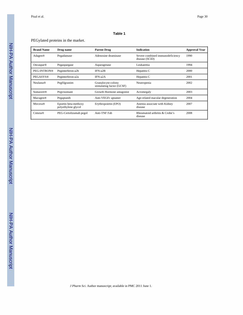
NIH
-PA Author Manuscript
NIH
-PA Author Manuscript
NIH
-PA Author Manuscript
Pisal et al. Page 30
Table 1
PEGylated proteins in the market.
Brand Name Drug name Parent Drug Indication Approval Year
Adagen® Pegadamase Adenosine deaminase Severe combined immunodeficiencydisease (SCID)
1990
Oncaspar® Pegaspargase Asparaginase Leukaemia 1994
PEG-INTRON® Peginterferon-a2b IFN-a2B Hepatitis C 2000
PEGASYS® Peginterferon-a2a IFN-a2A Hepatitis C 2001
Neulasta® Pegfilgrastim Granulocyte-colonystimulating factor (GCSF)
Neutropenia 2002
Somavert® Pegvisomant Growth Hormone antagonist Acromegaly 2003
Macugen® Pegaptanib Anti-VEGFc aptamer Age-related macular degeneration 2004
Mircera® Epoetin beta-methoxypolyethylene glycol
Erythropoietin (EPO) Anemia associate with Kidneydisease
2007
Cimzia® PEG-Certolizumab pegol Anti-TNF Fab Rheumatoid arthritis & Crohn’sdisease
2008
J Pharm Sci. Author manuscript; available in PMC 2011 June 1.

NIH
-PA Author Manuscript
NIH
-PA Author Manuscript
NIH
-PA Author Manuscript
Pisal et al. Page 31
Table 2
Relative changes in preclinical phamacokinetic properties of various macromolecular drugs and theirPEGylated conjugates [adapted from9,10,40]
Therapeutic class/PEGylated DrugPharmacokinetic (PK) effect (circulation half life t1/2) Pharmacodynamic (PD) effect
Parent drug t1/2(h) PEG-drug t1/2(h) Increased Fold DifferenceIn vitro activity (% Activityretained)
Antibodies
Fab’ fragments 0.33 9.05 27 100
Enzymes
PEG-arginine deiminase 2.8 50 18 48
PEG-methioninase 2 38 19 70
PEG-superoxide dismutase 0.01 38 380 51
PEG uricase 3 72 24 100
Cytokines
PEG-Granulocyte ColonyStimulating Factor
1.8 7.0 3.9 41
PEG-IFN-α2a 0.7 51 73 7
PEG-IFN-β1a 0.98 13 13 50
PEG-interleukin-6 0.05 48 960 51
PEG-interleukin-2 0.73 4.26 6 NA
Hormones
PEG-calcitonin 3.31 15.4 4.6 50
PEG-human Growth Hormone 0.34 10 29 24
Nucleic acids
Anti-VEGF RNA aptamer 24 94 3.91 25
J Pharm Sci. Author manuscript; available in PMC 2011 June 1.

NIH
-PA Author Manuscript
NIH
-PA Author Manuscript
NIH
-PA Author Manuscript
Pisal et al. Page 32
Table 3
List of well accepted protein/peptide drug formulations based on biodegradable microparticles in the market.
Brand Name Type of Particulate System Parent Drug Indication Approval Year
Lupron Depot® PLA Microspheres Leuprolide acetate Prostate cancer 1993
Nutropin Depot® PLGA Microspheres Growth hormone (Somatropinrecombinant)
Pediatric growth hormonedeficiency
1999
Sandostatin LAR® Depot PLGA Microspheres Octreotide acetate Acromegaly 1988
Somatuline® LA PLGA microspheres Lanreotide Acromegaly 2007
Trelstar™ Depot PLGA microsphere Triptorelin pamoate Prostate cancer 2008
J Pharm Sci. Author manuscript; available in PMC 2011 June 1.

NIH
-PA Author Manuscript
NIH
-PA Author Manuscript
NIH
-PA Author Manuscript
Pisal et al. Page 33
Table 4
The association efficiency in solid lipid nanoparticles and liposomes. (adapted from122).
Protein/peptide Solid Lipid Nanoparticle Lipososmes
Production procedure Association efficiency (%) Production procedure Association efficiency (%)
Bovine serum albumin Hot HPHa technique 100 Reversal evaporation 25–71
Calcitonin w/o/w double emulsion 75–90 Dry lipid hydration 20
Human recombinant Microemulsion technique -- Freeze- thawing extrusion 20–30
Epidermal GrowthFactor (EGF)
Insulin w/o/w double emulsion 27–68, 35–45 Reverse phase 30–82
J Pharm Sci. Author manuscript; available in PMC 2011 June 1.