yjsbi 5176 no. of pages 13;4c:3; model 5+ article in press
TRANSCRIPT

Journal of Structural Biology xxx (2006) xxx–xxx
www.elsevier.com/locate/yjsbi
YJSBI 5176 No. of Pages 13;4C:3; Model 5+ARTICLE IN PRESS
6 November 2006 Disk Used Aranganathan (CE) / Vijayakumar (TE)
ORRECTED PROOF
Spatial distribution and orientation of dermatan sulfate in human medial collateral ligament
Heath B. Henninger a,b, Steve A. Maas a,b, Clayton J. Underwood a,b, Ross T. Whitaker b, JeVrey A. Weiss a,b,c,¤
a Department of Bioengineering, University of Utah, 50 S Central Campus Drive, Rm. 2480, Salt Lake City, UT 84112, USAb ScientiWc Computing and Imaging Institute, University of Utah, 50 S Central Campus Drive, Rm. 3490 Salt Lake City, UT 84112, USA
c Department of Orthopaedics, University of Utah, 30 North 1900 East, Rm. 3B165, Salt Lake City, UT 84132, USA
Received 26 April 2006; received in revised form 1 September 2006; accepted 5 October 2006
Abstract
The proteoglycan decorin and its associated glycosaminoglycan (GAG), dermatan sulfate (DS), regulate collagen Wbril formation,control Wbril diameter, and have been suggested to contribute to the mechanical stability and material properties of connective tissues.The spatial distribution and orientation of DS within the tissue are relevant to these mechanical roles, but measurements of length andorientation from 2D transmission electron microscopy (TEM) are prone to errors from projection. The objectives of this study were toconstruct a 3D geometric model of DS GAGs and collagen Wbrils, and to use the model to interpret TEM measurements of the spatial ori-entation and length of DS GAGs in the medial collateral ligament of the human knee. DS was distinguished from other sulfated GAGsby treating tissue with chondroitinase B, an enzyme that selectively degrades DS. An image processing pipeline was developed to analyzethe TEM micrographs. The 3D model of collagen and GAGs quantiWed the projection error in the 2D TEM measurements. Model pre-dictions of 3D GAG orientation were highly sensitive to the assumed GAG length distribution, with the baseline input distribution of69§ 23 nm providing the best predictions of the angle measurements from TEM micrographs. The corresponding orientation distributionfor DS GAGs was maximal at orientations orthogonal to the collagen Wbrils, tapering to near zero with axial alignment. Sulfated GAGsthat remained after chondroitinase B treatment were preferentially aligned along the collagen Wbril. DS therefore appears more likely tobridge the interWbrillar gap than non-DS GAGs. In addition to providing quantitative data for DS GAG length and orientation in thehuman MCL, this study demonstrates how a 3D geometric model can be used to provide a priori information for interpretation of geo-metric measurements from 2D micrographs.© 2006 Published by Elsevier Inc.
Keywords: Ligament; Decorin; Glycosaminoglycan; Dermatan sulfate; Geometric model
UNC1. Introduction
Ligaments are collagen-based tissues that resist abnor-mal joint motions by connecting bone to bone. Ligament isapproximately 70% Type I collagen by dry weight (Amielet al., 1990), with the balance of the hydrated tissue consist-ing of “ground substance”, which is a gel-like mixture ofproteins, proteoglycans (PGs), glycosaminoglycans
* Corresponding author. Tel.: +1 801 587 7833; fax: +1 801 585 5361.E-mail address: [email protected] (J.A. Weiss).
1047-8477/$ - see front matter © 2006 Published by Elsevier Inc.doi:10.1016/j.jsb.2006.10.008
Please cite this article in press as: Henninger, H.B. et al., Spatial distlateral ligament, J. Struct. Biol. (2006), doi:10.1016/j.jsb.2006.10.008
(GAGs), and water surrounding the ordered collagenWbrils. GAGs in ligament constitute 0.2–5.0% of the totaldry weight (Amiel et al., 1984; Gillard et al., 1977).Although only a small percentage of tissue, understandingthe distribution and structural organization of GAGs inligament may shed light on the role of these important mol-ecules.
Decorin, a small leucine-rich PG, has been demonstratedto play diverse roles in connective tissues, ranging from reg-ulation of collagen Wbril formation (Vogel et al., 1984) toaVecting the mechanical properties of the tissue (Danielsonet al., 1997; Robinson et al., 2005). Decorin is the most
1
2
34
567
89
10
11121314151617181920212223242526
27
28
293031323334
ribution and orientation of dermatan sulfate in human medial col-
353637383940414243444546

2 H.B. Henninger et al. / Journal of Structural Biology xxx (2006) xxx–xxx
YJSBI 5176 No. of Pages 13;4C:3; Model 5+ARTICLE IN PRESS
6 November 2006 Disk Used Aranganathan (CE) / Vijayakumar (TE)
UNCORREC
prevalent PG species in ligaments in terms of molar quan-tity (Ilic et al., 2005) and it localizes to (or “decorates”) thesurface of collagen Wbrils in a repeating fashion (Pringleand Dodd, 1990). The decorin core protein is thought to beeither a horseshoe- or banana-shaped molecule and tostraddle a single collagen triple helix, binding every 67 nmalong the Wbril surface at the D-period band gap in the tro-pocollagen quarter-stagger pattern (Scott, 1996; Scott andOrford, 1981; Scott et al., 2004). The exact 3D conforma-tion of decorin, including whether it functions as a mono-mer or dimer, remains a subject of debate (Goldoni et al.,2004; Scott, 2003; Scott et al., 2004; Weber et al., 1996).Depending on the tissue, either a single dermatan sulfate(DS) or a single chondroitin sulfate (CS) GAG side chain iscovalently bound near the amino terminus of the decorincore protein (Chopra et al., 1985). This binding site allows asingle DS chain to be exposed to the interWbrillar space.Conformational Xexibility within the iduronate residuesalong the DS backbone may yield the Xexibility to align theGAG in many possible orientations with respect to the col-lagen Wbril (Scott, 1992; Venkataraman et al., 1994). DSchains can self-associate under physiological conditions,containing up to 10 or more individual GAG chains as anaggregate (Ernst et al., 1995; Scott, 1992). It should benoted that the biglycan PG, although signiWcantly less prev-alent than decorin in ligament (Ilic et al., 2005), containstwo GAG side chains, either DS and/or CS (Trowbridgeand Gallo, 2002).
The GAG side chain of decorin has been described asforming interWbrillar proteoglycan bridges by aggregationof GAG chains from adjacent collagen Wbrils (Scott, 2001;Scott and Thomlinson, 1998) and the bridges have beensuggested to play a direct role in the mechanical integrity oftendons (Liu et al., 2005; Redaelli et al., 2003; Vesentiniet al., 2005). The interWbrillar bridges have been proposedto elastically sustain mechanical stresses in Wbrous colla-gen-based tissues through a reversible longitudinal slippagemodel (Scott and Thomlinson, 1998). Within that model,GAG chains may act as a lubricant between adjacent colla-gen Wbrils, cushioning compressive forces (Scott, 2003).Recent research has attempted to quantify mechanicalinteractions in the collagen–decorin–DS bond chain (Liuet al., 2005). Liu quantiWed the GAG–GAG association tobe the weakest link, overcome by strong attractive forcesbetween the decorin core protein and tropocollagen mole-cule, and weaker still than the decorin–DS bond at the pro-tein–GAG interface, supporting the mechanism of thelongitudinal slippage model.
Although DS GAGs have been qualitatively describedas oriented roughly orthogonal to collagen Wbrils in varioustissues (Cribb and Scott, 1995; Kuwaba et al., 2001; Rasp-anti et al., 1997, 2002; Scott, 1988; Scott and Orford, 1981;Scott and Thomlinson, 1998; Van Kuppevelt et al., 1987),quantitative data for their spatial distribution and orienta-tion in any connective tissue are unavailable. Further,although other species of large sulfated GAGs have beenshown interspersed between and along the collagen Wbrils
Please cite this article in press as: Henninger, H.B. et al., Spatial distlateral ligament, J. Struct. Biol. (2006), doi:10.1016/j.jsb.2006.10.008
TED PROOF
(Raspanti et al., 1997, 2002; Van Kuppevelt et al., 1987), therelative proportions and species of sulfated GAGs at eachorientation have yet to be determined. Understanding howGAGs are oriented and distributed with respect to collagenWbrils has implications ranging from tissue modeling togaining a broader understanding of molecular-level mate-rial symmetries and mechanics.
To date, existing models of GAG mechanics withinWbrous connective tissues have assumed perfectly orthogo-nal GAG symmetry with respect to neighboring collagenWbrils (Redaelli et al., 2003). Measurements of three-dimen-sional (3D) GAG orientation are complicated by the factthat the primary method of viewing has been with two-dimensional (2D) TEM micrographs. When GAG structureand orientation are examined in 2D TEM micrographs,information is lost. The viewer is unable to tell if a GAG isleaving the viewing plane or is contained wholly within thepreparation, if a GAG overlaps another in the viewingplane, or the exact three-dimensional orientations withrespect to the neighboring collagen Wbrils.
The objectives of this study were to quantitatively deter-mine the quantity, length, and 3D orientation of DS GAGsin the human medial collateral ligament (MCL). To achievethis goal, we created a 3D geometric model of sulfatedGAGs interspersed within an array of collagen Wbrils, andused this model to interpret measurements of apparentquantity, length, and orientation from 2D TEM images.Digestion with chondroitinase B (ChB, an enzyme that spe-ciWcally degrades DS) was used to distinguish DS GAGsfrom non-DS GAGs. Custom image processing softwarewas written to allow reliable segmentation of stainedGAGs in 2D TEM images.
2. Methods
2.1. Collagen/GAG model inputs
A 3D geometric model of collagen and GAGs in tendonand ligament was created using parameters for collagenWbril diameter, Wbril area (cross-section), GAG length, andGAG distribution. It should be noted that when DS GAGsare described in the models and 2D TEM images, thesestructures are likely aggregates of at least two GAGs. Col-lagen Wbril diameter varies by tissue but not necessarily byspecies (Fung et al., 2003). Measurements of collagen Wbrildiameter from various tendons and ligaments place therange from 50 to 200 nm and measurements of the ratio ofcollagen cross-sectional area to total transverse cross-sec-tional area range from 40 to 70% (Baek et al., 1998; Franket al., 1989; Fung et al., 2003; Hart et al., 1999; Lo et al.,2004; Silver et al., 2003). In the baseline model, collagenWbrils were assigned a diameter range of 90–110 nm andcollagen area as a proportion of total transverse cross-sec-tional area was targeted at 50–65%.
A study of decorin PGs using rotary shadowing electronmicroscopy provides realistic measurements of their 3Dlength. Decorin PGs from bovine tendon were isolated onto a
4748495051525354555657585960616263646566676869707172737475767778798081828384858687888990919293949596979899100101102103
ribution and orientation of dermatan sulfate in human medial col-
104105106107108109110111112113114115116117118119120121122123124125126127128129130131132133134135
136
137
138139140141142143144145146147148149150151152153154155156157

H.B. Henninger et al. / Journal of Structural Biology xxx (2006) xxx–xxx 3
YJSBI 5176 No. of Pages 13;4C:3; Model 5+ARTICLE IN PRESS
6 November 2006 Disk Used Aranganathan (CE) / Vijayakumar (TE)
UNCORREC
Wxed plane before imaging (Morgelin et al., 1989). Small singleGAG chain PGs, likely decorin, assumed a near-Gaussianlength distribution with a mean of 69§23nm. The core pro-tein appeared to be only a small fraction of the overall length.This proWle of GAG lengths was used in the baseline geomet-ric model to represent the DS population in ligament.
The spatial distribution of GAGs in the model wasquasi-random with respect to the collagen Wbrils. GAGangle was not constrained, rather GAGs were allowed toproject from collagen Wbrils at any angle so long as they didnot occupy the space taken up by another GAG or a colla-gen Wbril.
2.2. Collagen/GAG model generation
Given the parameters stated previously, a highly control-lable and repeatable 3D computer model of a 3D collagen/GAG matrix was programmed using C++. Collagen wasgenerated in a quasi-hexagonal packing scheme, using a“jitter” parameter to introduce deviations from perfectcrystalline organization. A seed (random number) created aunique collagen pattern and therefore a unique GAG dis-tribution. Any given model could be repeated by using thesame seed. An executable version of the software, whichruns under the Microsoft WindowsXP operating system, isavailable for download in the Supplementary Material sec-tion on the Journal web page.
GAG length in the 3D geometric model was varied asdeWned by an input distribution (mean§standard deviation).D-period bands were labeled every 67nm along the length ofthe collagen Wbrils and their location was not dependent onbands of neighboring Wbrils. GAGs were generated by Wrstchoosing a collagen Wbril and D-period band, then projectinga GAG from the band at an angle such that the GAG linkedto another D-period band on a neighboring Wbril. This con-struction is consistent with the often hypothesized aggregationof GAG chains from adjacent Wbrils to create interWbrillarcross-links. GAGs were not allowed to pass through the spaceoccupied by other GAGs or collagen Wbrils (Fig. 1A).
2.3. Creation of 2D synthetic TEM micrographs from the 3D geometric model
A sectioning volume was created in the software to simu-late the thickness of our TEM preparations (70 nm). The
Fig. 1. Three-dimensional geometric model of a collagen and GAG matrix.(A) Example of a collagen matrix volume generated with GAGs dispersedthroughout. The range of collagen Wbril diameters and spacing are exag-gerated to assist visualization. (B) A representative sectioning plane fromthe matrix in (A). GAGs within section plane are still three dimensional,but collagen and GAGs outside the section have been removed for clarity.(C) Two-dimensional projection of the three-dimensional sectioning plane.Note that discrete GAGs in three dimensions sometimes appear to overlapone another and appear shorter than their three-dimensional length.Model and sectioning plane are 1300 £ 1300 nm, simulating overall dimen-sions of a TEM image. Arrow denotes viewing plane from which measure-ments were taken. Scale bars D 200 nm.
Please cite this article in press as: Henninger, H.B. et al., Spatial distlateral ligament, J. Struct. Biol. (2006), doi:10.1016/j.jsb.2006.10.008
TED PROOF
sectioning plane could be centered at any point into thedepth of Weld of the model. A clipping mode allowed theuser to turn oV display of the collagen Wbrils and GAGsoutside the sectioning plane to visualize the GAG geome-tries within the plane (Fig. 1B). A projection feature wasused to create synthetic two-dimensional TEM micro-
A
B
C
ribution and orientation of dermatan sulfate in human medial col-
158159160161162163164165166167168169
170
171172173174175176177178179180181182183184185186187188189190191192193194
195196
197198
199200201202203204
4 H.B. Henninger et al. / Journal of Structural Biology xxx (2006) xxx–xxx
YJSBI 5176 No. of Pages 13;4C:3; Model 5+ARTICLE IN PRESS
6 November 2006 Disk Used Aranganathan (CE) / Vijayakumar (TE)
UNCORREC
graphs by projecting the three-dimensional GAGs withinthe sectioning plane to the viewing plane (Fig. 1C).
Outputs from the software included GAG angle withrespect to collagen Wbrils and GAG length. These data wereavailable for the entire 3D volume, within a 3D sectioningvolume, and for the 2D projection planes. The synthetic two-dimensional TEM micrographs were saved for later process-ing. For the baseline model, 30 section planes were analyzed.
2.4. Model sensitivity studies
Sensitivity studies were conducted to test the relativeimpact of the input parameters to the 3D geometric modelon the resultant GAG geometries. Seed number (nine diVer-ent values), range of collagen diameter (70–90, 110–130,and 70–130 nm), GAG length (means of 50, 100, and250 nm), and GAG concentration (three diVerent values)were varied from the values used in the baseline model.Thus, a total of 19 diVerent models were analyzed. Thirtysectioning volumes were captured from each of the 19 mod-els and their GAG angle/length data and an image of eachtwo-dimensional projection were saved to Wles. It is impor-tant to note that GAG concentration merely scales themodel outputs. As such, all data were normalized for com-parative purposes.
2.5. Tissue sample selection
Five human knees were obtained from Wve separatedonors (age 47–64, median 53). Specimens remained frozenuntil the time of dissection. Knees were allowed to thawand were dissected free of fat and extraneous soft tissue.Knees with surgical scars, ligament injury or cartilagedegeneration characteristic of osteoarthritis were elimi-nated. The medial collateral ligament (MCL) was removedat both the femoral and tibial insertion sites and Wne dis-sected to remove any overlying fascia. The ligaments werekept hydrated with normal saline throughout the tissue iso-lation. Bulk tissue was trimmed to remove four samplesfrom the mid-substance of each MCL.
2.6. Glycosaminoglycan digestion
To isolate DS proteoglycans from other sulfated proteo-glycans, enzymatic treatment with chondroitinase B (ChB)was performed on two of the four samples (randomly cho-sen) from each MCL, while the other two samples wereused as controls. ChB speciWcally degrades DS (Ernst et al.,1995). All samples were equilibrated for 1 h in 15 ml ofbuVer (20 mM Tris, pH 7.5, 150 mM NaCl, and 5 mMCaCl2) and with one tablet of Mini-Complete proteaseinhibitor (Roche) per 10 ml of buVer. Following the 1-hequilibration, the control samples were soaked in 15 ml ofthe same buVer, without protease inhibitors, for 6 h at RT.The enzymatic treatment group was soaked in 15 ml of thesame buVer with 1.0 U/ml of ChB, and then soaked in astop buVer to inhibit further ChB activity (20 mM, Tris pH
Please cite this article in press as: Henninger, H.B. et al., Spatial distlateral ligament, J. Struct. Biol. (2006), doi:10.1016/j.jsb.2006.10.008
TED PROOF
7.5, 150 mM NaCl, and 10 mM EDTA). EDTA sequestersresidual calcium and inhibits ChB activity, as it is a cal-cium-dependent enzyme (Michel et al., 2004). All buVertreatments were performed with gentle agitation using anorbital shaker. To obtain suYcient enzyme for use in thisstudy, ChB was cloned in Flavobacterium heparinium aspreviously described (Pojasek et al., 2001).
2.7. SpeciWcity of chondroitinase B
The speciWcity of ChB for DS was veriWed by incubatingstock solutions of sulfated GAGs found in ligament withChB and then quantifying the concentration of the GAGsusing an improved 1,9-dimethylmethylene blue (DMB)assay (Farndale et al., 1986). Individual reactions (30 �l,nD6 for each condition) were set up containing 1.0 U/mlChB and 500 �g/ml of puriWed GAG (DS, chondroitin sul-fate A and C, heparin sulfate, or keratin sulfate). Controlreactions were set up containing GAGs and buVer only(20 mM Tris–HCl (pH 7.5), 150 mM NaCl, and 5 mMCaCl2). Reactions were allowed to proceed for 6 h at RT.GAG concentrations were then quantiWed using the DMBassay. Five microliters of each reaction (diluted 2-fold) wastransferred to a 96-well plate in duplicate, along with GAGstandards. Two hundred microliters of dimethylmethyleneblue reagent was added to each well, and the absorbancewas immediately read in a plate reader (Synergy HT, Bio-tek) at 530 and 590 nm. GAG concentrations wereexpressed as a percentage of control reactions.
2.8. Chondroitinase B activity and removal of dermatan sulfate
One control and one ChB-treated sample from eachknee were used to verify the removal of DS using the DMBassay. Samples were lyophilized overnight and dry weightswere obtained. The samples were then incubated with 20volumes (based on dry weight) of papain buVer for 4 h at60 °C until the tissue was completely digested. Papain buVerconsisted of 50 �g/ml papain, 50 mM sodium acetate, pH5.5, 2 mM dithiothreitol, and 2 mM EDTA. Each extractwas divided into a control and a ChB group. Twenty-Wvemicroliters of extract was mixed with 25 �l of 2£ ChBbuVer consisting of 0.1 U/ml ChB, 30 mM Tris, pH 8.1,10 mM NaCl, 25 mM acetate buVer, and 5 mM CaCl2. Con-trol reactions contained the same buVer but lacked ChB.DS digestion was allowed to proceed for 2 h at RT. TotalGAG content was quantiWed using the DMB assay in 96-well plates as described previously. The DS content of thepapain extracts was calculated by subtracting the amountof GAG in the extract treated with additional ChB fromthat of the extract treated with additional buVer only.
2.9. Transmission electron microscopy
The remaining control and ChB-treated specimens fromeach knee were used for transmission electron microscopy
205206207208209210211212
213
214215216217218219220221222223224225226227
228
229230231232233234235236237238239240
241
242243244245246247248249250251252253254255
ribution and orientation of dermatan sulfate in human medial col-
256257258259260261262
263
264265266267268269270271272273274275276277278279280281282
283284
285286287288289290291292293294295296297298299300301302303
304
305306
H.B. Henninger et al. / Journal of Structural Biology xxx (2006) xxx–xxx 5
YJSBI 5176 No. of Pages 13;4C:3; Model 5+ARTICLE IN PRESS
6 November 2006 Disk Used Aranganathan (CE) / Vijayakumar (TE)
UNCORREC
(TEM). TEM allows the visualization of sulfated GAGs byselectively targeting them with an electron-dense stain(Cribb and Scott, 1995; Haigh and Scott, 1986). Tissue wasWxed in 4% paraformaldehyde with 2% glutaraldehydeovernight at 4 °C with agitation. Twenty micron sectionsalong the sagittal plane of the ligament were obtained on acryostat (Leica CM3050S, Exton PA) at ¡25 °C. Sectionswere mounted on slides, Wxed for 30 min (2.5% glutaralde-hyde, 25 mM sodium acetate, pH 5.8) and stained at criticalelectrolyte concentration (CEC) with 0.05% CupromeronicBlue (CB) (2.5% glutaraldehyde, 25 mM sodium acetate,0.1 M MgCl2, and 0.05% CB, pH 5.8) for 3 h at 37 °C. AtCEC, the CB selectively binds to sulfated GAGs, acting as ascaVold to support the native conformation of the moleculewhile reducing translocation of the molecules during fur-ther processing (Scott, 1985). Slides were contrasted for30 min in 0.5% aqueous sodium tungstate to amplify theelectron density of the CB scaVold. Specimens were thendehydrated through graded ethanol and encapsulated usingSpurr’s resin. Ultrathin sections (»70 nm) were obtainedvia ultramicrotome (Leica Ultracut UCT, Exton PA) with adiamond knife (Diatome 45°, HatWeld PA) and ribbonswere Xoated onto 135 Hex copper grids. Grids were con-trasted in aqueous uranyl acetate to visually enhance thecollagen Wbrils.
Digital images were collected on a Hitachi H7100 TEMwith a LAB6 Wlament. Fields of view were selected at lowmagniWcation (1000–5000£) where intact areas of ligamentwere visible in the microscopic Weld. Edges of tissue andareas with Wne sectioning artifacts were avoided. MagniW-cation was ampliWed to 50 000£, the microscope wasfocused, and an image was collected with the integratedCCD camera. A minimum of Wve digital images were cap-tured from diVerent areas of tissue on a single grid so thatan average GAG distribution could be obtained for eachsample.
2.10. Image processing
To locate GAGs in the 2D TEM micrographs and todetermine their apparent length and apparent orientationwith respect to the local collagen Wbrils, an image process-ing software pipeline was developed in Matlab using func-tions in the Image Processing Toolbox (Mathworks,Natick, MA). The Matlab input Wle is available for down-load as Supplementary Material on the Journal web page.The collagen Wbril axis was determined by interactively dig-itizing four vectors along the predominant Wbril axis usingImageJ (National Institutes of Health). The average angleof the collagen Wbrils with respect to the horizontal wassaved to a data Wle.
A series of morphological operations were used to elimi-nate background noise and reduce each detected sulfatedGAG to a branched binary wireframe element, one pixelwide (Fig. 2). It should be noted that sulfated GAGs arelikely aggregates of smaller GAG chains (Ernst et al., 1995;Scott, 1992). Branched GAGs are therefore either GAGs
Please cite this article in press as: Henninger, H.B. et al., Spatial distlateral ligament, J. Struct. Biol. (2006), doi:10.1016/j.jsb.2006.10.008
TED PROOF
projecting oV the predominant chain of the main GAGaggregate or two or more GAGs overlapping through thethickness of the specimen. As these scenarios could not bediscerned using two-dimensional data, branches were bro-ken from the main chain keeping only the longest continu-ous chain of pixels to represent the GAG. A size Wlter wasapplied to remove extremely small objects (610 nm). Thesesmall objects were typically residual noise artifacts orGAGs exiting the viewing plane directly. In either case,such small units are subject to error in orientation process-ing and thus eliminated from all images.
Once wireframe elements were isolated, a principal com-ponent analysis was performed on the coordinates of thepixels of each object. This yielded a covariance matrix thatwas then transformed into the eigenvectors of the GAG.Taking the maximum eigenvector of each object as the deW-nition of its orientation, the vectors associated with theGAGs were compared to the collagen Wbril direction and aunique angle was quantiWed for each GAG with respect tothe collagen Wbril axis.
The synthetic 2D TEM images from the 3D geometricmodel were analyzed with the same image processingpipeline. Therefore, analysis of the simulation data wassubject to the same overlap, branching, and length errorsfound in TEM images. Output data from simulationsections were the same as TEM data, including GAGlengths and orientations with respect to local collagendirection.
2.11. Statistical analysis
Data from the geometric models were analyzed usingANOVA with Tukey post hoc tests. Data for GAG orienta-tion with respect to collagen Wbrils and GAG length datawere collapsed into discrete bins (0–10°, 11–20°, etc.) andhistograms were generated. The overall number of GAGsin a simulation section and the number of GAGs at eachangle and length bin were compared between and withinmodels. SigniWcance was set at �D0.05.
Results from TEM image measurements were ana-lyzed with Kolmogorov–Smirnov (KS) tests to assessnormality of data distribution. Parametric statisticalanalyses were preferred, but in cases with small samplesizes and/or non-normal distribution per the KS, non-parametric alternatives to the parametric tests werechosen. SigniWcance for all tests was set at �D 0.05.Independent t-tests were used to detect signiWcantchanges in the overall number of GAGs for the DMBassays as well as the image processing runs. Data forGAG orientation with respect to collagen Wbrils were col-lapsed into discrete angle bins (0–10°, 11–20°, etc.) andhistograms were generated. Independent t-tests were usedto detect signiWcant changes in the total number of GAGsat each individual angle bin between control and ChB-treated groups. Control and treated images for individualknees were compared using the Mann–Whitney U due tonon-normal data distributions.
307308309310311312313314315316317318319320321322323324325326327328329330331332333334335336337338339340341342
343
344345346347348349350351352353354355356357358359360361
ribution and orientation of dermatan sulfate in human medial col-
362363364365366367368369370371372373374375376377378379380381382383384385386387388389
390
391392393394395396397398399400401402403404405406407408409410411412413414415416

6 H.B. Henninger et al. / Journal of Structural Biology xxx (2006) xxx–xxx
YJSBI 5176 No. of Pages 13;4C:3; Model 5+ARTICLE IN PRESS
6 November 2006 Disk Used Aranganathan (CE) / Vijayakumar (TE)
UNCORREC3. Results
3.1. 3D geometric model and sensitivity studies
The average collagen radius for the baseline model was92.5§ 0.89 nm, with a collagen cross-sectional area ratio of63.3§ 1.6%. The seed number had no signiWcant eVect onGAG quantity, length, or orientation between or withinmodels (all p-values 7 0.125). Based on this Wnding, theresults of the 10 models examining seed number were aver-aged to create a master baseline model.
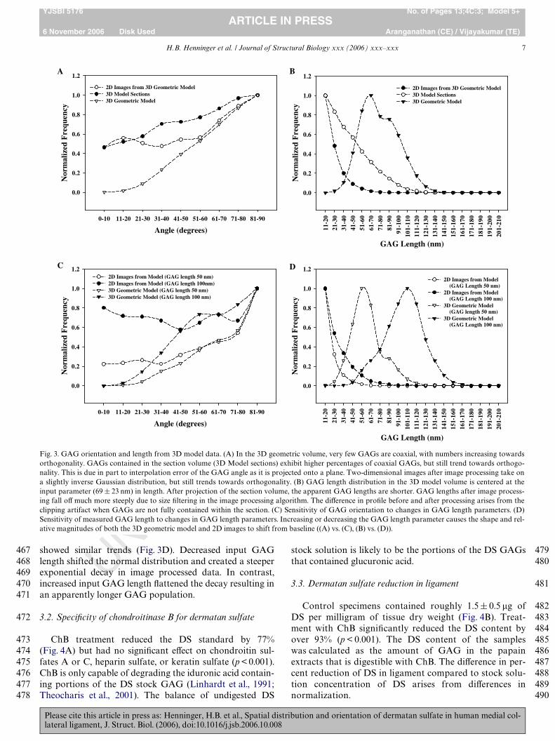
Results for GAG angle with respect to the collagen Wbrilfrom the geometric models were plotted as a histogram(Fig. 3A). The true 3D orientation of the GAGs had veryfew coaxially aligned GAGs (0–10°) but increased linearlytowards orthogonal orientations (81–90°). The alignmentof GAGs occupying the sectioning volumes, measured withrespect to the viewing plane, showed a higher percentage atcoaxial orientations, but a nearly linear increasing trendtowards orthogonal orientations. The orientations in 2D ofGAGs projected onto the viewing plane were determinedusing the TEM image analysis algorithm. These GAGswere also measured with respect to the viewing plane andshowed higher concentrations at coaxial orientations, butdid not begin to increase in number until near orthogonalorientations (51–60°), creating a bowl-shaped distribution.
Please cite this article in press as: Henninger, H.B. et al., Spatial distlateral ligament, J. Struct. Biol. (2006), doi:10.1016/j.jsb.2006.10.008
GAG length was plotted as a histogram (Fig. 3B). Thetrue 3D orientations of the GAGs in the baseline geometricmodel exhibited a nearly Gaussian distribution, centered at69 nm. This result is consistent with the input parameter tothe baseline model. The apparent lengths of GAGs occupy-ing the sectioning volumes, measured with respect to theviewing plane, were shorter due to projection. The lengthsdecayed steadily from a maxima at the shortest measure-ment of 11 nm. The lengths of GAGs projected onto theviewing plane determined using the TEM image analysisalgorithm also exhibited a minima at the shortest length.The proWle decayed exponentially with increasing GAGlength.
Changes to collagen diameter and GAG concentrationhad no signiWcant eVect on the GAG length or orientationdistributions (all p-values 70.125). In contrast, changingthe GAG length distribution had a dramatic eVect on theGAG orientation and length proWles and magnitudes (all p-values 60.05). Fig. 3C and D shows the angle and lengthpredictions for two diVerent GAG length input distribu-tions (50, 100 nm). With respect to angle (Fig. 3C), when theprescribed GAG length input distribution was shortened,larger orthogonal populations and smaller coaxial popula-tions of GAGs were created. Increased input GAG lengthhad the opposite eVect, increasing the entire angle distribu-tion, especially coaxial orientations. Apparent GAG length
TED PROOF
Fig. 2. Demonstration of the image processing procedure used to isolate sulfated GAGs in the TEM images and to determine their orientation angles. (A)Portion of sample image (three representative stained GAGs circled). (B) Sample image after high pass and background intensity Wltering. (C) Convolvedbinary image improves GAG internal connectivity. (D) Wire frame representation used to calculate orientation unit vectors (arrows) of stained GAGs viaprinciple component analysis with respect to collagen Wbril axis (large arrow, (A)). As dermatan sulfate is a single polysaccharide chain, branched wireframe objects were broken and only the longest continuous chain was retained. Bar D 50 nm.
417
418
419420421422423424425426427428429430431432433434435436437438439440
ribution and orientation of dermatan sulfate in human medial col-
441442443444445446447448449450451452453454455456457458459460461462463464465466
H.B. Henninger et al. / Journal of Structural Biology xxx (2006) xxx–xxx 7
YJSBI 5176 No. of Pages 13;4C:3; Model 5+ARTICLE IN PRESS
6 November 2006 Disk Used Aranganathan (CE) / Vijayakumar (TE)
UNshowed similar trends (Fig. 3D). Decreased input GAGlength shifted the normal distribution and created a steeperexponential decay in image processed data. In contrast,increased input GAG length Xattened the decay resulting inan apparently longer GAG population.
3.2. SpeciWcity of chondroitinase B for dermatan sulfate
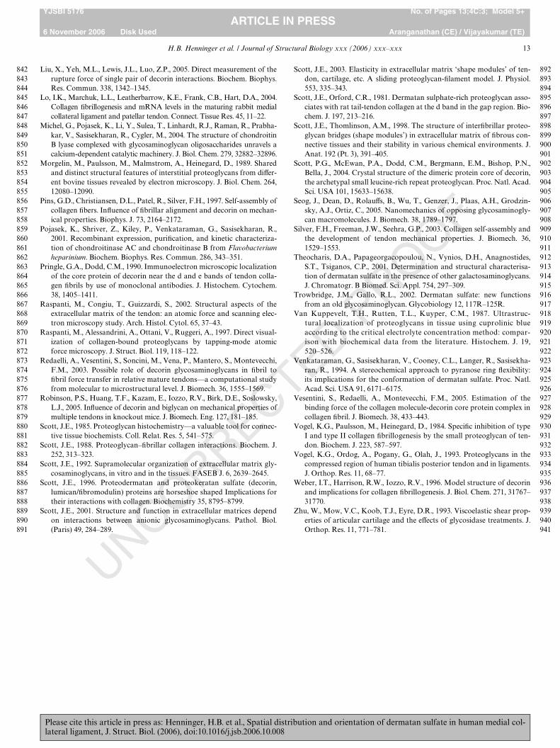
ChB treatment reduced the DS standard by 77%(Fig. 4A) but had no signiWcant eVect on chondroitin sul-fates A or C, heparin sulfate, or keratin sulfate (p < 0.001).ChB is only capable of degrading the iduronic acid contain-ing portions of the DS stock GAG (Linhardt et al., 1991;Theocharis et al., 2001). The balance of undigested DS
Please cite this article in press as: Henninger, H.B. et al., Spatial distlateral ligament, J. Struct. Biol. (2006), doi:10.1016/j.jsb.2006.10.008
stock solution is likely to be the portions of the DS GAGsthat contained glucuronic acid.
3.3. Dermatan sulfate reduction in ligament
Control specimens contained roughly 1.5§ 0.5 �g ofDS per milligram of tissue dry weight (Fig. 4B). Treat-ment with ChB signiWcantly reduced the DS content byover 93% (p < 0.001). The DS content of the sampleswas calculated as the amount of GAG in the papainextracts that is digestible with ChB. The diVerence in per-cent reduction of DS in ligament compared to stock solu-tion concentration of DS arises from diVerences innormalization.
CORRECTED PROOF
Fig. 3. GAG orientation and length from 3D model data. (A) In the 3D geometric volume, very few GAGs are coaxial, with numbers increasing towardsorthogonality. GAGs contained in the section volume (3D Model sections) exhibit higher percentages of coaxial GAGs, but still trend towards orthogo-nality. This is due in part to interpolation error of the GAG angle as it is projected onto a plane. Two-dimensional images after image processing take ona slightly inverse Gaussian distribution, but still trends towards orthogonality. (B) GAG length distribution in the 3D model volume is centered at theinput parameter (69 § 23 nm) in length. After projection of the section volume, the apparent GAG lengths are shorter. GAG lengths after image process-ing fall oV much more steeply due to size Wltering in the image processing algorithm. The diVerence in proWle before and after processing arises from theclipping artifact when GAGs are not fully contained within the section. (C) Sensitivity of GAG orientation to changes in GAG length parameters. (D)Sensitivity of measured GAG length to changes in GAG length parameters. Increasing or decreasing the GAG length parameter causes the shape and rel-ative magnitudes of both the 3D geometric model and 2D images to shift from baseline ((A) vs. (C), (B) vs. (D)).
Angle (degrees)
0-10 11-20 21-30 31-40 41-50 51-60 61-70 71-80 81-90
Nor
mal
ized
Fre
quen
cy
0.0
0.2
0.4
0.6
0.8
1.0
1.2
2D Images from 3D Geometric Model 3D Model Sections 3D Geometric Model
GAG Length (nm)
11-2
021
-30
31-4
041
-50
51-6
061
-70
71-8
081
-90
91-1
0010
1-11
011
1-12
012
1-13
013
1-14
014
1-15
015
1-16
016
1-17
017
1-18
018
1-19
019
1-20
020
1-21
0
0.0
0.2
0.4
0.6
0.8
1.0
1.2
2D Images from 3D Geometric Model3D Model Sections 3D Geometric Model
Angle (degrees)
0-10 11-20 21-30 31-40 41-50 51-60 61-70 71-80 81-90
0.0
0.2
0.4
0.6
0.8
1.0
1.22D Images from Model (GAG length 50 nm) 2D Images from Model (GAG length 100nm) 3D Geometric Model (GAG length 50 nm) 3D Geometric Model (GAG length 100 nm)
GAG Length (nm)
11-2
0
21-3
031
-40
41-5
0
51-6
061
-70
71-8
081
-90
91-1
0010
1-11
011
1-12
012
1-13
013
1-14
014
1-15
015
1-16
016
1-17
0
171-
180
181-
190
191-
200
201-
210
0.0
0.2
0.4
0.6
0.8
1.0
1.2
2D Images from Model (GAG Length 50 nm) 2D Images from Model (GAG Length 100 nm) 3D Geometric Model (GAG length 50 nm) 3D Geometric Model (GAG Length 100 nm)
Nor
mal
ized
Fre
quen
cy
Nor
mal
ized
Fre
quen
cy
Nor
mal
ized
Fre
quen
cy
A
C
B
D
467468469470471
472
473474475476477478
ribution and orientation of dermatan sulfate in human medial col-
479480
481
482483484485486487488489490

8 H.B. Henninger et al. / Journal of Structural Biology xxx (2006) xxx–xxx
YJSBI 5176 No. of Pages 13;4C:3; Model 5+ARTICLE IN PRESS
6 November 2006 Disk Used Aranganathan (CE) / Vijayakumar (TE)
UNCOR3.4. Transmission electron microscopy
Images of control tissue collected via TEM exhibitedtypical Cupromeronic Blue staining of sulfated GAGs(Cribb and Scott, 1995; Scott, 1988; Scott and Thomlinson,1998) (Fig. 5A). The apparent orientation of the stainedGAGs was predominantly orthogonal and coaxial to theWbrils, although orientations were distributed in betweenthese extremes as well. The apparent GAG length in controlimages ranged from approximately 10 nm up to 400 nm inthe most extreme cases (mean 31§22 nm). In general, theextremely long GAGs were found along collagen Wbrils inthe interWbrillar space between adjacent Wbrils. Spacingbetween neighboring sulfated GAGs was observed in theexpected range of the D-period band gap, roughly 60–70 nm (Pringle and Dodd, 1990; Scott, 1988; Scott andOrford, 1981). Treatment with ChB reduced the overallnumber of stained sulfated GAGs by 86% (p < 0.001)(Fig. 5B). GAGs visible after ChB digestion were generallylonger than those in control images and preferentially
Please cite this article in press as: Henninger, H.B. et al., Spatial distlateral ligament, J. Struct. Biol. (2006), doi:10.1016/j.jsb.2006.10.008
aligned coaxial to the collagen Wbrils (mean 45§13 nm,range 10–350 nm).
3.5. Orientation of sulfated GAGs via image processing
The apparent angular orientation of sulfated GAGswith respect to collagen Wbrils was signiWcantly altered byChB treatment (Fig. 6). The control tissues exhibited aninverse Gaussian distribution with relative peaks at coaxial(0–10°) and orthogonal (80–90°) orientations (medianangleD47.0°) (Fig. 6A). After ChB treatment, the apparentorientation of the remaining (non-DS) stained GAGsshifted to a positively skewed distribution (medianangleD19.7°) and showed a signiWcant decrease in the totalnumber of stained GAGs (p < 0.001).
Control and treated GAG orientation proWles were com-pared for each discrete angle bin. Across all knees, coaxialorientations showed a signiWcant 69% decrease in the num-ber of GAGs after treatment with ChB (p < 0.001). The per-centage increased linearly with angle towards a 96%
OFFig. 4. EVect of chondroitinase B treatment on stock GAG solutions and treated ligament specimens. (A) Chondroitinase B (1.0 U/ml) was incubated withglycosaminoglycans (500 �g/ml) for 6 h. GAG concentration was determined using the DMB assay. Concentrations were normalized to control reactions,which did not contain ChB. DS, dermatan sulfate; CsA,C, equal mixture of chondroitin sulfates A and C; HS, heparin sulfate; KS, keratin sulfate. N D 6,error bars D standard deviation. (B) Reduction in dermatan sulfate concentration from control to treated ligament specimens was 93.3% (p < 0.001). DScontent was normalized to dry weight of the specimen. N D 5, error bars D standard deviation.
DS CsA,C HS KS
Per
cent
of
Con
trol
0
20
40
60
80
100
120
Control ChB Treated
μg D
S / m
g of
dry
tis
sue
0.0
0.5
1.0
1.5
2.0
2.5A B
RECTED PRO
Fig. 5. Representative TEM images of medial collateral ligament stained with Cupromeronic Blue (large arrow denotes collagen Wbril direction). (A) Con-trol tissue with darkly stained sulfated GAGs. (B) Tissue treated with Chondroitinase B. Note the decrease in the number of Wbril spanning sulfated GAGsand the preferred orientation of remaining GAGs along the collagen Wbril direction. Bar D 200 nm.
491
492493494495496497498499500501502503504505506507508509
ribution and orientation of dermatan sulfate in human medial col-
510511
512
513514515516517518519520521522523524525526527

H.B. Henninger et al. / Journal of Structural Biology xxx (2006) xxx–xxx 9
YJSBI 5176 No. of Pages 13;4C:3; Model 5+ARTICLE IN PRESS
6 November 2006 Disk Used Aranganathan (CE) / Vijayakumar (TE)
UNCOreduction at orthogonal orientations. Analysis of the GAGreduction by angle bin in individual knees showed a similartrend. Of the Wve knees examined, four knees exhibited asigniWcant decrease in all angle bins (p < 0.001). The Wnalknee showed signiWcant decreases in the number of GAGsin all angle bins except one, the coaxial orientation(p < 0.001 and pD 0.81, respectively).
The apparent orientation of DS with respect to collagenWbrils was derived by subtracting the treated distributionproWle from the control distribution proWle (Fig. 6B). DSalone exhibited a less dramatic inverse Gaussian distribu-tion than control samples. DS distribution was negativelyskewed (median angleD55.0°) with a minor peak at coaxialalignment and a predominant peak at orthogonal orienta-tions. From this proWle it was estimated that nearly 60% ofDS GAGs are oriented at angles greater than 45° withrespect to collagen Wbrils.
Please cite this article in press as: Henninger, H.B. et al., Spatial distlateral ligament, J. Struct. Biol. (2006), doi:10.1016/j.jsb.2006.10.008
4. Discussion
Interpretation of the results from the analysis of the 3Dbaseline geometric model provides an understanding of theerrors in measurement of apparent length and orientationfrom 2D TEM micrographs due to projection. The stainedGAGs in 2D TEM micrographs may be oriented so thatthey leave the plane of section. Measurement of the 2D in-plane GAG length underestimates the true length by somefactor of the cosine of the through-thickness angle withrespect to the plane. Also, TEM sections are simply slicesthrough native tissue without regard to the position of thestructure in the depth of Weld. Without a priori knowledge ofthe underlying geometric structure, one cannot presume toknow which stained GAGs were cleaved in the sectioningprocess and which were contained wholly within the prepa-ration. Modeling the collagen/GAG geometry in 3D pro-
RRECTED PROOF
Fig. 6. Distribution of sulfated GAG angles with respect to collagen Wbril orientation. (A) Sulfated GAGs in control images exhibit an inverse Gaussiandistribution (median 47.0°) with relative peaks at coaxial (0–10°) and orthogonal orientations (81–90°). Non-dermatan sulfate GAGs shift to a positivelyskewed coaxial alignment (median 19.7°). Treatment with ChB resulted in an 86% reduction in the number of sulfated GAGs (p < 0.001). GAGs were sig-niWcantly reduced by a minimum of 69% (coaxial) up to roughly 95% at orthogonal orientations (p < 0.001). (B) Distribution of dermatan sulfate anglewith respect to collagen Wbril orientation. Dermatan sulfate exhibited an inverse Gaussian distribution (median 55°) trending from a minor peak at coaxialorientation to a predominant peak at orthogonal orientation. N D 5, error bars D standard deviation.
Angle (degrees)
0-10 11-20 21-30 31-40 41-50 51-60 61-70 71-80 81-90
TO
TA
L G
AG
(#
in f
ield
of
view
)
0
20
40
60Control (Total Sulfated GAG)Treated (Non-DS GAG)
Angle (degrees)
0-10 11-20 21-30 31-40 41-50 51-60 61-70 71-80 81-90
DS
GA
G (
# in
fie
ld o
f vi
ew)
0
20
40
60 Dermatan Sulfate (Control - Treated)
A
B
528529530531532533534535536537538539540541542543544
ribution and orientation of dermatan sulfate in human medial col-
545
546547548549550551552553554555556557558559560

10 H.B. Henninger et al. / Journal of Structural Biology xxx (2006) xxx–xxx
YJSBI 5176 No. of Pages 13;4C:3; Model 5+ARTICLE IN PRESS
6 November 2006 Disk Used Aranganathan (CE) / Vijayakumar (TE)
UNCORREC
vides the necessary a priori information to interpret the 2DTEM image data. As shown in Fig. 3, the distributions ofapparent GAG angle and length change when the imagedata are projected to two dimensions. The apparent numberof GAGs aligned at coaxial orientations increases dramati-cally after 2D projection, both in simple projection and afterimage processing of the planar projection (Fig. 3A). Sincethe true orientation of the GAG population was much moreorthogonal than coaxial as shown by the volume data(Fig. 3A, Wlled triangles), this is an artifact. Within the entirevolume, GAG angle was measured with respect to the colla-gen Wbril from which it originated. A GAG extending froma Wbril parallel to the viewing plane would retain the trueangle since projection simply maps it onto the viewing plane(Fig. 7A). In contrast, a GAG extending from a Wbril towardthe viewing plane would appear more vertical with therespect to the viewing plane (Fig. 7B). Therefore, the projec-tion error is primarily due to the orientation of GAGs rela-tive to the viewing plane rather than their orientation withrespect to the collagen Wbril.
When the lengths of GAGs in the 3D geometric modelwere analyzed, the GAG length distribution was asdescribed by the input parameter in a nearly Gaussian pro-Wle (Fig. 3B, Wlled triangles). Projection onto a 2D planesigniWcantly shortened the apparent GAG lengths untiltheir maxima were at the shortest measurable length(Fig. 3B, open circles). Further image processing of the pro-jected planar image intensiWed this eVect, creating a max-ima at the shortest length and an exponential decay infrequency with increasing length (Fig. 3B, Wlled circles). Inorder to reduce noise artifacts in measures of angle andGAG length in TEM images, a size Wlter is applied toremove any GAGs below 10 nm since they are more proneto misinterpretation against background noise.
Fig. 7. Schematic of errors in measurement of apparent orientation andlength due to projection onto a viewing plane. (A) Collagen Wbril with aGAG parallel to the viewing plane. When projected, the GAG length andorientation in the 2D plane will reXect the true length (Y) and true angle(�) with respect to the Wbril. (B) Rotate the same collagen Wbril and GAG90° so the GAG is extended into the viewing plane. The GAG has thesame length and angle in three dimensions, but the projected length andorientation are shorter (y) and more coaxial (�) with respect to the Wbril.
Please cite this article in press as: Henninger, H.B. et al., Spatial distlateral ligament, J. Struct. Biol. (2006), doi:10.1016/j.jsb.2006.10.008
TED PROOF
Pertaining to MCL specimens, the biochemical analysesdemonstrated that ChB treatment was eVective in eliminat-ing DS GAGs, allowing us to examine their orientation as asubpopulation when compared to all sulfated GAGs. Up to96% of DS in human MCL specimens was removed byenzymatic digestion, while TEM image processing resultedin a total sulfated GAG reduction on the order of 86% (interms of number, not weight). This demonstrates that DS isthe predominant sulfated GAG in the human MCL. Previ-ous studies have reported that, purely by number, themajority of PGs in connective tissue are decorin with itsassociated DS side chain (Amiel et al., 1990; Raspanti et al.,1997; Scott, 1988; Vogel et al., 1993). Studies in bovine liga-ment and human tendon have shown that while decorin/DScomprise up to 90% of the PGs in ligament by number,smaller concentrations of biglycan and versican are present(Campbell et al., 1996; Ilic et al., 2005; Vogel et al., 1993).These proteoglycans may both contain GAG chains ofchondroitin sulfates A and C, the likely sulfated GAGspresent in ligament after ChB treatment. Traces of aggre-can, a PG that contains numerous keratin sulfate and chon-droitin sulfate side chains, may also be found in extremelysmall concentrations but are much more prevalent in othermusculoskeletal soft tissues such as articular cartilage.
The apparent orientation and length data obtained fromanalysis of the 2D TEM sections can be interpreted in thecontext of the 3D geometric model to determine the appro-priateness of the assumptions of the 3D geometric modeland to interpret the results of the 2D TEM measurements.Analysis of the 2D TEM images showed that DS GAGswere apparently oriented at all angles with respect to thecollagen Wbrils (Fig. 6A and B). However, these data aresubject to the projection errors discussed previously. Fig. 8shows the GAG angle distribution from the 2D TEMimages from Fig. 6B and the GAG length distribution fromthe 2D TEM images (not shown previously), plotted withthe results from the 3D geometric model from Fig. 3. Simu-lation data from Fig. 3 were generated using a populationwith a GAG length describing only DS, not the longer non-DS GAG chains. The GAG apparent length and angle datafrom the 2D TEM images are in very good agreement withthe projected and processed data from the 3D model inboth angle (Fig. 8A) and length (Fig. 8B). In both cases, thedistributions from the 2D TEM images are nearly identicalto the distributions from the baseline 3D geometric model.This strongly suggests that the length distribution used inthe baseline model (69§ 23 nm, (Morgelin et al., 1989)) pro-vides a realistic description of DS length distribution inhuman MCL. Furthermore, it is clear from Fig. 3C and Dthat the angle and length distributions from the 3D modelare highly sensitive to the assumed GAG length distribu-tion, further supporting the interpretation of our experi-mental TEM data with the results of the baseline 3Dgeometric model.
Analysis of the GAG orientations with respect to thecollagen Wbrils demonstrated distinct diVerences betweenDS and non-DS species within human ligament. Non-DS
561562563564565566567568569570571572573574575576577578579580581582583584585586587588589590591592593594
ribution and orientation of dermatan sulfate in human medial col-
595596597598599600601602603604605606607608609610611612613614615616617618619620621622623624625626627628629630631632633634635636637638639640641642643644645646647648649650651

H.B. Henninger et al. / Journal of Structural Biology xxx (2006) xxx–xxx 11
YJSBI 5176 No. of Pages 13;4C:3; Model 5+ARTICLE IN PRESS
6 November 2006 Disk Used Aranganathan (CE) / Vijayakumar (TE)
UNCORREC
GAGs preferred coaxial alignment with collagen Wbrils andwere longer than DS GAGs, whereas DS GAGs trendedtowards orthogonality but were found in all orientationswith some regularity. In a study by Morgelin et al. (1989),average decorin GAG chain length was 69 nm, while agroup of large PGs, when extended, had core proteinlengths up to 300 nm. The results of a study of bovine ten-don and ligament using TEM agree with the sulfated GAGlength ranges stated previously, as well as the observationthat smaller GAGs spanned Wbrils while larger GAGs weretypically found between and along collagen Wbrils (VanKuppevelt et al., 1987). The molecular weight range of the
Fig. 8. Data from the 3D geometric model and 2D TEM images for GAGangle and length. (A) Histograms of GAG angle with respect to collagenWbril. Measurements from the 2D synthetic images generated from the 3Dgeometric model (open circles) are in very good agreement with the 2DTEM data (closed circles). (B) Histograms of GAG length. Again the mea-surements from the 2D synthetic images generated using the 3D geometricmodel are in excellent agreement with measurements from the 2D TEMimages, with a peak at the smallest length and exponential decay as lengthincreases. The agreement between the measurements from the 2D syn-thetic images and the 2D TEM images demonstrates that the baselinemodel accurately describes GAG length and distribution in the collagenmatrix. Thus, the true underlying distributions are similar to the predic-tions from the baseline 3D geometric model (open triangles).
GAG Length (nm)
11-2
0
21-3
0
31-4
0
41-5
0
51-6
0
61-7
0
71-8
081
-90
91-1
00
101-
110
111-
120
121-
130
131-
140
141-
150
151-
160
161-
170
171-
180
181-
190
191-
200
201-
210
0.0
0.2
0.4
0.6
0.8
1.0
1.2
2D Images from 3D Geometric Model 2D TEM Images 3D Geometric Model
Angle (degrees)
0-10 11-20 21-30 31-40 41-50 51-60 61-70 71-80 81-90
Nor
mal
ized
Fre
quen
cy
0.0
0.2
0.4
0.6
0.8
1.0
1.2
2D Images from 3D Geometric Model 2D TEM Images 3D Geometric Model
Nor
mal
ized
Fre
quen
cy
A
B
Please cite this article in press as: Henninger, H.B. et al., Spatial distlateral ligament, J. Struct. Biol. (2006), doi:10.1016/j.jsb.2006.10.008
TED PROOF
larger GAGs in their study was 165–200 kDa, consistentwith previous observations of aggrecan.
Until this study, most observations of the distribution ofDS focused on GAG species determination and the bindingsite between decorin and collagen Wbrils (Cribb and Scott,1995; Kuwaba et al., 2001; Scott and Orford, 1981; Scottand Thomlinson, 1998; Van Kuppevelt et al., 1987). Thesestudies qualitatively described the sulfated GAGs asorthogonal to the collagen Wbrils, but did not necessarilydistinguish between the subpopulations of sulfated GAGsand their speciWc orientations. Although our results demon-strate that DS does indeed prefer orthogonal orientations,almost 40% of DS GAGs were oriented across a range ofangles around and along the collagen Wbril, a signiWcantproportion of the overall DS population that had previ-ously been overlooked. This is in agreement with qualita-tive observations from atomic force and scanning electronmicroscopy of the rat tail tendon (Raspanti et al., 1997,2002), which demonstrated that a network of thin Wlamentswrap along the surface or span between neighboring colla-gen Wbrils. In these studies, treatment with chondroitinaseABC removed these thin Wlaments, suggesting that theGAGs were DS and/or chondroitin sulfates A and C.
The observed size and orientation of the sulfated GAGsmay be indicative of their potential functional roles. Largeproteoglycans such as versican and aggrecan are associatedwith large numbers of peripheral GAGs like chondroitinsulfates A and C and keratin sulfate. The large number oflocalized GAGs and their high electronegativity allowsthem to trap large amounts of water. They are found inlarge quantities in highly hydrated tissues like articular car-tilage that experience primarily hydrostatic loading condi-tions (Basalo et al., 2004; Frank et al., 1987; Seog et al.,2005; Theocharis et al., 2001; Zhu et al., 1993). The presenceof versican and aggrecan in ligament may suggest similarphysiological roles. As they appear to be found betweencollagen Wbrils and orientated coaxially, the presence of ahighly hydrated PG would provide resistance to watermovement through the tissue while possibly acting as alubricant as adjacent collagen Wbrils slide relative to oneanother.
In contrast, DS GAGs were much smaller than the largenon-DS GAGs in the TEM images and they were preferen-tially orientated in a Wbril spanning position, although theycould be found at all angles with respect to collagen. Therewere signiWcantly more DS molecules than non-DS mole-cules. Decorin/DS has been implicated in limiting collagenWbril diameter and controlling Wbril spacing (Iozzo, 1998),and it has been suggested that the DS GAGs may link adja-cent Wbrils and transmit mechanical forces in variousWbrous tissues (Danielson et al., 1997; Liu et al., 2005; Pinset al., 1997; Redaelli et al., 2003; Robinson et al., 2005;Scott, 2003; Scott and Thomlinson, 1998; Vesentini et al.,2005). DS may act as a spacer between Wbrils by surround-ing the collagen Wbril bundles, ensuring repeatable spacing,while self-associating with other DS molecules from neigh-boring Wbrils to keep the network intact.
652653654655656657658659660661662663
ribution and orientation of dermatan sulfate in human medial col-
664665666667668669670671672673674675676677678679680681682683684685686687688689690691692693694695696697698699700701702703704705706707708709710711712713714715716717718719720

12 H.B. Henninger et al. / Journal of Structural Biology xxx (2006) xxx–xxx
YJSBI 5176 No. of Pages 13;4C:3; Model 5+ARTICLE IN PRESS
6 November 2006 Disk Used Aranganathan (CE) / Vijayakumar (TE)
UNCORREC
A potential limitation, artifacts arising from the imageprocessing pipeline may inXuence the distribution ofGAGs, as the algorithms are not without bias. The “art” ofimage processing has no gold standard, since even rawimages must Wrst be interpreted by a human to determinewhat data in the Weld of view are valid. That said, algo-rithms to remove background noise and to detect stainedGAGs can only be evaluated by simple comparison of rawimages with processed images. From there it is the observa-tion and interpretation of the viewer that determines whatis an acceptable result. In this study, a small population ofraw images was manually compared to their line elementcounterparts. Features under scrutiny were the shape, ori-entation, and position of GAGs with respect to their neigh-bors. It was found that all stated characteristics of detectedGAGs agreed very well against raw images, but up to 5% ofGAGs were either not detected or were detected errone-ously. These errors would be expected to be found in anyset of images, and as this was a comparative study, thediVerence between control and treated cases would essen-tially result in error cancellation.
In conclusion, a 3D geometric model of collagen Wbrilsand GAGs was constructed, analyzed, and then used tointerpret the results of measurements of sulfated GAGs inhuman medial collateral ligament from TEM micrographs.An image processing pipeline was developed and used tosegment sulfated GAGs in digital TEM images, to deter-mine their orientation with respect to their local collagenWbrils, and to quantify their apparent length. The 3D modelallowed accurate interpretation of geometric measurementsfrom 2D TEM images by interpolating the diVerencesbetween volumetric orientation and geometries and theirrespective projections into two dimensions. DS was the pre-dominant sulfated GAG in the mid-substance of humanMCL by number, and it was most often found in orienta-tions spanning adjacent Wbrils. A signiWcant proportion ofDS GAGs, up to 40%, were found at angles not associatedwith Wbril spanning orientations. Non-DS sulfated GAGs,however, were oriented almost exclusively along the longaxis of the collagen Wbrils. These data provide a foundationfor improvements of models simulating the Wbril linkingcapabilities of DS GAGs, which to date have only assumeda simpliWed orthogonal GAG arrangement. However, onlydirect mechanical experimentation of the microenvironmentof connective tissues will be able to conclusively determine ifDS provides structural support in connective tissues.
Acknowledgment
Financial support from NIH #AR47369 is gratefullyacknowledged.
References
Amiel, D., Billings, E., Akeson, W., 1990. Ligament structure, chemistry,and physiology. In: Daniel, D. (Ed.), Knee Ligaments Structure, Func-tion, Injury, and Repair. Raven Press, New York, pp. 71–91.
Please cite this article in press as: Henninger, H.B. et al., Spatial distlateral ligament, J. Struct. Biol. (2006), doi:10.1016/j.jsb.2006.10.008
TED PROOF
Amiel, D., Frank, C., Harwood, F., Fronek, J., Akeson, W., 1984. Tendonsand ligaments: a morphological and biochemical comparison. J. Ort-hop. Res. 1, 257–265.
Baek, G.J., Carlin, G.H., Vogrin, T.M., Woo, S.L., Harner, C.D., 1998.Quantitative analysis of collagen Wbrils of human cruciate and menis-cofemoral ligaments. Clin. Orthop. Relat. Res., 205–211.
Basalo, I.M., Mauck, R.L., Kelly, T.A., Nicoll, S.B., Chen, F.H., Hung,C.T., Ateshian, G.A., 2004. Cartilage interstitial Xuid load support inunconWned compression following enzymatic digestion. J. Biomech.Eng. 126, 779–786.
Campbell, M.A., Tester, A.M., Handley, C.J., Checkley, G.J., Chow, G.L.,Cant, A.E., Winter, A.D., Cain, W.E., 1996. Characterization of a largechondroitin sulfate proteoglycan present in bovine collateral ligament.Arch. Biochem. Biophys. 329, 181–190.
Chopra, C.H., Pearson, R.K., Pringle, G.A., Fackre, D.S., Scott, P.G., 1985.Dermatan sulphate is located on serine-4 of bovine skin proteoderma-tan sulphate. Demonstration that most molecules possess only one gly-cosaminoglycan chain and comparison of amino acid sequencesaround glycosylation sites in diVerent proteoglycans. Biochem. J. 232,277–279.
Cribb, A.M., Scott, J.E., 1995. Tendon response to tensile stress: an ultra-structural investigation of collagen:proteoglycan interactions instressed tendon. J. Anat. 187 (Pt. 2), 423–428.
Danielson, K.G., Baribault, H., Holmes, D.F., Graham, H., Kadler, K.E.,Iozzo, R.V., 1997. Targeted disruption of decorin leads to abnormalcollagen Wbril morphology and skin fragility. J. Cell Biol. 136, 729–743.
Ernst, S., Langer, R., Cooney, C.L., Sasisekharan, R., 1995. Enzymatic deg-radation of glycosaminoglycans. Crit. Rev. Biochem. Mol. Biol. 30,387–444.
Farndale, R.W., Buttle, D.J., Barrett, A.J., 1986. Improved quantitationand discrimination of sulphated glycosaminoglycans by use of dimeth-ylmethylene blue. Biochim. Biophys. Acta 883, 173–177.
Frank, C., Bray, D., Rademaker, A., Chrusch, C., Sabiston, P., Bodie, D.,Rangayyan, R., 1989. Electron microscopic quantiWcation of collagenWbril diameters in the rabbit medial collateral ligament: a baseline forcomparison. Connect. Tissue Res. 19, 11–25.
Frank, E.H., Grodzinsky, A.J., Koob, T.J., Eyre, D.R., 1987. Streamingpotentials: a sensitive index of enzymatic degradation in articular carti-lage. J. Orthop. Res. 5, 497–508.
Fung, D.T., Ng, M.C., Leung, G.Y., Tay, D.K., 2003. Investigation of thecollagen Wbril distribution in the medial collateral ligament in a ratknee model. Connect. Tissue Res. 44, 2–11.
Gillard, G.C., Merrilees, M.J., Bell-Booth, P.G., Reilly, H.C., Flint, M.H.,1977. The proteoglycan content and the axial periodicity of collagen intendon. Biochem. J. 163, 145–151.
Goldoni, S., Owens, R.T., McQuillan, D.J., Shriver, Z., Sasisekharan, R.,Birk, D.E., Campbell, S., Iozzo, R.V., 2004. Biologically active decorinis a monomer in solution. J. Biol. Chem. 279, 6606–6612.
Haigh, M., Scott, J.E., 1986. A method of processing tissue sections forstaining with cu-promeronic blue and other dyes, using CEC tech-niques, for light and electron microscopy. Basic. Appl. Histochem. 30,479–486.
Hart, R.A., Akeson, W.H., Spratt, K., Amiel, D., 1999. Collagen Wbrildiameter distributions in rabbit anterior cruciate and medial collateralligaments: changes with maturation. Iowa Orthop. J. 19, 66–70.
Ilic, M.Z., Carter, P., Tyndall, A., Dudhia, J., Handley, C.J., 2005. Proteo-glycans and catabolic products of proteoglycans present in ligament.Biochem. J. 385, 381–388.
Iozzo, R.V., 1998. Matrix proteoglycans: from molecular design to cellularfunction. Annu. Rev. Biochem. 67, 609–652.
Kuwaba, K., Kobayashi, M., Nomura, Y., Irie, S., Koyama, Y., 2001. Elon-gated dermatan sulphate in post-inXammatory healing skin distributesamong collagen Wbrils separated by enlarged interWbrillar gaps. Bio-chem. J. 358, 157–163.
Linhardt, R.J., al-Hakim, A., Liu, J.A., Hoppensteadt, D., Mascellani, G.,Bianchini, P., Fareed, J., 1991. Structural features of dermatan sulfatesand their relationship to anticoagulant and antithrombotic activities.Biochem. Pharmacol. 42, 1609–1619.
721722723724725726727728729730731732733734735736737738739740741742743744745746747748749750751752753754755756757758759760761762763764765766
767
768769
770
771772773
ribution and orientation of dermatan sulfate in human medial col-
774775776777778779780781782783784785786787788789790791792793794795796797798799800801802803804805806807808809810811812813814815816817818819820821822823824825826827828829830831832833834835836837838839840841
H.B. Henninger et al. / Journal of Structural Biology xxx (2006) xxx–xxx 13
YJSBI 5176 No. of Pages 13;4C:3; Model 5+ARTICLE IN PRESS
6 November 2006 Disk Used Aranganathan (CE) / Vijayakumar (TE)
CORREC
Liu, X., Yeh, M.L., Lewis, J.L., Luo, Z.P., 2005. Direct measurement of therupture force of single pair of decorin interactions. Biochem. Biophys.Res. Commun. 338, 1342–1345.
Lo, I.K., Marchuk, L.L., Leatherbarrow, K.E., Frank, C.B., Hart, D.A., 2004.Collagen Wbrillogenesis and mRNA levels in the maturing rabbit medialcollateral ligament and patellar tendon. Connect. Tissue Res. 45, 11–22.
Michel, G., Pojasek, K., Li, Y., Sulea, T., Linhardt, R.J., Raman, R., Prabha-kar, V., Sasisekharan, R., Cygler, M., 2004. The structure of chondroitinB lyase complexed with glycosaminoglycan oligosaccharides unravels acalcium-dependent catalytic machinery. J. Biol. Chem. 279, 32882–32896.
Morgelin, M., Paulsson, M., Malmstrom, A., Heinegard, D., 1989. Sharedand distinct structural features of interstitial proteoglycans from diVer-ent bovine tissues revealed by electron microscopy. J. Biol. Chem. 264,12080–12090.
Pins, G.D., Christiansen, D.L., Patel, R., Silver, F.H., 1997. Self-assembly ofcollagen Wbers. InXuence of Wbrillar alignment and decorin on mechan-ical properties. Biophys. J. 73, 2164–2172.
Pojasek, K., Shriver, Z., Kiley, P., Venkataraman, G., Sasisekharan, R.,2001. Recombinant expression, puriWcation, and kinetic characteriza-tion of chondroitinase AC and chondroitinase B from Flavobacteriumheparinium. Biochem. Biophys. Res. Commun. 286, 343–351.
Pringle, G.A., Dodd, C.M., 1990. Immunoelectron microscopic localizationof the core protein of decorin near the d and e bands of tendon colla-gen Wbrils by use of monoclonal antibodies. J. Histochem. Cytochem.38, 1405–1411.
Raspanti, M., Congiu, T., Guizzardi, S., 2002. Structural aspects of theextracellular matrix of the tendon: an atomic force and scanning elec-tron microscopy study. Arch. Histol. Cytol. 65, 37–43.
Raspanti, M., Alessandrini, A., Ottani, V., Ruggeri, A., 1997. Direct visual-ization of collagen-bound proteoglycans by tapping-mode atomicforce microscopy. J. Struct. Biol. 119, 118–122.
Redaelli, A., Vesentini, S., Soncini, M., Vena, P., Mantero, S., Montevecchi,F.M., 2003. Possible role of decorin glycosaminoglycans in Wbril toWbril force transfer in relative mature tendons—a computational studyfrom molecular to microstructural level. J. Biomech. 36, 1555–1569.
Robinson, P.S., Huang, T.F., Kazam, E., Iozzo, R.V., Birk, D.E., Soslowsky,L.J., 2005. InXuence of decorin and biglycan on mechanical properties ofmultiple tendons in knockout mice. J. Biomech. Eng. 127, 181–185.
Scott, J.E., 1985. Proteoglycan histochemistry—a valuable tool for connec-tive tissue biochemists. Coll. Relat. Res. 5, 541–575.
Scott, J.E., 1988. Proteoglycan–Wbrillar collagen interactions. Biochem. J.252, 313–323.
Scott, J.E., 1992. Supramolecular organization of extracellular matrix gly-cosaminoglycans, in vitro and in the tissues. FASEB J. 6, 2639–2645.
Scott, J.E., 1996. Proteodermatan and proteokeratan sulfate (decorin,lumican/Wbromodulin) proteins are horseshoe shaped Implications fortheir interactions with collagen. Biochemistry 35, 8795–8799.
Scott, J.E., 2001. Structure and function in extracellular matrices dependon interactions between anionic glycosaminoglycans. Pathol. Biol.(Paris) 49, 284–289.
UN
Please cite this article in press as: Henninger, H.B. et al., Spatial distlateral ligament, J. Struct. Biol. (2006), doi:10.1016/j.jsb.2006.10.008
TED PROOF
Scott, J.E., 2003. Elasticity in extracellular matrix ‘shape modules’ of ten-don, cartilage, etc. A sliding proteoglycan-Wlament model. J. Physiol.553, 335–343.
Scott, J.E., Orford, C.R., 1981. Dermatan sulphate-rich proteoglycan asso-ciates with rat tail-tendon collagen at the d band in the gap region. Bio-chem. J. 197, 213–216.
Scott, J.E., Thomlinson, A.M., 1998. The structure of interWbrillar proteo-glycan bridges (shape modules’) in extracellular matrix of Wbrous con-nective tissues and their stability in various chemical environments. J.Anat. 192 (Pt. 3), 391–405.
Scott, P.G., McEwan, P.A., Dodd, C.M., Bergmann, E.M., Bishop, P.N.,Bella, J., 2004. Crystal structure of the dimeric protein core of decorin,the archetypal small leucine-rich repeat proteoglycan. Proc. Natl. Acad.Sci. USA 101, 15633–15638.
Seog, J., Dean, D., RolauVs, B., Wu, T., Genzer, J., Plaas, A.H., Grodzin-sky, A.J., Ortiz, C., 2005. Nanomechanics of opposing glycosaminogly-can macromolecules. J. Biomech. 38, 1789–1797.
Silver, F.H., Freeman, J.W., Seehra, G.P., 2003. Collagen self-assembly andthe development of tendon mechanical properties. J. Biomech. 36,1529–1553.
Theocharis, D.A., Papageorgacopoulou, N., Vynios, D.H., Anagnostides,S.T., Tsiganos, C.P., 2001. Determination and structural characterisa-tion of dermatan sulfate in the presence of other galactosaminoglycans.J. Chromatogr. B Biomed. Sci. Appl. 754, 297–309.
Trowbridge, J.M., Gallo, R.L., 2002. Dermatan sulfate: new functionsfrom an old glycosaminoglycan. Glycobiology 12, 117R–125R.
Van Kuppevelt, T.H., Rutten, T.L., Kuyper, C.M., 1987. Ultrastruc-tural localization of proteoglycans in tissue using cuprolinic blueaccording to the critical electrolyte concentration method: compar-ison with biochemical data from the literature. Histochem. J. 19,520–526.
Venkataraman, G., Sasisekharan, V., Cooney, C.L., Langer, R., Sasisekha-ran, R., 1994. A stereochemical approach to pyranose ring Xexibility:its implications for the conformation of dermatan sulfate. Proc. Natl.Acad. Sci. USA 91, 6171–6175.
Vesentini, S., Redaelli, A., Montevecchi, F.M., 2005. Estimation of thebinding force of the collagen molecule-decorin core protein complex incollagen Wbril. J. Biomech. 38, 433–443.
Vogel, K.G., Paulsson, M., Heinegard, D., 1984. SpeciWc inhibition of typeI and type II collagen Wbrillogenesis by the small proteoglycan of ten-don. Biochem. J. 223, 587–597.
Vogel, K.G., Ordog, A., Pogany, G., Olah, J., 1993. Proteoglycans in thecompressed region of human tibialis posterior tendon and in ligaments.J. Orthop. Res. 11, 68–77.
Weber, I.T., Harrison, R.W., Iozzo, R.V., 1996. Model structure of decorinand implications for collagen Wbrillogenesis. J. Biol. Chem. 271, 31767–31770.
Zhu, W., Mow, V.C., Koob, T.J., Eyre, D.R., 1993. Viscoelastic shear prop-erties of articular cartilage and the eVects of glycosidase treatments. J.Orthop. Res. 11, 771–781.
842843844845846847848849850851852853854855856857858859860861862863864865866867868869870871872873874875876877878879880881882883884885886887888889890891
ribution and orientation of dermatan sulfate in human medial col-
892893894895896897898899900901902903904905906907908909910911912913914915916917918919920921922923924925926927928929930931932933934935936937938939940941