yetişkin bir olguda pulmoner sekestrasyon - jcam.com.tr · pdf fileakın yıldızhan,...
TRANSCRIPT
Journal of Clinical and Analytical Medicine
DOI: 10.4328/JCAM.244 Received: 31.03.2010 Accepted: 21.04.2010 Printed: 01.09.2011Corresponding Author: Akın Yıldızhan, GATA Haydarpaşa Training Hospital, 34100, İstanbul, Türkiye.E-mail : [email protected]
Trakeobronşiyal Sisteme Yerleştirilmiş Nazogastrik Tüp / Tracheobronchially Placed Nasogastric Tube
Akın Yıldızhan, Nurettin Yiyit, Ahmet Rauf GörürGATA Haydarpaşa Training Hospital, İstanbul, Türkiye.
Yetişkin Bir Olguda Pulmoner Sekestrasyon
Pulmonary Sequestration in a Middle Age Man
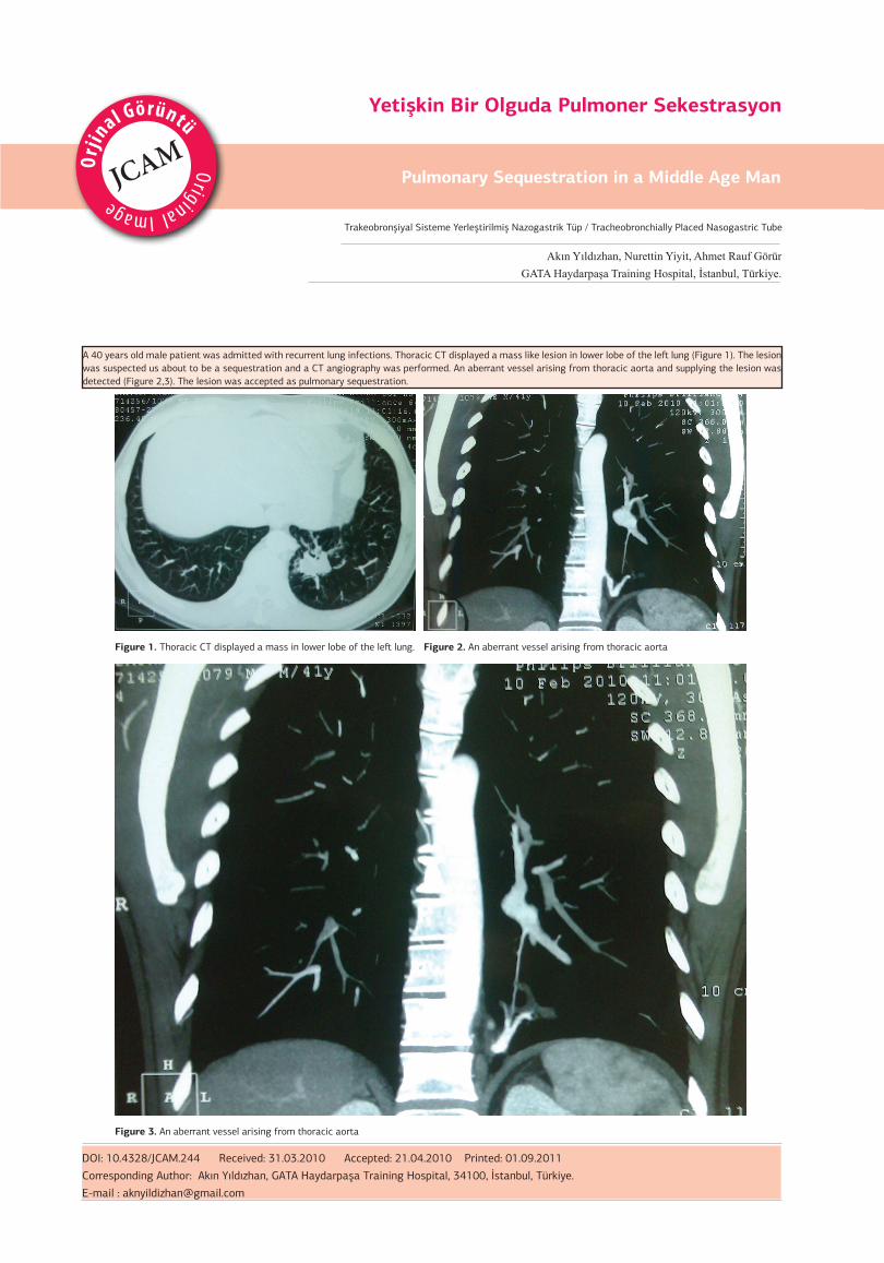
Figure 1. Thoracic CT displayed a mass in lower lobe of the left lung. Figure 2. An aberrant vessel arising from thoracic aorta
Figure 3. An aberrant vessel arising from thoracic aorta
A 40 years old male patient was admitted with recurrent lung infections. Thoracic CT displayed a mass like lesion in lower lobe of the left lung (Figure 1). The lesion was suspected us about to be a sequestration and a CT angiography was performed. An aberrant vessel arising from thoracic aorta and supplying the lesion was detected (Figure 2,3). The lesion was accepted as pulmonary sequestration.