yeast response to prolonged activation of the spindle assembly … · lo spindle assembly...
TRANSCRIPT

UNIVERSITA’ DEGLI STUDI DI MILANO-BICOCCA Facoltà di Scienze Matematiche, Fisiche e Naturali
Dipartimento di Biotecnologie e Bioscienze Dottorato in Biotecnologie Industriali, XXIII ciclo
Yeast response to prolonged activation of the spindle assembly checkpoint
Elena Galati
Anno Accademico 2009-2010

2

3
Yeast response to prolonged activation
of the spindle assembly checkpoint
Elena Galati
Maricola 049326
Tutor: Simonetta Piatti
Università degli Studi di Milano-Bicocca
Piazza dell’Ateneo Nuovo 1, 2126 Milano
Dipartimento di Biotecnologie e Bioscienze
Piazza della Scienza 2, 20126 Milano

4

5
CONTENTS
ABSTRACT 7 RIASSUNTO 11 INTRODUCTION 14 Kinetochore-microtubule attachment 14 The Spindle Assembly Checkpoint 18 How the SAC inhibits the APC 19 Turning-off the SAC 22 Adaptation to the SAC 24 Closing the cell cycle 32 Exit from mitosis in budding yeast: the FEAR and the MEN pathways 33 Mitotic exit in higher eukaryotes 38 RESULTS 40 The RSC chromatin-remodelling complex influences mitotic exit and adaptation to the SAC by controlling the Cdc14 phosphatase 40 - MAD2 overexpression as a tool to study adaptation to the SAC 42 - Characterization of SAC adaptation in yeast 42 - Adaptation to the SAC requires cyclin B degradation, Cdc20, the polo kinase Cdc5 and Cdc14 nucleolar release 45 - The chromatin-remodeling RSC complex is involved in adaptation to the SAC 46 - RSCRsc2 inactivation prevents mitotic exit of SAC-deficient mutants in the presence of microtubule-depolymerizing drugs 47 - Lack of Rsc2 impairs Cdc14 release from the nucleolus at the metaphase-to-anaphase transition 47 - Deletion of RSC2 has synthetic effects with mutations affecting the MEN 48 - Rsc2 interacts with the polo kinase Cdc5 and contributes to timely Net1 phosphorylation 49 The ATPase activity of the RSC complex is required for mitotic exit of nocodazole-treated SAC mutant cells 58 Loss of viability of GAL1-MAD2 rsc2∆ cells is independent of caspase-dependent cell death pathways 60 Cells divide nuclei and spindle pole bodies (SPBs) in presence of benomyl in an actin-independent manner 63 Involvement of other factors in adaptation to the spindle assembly checkpoint 67

6
1- Microtubule regulators are involved in SAC adaptation 68 2- The SAGA complex plays a role in mitotic slippage 71 3- The cdc5-ad allele has no effect on SAC adaptation 75 MATERIALS AND METHODS 77 DISCUSSION 90 Tools to characterize SAC adaptation in yeast 90 What happens when yeast cells adapt to the SAC 91 Adaptation versus apoptosis in yeast: preliminary observations 92 The RSC complex regulates mitotic exit by promoting the early release of Cdc14 92 The SAGA complex regulates SAC adaptation 94 Microtubule dynamics and SAC adaptation 95 REFERENCES 97
7
ABSTRACT
Faithful chromosome segregation during mitosis is fundamental for
cell viability and genome stability. For a correct division, all
kinetochores must be attached to the mitotic spindle and cohesion
must be timely removed. Anaphase is triggered by the Anaphase
Promoting Complex bound to its regulatory subunit Cdc20 (APC-
Cdc20) that polyubiquitylates securin (Pds1 in budding yeast), whose
role is to maintain inactive the protease separase (Esp1 in budding
yeast) until anaphase onset. Once active, separase cleaves cohesin,
thus triggering sister chromatid separation. Separase also promotes
cyclinB proteolysis and mitotic exit due to its involvement in the
Cdc14-early anaphase release (FEAR) pathway that promotes a partial
activation of the Cdc14 phophatase, which is in turn key for CDK
inactivation and mitotic exit. Cdc14 is maintained inactive throughout
most of the cell cycle bound to its inhibitor Net1/Cfi1 and trapped in
the nucleolus. At the beginning of anaphase Cdc14 is released from
the nucleolus into the nucleus by the FEAR pathway; subsequently,
Cdc14 is released also in the cytoplasm by the MEN (Mitotic Exit
Network) pathway. In this way Cdc14 is fully active and can trigger
mitotic exit by cyclinB-CDK inactivation.
The Spindle Assembly Checkpoint (SAC) is a surveillance mechanism
conserved in all eukaryotic organisms that ensures the correct
segregation of the genetic material. In fact, it inhibits the metaphase to
anaphase transition until all kinetochores are properly attached to the
mitotic spindle by inactivating the APC-Cdc20 complex, thus
providing the time for error correction.

8
Cells do not arrest indefinitely upon SAC activation. After a variable
period of time cells escape from the metaphase arrest also in the
presence of a damaged mitotic spindle or faulty kinetochore
attachments to spindle microtubules. This process is referred to as
adaptation or mitotic slippage and is often involved in the resistance to
chemotherapeutic compounds that target the mitotic spindle. In spite
of its importance, the adaptation process is still little known.
Within this context, the goals of my Ph.D. were: (1) to characterize
the molecular mechanisms underlying SAC adaptation and (2) to
search for factors involved in this process. For these purposes we used
the yeast Saccharomyces cerevisiae as a model organism.
(1) We characterized the adaptation process in either the presence
or the absence of mitotic spindle perturbations. We depolymerized
spindles by using two different drugs that alter microtubule dynamics,
i.e. nocodazole and benomyl, whereas we induced SAC
hyperactivation without spindle damage by overproducing Mad2
(GAL1-MAD2 cells), one of the key proteins for SAC signal
generation and maintenance. We observed that in all the conditions
cells are able to adapt, but with different kinetics. In particular, cells
adapt faster in benomyl, while in nocodazole and with high levels of
Mad2 cells need more time to slip out of mitosis. The few data
available about SAC adaptation in higher eukaryotes indicate that
SAC adaptation is accompanied by chromatid separation, a decrease
in mitotic CDK activity and mitotic exit. Indeed, like in mammalian
cells, yeast securin and cyclinB are degraded and sister chromatids are
separated during adaptation. In addition, cyclinB stabilization, as well
as Cdc20 and Cdc5 (polo kinase) inactivation, markedly delay

9
adaptation, while the only yeast CKI (Sic1) is not involved in this
process. Finally, when yeast cells adapt the SAC is likely to be turned
off, as shown by the disassembly of the Mad1/Bub3 checkpoint
complex.
(2) To search for factors involved in SAC adaptation, we
performed a genetic screen using GAL1-MAD2 cells. In particular, we
screened for mutants that would remain arrested for prolonged times
in mitosis upon MAD2 overexpression. We identified Rsc2, a non-
essential component of the RSC chromatin remodelling complex, as a
regulator of SAC adaptation in yeast. We demonstrated that RSCRsc2
is involved in fine tuning mitotic exit during the unperturbed cell
cycle. Its activity becomes particularly important in conditions that
would activate the SAC, as it contributes to cyclinB degradation. In
the absence of Rsc2 Net1 phosphorylation and the early anaphase
release of Cdc14 from the nucleolus are impaired, whereas expression
of a dominant allele of CDC14 that loosens Net1 inhibition
(CDC14TAB6-1) is sufficient to restore mitotic exit in conditions where
Rsc2 becomes essential for this process. We further demonstrated that
the ATPase activity of RSC is required for mitotic exit regulation,
suggesting that its chromatin-remodelling activity is involved in this
process. By studying possible genetic interactions between the RSC2
deletion and FEAR or MEN mutations, we found that RSC2 deletion
confers synthetic lethality or sickness to MEN but not to FEAR
mutants. Altogether, our data suggest that RSCRsc2 is a novel
component of the FEAR pathway. Finally, we demonstrated that Rsc2
interacts in vivo and in vitro with the polo kinase Cdc5, which
controls mitotic exit at different levels.

10
Since RSC binds to acetylated histone tails, it is possible that histone
transacetylases are also involved in SAC adaptation. We tested if the
SAGA (Spt-Ada-Gcn5 Acetyltransferase) complex is involved in SAC
adaptation by deleting ADA2 or GCN5 in yeast. Indeed, SAGA seems
involved in adaptation, although the contribution of Ada2 and Gcn5 in
the process differs depending on the conditions used to activate the
SAC.
Finally, since we found that upon treatment with benomyl (a
microtubule destabilizer) cells adapt dividing nuclei, we wondered if
SAC adaptation could be linked to the presence of cytoplasmic
microtubules that are still partially detectable in these conditions. We
therefore asked whether motor proteins and microtubule regulators are
involved in mitotic slippage. Indeed, we found that in the absence of
Kip2 and Bik1, which specifically bind to cytoplasmic microtubules,
cells divide nuclei and exit mitosis slower than wild type cells,
demonstrating that cytoplasmic microtubules and associated proteins
could accelerate SAC adaptation.
In conclusion, SAC adaptation is a very complex process whose
timing probably depends on the interplay between different
mechanisms. An important aim for a complete comprehension of this
process, as well as for the development of new and more efficient
cancer therapies, will be to identify novel factors implicated in
adaptation and clarify how their function might be linked to one
another.

11
RIASSUNTO La corretta segregazione dei cromosomi durante la mitosi è fondamentale per la vitalità cellulare e la stabilità del genoma. Al fine di avere una corretta divisione, tutti i cinetocori devono essere correttamente attaccati al fuso mitotico e la coesione deve essere tempestivamente rimossa. L’anafase è promossa dall’Anaphase Promoting Complex legato alla suo regolatore Cdc20 (APC-Cdc20) che poliubiquitina la securina (Pds1 nel lievito), che ha il ruolo di mantenere inattiva la proteasi separasi (Esp1 nel lievito) fino all’inizio dell’anafase. Una volta attiva, la separasi taglia la coesina, promuovendo così la separazione dei cromatidi fratelli. Inoltre, la separasi è in grado di promuovere la proteolisi delle cicline mitotiche e l’uscita dalla mitosi come componente nel FEAR pathway, una via di trasduzione del segnale che controlla il rilascio parziale di Cdc14 dal nucleolo al nucleo che permette un’attivazione parziale di Cdc14. Cdc14 è la fosfatasi chiave che promuove l’inattivazione delle CDK e l’uscita dalla mitosi; essa è mantenuta inattiva e segregata nel nucleolo per la maggior parte del ciclo cellulare grazie all’interazione col suo inibitore Net1/Cfi1. All'inizio dell’anafase, Cdc14 viene rilasciata dal nucleolo al nucleo grazie all’attivazione del FEAR pathway; successivamente, Cdc14 viene rilasciato anche nel citoplasma dal MEN (Mitotic Exit Network) pathway. In questo modo Cdc14 è completamente attiva e quindi in grado di promuovere l’uscita dalla mitosi mediante l’inattivazione delle cicline mitotiche. Lo Spindle Assembly Checkpoint (SAC) è un meccanismo di sorveglianza del ciclo cellulare altamente conservato in tutte le cellule eucariotiche che permette la corretta segregazione dei cromosomi. Infatti, esso è in grado di inibire la transizione metafase-anafase fino alla formazione dell’attacco bipolare tra il fuso mitotico e cinetocori inibendo il complesso APC-Cdc20. Ciò nonostante, il SAC non è in grado di mantenere le cellule arrestate in metafase per un tempo indefinito; infatti, dopo un certo periodo di tempo le cellule progrediscono nel ciclo cellulare sia in presenza di un fuso mitotico danneggiato che in assenza di un attacco corretto tra fuso mitotico e cinetocori. Questo processo è noto col nome di adattamento o “mitotic slippage” ed è spesso responsabile del fenomeno di resistenza ai chemioterapici che hanno come bersaglio il fuso mitotico. Nonostante la sua importanza dal punto di vista biologico, il processo di adattamento è ancora poco caratterizzato.

12
In questo contesto, i principali obiettivi del mio dottorato di ricerca sono stati: (1) caratterizzare i meccanismi molecolari alla base dell’adattamento e (2) cercare fattori coinvolti in questo processo. Per fare questo, abbiamo deciso di utilizzare l’organismo modello Saccharomyces cerevisiae. (1) Abbiamo caratterizzato l’adattamento sia in presenza sia in assenza del fuso mitotico. In particolare, abbiamo depolimerizzato il fuso mitotico mediante l’utilizzo di due droghe che alterano la dinamica dei microtubuli, il nocodazolo e il benomyl, mentre abbiamo iperattivato il SAC senza danneggiare il fuso mitotico mediante la sovraespressione di MAD2 (cellule GAL1-MAD2), una delle proteine fondamentali per la generazione e il mantenimento del segnale del SAC. Abbiamo osservato che le cellule sono in grado di adattarsi in tutte le condizioni analizzate, ma con cinetiche differenti. In particolare, le cellule si adattano più velocemente in benomyl, mentre in nocodazolo e in presenza di alti livelli di Mad2 le cellule hanno bisogno di più tempo. I dati presenti in letteratura sul processo di adattamento al SAC nelle cellule di mammifero indicano che l'adattamento è correlato con la separazione dei cromatidi fratelli, con una diminuzione dell’attività delle CDK mitotiche e con l'uscita dalla mitosi. In lievito abbiamo verificato che durante l'adattamento avviene la degradazione della securina e della ciclina B e che i cromatidi fratelli vengono separati; inoltre, la stabilizzazione della ciclina B (Clb2) e l’inattivazione di Cdc20 e della polo chinasi Cdc5 ritarda notevolmente l’adattamento, mentre l’inibitore delle CDK mitotiche (Sic1) non è coinvolto in questo processo. Infine, durante il processo di adattamento le cellule di lievito spengono il SAC; infatti, in queste cellule il complesso Mad1/Bub3 viene disassemblato. (2) Per cercare fattori coinvolti nell’adattamento al SAC, abbiamo eseguito uno screening genetico utilizzando cellule GAL1-MAD2. In particolare, eravamo alla ricerca di mutanti che rimanevano arrestati in mitosi più a lungo. Dallo screening abbiamo identificato Rsc2, una componente non essenziale del complesso RSC, coinvolto nel rimodellamento della cromatina. Abbiamo dimostrato che RSCRsc2 è coinvolto nella regolazione dell’uscita dalla mitosi durante un ciclo cellulare normale. Infatti, in assenza di Rsc2 la fosforilazione di Net1 e il rilascio parziale di Cdc14 dal nucleolo sono compromessi; è però sufficiente l’espressione di un allele dominante di CDC14 che si lega più labilmente all’inibitore Net1 (CDC14TAB6-1) per ripristinare l’uscita dalla mitosi in condizioni in cui Rsc2 risulta essenziale per questo processo. Abbiamo inoltre dimostrato che l’attività ATPasica del
13
complesso RSC è importante per la regolazione dell’uscita dalla mitosi; per questo, abbiamo ipotizzato che l’attività di rimodellamento della cromatina è richiesta per questo processo. Studiando le possibili interazioni genetiche tra la delezione di RSC2 e mutazioni che affliggono il FEAR o il MEN, abbiamo dimostrato che la delezione di RSC2 provoca letalità sintetica o forti difetti di crescita se combinata a mutazioni che affliggono il MEN, ma non il FEAR. In conclusione, i nostri dati suggeriscono che RSCRsc2 potrebbe essere una nuova componente del FEAR pathway. Infine, abbiamo dimostrato che Rsc2 interagisce in vivo e in vitro con la polo chinasi Cdc5, una proteina che controlla l’uscita dalla mitosi a diversi livelli. Dato che il complesso RSC si lega in particolare a regioni di DNA con istoni acetilati, è possibile che anche le acetilasi siano coinvolte nel processo di adattamento al SAC. Abbiamo testato se il complesso SAGA (Spt-Ada-Gcn5 Acetyltransferase) fosse coinvolto nell’adattamento mediante la delezione dei geni ADA2 e GCN5. Dai dati preliminari ottenuti sembra che il complesso SAGA possa regolare l’adattamento, anche se il contributo di Ada2 e Gcn5 dipende dalle condizioni analizzate. Infine, dato che abbiamo osservato che cellule trattate col benomyl (una droga che destabilizza i microtubuli) si adattano dividendo i nuclei, abbiamo ipotizzato che l’adattamento potrebbe dipendere dalla presenza di microtubuli citoplasmatici che sono parzialmente presenti in queste condizioni. Per questo motivo, ci siamo domandati se proteine motrici o proteine associate ai microtubuli potessero essere coinvolte nel processo di adattamento. In assenza di Kip2 e Bik1, che si legato specificamente ai microtubuli citoplasmatici, le cellule dividono i nuclei ed escono dalla mitosi più lentamente delle cellule selvatiche, dimostrando che i microtubuli citoplasmatici accelerano il processo di adattamento. In conclusione, l’adattamento al SAC è un processo molto complesso che viene probabilmente regolato a diversi livelli.

14
INTRODUCTION
Correct segregation of duplicated chromosomes to daughter cells
during cellular division is fundamental for the faithful inheritance of
genetic material. This process needs that all chromosomes achieve bi-
orientation (also called amphitelic attachment), i.e. they attach in a
bipolar way to microtubules emanating from opposite spindle poles
through their kinetochores, which are protein assemblies that reside at
the centromere of chromosomes. As microtubule attachment is a trial
and correction process, incorrect attachments can occur. If cells were
to undergo anaphase in the presence of erroneous attachments,
chromosomes would be segregated randomly, thus generating cells
with aberrant chromosome numbers known as aneuploidies. In order
to avoid this dramatic event, cells evolved a surveillance mechanism,
the spindle assembly checkpoint (SAC) that delays cell cycle
progression in mitosis until all chromosomes have reached a bipolar
attachment and are aligned on the metaphase plate. Dysfunction in this
machinery has been implicated in genetic diseases, such as the Down
syndrome, and in tumor progression. (from Tanaka and Hirota, 2009).
Kinetochore-microtubule attachment
The segregation of sister chromatids during mitosis mainly depends
on the forces generated by microtubules (MTs) that attach to
kinetochores (Tanaka and Desai, 2008). For proper chromosome
segregation, kinetochores must capture spindle MTs and properly
align on the mitotic spindle before anaphase onset. Cells undergo
these processes in a step-wise manner as follows (Tanaka, 2008): (1)
the kinetochore initially interacts with the lateral surface of a single

15
MT extending from a spindle pole (spindle-pole MT) (fig. 1, step 1);
(2) the kinetochore is transported along the spindle-pole MT towards a
spindle pole (fig. 1, step 2); (3) the kinetochore is tethered at the plus
end of a spindle-pole MT (conversion from the lateral to the end-on
attachment) (fig. 1, step 2); (4) sister kinetochores attach to MTs
extending from opposite spindle poles (sister kinetochore bi-
orientation). If sister kinetochores attach to MTs with aberrant
orientation, such errors must be corrected by turnover of the
kinetochore-microtubule attachment (error correction), before
anaphase onset (fig. 1, steps 3 and 4). All these steps, that are
conserved in all eukaryotes (from yeast to human), are crucial to
ensure high-fidelity chromosome segregation in the anaphase (fig. 1,
step 5).
Here is the detailed description of the first two steps of the process:
- Step 1, initial kinetochore-microtubule interaction: this event
occurs in a different cell cycle stage depending on the organism. In
fact, in metazoan cells in interphase the MT-organizing centres
(MTOCs) are outside of the nucleus and MTs can interact with
kinetochores only when the nuclear envelope is broken down at the
onset of mitosis; on the other hand, in budding yeast the kinetochores
are connected to MTOCs (called spindle-pole bodies or SPBs) by
MTs throughout most of the cell cycle (Winey and O’Toole, 2001).
In all eukaryotic cells, however, kinetochores initially attach to the
lateral side of a single microtubule (Hayden et al., 1990; Tanaka et
al., 2005) because the lateral side provides a much larger surface
compared with microtubule tips, thus providing a more efficient
initial attachment.

16
- Step 2, conversion from lateral to end-on attachment: once bound
to the microtubule lateral face (lateral attachment), kinetochores are
transported towards a spindle pole along the microtubule (fig. 1, step
2). The kinetochore sliding along microtubule is promoted by minus
end-directed motor proteins, i.e. dynein in vertebrate cells (Yang et
al., 2007) and Kar3 in budding yeast (Tanaka et al, 2005). While the
kinetochore is associated with the microtubule lateral face, the plus
end of the shrinking microtubule often catches up with the
kinetochore, leading the kinetochore to become tethered at the
microtubule plus end (end-on attachment) and being pulled further
towards a spindle pole as the microtubule shrinks (Tanaka et al.,
2007) (fig. 1, step 2). (from Tanaka, 2010)

17
Figure 1. Kinetochore-microtubule interaction in eukaryotic cells. The figure shows kinetochore–microtubule interactions during prometaphase (steps 1–3), metaphase (step 4) and anaphase (step 5) (from Tanaka, 2010).
Since kinetochore-microtubule attachment is a stochastic mechanism,
erroneous attachments can be generated. Different kinds of
attachments are possible (fig. 2): (1) amphitelic arrangement with the
two sister kinetochores bi-oriented is the only correct attachment and
allows proper chromosome segregation; (2) monotelic attachment, in
which only one kinetochore is attached to mitotic spindle; (3) syntelic
attachment, in which both kinetochores are attached to microtubules
arising from the same spindle pole; (4) merotelic attachment, in which
one kinetochore is correctly attached while the other one is attached to
microtubules arising from the opposite poles of the mitotic spindle.
Merotelic attachments are only possible in organisms where
kinetochores contact simultaneously several microtubules (e.g. not in
budding yeast). Finally, since the pulling forces exerted by spindle
microtubules on bi-oriented kinetochores are counteracted by the
cohesive forces that maintain sister chromatids together until the onset
of anaphase, in the amphitelic configuration kinetochores are under
tension. Establishment of kinetochore tension is indeed a key feature
of chromosome segregation. (from Musacchio and Salmon, 2007)

18
Figure 2. Different kind of kinetochore-microtubule attachments Correct and incorrect attachments can occur during mitosis. Monotelic attachment is a normal condition during prometaphase before bi-orientation. In syntelic attachment, both sisters in a pair connect to the same pole. Merotelic attachment occurs quite frequently. The SAC is able to sense monotelic syntelic attachment, but it is unable to detect merotelic attachment, since in these condition kinetchores are under tension (from Musacchio and Salmon, 2007).
THE SPINDLE ASSEMBLY CHECKPOINT
The spindle assembly checkpoint (or SAC) inhibits the metaphase-to-
anaphase transition until all kinetochores are correctly bi-oriented on
the spindle. It is known that the SAC signal originates from
unattached kinetochores and that one single unattached kinetochore is
sufficient to sustain the checkpoint signal (Rieder at al, 1995). The

19
target of the SAC is the anaphase promoting complex (APC) bound to
its coactivator Cdc20. The APC is an E3 ubiquitin ligase that is
absolutely necessary for sister chromatid separation in anaphase and
for mitotic exit by targeting some key proteins to degradation (Peters,
2006). When all chromosomes achieve bi-orientation, the activity of
the SAC is silenced and the APC triggers degradation of its substrates,
including cyclinB and securin: this event promotes anaphase (Clute
and Pines, 1999; Hagting et al, 2002). Degradation of securin allows
activation of the protease separase, which cleaves cohesion, thereby
triggering sister chromatid separation. On the other hand, degradation
of cyclinB causes inactivation of CDK activity and induces mitotic
exit (Peters, 2006). (from Tanaka and Hirota, 2009)
How the SAC inhibits the APC
The key components of the SAC (the Mad and Bub proteins) were
discovered by genetic screens in budding yeast (Li and Murray, 1991;
Hoyt et al, 1991). As Mad2 is enriched at unattached kinetochores and
it binds directly to Cdc20, Mad2 is thought to play a crucial role in
SAC activation and maintenance (Musacchio and Salmon, 2007; Yu,
2006; Sudakin et al., 2001). Consistent with this idea, suppression of
Mad2 depletes the checkpoint signal. The finding that only a fraction
of Mad2 is stably bound to unattached kinetochores, whereas the other
fraction is highly dynamic and turns over with the cytoplasmic pool,
led to the idea that Mad2 is activated by cycling through unattached
kinetochores (Howell et al, 2000; Howell et al, 2004; Shah et al,
2004). Structural analysis has identified two different conformations
of Mad2 (O for open and C for closed), with different activities

20
(Sironi et al, 2002). The current model for Mad2 regulation is that
Mad2 stably binds to Mad1 at unattached kinetochores (C-Mad2) and
the resulting C-Mad2-Mad1 complex works as a receptor that binds
and converts cytoplasmic free O-Mad2 from an inactive conformation
(open) to an active form (closed), which is able to bind and inhibit
Cdc20 and, consequently, the APC. The Cdc20-bound C-Mad2
probably activates other molecules of O-Mad2, thereby amplifying the
signal (fig. 3A and B). This mechanism, called “template model”,
explains how the SAC signal is created and amplified and how a
single unattached kinetochore can generate a strong SAC signal
(Musacchio and Salmon, 2007).
Does Mad2 directly inhibit Cdc20? First, it is known that another
checkpoint protein called BubR1 (or Mad3 in yeast) can directly bind
Cdc20 and inhibit APC/C activity without Mad2 (Tang et al, 2001).
Second, the MCC (mitotic checkpoint complex), composed by Mad2,
BubR1 (or Mad3), Bub3 and Cdc20, is a stronger inhibitor for APC
than Mad2 alone (Sudakin et al, 2001). Thus, a plausible hypothesis is
that BubR1/Mad3, and not Mad2, is the direct inhibitor of the APC
and might act through the MCC. Indeed, budding yeast Mad3 was
shown to inhibit the activity of APC/Cdc20 by acting as
pseudosubstrate (Burton and Solomon, 2007). In addition, it promotes
Cdc20 degradation (King et al, 2007). Therefore, the most recent
models for SAC signalling envision BubR1/Mad3 as direct inhibitor
of Cdc20, with BubR1 binding to Cdc20 significantly enhanced by
Mad2-Cdc20 (fig. 3C), which takes into account the key role of Mad2
in the SAC and the old observation that Mad2 is required for Mad3
association with Cdc20 (Hwang et al., 1998). Binding of BubR1 to
21
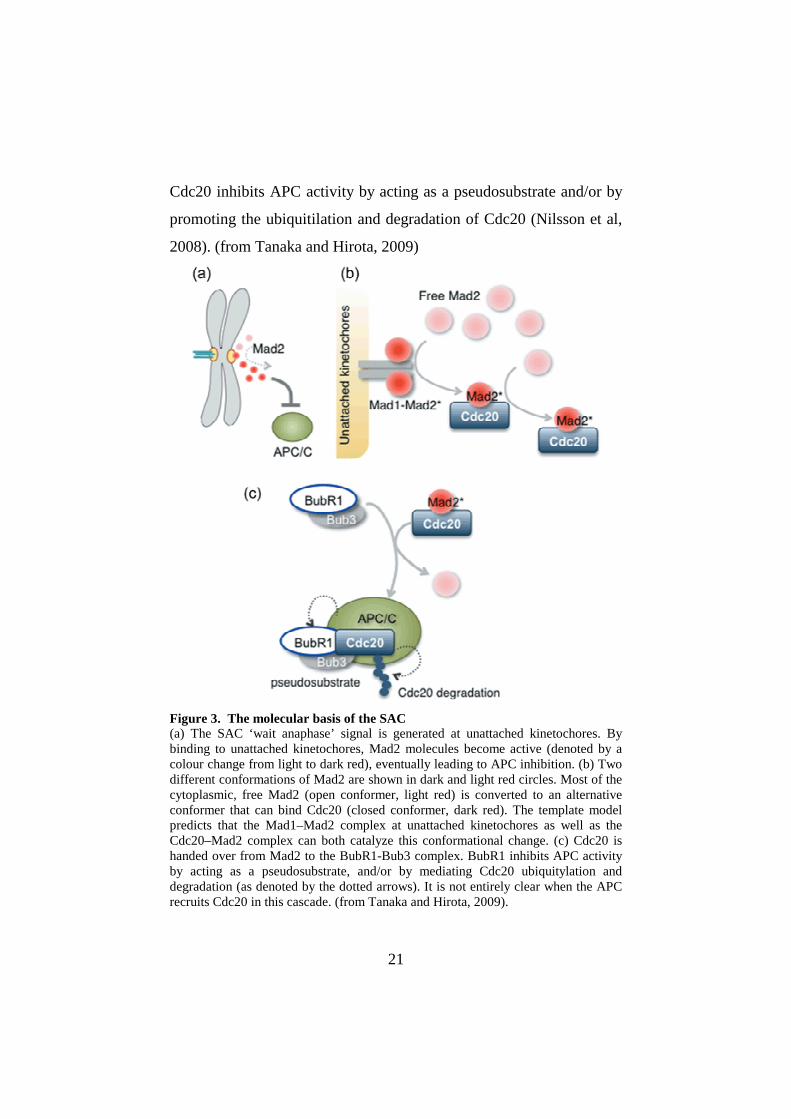
Cdc20 inhibits APC activity by acting as a pseudosubstrate and/or by
promoting the ubiquitilation and degradation of Cdc20 (Nilsson et al,
2008). (from Tanaka and Hirota, 2009)
Figure 3. The molecular basis of the SAC (a) The SAC ‘wait anaphase’ signal is generated at unattached kinetochores. By binding to unattached kinetochores, Mad2 molecules become active (denoted by a colour change from light to dark red), eventually leading to APC inhibition. (b) Two different conformations of Mad2 are shown in dark and light red circles. Most of the cytoplasmic, free Mad2 (open conformer, light red) is converted to an alternative conformer that can bind Cdc20 (closed conformer, dark red). The template model predicts that the Mad1–Mad2 complex at unattached kinetochores as well as the Cdc20–Mad2 complex can both catalyze this conformational change. (c) Cdc20 is handed over from Mad2 to the BubR1-Bub3 complex. BubR1 inhibits APC activity by acting as a pseudosubstrate, and/or by mediating Cdc20 ubiquitylation and degradation (as denoted by the dotted arrows). It is not entirely clear when the APC recruits Cdc20 in this cascade. (from Tanaka and Hirota, 2009).

22
Turning-off the SAC
Several mechanisms contribute to the inactivation of the SAC after
chromosome bi-orientation during normal cell cycle.
First, one of the key processes that allows SAC inactivation is the
“stripping” of Mad1, Mad2 and other SAC proteins from
kinetochores, which results in their redistribution to the spindle poles
(fig. 4). In metazoans, this process depends on dynein-dependent
motility along microtubules (Howell et al, 2001; Wojcik et al, 2001).
While Mad1-Mad2 are inactivated by dynein that removes this
complex from attached kinetochores, the regulation of BubR1 kinase
activity seems to depend on the kinetochore motor protein CENP-E
(Mao et al, 2005). However, this second mechanism is less defined at
the molecular level, also because BubR1 kinase activity does not seem
to be necessary for the SAC (Elowe et al., 2010). Both these “SAC
inactivation pathways” were identified in metazoans and depend on
kinetochore-microtubule interactions and motor activity. No nuclear
dynein or clear CENP-E homologue exist in yeast S. cerevisiae, and
therefore whether similar mechanisms act also in yeast is unknown at
the moment. In the yeasts S. cerevisiae and S. pombe
dephosphorylation events carried out by protein phosphatase PP1
seem necessary for SAC silencing (Pinsky et al., 2009; Vanoosthuyse
and Hardwick, 2009).
Another mechanism of SAC inactivation in metazoans is based on the
protein p31comet. p31comet works as a brake for the positive-feedback
loop based on C-Mad2 (Vink et al, 2006) and it is possible (but has
not been yet demonstrated) that kinetochores modify this protein to
temporarily prevent it from carrying out its function. As part of the

23
Mad2-template model, it is speculated that the reactivation of
p31comet upon disappearance of unattached kinetochores silences the
SAC by turning down the catalytic activation of Mad2 (fig. 4). In
practice, p31comet competes with free Mad2 for binding either to
Mad1/Mad2 at unattached kinetochores or to Cdc20/Mad2.
Another mechanism of SAC inactivation is based on Mad2/Cdc20
dissociation. Although the Mad2/Cdc20 dissociation occurs
spontaneously, this process is slow; probably, a source of energy is
required to trigger this dissociation during the cell cycle. A non-
degradative ubiquitylation of Cdc20 has been proposed as a possible
active mechanism for dissociation of the Mad2-Cdc20 complex
(Reddy et al., 2007; Stegmeier et al., 2007). Consistently, the de-
ubiquitylating (DUB) enzyme “protectin” antagonizes this reaction
and is required to sustain the SAC (Reddy et al., 2007; Stegmeier et
al., 2007) (fig. 4). Therefore, SAC maintenance might be a dynamic
process in which the MCC and Mad2–Cdc20 subcomplexes are
continuously actively dissociated and recreated by unattached
kinetochores.
Another mechanism that contributes to maintain the SAC inactive in
anaphase is APC-induced proteolysis itself. In anaphase, tension at
kinetochores is lost but the SAC is not reactivated, probably because
the SAC is inhibited in anaphase and this event is important for cell
cycle progression. Proteolysis of cyclinB and subsequent CDK
inactivation probably play an important role in SAC inhibition
(D’Angiolella et al, 2003). In fact, high levels of cyclinB/CDK are
required to sustain the SAC in many organisms (Kitazono et al, 2003;
Li and Cai, 1997).

24
Finally, Mad2 phosphorylation has been proposed to contribute to
SAC silencing by preventing its conformational change from open
into close configuration (Wassmann et al., 2003; Kim et al., 2010).
Altogether, these mechanisms contribute to turn the SAC off when the
checkpoint is satisfied. (from Musacchio and Salmon, 2007)
Figure 4. How the SAC is inactivated Only three regulatory aspects that might favour Mad2–Cdc20 dissociation at anaphase are depicted. First, disappearance of unattached kinetochores might result in reactivation of the capacity of p31comet to inhibit the closed-Mad2/open-Mad2 interaction and thereby to inhibit the catalytic amplification of the SAC signal that is predicted by the “template model”. Second, non-degradative ubiquitylation of Ccd20 in a reaction that involves the E2 enzyme UbcH10 and the de-ubiquitylating protein (DUB) protectin might accelerate the dissociation of Mad2–Ccd20. Third, the dynein–dynactin complex ‘strips’ Mad1–Mad2 and other proteins from kinetochores on formation of kinetochore microtubules, decreasing the ability to form new Mad2–Cdc20 complexes. A forth mechanism based on Mad2 phosphorylation is not depicted (see text) (from Musacchio and Salmon, 2007).
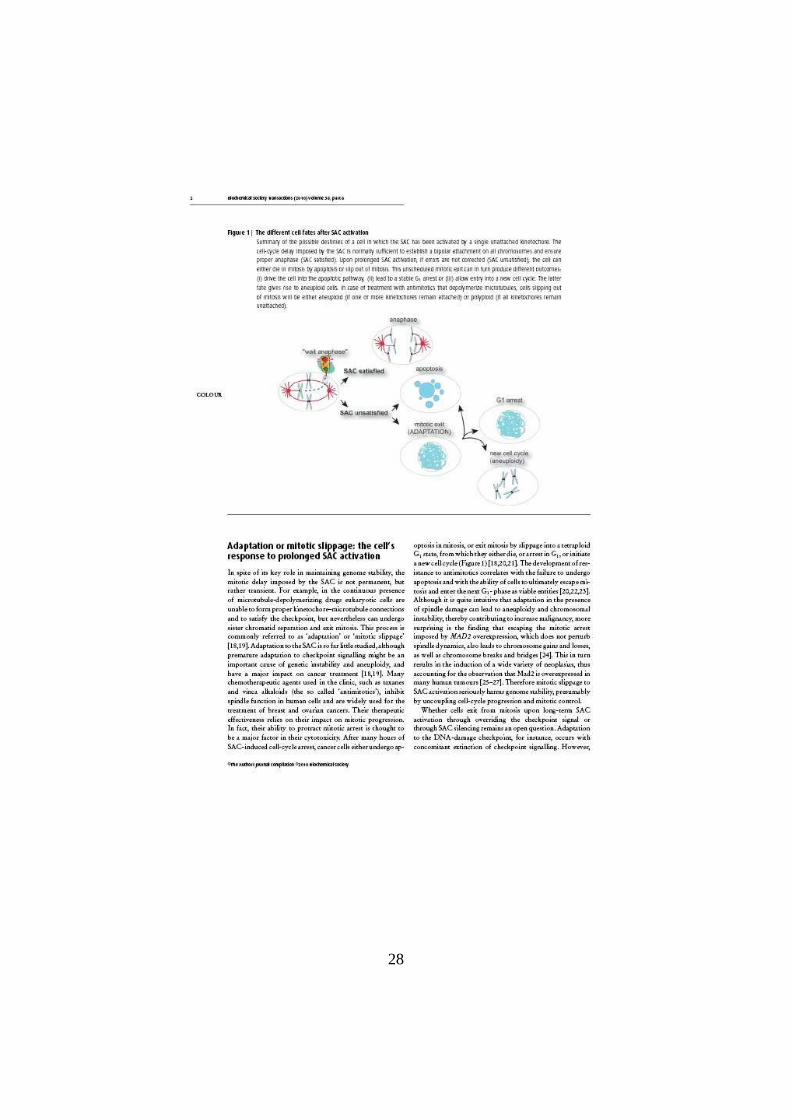
Adaptation to the SAC
As described above, the SAC is activated when at least one
kinetochore is not correctly attached to mitotic spindle and it is
inactivated when a bipolar attachment between kinetochores and the
mitotic spindle is generated. Satisfying the SAC is not always

25
possible. Upon prolonged SAC activation a mechanism called
adaptation or mitotic slippage allows cell cycle progression also in
absence of correct kinetochore-microtubule attachments.
Understanding how adaptation to the SAC takes place is of primary
importance for cancer research and for developing more efficient
drugs for cancer therapy (Rieder and Maiato, 2004; Weaver and
Cleveland, 2005). In fact, some chemotherapeutic agents used in
clinics called antimitotics, e.g. taxanes, inhibit mitotic spindle
dynamics thereby causing a prolonged activation of the SAC. After a
prolonged activation of the SAC, cancer cells can undergo apoptosis
in mitosis or exit from mitosis by adaptation. Cells that adapt in turn
can: (1) begin a new cell cycle; (2) arrest in G1; (2) die by apoptosis
(Rieder and Maiato, 2004; Gascoigne and Taylor, 2008). Cancer cells
often become resistant to antimitotics, likely because they bypass SAC
activation through the adaptation process and fail to activate the cell
death pathways.
In the last few years the molecular mechanisms at the basis of mitotic
slippage have started to be defined. At the moment, it is known that
cells undergo a progressive decrease in cyclinB/CDK activity that
leads to adaptation (Gascoigne and Taylor, 2008; Brito and Rieder,
2006) and that this event occurs by cyclinB degradation. It is not clear
yet if cells that adapt turn off or override the SAC. It has been
proposed that cells undergo adaptation in the presence of continuous
SAC signalling, as SAC proteins remain stably bound at kinetochores
during mitotic slippage in the presence of microtubule depolymerizers
(Brito and Rieder, 2006). However, since CDK activity is required to
sustain the SAC, it is possible that during adaptation the SAC is
26
actually switched-off and the persistence of SAC proteins at
kinetochores is simply due to the lack of poleward stripping when
microtubules are depolymerized.
What happens when cells are treated for prolonged times with
microtubule poisons? Recent data demonstrate that cells can undergo
two alternative and competing pathways: (1) die by apoptosis or (2)
slip out of mitosis through cyclinB proteolysis. Interfering with either
one of the two pathways conveys cells into the second one (Gascoigne
and Taylor, 2008). Therefore, the identification of factors influencing
SAC adaptation would have a major impact on cancer research, as it
would help designing more efficient therapeutic strategies.
Further details on the adaptation process are reported in the following
review.

27
28
29

30

31

32
CLOSING THE CELL CYCLE
The central components that coordinate cell cycle progression are
CDKs, and their activity is regulated by cyclins, their regulatory
subunits. In particular, mitotic CDKs drive the events of early mitosis:
chromosome condensation and resolution, nuclear envelope
breakdown and assembly of the mitotic spindle. To end the cell cycle,
eukaryotic cells must inactivate the mitotic CDKs. This event, referred
to as mitotic exit, includes a number of processes that strictly require
the inactivation of mitotic CDKs, such as chromosome
decondensation, spindle disassembly, formation of pre-replicative
complexes at replication origins and cytokinesis. In all eukaryotes
mitotic CDK inactivation depends on cyclinB proteolysis promoted by
APC/C and this event allows phosphatases to dephosphorylate the
CDK substrates. In general, the final stages of mitosis are governed by
two mechanisms: (1) dephosphorylation of CDK substrates and (2)
ubiquitylation of APC substrates (fig. 5). Out of APC substrates,
cyclinB is clearly the most critical protein that needs to be degraded in
order for cells to get out of mitosis. (from Sullivan and Morgan, 2007)

33
Figure 5. What happens when cells exit from mitosis Progression through mitosis is shown by the cells along the bottom of the figure. The transition from metaphase to anaphase is triggered by an increase in the activity of the anaphase-promoting complex (APC) (green line; top of figure), a ubiquitin protein ligase that promotes the assembly of chains of ubiquitin (Ub) on its substrates, thereby targeting them for destruction by the proteasome. The main APC targets are securin, the destruction of which leads to sister-chromatid separation, and cyclins, the destruction of which results in a drop in CDK activity (blue line). CDK inactivation allows cellular phosphatases to dephosphorylate CDK substrates during late mitosis. CDK-substrate dephosphorylation is required for the events of anaphase and telophase (not shown) (from Sullivan and Morgan, 2007).
Exit from mitosis in budding yeast: the FEAR and MEN pathways
In budding yeast, where the molecular mechanisms at the basis of
mitotic exit have been extensively studied, mitotic exit depends on the
key protein phosphatase Cdc14, which promotes directly cyclinB
degradation and CDK inactivation and at the same time reverses the
phosphorylation events carried out by CDKs in mitosis (Sullivan and
Morgan, 2007; Queralt and Uhlmann, 2008). Two pathways
contribute to Cdc14 activation: the Cdc-fourteen early anaphase
release (FEAR) pathway and the mitotic exit network (MEN).
At the onset of anaphase sister chromatids begin to be separated. This
event is triggered by the APC complex (Stegmeier et al., 2002). The
FEAR is a non-essential pathway that involves many proteins, such as
the separase Esp1, the polo kinase Cdc5, the kinetochore protein
Slk19 as well as the homologous proteins Spo12 and Bns1. In
addition, the FEAR is negatively regulated by Fob1 and the PP2ACdc55
phosphatase complex (reviewed by D’Amours and Amon, 2004). The
FEAR has a dual role: it is required for completion of chromosome
segregation (D’Amours et al, 2004) by the separase Esp1 and also
causes a partial and transient release of the phosphatase Cdc14 from
the nucleolus (fig. 6) (Stegmeier et al., 2002; Azzam et al., 2004;

34
D’Amours and Amon, 2004). In fact, Cdc14 is kept imprisoned in the
nucleolus by binding to the nucleolar protein Net1/Cfi1 until
metaphase (reviewed in Queralt and Uhlmann, 2008). At the anaphase
onset, separase activation not only leads to proteolytic cleavage of
cohesin, but also it utilises a second non-proteolytic activity to
activate Cdc14 (Sullivan and Uhlmann, 2003). At this stage, high
mitotic CDK activity triggers Cdc14 release by phosphorylating at
least 6 CDK recognition sites on Net1 (Azzam et al., 2004). The link
between separase and CDK-dependent phosphorylation comes
through the PP2A phosphatase bound to its Cdc55 regulatory subunit,
which until metaphase counteracts Net1 phosphorylation (Queralt et
al., 2006) (fig. 7). Separase-dependent downregulation of PP2ACdc55 at
anaphase onset allows Net1 phosphorylation, but the mechanisms by
which the separase inhibits PP2ACdc55 are yet not understood. Two
proteins that are tightly bound to PP2ACdc55, Zds1 and Zds2, are
required downstream of separase for timely phosphorylation of Cdc14
(Queralt and Uhlmann, 2008), suggesting that they might act as
inhibitors of PP2ACdc55. The polo kinase Cdc5 also plays a crucial role
on the FEAR pathway, likely by contributing to full Net1
phosphorylation (Visintin et al., 2008). The mechanism by which
other FEAR proteins promote Cdc14 activation is poorly understood.
However, Slk19, as well as separase, binds to Cdc5 and might
therefore modulate its FEAR function (Rahal and Amon, 2008).
As mentioned above, FEAR triggers only a transient activation of
Cdc14. To exit from mitosis, cells need that another pathway, the
MEN, is activated (McCollum and Gould, 2001). Unlike the FEAR,
the MEN is an essential pathway for mitotic exit composed by a Ras-

35
like GTPase called Tem1 and its downstream kinases Cdc15 and
Mob1/Dbf2 (Jaspersen et al., 1998; Lee et al., 2001; Visintin et al.,
2001) (fig. 7). Tem1 acts at the top of this signalling cascade and is
the target of the spindle position checkpoint (SPOC, fig. 8), a
surveillance mechanism that delays mitotic exit until the spindle is
properly positioned relative to the mother-bud axis. Tem1 is
concentrated on the daughter-bound spindle pole in mitosis, and the
elongation of the mitotic spindle into the daughter cell brings Tem1
away from its inhibiting kinase Kin4 that is concentrated on the
mother-cell cortex and mother-bound spindle pole (fig. 8). For many
years, people believed that Lte1 could be the putative GEF of Tem1
(Bardin et al., 2000; Pereira et al., 2000), but during these last years
this hypothesis has been disproved (Geymonat et al., 2009). At the
moment, how MEN is activated is still unclear. Counteracting Tem1
activation is the GTPase-activating protein (GAP) complex
Bfa1/Bub2 (Geymonat et al., 2003), a negative regulator of mitotic
exit. Its inhibitory effect on Tem1 is switched off by the
phosphorylation of Bfa1 by Cdc5 (Hu et al., 2001; Geymonat et al.,
2003), while the Kin4 kinase counteracts this phosphorylation and
maintains Bfa1/Bub2 capable of inhibiting Tem1 (Maekawa et al.,
2007). Active Tem1 bound to GTP interacts with the Cdc15 kinase to
activate the MEN. Cdc15 is in turn able to activate the Mob1/Dbf2
kinase complex (Asakawa et al., 2001; Mah et al., 2001). Besides
phosphorylating and inhibiting Bfa1/Bub2, Cdc5 has also a more
direct role in the activation of Mob1/Dbf2 (Lee et al., 2001). How
Dbf2 finally promotes Cdc14 nucleolar release is not clear, but one
hypothesis is that Dbf2 directly phosphorylates Net1 to promote

36
Cdc14 dissociation (Mah et al., 2005). So, when the spindle is
correctly positioned MEN promotes a total activation of Cdc14 (fig.
6). The full activation of Cdc14 leads to mitotic exit through
dephosphorylation of Cdh1, another APC coactivator factor, and Sic1,
an inhibitor of mitotic CDKs (Prinz and Amon, 1999). APC/Cdh1 and
Sic1 together totally inactivate CDK activity, allowing cells to exit
from mitosis and enter in a new cell cycle (D’Amours and Amon,
2004).
Figure 6. Regulation of the Cdc14 phosphatase in budding yeast Cdc14 activity is regulated by changes in its intracellular localization. From G1 to metaphase Cdc14 is sequestered in the nucleolus by its inhibitor Net1/Cfi1. Cdc14 release from Net1 requires the action of two consecutive networks, FEAR and MEN. (from De Wulf et al., 2009).

37
Figure 7. A model for mitotic exit in budding yeast. At the heart of the scheme is the reversible phosphorylation and dephosphorylation of the Cdc14 phosphatase inhibitor Net1. At anaphase onset, separase-dependent downregulation of PP2ACdc55 promotes CDK-dependent phosphorylation of Net1, amplified by Cdc5. This leads to Cdc14 early anaphase release from nucleolar inhibition by Net1 (FEAR). Inhibition of Bub2/Bfa1, along with Lte1-dependent activation of Tem1, brings about activation of the mitotic exit network (MEN). Kinases of the MEN cascade (e.g. Dbf2) may maintain Cdc14 release by sustaining Cdc5-amplified Net1 phosphorylation. (from Queralt and Uhlmann, 2008) Cdc5 has therefore emerged as a key factor in mitotic exit regulation,
as it is the only protein that is involved in both the FEAR and the
MEN pathway (Jaspersen et al., 1998; Stegmeier at al., 2002). One of
the best characterized roles of Cdc5 in regulating mitotic exit is the
inhibition of Bfa1, thus contributing to Tem1 activation (Geymonat et
al., 2003). Bfa1 phosphorylation is counteracted by PP2ACdc55 during
metaphase, so the downregulation of PP2ACdc55 at the onset of
anaphase promotes not only Net1 phosphorylation but also MEN
activation (Queralt et al., 2006). A direct target of Cdc5 could be the
inhibitor Net1 (Show et al., 2002; Yoshida and Toh-e, 2002), both
early and late in anaphase. In addition, Cdc5 contributes to the build-
up of CDK activity during mitotic entry, a pre-requisite for CDK-
dependent Net1 phosphorylation (Azzam et al., 2004). To work
properly, Cdc5 needs that its target undergoes a priming
phosphorylation event by another kinase. This priming
phosphorylation is important for substrate specificity (Elia et al.,
2003). Because of this peculiarity, Cdc5 might not have a specific
function in regulating mitotic exit but could act as a generic
phosphorylation amplifier. While Cdc5 may, therefore, not be
regulating the onset of Cdc14 activation, it remains a limiting factor
for Cdc14 release throughout anaphase, and APC/Cdh1-mediated
Cdc5 destruction at the end of mitosis is sufficient to cause Cdc14 re-

38
sequestration in the nucleolus (Visintin et al., 2008). (from Sullivan
and Morgan, 2007; Queralt and Uhlmann, 2008; Bosl and Li, 2005).
Figure 8. Regulation of the MEN by the SPOC The MEN proteins and their spatial localization is fundamental for the SPOC. Only when one of the SPBs migrates into the daughter cell, Tem1 can be activated by Lte1 triggering MEN activation, while when the mitotic spindle is mispositioned Tem1 is kept inactive by the Bfa1/Bub2 complex, which is in turn maintained active by the Kin4 kinase (from Fraschini et al., 2008, modified).
Mitotic exit in higher eukaryotes
CDK inhibition and dephosphorylation of CDK substrates promote
mitotic exit in all eukaryotes. In fact, although most of the substrates
that must be dephosphorylated for triggering mitotic exit are still
unknown, persistent CDK activity prevents mitotic exit in all
eukaryotes studied. Many of the dephosphorylation-dependent
anaphase events take place in a similar manner in yeast and other
eukaryotes. These include stabilisation of microtubules at anaphase
onset, relocalisation of the Aurora B kinase complex, spindle midzone

39
assembly and, upon completion of chromosome segregation, spindle
disassembly, cytokinesis and cell abscission. Some examples of CDK
substrates that are dephosphorylated in higher eukaryotes are the C.
elegans kinesin ZEN-4 (Mishima et al, 2004), and the human Ase1
orthologue PRC1 (Zhu et al, 2006), both involved in the assembly of
the anaphase spindle midzone. It is known that ZEN-4
dephosphorylation requires CDC-14, the non-essential C. elegans
Cdc14 orthologue. Instead, vertebrates have two Cdc14 orthologues,
Cdc14A and Cdc14B. Introduction of both human Cdc14A and
Cdc14B into budding yeast rescues cdc14∆ yeast mutant phenotypes,
suggesting that there is some functional conservation between these
phosphatases (Li et al, 1997).
Also fission yeast has a Cdc14 homologue, Clp1, that is not essential
for normal cell cycle progression. It is known that fission yeast cells
possess a signalling cascade similar to the budding yeast MEN, the
Septation Initiation Network (SIN). The SIN controls cytokinesis and
its downstream effectors are not yet known. Unlike the MEN, the SIN
is not required for Clp1 activation, but only contributes to sustain Clp1
nucleolar release until the completion of cytokinesis (Trautmann et al,
2001). Orthologues of the MEN kinases Cdc15 and Mob1/Dbf2 are
also found in higher eukaryotes, while no Cdc14 orthologues have
been found encoded in the genomes of higher plants (Kerk et al,
2008). If the processes that control the mitotic exit are partially
conserved in eukaryotes, in many organisms the Cdc14 phosphatase is
not essential for mitotic exit, so other unknown phosphatases must be
required. (from Queralt and Uhlmann, 2008)

40
RESULTS
The RSC chromatin-remodelling complex influences mitotic exit
and adaptation to the SAC by controlling the Cdc14 phosphatase
As mentioned in the introduction, the factors influencing the rate of
adaptation to the SAC are currently unknown, in spite of their obvious
relevance for cancer research. We decided to use budding yeast as
model system to study SAC adaptation. We have verified that yeast
cells adapt to SAC activation under different conditions by using the
normal regulatory networks that govern mitotic exit in unperturbed
conditions, leading to destruction of securin and cyclinB. Unlike
previously reported for mammalian cells (Brito and Rieder, 2006), we
found that SAC adaptation in yeast is accompanied by silencing of
SAC signalling and disassembly of checkpoint protein complexes. By
using a yeast strain that hyperactivates the SAC through MAD2
overexpression and, therefore, in the absence of spindle damage or
alterations in kinetochore-microtubule attachments, we carried out a
genetic screen for factors involved in SAC adaptation. We found the
yeast RSC (Remodel the Structure of Chromatin) complex, and in
particular its form bound to the accessory Rsc2 subunit, regulates
mitotic slippage. Further analyses showed that RSCRsc2 controls
mitotic exit by regulating the early release of the Cdc14 phosphatase
from the nucleolus, probably acting as a novel component of the
FEAR pathway. In addition, our data indicate that the Rsc2 interacts
with the polo kinase Cdc5, which is essential for Cdc14 activation and
mitotic exit regulation.
The data obtained from our studies are reported in the following
article.

41

42

43

44

45

46

47

48

49

50

51

52

53

54

55

56

57

58
The ATPase activity of the RSC complex is required for mitotic
exit of nocodazole-treated SAC mutant cells
In Rossio et al. (2010) we observed that the RSCRsc2 complex is
required for the mitotic exit of SAC-deficient mutants in the presence
of microtubule-depolymerizing drugs. To remodel the structure of
chromatin the RSC complex uses the ATPase activity provided by the
core catalytic subunit Sth1 (Du et al, 1998). To determine if the
ATPase activity of the RSC complex is involved in promoting mitotic
exit in SAC mutants treated with spindle destabilizers, we introduced
in the mad2∆ sth1td strain (where a temperature-sensitive degron
(sth1td) inactivates Sth1 at high temperatures) a plasmid carrying the
ATPase-defective allele sth1-K501R (Laurent et al., 1992; Du et al.,
1998). We then analyzed the effects of this mutation on the ability of
mad2∆ cells to escape from mitosis upon nocodazole treatment and re-
replicate their DNA. As expected, mad2∆ cells re-replicated their
DNA, while most mad2∆ sth1td cells arrested with 2C DNA contents
in these conditions. Similarly, mad2∆ sth1td sth1-K501R cells arrested
in mitosis (fig. 9), demonstrating that the lack of RSC ATPase activity
is sufficient to prevent mitotic exit of SAC-defective mutants in
presence of nocodazole. We can therefore envision that the role of the
RSCRsc2 complex in regulating adaptation and mitotic exit needs
chromatin remodelling.

59

60
Loss of viability of GAL1-MAD2 rsc2∆ cells is independent of
caspase-dependent cell death pathways
It is known that after a prolonged SAC activation mammalian cells
either slip out mitosis or die by apoptosis (Rieder and Maiato, 2004;
Gascoigne and Taylor, 2008). These alternative fates are dictated by
the speed at which cells undergo apoptosis relative to mitotic exit, the
latter of which depends on a progressive decline in cyclinB levels
(Brito and Rieder, 2006; Gascoigne and Taylor, 2008). Preventing
apoptosis through inhibition of caspase favors mitotic slippage,
whereas disabling cyclinB proteolysis channels cells into the apoptotic
pathway (Gascoigne and Taylor, 2008). In Rossio et al. (2010) we
found that GAL1-MAD2 rsc2∆ cells lost viability in inducing
conditions (i.e. presence of galactose in the medium). In yeast the only
caspase known is encoded by YCA1, and its deletion is sufficient to
abolish caspase activity (Madeo et al., 2002 Madeo et al., 2009). To
understand if the decrease of viability of GAL1-MAD2 rsc2∆ cells is
due to a caspase-dependent cell death pathway, we analyzed the
effects of YCA1 deletion on the ability of GAL1-MAD2 rsc2∆ cells to
undergo adaptation using a microcolony assay (see Materials and
Methods). GAL1-MAD2, GAL1-MAD2 yca1∆, GAL1-MAD2 rsc2∆
and GAL1-MAD2 rsc2∆ yca1∆ cells were grown in uninduced
conditions, arrested in G1 with alpha factor (unbudded cells) and
spotted on galactose-containing plates, which were incubated at 30°C.
Formation of microcolonies was scored with a transmission
microscope at different times. In particular, we divided cells in four
classes: (1) unbudded cells, (2) budded cells, (3) microcolonies
formed by four cells and (4) microcolonies formed by more than four

61
cells. As expected, GAL1-MAD2 cells accumulated as budded cells for
a few hours and after 8-10 hours from the release some cells adapted
and started a new cell cycle. GAL1-MAD2 yca1∆ cells behaved
similarly. In contrast, most GAL1-MAD2 rsc2∆ cells remained
arrested as budded cells for all the experiment (fig. 10). Likewise, the
GAL1-MAD2 rsc2∆ yca1∆ triple mutant remained arrested in mitosis,
suggesting that YCA1 is not required for the prolonged mitotic arrest
caused by the lack of Rsc2 when the SAC is hyperactive. Thus, the
inability of GAL1-MAD2 rsc2∆ cells to adapt is not due to the
caspase-dependent apoptotic pathways.

62

63
Cells divide nuclei and spindle pole bodies (SPBs) in presence of
benomyl in an actin-independent manner.
In Rossio et al. (2010) we found that the time for yeast cells to
undergo adaptation is highly variable, depending on the conditions. In
particular, we found that mitotic slippage was fastest in the presence
of benomyl and this correlated with the ability of cells to form
cytoplasmic microtubules and undergo an aberrant nuclear division in
the absence of mitotic spindles. In order to better characterize this
phenotype, we first decided to verify if these cells also undergo
spindle pole separation. To study this process, we expressed a used
Spc42-GFP protein fusion in wild type cells (Spc42 is a structural
component of the yeast microtubule organizing center or SPB; Adams
and Kilmartin, 1999). We synchronized wild type cells in G1 with
alpha factor and released them in presence of benomyl at 30°C. We
then analyzed DNA contents by FACS analysis at the indicated time
points, as well as the kinetics of budding, nuclear division, SPB
separation and bipolar spindle formation (fig. 11A). Consistent with
our previous observations, cells divided nuclei in absence of mitotic
spindles. In these conditions, cells also separated SPBs, albeit with a
seemingly slower kinetics than nuclear division. In particular, after 3
hours from the release about 50% of the cells had undergone some
kind of nuclear division, while they barely started separating SPBs
(fig. 11A and B). Therefore, cells are able to undergo an aberrant
nuclear division in the complete absence of bipolar spindles that is
also likely independent of spindle pole body separation.
Recently, fission yeast cells have been shown to divide nuclei in the
absence of mitotic spindles through an actin-dependent mechanism

64
(Castagnetti et al., 2010). We therefore decided to test whether the
actin cytoskeleton is required for the aberrant nuclear division of
budding yeast cells treated with benomyl. To this end, we
synchronized wild type cells in G1 with alpha factor and released
them in presence of benomyl at 30°C. After 90 minutes we added
either DMSO or Latrunculin B (a compound that disassembles the
actin cytoskeleton); after 120 minutes alpha factor was re-added to
arrest again cells in the next G1 phase. At different time points cell
samples were withdrawn for FACS analysis of DNA contents and to
measure the kinetics of budding and nuclear division (fig. 12). We
found that cells divided nuclei with the same kinetics in the absence
and in the presence of Latrunculin B, indicating that the aberrant
nuclear division taking place in the presence of benomyl is actin-
independent. In spite of that, cells treated with Latrunculin B did not
divide and did not re-accumulate with 1C DNA contents because actin
is required for cytokinesis.

65

66

67
Involvement of other factors in adaptation to the spindle assembly
checkpoint
Our genetic screen for factors involved in the adaptation to the spindle
assembly checkpoint was not saturated. While we plan to re-do the
screen in the future using a different strategy, we investigated the
possible involvement of candidate factors in this process. In particular,
we decided to investigate the possible role of three classes of proteins
in this process: (1) spindle motors and microtubule-binding proteins;
(2) the acetyl-transferase SAGA complex; (3) the polo kinase. The
rationale behind this choice is the following. (1) In higher eukaryotic
cells, dynein has been shown to silence the spindle assembly
checkpoint by mediating the poleward movement of checkpoint
proteins from kinetochores to spindle poles using its minus-end
directed motor activity (Howell et al, 2001; Wojcik et al, 2001).
Budding yeast seems to lack nuclear dynein. However, other motors
or microtubule-binding proteins might be involved in a similar
process. (2) The RSC complex is known to bind to acetylated histone
tails to move nucleosomes along chromatin through its ATPase-
dependent remodelling activity (Cairns et al, 1996). Since the SAGA
complex is the major acetyltransferase of budding yeast, we asked
whether its activity is required, like that of RSC, for adaptation to the
SAC; (3) We have shown that the polo kinase Cdc5 is required for
adaptation to the SAC as it is for mitotic exit (Rossio et al., 2010). A
particular allele of CDC5, cdc5-ad, is defective in adaptation to the
DNA damage checkpoint (Toczyski et al., 1997). However, it is
perfectly proficient for mitotic exit during the unperturbed cell cycle.

68
Thus, we decided to investigate whether cdc5-ad delays adaptation to
the SAC.
1. Microtubule regulators are involved in SAC adaptation
Like all eukaryotic cells, S. cerevisiae has motor proteins that move
along microtubules (Hildebrandt et al, 2000). Budding yeast has only
a reduced number of motor proteins: six kinesins (Cin8, Kar3, Kip1,
Kip2, Kip3, and Smy1) and a single cytoplasmatic dynein heavy
chain, Dyn1 (Chervitz et al., 1998). Microtubule motors and
microtubule-associated proteins are involved in microtubule
dynamics, as well as in mitotic spindle formation, elongation and
positioning. In particular, Cin8, Kar3 and Kip1 are involved in bipolar
spindles formation and maintenance (Saunders et al., 1992; Saunders
et al., 1997; Hoyt et al., 1993), whereas Kip2, Kip3 and Dyn1 are
required for spindle positioning (Yeh et al., 1995; Miller et al., 1998;
Miller et al., 1998b). Since upon benomyl treatment some cytoplasmic
microtubules were still detectable, we hypothesized that they could
drive the abnormal chromosome segregation and nuclear division that
we observe in these conditions, thus favoring mitotic slippage. We
therefore investigated whether adaptation to the SAC was impaired by
lack of microtubule motors or microtubule-binding proteins. In
particular, we decided to analyze the three microtubule motors
localized on cytoplasmic microtubules (Kip2, Kip3 and Dyn1) and
Bik1, a protein that forms a complex with Kip2 and associates to the
plus-ends of microtubules (Carvalho et al., 2004). Wild type, bik1∆,
kip3∆, dyn1∆ and kip2∆ cells were synchronized in G1 with alpha
factor and released in presence of benomyl at 30°C. After 120 minutes

69
from the release alpha factor was re-added to re-accumulate cells in
the next G1. We then analyzed DNA contents by FACS analysis and
kinetics of budding and nuclear division at different time points (fig.
13). dyn1∆ and kip3∆ cells showed no defect in mitotic slippage in
these conditions relative to wild type cells, although kip3∆ cells
underwent nuclear division somewhat more inefficiently. In sharp
contrast, kip2∆ and bik1∆ cells divide nuclei slowly and inefficiently
relative to wild type cells and most of them remained arrested as
dumbbell-shaped, indicating a strong defect in adaptation. Thus, Kip2
and Bik1 seem to be required for adaptation to the SAC in presence of
benomyl.

70

71
2. The SAGA complex plays a role in mitotic slippage
The SAGA complex is a chromatin-modifying complex composed by
21 widely conserved proteins that contains two distinct enzymatic
activities, Gcn5 and Ubp8, through which it acetylates and de-
ubiquitylates histones, respectively, thus regulating gene expression
(Rodriguez-Navarro, 2009).
Since RSC binds to acetylated histone tails (Cairns et al, 1996), we
tested if the SAGA complex, like RSC, is involved in SAC adaptation.
To assess the effects of SAGA inactivation on adaptation, we deleted
either GCN5 or ADA2, the latter of which encodes for another subunit
of the complex. We synchronized wild type, ada2∆ and gcn5∆ cells in
G1 with alpha factor and released them in presence of benomyl at
30°C. We then analyzed DNA contents by FACS analysis at different
time points, as well as the kinetics of budding and nuclear division
(fig. 14). Both ada2∆ and gcn5∆ cells were delayed in nuclear
division and mitotic exit relative to wild type cells.
We also analyzed microcolony formation of GAL1-MAD2, GAL1-
MAD2 ada2∆ and GAL1-MAD2 gcn5∆ after release from G1 in the
presence of galactose. Surprisingly, under these conditions ada2∆ and
gcn5∆ mutants behaved differently: while GAL1-MAD2 ada2∆ cells
formed microcolonies with a delay compared to GAL1-MAD2 cells,
GAL1-MAD2 gcn5∆ cells formed microcolonies slightly faster than
GAL1-MAD2 cells (fig. 15A and 15B).
These data suggest that in the absence of Ada2 cells delay adaptation
in the presence of both microtubule destabilizers and high levels of
Mad2, whereas the absence of Gnc5 only delays mitotic slippage in

72
benomyl. Further analysis will be needed to clarify the role of Gcn5 in
this process.

73

74
75
3. The cdc5-ad allele has no effect on SAC adaptation
Recent works demonstrate that there are crosstalks between the SAC
and the DNA damage checkpoint (Dotiwala et al, 2010; Kim and
Burke, 2008). It is therefore possible that the cdc5-ad allele that is
defective in adaptation to the DNA damage checkpoint is also
defective in adaptation to the SAC. To test this hypothesis, we tested
SAC adaptation of cdc5-ad and wild type cells in presence of either
benomyl or high levels of Mad2 (Rossio et al., 2010). For the
experiment in benomyl, we synchronized wild type and cdc5-ad cells
in G1 with alpha factor and released them in presence of benomyl at
30°C. After 120 minutes from the release we re-added alpha factor to
re-accumulate cells in the next G1 phase. We then analyzed DNA
contents by FACS analysis and the kinetics of budding and nuclear
division at different time points (fig. 16A). Under these conditions,
cdc5-ad cells divided nuclei and exited mitosis with kinetics similar to
wild type cells. To check whether cdc5-ad cells slowed down
adaptation upon MAD2 overexpression, GAL1-MAD2 and GAL1-
MAD2 cdc5-ad cells were grown in uninduced conditions, arrested in
G1 with alpha factor (unbudded cells) and spotted on galactose-
containing plates to follow the formation of microcolonies.
Surprisingly, GAL1-MAD2 cdc5-ad cells escaped from the mitotic
arrest and formed colonies even faster than GAL1-MAD2 cells (fig.
16B). Altogether, these data suggest that the cdc5-ad allele does not
affect adaptation to the SAC.

76
77
MATERIALS AND METHODS
Abbreviations
APC: Anaphase Promoting Complex; CDKs: cyclin-dependent
kinases; DMSO: dimethyl sulfoxide; FEAR: cdc-Fourteen Early
Anaphase Release; MEN: Mitotic Exit Network; MTs: microtubules;
RSC: Remodel the Structure of Chromatin; SAC: Spindle Assembly
Checkpoint; SAGA: Spt-Ada-Gcn5 acetyltransferase; SPBs: spindle
pole bodies.
Strains and plasmids
Bacterial strains
E. coli DH5α TM (F -, 80dlaZ∆M15, ∆lacZTA-argF) U169, deoR,
recA1, endA1, hsdR17, (rK-mK+), supE44, thi1, gyrA96, relA1) strain
was used as bacterial host for plasmid construction and amplification.
Transformation competent bacterial cells were provided by Invitrogen.
Plasmids
- YCplac111 (Gietz and Sugino, 1998) LEU2
- pSP773: YCplac111 (Gietz and Sugino, 1998) LEU2 carrying the
allele sth1K501R
Yeast strains
The genotypes of all the yeast strains used in this study are listed in
Table 1. Unless differently stated, all yeast strains were derivatives of
W303 (MATa or MATα ade2-1 can1-100 trp1-1 leu2-3,112 his3-11,15
ura3).

78
Name Relevant genotypes
ySP220 MATa bar1::URA3
ySP6170 MATa ura3::URA3::GAL1-MAD2 (m.c.)
ySP6273 MATalpha ura3::URA3::GAL1-MAD2 (m.c.)
ySP6850 MATa ura3::URA3::GAL1-MAD2 (m.c.) rsc2::kanMX
ySP6873 MATa ura3::URA3::GAL1-MAD2 (m.c.) ada2::kanMX
ySP6890 MATa TRP1::SPC42-GFP (m.c.)
ySP8722 MATa ura3::URA3::GAL1-MAD2 (m.c.) gcn5::hph
ySP8736 MATa ura3::URA3::GAL1-MAD2 (m.c.) rsc2::kanMX
yca1::kanMX
ySP8737 MATa ura3::URA3::GAL1-MAD2 (m.c.) yca1::kanMX
ySP8745 MATa bik1::TRP1 bar1::kanMX
ySO8746 MATa kip3::HIS3 bar1::kanMX
ySP8747 MATa cdc5-ad bar1::kanMX
ySP8749 MATa ura3::URA3::GAL1-MAD2 (m.c.) cdc5-ad
ySP8750 MATa dyn1::kanMX bar1::URA3
ySP8752 MATa gcn5::hph bar1::kanMX
ySP8768 MATa ada2::kanMX bar1::URA3
ySP8770 MATa kip2::kanMX bar1::URA3
ySP8789 MATa mad2::TRP1 YCplac111
ySP8795 MATa mad2::TRP1 GAL1-UBR1::HIS3 sth1::URA3
CUP1-1XHA-STH1td YCplac111
YSP8796 MATa mad2::TRP1 GAL1-UBR1::HIS3 sth1::URA3
CUP1-1XHA-STH1td pSP773
Table 1. Yeast strains used in this work. (m.c.), multiple copy integration.

79
Growth media
All media are autoclaved-sterilized and stored at room temperature.
Media for E. coli
LD 1% Bactotryptone
0.5% Yeast Extract
0.5% NaCl (pH 7.25)
LD amp LD + ampicillin (2.5 g/L)
Agar to 1% was added in order to obtain solid E. coli media.
Media for S. cerevisiae
YEP 1% Yeast extract
2% Bactopeptone
50 mg/l adenine (pH 5.4)
Before using, YEP medium was supplemented with different carbon
source: 2% glucose (YEPD), 2% raffinose (YEPR) or 2% raffinose
and 1% galactose (YEPRG).
Synthetic Medium 0.7% Yeast nitrogen base (YNB)
without aminoacids (pH 5)
Before using, synthetic medium was supplemented with 2% glucose
(YEPD), 2% raffinose (YEPR) or 2% raffinose and 1% galactose
(YEPRG) and 25 µg/ml of all aminoacids and nitrogen bases, unless
differently indicated.
5-FOA medium: it was obtained by adding 1g/l 5-FOA, 50 mg/l uracil
to synthetic complete medium.
Sporulation medium (VB) 1.36% CH3COONa3H2O
0.19% KCl
0.0035% MgSO47H2O
0.12% NaCl (pH 7)

80
Agar to 2% was added in order to obtain solid yeast media.
Buffers
SDS-PAGE running buffer 5X: 2M Glycin, 0.25M TRIS, 0.02M SDS,
pH 8.3
STET: 8% sucrose, 5% triton X100, 50mM EDTA, 50mM TRIS-HCl,
pH 8
TAE: 0.04M TRIS acetate, 0.001M EDTA
TE: 10mM TRIS-HCl, 1mM EDTA, pH 7.4
Laemly buffer: 0.62M TRIS, 2% SDS, 10% Glycine, 0.001% BFB,
100mM DTT
TBS 10X: 1.5M NaCl, 0.5M TRIS-HCl, pH 8
BLUE 6X: 0.2% bromofenol blue in 50% glycerol
Methods
Generation of diploid strains and sporulation
Diploid strains were generated by crossing the appropriate haploid
strains on YEPD plates. When diploid cell selection was possible,
crosses were transferred after 24 hours at 25°C to selective media
and/or temperatures allowing only diploid cells growth. Diploid cells
were allowed to sporulate by transferring them onto VB sporulation
medium. These plates were then incubated for 2 days at 25°C. After
zymolyase digestion of the cell wall, tetrads were dissected with an
optical micromanipulator on the appropriate agar medium.
Yeast transformation
Cells were inoculated o/n at 25°C YEP medium containing the
appropriate sugar, allowing them to reach stationary phase. Cell

81
cultures were then diluted and allowed to grow for at least 2 hours,
until they reached a concentration between 1x10⁶ and 1x10⁷ cells/ml.
10 ml of each culture were then centrifuged for 5 minutes at room
temperature and pellets were washed with 1 ml of LiAc 0.1 M to
completely eliminate the growth medium. Each pellet was then
resuspended in 500 µl of 0.1 M LiAc, and 1-2 µg of DNA were added
to 100 µl of cells suspension (sufficient for one transformation),
together with 16 µg of carrier DNA (salmon sperm DNA) and 45 µl of
PEG 4000 50%. After gently mixing, the tubes were incubated 30’-60’
at 25°C. Subsequently, 6 µl of glycerol 60%was added to the cell
suspension, followed by incubation a 25°C for 30’-60’. After a 5’ heat
shock at 42°C, cells were finally plated on selective medium.
Transposon insertion screen
We mutagenized the yeast genome by random transposon insertions
using a yeast genomic DNA library carrying random insertions of a
bacterial transposon into the yeast DNA (Kumar et al., 2002) (from M.
Snyder, Yale University). The bacterial transposon carries the lacZ
gene, the yeast marker LEU2 and the tetR gene that confers
tetracycline resistance to E. coli. We digested the mTn3-mutagenized
genomic library with NotI in order to obtain linear DNA fragments
carrying the bacterial transposon flanked by highly recombinant yeast
sequences that directed their insertion in the yeast genome by
homologous recombination, thereby replacing the corresponding wild
type locus. MATa and MATα GAL1-MAD2 cells (ySP6170 and
ySP6273) were transformed with the linearized transposon library.
After transformation on leucine lacking selective plates containing
raffinose, we obtained a collection of 3.2x10⁴ Leu+ GAL1-MAD2

82
clones carrying the transposon randomly inserted in the genome.
These clones were replica-plated on galactose selective medium in
order to identify those growing slower than the majority in these
conditions. 800 clones were selected in the first step. To discard the
clones that were slowly growing due to defects in galactose
metabolism, we streaked out the selected clones on 5-FOA (5-fluoro-
orotic acid) plates to select for their derivatives which had lost the
GAL1-MAD2 construct marked by the URA3 gene, based on the
toxicity of 5-FOA for URA3 cells, and chose for further analysis only
the clones whose ura3 derivatives did not show any growth defect on
galactose. To identify the transposon insertion site in the clones of
interest, we transformed them with the recovery plasmid pRSQ2-
URA3 (Kumar et al., 2002) that replaces part of the transposon.
Cutting the genomic DNA from each of the transformed clones with
BamHI, followed by ligation and E. coli transformation, allowed the
recover plasmids where the transposon was flanked by the adjacent
yeast DNA, whose sequence could be determined using a primer
complementary to 5’ end of the transposon, in order to identify the
precise point of transposon insertion for each clone.
E. coli transformation
DH5αTM-GIBCO BRL competent cells, kept at -80°C, were thawed
in ice, and 50-100 µl of cell suspension were used for each reaction.
After incubation in ice for 30 minutes, 1-10 ng of DNA were added to
the cells. After a further incubation in ice for 30 minutes, cells were
subjected to a 20-45 seconds heat shock at 37°C, followed by
incubation in ice for 2 minutes. Finally, 900-950 µl of LD medium
were added to each reaction tube. Cell suspension was then shacked

83
for one hour at 37°C before plating on selective medium and
incubation at 37°C.
Preparation of plasmid DNA from E. coli
Two different techniques were used, depending on the amount and
the quality of the DNA to be obtained:
1) Minipreps boiling: E. coli cells (2 ml overnight culture) are
harvested by centrifugation and resuspended in 500 µl STET buffer.
Bacterial cell wall is digested boiling the samples for 2 minutes with 1
mg/ml lysozyme. Cellular impurities are removed by centrifugation
and DNA is precipitated with isopropanol and resuspended in the
appropriate volume of water or TE.
2) Qiagen columns© kit: this protocol allows the purification of
up to 20 µg high copy plasmid DNA from 1-5 ml overnight E. coli
culture in LD medium. Cells are pelleted by centrifugation and
resuspended in 250 µl buffer P1. After addition of 250 µl buffer P2 the
solution is mixed thoroughly by inverting the tube 4-6 times, and the
lysis reaction occurs. 350 ml N3 buffer are added to the solution,
which is then centrifuged for 10 minutes. The supernatant is applied to
a QIAprep spin column which is washed once with PB buffer and
once with PE buffer. The DNA is eluted with EB buffer or water.
Synchronization with alpha-factor
MATa cells were inoculated in YEP medium supplemented with the
appropriate sugar, allowing them to reach a concentration of 5x10⁶
cells/ml. alpha-factor was then added to the final concentration of 2
µg/ml for BAR1 cells and to the final concentration of 0.2 µg/ml for
bar1∆ strains, and the percentage of budded cells was scored 2 hours
later. When more than 95% of cells arrested as unbudded (G1-arrested

84
cells), the pheromone was removed and cells were washed once with
fresh medium and then re-suspended and incubated in fresh medium.
Unless otherwise stated, synchronizations were performed at 25°C and
galactose, when required, was added half an hour before the release
from alpha-factor.
Nocodazole and benomyl response
Nocodazole and benomyl allow yeast cell synchronization in G2/M
transition by causing microtubule depolymerization, thus activating
the SAC, which in turn arrests cells at the metaphase to anaphase
transition. For nocodazole response, cells were released from G1
arrest in the presence of 15 µg/ml nocodazole (USBio) that was
dissolved in DMSO while for benomyl response, cells were released
from G1 arrest in the presence of 80 µg/ml benomyl (dissolved in
DMSO too). The final DMSO percentage in the growth medium was
1%.
Protein extracts
Cells were collected by centrifugation, washed once with 1 ml TRIS
10 mM pH 7.5 cold and then resuspended in two volumes of breaking
buffer (TRIS 50 mM pH 7.5 and protease inhibitor cocktail by
Boehringer Mannheim), using Eppendorf tubes that were always kept
in ice. Equal amount of acid-treated glass-beads were then added to
each suspension, and cells were broken by vortexing for 7 minutes.
Samples were then transferred into new tubes and clarified. 1 µl of
each protein extract was diluted in 1 ml of Biorad Protein Assay
(Biorad) for spectrophotometric protein quantification at 595 nm
wavelength. Clarified extracts were resuspended in 50 µl Lammli
buffer, boiled for 3 minutes and loaded on polyacrylamide gel.

85
Coimmunoprecipitation
Cells from 50 ml of a 1x10⁷ cells/ml concentration culture were
collected by centrifugation and rinsed. All the immunoprecipitations
have been performed with the same Immunoprecipitation buffer (50
mM HEPES pH 7.4, 75 mM KCl, 1 mM MgCl2, 1 mM sodium
orthovanadate, 5 mM β-glycerolphosphate, 1 mM EGTA pH 8, 0.1%
Triton X-100 and a protease inhibitor cocktail by Boehringer
Mannheim). For Mad1-Bub3 coimmunoprecipitations and pull-downs,
cells were resuspended in one volume of the Immunoprecipitation
buffer; an equal amount of acid-treated glass-beads was then added to
the suspension, and cells were broken by 7 cycles of 30’’ vortexing.
The sample was then transferred into a new tube and then clarified.
2µl of protein extract were taken for spectrophotometric protein
quantification at 280 nm UV wavelength. Mad1-Bub3
coimmunoprecipitation were performed as described in Brady and
Hardwick (2000), while pull-down were performed as described in
Donniani et al.(2010). For Rsc2-Cdc5 coimmunoprecipitation, cells
were lysed with zymolyase 20T at 30°C (1.2 M sorbitol, 0.1 M K-
phosphate, pH 6.4, 0.5 mM MgCl2, 0.6% β-mercaptoethanol and 600
µg/ml zymolyase). Spheroplasts were washed twice with the same
buffer and incubated in Immunoprecipitation buffer at 4°C for 30
minutes. 1-2 mg of cleared extracts were incubated for 30 minutes
with protein A-Sepharose and 1 hour with anti-HA antibodies
(12CA5). Protein A-Sepharose was then added to
immunoprecipitation and incubated for 30 minutes. The slurry was
washed four times with Immunoprecipitation buffer and twice with

86
PBS. The pellet is resuspended in 50 µl Laemmly buffer , boiled for 3
minutes and finally clarified by centrifugation before loading.
Western blot analysis
Protein were separated based on their molecular weight on 10% or
12.5% polyacrylamide gel in SDS-PAGE. Proteins were transferred
on nitrocellulose filters by 200 mA o/n. In order to preliminarily
quantify the total amount of transferred proteins, the filters were
stained with Ponceau S (Sigma). After destaining with PBS 1X, filters
were incubated for one hour at room temperature in non-fat 4% or 5%
dust milk in TBS 1X and 0.2% Triton X-100. Filters were then
incubated 2 hours with primary antibodies directly diluted in 4% milk.
To detect HA tagged proteins, we used 1:3000 dilutions of
monoclonal antibodies 12CA5; to detect myc-tagged proteins, we
used 1:2000 dilutions of monoclonal antibodies 9E10; to detect flag-
tagged proteins, we used 1:2000 dilutions of anti-flag M2 mAb
(Sigma-Aldrich); for Clb2 we used anti-Clb2 polyclonal antibodies
kindly supplied by E. Schwob. Filters were then washed three times in
TBS 1X for 10 minutes, before incubating them for an hour at room
temperature with properly diluted secondary antibodies (anti-mouse
IgG against monoclonal antibodies and anti-rabbit IgG for polyclonal
antibodies). These secondary antibodies, provided by Amersham in
the kit ECL, are conjugated with the peroxydase enzyme. Filters were
finally washed three times for 10 minutes, dried on 3MM paper and
carefully dipped in a mix composed by equal volumes of the two ECL
solutions. After a new drying, the filters were exposed for different
times to a film; peroxydase, together with its reagents on the film

87
catalyses a reaction which emits light, which is detectable on the film
filter after treatment with developing and fixing solution.
FACS analysis, budding and nuclear division
For FACS (Fluorescence-Activated Cell Sorter) analysis, 5x10⁶ cell
samples were collected by centrifugation and then resuspended in 1 ml
of 70% ethanol, prior to 1 hour of incubation at room temperature.
Cells were then washed once with 1 ml of Tris 50 mM pH 7.5 and the
pellet was then resuspended in 0.5 ml of Tris mM pH 7.5 containing 1
mg/ml RNase. After incubation overnight at 37°C, cells were
collected by centrifugation, and pellets were resuspended in 0.5 ml of
pepsin 5 mg/ml, dissolved in 55 mM HCl, and incubated 30 minutes at
37°C. Cells were than washed once with 1 ml of FACS buffer (200
mM Tris pH 7.5, 200 mM NaCl, 78 mM MgCl2) and resuspended in
the same buffer containing 50 µg/ml Propidium Iodide. Samples were
finally analysed with a FACS-Scan device provided by Becton
Dickinson. Budding and nuclear division were scored microscopically
on FACS analysis samples.
In situ immunofluorescence and DAPI-staining
Cells were fixed in 1 ml IF buffer (0.1 M K+/PO43- buffer pH 6.4, 0.5
mM MgCl2) containing 3.7% formaldehyde at 4°C overnight.
Samples were washed three times in IF buffer and once in IF buffer
containing 1.2 M sorbitol. Cells were spheroplasted by incubating for
about 45-60 minutes at room temperature in 0.2 ml spheroplasting
buffer (1.2 M sorbitol, 0.1 M K+/PO43- buffer pH 6.4, 0.5 mM
MgCl2, 0.2% β-mercaptoethanol , 250 µg/ml zymolyase).
Spheroplasting was monitored by mixing a drop of cells in

88
spheroplasting buffer with an equal amount of 10% SDS. When cells
lysed under these conditions the spheroplasts were washed once in IF
buffer containing 1.2 M sorbitol and resuspended in 20-200 µl of the
same solution depending on the amount of cells. A 30-well slide was
coated with 0.1% polylysine, rinsed with mQ H2O and dried. A drop
of cell suspension of medium density was added to each well, and cell
were allowed to attach to the slide surface for 10-20 minutes. The cell
suspension was then removed, and slides were put in a methanol bath
at -20°C for 6 minutes, and in an acetone bath at -20°C for 30
seconds. Next, the slide were dried. Primary antibody was added to
each well and incubated for two hours at room temperature. Then the
primary antibody was aspired off, each well was washed three times
with BA-PBS followed by addition of secondary antibody. Slides
were incubated in he dark for 2 hours, then the secondary antibody
was aspired off, and each well was washed four times with BSA-PBS.
For DAPI staining, a drop of pd-DAPI (0.25 µg/ml DAPI (4,6
diamino-2-Phenylindolo), 0.1% p-phenylenediamine, and 10% PBS
pH 8.0 in glycerol) was added to each well, a coverslip was placed
over, and the coverslip was sealed using nail hardener. Slides were
stored at -20°C in the dark until use. Immunostaining of alpha-tubulin
was performed with the YOL34 mAb (Serotec) that was diluted 1:100
in BSA-PBS, followed by indirect immunofluorescence using
rhodamine-conjugated anti-rat Ab (1:100 Pierce Chemical Co.).
Immunostaining of Pds1-myc18 was detected by incubation with the
9E10 mAb followed by indirect immunofluorescence usingCY3-
conjugated goat anti-mouse antibody (1:500; GE Healthcare).
Immunostaining of Cdc14 was performed with anti-Cdc14 antibody

89
(supplied by Rosella Visintin) that was diluted 1:300 in ovalbumin-
PBS followed by indirect immunofluorescence using CY3-conyugated
goat anti-rabbit antibody (1:500).
Digital images were acquired on a fluorescent microscope (Eclipse
E600; Nikon) equipped with a change-coupled device camera
(DC350F; Leica) at room temperature with an oil 100x 1.3 Plan Fluor
objective (Nikon), using FW4000 software (Leica).
Drop test
For drop test analysis, cell cultures were grown overnight to a 108
cells/ml concentration and then diluted to 1.6 x 10⁶, 1.6 x 10⁵, 1.6 x
104 and 1.6 x 10³ cells/ml respectively. For each dilution, 5 µl of
cellular suspension were spotted on plates that were incubated at the
appropriate temperatures.
Microcolony assay
Cells were grown in uninduced conditions, arrested in G1 with alpha-
factor (unbudded cells) and spotted on glucose- or galactose-
containing plates, which were then incubated at 30°C (1% of galactose
was added to the cultures 30 minutes before the release to pre-induce
the GAL1 promoter). At different time points formation of
microcolonies was scored with a transmission microscope. We
divided cells into four classes: (1) unbudded cells, (2) budded cells,
(3) microcolonies formed by four cells and (4) microcolonies formed
by more than four cells.

90
DISCUSSION
Tools to characterize SAC adaptation in yeast
Adaptation to checkpoint activation is an important process that gives
to cells the possibility to proceed into the cell cycle in critical
situations, i.e. in the absence of kinetochore-microtubule attachments
or in presence of DNA damage. In fact, it provides the cells with a
chance to survive in the presence of irreparable damages or errors that
might be corrected during the following cell cycle. Adaptation is also
a dangerous process as a potential source of aneuploidy if errors and
damages are not corrected (Rieder and Maiato, 2004). Since at the
moment little is known about the mechanisms that promote SAC
adaptation, we chose to use a genetically tractable model system, such
as budding yeast, to identify factors influencing the rate of adaptation.
We found two conditions suitable to study what happens when yeast
cells adapt to the SAC. First, we hyperactivated the SAC
overproducing Mad2, one of the key SAC proteins; in this situation
cells arrest in metaphase with normal bipolar attachments (i.e. absence
of errors). Second, we induced SAC activation by depolymerisation of
the mitotic spindle with nocodazole or benomyl. Consistent with
previous studies, we found that adaptation to the SAC takes a variable
amount of time depending on the conditions. In particular, we found
that yeast cells adapt the fastest in benomyl (3-4 hours), somewhat
slower in the presence of high levels of Mad2 (4-5 hours) and the
slowest in nocodazole (6-8 hours). The reasons for these differences
are unclear at the moment. One difference between nocodazole and
benomyl treatment is that the former completely depolymerizes both
spindle and cytoplasmic microtubules, whereas in the latter some

91
cytoplasmic microtubules are still detectable in the absence of mitotic
spindles. However, since cytoplasmic microtubules should also be
unaffected by Mad2 overproduction, this is unlikely to be the main
reason for the different adaptation timing between nocodazole and
benomyl. Further studies to identify factors that promote adaptation to
the SAC in the different conditions will help shedding light on this
process.
What happens when yeast cells adapt to the SAC
Like in mammalian cells, we found that yeast cells undergo SAC
adaptation with sister chromatid separation and mitotic exit that are
accompanied by degradation of the securin Pds1 and the mitotic cyclin
Clb2. Unlike in animal cells, however, where adaptation to the SAC
takes place in the continuous presence of SAC signalling (Brito and
Rieder, 2006), in yeast it occurs concurrently with Mad1/Bub3
dissociation, suggesting that the SAC signal is extinct. Whether SAC
silencing is the cause or the consequence of adaptation is unclear at
the moment. Beside that, other aspects of SAC adaptation seem to be
conserved. For instance, expression of a non-degradable form of Clb2
or inactivation of Cdc20 delay adaptation. In addition, we found that
the polo kinase Cdc5, which is a key regulator of cyclin B proteolysis
and mitotic exit (Queralt and Uhlmann, 2008; Geymonat et al., 2003;
Liang et al., 2009; Yoshida et al., 2006), is involved in SAC
adaptation. On the contrary, deletion of the CDK inhibitor Sic1 did
not show any effect on the timing of adaptation. Altogether, our data
demonstrate that the modality by which cells undergo SAC adaptation
is similar from yeast to human, although the molecular mechanisms

92
underlying this process might differ. In particular, our data raise the
possibility that, like for adaptation to DNA damage (reviewed in
Clemenson and Marsolier-Kergoat, 2009), SAC adaptation might be
indistinguishable from checkpoint silencing.
Adaptation versus apoptosis in yeast: preliminary observations
We observed that the absence of RSC2 caused a prolonged arrest in
mitosis upon engagement of the SAC by either microtubule
depolymerization or MAD2 overexpression. When eukaryotic cells
escape from prolonged SAC activation they either undergo adaptation
and proceed into the next cell cycle or die by apoptosis (Rieder and
Maiato, 2004; Gascoigne and Taylor, 2008). We therefore asked if the
persistent cell cycle arrest caused by RSC2 deletion was due to cell
death. We verified that in absence of the only known yeast
metacaspase (Yca1) rsc2∆ cells remained arrested in mitosis upon
SAC hyperactivation. These preliminary data suggest that rsc2∆ cells
do not die in mitosis by activation of the apoptotic caspase-dependent
pathway. However, we cannot exclude that these cells die due to
caspase-independent pathways. We are in the process of testing
whether in more general terms adaptation to the SAC is accompanied
by apoptotic features.
The RSC complex regulates mitotic exit by promoting the early
release of Cdc14
The RSC complex is composed by many proteins; in particular, in
yeast RSC can bind to two alternative regulatory subunits, Rsc1 and
Rsc2 (Cairns et al., 1999). Our experimental data show a new role of

93
the RSCRsc2, but not RSCRsc1, in mitotic exit regulation. In particular,
we demonstrated that RSC2 deletion in cells with hyperactivated SAC
by MAD2 overexpression strongly delays adaptation. Moreover, in the
absence of RSC2 SAC-deficient mutants treated with microtubule
drugs are unable to exit from mitosis. These cells degrade Pds1 but
maintain a fraction of the mitotic cyclin Clb2 stable and do not
accumulate the CDK inhibitor Sic1. These phenotypes have been
observed also in absence of the Sth1 catalytic subunit of RSC, or
when a ATPase-defective form of Sth1 (sht1-K501R) was expressed
as sole source of Sth1 in the cells. We found that also during the
unperturbed cell cycle, where RSCRsc2 has a minor role in mitotic
exit, the early anaphase release of Cdc14 was impaired. In addition,
we observed that the early release of Cdc14 from the nucleolus was
strongly impaired by the lack of Rsc2 in SAC-deficient mutants in the
presence of nocodazole. Expression of a dominant allele of Cdc14 that
loosens Net1-dependent inhibition (CDC14TAB6-1, Shou et al, 2001) is
sufficient to restore the ability of these cells to escape mitosis in these
conditions. Altogether, these data demonstrate that RSCRsc2 is
involved in the regulation of Cdc14 and mitotic exit and this role
requires its ATPase activity, suggesting that Cdc14 nucleolar release
and mitotic exit regulation presumably depend to a certain extent on
chromatin-remodelling. Interestingly, a recent report implicated also
some histone modifications in the FEAR pathway of Cdc14 activation
(Hwang and Madhani, 2009). Our data strongly indicate that RSCRsc2
might be a component of the FEAR pathway. This idea is supported
by the fact that deletion of RSC2 shows synthetic growth defects when
combined with mutations affecting the MEN but not the FEAR

94
pathway. In addition, deletion of FEAR genes in SAC-deficient cells
treated with nocodazole shows the same phenotype of RSC2 deletion,
i.e. cells are unable to exit mitosis and re-replicate their DNA.
How could RSCRsc2 affect Cdc14 release from the nucleolus? We
showed that Rsc2 interacts with the polo kinase Cdc5 that regulates
both the FEAR and the MEN pathway and phosphorylates in vitro the
Cdc14 inhibitor Net1 (Shou et al., 2002). Indeed, we found that in the
absence of RSC2 Net1 phosphorylation is impaired. Therefore,
RSCRsc2 could stimulate Cdc5 activity for what concerns its
function(s) in Cdc14 regulation. Alternatively, it is Cdc5 that
modulates the function of RSCRsc2 to allow it to perform its FEAR
function. In this scenario, RSCRsc2 could regulate more directly
Cdc14 release from the nucleolus, perhaps by loosening the chromatin
structure at rDNA and/or by modulating the accessibility of Net1 to
CDK phosphorylation.
The SAGA complex regulates SAC adaptation
Since chromatin-remodelling seems important for SAC adaptation,
other factors that regulate the structure of chromatin could be involved
as well. In particular, since RSC binds to acetylated histone tails to
promote nucleosome positioning, it is possible that histone
transacetylases are involved together with RSC in adaptation. We
decided to test if the SAGA, a protein complex that acetylates histones
thereby promoting chromatin restructuring, could have a role in SAC
adaptation. Indeed, we observed that elimination of two SAGA
components, Ada2 and Gcn5, conferred adaptation defects in presence
of benomyl, while the lack of Ada2 only, but not of Gcn5, affected

95
adaptation in the presence of high levels of Mad2. These preliminary
data suggest that the SAGA complex might be involved in SAC
adaptation. The fact that Gcn5 and Ada2 do not show the same
phenotypes could indicate that these two proteins play different roles
in this process. SAGA components have been previously shown to be
differentially required for other processes. For example, a recent
report demonstrates that Ada2 promotes transcriptional silencing at
telomeres and ribosomal DNA in a Gcn5-independent manner
(Jacobson and Pillus, 2009). Further investigation will be necessary to
better understand the role of the SAGA complex in SAC adaptation.
Microtubule dynamics and SAC adaptation
When we characterised adaptation using benomyl or nocodazole to
depolymerise the mitotic spindle we observed that an abnormal
nuclear division occurred only in cells treated with benomyl. The
main difference between these two drug treatments is that in the
presence of nocodazole no microtubules are present in the cells, while
in the presence of benomyl some cytoplasmic microtubules can be still
detectable. For this reason, we supposed that the nuclear division
observed in benomyl could depend on cytoplasmic microtubules.
Microtubules dynamics is controlled by motor proteins and
microtubule regulators. Among the ones that we tested, we found that
in the absence of the kinesin Kip2 or its associated microtubule
binding protein Bik1, nuclear division is delayed upon benomyl
treatment. In addition, mitotic slippage is also impaired in kip2∆ and
bik1∆ mutants, suggesting that the aberrant nuclear division might be
linked to mitotic exit. Since Kip2 and Bik1 regulate the dynamics of

96
cytoplasmic microtubules, these observations reinforce the hypothesis
that nuclear division might be triggered by cytoplasmic microtubules.
In the future, it will be interesting to investigate more extensively the
role of Kip2 and Bik1 in SAC adaptation, as well as to establish
whether other mutants that specifically destabilise cytoplasmic
microtubules affect this process.

97
REFERENCES - Adams IR, Kilmartin JV (1999). Localization of core spindle pole
body (SPB) components during SPB duplication in Saccharomyces cerevisiae. J. Cell Biol. 145:809-823
- Asakawa K, Yoshida S, Otake F, Toh-e A (2001). A novel fuctional domain of Cdc15 kinase is required for its interaction with Tem1 GTPase in Saccharomyces cerevisiae. Genetics 157:437-1450.
- Azzam R, Chen SL, Shou W, Mah AS, Alexandru G, Nasmyth K, Annan RS, Carr SA, Dashaies RJ (2004). Phosphorylation by cyclin B-Cdk underlies release of mitotic exit activator Cdc14 from the nucleolus. Science 305:516-519
- Bardin AJ, Visintin R, Amon A (2000). A mechanism for coupling exit from mitosis to partitioning of the nucleus. Cell 102:21-31
- Brady DM, Hardwick KG (2000). Complex formation between Mad1p, Bub1p and Bub3p is crucial for spindle checkpoint function. Curr. Biol. 10:675-678
- Brito DA, Rieder CL (2006). Mitotic checkpoint slippage in human occurs via cyclin B destruction in the presence of an active checkpoint. Curr. Biol. 16:1194-1200
- Burton JL, Solomon MJ (2007). Mad3p, a pseudosubstrate inhibitor of APCCdc20 in the spindle assembly checkpoint. Genes Dev. 2:655-667
- Cairns BR et al (1996). RSC, an essential, abundant chromatin-remodeling complex. Cell 87:1249-1260
- Cairns BR, Schlichter A, Erdjument-Bromage H, Tempts P, Kornberg RD, Winston F (1999). Two functionally distinct forms of the RSC nucleosome-remodeling complex, containing essntial AT hook, BAH, and bromodomains. Mol. Cell 4:715-723
- Carvalho P, Gupta ML Jr, Hoyt MA, Pellman D (2004). Cell cycle control of kinesin-mediated transport of Bik1 (CLIP-170) regulates microtubule stability and dynein activation. Dev. Cell 6:815-829
- Castagnetti S, Oliferenko S, Nurse P (2010). Fission yeast cells undergo nuclear division in the absence of spindle microtubules. PLos Biol. 8(10)
- Chervitz SA et al (1998). Comparison of the complete protein sets of worm and yeast : orthology and divergence. Science 282:2022:2028
98
- Clemenson C, Marsolier-Kergoat MC (2009). DNA damage checkpoint inactivation: adaptation and recovery. DNA Repair (Amst) 8:1101-1109
- Clute P, Pines J (1999). Temporal and spatial control of the cyclin B1 destruction in metaphase. Nat. Cell Biol. 1: 82-87
- De Wulf P, Montani F, Visintin R (2009). Protein phosphatases take the mitotic stage. Curr. Opin. Cell Biol. 21:806:815
- D’Amours D, Stegmeier F, Amon A (2004). Cdc14 and condensin control the dissolution of cohesin-independent chromosome linkages at repeated DNA. Cell 117:455-469
- D’Amours D, Amon A (2004). At the interface between signalling and executing anaphase-Cdc14 and the FEAR network. Genes Dev. 18:2581-2595
- D’Angiolella V, Mari C, Nocera D, Rametti L, Grieco D. (2003). The spindle checkpoint requires cyclin-dependent kinase activity. Genes Dev. 17:2520-2525
- Dotiwala F, Harrison JC, Jain S, Sugawara N, Haber JE (2010). Mad2 prolongs DNA damage checkpoint arrest caused by a double-strand break via a centromere-dependent mechanism. Curr. Biol. 20:328-332
- Du J, Nasir I, Benton BK, Kladde MP, Laurent BC (1998). Sth1p, a Saccharomyces cerevisiae Snf2p/Swi2P homolog, is an essential ATPase in RSC and differs from Snf/Swi in its interactions with histones and chromatin-associated proteins. Genetics 150:987-1005
- Elia AE, Cantley LC, Yaffe MB (2003). Proteomic screen finds pSer pThr-binding domain localizing Plk1 to mitotic substrates. Science 299.1228-1231
- Elowe S, Dulla K, Uldschmid A, Li X, Dou Z, Nigg EA (2010). Uncoupling of the spindle-checkpoint and chromosome-congression functions of BubR1. J. Cell Sci. 123:84-94
- Fraschini R, Venturetti M, Chiroli E, Piatti S (2008). The spindle position checkpoint: how to deal with spindle misalignment during asymmetric cell division in budding yeast. Biochem. Soc. Trans. 36:416-420
- Gascoigne KE, Taylor SS (2008). Cancer cells display profound intra- and interline variation following prolonged exposure to antimitotic drugs. Cancer Cell 14:111-122

99
- Geymonat M, Spanos A, Walker PA, Johnston LH, Sedgwick SG (2003). In vitro regulation of budding yeast Ba1/Bub2 GAP activity by Cdc5. J. Biol. Chem. 278:14591-14594
- Geymonat M, Spanos A, de Bettignies G, Sedgwick SG (2009). Lte1 contributes to Bfa1 localization rather than stimulating nucleotide exchange by Tem1. J. Cell Biol. 187: 497-511
- Hagting A, Den Elzen N, Vodermaier HC, Waizenegger IC, Peters JM Pines J (2002). Human securin proteolysis is controlled by the spindle checkpoint and reveals when the APC/C switches from activation by Cdc20 to Cdh1. J. Cell Biol. 157:1125-1137
- Hayden JH, Bowser SS, Rieder CL (1990). Kinetochores capture astral microtubules during chromosome attachment to the mitotic spindle: dirct visualization in live newt lung cells. J. Cell Biol. 111:1039-1045
- Hildebrandt ER, Hoyt MA (2000). Mitotic motors in Saccharomyces cerevisiae. Biochim.Biophys. Acta. 1496:99-116
- Howell BJ, Hoffman DB, Fang G, Murray AW, Salmon ED (2000). Visualization of Mad2 dynamics at kinetochores, along spindle fibers, and at spindle pole in living cells. J. Cell Biol. 150:1233-1250
- Howell BJ et al. (2001). Cytoplasmic dynein/dynactin drives kinetochore protein transport to the spindle poles and has a role in mitotic spindle checkpoint inactivation. J. Cell Biol. 155:1159-1172
- Howell BJ, Moree B, Farrar EM, Stewarts S, Fang G, Salmon ED (2004). Spindle checkpoint protein dynamics at kinetochores in living cells. Curr. Biol. 14:953-964
- Hoyt MA, Totis L, Roberts BT (1991). S. cerevisiae genes required for cell cycle arrest in response to loss of microtubule function. Cell 66:507-517
- Hoyt MA, He L, Totis L, Saunders WS (1993). Loss of function of Saccharomyces cerevisiae kinesi-related CIN8 and KIP1 is suppressed by KAR3 motor domain mutations. Genetics 135:35-44
- Hu F, Wang Y, Liu D, Li Y, Qin J, Elledge SJ (2001). Regulation of the Bub2/Bfa1 GAP complex by Cdc5 and cell cycle checkpoins. Cell 107:655-665
- Hwang LH, Lau LF, Smith DL, Mistrot CA, Hardwick KG, Hwang ES, Amon A, Murray AW (1998). Budding yeast Cdc20: a target of the spindle checkpoint. Science 279:1041-1044

100
- Hwang WW, Madhani HD (2009). Nonredundant requirement for multiple histone modifications for the early anaphase release of the mitotic exit regulaor Cdc14 from nucleolar chromatin. PLoS Genet. 5:e1000588
- Jacobson S, Pillus L (2009). The SAGA subunit Ada2 functions in transcriptional silencing. Mol. Cell Biol. 29:6033-6045
- Jaspersen SL, Charles JF, Tinker-Kulberg RL, Morgan DO (1998). A late mitotic regulatory network controlling cyclin destruction in Saccharomyces cerevisiae. Mol. Biol. Cell 9:2803-2817
- Kerk D, Templeton G, Moorhead GBG (2008). Evolutionary radiation pattern of novel protein phosphatases revealed by analysis of protein data from the completely sequenced genomes of humans, green algae and higher plants. Plant Physiol. 146:351-367
- Kim EM, Burke DJ (2008). DNA damage activates the SAC in an ATM/ATR-dependent manner, independently of the kinetochore. PLos Genet.
- Kim S, Sun H, Ball HL, Wassmann K, Luo X, Yu H (2010). Phosphorylation of the spindle checkpoint protein Mad2 regulates its conformational transition. Proc. Natl. Acad. Sci. USA 107:19772-19777
- King EM, van der Sar SJ, Hardwick KG (2007). Mad2 KEN boxes mediate both Cdc20 and Mad3 turnover, and are critical for the spindle checkpoint. PLoS One 2:e342
- Kitazono AA, Garza DA, Kron SJ (2003). Mutations in the yeast cyclin-dependent kinase Cdc28 reveal a role in the spindle assembly checkpoint. Mol. Genet. Genomics 269:672-684
- Kumar A, Vidan S, Snyder M (2002). Insertional mutagenesis: transposon-insertion libraries as mutagens in yeast. Methods Enzymol. 350:219-229
- Laurent BC, Yang X, Carlson M (1992). An essential Saccharomyces cerevisiae gene homologous to SNF2 encodes a helicase-related protein in a new family. Mol. Cell Biol. 12:1893-1902
- Lee SE, Frenz LM, Wells NJ, Johnson AL, Johnson LH (2001). Oder of function of the budding yeast mitotic exit-network proteins Tem1, Cdc15, Mob1, Dbf2, and Cdc5. Curr. Biol. 11:784-788
- Li L, Ernsting BR, Wishart MJ, Lohse DL, Dixon JE (1997). A family o putative tumor suppressors is structurally and functionally conserved in humans and yeast.J. Biol. Chem. 272:29403-29406

101
- Li R, Murray AW (1991). Feedback control of mitosis in budding yeast. Cell 66:519-531
- Li X, Cai M (1997). Inactivation of the cyclin-dependent kiase Cdc28 abrogates cell cycle arrest induced by DNA damage and disassebly of mitotic spindles in Saccaromyces cerevisiae. Mol. Cell Biol. 17:2723-2734
- Liang F, Jin F, Liu H, Wang Y (2009). The molecular function of the yeast polo-like kinase Cdc5 in Cdc14 release during early anaphase. Mol. Biol. Cell 20:3671-3679
- Madeo F et al (2002). A caspase-related protease regulates apoptosis in yeast. Mol. Cell 9:911-917
- Madeo F, Herker E, Wissing S, Jungwirth H, Eisenberg T, Frohlich KU (2004). Apoptosis in yeast. Curr. Opin. Microbiol. 7: 655-660
- Madeo F, Carmona-Gutierrez D, Ring J, Buttner S, Eisenberg T, Kroemer G (2009). Caspase-dependent and caspase-indpendent cell death pathways in yeast. Biochem. Biophys. Res. Commun. 382:227-231
- Maekawa H, Priest C, Lechner J, Pereira G, Schiebel E (2007). The yeast centrosome translates the positional information of the anaphase spindle into a cell cycle signal. J. Cell Biol. 179:423-436
- Mah AS, Jang J, Deshaies RJ (2001). Protein kinase Cdc15 activates the Dbf2-Mob1 kinase complex. Proc. Natl. Acad. Sci. USA 98:7325-7330
- Mah AS, Elia AEH, Devgan G, Ptacek J, Schutkowski M, Snyder M, Yaffe MB, Deshaies RJ (2005). Substrate specificity analysis of protein kinase complex Dbf2-Mob1 by peptide library and proteome array screening. BMC Biochem. 6:22
- Mao Y, Desai A, Cleveland DW (2005). Microtubule capture by CENP-E silences BubR1-dependent mitotic checkpoint signaling. J. Cell Biol. 170:873-880
- McCollum D, Gould KL (2001). Timing is everything: rgulation of the mitotic exit and cytokinesis by the MEN and the SIN. Trends Cell Biol. 11:89-95
- Miller et al (1998). The kinesin-related proteins, Kip2p and Kip3p, function differently in nuclear migration in yeast. Mol. Cell Biol. 9:2051-2068
- Miller RK, Rose MD (1998b). Kar9p is a novel cortical protein required for cytoplasmic microtubule orientation in yeast. J. Cell Biol. 140:377-390

102
- Mishima M, Pavicic V, Gruneberg U, Nigg EA, Glotzer M (2004). Cell cycle regulation of central spindle assembly. Nature 430:908-913
- Musacchio A, Salmon ED (2007). The spindle-assembly checkpoint in space and time. Nat. Rev. Mol. Cell Biol. 8:379-393
- Nilsson J, Yekezare M, Minshull J, Pines J (2008). The APC/C maintains the spindle assembly checkpoint by targeting Cdc20 for destruction. Nat. Cell Biol. 10:1411-1420
- Pereira G, Hofken T, Grindlay J, Manson C, Schiebel E (2000). The Bub2p spindle checkpoint links nuclear migration with mitotic exit. Mol. Cell. 6:1-10
- Peters JM (2006). The anaphase promoting complex/cyclosome: a machine designed to destroy. Nat. Rev. Mol. Cell Biol. 7: 644-656
- Pinsky BA, Nelson CR, Biggins S (2009). Protein phosphatase 1 regulates exit from the spindle checkpoint in budding yeast. Curr. Biol. 19:1182-1187
- Printz S, Amon A (1999). Dual control of the mitotic exit. Nature 402:133-135
- Queralt E, Lehane C, Novak B, Uhlmann F (2006). Downregulation of PP2ACdc55 phosphatase by separase initiates mitotic exit in budding yeast. Cell 125:719-732
- Queralt E, Uhlmann F (2008). Cdk-counteracting phosphatase unlock mitotic exit. Curr. Opin. Cell Biol. 20:661-668
- Rahal R, Amon A (2008). The Polo-like kinase Cdc5 interacts with FEAR network components and Cdc14. Cell Cycle 7:3262-3272
- Reddy SK, Rape M, Margansky WA, Kirschner MW (2007). Ubiquitynation by the anaphase-promoting complex drives spindle checkpoint inactivation. Nature 446:921-925
- Rieder CL, Cole RW, Khodjakov A, Sluder G (1995). The checkpoint delaying anaphase in response to chromosome monoorientation is mediated by an inhibitory signal produced by unattached kinetochores. J Cell Biol. 130:941-948
- Rieder CL, Maiato H (2004). Stuck in division or passing through: what happens when cells cannot satisfy the spindle assembly checkpoint. Dev. Cell. 7:637-651
- Rodriguez-Navarro S (2009). Insights into SAGA function during gene expression. EMBO rep. 10:843-850
- Saunders WS, Hoyt MA (1992). Kinesin-related proteins required for structural integrity of the mitotic spindle. Cell 70:451-458

103
- Saunders W, Hornack D, Lengyel V, Deng C (1997). The Saccaromyces cerevisiae kinesi-related motor Kar3p acts at preanaphase spindle poles to limit the number and length of cytoplasmic mictitubules. J. Cell Biol. 137:417-431
- Shah JV, Botvinick E, Bonday Z, Furnari F, Berns M, Cleveland DW (2004). Dynamics of centromere and kinetochore proteins; implications for checkpoint signaling and silencing. Curr. Biol. 14:942-952
- Shou W, Sakamoto KM, Keener J, Morimoto KW, Traverso EE, Azzam R, Hoppe RS, Feldman RM, DeModena J, Moazed D (2001). Net1 stimulates RNA polymerase I transcription and regulates nucleolar structure independently of controlling mitotic exit. Mol. Cell 8:45-55
- Shou W, Azzam R, Chen SL, Huddleston MJ, Baskerville C, Charbonneau H, Annan RS, Carr SA, Deshaies R (2002). Cdc5 influences phosphorylation of Net1 and disassembly of the RENT complex. BMC Mol. Biol. 3:3
- Sironi L, Mapelli M, Knapp S, De Antoni A, Jeang KT, Musacchio A (2002). Crystal structure of the tetrameric Mad1-Mad2 core complex: implications of a “safety belt” binding mechanism for the spindle checkpoint. EMBO J 21:2496-2506
- Stegmeier F, Visintin R, Amon A (2002). Separase, polo kinase, the kinetochore protein Slk19, and Spo12 function in a network that controls Cdc14 localization during early anaphase. Cell 108:207-220
- Stegmeier F et al. (2007). Anaphase initiation is regulated by antagonistic ubiquitination and deubiquitination activities. Nature 446:876-881
- Sudakin V, Chan GK, Yen TJ (2001). Checkpoint inhibition of the APC/C in Hela cells is mediated by a complex of BUBR1, BUB3, CDC20 and MAD2. J. Cell Biol. 154:925-936
- Sullivan M, Uhlmann F (2003) A non-proteolytic function of separase links the onset of anaphase to mitotic exit. Nat. Cell Biol 5:249-254
- Sullivan M, Morgan DO (2007). Finishing mitosis, one step at time. Nat. Rev. Mol. Cell Biol. 8:894-903
- Tanaka K, Mukae N, Dewar H, van Breugel M, James EK, Prescott AR, Antony C, Tanaka TU (2005). Molecular mechanisms of kinetochore capture b spindle microtubules. Nature 434:987-994

104
- Tanaka K, Kitamura E, Kitamura Y, Tanaka TU (2007). Molecular mechanisms of microtubule-dependent kinetochore transport toward spindle poles. J. Cell Biol. 178:269-281
- Tanaka K, Hirota T (2009). Chromosome segregation machinery and cancer. Cancer Sci. 100:1158-1165
- Tanaka TU (2008). Bi-orienting chromosomes: acrobatics on the mitotic spindle. Chromosoma 117:521-533
- Tanaka TU, Desai A (2008). Kinetochore-microtubule interactions: the means to the end. Curr. Opin. Cell Biol. 20:53-63
- Tanaka TU (2010). Kinetochore-microtubule interactions: steps towards bi-orientation. EMBO J 29:4070-4082
- Tang Z, Bharadwaj R, Li B, Yu H (2001). Mad2-independent inhibition of APCCcd20 by the mitotic checkpoint protein BubR1. Dev. Cell 1:227-237
- Toczyski DP, Galgoczy DJ, Hartwell LH (1997). CDC5 and CKII control adaptation to the yeast DNA damage checkpoint. Cell 90:1097-1106
- Trautmann S, Wolfe BA, Jorgensen P, Tyers M, Gould KL, McCollum D (2001). Fission yeast Clp1p phosphatase regulates G2/M transition and coordination of cytokinesis with cell cycle progression. Curr. Biol. 11:931-940
- Vanoosthuyse V, Hardwick KG (2009). Overcoming inhibition in the spindle checkpoint. Genes Dev. 23:2799-2805
- Vink M et al. (2006). In vitro FRAP identifies the minimal requirements for Mad2 kinetochore dynamics. Curr. Biol. 16:755-766
- Visintin C, Tomson BN, Rahal R, Paulson J, Cohen M, Taunton J, Amon A, Visintin R (2008). APC/C–Cdh1-mediated degradation of the Polo kinase Cdc5 promotes the return of Cdc14 into the nucleolus. Genes Dev. 22:79-90
- Visintin R, Amon A (2001). Regulation of the mitotic exit protein kinases Cdc15 and Dbf2. Mol. Biol. Cell 12:2961-2974
- Wassmann K, Liberal V, Benezra R (2003). Mad2 phosphorylation regulates its association with Mad1 and the APC/C. EMBO J 22:797-806
- Weaver BA, Cleveland DW (2005). Decoding the links between mitosis, cancer, and chemotherapy: the mitotic checkpoint, adaptation, and cell death. Cancer Cell 8:7-12

105
- Winey M, O’Toole ET (2001). The spindle cycle in budding yeast. Nat. Cell Biol. 3:E23-E27
- Wojcik E et al (2001). Kinetochore dynein: its dynamics and role in the transport of the Rough deal checkpoint protein. Nature Cell Biol. 3:1001-1007
- Yang Z, Tulu US, Wadsworth P, Rieder CL (2007). Kineochore dynein is required for chromosome motion and congression independent of the spindle checkpoint. Curr. Biol. 17:973-980
- Yeh E, Skibbens RV, Cheng JW, Salmon ED, Bloom K (1995). Spindle dynamics and cell cycle regulation of dynein in the budding yeast, Saccharomyces cerevisiae. J. Cell Biol. 130:687-700
- Yoshida S, Toh-e A (2002). Budding yeast Cdc5 phosphorylates Net1 and assists Cdc14 release from the nucleolus. Biochem Biophys. Res. Commun. 294:687-691
- Yoshida S, Kono K, Lowery DM, Bartolini S, Yaffe MB, Ohya Y, Pellman D (2006). Polo-like kinase Cdc5 controls the local activation of Rho1 to promote cytokinesis. Science 313:108-111
- Yu H (2006). Structural activation of Mad2 in the mitotic spindle checkpoint: the two-state Mad2 model versus the Mad2 template model. J. Cell Biol. 173:153-157
- Zhu C, Lau E, Schwarzenbacher R, Bossy-Wtzel E, Jiang W (2006). Spatiotemporal control of spindle midzone formation by PRC1 inhuman cells. Proc. Natl. Acad. Sci. USA 103:6196-6201