xviith international conference on bioencapsulation ... · islet allotransplants without immune...
TRANSCRIPT

Pancreatic cell encapsulation by alginate emulsion and internal gelation
Hoesli C.A. 1#
, Raghuram K.1, Donald C.
2, Kiang R.
1, Kieffer T.J.
2 and
Piret J.M. 1*
1 Department of Chemical and Biological Engineering & Michael Smith Labs,
University of British Columbia (UBC), Vancouver, BC, Canada 2 Departments of Cellular & Physiological Sciences and Surgery, Laboratory of
Molecular and Cellular Medicine, UBC, Vancouver, Canada
* supervisor # contact email: [email protected]
INTRODUCTION
Diabetes affects ~3% of the worldwide population. In Type 1 diabetes, the insulin-producing !-cells
found in cell clusters termed islets of Langerhans are destroyed by the patient’s immune system.
The current therapy of blood glucose monitoring and insulin injections results in imperfect blood
glucose control and long-term complications such as kidney disease and heart failure. Islet
transplantation has emerged as an alternative treatment, allowing >80% of patients to become
insulin-independent for >1 year (Ryan et al. 2002). The islets used to treat one patient are usually
procured from 2 to 3 cadaveric donors. Widespread access to this treatment would require an
approximate 100-fold increase in tissue availability (Ricordi 2003). Potential alternatives include
the use of insulin-producing cells generated by expanding and differentiating adult pancreatic
progenitor cells. Several methods to induce insulin expression in expanded pancreatic tissue involve
matrix-immobilized cell culture, for instance in alginate (Tatarkiewicz et al. 2001; Tsang et al.
2007). Alginate encapsulation has also been broadly studied as a method to protect allogeneic and
xenogeneic islet grafts from immune rejection (de Vos et al. 2006). Clinical trials of encapsulated
islet allotransplants without immune suppression are ongoing (Calafiore et al. 2007).
To meet the demand of ~ 1 million islets to treat one patient, progenitors isolated from one pancreas
would need to generate ~ 100 million islet-like cell clusters. The highest throughput reported for a
single nozzle encapsulator is 330 mL/h (Schwinger et al. 2002). At this flowrate, > 30 hours would
be required to generate the ~ 10 L alginate bead volumes needed per batch. This work describes the
adaptation of a highly scaleable emulsion and internal gelation (EM/IG) process to pancreatic cell
immobilization, culture and transplantation. EM/IG has been used to encapsulate biological
molecules, bacteria, yeast and plant cells (Lencki et al. 1989), but it has never been described for
mammalian cell immobilization.
MATERIALS AND METHODS
Alginate and cells: The MIN6 and !TC3 cell lines were maintained in DMEM + 10% fetal bovine
serum (medium). The alginate was Sigma A0682 for process optimization, while a higher grade
alginate (50/50 mixture of LVM/MVG alginates, Pronova) was used for the transplantation.
EM/IG process: The initial non-optimized EM/IG process was based on the method described by
Poncelet et al. (1992) for enzyme immobilization, albeit with reduced processing times and 10 mM
HEPES buffering. Briefly, 1 volume cell stock was added to 9 volumes alginate and 0.5 volumes of
CaCO3 to obtain a final concentration of 1.5% alginate. Saline (10 mM HEPES, 170 mM NaCl, pH
7.4) was initially used to dissolve the alginate and suspend the CaCO3, but was replaced by 60 mM
XVIIth International Conference on Bioencapsulation, Groningen, Netherlands ; September 24-26, 2009
Oral 10-4 – page 1
MOPS, 127 mM NaCl, pH 7.6 after process optimization. Then, 10.5 mL of the alginate, cell and
CaCO3 mixture was added to 20 mL of light mineral oil stirred at 500 rpm. After 12 min (reduced
to 3 min after optimization) of emulsification, 10 mL oil + 40 !L of acetic acid was added for 8 min
(reduced to 1 min after optimization) to trigger internal Ca2+
release. Immediately after gelling, a
higher pH was re-established by adding 40 mL of saline containing 10% medium, leading to phase
inversion. The agitation was ceased and the entire mixture was centrifuged 3 minutes at 400 x g to
accelerate phase separation. The oil and excess saline were removed by aspiration, followed by two
washes with medium. For cell growth, half medium changes were performed every second day.
Vibrating nozzle encapsulation or slab formation by external gelation (EG): For the generation of
EG alginate beads, the alginate and cell mixture was prepared as above, but process buffer replaced
the CaCO3 suspension. Beads were generated by extrusion at 6 mL/h through a 250 !m diameter
nozzle using an Inotech encapsulator following the manufacturer’s instructions. The beads were
agitated 10 minutes in gelling solution (75 mM CaCl2, 10 mM HEPES, 75 mM NaCl, pH 7.4),
followed by 5 minutes in saline. The beads were then transferred to a biological safety cabinet and
washed twice with medium. For the alginate slabs, the alginate and cell mixture (without CaCO3)
was spread at 0.44!L/cm2 on the bottom of a Petri dish and dropwise addition of gelling solution.
!TC3 transplantation: !TC3 cells were encapsulated at 5e6 cells/mL alginate via the EM/IG or EG
processes and kept in culture overnight. The next day, the beads were washed 5 times in saline. The
viable cell yield was determined by transferring 1 mL beads into degelling solution. The equivalent
of 2.3 e6 cells in beads were transplanted per mouse. The diabetic control mice received 2.3 e6 non-
encapsulated !TC3 cells in saline instead. The recipients were male C57BL/6 mice (Jackson) that
had been treated with streptozotocin (STZ) 7 days prior to transplantation (Tx). Blood glucose and
body weight were monitored 2-3 times per week for 2 weeks before and after the transplantation.
Cell enumeration: To retrieve cells from gelled alginate, 1 volume of gel was added to 4 volumes
degelling solution (55 mM citrate, 10 mM HEPES, 90 mM NaCl, pH7.4) and left 15 min on ice on
a rotary shaker. Single cells were counted based on trypan blue exclusion by a CEDEX automated
cell counter. If cells were in aggregates, the packed cell volume (PCV) was measured instead.
RESULTS AND DISCUSSION
The application of EM/IG to mammalian cell encapsulation has enormous potential due to the
scalability and robustness of the process compared to nozzle-based strategies. However, the effect
of the required processing steps (contact with CaCO3, emulsification, acidification) on these
sensitive cells was unknown. We first sought to determine whether mammalian cells could
withstand these conditions and to identify the processing variables that had the most impact on
viable cell recovery. The initial process led to less than 40% cell viability following the addition of
acid to the emulsion (Figure 1). This was not surprising as the final pH in the alginate beads was ~
5.0 while the MIN6 viability was shown to be significantly affected after 30 min exposure to pH <
6.5 (data not shown).
Following these observations, the process was improved by 1) adding 10% medium to the citrate
degelling solution, 2) minimizing the emulsification and acidification time without affecting the
bead size distribution or chelation kinetics and 3) by using a 60 mM MOPS buffer to target a final
pH of 6.5 after the addition of the same amount of acid. As a result, the MIN6 cell survival was
increased to 90 ± 1%.
Oral 10-4 – page 2
XVIIth International Conference on Bioencapsulation, Groningen, Netherlands ; September 24-26, 2009
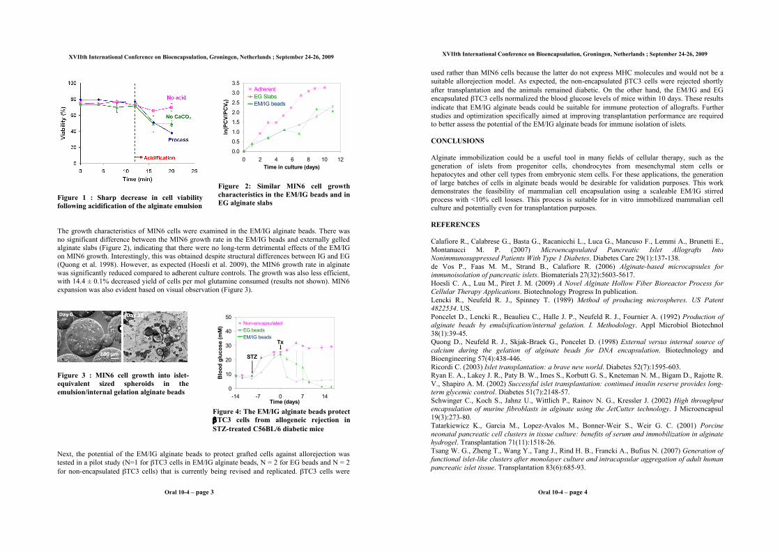
Figure 1 : Sharp decrease in cell viability
following acidification of the alginate emulsion
0.0
0.5
1.0
1.5
2.0
2.5
3.0
3.5
0 2 4 6 8 10 12
Time in culture (days)
ln(P
CV
/PC
V0)
Adherent
EG Slabs
EM/IG beads
Figure 2: Similar MIN6 cell growth
characteristics in the EM/IG beads and in
EG alginate slabs
The growth characteristics of MIN6 cells were examined in the EM/IG alginate beads. There was
no significant difference between the MIN6 growth rate in the EM/IG beads and externally gelled
alginate slabs (Figure 2), indicating that there were no long-term detrimental effects of the EM/IG
on MIN6 growth. Interestingly, this was obtained despite structural differences between IG and EG
(Quong et al. 1998). However, as expected (Hoesli et al. 2009), the MIN6 growth rate in alginate
was significantly reduced compared to adherent culture controls. The growth was also less efficient,
with 14.4 ± 0.1% decreased yield of cells per mol glutamine consumed (results not shown). MIN6
expansion was also evident based on visual observation (Figure 3).
100 !m
Day 0
Figure 3 : MIN6 cell growth into islet-
equivalent sized spheroids in the
emulsion/internal gelation alginate beads
0
10
20
30
40
50
-14 -7 0 7 14Time (days)
Blo
od
glu
co
se (
mM
)
Non-encapsulated
EG beads
EM/IG beads
STZ
Tx
Figure 4: The EM/IG alginate beads protect
!TC3 cells from allogeneic rejection in
STZ-treated C56BL/6 diabetic mice
Next, the potential of the EM/IG alginate beads to protect grafted cells against allorejection was
tested in a pilot study (N=1 for !TC3 cells in EM/IG alginate beads, N = 2 for EG beads and N = 2
for non-encapsulated !TC3 cells) that is currently being revised and replicated. !TC3 cells were
Oral 10-4 – page 3
XVIIth International Conference on Bioencapsulation, Groningen, Netherlands ; September 24-26, 2009
used rather than MIN6 cells because the latter do not express MHC molecules and would not be a
suitable allorejection model. As expected, the non-encapsulated !TC3 cells were rejected shortly
after transplantation and the animals remained diabetic. On the other hand, the EM/IG and EG
encapsulated !TC3 cells normalized the blood glucose levels of mice within 10 days. These results
indicate that EM/IG alginate beads could be suitable for immune protection of allografts. Further
studies and optimization specifically aimed at improving transplantation performance are required
to better assess the potential of the EM/IG alginate beads for immune isolation of islets.
CONCLUSIONS
Alginate immobilization could be a useful tool in many fields of cellular therapy, such as the
generation of islets from progenitor cells, chondrocytes from mesenchymal stem cells or
hepatocytes and other cell types from embryonic stem cells. For these applications, the generation
of large batches of cells in alginate beads would be desirable for validation purposes. This work
demonstrates the feasibility of mammalian cell encapsulation using a scaleable EM/IG stirred
process with <10% cell losses. This process is suitable for in vitro immobilized mammalian cell
culture and potentially even for transplantation purposes.
REFERENCES
Calafiore R., Calabrese G., Basta G., Racanicchi L., Luca G., Mancuso F., Lemmi A., Brunetti E.,
Montanucci M. P. (2007) Microencapsulated Pancreatic Islet Allografts Into
Nonimmunosuppressed Patients With Type 1 Diabetes. Diabetes Care 29(1):137-138.
de Vos P., Faas M. M., Strand B., Calafiore R. (2006) Alginate-based microcapsules for
immunoisolation of pancreatic islets. Biomaterials 27(32):5603-5617.
Hoesli C. A., Luu M., Piret J. M. (2009) A Novel Alginate Hollow Fiber Bioreactor Process for
Cellular Therapy Applications. Biotechnology Progress In publication.
Lencki R., Neufeld R. J., Spinney T. (1989) Method of producing microspheres. US Patent
4822534. US.
Poncelet D., Lencki R., Beaulieu C., Halle J. P., Neufeld R. J., Fournier A. (1992) Production of
alginate beads by emulsification/internal gelation. I. Methodology. Appl Microbiol Biotechnol
38(1):39-45.
Quong D., Neufeld R. J., Skjak-Braek G., Poncelet D. (1998) External versus internal source of
calcium during the gelation of alginate beads for DNA encapsulation. Biotechnology and
Bioengineering 57(4):438-446.
Ricordi C. (2003) Islet transplantation: a brave new world. Diabetes 52(7):1595-603.
Ryan E. A., Lakey J. R., Paty B. W., Imes S., Korbutt G. S., Kneteman N. M., Bigam D., Rajotte R.
V., Shapiro A. M. (2002) Successful islet transplantation: continued insulin reserve provides long-
term glycemic control. Diabetes 51(7):2148-57.
Schwinger C., Koch S., Jahnz U., Wittlich P., Rainov N. G., Kressler J. (2002) High throughput
encapsulation of murine fibroblasts in alginate using the JetCutter technology. J Microencapsul
19(3):273-80.
Tatarkiewicz K., Garcia M., Lopez-Avalos M., Bonner-Weir S., Weir G. C. (2001) Porcine
neonatal pancreatic cell clusters in tissue culture: benefits of serum and immobilization in alginate
hydrogel. Transplantation 71(11):1518-26.
Tsang W. G., Zheng T., Wang Y., Tang J., Rind H. B., Francki A., Bufius N. (2007) Generation of
functional islet-like clusters after monolayer culture and intracapsular aggregation of adult human
pancreatic islet tissue. Transplantation 83(6):685-93.
Oral 10-4 – page 4
XVIIth International Conference on Bioencapsulation, Groningen, Netherlands ; September 24-26, 2009