xth - splc-crs · 2 xth spanish-portuguese conference on controlled drug delivery drug delivery...
TRANSCRIPT

1
2
Xth
Spanish-Portuguese Conference on Controlled Drug Delivery
Drug Delivery Systems from Lab to Clinic. New trends and Opportunities.
Centro de Investigación Príncipe Felipe (CIPF), Valencia, Spain
Sunday 10th November – Tuesday 12th November 2013
Chairman
María J. Vicent (CIPF, Valencia)
Organising Committee: Emilia Barcia (Univ. Complutense de Madrid)
Dolores Torres (Univ. Santiago de Compostela)
Mª Adolfina Ruíz (Univ. Granada)
Maribel Bento (Instituto Superior de Ciências da Saúde Granda, Portugal)
Young Organising Committee:
Aroa Duro (CIPF, Valencia)
Amaya Niño (CIPF, Valencia)
Lucía Martín (Univ. Sevilla)
Ana Cadete (Univ. Santiago de Compostela)
Sonia Reimondez (Univ. Santiago de Compostela)
Spanish-PortugueseLocal Chapter
Spanish-PortugueseLocal Chapter
3
____________________________________
International Advisory Board
M.J. Alonso (Univ. Santiago Compostela, Spain)
A. Almeida (Univ. Lisbon, Portugal)
M. Bermejo (Univ. Miguel Hernández)
M. Blanco (Univ. Navarra, Spain)
P. Caliceti (Univ. Padua, Italy)
P. Calvo (Pharmamar, Spain)
R. Duncan (Cardiff, UK)
J. M. de la Fuente (Inst. Nanociencias Aragón, Spain)
E. Fattal ( Univ. Paris-sud, France)
A. Gabizon (Jerusalem, Israel)
R. Gaspar (Univ. Lisbon, Portugal)
M. Guzman (Univ. Alcalá de Henares, Spain)
A. Kabanov (Univ. Chapel Hill, USA)
L. Mayer (Celator Pharm., Canada)
S. Muro (Univ. Maryland, USA)
J. Nuno Moreira (Univ. Coimbra, Portugal)
L. Pradella (Z-Cube, Italy)
J.L. Pedraz (Univ. País Vasco, Spain)
N.A. Peppas (Univ Texas at Austin, USA)
G. Storm (Univ. Utrecht, NL)
A. Torres (Univ. Complutense Madrid, Spain)
A.M. Rabasco (Univ. Sevilla, Spain)
D. Shabat (Tel Aviv Univ, Israel)
R. Satchi-Fainaro (Tel Aviv Univ, Israel)
J. San Roman (CSIC, Spain)
V. Torchilin (Mass Gen Hospital, USA)

4
SPONSORS
SCOPE
A growing number of drug delivery technologies have been approved by Regulatory Authorities for routine clinical use and others are progressing through clinical trials as single agents or as components of combination therapy regimes.
Novel carriers and synthetic technologies are being developed for different applications ranging from
macro- to nano-sized controlled drug delivery technologies. Ever more sophisticated synthetic chemistry is leading to complex three dimensional polymeric architectures, hybrid constructs and self-assembling micro and nano-sized particles. Many carriers and hybrid systems are being developed as imaging agents and theranostics.
As clinical applications broaden to include treatments for infectious and inflammatory diseases,
tissue repair and regeneration (including cell therapy), and diseases of the ageing population. Importance will be given to combination therapy and the key issues of the manufacture/development process to achieve transfer from lab. to clinics.

5
INDEX
Page No
Programme 6-9
Abstracts of Plenary Lectures 10-14
Abstracts of Invited Lectures 15-31
Abstracts of Oral Introduction to Posters 32-66
Abstracts of Poster Presentation Only 67-108
Authors Index 108-116
Participant Contact Details 117-121
6
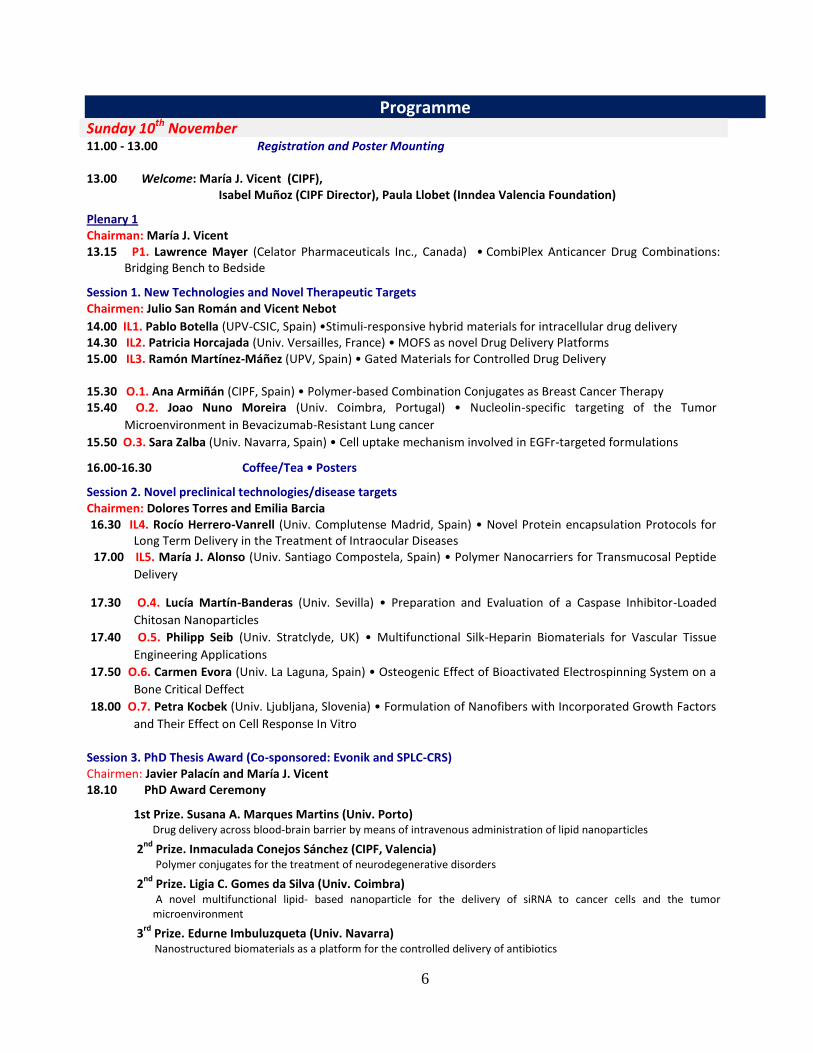
Programme Sunday 10th November 11.00 - 13.00 Registration and Poster Mounting 13.00 Welcome: María J. Vicent (CIPF),
Isabel Muñoz (CIPF Director), Paula Llobet (Inndea Valencia Foundation)
Plenary 1 Chairman: María J. Vicent 13.15 P1. Lawrence Mayer (Celator Pharmaceuticals Inc., Canada) • CombiPlex Anticancer Drug Combinations:
Bridging Bench to Bedside
Session 1. New Technologies and Novel Therapeutic Targets Chairmen: Julio San Román and Vicent Nebot
14.00 IL1. Pablo Botella (UPV-CSIC, Spain) •Stimuli-responsive hybrid materials for intracellular drug delivery 14.30 IL2. Patricia Horcajada (Univ. Versailles, France) • MOFS as novel Drug Delivery Platforms 15.00 IL3. Ramón Martínez-Máñez (UPV, Spain) • Gated Materials for Controlled Drug Delivery 15.30 O.1. Ana Armiñán (CIPF, Spain) • Polymer-based Combination Conjugates as Breast Cancer Therapy 15.40 O.2. Joao Nuno Moreira (Univ. Coimbra, Portugal) • Nucleolin-specific targeting of the Tumor
Microenvironment in Bevacizumab-Resistant Lung cancer
15.50 O.3. Sara Zalba (Univ. Navarra, Spain) • Cell uptake mechanism involved in EGFr-targeted formulations
16.00-16.30 Coffee/Tea • Posters
Session 2. Novel preclinical technologies/disease targets Chairmen: Dolores Torres and Emilia Barcia 16.30 IL4. Rocío Herrero-Vanrell (Univ. Complutense Madrid, Spain) • Novel Protein encapsulation Protocols for
Long Term Delivery in the Treatment of Intraocular Diseases 17.00 IL5. María J. Alonso (Univ. Santiago Compostela, Spain) • Polymer Nanocarriers for Transmucosal Peptide
Delivery
17.30 O.4. Lucía Martín-Banderas (Univ. Sevilla) • Preparation and Evaluation of a Caspase Inhibitor-Loaded
Chitosan Nanoparticles
17.40 O.5. Philipp Seib (Univ. Stratclyde, UK) • Multifunctional Silk-Heparin Biomaterials for Vascular Tissue
Engineering Applications
17.50 O.6. Carmen Evora (Univ. La Laguna, Spain) • Osteogenic Effect of Bioactivated Electrospinning System on a
Bone Critical Deffect
18.00 O.7. Petra Kocbek (Univ. Ljubljana, Slovenia) • Formulation of Nanofibers with Incorporated Growth Factors
and Their Effect on Cell Response In Vitro
Session 3. PhD Thesis Award (Co-sponsored: Evonik and SPLC-CRS) Chairmen: Javier Palacín and María J. Vicent 18.10 PhD Award Ceremony
1st Prize. Susana A. Marques Martins (Univ. Porto) Drug delivery across blood-brain barrier by means of intravenous administration of lipid nanoparticles
2nd
Prize. Inmaculada Conejos Sánchez (CIPF, Valencia) Polymer conjugates for the treatment of neurodegenerative disorders
2nd
Prize. Ligia C. Gomes da Silva (Univ. Coimbra) A novel multifunctional lipid- based nanoparticle for the delivery of siRNA to cancer cells and the tumor microenvironment
3rd
Prize. Edurne Imbuluzqueta (Univ. Navarra) Nanostructured biomaterials as a platform for the controlled delivery of antibiotics
7
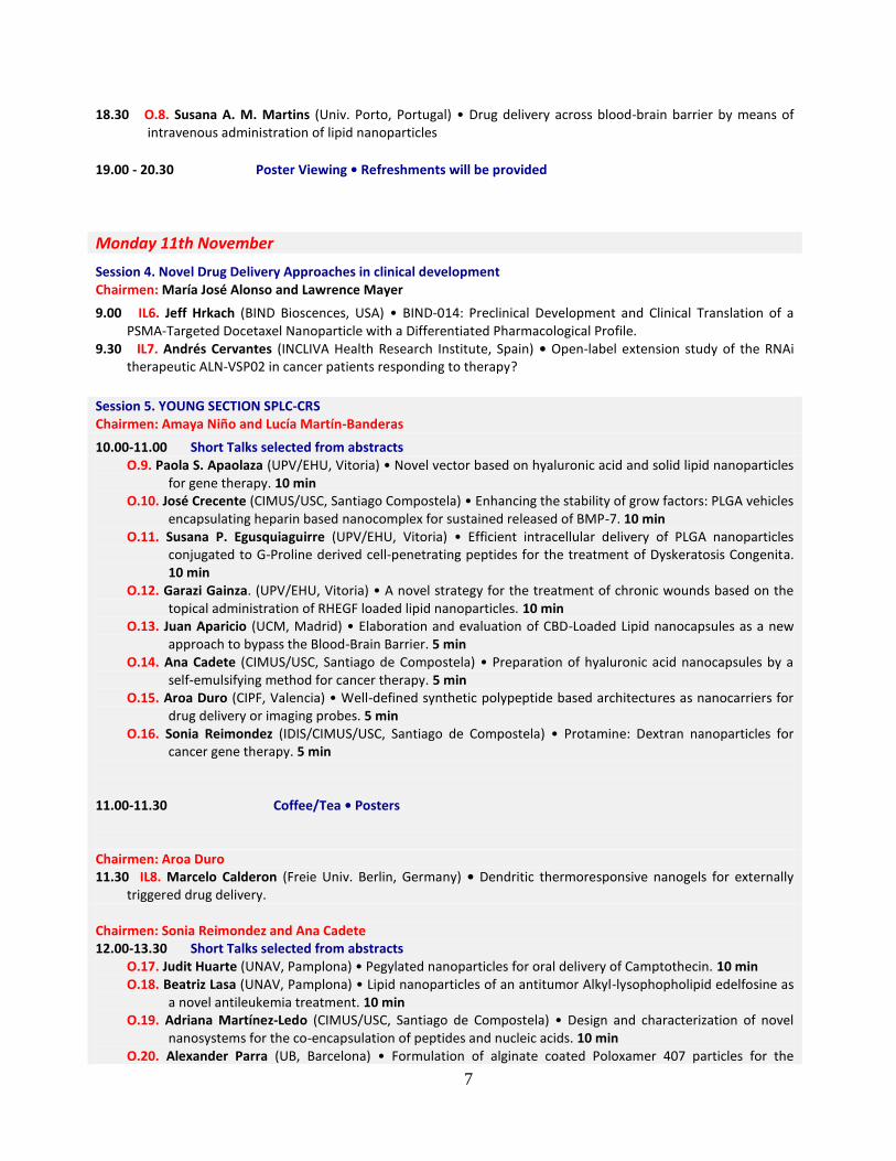
18.30 O.8. Susana A. M. Martins (Univ. Porto, Portugal) • Drug delivery across blood-brain barrier by means of intravenous administration of lipid nanoparticles
19.00 - 20.30 Poster Viewing • Refreshments will be provided
Monday 11th November
Session 4. Novel Drug Delivery Approaches in clinical development Chairmen: María José Alonso and Lawrence Mayer
9.00 IL6. Jeff Hrkach (BIND Bioscences, USA) • BIND-014: Preclinical Development and Clinical Translation of a PSMA-Targeted Docetaxel Nanoparticle with a Differentiated Pharmacological Profile.
9.30 IL7. Andrés Cervantes (INCLIVA Health Research Institute, Spain) • Open-label extension study of the RNAi therapeutic ALN-VSP02 in cancer patients responding to therapy?
Session 5. YOUNG SECTION SPLC-CRS Chairmen: Amaya Niño and Lucía Martín-Banderas
10.00-11.00 Short Talks selected from abstracts O.9. Paola S. Apaolaza (UPV/EHU, Vitoria) • Novel vector based on hyaluronic acid and solid lipid nanoparticles
for gene therapy. 10 min O.10. José Crecente (CIMUS/USC, Santiago Compostela) • Enhancing the stability of grow factors: PLGA vehicles
encapsulating heparin based nanocomplex for sustained released of BMP-7. 10 min O.11. Susana P. Egusquiaguirre (UPV/EHU, Vitoria) • Efficient intracellular delivery of PLGA nanoparticles
conjugated to G-Proline derived cell-penetrating peptides for the treatment of Dyskeratosis Congenita. 10 min
O.12. Garazi Gainza. (UPV/EHU, Vitoria) • A novel strategy for the treatment of chronic wounds based on the topical administration of RHEGF loaded lipid nanoparticles. 10 min
O.13. Juan Aparicio (UCM, Madrid) • Elaboration and evaluation of CBD-Loaded Lipid nanocapsules as a new approach to bypass the Blood-Brain Barrier. 5 min
O.14. Ana Cadete (CIMUS/USC, Santiago de Compostela) • Preparation of hyaluronic acid nanocapsules by a self-emulsifying method for cancer therapy. 5 min
O.15. Aroa Duro (CIPF, Valencia) • Well-defined synthetic polypeptide based architectures as nanocarriers for drug delivery or imaging probes. 5 min
O.16. Sonia Reimondez (IDIS/CIMUS/USC, Santiago de Compostela) • Protamine: Dextran nanoparticles for cancer gene therapy. 5 min
11.00-11.30 Coffee/Tea • Posters Chairmen: Aroa Duro 11.30 IL8. Marcelo Calderon (Freie Univ. Berlin, Germany) • Dendritic thermoresponsive nanogels for externally
triggered drug delivery. Chairmen: Sonia Reimondez and Ana Cadete 12.00-13.30 Short Talks selected from abstracts
O.17. Judit Huarte (UNAV, Pamplona) • Pegylated nanoparticles for oral delivery of Camptothecin. 10 min O.18. Beatriz Lasa (UNAV, Pamplona) • Lipid nanoparticles of an antitumor Alkyl-lysophopholipid edelfosine as
a novel antileukemia treatment. 10 min O.19. Adriana Martínez-Ledo (CIMUS/USC, Santiago de Compostela) • Design and characterization of novel
nanosystems for the co-encapsulation of peptides and nucleic acids. 10 min O.20. Alexander Parra (UB, Barcelona) • Formulation of alginate coated Poloxamer 407 particles for the

8
treatment of vulvovaginal candidiasis. 10 min O.21. Teresa Simon (UNAV, Pamplona) • PLGA microparticles carrying VEGF: Preparation and efficacy studies
in combination with COQ10 PLGA nanoparticles in an animal model of myocardial ischemia. 10 min O.22. María J. Martin (UGR, Granada) • Viability of lactobacillus fermentum cect5716 encapsulated in
gelatine and gastroresistant capsules 10 min
O.23. Ana D. Bonillo (UGR, Granada) • Influence of Compritol on the controlled release of Dexketoprofen Trometamol from matrices containing polyesteramide PADAS. 5 min
O.24. Patricia Marcianes (UCM, Madrid) • Multiparticulate controlled delivery systems of Tolcapone for Parkinson’s disease. 5 min
O.25. Rebeca Peñalva (UNAV, Pamplona) • Encapsulation of resveratrol in polymeric nanoparticles to improve its oral bioavailability. 5 min
O.26. Juan P. Sánchez (UV, Valencia) • Evaluation of mesoporus silicon particles: studies in vivo with insulin. 5 min
O.27. Edorta Santos (UPV/EHU, Vitoria) • Inactivation of encapsulated cells and their therapeutic effects by means of TK-GFP-Luciferase plasmid. 5 min
O.28. Martha L. Vázquez (UB, Barcelona) • Enhance of hyaluronic acid release: development of the transformation of liposomes into planar lipid bilayers. 5 min
13.30-14.45 Lunch • Posters Plenary 2 Chairman: Ruth Duncan 14.45 P2. Collen Masimirembwa (African Institute of Biomedical Science & Technology, Zimbabwe) • The challenge
of diseases of poverty in Africa and what Drug Delivery Technologies can achieve Session 6. Tools to optimise the safety and efficacy of novel DDS Chairmen: Antonio Pineda-Lucena and Marianne Ashford 15.30 IL9. Iola Duarte (CICECO, Univ. Aveiro Portugal) • Metabolomics as a new tool to monitor safety and efficacy
of nanomedicines 16.00 IL10. Vicent Nebot (Polypeptide Therapeutic Solutions SL, Spain) •Well-Defined Synthetic Drug Delivery
Carriers 16.15 O.29. Martina Palomino (CIPF, Spain) • A metabolomics perspective of controlled drug delivery 16.25-16.50 Coffee/Tea • Posters 16.50 O.30. Jennifer Hare (AstraZeneca, UK) • Improving the Pre-clinical to Clinical Translatability of Nanomedicines:
Re-Investigating the EPR Effect Across Solid Tumours 17.20-18.30 SPLC-CRS GENERAL ASSEMBLY 19.30 Turistic Tour Valencia City Center Meeting Point: Torres de Serrano (to finalise at ‘La Embajada’ for dinner) 21.00 Conference Dinner • La Embajada Pl. Alfonso el Magnánimo 7, 1º

9
Tuesday 12th November Plenary 3 Chairman: María Blanco 9.15 P3. Felipe Prosper (Univ. Navarra, Spain) • Cell Therapy and Controlled Release Materials for Tissue
Regeneration Session 7: Novel preclinical technologies/disease targets Chairmen: Julie Movellan and Rogerio Gaspar
10.00 IL11. Damiá Tormo (BiOncotech Pharmaceuticals SL, Spain) • BO-110 an RNA based therapeutic from Lab to Clinic
10.30 IL12. Africa González (Univ. Vigo, Spain) • Drug Delivery Technologies interplay with the immune systems
11.00-11.30 Coffee/Tea • Poster Session 8: Challenges for development of ‘DDS from Lab to Clinic’ Chairmen: Ruth Duncan
11.30 IL13. Rogerio Gaspar (Univ. Lisbon, Portugal) • Biosimilars and Generics. Issues for development and approval.
12.00 IL14. Dolores Hernan (EMA) • How does the EU facilitate the development of nanomedicines? Recent guidance.
12.30 ROUND TABLE DISCUSSION. Coordinator: Ruth Duncan (Cardiff, UK)
13.15 Reflections on SPLC-CRS Local Chapter. The 10
th Conference Anniversary.
Where are we now and where are we going? 13.30 Oral and Poster Communication Prizes Concluding Remarks

10
ABSTRACTS OF PLENARY LECTURES

11
Page
PL_1 CombiPlex Anticancer Drug Combinations: Bridging Bench to Bedside.
Lawrence D. Mayer
12
PL_2 The challenge of diseases of poverty and what drug discovery technologies can
achieve. Collen Masimirembwa
13
PL_3 Cell Therapy and Controlled Release Materials for Tissue Regeneration.
Felipe Prosper
14

12
CombiPlex Anticancer Drug Combinations: Bridging Bench to Bedside
Lawrence D. Mayer, Ph.D.
President and Chief Scientific Officer
Celator Pharmaceuticals, Ewing, NJ and Vancouver, BC
Abstract
Anticancer drug combinations can act synergistically or antagonistically against tumor cells in vitro
depending on the ratios of the individual agents comprising the combination. The ability to take
advantage of this relationship in vivo requires that drug ratios be controlled after administration.
Nano-scale drug delivery vehicles are well suited for this application since they can be designed to
coordinate the release of drug combinations after injection so that synergistic drug ratios can be
delivered to tumors. This fixed-ratio dosing approach, referred to as CombiPlex, leads to dramatic
increases in preclinical efficacy and clinical trials with CPX-351, a co-encapsulated liposome
formulation of cytarabine:daunorubicin at a synergistic 5:1 molar ratio, have revealed marked
efficacy improvements in certain AML populations. In addition, such formulations can be
reproducibly manufactured at commercial scale and exhibit robust pharmaceutical stability. Nano-
scale drug delivery vehicles allow in vitro drug ratio-dependent synergy informatics to be translated
in vivo and these relationships appear to further translate from preclinical models to the clinic.
Ratiometric dosing using CombiPlex technology allows drug combination synergy to be fully
exploited in vivo, thereby increasing the likelihood of favorable therapeutic outcomes in patients.
PL_1

13
THE CHALLENGE OF DISEASES OF POVERTY AND WHAT DRUG DISCOVERY
TECHNOLOGIES CAN ACHIEVE
Collen Masimirembwa
African Institute of Biomedical Science and Technology, Zimbabwe
INTRODUCTION
Poor economic development in African countries has meant that diseases peculiar to this continent do not
receive attention from the pharmaceutical industry with respect to the discovery of new drugs hence their
being referred to as poverty related diseases (PRD) among which are tuberculosis, malaria, trypanosomiasis,
and schistosomiasis. Most of the drugs in use for the treatment or prevention of these diseases were
discovered more than 50 years ago and are either unsafe, have variable efficacy, or are bring lost to drug
resistance. The major cause of these liabilities has been shown to be inadequate pharmacokinetics. Advances
in drug delivery systems are showing promise in addressing the DMPK parameters important for the safe and
effective use of these drugs. In this presentation, cases were such exploratory work has or is being done will
be highlighted.
CASES & OPPORTUNITIES
Malaria: In the treatment of malaria, traditional successful drugs such as chloroquine and fansider have been
lost to drug resistance. We are now left with the artemisinin based combination therapy (ACTs) for which
there are reports of emerging drug resistance and/or poor effectiveness in some parts of Africa. The highly
potent artemisinins (artemisinin, artemether, artesunate or dihydroartemisinin) have the limitation of short
half lives associated with incomplete cure rates. Studies to improve their half lives through use of liposomes
(1) and their absorption using pheroids have been reported (2).
Tuberculosis: Treatment of tuberculosis is with a cocktail of drugs which include key components such as
rifampicin and isoniazid. Three to six months of continuous daily drug intake is required for complete
treatment. This long time and the tablet burden are associated with poor patient compliance. Rifampicin has
additionally been associated with many undesirable drug-drug interactions due to its inductory effects on
drug metabolizing enzymes. Nano-formulations of anti-TB drugs are showing promising results to reduce the
dosing regimens and the length of treatment (3,4).
Schistosomiasis: The only drug effective against all forms of schistosomiasis is praziquantel. The drug has
however been shown to exhibit variable cure rates. This has been attributed to its variable bioavailability
which in turn is due to its poor solubility and high metabolic clearance. Nano-formulation is being explored
to address these limitations (5,6).
Conclusion: Whilst none of the above formulations and delivery systems for drugs against PRDs are in
clinical use, the efforts of some dedicated research groups in Africa and their international collaborators
promise to yield tangible results in the near future.
REFERENCES 1. Isacchi, B., Arrigucci, S., Marca, G.L., Bergonzi, M.C., Vannucchi, M.G., Novelli, A. & Bilia, A.R. 2011.
Conventional and long-circulating liposomes of artemisinin: Preparation, characterization, and pharmacokinetic profile
in mice. Journal of liposome research, 21(3):237-244.
2. Steyn, J.D., Wiesner, L., DU Plessis, L.H., Grobler, A.F., Smith, P.J., Chan, W., Haynes, R.K. & Kotzé, A.F. 2011.
Absorption of the novel artemisinin derivatives artemisone and artemiside: Potential application of pheroid™
technology. International journal of pharmaceutics, 414(1):260-266.
3. Choomara YE, Pillay V, Ndesendo VMK et al., 2011. Polymeric emulsion and crosslink-mediated synthesis of super-
stable nanoparticles as sustained-release anti-tuberculosis drug carriers. Colloids Surf B biointerfaces 87:243-254.
4. Swai H, Semete B, Kalombo L et al., 2008. Potential of treating tuberculosis with a polymeric nano-drug delivery
system. J Control Release 132:e48.
5. Xie S, Pan B, Shi B, Zhang Z, Zhang X, and Wang M, 2011. Solid lipid nanoparticle suspension enhanced the
therapeutic efficacy of praziquantel against tapeworm. International Journal of Nanomedicine 2011:6 2367–2374
PL_2

14
Cell Therapy and Controlled Release Materials for Cardiac Regeneration
FR Formiga1, JJ Gavira
2, G Abizanda
2, E Garbayo
1, B Pelacho
2, MJ Blanco-Prieto
1 and F Prosper
2
1Department of Pharmacy and Pharmaceutical Technology, School of Pharmacy, University of Navarra, Pamplona, Spain.
2Hematology, Cardiology and Cell Therapy, Clínica Universidad de Navarra and Foundation for Applied Medical
Research, University of Navarra, Pamplona, Spain
INTRODUCTION
Cardiovascular diseases remain the first cause of morbidity and mortality in the developed countries accounting
for almost 30% of all deaths (1). Despite recent evidence indicating that the heart is endowed with a regerative
potential based on the presence of cardiac progenitors/stem cells, this is insufficient overall to prevent the
development of cardiac failure after MI in the majority of patients (2). While heart transplant remains the only
curative option for patients with end-stage heart failure, new approaches such as gene and stem cell therapy (3)
or even the direct administration of pro-angiogenic growth factors have been explored in recent years (4). In the
case of cell therapy, the current view suggests that stem cells contribute to cardiac repair through a paracrine
effect associated with the release of growth factors rather than by directly contributing to tissue regeneration (5).
RESULTS AND DISCUSSION
We have assessed the potential of administration of different growth factors using DDS based of PLGA
microparticles in models of myocardial infarction, including small (rat) as well as large preclinical models (pig).
As a proof of concept we employed (PLGA) microparticles loaded with VEGF165 and compared with free
VEGF or control non-loaded microparticles in a rat model of ischemia-reperfusion. VEGF165 loaded
microparticles could be detected in the myocardium of the infarcted animals for more than a month after
transplant and provided sustained delivery of active protein in vitro and in vivo. One month after treatment, an
increase in angiogenesis, arteriogenesis along with a positive remodeling of the heart was detected in the VEGF-
microparticle group. This was associated with an improvement in cardiac function (6). These results led us to
initiate further studies using growth factors involved not only in angiogenesis but also in cardyogenesis and
cardiomyocyte survival such as Neurogeulin 1 (NRG1) and FGF1 in a rat and a pig model of MI.
In order to characterize the mechanisms by which the cytokine loaded MP exerted its action, tissue
cardiomyocyte proliferation was examined by double immunostaining for cTnT and Ki-67+. A significant
increase in the number of adult cardiomiocytes Ki-67+ was detected in the hearts treated with NRG-1, 3 months
post-treatment. Furthermore, c-Kit+CD45- progenitor cells were also detected in the myocardium, although
tissue quantification did not show significant differences among groups, 3 months post-injection. However, a
significant increase was detected 1 week post-implantation in the NRG-1-MP group in comparison with the NL-
MP control group. Finally, growth factor loaded MP used in rats proved to contribute to an improvement in
cardiac function. The strategy was successfully scaled-up to be tested in an ischemia-reperfusion porcine model.
NRG1-MP, FGF1-MP and non-loaded MP were next injected locally into the infarcted myocardium using
NOGA® XP Cardiac Navigation System, a highly accurate electro-guided methodology which creates precise
3-D heart images, 1 week after the infarct. NRG-1-MP, FGF-1-MP and non-loaded MP were injected into the
infarcted myocardium using NOGA® XP Cardiac Navigation System 1 week after the infarct. The long-term
functional efficacy of growth factor-loaded MP treatment is currently being evaluated. The validation of this
therapeutic approach could make significant progress for patients with ischemic heart disease.
REFERENCES
1. Chockalingam A, et al (2000). J Hypertens 18: 1705-1708.
2. Bergmann O, et al. (2009). Science 324: 98-102.
3. Passier R, et al (2008). Nature 453: 322-329.
4. Maulik N, et al (2008). J Mol Cell Cardiol 44: 219-227.
5.Gnecchi M, et al. (2005). Nat Med 11: 367-368
6.Formiga FR, et al. J Control Release. 2010; 147:30-7.
PL_3

15
ABSTRACTS OF INVITED LECTURES

16
Page
IL_1 Stimuli-responsive hybrid materials for intracellular drug delivery P. Botella
18
IL_2 Metal Organic frameworks as novel drug delivery platforms
Patricia Horcajada
19
IL_3 Gated materials for controlled drug delivery
Ramón Martínez-Máñez
20
IL_4 Novel protein encapsulation protocols for long term delivery in the
treatment of intraocular diseases
Rocío Herrero-Vanrell, Irene Bravo-Osuna, Patricia Checa-Casalengua, Cristina
Garcia-Caballero, Budd Tucker, Michael J. Young, Irene Molina-Martínez.
21
IL_5 Polymer nanocarriers for transmucosal peptide delivery
Maria José Alonso, Noemi Csaba, Jorge Pinto, Ma Luo
22
IL_6 BIND-014: Preclinical Development and Clinical Translation of a PSMA-
Targeted Docetaxel Nanoparticle with a Differentiated Pharmacological
Profile
J. Hrkach and the entire BIND Team
23
IL_7 Open-label extension study of the rnai therapeutic ALN-VSP02 in cancer
patients responding to therapy
Andrés Cervantes
24
IL_8 Dendritic thermoresponsive nanogels for externally triggered drug delivery
Marcelo Calderón
25
IL_9 Metabolomics as a new tool in nanotoxicology and nanomedicine:
Validation in vitro using human keratinocytes exposed to silver
nanoparticles
Iola F. Duarte, Joana Carrola, Ana L. D. Silva, Eliana Malheiro, Verónica
Bastos, Tiago Pedrosa, Helena Oliveira, José M. Oliveira, Conceição Santos,
Ana M. Gil
26
IL_10 Well-defined polymers for therapeutic applications: versatile
polyglutamates via nca polymerisation
V.J. Nebot, A. Duro-Castano, I. Conejos-Sanchez, R.M. England, M.J. Vicent
27

17
IL_11 BO-110, A new concept of anticancer therapy
Damiá Tormo
28
IL_12 Drug Delivery Technologies interplay with the immune system
África González-Fernández
29
IL_13 Biosimilars and Generics. Issues for development and approval.
R. Gaspar
30
IL_14 How does the EU facilitate the development of nanomedicines? recent
guidance.
Dolores Hernán Pérez de la Ossa
31

18
Stimuli-responsive hybrid materials for intracellular drug delivery P. Botella
Instituto de Tecnología Química (UPV-CSIC), Av. Los Naranjos s/n, 46022 Valencia, Spain.
INTRODUCTION In cancer therapy, to achieve complete eradication of tumors, cytotoxic drugs must be administrated
systematically in high dose. However, this may lead to severe side-effects due to non-specific uptake of
antitumor drugs by healthy tissues/organs.1 In this sense, an ideal drug carrier should deliver a significant
quantity of the pharmaceutical payload with no premature release of the drug prior to reaching the targeted
cells.2 Therefore, it is compulsory to develop drug delivery systems (DDS) able to discharge their cargo
under specific stimuli that may trigger drug release under the internalization process takes place within
cancer cells. Here, organic/silica hybrid materials have been considered to be excellent candidates for the
preparation of DDS, as their textural properties favor the loading of important amounts of therapeutic
molecules and their silanol-containing surface can be easily functionalized, introducing additional features
that allow imposing a stimuli-responsive controlled drug release.3,4
Herein, we present some novel
organic/silica nanohybrids for the intracellular delivery of anticancer drugs that can release the therapeutic
load under specific stimuli.
RESULTS AND DISCUSSION We have developed DDS responsive to physical (vis/NIR light), chemical (glutathione) or biological
(esterases) stimuli. In the first case, the therapeutic nanoplatform associates the optical activity of gold
nanoclusters with the cytotoxicity of camptothecin (CPT).5 Gold nanoparticles were assembled into stable
clusters with a tailored absorption cross-section in the vis/NIR spectrum. These clusters were further
encapsulated in a mesoporous silica coating containing CPT. After internalization in 42-MG-BA human
glioma cells, these protected gold nanoclusters produced photothermolysis under fs pulse laser irradiation of
790 nm. Moreover, incorporated CPT was released during the process, provoking significant cell death
increase. In the second example, a (pyridin-2-yldisulfanyl)alkyl carbonate CPT derivative was directly
coupled with thiol groups of silica hybrid nanoparticles containing a non-porous core and a mesoporous
shell. Upon internalization in HeLa cells, the reducing activity of cytosolic gluthathione provoked disulfide
bridge cleavage, releasing the naked drug after an intramolecular cyclization mechanism.7 Finally,
amorphous non-porous silica nanoparticles were surface-modified to covalently link CPT through an ester
bond.8 Direct coupling of 20-O-trifluoroglycinylcamptothecin with carboxylate groups of silica nanoparticles
gave a highly stable in plasma nanodrug. After uptake in HeLa and HT29 cells, CPT release took place due
to the activity of cytosolic esterases.
Overall, we foresee a great future for these multifunctional hybrid materials as novel DDS, as their tailored
physical and chemical properties allow imposing controlled release over delivered compounds under specific
stimuli.
REFERENCES 1. Chen Y., Chen H., Deping Z., Tian Y., Chen F., Feng J., Shi J. (2010) ACS Nano 4, 6001-6013.
2. Slowing I.I., Vivero-Escoto, J.L., Wu C.-W., Lin V.S.-Y. (2008) Adv. Drug Deliv. Rev. 60, 1278-1288.
3. Yang P., Gai S., Lin J. (2012) Chem. Soc. Rev. 41, 3679-3698.
4. Coll C., Bernardos A., Martinez-Mañez R., Sancenon F. (2013) Acc. Chem. Res. (2013) 46, 339-349.
5. Botella P., Ortega I. et al. (2012) Dalton Transactions 41, 9286–9296.
6. Muniesa C, Vicente V, Quesada M., Saez-Atienzar S., Blesa J.R., Abasolo I., Fernandez Y., Botella P. (2013) RSC Adv. 3,
15121–15131.
7. Botella P., Abasolo I., et al. (2011) J. Controlled Release 156, 246-257
IL_1

19
Metal Organic frameworks as novel drug delivery platforms
Patricia Horcajada
Institut Lavoisier, CNRS UMR 8180, Université de Versailles, 45 Avenue des Etats Unis, 78035
Versailles Cedex, France
Porous metal-organic frameworks (MOF) belong to a fascinating class of porous crystalline
materials and currently receive much attention in regard to their potential applications in strategic
fields such as gas storage, separation, sensing or heterogeneous catalysis.[1] Among these,
bioapplications have emerged in 2006 as a very promising field,[2,3] due to their high and regular
porosity and their highly versatile chemical composition. In particular, biocompatible and non toxic
nanoparticles of porous iron(III) carboxylates have shown exceptional loadings of different
challenging drugs (antitumoral, antiretroviral, cosmetics) with controlled releases of the active form
of the therapeutic molecule.[4]
This talk discloses an overview of the most interesting MOF structures developed at the
Institut Lavoisier of Versailles as well as some of their competitive performances and/or limitations
in the biomedical field.
References:
1. See special issues: (a) Chem. Soc. Rev., 2009, 38, 1201; (b) Chem. Rev., 2012, 112, 673; (c)
Acc. Chem. Res., 2005, 38, 215.
2. P. Horcajada et al. Metal-organic frameworks as efficient materials for drug delivery. Angew.
Chem. Int. Ed. 2006, 45, 5974-5978.
3. W.J. Rieter et al. Nanoscale metal-organic frameworks as potential multimodal contrast
enhancing agents. J. Am. Chem. Soc. 2006, 128, 9024-9025
4. P. Horcajada et al., Porous metal organic framework nanoscale carriers as a potential platform for
drug delivery and imagin, Nature Mater., 2010, 9, 172-178.
IL_2
20
GATED MATERIALS FOR CONTROLLED DRUG DELIVERY
Ramón Martínez-Máñez1,2,3
1 Centro de Reconocimiento Molecular y Desarrollo Tecnológico (IDM), Unidad Mixta Universitat
Politècnica de Valencia - Universitat de Valencia, Spain 2
Departamento de Química, Universitat Politècnica de Valencia, Camino de Vera s/n, 46022,
Valencia, Spain 3
CIBER de Bioingeniería, Biomateriales y Nanomedicina (CIBER-BBN)
INTRODUCTION Gated nanochemistry, although highly topical and rapidly developing, is still in its infancy.
Recently it has been demonstrated the possible incorporation of “gates” into mesoporous supports.
In this field, molecular or supramolecular gates can be defined as nanoscopic-based devices in
which mass transport can be triggered by target external stimuli that can control the state of the
gate; i.e., closed or open. In fact in the last few years, nano-containers bearing gated scaffoldings
have proved to be excellent candidates for the design of controlled-release “nano-machines” at
different levels. In this area, the use of gated ensembles built up using silica mesoporous materials
containing on-off triggered gated systems have proved their suitability. These systems show an
ideal “zero release” until opened via suitable stimuli. Mesoporous supports show stable structures
(pores of ca. 2-3 nm), large surface areas (up to 1200 m2/g), tunable pore sizes and volumes, and
well-defined surface properties for hosting molecules and for site-specific delivery. The
mesoporous support can additionally be obtained in a nanometric size, resulting in suitable
materials for the design of “nanodevices” for the controlled delivery of drugs and other species.
Moreover, a second novel application involves the use of gated material in sensing protocols.
RESULTS AND DISCUSSION
Selected examples of triggered silica mesoporous gated materials able to deliver their cargo by
changes in temperature,[1] irradiation with light,[2] and by the presence of small molecules or
biomolecules [3,4,5] will be shown.
REFERENCES 1. E. Aznar, L. Mondragón, J. V. Ros-Lis, F. Sancenón, M. D. Marcos, R. Martínez-Máñez, J. Soto,
E. Pérez-Payá, P. Amorós (2011) Finely tuned temperature-controlled cargo release using paraffin-
capped mesoporous silica nanoparticles. Angew. Chem. Int. Ed., 50, 11172.
2. E. Aznar, M. D. Marcos, R. Martínez-Máñez, F. Sancenón, J. Soto, P. Amorós, C. Guillem
(2009) pH- and photo-switched release of guest molecules from mesoporous silica supports. J. Am.
Chem. Soc., 131, 6833.
3. A. Agostini, L. Mondragón, A. Bernardos, R. Martínez-Máñez, M.D. Marcos, F. Sancenón, J.
Soto, A. Costero, C. Manguan-García, R. Perona, M. Moreno-Torres, R. Aparicio-Sanchis, J.R.
Murguía (1012) Targeted cargo delivery in senescent cells using capped mesoporous silica
nanoparticles. Angew. Chem. Int. Ed., 51, 10556
4. E. Climent, L. Mondragón, R. Martínez-Máñez, F. Sancenón, M.D. Marcos, J.R. Murguía, P.
Amorós, K. Rurack. E. Pérez-Payá, (2013) Selective, highly sensitive and rapid detection of
genomic DNA using gated materials: the example of Mycoplasma detection. Angew. Chem. Int.
Ed., in press
IL_3

21
NOVEL PROTEIN ENCAPSULATION PROTOCOLS FOR LONG TERM DELIVERY IN
THE TREATMENT OF INTRAOCULAR DISEASES
Rocío Herrero-Vanrell1,2
, Irene Bravo-Osuna1,2
, Patricia Checa-Casalengua1, Cristina Garcia-
Caballero1,2
, Budd Tucker4, Michael J. Young
3, Irene Molina-Martínez
1,2.
1Department of Pharmacy and Pharmaceutical Technology. School of Pharmacy. Complutense
University. 28040 Madrid. Spain. 2IdISSC, Hospital Clínico San Carlos, Profesor Martín Lago s/n,
28040 Madrid. 3Schepens Eye Research Institute, Dep. of Ophthalmology, Harvard Medical School,
Boston, MA, EEUU. 4Institute for Vision Research, Dep. of Ophthalmology, Carver College of
Medicine, University of Iowa, Iowa, IA, EEUU.
INTRODUCTION
Chronic and multifactorial pathologies affecting the back of the eye represent visual
impairment and blindness. Biotechnological products are often employed for the treatment of these
posterior segment disorders. Treatments are performed by intravitreal injections due to the difficulty
in delivering effective doses of these active molecules to intraocular target tissues. This therapeutic
approach allow to reaching effective tissue drug levels. However, successive intraocular injections
are associated to side effects (retinal detachment, hemorrhage, endophthalmitis, and cataract) and
the risk increases with the frequency of administrations. Intraocular drug delivery systems (IODDS)
are under evaluation to avoid successive injections. Depending on their size, the devices can be
implanted through a relatively large surgical incision or through a smaller tissue perforation.
Among the IODDS, microparticles (1-1000µm size) are emerging therapeutic tools for the
treatment of posterior segment diseases as they are able to release the active substance during weeks
or months. Biodegradable microspheres can be injected through small gauge needles avoiding
surgical procedures and disappear from the site of injection after delivering the drug. Furthermore,
they can be used in personalized medicine as different amounts of microspheres can be injected for
individualized therapy. Since several years ago, the encapsulation of proteins has been one of the
most interesting challenges in the field of pharmaceutical technology. The encapsulation technique
should guarantee the maintenance of the biological activity of the product throughout
manufacturing, storage and use.
RESULTS AND DISCUSSION
Preservation of protein biological activity can be achieved by the use of a novel protein
encapsulation procedure based on a solid-in-oil-water (S/O/W) emulsion technique. To this, the
bioactive substance is encapsulated without any preliminary manipulation. With this technique, the
use of additives promotes additional protection of the biological product before and after
sterilization. Protein is released from microspheres in its active form for long periods of time. This
technological strategy has been applied to the encapsulation of a neurotrophic factor (Glial-cell
derived neurotrophic factor) in PLGA microspheres. Efficacy studies in an animal model of
glaucoma have confirmed the activity of the GDNF released from the microspheres after 12 weeks
of their intravitreal injection.
ACKNOWLEDGEMENTS. FP7 program from EU (NMP4-SL-2010-24618), UCM
Research Group 920415, RETICS RD 12/0034 and MAT 2010-18242.
IL_4
22
POLYMER NANOCARRIERS FOR TRANSMUCOSAL PEPTIDE DELIVERY
Maria José Alonso, Noemi Csaba, Jorge Pinto, Ma Luo
Research Center on Molecular Medicine (CIMUS), School of Pharmacy, University of Santiago de
Compostela, Spain
Email: [email protected]
Our group, being committed with the translation of ideas from the university through novel
pharmaceutical technology, has designed novel polymer nanocarriers intended to transport peptides
and proteins across biological barriers and to deliver them to the target tissue. In particular, we
pioneered the development of nanoparticles and nanocapsules made of chitosan in combination with
other biomaterials, such as glucomanan, hyaluronic acid, cyclodextrins, phospholipids and oils.
During my presentation I would like to focus on the analysis of the potential of a variety of
nanocarriers for the oral and nasal administration of therapeutic peptides, i.e. insulin and salmon
calcitonin. Within this frame, I will also describe the activities being implemented within TRANS-
INT European Consortium and those planned for the coming years (for more information visit
www.trans-int.eu). Finally, I will present the potential of different types of nanoparticles for
intranasal immunization using deferent protein and peptide antigens. As a critical example, I will
describe our recent advances towards the development of a peptide-based nasal HIV vaccine, in
collaboration with the University of Manitoba. Overall, the results have shown that packaging 12
peptide antigens within nanoparticles is a powerful strategy for achieving significant immune
responses following nasal administration to macaques.
More information about these applications and the literature associated to them can be found at:
http://webspersoais.usc.es/mariaj.alonso
IL_5
23
BIND-014: Preclinical Development and Clinical Translation of a PSMA-Targeted Docetaxel
Nanoparticle with a Differentiated Pharmacological Profile
J. Hrkach and the entire BIND Team
BIND Therapeutics. 325 Vassar Street, Cambridge, MA, 02139 USA.
INTRODUCTION
The challenge for all drugs is to maximize the net clinical benefit by increasing the desired
therapeutic effect and reducing adverse effects. This is especially difficult in cancer, where the goal
is to destroy or inhibit growth of cancer cells without damaging similar healthy cells. Accurins™
are polymeric nanoparticles that incorporate a therapeutic payload and are designed to have
prolonged circulation within the bloodstream, enable targeting of the diseased tissue or cells, and
provide for the controlled and timely release of the therapeutic payload. Accurins are designed with
specified physical and chemical characteristics to target specific cells or tissues and concentrate a
therapeutic payload at the site of disease to enhance efficacy while minimizing adverse effects on
healthy tissues.
RESULTS AND DISCUSSION
BIND-014 is a prostate-specific membrane antigen, or PSMA, targeted Accurin that contains
docetaxel. PSMA is a clinically-validated tumor marker expressed on prostate cancer cells and the
blood vessels of many types of non-prostate solid tumors, including non-small cell lung cancer.
We compared the pharmacokinetics of BIND-014 and Taxotere in cynomolgus monkeys, with
equal doses of BIND-014 or Taxotere administered and blood samples collected at various times
over a 70-hour period to measure the total docetaxel concentration. The docetaxel concentration
was approximately 10 to 100 times higher with BIND-014 than Taxotere for the entire duration of
the experiment. We also studied the levels of accumulation of docetaxel in a mouse model of
human prostate cancer when treated with BIND-014 or Taxotere. After 12 hours, the docetaxel
concentration was more than seven times higher in the animals treated with BIND-014. We also
compared the efficacy of BIND-014, a version of BIND-014 without targeting ligand, which we
refer to as PTNP, and docetaxel in a mouse model using the LNCaP human prostate cancer cell line.
BIND-014 treatment resulted in significantly increased shrinkage of the tumors when compared to
docetaxel and was also significantly more effective than PTNP.
To date, we have clinically tested BIND-014 in over 45 patients with advanced or metastatic cancer
who failed prior therapies. In our Phase 1 clinical trial, of the 28 patients who received BIND-014
once every three weeks, to date there have been one complete response in a patient with cervical
cancer and three partial responses in patients with NSCLC, mCRPC and ampullary cancer. Five
additional patients had stable disease lasting longer than 12 weeks. BIND-014 is in Phase 2 clinical
trials for non-small cell lung cancer and metastatic castrate-resistant prostate cancer.
REFERENCES
1. Hrkach J., et al. (2012) Sci. Transl. Med. 4(128):128ra39
2. Summa J., et al. (2012) Abstract 9660, AACR Annual Meeting
IL_6

24
OPEN-LABEL EXTENSION STUDY OF THE RNAi THERAPEUTIC ALN-VSP02 IN
CANCER PATIENTS RESPONDING TO THERAPY
Andrés Cervantes
INCLIVA Health Research Institute, Spain
IL_7

25
DENDRITIC THERMORESPONSIVE NANOGELS FOR EXTERNALLY TRIGGERED
DRUG DELIVERY
Marcelo Calderón
Institut für Chemie und Biochemie, Freie Universität Berlin, Takustr. 3, 14195 Berlin, Germany.
INTRODUCTION
The effort to design and develop materials on the nanometer scale has been accelerating at a fast
pace in recent decades. In the field of biomedicine, stimuli-responsive and biocompatible materials
have emerged as the new generation of smart molecules/materials. In general, the operating
principle behind responsive architectures lies in the fact that different environmental triggers can
lead to structural/chemical changes within the scaffold of such materials. This unique feature
enables their use in diversified biomedical applications.[1]
In an attempt to create very well-defined, monodisperse, stable nanostructures at the molecular
level, highly branched dendritic polymers have been used for last couple of decades. Special interest
has been devoted to the fabrication of nanogels which are high molecular weight cross-linked
polymers that combine the characteristics of dendritic polymers with that of cross-linked
macroscopic gels, to yield soluble particles within the useful size range between 20 and 200 nm.
Stimuli-sensitive nanogels can shrink or swell rapidly by expelling or absorbing water in response
to external stimuli such as temperature, pH, electrical, and magnetic fields.[2] The combination of
nanogel properties and thermo-responsiveness generates a promising candidate for the development
of smart nanocarrier systems. Their potential properties can be influenced by temperature changes
with high responsiveness, reveal high loading capacity, can improve drug stability, and thus can be
used for stimuli-controlled drug release.
RESULTS AND DISCUSSION
Our hypothesis treats the preparation of thermoresponsive glycerol-based nanogels and the
investigation of their phase behavior with respect to their potential biomedical applications. Initial
focus was given to fabricate nanogels with size control over the range of 50 and 200 nm and narrow
size distributions.[3] Preliminary results about their low cytotoxicity, their capability to penetrate
cell membranes, and their potential to delivery and release bioactives upon external triggers like
temperature and light will be presented.
REFERENCES
1. M. Calderón, M. Quadir, M. Strumia, R. Haag, Functional dendritic polymer architectures as
stimuli-responsive nanocarriers, Biochimie, 92 (2010), 1242-1251.
2. M. Asadian, A. Souza, D. Steinhilber, J. Cuggino, M. Calderón, Functional nanogels for
biomedical applications, Current Medicinal Chemistry 19 (2012), 5029-5043.
3. J. Cuggino, C.I. Alvarez I., M. Strumia, P. Welker, K. Licha, D. Steinhilber, R.-C- Mutihac, M.
Calderón, Thermosensitive nanogels based in dendritic polyglycerol and N-isopropylacrylamide for
biomedical applications, Soft Matter 7 (2011), 11259-11266.
IL_8

26
Metabolomics as a new tool in nanotoxicology and nanomedicine: Validation in vitro using
human keratinocytes exposed to silver nanoparticles Iola F. Duarte
1, Joana Carrola
1, Ana L. D. Silva
1, Eliana Malheiro
1, Verónica Bastos
2, Tiago
Pedrosa2, Helena Oliveira
2, José M. Oliveira
2, Conceição Santos
2, Ana M. Gil
1
1 CICECO, Department of Chemistry, University of Aveiro, 3810-193 Aveiro, Portugal.
2 CESAM
& Laboratory of Biotechnology and Cytomics, Department of Biology, University of Aveiro, 3810-
193 Aveiro, Portugal.
INTRODUCTION
There is growing concern regarding the potential toxicity of nanomaterials in respect of
accidental human exposure and also during their development as nanomedicines. Silver
nanoparticles (Ag-NPs) are among the nanomaterials with highest propensity for human exposure,
arising from their established use in wound dressings and increasing incorporation into consumer
products (e.g. clothing, food packaging), mainly due to their remarkable antimicrobial properties.
However, there is a narrow window between bactericidal activity of Ag-NPs and their toxicity to
human cells1, making the further understanding of their biological effects a relevant up-to-date
subject. Development of metabolic profiling (metabolomics) strategies for assessing the cellular
effects of these nanoparticles may provide a unique and important tool that can be broadly applied
in the areas of nanotoxicology and nanomedicine2.
RESULTS AND DISCUSSION
Human epidermis keratinocytes (HaCaT cell line) grown in DMEM medium, have been
exposed for 24 and 48h to well-characterised Ag-NPs of different average diameters (10, 30 and 60
nm) at (sub)toxic doses, and their endo- and exo-metabolomes characterised by 1H Nuclear
Magnetic Resonance (NMR) spectroscopy, in tandem with multivariate analysis. Cell proliferation
and cytotoxicity evaluations were conducted by microscopic evaluation and MTT cell viability
assay. A number of metabolites involved in different biochemical processes (e.g. antioxidative
response, aminoacid and lipid metabolisms) were found to be altered upon Ag-NPs exposure, in a
dose- and time-dependent manner. Ag-NPs size and aggregation pattern were also seen to influence
the metabolic responses. The results show the potential of NMR metabolic profiling for highlighting
new endpoint markers of Ag-NPs effects, thus demonstrating the value of metabolomics as a novel
tool in the area of in vitro nanotoxicology.
REFERENCES
1. Kim S. and Ryu D.Y. (2013) Silver nanoparticle-induced oxidative stress, genotoxicity and
apopatosis in cultured cells and animal tissues. J. Appl. Toxicol. 33, 78.
2. Duarte I.F. (2011) Following dynamic biological processes through NMR-based metabonomics:
a new tool in nanomedicine? J. Control. Release, 153(1), 34.
IL_9
27
WELL-DEFINED POLYMERS FOR THERAPEUTIC APPLICATIONS: VERSATILE
POLYGLUTAMATES VIA NCA POLYMERISATION
V.J. Nebot,1,2
A. Duro-Castano,1 I. Conejos-Sanchez,
1 R.M. England,
2
M.J. Vicent1,2
1 Centro de Investigación Príncipe Felipe. Polymer Therapeutics Laboratory. Av. Eduardo Primo
Yúfera 3, 46012 Valencia, Spain. 2 Polypeptide Therapeutic Solutions SL. (PTS). Av. Eduardo
Primo Yúfera 3, 46012 Valencia, Spain.
INTRODUCTION
Polymer therapeutics have demonstrated excellent potential as drug delivery vehicles for localised
delivery of cytotoxic drugs, with several carriers now in the marketplace or clinical trials as
treatments for cancer and other medical conditions.1 Biopersistent carriers (PEG, HPMA) present
disadvantages if chronic parenteral administration and/or high doses are required as there is the
potential to generate 'lysosomal storage disease' syndrome. Therefore, there is a need for the
development of new synthetic strategies to access biodegradable carriers with well-defined
architectures. On this context, the use of biodegradable polypeptides is gaining interest within the
field due to their molecular diversity and multifunctionality.2
RESULTS AND DISCUSION
Here we present a wide and versatile family of polyglutamates with well-defined architectures
obtained via ring opening polymerisation of N-Carboxyanhydrides. To this end, we have developed
new synthetic strategies allowing the access to several polyglutamate architectures including linear,
branched and star homopolymers but also block copolymers with narrow polydispersities and batch
to batch reproducibility. The use of amphiphilic block copolymers opens the possibility to design
polymeric micelles for different applications. An exhaustive study on solution conformation of the
final carriers is of key importance to value their possible therapeutic output. Interestingly, the post-
polymerisation modification of the polymers has proven to be an effective and straightforward
strategy towards the preparation of multifunctional polyglutamates with potential applications as
carriers in drug delivery approaches.3,4
Finally, it is worth mentioning that PTS has recently developed a hybrid drug delivery system
combining a PGA-derived hydrogel including polymeric micelles that opens the possibility for local
treatment, with particular interest on tissue regeneration.
REFERENCES
1. R. Duncan and M. J. Vicent, Advanced Drug Delivery Reviews, 2013, 65, 60-70.
2. M. Barz, R. Luxenhofer, R. Zentel and M. J. Vicent, Polymer Chemistry, 2011, 2, 1900-1918.
3. I. Conejos-Sanchez, A. Duro-Castano, A. Birke, M. Barz and M. J. Vicent, Polymer Chemistry,
2013, 4, 3182-3186.
4. M. Barz, A. Duro-Castano and M. J. Vicent, Polymer Chemistry, 2013, 4, 2989-2994.
IL_10

28
BO-110, A NEW CONCEPT OF ANTICANCER THERAPY
Damiá Tormo
BiOncotech Therapeutics, Parc Científic Universitat de Valencia c/ Catedrático Agustín Escardino,
9. 46980 Paterna, Valencia
BO-110 is an antitumoral drug product for parenteral administration with high efficacy and a broad
spectrum of action that eludes mechanisms of cell survival in malignant cells. BO-110 is formed by
combination of a RNA and a polycationic polymer and exhibits a multiple mechanism of action in
tumor cells with a solid proof of concept. Bioncotech Therapeutics supposes a platform for
chemistry, manufacturing and control of BO-110 drug product. In addition, Bioncotech is now
involved in nonclinical and clinical evaluation of the drug.
REFERENCES
1. Self-killing of melanoma cells by cytosolic delivery of dsRNA: wiring innate immunity for a
coordinated mobilization of endosomes, autophagosomes and the apoptotic machinery in tumor
cells. Alonso-Curbelo D, Soengas MS. Autophagy. 2010 Jan;6(1):148-50.
2. Targeted activation of innate immunity for therapeutic induction of autophagy and apoptosis in
melanoma cells.Tormo D, Checińska A, Alonso-Curbelo D, Pérez-Guijarro E, Cañón E, Riveiro-
Falkenbach E, Calvo TG, Larribere L, Megías D, Mulero F, Piris MA, Dash R, Barral PM,
Rodríguez-Peralto JL, Ortiz-Romero P, Tüting T, Fisher PB, Soengas MS. Cancer Cell. 2009 Aug
4;16(2):103-14
IL_11

29
DRUG DELIVERY TECHNOLOGIES INTERPLAY WITH THE IMMUNE SYSTEM
África González-Fernández
Immunology, Institute of Biomedical Research (IBIV), Biomedical Research Center (CINBIO),
University of Vigo, Vigo, Spain.
INTRODUCTION
The potential toxicity of nanomaterials (NMs) has raised many concerns in the nanotechnology
research field and in the regulatory/advisory committees. Nanostructures can interact with
biological systems, such as the immune system, which includes a large variety of cells and soluble
elements, being its main role to detect and to eliminate foreign elements. The understanding of the
interaction between NMs with biological fluids (such as serum) and the immune system is essential
(1). The interaction of serum proteins with the NMs can have negative consequences, such as the
induction of conformational changes, leading to functional loss or important modifications in some
proteins. NMs can induce immune responses such as complement activation, phagocytosis,
induction of oxidative stress, and the activation or inhibition of the immune cells. We have tested
(in vitro and in vivo) the effect induced by non-biodegradable and biodegradable NMs on the
immune system and biological fluids.
RESULTS AND DISCUSSION
The interaction of four different metal oxide Nps (ZnO, TiO2, CeO2 and Al2O3) with human
albumin, fibrinogen and globulins showed that for ZnO Nps, a strong interaction was observed,
which induced a decrease in the thermal stability of both fibrinogen and albumin at a low
temperature, interfering with the clotting activity of fibrinogen. TiO2 and CeO2 Nps showed lower
effects, while for Al2O3 Nps only a slight interaction was observed (2). Thus, some metal oxide
nanoparticles induce conformational changes in the secondary structure of human serum proteins.
None of the four Np tested induced complement activation, oxidative stress or in vivo
immunotoxicity, although ZnO Nps showed high cytotoxicity in vitro (3).
We also tested several prototypes of biodegradable nanostructures containing chitosan (in the
presence or not of a toll like receptor 7 ligand) for improving immunization against hepatitis B
infection (using recombinant hepatitis B surface antigen). Comparison with the conventional alum-
rHBsAg vaccine was performed. The results indicate that a polymer/oil based nanovaccine can be
used as a single-dose immunization approach eliciting long lasting and protective immune
responses (4), and that the co-delivery of viral proteins and a TLR7 agonist from polysaccharide
nanocapsules, can be useful in a needle free vaccination strategy (5).
REFERENCES
1. Zolnik, BS., et al. Endocrinology 151(2): 458-465 (2010)
2. Simón, R., et al. Colloids and surfaces B: Biointerfaces (in press, 2013)
3. Lozano, T., et al. Journal of Physics: Conference series 304: 1-8 (2011)
4. Vicente,S., et al. Plos One 8 (4): 1-8 (2013)
5. Vicente, S., et al. J Controlled Release (in press 2013)
IL_12

30
Biosimilars and Generics. Issues for development and approval.
R. Gaspar1
1 IntraCellular Trafficking Modulation for Advanced Drug Delivery research group
(InTraCell_ADD) at the Medicines Research Institute (iMed.UL), Faculty of Pharmacy, University
of Lisboa (Portugal).
INTRODUCTION
The increased complexity of factors involved in drug discovery, design, development and usage
(3DU) makes way for new approaches that can integrate Science & Technology serving the needs
of patients1. Among those approaches the need to establish more efficient strategies to transform
Science in better Healthcare faces challenges from Industrial organization (and business model),
from Science gaps (in certain areas evident lack of adequate models for translation to first in man or
clinical trials) and from non-harmonized views in certain areas between regulators and scientists.
Systems approaches will become more common through the introduction of complex analytical and
predictive tools, by opening new doors for systems toxicology (allowing room for the introduction
of modern toxicology methods), systems pharmacology (a whole new paradigm currently addressed
by a number of NIH initiatives), systems therapeutics (integrating also pharmacoepidemiology and
pharmacogenetics, as well as efficacy and effective evaluation tools, paving the way for health
technologies assessment with a better scientific base), systems technologies (Quality by Design or
QbD approaches, through the use of PAT), and complex systems, through the development and use
of new hybrid and increasingly complex structures that will allow to combine different therapeutic
targets with a combination of diagnostics, therapeutics and monitoring. These five systems
approaches are the Science base for modern Regulatory Science2.
Among different innovative approaches for development of better medicinal products based in
existing technologies a number of issues have been arising regarding the changes in biosimilar
medicinal products introduced in Europe in 2004 and the current discussions on follow-on non-
biological complex drugs.
An overview of major problems arising in this particular area will be established according to
current scientific and regulatory discussions3.
REFERENCES
1.Gaspar R (2010) – Therapeutic products: regulating drugs and medical devices. Chapter 14 in
“International Handbook on Regulating Nanotechnologies”, editors Graeme Hodge, Diana Bowman and
Andrew Maynard, Edward Elgar Publishing, pp 291- 320, ISBN 978-1-84844-673-1.
2. Gaspar R et al. (2012) European Journal of Pharmaceutical Sciences 47 (2012): 979-987, doi
10.1016/j.ejps.2012.09.020
3. Schellekens H et al, AAPS J (In Press) doi: 10.1208/s12248-013-9533-z
IL_13

31
HOW DOES THE EU FACILITATE THE DEVELOPMENT OF NANOMEDICINES?
RECENT GUIDANCE.
Dolores Hernán Pérez de la Ossa1 1European Medicines Agency. 7 Westferry Circus, Canary Wharf E14 4HB London, UK
INTRODUCTION
Nanomedicine is an emerging interdisciplinary scientific research field with a wide applicability in
the context of the development of medicinal products. It can offer many advantages over
conventional medicines, including target-specific delivery, improved solubility and bioavailability,
and reduced adverse effects.
Over the last three decades several first-generation nanomedicines have successfully entered routine
clinical use. As this science evolves, several “follow-on” nanomedicines (“nanosimilars”) and
second generation nanomedicines are being developed. To ensure a timely introduction of high
quality, safe and efficacious medicinal products it is important to identify and address gaps in
scientific knowledge and to prepare for their evaluation.
DISCUSSION
Over the last decade the European Medicines Agency (EMA) has established different initiatives to
support innovation of nanomedicines and protect public health.
The EMA, in collaboration with its network of experts, has published several guidance documents
to provide appropriate regulatory guidance and assist researchers and companies interested in the
development of nanomedicines for clinical applications. The presentation will focus on the key
aspects of the recently published reflection papers for liposomes; block copolymer micelles and iron
nanoparticles.
The accumulation of experience is allowing, on an on-going basis, to assess the need for further
guidance specific to nanomedicines or for the update of existing ones.
Applicants developing nanomedicinal products are encouraged to establish a dialogue with the
EMA from the early stages of development through the EMA Innovation Task Force and/or the
Scientific Advice/Protocol Assistance procedure.
By establishing expert groups, international collaborations and convening stakeholders in public
workshops, the EMA continues to adapt and prepare the regulatory system for the development,
evaluation and successful market entry of nanomedicines for the benefit of patients.
REFERENCES
1. Reflection paper on nanotechnology-based medicinal products for human use.
2. Reflection paper on data requirements for intravenous liposomal products develioed with
reference to an innovator liposomal product (CHMP/806058/2009/Rev.02)
3. Draft joint Ministry of Health, Labour and Welfare / European Medicines Agency reflection
paper on the development of block-copolymer-micelle medicinal products (CHMP/13099/2013).
4. Reflection paper on Non-clinical studies for generic nanoparticle iron medicinal product
applications SWP/100094/2011
5. Ehmann, F et al. (2013). Next-generation nanomedicines and nanosimilars: EU regulators'
initiatives relating to the development and evaluation of nanomedicines. Nanomedicine (Lond);
8(5):849-56.
IL_14

32
ABSTRACTS OF ORAL INTRODUCTION TO POSTERS

33
Page
O_1 Polymer-based combination conjugates as breast cancer therapy
C. Deladriere, A. Arminan, E. Masiá, R. Lucas and M.J. Vicent
37
O_2 Nucleolin-specific targeting of the tumor microenvironment
in bevacizumab-resistant lung cancer
Ângela Valério-Fernandes, Nuno Fonseca, Vera Moura, Ana Ladeirinha, Teresa
Ferreira, Ana Alarcão, Lina Carvalho, Sérgio Simões, João Nuno Moreira
38
O_3 Cell uptake mechanism involved in EGFr-targeted formulations
Sara Zalba, Ana Margarita Contreras, María Jesús Garrido
39
O_4 Preparation and Evaluation of a Caspase Inhibitor-loaded Chitosan
Nanoparticles
L. Martín-Banderas, M. Durán-Lobato, A. M. Espinosa-Oliva, A. García-
Quintanilla, J. L. Venero, M. Fernández-Arévalo.
40
O_5 Multifunctional silk-heparin biomaterials for vascular tissue engineering
applications
F. Philipp Seib, Manuela Herklotz, Kelly A. Burke, Manfred F. Maitz, Carsten
Werner, David L. Kaplan
41
O_6 Osteogenic effect of bioactived electrospinning system on a bone critical
defect
Carmen Évora, María Rguez-Évora, Emiliano García, Carlos del Rosario,
Ricardo Reyes, Araceli Delgado
42
O_7 Formulation of nanofibers with incorporated growth factors and their
effect on cell response in vitro
Petra Kocbek, Valentina Bertoncelj, Jan Pelipenko, Julijana Kristl
43
O_8 Drug delivery across blood-brain barrier by means of intravenous
administration of lipid nanoparticles
Susana Martins
44
O_9 Novel vector based on hyaluronic acid and solid lipid nanoparticles for
gene therapy
PS Apaolaza, D Delgado, A del Pozo-Rodríguez, A Rodríguez-Gascón, MA
Solinís
45

34
O_10 Enhancing the stability of growth factors: PLGA vehicles encapsulating
heparin based nanocomplex for sustained release of BMP-7
Crecente-Campo J., Borrajo, E., De la Fuente M., Vidal A., Garcia-Fuentes M
46
O_11 E -
proline derived cell-penetrating peptides for the treatment of dyskeratosis
congenita
S.P. Egusquiaguirre, C. Manguán-García, L. Pintado-Berninches, R. Perona, D.
Carbajo, F.Albericio, M. Royo, M. Igartua, R.M. Hernández, J.L. Pedraz
47
O_12 A novel strategy for the treatment of chronic wounds based on the topical
administration of RHEGF loaded lipid nanoparticles
Garazi Gainza, Marta Pastor, Francisco Borja Gutierrez, Jose Javier Aguirre,
Jose Luis Pedraz, Rosa María Hernández, Manoli Igartua
48
O_13 Elaboration and evaluation of CBD-loaded lipid nanocapsules as a new
approach to bypass the blood-brain barrier
Juan Aparicio Blanco, Elena González Burgos, Luna Prieto García, Pilar
Gómez-Serranillos Cuadrado and Ana Isabel Torres-Suárez
49
O_14 Preparation of hyaluronic acid nanocapsules by a self-emulsifying method
for cancer therapy
Ana Cadete, Marcos Garcia-Fuentes, Maria Jose Alonso, Dolores Torres
50
O_15 Well-defined synthetic polypeptide based architectures as nanocarriers for
drug delivery or imaging probes
A. Duro-Castano, I. Conejos-Sánchez, R. England, M. Oteo, E. Romero, D.
Razola ,M.A.Morcillo, MJ Vicent
51
O_16 Protamine:dextran nanoparticles for cancer gene therapy
Reimóndez-Troitiño S, de la Fuente M, García-Fuentes M, Csaba N
52
O_17 Pegylated nanoparticles for oral delivery of camptothecin
J. Huarte, R. Peñalva, S. Espuelas, Yusi Lai, Bin He, J.M. Irache
53
O_18 Lipid nanoparticles of an antitumor alkyl-lysophospholipid edelfosine as a
novel antileukemia treatment
Beatriz Lasa-Saracíbar, Ander Estella-Hermoso de Mendoza, M. Ángela Aznar,
Melissa Guada, Hugo Lana, Amaia Vilas-Zornoza, Xabier Agirre, Felipe
Prósper, María J. Blanco-Prieto
54
35
O_19 Design and characterization of novel nanosystems for the co-encapsulation
of peptides and nucleic acids
A. Martínez Ledo, M. S. Sasso, I. Marigo, V. Bronte, A. McGlone, M. Garcia-
Fuentes, M. J. Alonso Fernández
55
O_20 Formulation of alginate coated Poloxamer 407 particles for the treatment
of vulvovaginal candidiasis
Alexander Parra, María José Martín-Villena, Ana Cristina Calpena-
Campmany,Patricia Gálvez-Martín, Francisco Fernández-Campos, Beatriz
Clares-Naveros
56
O_21 PLGA microparticles carrying VEGF: preparation and efficacy studies in
combination with CoQ10 PLGA nanoparticles in an animal model of
myocardial ischemia
Teresa Simón-Yarza, Esther Tamayo, Carolina Benavides, Hugo Lana, Fabio R.
Formiga, Beatriz Pelacho, Charitra N. Grama, Carlos Ortiz-de-Solorzano,
Felipe Prosper, M.N.V.Ravi Kumar, Maria J. Blanco-Prieto
57
O_22 Viability of Lactobacillus Fermentum CECT 5716 encapsulated in gelatin
and gatro-resistant capsules
María Adolfina Ruiz, María José Martín-Villena, Federico Lara-Villoslada,
María Encarnación Morales
58
O_23 Influence of Compritol on the controlled release of Dexketoprofen
Trometamol from matrices containing polyesteramide PADAS.
Inés C. Rodríguez, M. Victoria Margarit, Ana D. Bonillo, Alfonso Rodríguez
59
O_24 Multiparticulate controlled delivery systems of Tolcapone for Parkinson´s
Disease
P. Marcianes-Moreno, S. Negro, M. Vera-Sarría, E. Barcia, A. Fernández-
Carballido
60
O_25 Encapsulation of resveratrol in polymeric nanoparticles to improve
its oral bioavailability
R. Peñalva, I.Esparza, E. Rincón, M. Agüeros, J.M. Irache
61
O_26 Evaluation of mesoporus silicon particles: studies in vivo with insulin
JP Sánchez-Rivera, Teresa Micó-Albiñana, Ester Pastor, Teresa M Garrigues,
Eugenia Matveeva
62

36
O_27 Inactivation of encapsulated cells and their therapeutic effects by
means of TK-GFP-luciferase plasmid
E. Santos, L. Larzabal, A. Calvo, R.M. Hernandez, G. Orive, J.L. Pedraz
63
O_28 Enhance of hyaluronic acid release: development of the
transformation of liposomes into planar lipid bilayers
Vázquez-González M.L, Calpena AC, Domenech O, Bernad R, Hernandez-
Borell J
64
O_29 A metabolomics perspective of controlled drug delivery
M. Palomino-Schätzlein, A. Armiñán, M. J. Vicent,A. Pineda-Lucena
65
O_30 Improving the pre-clinical to clinical translatability of nanomedicines: re-
investigating the epr effect across solid tumours
Jennifer I. Hare, Gert Storm, Sanyogitta Puri, Simon T. Barry
66

37
Polymer-based combination conjugates as breast cancer therapy
C. Deladriere1, A. Armiñán
1, E. Masiá
2, R. Lucas
1 and M.J. Vicent
1,2
1 Polymer therapeutics Laboratory and
2 Screening Platform.
Centro de Investigación Príncipe Felipe. Av.
Autopista del Saler,16, 46012 Valencia, Spain.
INTRODUCTION
Due to the molecular complexity of cancer, the use of polymer-drug conjugates in combination
therapy represents an important opportunity to enhance tumour response rates.1 The polymeric
carrier provides an ideal platform for the simultaneous delivery of drug cocktails.2 In the treatment
of hormone-dependent cancer, it has been demonstrated that the combination of endocrine therapy
with a chemotherapeutic agent could bring significant advantages.3,4
This novel approach includes
drug synergism and patience compliance. We have previously reported the first endocrine-
chemotherapy combination HPMA copolymer-AGM-Dox conjugate.3,4
The conjugate containing
both drugs showed markedly enhanced cytotoxicity compared with HPMA copolymer-Dox which
has already showed clinical activity in breast cancer patients4. Currently our efforts are directed
towards the understanding of the molecular mechanisms of action for the combination vs. single
Dox conjugates and the achievement of the in vivo proof for synergism in breast cancer mice
models.
RESULTS AND DISCUSSION
To evaluate the HPMA combination conjugates, an orthopic breast cancer mice model was
optimised.5 In 6 weeks balb/c mice female, 5 million of 4T1 murine breast cancer cells were
injected in the third breast. Tumours were analysed in order to control the aromatase enzyme levels
together with the tumour vascularisation. EPR effect can significantly vary between tumour type
and stage, therefore is highly important to properly characterise the in vivo models used in order to
evaluate the therapeutic value of a conjugate based on passive targeting. The vascular permeability
was studied using BSA-Evans blue.5 We were able to determine 0.1 cm
3 as the maximum
accumulation of Evans Blue in tumours. After 8 days (0.1 cm3 tumour size) HPMA-AGM-Dox
combination conjugate, HPMA-Dox conjugate, HPMA-Dox+ HPMA-AGM and free Dox were
injected i.v. at 5mg/mL Dox-equiv. 3 times every 3 days. Tumor growth and animal weigh were
measured daily until the end point. HPMA-Dox and HPMA-AGM-Dox diminished tumor growth
without any animal weigh loss in comparison to the Dox and control groups. Moreover, after the
second dose, the antitumour activity of HPMA copolymer-AGM-Dox conjugate compared to
HPMA copolymer-Dox conjugate was significantly greater. As expected, the combination of both
single conjugates (HPMA copolymer-Dox + HPMA copolymer-AGM) showed a different effect in
comparison with HPMA copolymer-AGM-Dox confirming the importance of conjugating both
drugs in the same polymer backbone.
The molecular mechanisms responsible for the observed antitumor synergism were also studied
using tumour tissues. The differences observed in cell death mechanisms (autophagy vs. apoptosis)
together with a different VEGF modulation seem to be the key factors responsible for a greater
antitumour effect with the combination conjugate.
REFERENCES
1. Herzog T., Barret R.J., Edwards R. and Oldham F.B. (2005) J. Clin. Oncol. 23 (16), 458S-458S.
2. Greco F. and Vicent MJ. (2009) Adv. Drug Deliv. Rev. 61(13), 1203-1213
3. Vicent M.J., Greco F., Nicholson R.I., Paul A., Griffiths PC, Duncan R. (2005) Angew. Chem. Int. Ed.44, 2-6
4. Greco F, Vicent MJ, Gee S, Jones AT, Gee J, Nicholson RI, Duncan R. (2007) J. Control Rel. 13, 459-470
5. Maeda.H. (2010), Bioconjugate Chem. 21, 797-802.
O_1

38
NUCLEOLIN-SPECIFIC TARGETING OF THE TUMOR MICROENVIRONMENT IN BEVACIZUMAB-RESISTANT LUNG CANCER
Ângela Valério-Fernandes1,2
, Nuno Fonseca1,3
, Vera Moura1,4
, Ana Ladeirinha5, Teresa Ferreira
5,
Ana Alarcão5, Lina Carvalho
5, Sérgio Simões
1,3, João Nuno Moreira
1,3
1CNC - Center for Neurosciences and Cell Biology, University of Coimbra, 3004–517 Coimbra, Portugal;
2IIIUC – Institute for Interdisciplinary Research, Polo II, University of Coimbra, 3030-789 Coimbra;
3FFUC
- Faculty of Pharmacy, Pólo das Ciências da Saúde, University of Coimbra, 3000-354 Coimbra, Portugal; 4TREAT U, SA (spin-off from UC/CNC);
5Institute of Anatomical Pathology, Faculty of Medicine, Polo I,
University of Coimbra, 3004-504 Coimbra, Portugal
INTRODUCTION In recent decades, the treatment options for lung cancer have expanded beyond traditional chemotherapy to
include targeted therapies that act specifically against key components involved in tumorigenesis.
Considering the limitations associated with chemotherapies and the possibility of interrupting a tumor
vascular network, there has been great interest in targeting the tumor vasculature and much effort has been
directed towards the development of agents that disrupt angiogenesis. In addition to the target accessibility,
the endothelial cells of the tumor-associated vasculature have greater genetic stability than cancer cells. Anti-
angiogenic drugs, such as bevacizumab, have become a standard treatment option in lung cancer. In spite
some clinical successes with these inhibitors, disappointing results have been reported, with a substantial
number of lung cancer patients who have become resistant to angiogenic inhibitors1-3
. Recently, a novel
nanotechnology-based strategy, using lipid-based nanoparticles containing doxorubicin (DXR) and targeted
to nucleolin (overexpressed on the surface of both cancer and endothelial cells from tumor blood vessels) has
achieved a significant antitumor effect in a murine model of human breast cancer4. Such promising results
led us to investigate the impact of this therapeutic approach against human lung cancer models resistant to
bevacizumab.
RESULTS AND DISCUSSION
In vitro studies demonstrated an improved association and intracellular delivery of encapsulated DXR in
liposomes targeted to nucleolin, leading to a significant impact on cell death. Along with this,
immunohistochemical analysis revealed that nucleolin was highly expressed in different cells in the tumor
microenvironment of patient-derived lung tumors, in a tumor-specific manner. The generated results render
an important indication of the therapeutic potential of the nucleolin-targeted strategy against lung cancer.
ACKNOWLEDGEMENTS This was work was supported by grants QREN/FEDER/COMPETE (Ref. 23240) and PEst-
C/SAU/LA0001/2011. Ângela Valério-Fernandes is a graduate student from the PhD programme on
Biomedicine and Experimental Biology from the Center for Neuroscience and Cell Biology, University of
Coimbra (FCT fellowship reference: SFRH/BD/51191/2010).
REFERENCES
1. Bergers, G. & Hanahan, D. Modes of resistance to anti-angiogenic therapy. Nature reviews. Cancer 8, 592-603
(2008).
2. Cascone, T., et al. Upregulated stromal EGFR and vascular remodeling in mouse xenograft models of angiogenesis
inhibitor-resistant human lung adenocarcinoma. The Journal of clinical investigation 121, 1313-1328 (2011).
3. Custodio, A., Mendez, M. & Provencio, M. Targeted therapies for advanced non-small-cell lung cancer: current
status and future implications. Cancer treatment reviews 38, 36-53 (2012).
4. Moura, V., et al. Targeted and intracellular triggered delivery of therapeutics to cancer cells and the tumor
microenvironment: impact on the treatment of breast cancer. Breast cancer research and treatment 133, 61-73 (2012).
O_2

39
Cell uptake mechanism involved in EGFr-targeted formulations Sara Zalba
1, Ana Margarita Contreras
1, María Jesús Garrido
1
1. Department of Pharmacy and Pharmaceutical Technology. School of Pharmacy. University of
Navarra. Irunlarrea,1 31008, Pamplona, Navarra (Spain)
Introduction Targeted therapy is the current strategy used in the treatment of several types of tumors that over-
express certain receptors, such as the endothelial growth factor receptor (EGFr). This strategy
consists in the use of ligands able to block those specific receptors involved in the growth and
progression of cancer cells. One example is the application of monoclonal antibodies (mAbs), like
Cetuximbab. This mAbs has higher affinity by EGFr than the endogenous factors, providing the
inhibition of the receptor-activity [1].
Nanomedicine applied in the cancer treatment, is focused in the development of targeted-
nanosystems to increase their selectivity by the tumor [2]. Therefore, the coupling of mAbs to the
surface of liposomes shows certain advantages at the therapeutic levels compared to stealth
liposomes or non-targeted liposomes [3]. However, the presence of Fc’ fragments of the mAb
induce a rapid withdrawal from circulation and produce several undesirable immunoreactions.
Thus, the current targeted therapies are based on the coupling of small molecules, like the Fab’
fragment of an antibody or others even smaller than Fab’[4]. On the other hand, the endogenous
ligand of EGFr, EGF, has been also conjugated to liposomes to obtain a more efficient gene
delivery in tumor cells that over-express this receptor [5].
Therefore, the aim of this work was to develop nanoliposomes coupled to EGF or Fab’ for EGFr
targeting and explore the mechanism involved in the cell uptake using four human colorectal cancer
cell lines with different expression level of the receptor.
Results and discussion The Fab’ fragment, obtained from Cetuximab by enzymatic digestion with pepsin, was purified for
the coupling to the surface of liposomes previously formulated with a fluorescent probe. EGF was
also covalent linked to liposomes by a previous modification of the protein. These ligands did not
influence parameters like particle size and polydispersity index, as was expected. In-vitro studies
showed that the time profile of the liposomes internalization for both targeted formulations, Fab’
and EGF, was significantly higher in comparison with non targeted liposomes. In order to explore
the role of the receptor in this process, a pretreatment with Cetuximab induced a reduction in the
uptake of the targeted formulation. Therefore, although the EGFr targeted strategy seems to be a
promising approach to increase the accumulation of these formulation in tumor cells, the
mechanism observed deserves to be investigated, especially in in-vivo studies.
References 1. Eng C. (2010) The evolving role of monoclonal antibodies in colorectal cancer: early presumptions and impact on
clinical trial development. Oncologist. 15(1):73-84.
2. Surendiran A et al. (2009). Novel applications of nanotechnology in medicine. Indian J Med Res. 130(6):689-701.
3. Koshkaryev A et al. (2013). Immunoconjugates and long circulating systems: origins, current state of the art and
future directions. Adv Drug Deliv Rev. 65(1):24-35.
4. Sapra P et al. (2004). Improved therapeutic responses in a xenograft model of human B lymphoma (Namalwa) for
liposomal vincristine versus liposomal doxorubicin targeted via anti-CD19 IgG2a or Fab' fragments. Clin Cancer Res.
1;10(3):1100-11.
5. Buñuales M, et al. (2011). Efficient gene delivery by EGF-lipoplexes in vitro and in vivo. Nanomedicine (Lond).
6(1):89-98.
O_3
40
PREPARATION AND EVALUATION OF A CASPASE INHIBITOR-LOADED CHITOSAN
NANOPARTICLES
L. Martín-Banderas1, M. Durán-Lobato
1, A. M. Espinosa-Oliva
2, A. García-Quintanilla
2, J. L.
Venero2, M. Fernández-Arévalo
1
1Department of Pharmacy and Pharmaceutical Technology,
2 Department of Biochemistry and
Molecular Biology Faculty of Pharmacy, University of Seville, c/ Profesor García González, nº 2,
41012 Sevilla, Spain
INTRODUCTION
Activation of microglia and inflammation-mediated neurotoxicity are suggested to play a decisive
role in the pathogenesis of several neurodegenerative disorders. Recently, it has been demonstrated
that caspase-8 and caspase-3/7 are involved in regulating microglia activation. Therefore, the
inhibition of these caspases in microglial cells could be neuroprotective 1. Chitosan is a linear
polysaccharide characterized by a high biodegradability with applications in the biomedical and
pharmaceutical fields. Chitosan nanoparticles play an important role as drug delivery to the brain
specially neuroptotection 2. To further enhance the stability and biocompatibility of chitosan
nanostructures in vivo, several authors have synthesized graft copolymers of chitosan with the
hydrophilic polymer PEG. The resulting PEG-chitosan graft copolymers have shown indeed
improved biocompatibility3. Here, we propose CS-PEG nanoparticles (NPs) as possible carriers for
the caspase-8 inhibitor Z-Ile-Glu(O-ME)-Thr-Asp(O-Me) fluoromethyl ketone (Z-IETD-FMK).
RESULTS AND DISCUSSION
Chitosan (CS) NPs (Protasan®
UP CL113, Novamatrix) were produced by ionic gelation with
sodium tripolyphosphate (TPP, Sigma-Aldrich) 2. Z-IETD-FMK loaded-nanoparticles were
produced by dissolving the inhibitor in DMSO at 571 ng/mg NPs. Particles were collected by
centrifugation (10,000 rpm, 4ºC, 30 min).
Cationic (zeta potential = +30 mV), monodisperse particles around 345 nm in diameter (Pdi ≈ 0.2)
were obtained. Drug content, was determined by measuring the amount of free drug in the
supernatant. For this purpose we have developed an LC-MS method for the quantification of the
peptide. A QTRAP: Hybrid triple quadrupole-linear ion trap mass spectrometry (QqQLIT)
technique was used. The loaded amount was 485.7 ± 86 ng/mg NPs with high encapsulation
efficiency (85%). Loaded NPs were in vitro evaluated and added to BV2 microglial cell cultures
and caspase-8 activity was measured (Caspase-Glo 8 Assay, Promega). Next, to study microglial
activation we analyzed iNOS, IL6 and TNFα levels by qPCR. In this case we used modified NPs
expressing PEG and biotin on their surface.
NPs loaded with Z-IETD-FMK inhibited caspase-8 activity and microglial activation induced by
LPS. This work is the first step on the application of Z-IETD-FMK loaded-CS NPs for their
targeted delivery into the CNS.
REFERENCES 1 M. A. Burguillos, T. Deierborg, E. Kavanagh, et al. Caspase signalling controls microglia activation and
neurotoxicity. Nature, 472, 319–324 2 L. Martin-Banderas, M. A. Holgado, J.L. Venero et al. Nanostructures for drug delivery to the brain. Current
Medicinal Chemistry. 2011,18(34), 5303-5321
3 E. Fernández-Megía, R. Novoa-Carballal, E. Quiñoá, R. Riguera. Conjugation of Bioactive Ligands to PEG-Grafted
Chitosan at the Distal End of PEG. Biomacromolecules 2007, 8, 833-842
O_4

41
MULTIFUNCTIONAL SILK-HEPARIN BIOMATERIALS FOR VASCULAR TISSUE
ENGINEERING APPLICATIONS
F. Philipp Seib1, Manuela Herklotz
2, Kelly A. Burke
3, Manfred F. Maitz
2, Carsten Werner
2, David
L. Kaplan3
(1) Strathclyde Institute of Pharmacy and Biomedical Sciences, University of Strathclyde, 161
Cathedral Street Glasgow G4 0RE, UK. (2) Leibniz Institute of Polymer Research Dresden,
Max Bergmann Centre for Biomaterials Dresden, Hohe Str. 6, Dresden 01069, Germany. (3) Tufts
University, Department of Biomedical Engineering, 4 Colby Street Medford, MA 02155, USA.
INTRODUCTION
Scaffold materials with suitable biological and mechanical properties are necessary for vascular
tissue engineering1. One potential scaffold material of interest is silk
1,2. Many in vitro studies have
proposed the use of silk for vascular applications; for example, as a stent coating for sustained drug
release3, in blood vessel engineering, and as a material for small vascular grafts
4. Silk sutures are
well tolerated in humans5, but the use of silk for vascular engineering applications still requires
extensive biocompatibility testing2. The aim of this study was to develop multifunctional silk-
heparin biomaterials and determine their hemocompatibility in a humanised in vitro test system.
RESULTS AND DISCUSSION
Over the past 30 years, silk has been proposed for numerous biomedical applications that go beyond
its traditional use as a suture material5. Some studies have indicated a need to modify silk to yield a
hemocompatible surface2,5
. This study examined the potential of low molecular weight heparin as a
material for refining silk properties by acting as a carrier for vascular endothelial growth factor
(VEGF) and improving silk hemocompatibility. Heparinized silk showed a controlled VEGF release
over 6 days; the released VEGF was bioactive and supported the growth of human endothelial cells.
Silk samples were then assessed using a novel humanized hemocompatibility system that employs
whole blood and endothelial cells. The overall thrombogenic response for silk was very low and
similar to the clinical reference material polytetrafluoroethylene. Despite an initial inflammatory
response to silk, apparent as complement and leukocyte activation, the endothelium was maintained
in a resting, anticoagulant state. The low thrombogenic response and the ability to control VEGF
release support the further development of silk for vascular applications.
REFERENCES
1. Kasoju N, Bora U. Silk fibroin in tissue engineering. Adv Healthc Mater. 2012;1:393-412.
2. Seib FP, Maitz MF, Hu X, Werner C, Kaplan DL. Impact of processing parameters on the haemocompatibility of
Bombyx mori silk films. Biomaterials. 2012;33:1017-23.
3. Wenk E, Merkle HP, Meinel L. Silk fibroin as a vehicle for drug delivery applications. J Control Release.
2011;150:128-41.
4. Domachuk P, Tsioris K, Omenetto FG, Kaplan DL. Bio-microfluidics: Biomaterials and Biomimetic Designs. Adv
Materials. 2010;22:249-60.
5. Seib FP, Kaplan DL. Silk for drug delivery applications: opportunities and challenges. Isr J Chem. 2013 submitted.
O_5

42
OSTEOGENIC EFFECT OF BIOACTIVED ELECTROSPINNING SYSTEM ON A BONE
CRITICAL DEFECT Carmen Évora, María Rguez-Évora, Emiliano García, Carlos del Rosario, Ricardo Reyes,
Araceli Delgado
Department of Chemical Engineering and Pharmaceutical Technology. Facultad de Farmacia. Instituto de
Tecnología Biomédica. Universidad de La Laguna, Spain
INTRODUCTION Constructs combining porous 3D scaffold, GFs and cells have been proposed to bone healing.
Electrospinning is a relatively simple and cost-effective technique that allows fabricating synthetic as well as
natural micro- and nanoscale fiber scaffolds for bone tissue engineering to mimic the native extracellular
matrix (ECM) [1]. Electrospun structures typically present a high specific surface area for cell attachment,
and porosity for improved cell infiltration and nutrient diffusion [2]. Technical parameters (polymer
concentration and molecular weight, flow rate, voltage, distance from the collector) are easily modified for
different geometries, structural properties, and application targets [3. The first part of the present study was
dedicated to the in vitro characterization of electrospun membranes of PLGA 75:25, its degradation over
time, biocompatibility, and promotion of rat mesenchymal stem cells (rMSC) proliferation and
differentiation. The objective of the in vivo part was to evaluate the osteogenic effect of a sandwich-like
system for implantation in a 8 mm rat calvarial critical-size defect. The system was bioactivated with BMP-2
in microspheres or rMSCs pre-seeded in the electrospun microfiber membranes at varying densities or
combinations of both.
RESULTS AND DISCUSSION Electrospun membranes of 120±10 μm thickness, uniform distribution of fibers (diameter 1.65±0.39μm) and
an interconnected porous 3D structure (70±3.9%) were obtained. The contact angle was 117º and dropped to
100º in 30 min. PLGA 75:25 membranes favored environmental conditions for osteogenic differentiation.
The in vivo analysis showed that the bone formation started on the defect borders and progressed into the
center, the void zone being frequently localized in the middle of the defect. The systems with the higher cell
density induced a more pronounced progress in growth than the one implanted with fewer cells. However,
the 40% of repair achieved after 12 weeks was still less than the 60% obtained in the BMP-2 group.
Combinations of rMSCs with BMP-2 did not further enhance the regeneration response obtained with solely
BMP-2. In addition, as the system disrupted the microspheres spread and interacted with the tissue, the rest
of membranes were progressively degraded and tissue was progressively integrated into the remaining fibers.
In conclusion, this study presents good bone defect repair outcomes within 8–12 weeks using an electrospun
system combined with BMP-2.
REFERENCES 1. Teo W.E. et al.,(2006) Electrospun scaffold tailored for tissue specific extracellular matrix. Biotechnology
Journal1:918-929.
2. Hsiu-Mei et al., (2012) Preparation and characterization of mesoporous bioactive glass polycaprolactone
nanofibrous matrix for bone tissues engineering. J Mater Sci Mater Med 23:2619-30.
3. Liang D. et al., (2007) Functional electrospun nanofibrous scaffolds for biomedical application. Adv Drug
Deliv Rev 59:1392-412.
ACKNOWLEDGMENTS We thank the National Research Council (MAT2011-23819) by financial support and the ITC by
electrospinning technical assistance. Maria Rguez-Evora was supported by “Beca CajaCanarias para
posgraduados.
O_6

43
FORMULATION OF NANOFIBERS WITH INCORPORATED GROWTH FACTORS AND
THEIR EFFECT ON CELL RESPONSE IN VITRO
Petra Kocbek1, Valentina Bertoncelj
1, Jan Pelipenko
1, Julijana Kristl
1
1University of Ljubljana, Faculty of Pharmacy, Aškerčeva 7, 1000 Ljubljana, Slovenia
INTRODUCTION
The ongoing demographic changes bring, beside other health related problems, increased
incidence of patients suffering from chronic wounds. Novel materials and therapeutic approaches
are therefore needed to achieve effective healing. Polymer nanofibers as a new class of
nanostructured materials show high potential as tissue scaffolds, modern wound dressings and new
drug delivery systems1. They mimic the fibrillar elements of natural extracellular matrix; therefore,
their application may induce better and faster tissue regeneration2. On the other hand, it is well
known from clinical practice that application of various growth factors enhances tissue
regeneration3. The combination of nanofibers with simultaneous delivery of growth factors is,
therefore, expected to exert synergistic effects in wound healing. Fabrication of nanofibers can be
performed using electrospinning. It is a modern, versatile, one-step and widely used method, which
enables the formation of nanofibers from numerous biocompatible polymers.
The objective of this research was formulation of chitosan-based nanofibers with
incorporated blood-derived growth factors and evaluation of in vitro cell response upon contact
with nanofibers.
RESULTS AND DISCUSSION
In the current study chitosan was selected as a biocompatible, and biodegradable material for
nanofiber formation, since it has been widely investigated for various biomedical applications, due
to its haemostatic activity, promotion of normal tissue regeneration, bacteriostatic and fungistatic
effects4. To improve its spinnability chitosan solution was supplemented with polyethylene oxide as
a spinnable carrier. The optimization of process and solution parameters enabled preparation of
electrospun bead-less chitosan-based nanofibers with incorporated blood-derived growth factors
with diameters ranging from ~70 nm to ~90 nm as determined by SEM analysis. Keratinocyte and
dermal fibroblasts were seeded on nanofibrillar supports. Evaluation of cell proliferation indirectly
revealed that bioactivity of growth factors was preserved in electrospinning process. However, the
cell mobility was hindered and cell morphology was changed, depending on concentration of
growth factors in the cell culture. To sum up, the results show that chitosan-based nanofibers with
incorporated growth factors support cell growth in vitro, therefore, showing a good potential for in
vivo application in management of chronic wounds.
REFERENCES
1. Rošic R. et al. (2013) Nanofibers and their biomedical use. Acta Pharm., in press.
2. Pelipenko J. et al. (2013). The topography of electrospun nanofibers and its impact on the growth
and mobility of keratinocytes. Eur. J. Pharm. Biopharm. 84, 401.
3. Koria P. (2012) Delivery of growth factors for tissue regeneration and wound healing. BioDrugs.
26, 163.
4. Bhattarai N. et al. (2005) Electrospun chitosan-based nanofibers and their cellular compatibility.
Biomaterials 26, 6176.
O_7
44
DRUG DELIVERY ACROSS BLOOD-BRAIN BARRIER BY MEANS OF
INTRAVENOUS ADMINISTRATION OF LIPID NANOPARTICLES
Susana Martins1
1 Laboratory of Pharmaceutical Technology, Faculty of Pharmacy, University of Porto. Rua de Jorge Viterbo
Ferreira, Porto, Portugal.
INTRODUCTION
The blood-brain-barrier (BBB) is a selective barrier that generally restricts the passage of drugs
setting serious limitations for CNS pharmacotherapy. Looking at high mortality of malignant
gliomas and the lack of efficient treatment, this field is a primary area for new intravenous
formulations with BBB crossing ability. The focus of this work was to use nanotechnology in
cancer therapy, based on the use of solid lipid nanoparticles (SLN) to assess the suitability of these
carriers for anticancer delivery to the brain. Bearing that in mind, novel formulations of SLN loaded
with camptothecin (CPT), a potent anticancer candidate, were designed. To assess the suitability of
the CPT-loaded SLN for brain drug delivery small and wide angle X-ray scattering (SAXS/WAXS)
analyses, cellular internalization studies (flow cell cytometry) and cytotoxicity studies (MTT) in
four human glioma cell lines and one human macrophage cell line were performed. Additionally, in
vivo fluorescence and biodistribution studies of injected rhodamine 123-loaded SLN and CPT-
loaded SLN were performed in rats.
RESULTS AND DISCUSSION
Particles with a mean particle size below 200 nm, a homogenous size distribution, high
encapsulation efficiency (> 90%) and high stability were developed1,2,3
. SAXS/WAXS studies
suggested favorable interactions of SLN and the cells lipid bilayers4. A higher affinity of the SLN to
the porcine brain capillary endothelial cells (BCEC) and gliomas was shown in comparison to
macrophages3,5
. Cytotoxicity studies revealed that CPT-loaded SLN induced glioma cell death with
the lowest maximal inhibitory concentration (IC50). The mechanism of internalization was found to
be mainly through a clathrin-dependent endocytic pathway. In this work it was demonstrated not
only the brain targeting ability of SLN coated with polysorbate 60 and 80, but also the superiority of
the antitumor activity of CPT-loaded SLN compared with CPT in solution/suspension or in physical
mixture with SLN4,6
. These studies indicate that the CPT-loaded SLN are a promising drug brain
delivery system worth to be exploited as a novel formulation for brain tumor therapy.
REFERENCES 1. .Martins, S., et al., (2011) Physicochemical properties of lipid nanoparticles: Effect of lipid and surfactant
composition. Drug Dev Ind Pharm, 37(7), 815.
2. Martins, S., et al., (2012) Multivariate design for the evaluation of lipid and surfactant composition effect for
optimisation of lipid nanoparticles. Eur J Pharm Sci, 45(5) 613.
3. Martins, S., et al., (2012) Brain delivery of camptothecin by means of solid lipid nanoparticles: Formulation design,
in vitro and in vivo studies. Int J Pharm, 439(1–2) 49.
4. Martins, S.M., et al., (2013) Brain targeting effect of camptothecin-loaded solid lipid nanoparticles in rat after
intravenous administration. Eur J Pharm Biopharm, doi: 10.1016/j.ejpb.2013.08.011
5. Martins, S., et al., (2012) Solid lipid nanoparticles as intracellular drug transporters: An investigation of the uptake
mechanism and pathway. Int J Pharm, 430(1-2) 216.
6. Martins, S.M., et al., (2012) Development and validation of a simple reversed-phase HPLC method for the
determination of camptothecin in animal organs following administration in solid lipid nanoparticles. J Chromatogr B,
880(1) 100.
O_8
45
NOVEL VECTOR BASED ON HYALURONIC ACID AND SOLID LIPID NANOPARTICLES FOR
GENE THERAPY
PS Apaolaza1, D Delgado
1, A del Pozo-Rodríguez
1, A Rodríguez-Gascón
1, MA Solinís
1
1 Pharmacokinetic, Nanotechnology and Gene Therapy Group (PharmaNanoGene). Faculty of Pharmacy. Centro de
investigación Lascaray ikergunea. University of the Basque Country UPV/EHU. Vitoria-Gasteiz (Spain)
INTRODUCTION
Non-viral gene therapy based on solid lipid nanoparticles (SLNs) is a promising alternative for the
treatment of several diseases1,2
. As non-viral vectors, their main advantage is the safety profile, but the low
efficiency of transfection of SLNs makes them to be far from an ideal vector. The incorporation of different
components to overpass the main limiting steps for transfection is one of the strategies used for the
improvement of their efficency.
In this work, we have designed and evaluated a new vector composed by SLNs, protamine (P) and
hyaluronic acid (HA) of different molecular weights. P is a potent DNA condenser that enhances the active
transport of DNA to the nucleus and also improves the intranuclear transcription. HA is a biocompatible anionic
biopolymer that provides the particles with desirable properties, rendering them non-immunogenic,
biocompatible, and biodegradable.
The SLNs were prepared by a solvent emulsification-evaporation technique previously described by del
Pozo-Rodríguez et al.3 .Firstly, a P-DNA complex was prepared. Then, an aqueous solution of HA was added to
form the HA-P-DNA complex, that was put in contact with a suspension of previously prepared SLNs.
Once the vectors were characterized, the nanoparticle transfection efficacy of the reporter plasmid pCMS-
EGFP and cell viability in HEK-293 cells were studied. Moreover, in order to insight into the mechanisms by
through the HA influences the transfection, we studied the cellular uptake and the endocytosis mechanism of the
vectors labelling the SLNs with Nile Red, and the intracellular disposition of DNA into the cytoplasm by using
EMA-labeled-DNA vectors.
RESULTS AND DISCUSSION The particle size of the vectors ranged from 240 nm to 340 nm and the surface charge varied from +30 mV
to +40 mV. All formulations were able to protect the DNA from the enzymatic degradation and to release it in
presence of SDS. The vectors were able to efficiently transfect HEK-293 cells (60% EGFP positive cells at 72
h), without compromising the cell viability. The cellular uptake was about 60% at 8h. The vectors were
internalized by both clathrin- and caveoale/lipid raft-mediated endocytosis, regardless of the presence of HA
and protamine on the surface of the nanoparticle. Since caveoale/lipid raft-mediated endocytosis is
quantitatively much more active than clathrin-mediated endocytosis in HEK-293 cells, to efficiently transfect
this cell line, vectors should be designed to once internalized, the transfection does not depend on the lysosomal
activity, being this the case of the HA-P-DNA-HA vectors. We have previously shown that when the vector was
prepared with protamine and SLNs but without HA, it was unable to transfect this cell line due to high
condensation of DNA by the protamine4. The intracellular disposition of EMA-labelled DNA indicated that the
HA is able to modulate the high degree of condensation of the DNA due to the protamine; this is especially
important if the vector is uptaken mainly by caveoale/lipid raft, since optimizing the formulation we can
influence on the decondensation of the DNA inside the cell, and therefore, on the transfection rate.
REFERENCES 1 Rodríguez-Gascón A.; Solinís, M.A.; del Pozo-Rodríguez, A.; Delgado, D.; Pedraz, J.L. Lipid nanoparticles for gene therapy. 2012,
US 20120183589 A1.
2 Rodríguez-Gascón A.; Solinís, M.A.; del Pozo-Rodríguez, A.; Delgado, D.; Fernández, E. Lipid nanoparticles for treating ocular
diseases. 2012, WO 2012085318 A1.
3 del Pozo-Rodríguez, A.; Delgado, D.; Solinís, M.A.; Gascón, A.R.; Pedraz, J.L. Solid lipid nanoparticles: Formulation factors
affecting cell transfection capacity. Int. J. Pharm. 2007, 339, 261-268.
4 Delgado, D.; del Pozo-Rodríguez, A.; Solinís, M.Á.; Rodríguez-Gascón, A. Understanding the mechanism of protamine in solid lipid
nanoparticle-based lipofection: The importance of the entry pathway. Eur. J. Pharm. Biopharm. 2011, 79, 495-502.
O_9
46
ENHANCING THE STABILITY OF GROWTH FACTORS: PLGA VEHICLES
ENCAPSULATING HEPARIN BASED NANOCOMPLEX FOR SUSTAINED RELEASE
OF BMP-7
Crecente-Campo J., Borrajo, E., De la Fuente M., Vidal A., Garcia-Fuentes M.
Center for Molecular Medicine and Chronic Diseases (CIMUS), Campus Vida, Universidad de
Santiago de Compostela, 15782, Spain
INTRODUCTION
Morphogens are naturally occurring compounds responsible for cellular differentiation. Their
function is to convert normal stem cells in differentiated cells of a specific tissue or organ. Bone
morphogenetic proteins (BMPs), a group of morphogens, are essential for the normal formation of
bone and cartilage. Moreover, BMPs has recently been pointed out as key components in some
pathological processes such as cancer1,2
, osteoporosis or osteoarthritis. However, the therapeutic use
of these proteins is limited by their high instability and low half life3. To overcome this problem,
several strategies have been proposed. Some of them make use of heparin, a natural polymer that
binds to BMPs protecting them from the environment4. In this work, we design a new system that
entraps nanocomplexes of BMP-7-heparin-Tetronic® in a PLGA matrix. In this way, a sustained
release of the BMP-7 is achieved in its antigenically-active form. This system could have a potential
impact in tissue engineering.
RESULTS AND DISCUSSION
Two different vehicles for growth factors were synthesised. Firstly, microparticles of PLGA were
prepared following a modified S/O/O emulsification method where the morphogen is introduced as
a complex with heparin-Tetronic®. With this strategy sustained release rates for, at least, one month
are achieved while preserving the antigenically-active form of the protein. The size and morphology
of microparticles were evaluated using scanning electron microscopy. Secondly, nanocomplexes of
growth factor-heparin-Tetronic® were also encapsulated in scaffolds of PLGA. These scaffolds
were synthesised using a solvent-casting salt-leaching method. Both platforms, microparticles and
scaffolds, were used to achieve the differentiation of human mesenchymal stem cells to
chondrocytes. The formation of cartilage cells was confirmed by histological analysis and RT-PCR
of cartilage specific genes.
REFERENCES 1. Chirasani, S. R. et al. Bone morphogenetic protein-7 release from endogenous neural precursor
cells suppresses the tumourigenicity of stem-like glioblastoma cells. Brain : a journal of neurology
133, 1961–72 (2010).
2. Piccirillo, S. G. M. et al. Bone morphogenetic proteins inhibit the tumorigenic potential of human
brain tumour-initiating cells. Nature 444, 761–5 (2006).
3. Bramono, D. S. et al. Bone marrow-derived heparan sulfate potentiates the osteogenic activity of
bone morphogenetic protein-2 (BMP-2). Bone 50, 954–64 (2012).
4. Jeon, O. et al. Enhancement of ectopic bone formation by bone morphogenetic protein-2 released
from a heparin-conjugated poly(L-lactic-co-glycolic acid) scaffold. Biomaterials 28, 2763–71
(2007).
O_10

47
EFFICIENT INTRACELLULAR DELIVERY OF PLGA NANOPARTICLES CONJUGATED
TO -PROLINE DERIVED CELL-PENETRATING PEPTIDES FOR THE TREATMENT OF
DYSKERATOSIS CONGENITA S.P. Egusquiaguirre
1,2, C. Manguán-García3, L. Pintado-Berninches3, R. Perona3, D. Carbajo
2,4,5, F.Albericio
2,4,M. Royo
2,5,
M. Igartua1,2
, R.M. Hernández1,2
, J.L. Pedraz1,2
1NanoBioCel Group, Laboratory of Pharmaceutics, University of the Basque Country, School of Pharmacy, Vitoria, 01006, Spain.
2Biomedical Research Networking Center in Bioengineering, Biomaterials and Nanomedicine (CIBER-BBN), Vitoria, 01006, Spain. 3Instituto de Investigaciones Biomédicas CSIC/UAM, IDIPaz and CIBER de Enfermedades Raras CIBERER, Madrid, Spain.
4Institut de Recerca Biomédica. Barcelona. 08028, Spain.5Unitat de Química Combinatòria, Parc Científic de Barcelona, Barcelona,
08028, Spain.
INTRODUCTION
Dyskeratosis Congenita (DC) is a rare, inherited, multi-systemic disorder exhibiting premature ageing,
bone marrow failure and increased cancer predisposition; initially described by the mucocutaneous
triad (nail dystrophy, oral leukoplakia and abnormal skin pigmentation). There is an association of DC
with defective telomerase, leading to the accumulation of short telomeres. Mutations in seven genes
encoding components for telomere maintenance have been identified, and are responsible to cause the
three different modes of inheritance of the disease1. It has been described that the peptide GSE 24-2,
corresponding to an internal domain of DCK1 gene (encoding a component of telomerase complex) is
able to reactivate the telomerase2. Unfortunately, peptides lose their therapeutic activity due to their
high susceptibility to degradation3. Furthermore, since GSE 24-2 target is localized within the nucleus,
it must be adequately internalized into cells and reach the nucleus. Therefore, in this study we propose
a method to reduce the mentioned inefficiencies, encapsulating GSE 24-2 into biodegradable poly-
lactic-co-glycolic acid nanoparticles (PLGA NPs) conjugated to different -proline derived cell-
penetrating peptides (CPPs) which display the capacity of cellular and nuclear internalization4.
RESULTS AND DISCUSSION
The average size of the NP formulations was around 220 nm and, according to the polydispersity
index, (PDI < 0.15), all formulations displayed a narrow size distribution. potential values were also
measured; showing that all CPPs conjugated NPs resulted in negative surface charge. Besides, the
encapsulation efficiencies and surface adsorbed peptide of the NPs were quite variable, depending on
the CPP attached. According to the cytotoxic assay, MEF cells maintained a moderate viability level,
especially at low concentrations of NPs. The intracellular uptake and the time course of internalization
of PLGA NPs bound to the different CPPs were evaluated in HeLa cells, showing that, depending on
the CPP, the uptake of the NPs into the nucleus and their internalization time varied. Finally, the
bioactivity of GSE 24-2 encapsulated into PLGA NPs was analyzed in vitro by the incubation of the
peptide with F9 A353V cells. As a result, it was observed that, GSE 24-2 incorporated into PLGA NPs
preserves the in vitro biological activity, rescuing DNA damage and therefore reactivating the
telomerase.
ACKNOWLEDGMENTS This Project was partially supported by the Basque Government (Consolidated Groups, IT-407-07) and “Ministerio de
Economía y Competitividad” (INNPACTO, IPT-2012-0674-090000).S.P. Egusquiaguirre thanks the “Basque Government”
for the Fellowship Grant.
REFERENCES: 1. Nelson, N.D. et al. (2012) Dyskeratosis congenita as a disorder of telomere maintenance. Mutation Research. 730, 43
2. Machado-Pinilla, R. et al. (2008) A dyskerin motif reactivates telomerase activity in X-linked dyskeratosis congenita and
in telomerase-deficient human cells. Blood. 111 (5), 2606
3. Yadav, S.C. et al (2011) Development of peptide and protein nanotherapeutics by nanoencapsulation and
nanobioconjugation. Peptides. 32 (1), 1734. Farrera-Sinfreu, J. et al. (2005) Cell-Penetrating cis--Amino-L-Proline-
Derived Peptides. J. AM. CHEM. SOC. 127, 9459
O_11
48
A NOVEL STRATEGY FOR THE TREATMENT OF CHRONIC WOUNDS BASED ON
THE TOPICAL ADMINISTRATION OF rhEGF LOADED LIPID NANOPARTICLES
Garazi Gainza1,2
, Marta Pastor1,2
, Francisco Borja Gutierrez3, Jose Javier Aguirre
3, Jose Luis
Pedraz1,2
, Rosa María Hernández1,2
, Manoli Igartua1,2
1NanoBioCel Group, Laboratory of Pharmaceutics, University of the Basque Country, School of
Pharmacy, Vitoria, 01006, Spain 2Biomedical Research Networking Center in Bioengineering, Biomaterials and Nanomedicine
(CIBER-BBN), Vitoria, 01006, Spain. 3Hospital Universitario de Álava (HUA) Txagorritxu, Vitoria, 01009, Spain
INTRODUCTION
The increasing incidence of suffering chronic wounds represents a major clinical challenge for
health care systems worldwide. Chronic wounds are characterised by the persistence of
inflammation, the disordered extracellular matrix and a lack of re-epithelialisation1. The epidermal
growth factor (EGF) plays an important role in tissue remodelation by stimulating dermal and
epidermal regeneration and granulation tissue formation. However, the short half-life of rhEGF in
vivo requires multiple administrations and this restricts its clinical application2. To overcome these
limitations, in the current work rhEGF loaded Solid lipid Nanoparticles (rhEGF-SLN) and
Nanostrucutres Lipid Carries (rhEGF-NLC) were prepared and their efficacy was tested in vitro in
BALB/c fibrosblast and after their topical administration in a full-thickness excisional wound model
in db/db mice.
RESULTS AND DISCUSSION
rhEGF-SLN and rhEGF-NLC were prepared by a simple emulsification-ultrasonication method.
Interestingly, the in vitro proliferation studies, evidenced that rhEGF-SLN and rhEGF-NLC
bioactivity was even higher than free rhEGF, suggesting that nanoencapsulation of rhEGF may
promote its affinity to EGF receptors, and/or may enhance nanoformulations cell internalization.
Finally, the in vivo studies revealed that 4 topical administrations of 20 µg rhEGF-NLC and rhEGF-
NLC reduced wound area by days 8 and 11 more than the control groups (untreated control and
blank nanoparticles) . Moreover, the re-epithelisation and the resolution of the inflammatory state
reached earlier for rhEGF-SLN and rhEGF-NLC groups in comparison with controls. Besides, it
should be pointed out that both 20 µg rhEGF-SLN and rhEGF-NLC improved the re-epithelisation
even more than 4 doses of 75 µg free rhEGF administrated intralesionally. Overall, we demonstrate
the promising effect of rhEGF-SLN and rhEGF-NLC to promote faster and more effective wound
healing, and suggest its possible application in chronic wounds treatment.
ACKNOWLEDGMENTS G. Gainza thanks the “Universidad del País Vasco- UPV-EHU” for the Fellowship Grant. The authors gratefully
acknowledge the support of the”Gobierno Vasco” (Consolidated Groups, IT-407-07) and “Ministerio de Economía y
Competitividad” (INNPACTO, IPT-2012-0602-300000).
REFERENCES 1. Mustoe, TA. et al. (2006). Chronic wound pathogenesis and current treatment strategies: a unifying hypothesis. Plast
Reconstr Surg. (7 Suppl):35S-41S.
2. Hardwicke, J. et al. (2008). Dextrin-rhEGF conjugates as bioresponsive nanomedicines for wound repair. J Control
Release. 130(3):275-83.
O_12

49
ELABORATION AND EVALUATION OF CBD-LOADED LIPID NANOCAPSULES AS A
NEW APPROACH TO BYPASS THE BLOOD-BRAIN BARRIER
Juan Aparicio Blanco1, Elena González Burgos
2, Luna Prieto García
1, Pilar Gómez-Serranillos
Cuadrado2 and Ana Isabel Torres-Suárez
1
1Departamento de Farmacia y Tecnología Farmacéutica.
2Departamento de Farmacología.
Facultad de Farmacia, Universidad Complutense de Madrid. Plaza Ramón y Cajal s/n 28040
Madrid, Spain.
INTRODUCTION
The poor access of the majority of therapeutic agents through the blood-brain barrier (BBB) often limits
the effective non-invasive treatment of diseases of the central nervous system (CNS). For this reason,
research has been focusing on ways to bypass such a barrier. In this context, the lipid nanocapsules arise as a
promising approach to achieve such an ambitious aim, since their small size (ranged from 18 to 70 nm) and
their lipophilic nature enable them to carry drugs into the CNS, otherwise unable to go through. The above
mentioned nanocarriers, obtained according to an expanded phase inversion-based method, are loaded with
cannabidiol (CBD). Phase inversion temperature was determined by conductimetry and drug stability during
heating cycles was evaluated in order to establish nanoparticle elaboration conditions. Nanoparticles
obtained were then characterized in terms of size and amount of drug entrapped. Afterwards, the
colorimetric MTT metabolic activity assay was used to measure cell viability/cytotoxicity of cannabidiol and
unloaded lipid nanocapsules on the rat brain RBE4 endothelial cells for 30 min, 3 h and 24 h.
RESULTS AND DISCUSSION
CBD inclusion to the mixture of lipids forming nanocapsules resulted in a slight decrease in the phase
inversion temperature (the change in conductivity was detected in the range of 67- 72ºC). After three
temperatures cycles from 40 to 90ºC, loss of drug lower than 20% was observed. Three batches of CBD-
loaded lipid nanocapsules were obtained, with average size of 22nm, 41nm and 75nm. These diameter values
were slightly higher when compared to unloaded nanoparticles, what is in agreement with the expected
behavior of CBD, soluble in the oily core (as a proof of it, such an increase in size was higher for those
formulations containing higher proportions of the drug). High encapsulation efficiency was obtained in all
cases. According to viability in vitro results, cannabidiol did not markedly affect RBE4 cell viability in the
concentration range of 10-40 µM at the different incubation periods assayed. On the other hand, the
treatment of RBE4 cells with unloaded lipid nanocapsules (from 0.1 mg/mL to 10 mg/mL) revealed that cell
viability was reduced in a concentration and time-dependent manner. The optimal concentration range for
unloaded lipid nanocapsules was established.
REFERENCES 1. Béduneau, A., Hindré, F., Clavreul, A., Leroux, J.C., Saulnier, P., Benoît, J.P., 2008. Brain targeting using novel
lipid nanovectors, Journal of controlled release 126, 44-49.
2. H. Yang, H., 2010. Nanoparticle-Mediated Brain-Specific Drug Delivery, Imaging and Diagnosis, Pharmaceutical
Research 27, 1759-1771. 3. Malzert-Fréon, A., Vrignaud, S., Saulnier, P., Lisowski, V., Benoît, J.P, Rault, S., 2006. Formulation of sustained
release nanoparticles loaded with a tripentone, a new anticancer agent. International Journal of Pharmaceutics 320, 157-
164.
4. Wong, H.L., Wu, X.Y., Bendayan R., 2012. Nanotechnological advances for the delivery of CNS therapeutics.
Advanced Drug Delivery Reviews 64, 686-700.
5. Mosmann, T., 1983. Rapid colorimetric assay for cellular growth and survival: application to proliferation and
cytotoxicity assays. Journal of Immunological Methods 65, 55–63.
O_13
50
PREPARATION OF HYALURONIC ACID NANOCAPSULES BY A SELF-
EMULSIFYING METHOD FOR CANCER THERAPY
Ana Cadete, Marcos Garcia-Fuentes, Maria Jose Alonso, Dolores Torres CIMUS - Center for Research in Molecular Medicine and Chronic Diseases
University of Santiago de Compostela
INTRODUCTION
Nanocapsules (NC) are flexible drug delivery systems with potential applications in several
medical conditions such as oncology.[1] Coating of NC with hyaluronic acid (HA) is an attractive
approach to achieve active targeting. In addition to its biocompatibility, non toxicity and
biodegradability, HA is effectively recognized by CD44 receptor, which is over-expressed in many
tumor cell types; consequently, HA-modified nanocarriers can increase the concentration of
anticancer drugs at the target site. [2] NC can be produced using different technologies, but
nowadays, special attention has been given to self-emulsifying methods that generate NC without
requiring organic solvent and heat. Self-emulsification is interesting for the pharmaceutical industry
since it does not require management of organic solvents, and it can be scaled-up with ease.
Furthermore, the production of NC by this method allows the incorporation of thermolabile drugs.
[3, 4] The aim of this study was to formulate HA NC using a self-emulsifying method and to
characterize the physicochemical properties of the resulting nanocarrier.
RESULTS AND DISCUSSION
Hyaluronic-acid nanocapsules (HA NC) were prepared by a self-emulsifying method, without the
use of solvents, at room temperature and gentle stirring. In a first step an O/W nanoemulsion (NE)
was designed and its surfactant concentration and oil/water ratio were optimized. The best NE
formulation was obtained by using a medium chain triglyceride and Tween80® as the oily phase and
an aqueous phase composed of Solutol HS 15®. HA NC were obtained by the addition of a cationic
surfactant to the oil phase and a 50KDa molecular weight HA to the aqueous phase. The optimized
formulation presented a particle size below 150 nm and negative zeta potential that confirmed the
effective HA coating. HA NC were stable at room temperature for 48h and in PBS 0,02M for 2h.
This versatile system offers several sites amenable for drug encapsulation: a core for hydrophobic
anticancer agents and a surface for loading hydrophilic drugs such as antibodies. Drug loading
studies are underway to investigate the full potential of this system.
REFERENCES 1. Wang, A.Z., R. Langer, and O.C. Farokhzad, Nanoparticle delivery of cancer drugs. Annu Rev Med, 2012. 63: p.
185-98.
2. Ossipov, D.A., Nanostructured hyaluronic acid-based materials for active delivery to cancer. Expert Opin Drug
Deliv, 2010. 7(6): p. 681-703.
3. Date, A.A., et al., Self-nanoemulsifying drug delivery systems: formulation insights, applications and advances.
Nanomedicine (Lond), 2010. 5(10): p. 1595-616.
4. Viehof, A., et al., Oral insulin delivery in rats by nanoparticles prepared with non-toxic solvents. Int J Pharm, 2013.
443(1-2): p. 169-74.
O_14

51
WELL-DEFINED SYNTHETIC POLYPEPTIDE BASED ARCHITECTURES AS
NANOCARRIERS FOR DRUG DELIVERY OR IMAGING PROBES
A. Duro-Castano1, I. Conejos-Sánchez
1, R. England
2, M. Oteo
3, E. Romero
3, D. Razola
3, M.A.Morcillo
3,
MJ Vicent1
1Polymer Therapeutics Laboratory, Centro de Investigación Príncipe Felipe, Valencia, Spain.
2Polypeptide Therapeutics Solutions S.L.,Valencia,Spain
3Unidad de Aplicaciones Biomédicas y Farmacocinética, Instalación Radiactiva IR-08
CIEMAT, Madrid
INTRODUCTION
A large variety of natural and synthetic polymers have been described as carriers for drug delivery or
imaging probes. Nevertheless, only a few have been successfully transferred into patients (mostly
poly(ethyleneglycol) (PEG), N-2-Hydroxy-propilmethacrylamide (HPMA) copolymers and poly-L-glutamic
acid (PGA) conjugates)1. Polyglutamates are highly biocompatible, biodegradable and multifunctional
polymers, which have been effectively used as building blocks in polymer drug conjugates and polymeric
micelles for various medical applications ranging from cancer2-4
to ischemic processes.5,6
Moreover, it is
expected its FDA approval after approval of PGA-paclitaxel conjugate, OpaxioTM
for the treatment of
various cancers alone or in combination (OpaxioTM
has been recently designated as orphan drug in
combination with radiotherapy for glioblastoma treatment).1,7,8
RESULTS AND DISCUSSION
We report the development of synthetic pathways to a plethora functional polyglutamates with well
defined structure, adjustable molecular weight and low dispersity (D = Mw/Mn <1.2) applying the ring
opening polymerization (ROP) of N-carboxyanhydrides (NCA) with novel initiators.9
Furthermore, a number of architectures based on PGA, including stars, grafts, and hybrid diblock
copolymers have been designed. In addition, a variety of functionalities such as alkyne, azides, reactive
disulphides, protected amines… can be easily introduced by “post-polymerization modification” reactions
yielding a set of orthogonal reactive attachment sides9 suitable for further bioconjugations. Those different
structures, after an adequate labelling with fluorescence/NIR probes or/and complexing agents for MRI
and/or PET techniques allowed us to study polymer in vivo fate (pK and biodistribution) in animal models.
Summarizing, new polymeric libraries with a range of attractive and sometimes unexpected
properties result of the contribution of the different architectures can be built, characterized and investigated
for different applications in the nanomedicine field. The knowledge of the structure/in vivo fate relationships
will allow us to regulate the polymer properties to each desired application.
REFERENCES
1. Vicent MJ. Ringsdorf H., Duncan R. Adv Drug Deliv Rev 2009, 61:1117-1120.
2. Vicent, MJ. Duncan, R. Trends Biotechnol. 2006, 24, 39-47.
3. Duncan, R. Nature Rev. Cancer 2006, 6, 668-701.
4. Vicent MJ. et al. Adv. Drug Deliv. Rev. 2009, 61(13), 1117-1120.
5. Vicent M.J. Pérez-Payá E. J. Med. Chem, 49, 3763-3765, 2006,
6. Mondragón, L., et al. J. Med. Chem 2008, 5, 521-529
7. Li C. Wallace S. Adv Drug Deliv Rev 2008, 60, 886-898
8. Chipman SD et al. Int J Nanomedicine 2006, 1, 375-383
9. a) Vicent MJ, Barz M, Canal F, Conejos Sanchez I, Duro Castano A, England R. P201131713.
b) Conejos-Sanchez I., Duro-Castano A. et al. Polym. Chem., 2013, 4 (11), 3182 – 3186
C) Barz M., Duro-Castano A. and Vicent MJ. Polym. Chem., 2013, 4 (10), 2989 – 2994
O_15

52
PROTAMINE:DEXTRAN NANOPARTICLES FOR CANCER GENE THERAPY
Reimóndez-Troitiño S, de la Fuente M, García-Fuentes M, Csaba N.
Health Research Institute of Santiago de Compostela (IDIS). Center for Research in Molecular and
Chronic Diseases (CIMUS), Department of Pharmaceutical Technology, University of Santiago de
Compostela. 15706 Santiago de Compostela, Spain
INTRODUCTION
Gene therapy is considered to be a highly potent tool including the regulation of certain
genes implicated in cancer processes. Developing an innovative and successful cancer treatment
based on gene therapy requires effective therapeutic genes and efficient, safe and cheap gene
delivery systems. Nanoparticles based on the combination of biodegradable polypeptides and
polysaccharides may be an interesting approach for the design of new nanomedicines [1].
Nanoparticles can efficiently associate different nucleic acids (pDNA and siRNA) and successfully
deliver them to cancer cells. The aim of this work was to develop and characterize
protamine:dextran-based nanoparticles, as delivery systems in gene therapy, using plasmid DNA
and interference RNA as model molecules.
RESULTS AND DISCUSSION
Protamine:dextran (Pr:Dx) nanoparticles were prepared in very mild conditions by ionic
gelation technique [2], which avoids the use of organic solvents and high energy sources. Therefore,
this technique is highly recommended for the encapsulation of labile molecules such as nucleic
acids. In a first step, different Pr:Dx ratios (8:1, 4:1, 2:1, 1:2, 1:4, 1:8; w/w) were studied in terms of
(i) physicochemical properties (ii) yield of reaction and (iii) nucleic acid association efficiency in
order to establish the optimal ratio between the components. The selected formulation was Pr:Dx
4:1 due to its high yield of reaction (75%±12), and good association of pDNA and siRNA
confirmed by agarose gel electrophoresis and UV spectroscopy (≥90%). The mean particle size of
the system was below 200 nm and it presented positive zeta potential. TEM images showed
homogenous populations of spherical nanostructures. The nanoparticles presented adequate stability
in cell culture medium and simulated physiological conditions.
In vitro studies were carried out in U87MG cancer cell line. Cellular uptake studies were
performed by confocal microscopy and showed that these nanoparticles have the capacity to interact
and be efficiently internalized by cells. Cell proliferation was evaluated by the colorimetric MTT
assay, which confirmed low/non-toxicity of the nanoparticles. The hemolytic capacity of the system
was also studied and no hemolytic effect was detected after the incubation of nanoparticles with red
blood cells. Additionally, transfected cells with pDNA encoding GFP-F were directly examined
under a fluorescence microscope.
Results evidenced the promise of these nanocarriers in gene therapy. In conclusion, the
developed nanosystem shows potential for cancer gene therapy and therefore, subsequent studies
will be carried out in the near future to explore their activity in tumour-bearing animal models.
REFERENCES 1. P. Midoux, C. Pichon, J. Yaouanc, P. Jaffrès. British Journal of Pharmacology (2009), 157, 166–178
2. Calvo, P., et al., Novel hydrophilic chitosan-polyethylene oxide nanoparticles as protein carriers. Journal of Applied
Polymer Science, 1997. 63(1): p. 125-132.
O_16

53
PEGYLATED NANOPARTICLES FOR ORAL DELIVERY OF
CAMPTOTHECIN J. Huarte
1, R. Peñalva, S. Espuelas
1, Yusi Lai
2, Bin He
2, J.M. Irache
1
1Dep. of Pharmacy and Pharmaceutical Technology, University of Navarra, Pamplona, 31008- Spain.
[email protected] 2National Engineering Research Center for Biomaterials, Sichuan University, Chengdu 610064, China.
INTRODUCTION
Camptothecin (CPT) is a Topoisomerase I inhibitor that was first isolated from the bark of
Camptotheca acuminata in 1966. Although it shows powerful anticancer activity, it has great
limitations such as poor solubility and stability [1]. When administered orally, camptothecin and its
analogues display a high variability and a low bioavailability due to their physicochemical
characteristics and their presystemic metabolism (P-glycoprotein and Cytochrome P450) [2].
Gantrez AN is a copolymer of methyl vinyl ether and maleic anhydride (PVM/MA) that shows
low toxicity and excellent biocompatibility. Polyethyleneglicols (PEGs) are hydrophilic polymers.
Pegylation can provide nanoparticle’s surface with slippery properties and, thus, the resulting
carriers would be capable to cross the mucus layer [3]. Interestingly, PEGs appears to inhibit the
intestinal P-gp and the CYP isoenzyme 3A4 [4]. Taking all this into account, it is reasonable to
think that pegylated polymeric nanoparticles can be suitable carriers to enhance the oral
bioavailability of camptothecin. These pegylated nanoparticles would conduct the loaded drug to
the surface of the enterocytes in which their cargo would be released. Then, the presence of the
PEG would inhibit the activity of both P-gp and cythocrome P450 complex allowing the absorption
of the anticancer drug.
RESULTS AND DISCUSSION
Nanoparticles were prepared by a solvent displacement method and subsequent lyophilization. The
resulting carriers displayed spherical shape, negative zeta potential, and a mean size of 195 nm. The
mean amount of CPT loaded in the nanoparticles was close to 10 g/mg and the encapsulation
efficiency was around 25 %. The in vitro release profile of the formulation exhibited a biphasic
pattern, characterized by an initial non release period when the nanoparticles were incubated in the
SGF followed by a rapid release of about 90% of the loaded drug when the nanoparticles were
dispersed in the SIF. The release then is complete after 14 hours of incubation.
Pharmacokinetic studies were carried out in male Wistar rats. All the animals received a single oral
dose of 1mg/kg of CPT, either encapsulated in nanoparticles or as a suspension. In addition, an
intravenous CPT suspension was administrated at the same dose. The amount of CPT in plasma was
quantified by HPLC. Results showed that the bioavailability of the encapsulated CPT was more
than 6 fold higher than the bioavailability of the oral suspension of the drug.
REFERENCES
1. Wall, M.E.W., et al., J. Am. Chem. Soc., 1966. 88: p. 3888–3890.
2. Drengler, R.L., et al., J.Clin. Oncol., 1999. 17 (2): p. 685-96.
3. Yoncheva, K., et al., J. Pharm. Sci., 2005. 24 (5): p.411-9.
4. Hugger, E.D., et al., J. Pharm., 2002. 242 (1-2): p. 129-36. Acknowledgements:
This work was supported by the 7th
Framework Programme’s Project 295218 of the IRSES Marie Curie Actions, the
Asociación de Amigos of the University of Navarra and the Spanish Ministry of Science and Innovation (Project
SAF2008-02538).
O_17

54
LIPID NANOPARTICLES OF AN ANTITUMOR ALKYL-LYSOPHOSPHOLIPID
EDELFOSINE AS A NOVEL ANTILEUKEMIA TREATMENT
Beatriz Lasa-Saracíbar1, Ander Estella-Hermoso de Mendoza
1, M. Ángela Aznar
1, Melissa Guada
1,
Hugo Lana1, Amaia Vilas-Zornoza
2, Xabier Agirre
2, Felipe Prósper
2, María J. Blanco-Prieto
1
1 Department of Pharmacy and Pharmaceutical Technology, School of Pharmacy, University of
Navarra, Pamplona, Spain. 2
Oncology Division, Center for Applied Medical Research (CIMA),
University of Navarra, Pamplona, Spain.
INTRODUCTION
Cancer is one of the leading causes of death worldwide and leukemia represents 3% of all cancer
cases1. Although there have been new advances in the treatment of these disease, current antitumor
therapy is not always effective and relapses are frequent. Edelfosine (ET) is a selective antitumor
against cancer cells2. Moreover, edelfosine has shown potent anticancer in vitro activity against
several cancer cell lines. However, despite its high in vitro efficacy, edelfosine present in vivo side
effects such as haemolysis when it is administered intravenously and gastrointestinal toxicity when
it is administered by the oral route. These disadvantages led us to the vehiculization of the drug
using nanotechnology. Edelfosine was encapsulated into lipid nanoparticles (LN) that are
constituted by biodegradable lipids that are solid at room and body temperature3. The
manufacturing process consists on a hot homogenization method that avoids the use of organic
solvents4. This work evaluates the efficacy of lipid nanoparticles containing edelfosine (ET-LN) in
treating leukemia5; gives further insight into lipid nanoparticles oral absorption and evaluates the in
vivo toxicity profile of the vehicle and the treatments (free and encapsulated ET).
RESULTS AND DISCUSSION
Lipid nanoparticles produced by the hot homogenization method consisting on high shear
homogenization and ultrasonication provided homogenous spherical nanoparticles with an average
size around 100 nm suitable for intraperitoneal, intravenous and oral administration. PDI index was
less than 0.3 in all cases and LN charge was negative and enough to maintain nanoparticle stability.
Nanoencapsulated edelfosine preserved the potent apoptotic effect that free ET has in sensitive
leukemia cells; moreover, only ET-LN were able to induce apoptosis in resistant leukemia cells.
Additionally, in vivo studies showed that ET-LN were able to protect animals from gastrointestinal
toxicity. Presently, efficacy of ET-LN is being evaluated in an in vivo xenogeneic model of human
ALL in immune-deficient mice.
REFERENCES 1. GLOBOCAN 2008 (IARC) Section of Cancer Information. Available at:
http://globocan.iarc.fr/factsheets/populations/factsheet.asp?uno=900 [last accessed 29 July 2013]. 2. Mollinedo F. et al. (1997) Selective induction of apoptosis in cancer cells by the ether lipid ET-18-OCH3
(edelfosine): Molecular structure requirements, cellular uptake, and protection by bcl-2 and bcl-X(L). Cancer
Res. 57, 1320.
3. Lasa-Saracibar B. et al. (2012) Lipid nanoparticles for cancer therapy: state of the art and future prospects.
Expert Opin Drug Deliv. 10, 1245.
4. Estella-Hermoso de Mendoza A. et al. (2008) Lipid nanoparticles for alkyl lysophospholipid edelfosine
encapsulation: Development and in vitro characterization. Eur. J. Pharm. Biopharm. 68, 207.
5. B. Lasa-Saracíbar et al. (2013) Edelfosine lipid nanosystems overcome drug resistance in leukemic cell
lines. Cancer Lett. 2, 302.
O_18

55
DESIGN AND CHARACTERIZATION OF NOVEL NANOSYSTEMS FOR THE CO-
ENCAPSULATION OF PEPTIDES AND NUCLEIC ACIDS
A. Martínez Ledo1, M. S. Sasso
2, I. Marigo
2, V. Bronte
3, A. McGlone
1, M. Garcia-Fuentes
1, M. J.
Alonso Fernández1
1 Center for Research in Molecular Medicine and Chronic Diseases (CIMUS), Institute of Sanitary
Investigation of Santiago de Compostela (IDIS). Department of Pharmacy and Pharmaceutical
Technology, Faculty of Pharmacy, University of Santiago de Compostela. 15705 Campus Vida,
Santiago de Compostela, Spain ([email protected]) 2 Istituto Oncologico Veneto, IRCCS, Via Gattamelata 64, 35128 Padova, Italy
3 Verona University Hospital and Department of Pathology, Immunology Section, University of
Verona, 37134 Verona, Italy
INTRODUCTION
Despite the immune system is traditionally considered to act as a barrier against the
formation and progression of tumors [1], recent experimental work shows that tumors are able to
recruit certain immune cells from immunocompetent individuals to generate an immunosuppressive
environment which allows their spread. Among these immune cells, one kind of partly
differentiated myeloid progenitors known as “myeloid-derived suppressor cells” (MDSCs), has
been identified as an important population [2].
In this work, we propose the use of gene medicines capable of promoting a differentiation of these
MDSCs towards non-immunosuppressive phenotypes, with the aim of rescuing the immune
response against the tumor. With that purpose, we have designed a nanocapsular system capable of
co-encapsulating two different drugs: the CCL2 chemokine in the core, used as chemoattractant of
immune cells [3], and a miR sequence capable of differentiating the MDSCs, complexed onto its
surface. It can be hypothesised that the combination of these two biomolecules within the same
platform could improve the targeting of the genetic therapy towards the myeloid cells, due to the
chemotactic effect of the chemokine. Besides, the nanostructured nature of the platform may favor
the accumulation of the treatment in the spleen, where most part of the MDSCs population remains.
RESULTS AND DISCUSSION
Polyarginine nanocapsules with a specific composition were developed for the appropriate
encapsulation of CCL2. On the other hand, the therapeutic miR was successfully adsorbed onto
their surface as demonstrated by agarose gel electrophoresis and UV absorption. Imaging and
microstructural analysis of the nanocapsules has been performed. In addition, a transwell migration
assay with RAW 264.7 macrophages [4] has been adapted in order to evaluate the functionality of
the encapsulated CCL2.
REFERENCES
1. M.W.L. Teng and J.B. Swann, J Leukoc Biol 84 (2008) 988–993
2. D. Hanahan and R. A. Weinberg, Cell 144 (2011) 646-674
3. C. E. Brown and R. P. Vishwanath, J Immunol 179 (2007) 3332-3341 4. C. E. Green and T. Liu, Plos One 4 (2009) e6713
O_19

56
FORMULATION OF ALGINATE COATED POLOXAMER 407 PARTICLES
FOR THE TREATMENT OF VULVOVAGINAL CANDIDIASIS
Alexander Parra1, María José Martín-Villena
2, Ana Cristina Calpena-Campmany
1,
Patricia Gálvez-Martín2, Francisco Fernández-Campos
1, Beatriz Clares-Naveros
2
1 Biopharmaceutical and Pharmacokinetics Unit, School of Pharmacy, University of Barcelona.
Avda. Joan XXIII, 08028 Barcelona, Spain. 2Pharmacy and Pharmaceutical Technology
Department, School of Pharmacy, University of Granada, Campus Cartuja s/n 18071 Granada,
Spain.
INTRODUCTION
Vulvovaginal candidosis is estimated to be the second most common cause of vaginitis after bacterial
vaginosis. Up to 75% of all women suffer at least one episode of this infection during their lifetime.
Most patients with Candida vaginitis respond to topical treatment with nystatin (NYS) or imidazoles.
However excessive use of azole antifungal drugs has increased the fungi resistances, and non-C. albicans
related disease is less likely to respond to azole therapy. Moreover, azoles have a fungistatic effect,
whereas Nystatine has both an antifungal and fungistatic activity1. NYS has been found to possess
broader spectrum of activity than the former drugs towards fungi in Candida vaginitis too. For these
reasons a special attention has been focused on the development of controlled release drug delivery
systems to provide a long term therapeutic concentration of NYS following the application of a single
dose in vagina2. In this sense, the use of microparticles have some advantages3. Thus based on these
considerations, the aims of this study were: (i) to develop a NYS loaded microcapsules by complex
coacervation coated with poloxamer 407 for the vaginal administration of nystatin, (ii) to characterize
and to determine the antifungal activity of the Nys loaded microcapsules against a strain of C. albicans.
RESULT AND DISCUSSION
Uncoated alginate and poloxamer 407 coated microparticles (PCM) were successfully prepared by an
emulsification/internal gelation method. Size of the systems increased with the incorporation of the drug,
but not after storage periods except for NYS loaded PCM. These microparticulate systems showed
spherical shape and slightly rough surface.PCM displayed some shape irregularities due to the sample
preparation method for SEM study. Optimal values of percentage yield, loading capacity and
encapsulation efficiency were obtained. Equally, release studies gave a good fit to first order kinetic
model indicating that NYS release from microcapsules followed a concentration gradient pattern, based
on the first Fick’s law where the released amounts are directly proportional to the amounts remaining
into the dosage form offering sustained release of drug. The ability of these systems, to adhere to the
vaginal mucosa has great appeal for the treatment of localized infection. Moreover, NYS loaded
microparticles exhibited a clear inhibition effect on the C. albicans growth, suggesting their clinical
potential use, once assured the security of the treatment by the permeation studies performed.
REFERENCES
1.Recamier, K. S. et al. (2010) Effect of membrane structure on the action of polyenes: I. Nystatin action
in cholesterol and ergosterol-containing membranes. J. Membr. Biol. 237, 31–40.
2.Mandal, T. K. (2000) Swelling-controlled release system for the vaginal delivery of miconazole. Eur.
J. Pharm. Biopharm. 50, 337–343.
3.Yuen, C. W. M et al. (2012). Chitosan microcapsules loaded with either miconazole nitrate or
clotrimazole, prepared via emulsion technique. Carbohyd. Polym. 89, 795–801
O_20

57
PLGA MICROPARTICLES CARRYING VEGF: PREPARATION AND EFFICACY STUDIES IN
COMBINATION WITH CoQ10 PLGA NANOPARTICLES IN AN ANIMAL MODEL OF MYOCARDIAL
ISCHEMIA
Teresa Simón-Yarza1, Esther Tamayo
1, Carolina Benavides
2, Hugo Lana
1, Fabio R. Formiga
1, Beatriz Pelacho
3,
Charitra N. Grama4, Carlos Ortiz-de-Solorzano
2, Felipe Prosper
3, M.N.V.Ravi Kumar
4, Maria J. Blanco-Prieto
1
1 Department of Pharmacy and Pharmaceutical Technology, School of Pharmacy, University of Navarra, Spain.
2 Imaging Laboratory, Foundation for Applied Medical Research, University of Navarra, Pamplona, Spain.
3
Hematology, Cardiology and Cell Therapy, Clínica Universidad de Navarra and Foundation for Applied Medical
Research, University of Navarra, Pamplona, Spain. 4 Strathclyde Institute of Pharmacy and Biomedical Sciences,
University of Strathclyde, Glasgow G4 0RE, UK.
INTRODUCTION
Myocardial ischemia is one of the leading causes of death worldwide1. VEGF therapy, with a proved angiogenic
activity, has been investigated as a promising strategy to overcome ischemia and its consequences. Nevertheless,
clinical trials involving VEGF suggested that incorporation of this cytokine into delivery systems could be an optimal
way to face limitations of protein therapy2, In a previous study, our group demonstrated the efficacy of VEGF polymeric
microparticles to promote neovascularization in an animal model of myocardial ischemia3.
On the other hand, antioxidant agents are gaining relevance in pathologies that are mediated by oxidative stress,
including heart ischemia4. CoQ10, also named ubiquinone, poses a unique role in the electron transport chain and thus
seems to be a good candidate. However new delivery strategies to increase its bioavailability are need, since it shows a
high lipophilic nature5. Also it is necessary to establish the role of this antioxidant in the heart damage arena, since very
few studies have been done to the date.
RESULTS AND DISCUSSION
VEGF stealth microparticles (VEGF-MP) and CoQ10 nanoparticles (CoQ10-NP) have been prepared and tested in an
animal model of myocardial ischemia. VEGF-MP (intramyocardial) and CoQ10-NP (oral) were administered to female
Sprague Dawley rats. 40 animals were divided into 8 different groups and the efficacy of the treatments was studied
both separately and in combination, including also appropriate controls. Cardiac function was followed up by measuring
ejection fraction before and after three months of therapy. Also animal hearts were analyzed by immunofluorescence
technique.
Results of this study show that both the treatment with VEGF-MP and with CoQ10-NP is effective when given
separately. They are able to statistically improve cardiac function and to promote neovascularization after three months.
On the other hand, the co-administration of the treatments did show neither synergistic effects nor benefits when
compared to control groups. Immunohistology demonstrated a reduction in the formation of new vessels in this group.
In conclusion, VEGF-MP and CoQ10-NP can be considered as promising strategies to help tissue regeneration after
cardiac ischemia. Combination of both agents needs to be studied in depth to establish a possible counteraction.
REFERENCES
1. WHO. Programs and projects: cardiovascular disease. (On line). <http://www.who.int/cardiovascular_diseases/en/>
(last visited: October 2012).
2. Benedict S et al. 2001. Therapeutic angiogenesis for ischemic cardiovascular disease. Journal of molecular and
cellular cardiology 33 (3): 379-393.
3. Formiga FR et al. 2010. Sustained release of VEGF through PLGA microparticles improves vasculogenesis and
tissue remodeling in an acute myocardial ischemia-reperfusion model. J Control Release. 1;147(1):30-7.
4. Littarru GP et al. 2011. Coenzyme Q10, endothelial function, and cardiovascular disease. Biofactors 37(5):366-73.
5. Hatanaka J et al. 2008. Physicochemical and pharmacokinetic characterization of water-soluble Coenzyme Q(10)
formulations. Int J Pharm 3;363(1-2):112-7.
O_21

58
VIABILITY OF Lactobacillus Fermentum CECT 5716 ENCAPSULATED IN GELATIN
AND GATRO-RESISTANT CAPSULES
María Adolfina Ruiz1, María José Martín-Villena
1, Federico Lara-Villoslada
2,
María Encarnación Morales1
1Department of Pharmacy and Pharmaceutical Technology, School of Pharmacy, University of
Granada, Campus of Cartuja s/n, 18071 Granada, Spain.2Nutrition and Food Safety, Lactalis
Puleva, Camino de Purchil nº 66, 18004 Granada, Spain.
INTRODUCTION
The intestinal microbiota plays an important role in human health as they contribute to
inhibiting pathogen colonization, boosting the immune system, and metabolizing nutrients1. On the
one hand, different factors like diet, antibiotics, and stress are reported to negatively influence
bacterial population in human gastrointestinal tract. On the other hand, several conditions influence
the bacterial survival during technological processes and the gastrointestinal transit.
The currently accepted definition of probiotics requires that the bacteria maintain its
viability from production to consumption2. Consequently, the industry demands technologies
ensuring probiotic stability for both economical and health reasons.
In this sense, the encapsulation of probiotics in capsules may provide an approach for
protecting probiotics. Thus based on these considerations, the goal of the present study was to
evaluate the survival of lactobacillus fermentum CECT 5716 stored into gelatin and gastro-resistant
capsules during a period of 3 months at room temperature (RT).
RESULT AND DISCUSSION
The gastro-resistant and gelatin capsules were dosed aseptically by means of Capsunorm®,
with a cell density of about 109 colony forming units (CFU). 2.7x10
9 5.7x10
8 CFU/conventional
capsule and 1x10⁹ ±
3,5x10⁸
CFU/gastro-resistant capsule, respectively. The capsules were placed in
flasks containing silica-gel desiccants and stored at RT under darkness. Technological properties
(mass uniformity, content uniformity and disintegration) were determined at time cero. The results
show that gelatin capsules fulfill all the experiments, meanwhile gastro-resistant capsules release
their content before 1 hour at gastric pH due to the opening of the capsules. The experiment was
repeated using discs and without the discs. The tightness of capsule was tested prior to performing
the assays. In both cases the capsule is opened before 1 hour.
The viability was also evaluated throughout a period of 3 months. The number of
encapsulated cells remained relatively constant after this period in all of the cases. No significant
differences (p>0.05) were found between samples at time cero and after 3 months. It means that
both capsules are able to maintain a therapeutical level of bacteria (109
CFU) during the storage.
However, future works are aimed at studying the functionality of the encapsulated probiotic and
whether gastro-resistant capsules improve their survival through gastrointestinal tract. To this end,
and to test the stability of the capsules at gastric pH, dissolution test will be carried out. Longer
stability studies will be also made to elucidate if viability remains in the therapeutical level at least
during 1 year.
REFERENCES 1. Kurmann, J. A. et al. (1991).Therapeutic properties of fermented milks; Robinson, R. K.,Ed.; Elsevier
Applied Science Publishers: London, UK, pp117-157. 2. FAO/ WHO (2001). Evaluation of health and nutritional properties of powder milk and live lactic
acid bacteria.Avalailable at: http:// www.fao.org/es/ESN/Probio/probio.htm>.
O_22

59
INFLUENCE OF COMPRITOL ON THE CONTROLLED RELEASE OF
DEXKETOPROFEN TROMETAMOL FROM MATRICES CONTAINING
POLYESTERAMIDE PADAS.
Inés C. Rodríguez1, M. Victoria Margarit
1, Ana D. Bonillo
1, Alfonso Rodríguez
2.
1 Department of Pharmacy and Pharmaceutical Technology. Faculty of Pharmacy, University of
Granada. 18071 Granada, Spain. 2 Department of Chemical Engineering, ETSEIB. University
Politecnic of Cataluña. Barcelona. Spain.
INTRODUCTION
Polyesteramide PADAS poly (L-alanine-dodecanediol-L-alanine-sebacic) has been technologically
characterized in previous studies (1) and applied as an inert matrix forming agent in tablets
containing AINEs, demonstrating its ability to retain and control the release of ketoprofen and
diclofenac sodium in different formulations (2, 3). However, for similar formulations containing
dexketoprofen trometamol (DK-T), the polymeric matrix produced conventional release profiles
(4). In order to prevent this drawback, this work has been designed to study the addition of a lipidic
component (glyceryl behenate), Compritol 888 ato (C), to the matrix tablets (PADAS+ DX-T),
representing respectively 3, 15, and 33% of the tablet weight. The influence of the amount of C on
the release profile and the thermal treatment of 80ºC/30 min that the tablets were submitted to were
assessed as a device to influence the porous matrix structure and, therefore, the release behavior of
DK-T. The interaction between all tablet components was studied using differential scanning
calorimetry (DSC), and environmental scanning electron microscopy (ESEM) was used to show
pictures of the tablets before and after the dissolution assay.
RESULTS AND DISCUSSION
The study of the release profiles of DK-T showed the amount of drug released from matrix system.
It has been observed that the presence of lipidic C improved drug retention, increasing the t80 value
from 6 h (tablets without any lipids) to 12 and 16 h in tablets with C. Those tablets which were
thermally treated experienced a higher retention of DK-T that the same formulations without this
treatment. The obtained results showed that the models that best fit the formulations without
thermal treatment followed the Higuchi and Korsmeyer-Peppas equations. On the other hand, the
thermal treatment produced significant differences in the release rate between those formulations
contained 3% of C (75,75% of the drug at 24 h) and those with a higher concentration (15 and 33
%) which release 56,24 % of the drug following Higuchi kinetics with a n value of 0.4 (Korsmeyer-
Peppas equation). The two latter have an identic dissolution profile, suggesting that from a certain
amount of C, this factor does not modify the release rate. Tablet components do not experience any
interactions produced by the thermal treatment, as can be seen with the analysis by DSC.
REFERENCES 1. Rodríguez I.C et al. (2010). Evaluation of the pharmacotecnics characteristics of the polyesteramide containnig L-
alanine (PADAS). XXIX Congreso Internacional Sociedad Farmacéutica Mediterráneo Latino. Granada, p. 151.
2. Margarit MV. et al. (2008). Controlled release of ketoprofen from inert matrices containing a polyesteramide
PADAS. 6th World Meeting on Pharmaceutics Biopharmaceutics and Pharmaceutical Technology. Barcelona, p. 33.
3. Rodríguez I.C. et al. (2009). Controlled release of diclofenac sodium on tablets elaborated with a no swelling
polyesteramide. IX Congreso de la Sociedad Española de Farmacia Industrial y Galénica. Pamplona, p. 209.
4. Margarit M.V. et al., (2013). Release of ketoprofen y dexketoprofen trometamol on matrix tablets with PADAS and
manitol. XI Congreso de la Sociedad Española de Farmacia Industrial y Galénica. Alicante, p. 123.
O_23

60
MULTIPARTICULATE CONTROLLED DELIVERY SYSTEMS OF TOLCAPONE FOR
PARKINSON´S DISEASE
P. Marcianes-Moreno1, S. Negro
1, 2, M. Vera-Sarría
1, E. Barcia
1, 2, A. Fernández-Carballido
1, 2
1Departamento de Farmacia y Tecnología Farmacéutica, Facultad de Farmacia,
2Instituto
Universitario de Farmacia Industrial, Universidad Complutense de Madrid, 28040, Madrid, Spain.
INTRODUCTION
Parkinson’s disease (PD) is the second most common neurodegenerative disorder after Alzheimer’s
disease which affects 1% of the population older than 60 years (1). Tolcapone (TC) is a selective
and reversible catechol-O-methyltransferase inhibitor that is used as adjunctive therapy to levodopa
and a peripheral dopa-decarboxylase inhibitor in the treatment of PD (2). TC improves the
pharmacokinetics of levodopa and increases the concentration of dopamine (3). Oral bioavailability
of TC is 65% with a short elimination half-life (2 hrs) (4). In this study we develop two types of
controlled release systems: microparticles (MPs) and nanoparticles (NPs). MPs were designed to
achieve controlled release of TC for as long as possible. On the other hand, as the blood-brain
barrier (BBB) plays an important role limiting strategies of therapy, NPs were developed to cross
the BBB. MPs and NPs were prepared using poly D, L-lactic-co-gycolic acid resomers (PLGA
502®) and prepared by the solvent evaporation technique from an O/W emulsion for MPs and
nanoprecipitation for NPs. The ratios of TC:PLGA used were 1:10 and 2:10 for MPs, and 0.5:10
and 1:10 for NPs. Particles were characterized by SEM, encapsulation efficacy and in vitro release
at pH 7,4. Quantification of TC was performed by HPLC and spectrophotometry at 286 nm.
RESULTS AND DISCUSSION
SEM revealed that MP and NP were spherical and with smooth surfaces. Mean particle sizes were
around 50 µm for MPs and 275 nm for the NPs prepared with a 0.5:10 TP:PLGA ratio. A 1:10
TC:PLGA ratio was not possible for the preparation of NPs. Encapsulation efficacy was higher than
70% for both systems. In vitro release profiles obtained from TC-MPs showed initial burst releases
(1h) of 7.5% and 18% for MPs prepared with 1:10 and 2:10 ratios, respectively. A better controlled
release of TC was obtained with the MPs prepared with a 2:10 ratio. In vitro release profiles
obtained from TC-NPs showed an initial burst release higher than 20%. Thereafter, two release
kinetics were observed. The first slower kinetics took place over 23 days, releasing 70% of drug.
The second faster kinetics occurred until 96% of the drug was released from the system.
REFERENCES 1. Wirdefeldt, K. et al. (2011). Epidemiology and etiology of Parkinson’s disease: a review of the evidence.European
journal of epidemiology, 26(1), 1-58.
2. Ries, V. et al. (2010). Replacing a Dopamine Agonist by the COMT-Inhibitor Tolcapone as an Adjunct to l-dopa in
the Treatment of Parkinson's Disease: A Randomized, Multicenter, Open-Label, Parallel-Group Study. Clinical
neuropharmacology, 33(3), 142-150.
3. Adamiak, U. et al (2010). Pharmacokinetic-pharmacodynamic modeling of levodopa in patients with advanced
Parkinson disease. Clinical neuropharmacology, 33(3), 135-141.
4. Keating, GM. et al. (2005) Tolcapone. A review of its use in the management of Parkinson’s disease. CNS Drugs.;29:
165–184.
ACKNOWLEDGEMENTS: Work founded by a research project Fundación Mutua Madrileña (FMM2012).
O_24

61
ENCAPSULATION OF RESVERATROL IN POLYMERIC NANOPARTICLES TO
IMPROVE ITS ORAL BIOAVAILABILITY
R. Peñalva, I.Esparza, E. Rincón, M. Agüeros, J.M. Irache. Department of Pharmacy and Pharmaceutical Technology, University of Navarra, Pamplona, Navarra, 31008, Spain
INTRODUCTION
Resveratrol (3,5,4’-trihydroxystilbene), a naturally occurring polyphenol, has attracted considerable
interest for its potential benefits for human health, which include anti-oxidant, anti-inflammatory,
cardioprotective and anti-tumor activities [1, 2]. However, the in vivo biological effects of
resveratrol appear strongly limited by its short biological half-life, labile properties and very low
bioavailability, which hamper the development of therapeutic applications. This low oral
bioavailability of resveratrol would be mainly due to its low aqueous solubility and is chemical
unstable, being rapidly and extensively metabolized and excreted [2].
In order to overcome this drawback, one possible strategy may be the use of nanoparticles capable
to carry the loaded drug till the surface of the mucosa and, once in it, control its release.
The aim of this work was to explore the potential of food protein nanoparticles as carriers for the
oral delivery of resveratrol. For this purpose two types of nanoparticles were used: casein (CS-NP)
and zein nanoparticles (Z-NP).
RESULTS AND DISCUSSION
Nanoparticles were prepared by coacervation and subsequent desiccation by Spray-drying
technique. The resulting carriers displayed, in all cases, a mean size of about 300 nm, spherical
shape and negative zeta potential. The amount of resveratrol incorporated in these nanoparticles was
dependent on the food protein used and ranged from 24 µg/mg CS-NP to 80 µg/mg Z-NP.
However, in both cases, the encapsulation efficiency was closed to 80 %. The in vitro release
profiles of resveratrol from CS-NP and Z-NP exhibited a gastroresistant behavior and when
nanoparticles were incubated in an intestinal simulated fluid, resveratrol was rapidly released.
Pharmacokinetic studies were carried out in male Wistar rats. All the animals received a single oral
dose of 10 mg/kg resveratrol, either encapsulated in nanoparticles or as PEG 400: water solution. In
addition, an intravenous resveratrol solution was administrated at the same dose. The amount of
resveratrol in plasma was performed by High-resolution liquid chromatography. Results showed an
increase in the plasma levels of resveratrol when encapsulated in polymeric nanoparticles,
compared to the free antioxidant. Additionally, the relative oral bioavailability of resveratrol
delivered in nanoparticles was calculated to be around 9% and 35% for CS-NP and Z-NP,
respectively. In all cases, these values were 13-40 fold higher (on average) than the bioavailability
estimated for the oral solution of the antioxidant (Fr=1%).
REFERENCES
1. A. Amri, et al., J. Control.Release (2011); 158(2): 182-193
2. J.M. Planas et al., Arch. Biochem. Biophys. (2012),527: 67-73
Acknowledgements This work is supported by ADICAP (INNPACTO Project, IPT-2011-1717) from the Ministery of Science and Innovation and the
Gobierno de Navarra in Spain. Rebeca Peñalva is financially supported by ADA.
O_25
62
EVALUATION OF MESOPORUS SILICON PARTICLES: STUDIES IN VIVO WITH INSULIN
JP Sánchez-Rivera1, Teresa Micó-Albiñana
1, Ester Pastor
1, Teresa M Garrigues
2, Eugenia Matveeva
1
1Department Pharmacy and Pharmaceutical Technology, Faculty of Pharmacy, University of Valencia.
Street Vicente Andrés S/N, 46100, Valencia, Spain. 2 EM-Silicon Nano-Technologies, S.L., Parque Tecnológico de la Universitat de València, C/ Catedrático A.
Escardino 9. 46980 Paterna (Valencia), Spain.
INTRODUCTION
Mesoporous silicon particles have proven to be an interesting delivery system for drugs (1). They present
important advantages as long term stability at low pH and large specific surface area (>300 m2/g), that make
them a suitable carrier for proteins as they can load them by adsorption while protecting their structure into
its small pores (2-50 nm). Some types of mesoporous silicon particles have already demonstrated
biocompatibility and biodegradability (2). Nevertheless, these properties are not common to all types, as their
properties rely on the particle structure (3). Actually, there are many sources of this kind of material. In our
study, we focused on the material produced by EM-Silicon (MPS). Previous work on it showed the ability of
easily loading high percentages of proteins (4). The objective of this part of the project was to test the
performance of the particles loaded with insulin to ascertain if the protein maintains the activity in vivo and
if the particles were able to enhance the oral absorption.
RESULTS AND DISCUSSION
Diabetic make rats were administered the particles, after an ip dose of glucose (1g/kg). Blood sampling was
maintained for 8h, and glycaemia measured in plasma by BCA™ Protein Assay 23225, Thermo Scientific®.
Four groups of animal were used: a) control; b) oral administration; c) ip administration; as blank MPS.
Animals of b) and c) group received MPS loaded with insulin. The area under the curve (AUC) was
estimated by the trapezoidal rule. The means of the two control groups (a and d) were compared by a t-
Student test and no differences were demonstrated (α=0.05), so that they were considered as a unique control
group. Considering this results, it can be concluded that the particles did not affect the glycaemia by
themselves. Thus, the three groups were compared by one way ANOVA and the post-hoc Dunett D3 test, by
means of SSPS v. 19®. Important features were noticed. The ip administration produces a significant effect,
demonstrating that the insulin remains active into the MPS. Nevertheless, further studies on stability should
be developed. Considering the oral group, even though it has a tendency to recover basal glycaemia faster
than the control one, it did not show statistical differences. This result leads to the conclusion that no effect is
attained by this route. Taking into account that the MSP show a good acidic resistance, it can be concluded
that nor the particles nor the insulin are able to be absorbed into the enterocytes. The distinction between
both possibilities would also lead to a better understanding of the biological interaction of mesoporous silica.
In summary, although more studies are needed, the MSP (produced by EM Silicon Nanotechnologies)
particles can be considered as an interesting carrier for protein delivery as they constitute a system of
protection against degradation. Results presented here do not suggest that these particles can permeate
through the intestinal wall.
REFERENCES
1. Salonen J. et al. 2008. J Pharm Sci 97 (2): 632-653
2. Canham LT. 1997. In: Canham LT, editor. Properties of porous silicon. EMIS Datareview series N. 18.
London. INSPEC pages 371-376
3. F. Wang et al. 2009. Mol Pharm, 7:227-236
4. Pastor E. et al. 2011. Colloids and Surfaces B: Biointerfaces 88: 601– 609
O_26

63
INACTIVATION OF ENCAPSULATED CELLS AND THEIR THERAPEUTIC EFFECTS
BY MEANS OF TK-GFP-LUCIFERASE PLASMID
E. Santos1,2
, L. Larzabal3, A. Calvo
3, R.M. Hernandez
1,2, G. Orive
1,2, J.L. Pedraz
1,2
1NanoBioCel, UPV/EHU, Vitoria-Gasteiz, Spain,
2Biomedical Research Networking Center in
Bioengineering, Biomaterials and Nanomedicine (CIBER-BBN), 3Oncology Division. CIMA
University of Navarra, Pamplona, Spain.
INTRODUCTION
Cell microencapsulation comprises the immunoisolation of non-autologus cells to supply the
lack of functional organs or target the sustained delivery of therapeutic factors. Nowadays,
biosecurity of these biosystems is clearly one of the prime concerns among scientist in this field.
The possibility to inactivate the implant once the therapy reaches its final goal and/or in case of
undesirable deleterious effects represents an attractive and necessary feature for cell
microencapsulation to be included. Recently, we proposed TGL triple reporter gene, which codifies
for green fluorescence protein (GFP), Firefly Luciferase and Herpes Simplex virus type 1
thymidine-kinase (HSV1-TK), as a promising alternative for such aim.1,2
However, its efficacy to
control the therapeutic effects of enclosed cells has not been assessed yet. GCV mediated apoptosis
has been well described for cells plated onto 2D surfaces, emphasizing its dependence on cell
proliferation and defining in detail the time intervals required for each phase.3 Nevertheless, due to
the remarkably different behaviour shown by cells within three-dimensional scaffolds, such
inactivation process must be re-characterized in order to understand and validate its use within these
platforms. The aim of this work is to confirm the inclusion of TGL triple reporter gene as a suitable
strategy to inactivate encapsulated cells and their therapeutic effect, following a comprehensive
characterization of this process both in vitro and in vivo.
RESULTS AND DISCUSSION
Myoblasts genetically engineered to secrete erythropoietin (EPO) were retroviraly transduced with
the SFGNESTGL plasmid to further characterize their ganciclovir (GCV)-mediated inactivation
process. GCV sensitivity of encapsulated cells was 100-fold lower when compared to 2D-plated
cells. However, the number of cells per capsule and EPO secretion decayed to less than 15% at the
same time that proliferation was arrested after 14 days of GCV treatment in vitro. In vivo, ten days
of GCV treatment was enough to restore the increased hematocrit levels of mice implanted with
encapsulated TGL-expressing and EPO-secreting cells. Altogether, these results show that TGL
triple-fusion reporter gene may be a good starting point in the search of a suitable biosafety strategy
to inactivate encapsulated cells and their therapeutic effects.
REFERENCES
1. Ponomarev, V. et al. (2004) A novel triple-modality reporter gene for whole-body fluorescent, bioluminescent, and
nuclear noninvasive imaging. Eur. J. Nucl. Med. Mol. I. 31(5): 740-51
2. Catena, R. et al. (2010) Improvement of the monitoring and biosafety of encapsulated cells using the SFGNESTGL
triple reporter system. J. Control. Rel. 146(1):93-8
3. Tomicic, M.T. et al. (2002) Ganciclovir-induced apoptosis in HSV-1 thymidine kinase expressing cells: critical role
of DNA breaks, Bcl-2 decline and caspase-9 activation. Oncogene 21(14): 2141-53
4. Lim, F., Sun. A. (1980) Microencapsulated islets as bioartificial endocrine pancreas. Science 210: 908-909
5. Santos, E. et al. (2013) Inactivation of encapsulated cells and their therapeutic effects by means of TGL triple-fusion
reporter/biosafety gene.Biomaterials 34(4):1442-51
O_27
64
ENHANCE OF HYALURONIC ACID RELEASE: DEVELOPMENT OF THE
TRANSFORMATION OF LIPOSOMES INTO PLANAR LIPID BILAYERS Vázquez-González M.L
1, Calpena AC
2, Domenech O
1, Bernad R
3, Hernandez-Borell J
1
1Departament of Physical Chemistry, Faculty of Pharmacy, Barcelona University, Barcelona, Spain;
2Department of
Pharmacy and Pharmaceutical Technology, Faculty of Pharmacy, University of Barcelona, Spain; 3Labiana
Pharmaceuticals S.L.U
INTRODUCTION
Liposomes are colloidal vesicles formed basically by phospholipids. Liposomes are capable to encapsulate
hydrophilic molecules in their internal aqueous space as well as lipophilic and amphiphilic molecules embedded into
the phospholipid bilayers1. The use of liposomes as drug delivery systems has been developed for many years. At
present several commercial formulations are available and, hence its potential rediscovered. Specifically, transdermal
delivery of drugs has shown to reduce the skin main barrier: the stratum corneum2.
Although a large number of transdermal studies have used liposomes with a variety of therapeutic agents, there
is no experimental evidence explaining how the liposomes release the encapsulated drugs and which underlying
mechanism can be related with the carrier structure3.
Hyaluronic acid is present in the intercellular matrix of most vertebrate connective tissues, especially in the
skin. Its biocompatibility facilitates its use in medical and pharmaceuticals applications, as a supplemental for fluid
arthritis patients and regeneration of surgical wounds4.
We have used the concept of supported lipid bilayer systems5 to enhance hyaluronic acid release by promoting
the transformation of liposomes into planar structures onto the skin. In this presentation we show how different
formulations of elastic liposomes have been prepared with the aim of enhancing skin drug delivery through the
formation of planar lipid bilayers. These formulations were formed from LUVs loaded with hyaluronic acid and small
amounts of various enhancers like Tween 65®, Tween 80
® and Transcutol
®. The formation of planar structures on the
clean skin surface was confirmed by Atomic Force Microscopy (AFM).
RESULTS AND DISCUSION
Understanding the kinetics of drug release is a prerequisite to improve or design topical drug delivery
formulations. In this study, we prepared liposomes containing hyaluronic acid with different surfactants. These
surfactants were incorporated to the liposomes with the aim to slightly destabilize the lipid membrane and to promote
the formation of supported lipid bilayer systems when spread onto the skin surface. To verify the formation of these
supported planar bilayers on the skin, AFM images with and without liposomes were achieved. Although liposomes
spread reasonably well on the skin, the addition of the surfactants facilitated the formation of the lipid planar structures.
According with the results from the release study the incorporation of surfactants to the liposomes promotes a
sustained release of the hyaluronic acid to the receptor media. The highest percentages of hyaluronic acid delivered
were obtained from the formulation with Transcutol® with 84 %. The values obtained were fitted to different kinetic
models. Best results were found fitting the hyaluronic acid release to a Korsmeyer-Peppas kinetic model. Because the
release exponent values were below 0.43, diffusion process was the principal mechanism of drug release from our
formulations.
REFERENCES
1. Shivakumar HN, Narasimha S. Topical and Transdermal Drug delivery. In: Vitthal S. Kulkarni, editor. Handbook of
non-invasive drug delivery systems and personal care products. 1st ed. USA: Elsevier; 2010. p. 1-36.
2. Pierre MB, Dos Santos Miranda Costa I. Liposomal systems as drug delivery vehicles for dermal and transdermal
applications. Arch Dermatol Res 2011 Nov;303(9):607-621.
3. Bahia AP, Azevedo EG, Ferreira LA, Frezard F. New insights into the mode of action of ultradeformable vesicles
using calcein as hydrophilic fluorescent marker. Eur J Pharm Sci 2010 Jan 31;39(1-3):90-96.
4. Brown, M. and Jones, S. (2005), Hyaluronic acid: a unique topical vehicle for the localized delivery of drugs to the
skin. Journal of the European Academy of Dermatology and Venereology, 19: 308–318.
5. Tanaka M, Sackmann E. Polymer-supported membranes as models of the cell surface. Nature 2005 Sep
29;437(7059):656-663.
O_28
65
A METABOLOMICS PERSPECTIVE OF CONTROLLED DRUG DELIVERY
M. Palomino-Schätzlein1, A. Armiñán
2, M. J. Vicent
2,A. Pineda-Lucena
1
1Structural Biochemistry Laboratory and
2Polymer Therapeutics Laboratory, Centro de Investigación
Príncipe Felipe (CIPF), Avda. Autopista del Saler 16, E-46012 Valencia, Spain.
INTRODUCTION Metabolomics encompasses the systematic study of metabolic profiles corresponding to the
different cellular processes taking place at a given time1. Metabolites are the end products of all
physiological transformations and offer a snapshot of the state of a biological system. In preclinical
and clinical studies, metabolomics provides information about key factors, including those related
to the toxicity or mode-of-action of a given treatment.
Nuclear Magnetic Resonance (NMR)-based metabolomics has proven a fast, effective,
nondestructive method of obtaining good quality structural and quantitative information about
metabolic processes.2 With this technique, a great variety of samples can be analyzed, including
biological fluids, tissues and cultured cells, allowing the study of the metabolic profile of samples
from in vitro as well as in vivo models.
In this study, a HPMA conjugate of doxorubicin for the treatment of breast cancer3 has been chosen
as a model system to prove the potential of metabolomics for characterizing the effect of controlled
drug delivery. To this end, the metabolic fingerprint of samples from cell cultures and animal
models after drug-treatment was determined by NMR.
RESULTS AND DISCUSSION Characteristic metabolic profiles were associated with the treatment of the conjugate and the free
drug in in vivo and in vitro preclinical models. Metabolite levels could be related to key parameters
such as efficacy, toxicity and mode of action, and associated with the different forms of drug
release.
Results show that NMR-based metabolomics is a powerful tool for studying controlled drug
delivery. Together with gene expression data and proteomic analyses it could be implemented on a
regular basis for the evaluation of drug therapies, and especially for the development of new drug
delivery systems on a preclinical stage.
REFERENCES 1. Fiehn O. (2002) Metabolomics--the link between genotypes and phenotypes. Plant Mol Biol., 48, 155-71.
2. Nicholson JK, Lindon JC (2008). Systems biology: Metabonomics. Nature 455 (7216): 1054–6.
3. Vicent, M.J. et al. (2005) Polymer Therapeutics designed as a Novel Combination Therapy for the
Treatment of Hormone-Dependent Cancer. Angew. Chem. Int. Edit. 44, 4061-6.
O_29

66
IMPROVING THE PRE-CLINICAL TO CLINICAL TRANSLATABILITY OF
NANOMEDICINES: RE-INVESTIGATING THE EPR EFFECT ACROSS SOLID TUMOURS Jennifer I. Hare1, Gert Storm2,3, Sanyogitta Puri1, Simon T. Barry1
1 Innovative Medicines – Oncology, AstraZeneca UK, Macclesfield, SK10 4TG, UK.
2 Pharmaceutics, Department of Pharmaceutical Sciences, Utrecht University, 3508 TB Utrecht, NL. 3
Targeted Therapeutics, MIRA Institute for Biomedical Technology and Technical Medicine, University
of Twente, 7500 AE Enschede, NL.
INTRODUCTION Many nanomedicinal anti-cancer drug formulations have shown substantial therapeutic gains in pre-
clinical models, but these successes have not translated to significant improvements in therapeutic
effect in the clinic1. Nanomedicines passively accumulate in solid tumours via the enhanced
permeability and retention (EPR) effect2; however, it has been recognized that the EPR effect is
highly variable in solid tumours in the clinic3, and may be heavily influenced by the tumour
microenvironment. Typical pre-clinical tumour xenograft models do not accurately recapitulate the
complexities of human disease, and as such, may lead to an over-simplification and over-estimation
of the EPR effect compared to the clinical situation. The distribution and density of the tumour
vasculature (CD31), stroma (alpha-smooth muscle actin), lymphatics (LYVE-1), and level of
immune infiltrate (CD68 or F4/80) are postulated to be key contributors to the EPR effect in human
solid tumours; we will study the effects of these features on the intratumoural distribution of a
liposomal formulation. We aim to use clinically relevant patient-derived tumour explant (PTX)
models to improve our understanding of the EPR effect to identify whether certain solid tumour
phenotypes are more (or less) amenable to treatment with nanomedicines, and compare these to
typical xenograft models that are often used to test the pre-clinical efficacy of nanomedicines.
RESULTS AND DISCUSSION Our tumour characterisation data have revealed substantial differences in the key EPR-related
structural features between various human clinical tumour types (and subtypes), typical pre-clinical
xenograft tumours, and pre-clinical PTX tumours. Compared to typical xenografts, PTX tumours
more closely recapitulated the complexities of the clinical tumour samples. Preliminary work has
been completed in typical xenografts and relevant PTX models, and the intratumoural distribution
of liposomes that was predicted for each model based on the tumour characterisation studies
showed a strong correlation with the distribution we observed in vivo. We will present further
analysis of different tumour models, assessing whether clinical tumour phenotypes are recapitulated
by pre-clinical disease models, and how the key EPR-related structural features influence the
intratumoural distribution of liposomes. Understanding how key tumour features affect the
intratumoural distribution and therapeutic effect of drug delivery systems is required to identify
more relevant pre-clinical models for the evaluation of nanomedicines for clinical use and to
improve their clinical translatability.
REFERENCES 1. Jain, R.K. and T. Stylianopoulos, 2010. Delivering nanomedicine to solid tumors. Nat. Rev. Clin. Oncol. 7(11), 653.
2. Matsumura, Y. and H. Maeda, 1986. A new concept for macromolecular therapeutics in cancer chemotherapy:
mechanism of tumoritropic accumulation of proteins and the antitumor agent smancs. Cancer Res. 46(12 Pt 1), 6387.
3. Harrington, K.J., et al., 2001. Effective targeting of solid tumors in patients with locally advanced cancers by
radiolabeled pegylated liposomes. Clin. Cancer Res. 7(2), 243
O_30

67
ABSTRACTS OF POSTER PRESENTATION ONLY
68
Page
P_1 Lipid nanocapsules: physical characterisation and evaluation of the role
played by different factors involved in their formulation
Juan Aparicio Blanco, Ana Isabel Torres Suárez
72
P_2 Optimization of quantitative tools to study cellular fate of Polymer
Therapeutics
A. Armiñán, C. Deladriere, M. Palomino-Schätzlein, A. Pineda-Lucena, and M.
J. Vicent
73
P_3 Development of multiparticulate systems of Celecoxib for glioblastoma
M. Vera, E. Barcia, S. Negro, P. Marcianes, C. Montejo, A. Fernández-
Carballido
74
P_4 A new controlled release system for Ropinirole
E. Barcia, A. Fernández, M. Artolazabal, L. Boeva, A. Salama, S. Negro
.
75
P_5 In vitro assessment of Azone® and oleic acid enhanced transdermal
delivery of Nadolol and Propranolol hydrochloride for the
prophylactic treatment of migraine
M.A. Calatayud Pascual, M. Sebastián Morelló, C. Balaguer Fernández,
A. López Castellano, M.B. Delgado-Charro, V. Merino
76
P_6 Polyether-based responsive dendritic nanogels with tunable sizes and
phase transition temperatures
Mazdak Asadian-Birjand, Marcelo Calderón
77
P_7 In vitro ocular biocompatibility of novel HP-Β-CD acetazolamide
complexes
Javier A. Calles, María J. Mora, Antonio Lopez-García, Santiago D.
Palma, Enrique M. Vallés, Marcela R. Longhi, Gladys E. Granero,
Yolanda Diebold
78
P_8 Evaluation of polymeric micellar carriers for Meglumine antimoniate
Alba Pujol-Brugués, Cristina Riera Lizandra, Roser Fisa Saladrigas, Ana
Cristina Calpena-Campmany, Pilar Hernández-Pastor, Beatriz Clares-Naveros
79
P_9 Magnetic liposomes as controlled release delivery systems of 5-
Fluorouracil
B. Clares, R. Biedma-Ortiz, E. Sáez-Fernández, J.L. Arias
80

69
P_10 Development and characterization of poly (D, L lactide-co-glycolide)
microparticles loaded with Cannabidiol. influence of polymer
molecular weight.
Ana Isabel Fraguas Sánchez, Ana María Fernández Carballido, Ana Isabel
Torres Suárez
81
P_11 Controlled intramyocardial delivery of neuregulin-1 and fibroblast
growth factor-1 from biodegradable microparticles in a large
preclinical myocardial infarction model
Elisa Garbayo, Juan José Gavira, Gloria Abizanda, Beatriz Pelacho, Edurne
Albiasu, Felipe Prósper, María José Blanco-Prieto
82
P_12 In vitro transdermal absorption studies with some ibuprofen
formulations using different model membranes
Isabel Arroyo, Verónica García, Damián Cordoba-Diaz, Manuel Cordoba-
Diaz
83
P_13 Mucoadhesive particles with enhancing intestinal absorption capacity as
drug delivery platform for vitamin E
Laura Redondo, Luca Casettari, Diego R. Perinelli, Marco Cespi, Giulia
Bonacucina, Giovanni F. Palmieri, Pilar Jiménez, Tomás Girbés, Verónica
García, Damián Cordoba-Diaz, Manuel Cordoba-Diaz
84
P_14 Development and validation of an analytical method by RP-HPLC to
simultanously quantify acetazolamide and timolol from transetosomes.
application of quality-by-design to design and development of optimized
formulation C. Pazmino, L. Cabanzo, M.J. Cózar-Bernal, A.M. Rabasco, M.L. González-
Rodríguez
85
P_15 Applying the molecular trojan horse technology to design
immunoliposomes for brain drug delivery
E. García-Esteban, M.J. Cózar-Bernal, A.M. Rabasco, M.L. González-
Rodríguez
86
P_16 Development and characterization of buccal bilayer tablets containing
microparticles of Ibuprofen
J. Conceição, M. Estanqueiro, M. H. Amaral, P. Lobão, P. Costa, J. M.
Sousa Lobo
87
P_17 Human serum albumine loaded microparticles to modulate
Dexamethasone drug release
Javier Rodríguez , Manuel Guzmán, Irene Bravo, Irene Molina, Rocio
Herrero
88
70
P_18 Increased antiparkinson efficacy by the combined administration of
VEGF- and GDNF-releasing nanospheres in a partial lesion model of
Parkinson’s Disease
E. Herrán, C. Requejo, J.A. Ruiz-Ortega, A. Aristieta, M. Igartua, H.
Bengoetxea, A. García-Blanco, L. Ugedo, J.L. Pedraz, J.V. Lafuente,
R.M. Hernández
89
P_19 In vitro dissolution/permeability assays with nimesulide nanoparticles
C. Huerta, M.R. Aberturas, J. Molpeceres
90
P_20 Pharmacokinetics and pharmacodynamics of hydrophobic Gentamicin-
loaded nanoparticles in Brucella-infected mice
Edurne Imbuluzqueta, Carlos Gamazo, Hugo Lana, Miguel Ángel Campanero,
David Salas, Ana Gloria Gil, Elisa Elizondo, Nora Ventosa, Jaume Veciana,
María J. Blanco-Prieto
91
P_21 Theranostic macromolecular prodrugs: fret for polymer therapeutics
Harald R. Krüger, Laura Vossen, Irene Schütz, Kai Licha, Pia Welker,
Volker Haucke, Marcelo Calderón
92
P_22 Design of new nanocarriers of histone deacetylase
M.López-Viota, J.A. Vidal, F. R. Rodriguez, J. Martin, Mª A. Ruiz, J. L.Viota
93
P_23 Optimised polymeric nanoparticles loaded with
antiinflamatory drug.
Mireia Mallandrich, Araceli Barreto, Alexander Parra, Francisco
Fernández-Campos, Nora Provenza, M. Antonia Egea, Maria Luisa
García, Ana C. Calpena
94
P_24 Δ9-THC-based nanoacarriers for oral administration: anticancer in
vitro evaluation
I. Muñoz-Rubio, L. Martín-Banderas,
Josefa Álvarez-Fuentes,
J.M.
Calderón-Montaño, M. López-Lázaro, Mª Ángeles Holgado, M.
Fernández-Arévalo
95
P_25 Polyethylenimine nanoconjutes as magnetic responsive nonviral
vectors for targeted gen delivery
R. Megías Iglesias, M. López-Viota, Mª. A. Ruiz, J. L. Arias.
96
P_26 Nanoprecipitation synthesis of all glycerol thermo-responsive
nanogels for encapsulation and release of doxorubicin
Maria Molina Soler, Michael Giulbudagian, Stefanie Wedepohl, Marcelo
Calderón
97

71
P_27 Preparation and characterization of polymeric microparticles
containing Ciprofloxacin
Begoña Muñoz, Mª Teresa Marín
98
P_28 Novel Zwitterionic Systems For Cytosolic Delivery
A Nino, C. Scholz, P. Kos, E. Wagner, M.J. Vicent
99
P_29 Cτ as a waiver for steady-state studies on oral extended-release drug
products
Paulo Paixão, Luís F.Gouveia, José A.G. Morais
100
P_30 Dialysis of membrane of freeze drying nanoparticles Carprofen
Parra A, Fernández F,. Mallandrich M, Abrego G. Alvarado H, González
E, García ML and Calpena AC
101
P_31 Development of a new combined formulation for ophthalmic
administration: Acetazolamide and Timolol loaded transethosomes
L. Cabanzo, C. Pazmiño, M.J. Cózar-Bernal, M.L. González-Rodríguez, A.M.
Rabasco Department
102
P_32 Timolol-loaded transfersomes for ophthalmic administration:
deformability studies from experimental design
C. Arroyo, M.J. Cózar-Bernal, A.M. Rabasco, M.L. González-Rodríguez
103
P_33 Role of endocytic uptake in transfection efficiency of solid lipid
nanoparticles-based vectors Aritz Perez Ruiz de Garibay, Marian Solinís Aspiazu, Alicia Rodríguez
Gascón, Renate Fuchs.
104
P_34 Release studies of Doxepin from mucoadhesives films
Mª Adolfina Ruiz, Herminia Castán, Beatriz Clares and Mª Encarnación
Morales
105
P_35 Evaluation of a liposomal active in the formulation of anti-ageing
cream: preliminary study "in vitro"
JP Sánchez-Rivera, M. Abad, A. Melero, R. Talens Visconti, TM
Garrigues
106
P_36 Multifunctional dendritic polyglycerol sulfate as a novel platform for
paclitaxel delivery
Ana Sousa-Herves, Pia Welker, Patrick Würfel, Kai Licha, Jayant
Khandare, Marcelo Calderón and Rainer Haag
107
P_37 PGA-based combination therapy for the treatment of breast cancer
C. Deladriere, J.J Arroyo, E. Masiá, A. Paul, A. Armiñán, M.J. Vicent
108

72
LIPID NANOCAPSULES: PHYSICAL CHARACTERISATION AND EVALUATION OF
THE ROLE PLAYED BY DIFFERENT FACTORS INVOLVED IN THEIR
FORMULATION
Juan Aparicio Blanco1, Ana Isabel Torres Suárez
1,2
1Departamento de Tecnología Farmacéutica.
2Instituto Universitario de Farmacia Industrial
Facultad de Farmacia, Universidad Complutense de Madrid. Plaza Ramón y Cajal s/n 28040
Madrid, Spain
INTRODUCTION
Lipid nanocapsules have been claimed to represent a promising platform for the controlled
delivery of lipophilic drugs, enabling them, by means of their efficient encapsulation, to be
administered intravenously. Moreover, these systems overcome some drawbacks of former
nanocarriers, since they are, unlike any other nano-cargo, obtained according to a solvent-free
process and have both a higher drug loading capacity and a longer term stability due to their inner
structure, when compared to liposomes. The lipid nanocapsules, prepared by an expanded phase
inversion-based method, are composed of a liquid core surrounded by a rigid shell of surfactants.
The aim of the present study is to evaluate the influence, on the physical characteristics of these
systems, of factors involved in their formulation as their composition and the temperature cycling.
Furthermore, the reproducibility of the technique and the aptness to freeze-drying are also assessed.
Lipid nanocapsules are characterized in terms of size, by dynamic light scattering, and are
observed both by atomic force microscopy (AFM) and transmission electronic microscopy (TEM).
RESULTS AND DISCUSSION
Varying constituents ratio lipid nanocapsules were obtained ranged in size from 18 nm to 90 nm
with narrow size distributions. These nanocapsules were observed by both AFM and TEM,
obtaining the best results by TEM when 1% phosphotungstic acid was used as dye, instead of
uranyl acetate, and without using p-formaldehyde as fixer. By increasing the amount of hydrophilic
surfactant, the nanocapsules average diameter is strongly decreased; on the contrary, by increasing
the amount of lipid, so does the average diameter. The temperature cycling did not prove to expand
the feasibility domain of the classical phase-inversion-temperature method. In all cases,
reproducibility was insured, being that higher, the smaller the size. A comparative study of freeze-
drying conditions revealed that the sample characteristics were preserved in the absence of any
additive, hence the use trehalose as cryoprotectant agent does not involve any significant advantage.
REFERENCES 1. Anton, N., Gayet, P., Benoît, J.P., Saulnier, P., 2007. An investigation on the role of the temperature cycling on the
emulsion phase inversion. International Journal of Pharmaceutics 344, 44-52.
2. Heurtault, B., Saulnier, P., Pech, B., Venier-Julienne, M.C., Proust, J.E., Phhan-Tan-Luu, R., Benoît, J.P., 2003. The
influence of lipid nanocapsule composition on their size distribution. European Journal of Pharmaceutical Sciences 18,
55-61.
3.Heurtault, B., Saulnier, P., Pech, B., Proust, J.E., Benoît, J.P., 2002. A Novel Phase Inversion-Based Process for the
Preparation of Lipid Nanocarriers. Pharmaceutical Research Vol. 19, No. 6, 875-880.
4. Huynh, N.T., Passirani, C., Saulnier, P., Benoît, J.P., 2009. Lipid naocapsules: A new plataform for nanomedicine.
International. Journal of Pharmaceutics 379, 201-209.
P_1
73
Optimization of quantitative tools to study cellular fate of Polymer Therapeutics
A. Armiñán1, C. Deladriere
1, M. Palomino-Schätzlein
2, A. Pineda-Lucena
2,3, and M. J. Vicent
1
1Polymer Therapeutics Lab.,
2NMR Service and
3Structural Biochemistry Lab. Centro de Investigación Príncipe Felipe (CIPF)
Avda. Autopista del Saler 16, E-46012 Valencia, Spain.
INTRODUCTION
The full understanding of polymer conjugate pharmacokinetics and fate at cellular level is crucial in order to
allow progress from bench to bedside. These macromolecules can interact with the cellular membrane allowing
entry into the cell by endocytosis. In this process most nacoconjugates are transported through the endosomes,
which are able to sort their content towards different destinations based on its intrinsic characteristics1. Classically,
the intracellular trafficking of polymer therapeutics has been carried out qualitatively, mainly, by flow cytometry
and confocal fluorescence microscopy. However, quantitative methodology is now needed to achieve the complete
knowledge of the therapeutic used in specific cell models. Not only cell uptake and distribution is required but also
the percentage of conjugate uptake in key organelles is highly desired to fully understand the internalization(s)
mechanism used and consequently the most suitable application for the studied conjugate2,3
. For this reason, we
have been working towards the optimization of semi-quantitative and more importantly, quantitative techniques
that could clearly complement the traditional knowledge on conjugate trafficking and that will allow moving a step
further on the design of more advanced polymer conjugates. Summarising, the goal of the present work was to
establish a battery of validated techniques (from qualitative to quantitative methods) to be used in order to fully
understand conjugate cellular fate even up to subcellular level. Poly-L-glutamic acid (PGA) conjugates, HPMA
copolymers and human breast cancer cells MCF-7 were chosen as model.
RESULTS AND DISCUSSION
Classical approaches to single-cell analysis have been used to determine the cellular trafficking of HPMA
copolymer-oregon green (OG) and PGA conjugates in MCF-7 cells. In addition, semi/quantitative techniques as
multispectral image-in-flow cytometry (MsIFC) and subcellular fractionation (SCF) have been established and
optimised in order to improve systems’ characterisation and to validate the more traditional approaches. MsIFC
combines the features of conventional flow cytometry and fluorescence microscopy allowing analyzed the polymer
cellular uptake and more interestingly, to quantify its intracellular localisation. Lysosomal co-localisation of PGA-
OG and HPMA-OG was corroborated with this system by the lysosomal marker dextran. IDEAS software was
used to perform the semi-quantitative analysis (BDS score) 4,5
. SCF is a very demanding technique that requires a
careful standardisation and validation, however, up to now is considered the most reliable technique to achieve
quantitative data. Herein cell breakage and organelle fractionation with MCF-7 cells was optimised. Differential
centrifugation allowed the purification of organelles according to their size. Enrichment of DNA in the nuclear
fraction and lactate dehydrogenase (LDH) in the soluble fraction indicate an optimal protocol standardisation.
Succinate dehydrogenase (SDH) activity was chosen as a mitochondrial marker and Hex A as a lysosomal marker;
as it was expected, both were enriched in the adequate organelle. Procedure standardisation was also supported by
electron microscopy. Having established the procedure for MCF-7 cells, intracellular trafficking of the conjugates
was determined. After incubating MCF-7 cells the fluorescence recovery in the lysosome was significantly higher
than in other fractions. In parallel, deuterated PGA as resonance probe was also used to corroborate and validate
the data obtained by fluorescence techniques. Preliminary data indicates that this novel approach is highly
promising as a robust and highly selective technique to allow quantification of conjugate trafficking overcoming
fluctuations induced by changes in intracellular pH or dye concentration.
REFERENCES
1. G. Sahay et al, J Control Rel (2010) 145(3):182-95
2. F.P. Seib et al. J Drug Targeting (2006) 14(6):375-390
3. M. Manunta et al. J Drug Targeting (2007)15(1):37-50
4. A. Eldar-Boock et al. Biomaterials (2011) 32(15):3862-3874
5. Y. Yan et al. ACS Nano (2010) 4(5):2928-2936
P_2
74
DEVELOPMENT OF MULTIPARTICULATE SYSTEMS OF CELECOXIB FOR
GLIOBLASTOMA
M. Vera1, E. Barcia
1, S. Negro
1, P. Marcianes
1, C. Montejo
2, A. Fernández-Carballido
1
1Universidad Complutense de Madrid, Departamento de Farmacia y Tecnología Farmacéutica,
Facultad de Farmacia, Ciudad Universitaria s/n, 28040 Madrid. 2Universidad CEU San Pablo, Área
de Farmacia y Tecnología Farmacéutica, Facultad de Farmacia, Ctra. Boadilla Km 5,300, 28668
Madrid.
INTRODUCTION
Glioblastoma multiforme (GBM) comprises a heterogeneous group of neoplasms that differ
in their location within the CNS; it is responsible for 51% of all primary gliomas in adults and
represents the second cause of cancer death in adults less than 35 years old [1]. A high level of
COX-2 expression is associated with clinically more aggressive tumours, such as GBM being a
strong predictor of poor survival. Considering the selectivity of celecoxib (CXB) as COX-2
inhibitor [2], the aim of this work is to develop 2 types of controlled delivery systems:
microparticles for intracranial administration when biopsy is practised, in order to reduce the
inflammation and angiogenesis [3] and, nanoparticles aimed to cross the BBB during the treatment
period. Microspheres were prepared by the solvent evaporation technique from an o/w emulsion
using 40 mg of CXB and 200 mg of PLGA Resomer® 503, and nanoparticles prepared by a
nanoprecipitation method using 5 mg of CXB and 50 mg of PLGA Resomer 502®. Particles were
characterized and aliquots from the in vitro release tests were used for cell culture studies. The
human glioblastoma cell line U373 was exposed to increasing concentrations of CXB, ranging from
60 to 120 µM, for a period of 24 h in order to determine the antiproliferative effect of CXB by flow
cytometry.
RESULTS AND DISCUSSION
All CXB micro- and nanospheres were spherical with smooth surfaces and particle sizes
around 60 μm and 173±44.90 nm, respectively. Mean EE were 74.57±2.15% for microspheres and
79.55±11.6% for nanospheres. In vitro release profiles obtained from CXB-loaded PLGA 503
microspheres showed constant release during 34 days with K0 = 37.90 µg/day/20 mg microspheres.
For CXB-loaded PLGA nanoparticles, the in vitro release profiles showed an initial burst release
(first day) higher than 20%. From this time on, two release kinetics occurred. In the first one 45% of
CXB was released within 30 days. The second phase was faster, with a drug release higher than
90% after 54 days. Zeta potential for the nanoparticles was -14.1 ±0.6 mv. After 24 h, a decrease in
cell viability was obtained in U373 cells incubated with increasing concentrations of CXB. This cell
viability decrease is dose-dependent.
REFERENCES
1 Chaudhry NS. et al. (2013). Predictors of long-term survival in patients with glioblastoma multiforme: advancements
from the last quarter century. Cancer Invest. 31(5):287-308.
2 Allhenn D et al. (2013). A "drug cocktail" delivered by microspheres for the local treatment of rat glioblastoma. J
Microencapsul. 527(2):62-70.
3 Gatson NN. et al. (2012). Anti-angiogenic gene therapy in the treatment of malignant gliomas. Neurosci Lett.
527(2):62-70.
ACKNOWLEDGEMENTS: Work found by UCM research group 910939.
P_3
75
A NEW CONTROLLED RELEASE SYSTEM FOR ROPINIROLE
E. Barcia, A. Fernández, M. Artolazabal, L. Boeva, A. Salama, S. Negro
Universidad Complutense de Madrid, Departamento de Farmacia y Tecnología Farmacéutica,
Facultad de Farmacia, Ciudad Universitaria s/n, 28040 Madrid
INTRODUCTION
Parkinson’s disease (PD) is one of the most prevalent neurodegenerative diseases which
affects around 2% of adults over the age of 60 years with an estimated 7-10 million people affected
worldwide (1). Levodopa coupled with a peripheral decarboxylase inhibitor remains the gold
standard of symptomatic treatment of motor features of PD (2). Dopamine agonists such as
ropinirole (RP) can be used as monotherapy to improve symptoms in early PD or as adjuncts to
levodopa in patients who are experiencing motor fluctuations. Oral bioavailability of RP is 50%
with a short elimination half-life (6 hrs) (3). A once-daily prolonged release formulation of RP has
shown comparable efficacy and tolerability to immediate release RP in early PD patients, with
significantly greater compliance (4). In our work we develop a new biodegradable controlled
release system of RP. Microspheres were prepared using two poly D,L-lactic-co-glycolic acid
resomers (PLGA 502®
or PLGA 502H®
) by the solvent evaporation technique from an O/W
emulsion. Ratios of RP/PLGA used were 40 mg/400mg, 80mg/400mg and 120mg/400mg. Three
batches of each formulation were prepared. Particles were characterized by SEM, process yield,
drug loading and in vitro release at pH 7.4. Quantification of RP was performed by
spectrophotometry at 254 nm.
RESULTS AND DISCUSSION
All RP microspheres were spherical with smooth surfaces. Particle sizes ranged between 10-
50 μm. Mean process yields ranged between 64.92-77.07% and 63.15-71.16 for RP-loaded PLGA
502 and 502H microspheres, respectively. Mean encapsulation efficiencies were higher than 82%
for all formulations. In vitro release profiles obtained from RP-loaded PLGA 502 microspheres
showed a low initial burst release (24 hrs) equivalent to less than 1% RP followed by a constant
drug release between days 1 and 21. Mean values of the zero-order release rate constants were
33.29 μg/day, 51.73 μg/day and 78.23 μg/day for the different RP:PLGA 502 ratios used. Complete
drug release occurred after 25 days. Regarding RP-loaded PLGA 502H microspheres, initial burst
release was lower than 20%, followed by constant release of RP for 11 days. Mean values of the
zero-order release rate constants calculated were 57.33 μg/day, 111.48 μg/day and 159 μg/day,
respectively. Complete drug release occurred after 14 days. RP-loaded PLGA 502 microspheres
showed the most promising results being the formulation destined to further investigation in an
animal model of PD.
REFERENCES
1 Muangpaisan W. et al. (2011). A systematic review of the worldwide prevalence and incidence of Parkinson's disease.
J. Med. Assoc. Thai. 94, 749.
2 Thobois S. et al. (2005). Treatment of motor dysfunction in Parkinsons disease: an overview. Clin. Neurol.
Neurosurg. 10, 269.
3 Nashatizadeh MM. et al. (2009). A review of ropinirole prolonged release in Parkinson’s disease. Clin. Interv. Aging
4, 179.
4 Stocchi F. et al. (2008). Ropinirole 24-hour prolonged release and ropinirole immediate release in early
Parkinson’s disease: a randomized, double-blind, non-inferiority crossover study. Curr. Med. Res. Opin. 24, 2883.
ACKNOWLEDGEMENTS: Work founded by a research project Fundación Mutua Madrileña (FMM2012).
P_4

76
IN VITRO ASSESSMENT OF AZONE®
AND OLEIC ACID ENHANCED TRANSDERMAL
DELIVERY OF NADOLOL AND PROPRANOLOL HYDROCHLORIDE
FOR THE PROPHYLACTIC TREATMENT OF MIGRAINE
M.A. Calatayud Pascual1,
M. Sebastián Morelló
1, C. Balaguer Fernández
1, A. López Castellano
1,
M.B. Delgado-Charro2, V. Merino
3
1Dpto. Farmacia. Fac. Ciencias de la Salud. Instituto Ciencias Biomédicas. CEU Cardenal Herrera. Av. Seminario s/n,
46113 Moncada, Valencia, Spain. 2Dept. Pharmacy and Pharmacology, University of Bath. Bath, UK.
3Instituto de
Reconocimiento Molecular y Desarrollo Tecnológico, Centro Mixto Universidad Politécnica de Valencia-Universidad
de Valencia. Dpto. Farmacia y Tecnología Farmacéutica. Fac. Farmacia. Universidad de Valencia. Av. Vicente Andrés
Estellés s/n, 46100 Burjasot, Valencia, Spain.
INTRODUCTION
Nadolol and propranolol are drugs used frequently in preventive migraine treatment but they have a low bioavailability
after oral administration1. For this reason, the development of new pharmaceutical formulations for alternative routes of
administration, as the transdermal route, becomes a target of interest. To this end, the dermatopharmacokinetics (DPK)
approach suggested by the FDA proposes to evaluate the level of topically applied drug in the stratum corneum (SC)
during its uptake so as to calculate classic pharmacokinetc parameters2. The objective of this study was to evaluate the
effect of two chemical enhancers on the skin transport of two beta-blockers drugs with different lipophilicity and
molecular size. Skin transport parameters were obtained via tape stripping and transepidermal water loss (TEWL)
measurements. In vitro experiments were performed using dermatomed porcine dorsal skin and side-by-side diffusion
cells. The permeation of nadolol and propranolol hydrochloride across skin pre-treated with Azone® and oleic acid [5%
(w/w) in ethanol] was investigated. After 3h of drug permeation the stratum corneum was sequentially stripped off with
adhesive tapes. TEWL was measured along the tape-stripping procedure to normalize the SC thickness. The “drug
concentration-SC depth” profiles built were fitted mathematically to the appropriate solution of Fick’s second law. The
SC-vehicle partition coefficient (K) and the diffusivity parameter (D/L2, where L=SC thickness) were derived from the
fitting, and the extent and rate of drug absorption across the skin were also calculated. Integration of the concentration
profiles yielded the total drug amount in the SC at the end of the application period.
RESULTS AND DISCUSSION
Pre-treatment of the skin with Azone® and oleic acid resulted in significantly enhanced fluxes of nadolol and
propranolol with respect to control experiments. However, in vitro data suggested that Azone® is the most effective,
with a value 7.18-fold (p <0.01) (nadolol) and 1.74-fold (propranolol) that of the control. The tape-stripping
experiments typically involved 15 tape-strips which removed efficiently most of the SC as shown by the TEWL
measurements. Two times more propranolol was retained in the SC (5.26 ± 1.42 µg/cm2) as compared to nadolol. Pre-
treatment of the skin significantly enhanced the SC accumulation of both beta-blockers as well as their SC-vehicle
partitioning of both drugs (p<0.05). Research evidence also suggests strongly that the partitioning parameter of
propranolol hydrochloride by passive diffusion has a high value (4.37 ± 0.62) but was in the presence of oleic acid
when it reached a higher value of K and amount of drug in the stratum corneum than other conditions (p<0.05) and
similar to Azone®. However, the transport parameter (D/L
2) was only increased for nadolol (p<0.05). This could be
related to the different lipophilicity of the drugs, with nadolol (log P=0.93) being more hydrophilic than propranolol
(log P=3.21)3. Meanwhile, the accumulated amounts of both beta-blockers in donor compartments after in vitro
transdermal absorption studies were compared and these were found to be lower in the experiments with nadolol. Tape-
stripping provides mechanistic understanding about the effect of chemical enhancers on the skin permeation of different
drugs with different physico-chemical properties. In the case of nadolol, the dermato-pharmacokinetic profile indicates
that nadolol is efficiently delivered into the stratum corneum of skin pre-treated with oleic acid. In the case of
propranolol, an increased accumulation of the drug into the SC is observed. Therapeutic concentrations of nadolol could
be attained based on these data and the drug clinical pharmacokinetic properties; however the effect of the skin model
and of the possible irritation caused by the enhancer should be considered.
REFERENCES 1. Evers S et al. Drug treatment of migraine. In: European Handbook of Neurological Management. 2011; 159-178.
2. FDA. Guidance for Industry: topical dermatological drug product NDAs and ANDAs-in vivo bioavailability,
bioequivalence, in vitro release, and associated studies. 1998.
3. Martindale: the complete drug reference. Sean C. Sweetman (ed.). Pharmaceutical Press. Londres. 2011.
P_5
77
POLYETHER-BASED RESPONSIVE DENDRITIC NANOGELS WITH TUNABLE SIZES
AND PHASE TRANSITION TEMPERATURES
Mazdak Asadian-Birjand, Marcelo Calderón
Institut für Chemie und Biochemie, Freie Universität Berlin, Takustr. 3, 14195 Berlin, Germany.
INTRODUCTION
Nanogels are water swollen nanosized networks composed of hydrophilic or amphiphilic polymer
chains. They are developed as carriers to transport small molecules as drugs and dyes, or
biomacromolecules like polynucletides and proteins. Thermoresponsive polymers are water soluble
systems which undergo a phase transition at a certain temperature, in aqueous media. They can
change their aggregation state, exhibit conformational change and undergo shrinking, swelling, or
micellization upon a thermal trigger.[1] The combination of nanogel properties and
thermoresponsiveness generates promising candidates for the development of smart nanocarrier
systems, which can reveal high loading capacity, can improve drug stability, and thus can be used
for stimuli-controlled release in drug delivery.[2]
RESULTS AND DISCUSSION
Herein we present the synthesis of thermoresponsive dendritic nanogels (dNG) through a
precipitation approach recently developed by our group.[3] The methodology affords nanogels with
tunable sizes over the range of 50 – 400 nm and adjustable phase transition temperatures in the
range of 23–74 °C. Acrylated dendritic polyglycerol (dPG-Ac), as water soluble cross-linker, and
oligo ethylene glycol (OEG), as thermoresponsive monomer, were chosen as building blocks to
combine their inherent multifunctionality, responsiveness, and biocompatibility.[4,5] Free radical
dispersion/precipitation polymerization was used for the formation of dNG in a single reaction step.
The variation of the monomer mol ratio for a long-chain OEG (OEG475) and a short-chain OEG
(OEG188), both different in their hydrophilic/hydrophobic behavior, yielded adjustable phase
transition temperatures for the dNGs. Different cross-linker concentration and cross-linker
functionalization were used to tune the dNG sizes. Both dynamic light scattering (DLS) and 1H-
NMR studies showed that decreasing the cross-linker density resulted in increased dNG sizes. The
simplicity of the synthetic approach enabled the tailoring of dNG properties, as size and transition
temperature, which highlights their strong potential for pharmaceutical and biomedical application.
REFERENCES
1. A. V. Kabanov, S. V. Vinogradov, Angewandte Chemie International Edition 2009, 48, 5418-
5429.
2. M. Asadian-Birjand, A. Sousa-Herves, D. Steinhilber, J. C. Cuggino, M. Calderon, Current
Medicinal Chemistry 2012, 19, 5029-5043.
3. J. C. Cuggino, C. I. Alvarez I, M. C. Strumia, P. Welker, K. Licha, D. Steinhilber, R.-C. Mutihac,
M. Calderon, Soft Matter 2011, 7, 11259-11266.
4. M. Calderón, M. A. Quadir, S. K. Sharma, R. Haag, Advanced Materials 2010, 22, 190-218.
5. R. K. Kainthan, D. E. Brooks, Biomaterials 2007, 28, 4779-4787.
P_6

78
IN VITRO OCULAR BIOCOMPATIBILITY OF NOVEL HP-β-CD ACETAZOLAMIDE
COMPLEXES Javier A. Calles
1, 2, María J. Mora
3, Antonio Lopez-García
4, Santiago D. Palma
3, Enrique M. Vallés
1,
Marcela R. Longhi3, Gladys E. Granero
3, Yolanda Diebold
4
1 PLAPIQUI – CONICET – Universidad Nacional del Sur. Camino La Carrindanga km7. 8000 Bahía
Blanca. Argentina. 2 Department of Biology, Biochemistry and Pharmacy – Universidad Nacional del Sur.
San Juan 670. 8000 Bahía Blanca. Argentina. 3 Faculty of Chemical Sciences – Universidad Nacional de
Córdoba. Córdoba. Argentina. 4 IOBA – University of Valladolid. Paseo de Belén, 17. 47011 Valladolid.
Spain.
INTRODUCTION
Cyclodextrins (CDs) are a group of natural products formed during bacterial digestion of starch.
These cyclic oligosaccharides consist of (α-1,4)-linked α-D glucopyranose units with a hydrophilic outer
surface and a lipophilic central cavity1. CDs are able to solubilize many lipophilic water-insoluble drugs
otherwise is hard to formulate in aqueous eyedrop solutions2,3
.
Acetazolamide (ACZ) is a carbonic anhydrase inhibitor used orally for the reduction of intraocular
pressure (IOP) in patients suffering from glaucoma4. The two major problems that hinder the topical
effectiveness of ACZ are its poor aqueous solubility (w0.7 mg/ml) and a low corneal permeability coefficient
of 4.1x10⁻⁶
cm/s5. In order to enhance the ocular bioavailability of ACZ, a multicomponent complex with
hydroxypropyl-ß-cyclodextrin (HP-ß-CD) and triethanolamine (TEA) was prepared for ocular topical
application.
The aim of this work was to evaluate the in vitro ACZ delivery performance and the bio-
compatibility between ocular surface cells and ACZ complexes as a first step in the design of a topical DDS
for ocular administration.
RESULTS AND DISCUSSION
Cell viability: Human corneal cells exposed to ACZ complexes (ACZ-HP-ß-CD and ACZ-HP-ß-
CD-TEA) in concentrations of 0,1%, 0,5% and 1% (ACZ), for 24 h exhibited viabilities around 100% in all
cases. Morphological details of exposed cells remained intact. Viability was not reduced when cells were
exposed to higher concentrations of complexes solutions.
Proliferation: Proliferation rate of human corneal cells exposed to 0,1% or 0,5% ACZ-CD
complexes was equivalent to that of controls. Cells exposed to ACZ-HP-ß-CD 1% solutions showed a
marked reduction in proliferation rate. Cells exposed to ACZ-HP-ß-CD-TEA 1% solutions also showed an
important reduction in proliferation through the first 24 h, although they recovered their normal proliferation
rate in the following 24 h.
Drug delivery: CD-free formulations showed a slower ACZ release profile than those of HP-ß-CD-
containing formulations, being the drug release from the physical mixture the lowest. This decrease in drug
release is probably a result from the formation of an ion pair between ACZ and TEA, whose hydrophilicity
and relatively larger size than the free drug might be responsible for its poor membrane permeability.
REFERENCES 1. T. Loftsson, T. Järvinen, Cyclodextrins in ophthalmic drug delivery, Adv. Drug Deliv. Rev. 36 (1999) 59–79.
2. T. Loftsson, E. Stefánsson, Cyclodextrins in eye drop formulations: enhanced topical delivery if corticosteroids to the
eye, Acta Ophthalmol. Scand. 80 (2002) 144–150.
3. M.E. Brewster, T. Loftsson, Cyclodextrins as pharmaceutical solubilizers, Adv. Drug Deliv. Rev. 59 (2007) 645–666.
4. I.P. Kaur, R. Smitha, D. Aggarwal, M. Kapil, Acetazolamide: future prespective in topical glaucoma therapeutics,
Int. J. Pharm. 248 (2002) 1-14.
5. M.W. Duffel, I.S. Ing, T.M. Segarra, J.A. Dixson, C.F. Barfkenecht, R.D. Schoenwald, N-Substituted sulfonamide
carbonic anhydrase inhibitors with topical effects on intraocular pressure, J. Med. Chem. 29 (1986) 1488-1494.
6. S. Cohen, E. Lobel, A. Trevgoda, Y. Peled, A novel in situ-forming ophthalmic drug delivery system from alginates
undergoing gelation in the eye, J. Control. Release 44 (1997) 201-208.
P_7

79
EVALUATION OF POLYMERIC MICELLAR CARRIERS FOR MEGLUMINE
ANTIMONIATE
Alba Pujol-Brugués1, Cristina Riera Lizandra
1, Roser Fisa Saladrigas
1, Ana Cristina
Calpena-Campmany2, Pilar Hernández-Pastor
3,
Beatriz Clares-Naveros
4
1Department of Health Microbiology and Parasitology, Laboratory of Parasitology,
School of
Pharmacy, University of Barcelona. 2
Unit of Biopharmaceutical and Pharmacokinetics, School of
Pharmacy, University of Barcelona, 1,2
Joan XXIII, 08028 Barcelona, Spain. 3Independent health
consultant in Bolivia for the Carmen Pampa Fund, 1821 University Avenue West, St Paul, MN
55113, United States. 4Department of Pharmacy and Pharmaceutical Technology, School of
Pharmacy, University of Granada, Campus Cartuja s/n 18071 Granada, Spain.
INTRODUCTION
The mainstays of therapy for leishmaniasis are the pentavalent antimony (SbV) compounds sodium
stibogluconate and meglumine antimoniate (MA). Parenteral administration of pentavalent
antimony organic compounds remains as the first choice drug in all leishmaniasis froms. In the case
of Cutaneous leishmaniasis (CL) pentavalent anitmonials can be administrated intralesionally.
However, resistances and frequent side effects are still relevant problems associated with this
treatment1. Topical treatment represents an interesting alternative. Formulations can be applied
either to thickened lesions (then the drug must be able to target the Leishmania parasites in the deep
dermal layer of the skin), or to open lesions, in which the epidermis have been completely lost (then
re-epithelization of open lesions must be achieved). In this way, poloxamer micellar carriers have
attracted particular interest in the design of dermal nanogels, with a view to promoting drug
permeation and possessing healing properties3. The present study is therefore aimed at developing a
novel nanogel composed of poloxamers and MA in order to build a new and efficient dermal drug
delivery system.
RESULTS AND DISCUSSION
The direct dissolution method was used to prepare the MA-nanogel. Results of drug content
uniformity test for the formulation indicated that the drug was properly and uniformly dispersed.
pH of formulations was 6.77, reflecting no risk of skin irritation. The resulted micelles had an
average diameter about 20 nm and narrow distribution. To corroborate the particle size and to
examine the morphology, the formulation was observed using transmission electron microscopy.
The temperature not significantly affected the size of the micelles. Rheological properties of the
nanogels exhibited an exclusive temperature-sensitivity. MA entrapment in the nanogel slowed
down the drug release showing a sustained release for about 5 h. Model fitting showed that
formulation followed a first order model, with a total release drug (%Q∞) of 100 %. The overall
results obtained corroborated that nanogel could be an interesting carrier of MA and a candidate for
the treatment of CL.
REFERENCES 1 Croft S.L. and Coombs G.H. (2003). Leishmaniasis–current chemotherapy and recent advances in the search for
novel drugs. Trends Parasitol, 19:502–508.
2 Kabanov A.V. and Vinogradov S.V. (2009). Nanogels as pharmaceutical carriers finite networks of infinite
capabilities, Angew. Chem. Int. Ed. 48: 5418–5429.
3Fowler E.B. et al. (2002). Evaluation of pluronic polyols as carriers for grafting materials: study in rat calvaria
defects. J Periodontol, 73:191–197.
ACKNOWLEDGMENT: Plan Propio de Investigación 2012. Programa 18. Acciones Especiales. We thanks to
Sanofi-Aventis for the kindly AM supply.
P_8

80
MAGNETIC LIPOSOMES AS CONTROLLED RELEASE DELIVERY SYSTEMS OF 5-
FLUOROURACIL
B. Clares, R. Biedma-Ortiz, E. Sáez-Fernández, J.L. Arias
Department of Pharmacy and Pharmaceutical Technology, School of Pharmacy, University of
Granada, Campus Cartuja s/n 18071 Granada, Spain.
INTRODUCTION
Colloids responsive to magnetic fields are different types of nanoplataforms designed for drug
delivery with potential applications in biomedicine1. In cancer therapy, they can be based on
biocompatible, biodegradable, non-toxic, and non-immunogenic nanosystems 2, consisting of
superparamagnetic iron oxide embedded into a lipid vesicle, obtaining nanoparticles so-called
magnetic liposomes or magnetoliposomes. The present research aims to develop a formulation of
magnetoliposomes as nanoplataforms for specific delivery of 5-fluorouracil (5-FU), an
antimetabolite of the pyrimidine analogue type which has recently exhibited limited activity against
advanced colorectal cancer (probably due to drug resistance). Hence, it could be expected that the
loading of 5-FU magnetic liposomes will optimize its therapeutic efficacy, thanks to a controlled
delivery at the cancer tissue, the reduction of the severe associated toxicity, and the optimization of
the poor pharmacokinetic profile (short biological half life, and extensive biodistribution)3.
RESULTS AND DISCUSION
Magnetoliposomes, composed of magnetite (Fe3O4) particles and phosphatidylcholine (PC),
Fe3O4:PC (3:4 mass ratio), were obtained through thin film hydration modified method 4. Geometry
analysis of magnetoliposomes was performed Transmission Electron Microscopy and Photon
Correlation Spectroscopy. The magnetic properties of magnetoliposomes were evaluated through
hysteresis cycle and visual observation of the particles suspension behavior under influence of a
permanent magnet. A positive effect of 5-FU concentration on the absorption efficiency into
magnetoliposome particles was clearly appreciated by UV-Vis spectrophotometry5. Compared to 5-
FU absorption, the incorporation of the chemotherapy agent exclusively onto the surface of
magnetoliposome particles (adsorption procedure) resulted in a dramatic reduction of the
entrapment efficiency (%). In vitro drug release under the physiological conditions (pH 7.4 ± 0.1
and 37.0 ± 0.5 ºC) showed a rapid desorption of 5-FU adsorbed onto magnetoliposomes. The
release of 5-FU entrapped into magnetic liposomes followed an initial fast burst effect in
drug release in 6 h and provided sustained drug release for 5 days.
REFERENCES 1. Arias, J.L. (2011) Drug targeting strategies in cancer treatment: an overview. Mini. Rev. Med. Chem. 11(1), 1–17.
2. Yoshida, M et al. (2012) Tumor local chemohyperthermia using docetaxel-embedded magnetoliposomes: interaction
of chemotherapy and hyperthermia. J. Gastroenterol. Hepatol. 27(2), 406–411.
3. Arias, J.L. (2008) Novel strategies to improve the anticancer action of 5-fluorouracil by using drug delivery systems.
Molecules. 13(10), 2340–2369.
4. Clares, B. et al. (2013) Nano engineering of 5-fluorouracil-loaded magnetoliposomes for combined hyperthermia and
chemotherapy against colon cancer. Eur. J. Pharm. Biopharm.
5. Holgado, M.A.et al. (2009) Protein-loaded PLGA microparticles engineered by flow focusing: physicochemical
characterization and protein detection by reversed-phase HPLC. Int. J. Pharm. 380(1-2), 147–154.
Acknowledgments: Grant FIS 11/02571 (Instituto de Salud Carlos III, España).
P_9
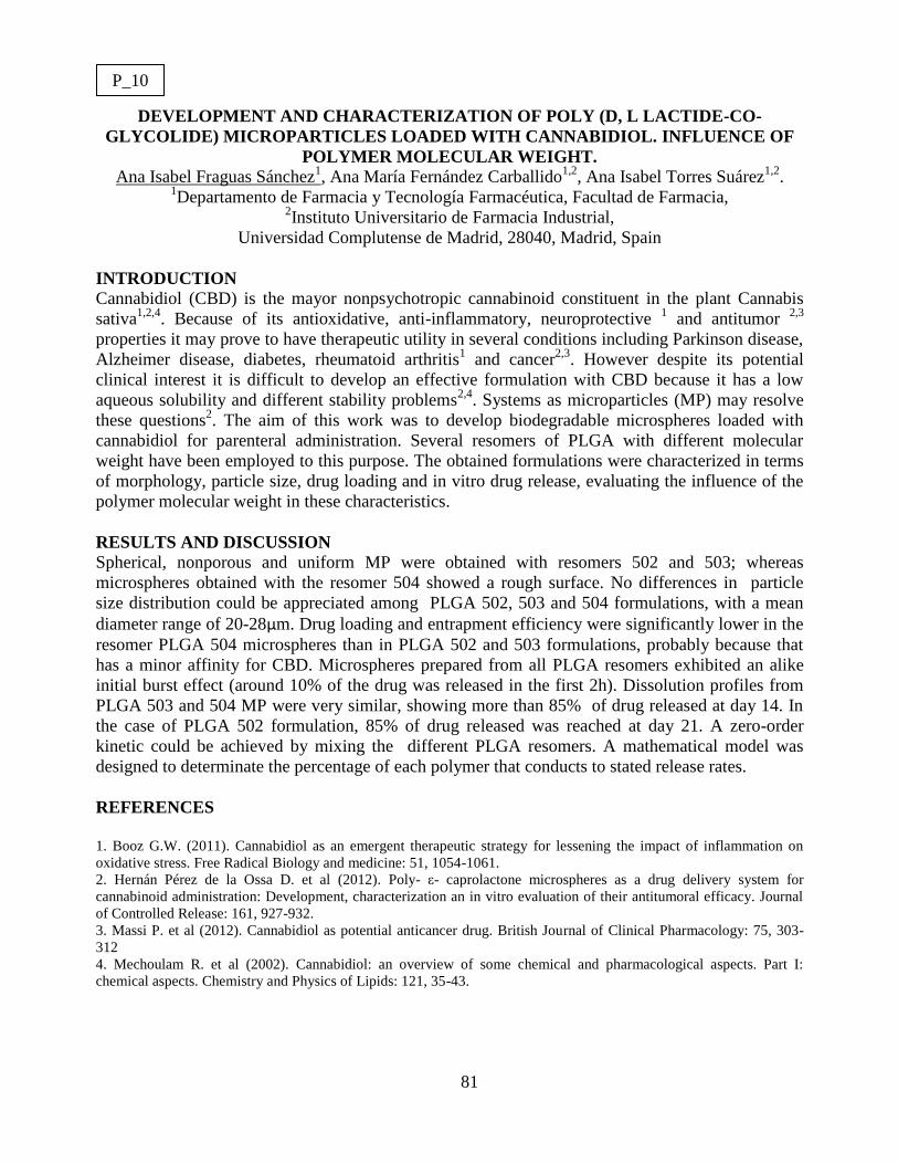
81
DEVELOPMENT AND CHARACTERIZATION OF POLY (D, L LACTIDE-CO-
GLYCOLIDE) MICROPARTICLES LOADED WITH CANNABIDIOL. INFLUENCE OF
POLYMER MOLECULAR WEIGHT.
Ana Isabel Fraguas Sánchez1, Ana María Fernández Carballido
1,2, Ana Isabel Torres Suárez
1,2.
1Departamento de Farmacia y Tecnología Farmacéutica, Facultad de Farmacia,
2Instituto Universitario de Farmacia Industrial,
Universidad Complutense de Madrid, 28040, Madrid, Spain
INTRODUCTION
Cannabidiol (CBD) is the mayor nonpsychotropic cannabinoid constituent in the plant Cannabis
sativa1,2,4
. Because of its antioxidative, anti-inflammatory, neuroprotective 1 and antitumor
2,3
properties it may prove to have therapeutic utility in several conditions including Parkinson disease,
Alzheimer disease, diabetes, rheumatoid arthritis1 and cancer
2,3. However despite its potential
clinical interest it is difficult to develop an effective formulation with CBD because it has a low
aqueous solubility and different stability problems2,4
. Systems as microparticles (MP) may resolve
these questions2. The aim of this work was to develop biodegradable microspheres loaded with
cannabidiol for parenteral administration. Several resomers of PLGA with different molecular
weight have been employed to this purpose. The obtained formulations were characterized in terms
of morphology, particle size, drug loading and in vitro drug release, evaluating the influence of the
polymer molecular weight in these characteristics.
RESULTS AND DISCUSSION
Spherical, nonporous and uniform MP were obtained with resomers 502 and 503; whereas
microspheres obtained with the resomer 504 showed a rough surface. No differences in particle
size distribution could be appreciated among PLGA 502, 503 and 504 formulations, with a mean
diameter range of 20-28µm. Drug loading and entrapment efficiency were significantly lower in the
resomer PLGA 504 microspheres than in PLGA 502 and 503 formulations, probably because that
has a minor affinity for CBD. Microspheres prepared from all PLGA resomers exhibited an alike
initial burst effect (around 10% of the drug was released in the first 2h). Dissolution profiles from
PLGA 503 and 504 MP were very similar, showing more than 85% of drug released at day 14. In
the case of PLGA 502 formulation, 85% of drug released was reached at day 21. A zero-order
kinetic could be achieved by mixing the different PLGA resomers. A mathematical model was
designed to determinate the percentage of each polymer that conducts to stated release rates.
REFERENCES
1. Booz G.W. (2011). Cannabidiol as an emergent therapeutic strategy for lessening the impact of inflammation on
oxidative stress. Free Radical Biology and medicine: 51, 1054-1061.
2. Hernán Pérez de la Ossa D. et al (2012). Poly- ε- caprolactone microspheres as a drug delivery system for
cannabinoid administration: Development, characterization an in vitro evaluation of their antitumoral efficacy. Journal
of Controlled Release: 161, 927-932.
3. Massi P. et al (2012). Cannabidiol as potential anticancer drug. British Journal of Clinical Pharmacology: 75, 303-
312
4. Mechoulam R. et al (2002). Cannabidiol: an overview of some chemical and pharmacological aspects. Part I:
chemical aspects. Chemistry and Physics of Lipids: 121, 35-43.
P_10

82
Controlled intramyocardial delivery of neuregulin-1 and fibroblast growth factor-1 from
biodegradable microparticles in a large preclinical myocardial infarction model
Elisa Garbayo1, Juan José Gavira
2, Gloria Abizanda
2, Beatriz Pelacho
2, Edurne Albiasu
2, Felipe
Prósper2, María José Blanco-Prieto
1
1Pharmacy and Pharmaceutical Technology Department, University of Navarra, Spain.
2Hematology, Cardiology and Cell Therapy, Clinica Universidad de Navarra and Foundation for
Applied Medical Research, University of Navarra, Spain.
INTRODUCTION
Growing evidence suggests that growth factor therapy is a promising approach to treat myocardial
infarction (MI). Among therapeutic cytokines, neuregulin-1 (NRG1) and acidic-fibroblast growth
factor (FGF1) are involved in cardiac repair after MI1,2
. However, limited protein stability is a
major challenge in growth factor therapy. The combination of proteins along with drug delivery
systems (DDS) represents a new therapeutic approach. Microparticles (MP), one of these DDS,
could protect proteins from degradation and ensure sustained release among time. Recently, our
group assessed the efficacy of biodegradable poly(lactic-co-glycolic acid) NRG1-MP and FGF1-
MP in a rat MI model demonstrating that MP promoted cardiac repair and improved cardiac
performance3. In order to further validate this clinically applicable platform for drug delivery, we
now propose to test the beneficial effect of NRG1-MP and FGF1-MP in a large preclinical
ischemia-reperfusion model.
RESULTS AND DISCUSSION
Growth factor loaded MP used in rats were successfully scaled-up to be tested in an ischemia-
reperfusion porcine model. MP were prepared by double-emulsion solvent evaporation technique
using the total recirculation one-machine system. The mean particle size measured by laser
diffractometry was 7.2±1.9 µm which is compatible with an intramyocardial administration.
Growth factors were efficiently encapsulated reaching values of 84.05±15.5% for FGF1 and
83.5±13.25% for NRG1. The released cytokines bioactivity was evaluated in vitro by determining
H9c2 cardiomyocyte proliferative capacity following growth factor treatment. A 1.3 and 2.1-fold
increase in cell density was observed when stimulated with FGF1 and NRG1 respectively.
Proliferation rates of cells treated with the free-cytokines were similar to the ones released by
microparticles, indicating that both cytokines retained its biological activity after
microencapsulation. NRG1-MP, FGF1-MP and non-loaded MP were next injected locally into the
infarcted myocardium using NOGA® XP Cardiac Navigation System, a highly accurate electro-
guided methodology which creates precise 3-D heart images, 1 week after the infarct. Preliminary
results showed that intramyocardial growth factor-MP injection has therapeutic effects and
improves cardiac function. Histological studies are currently been performed to fully characterize
treatment effectiveness. The validation of this therapeutic approach could make significant progress
for patients with ischemic heart disease.
REFERENCES 1. Liu X. et al. (2006) Neuregulin-1/erbB-activation improves cardiac function and survival in models of ischemic,
dilated, and viral cardiomyopathy. J Am Coll Cardiol. 48,1439.
2. Palmen M. et al. (2004) Fibroblast growth factor-1 improves cardiac functional recovery and enhances cell survival
after ischemia and reperfusion. J Am Coll Cardiol. 44,1113.
3. Formiga F.R. et al. Controlled delivery of fibroblast growth factor-1 and neuregulin-1 from biodegradable
microparticles promotes cardiac repair in a rat myocardial infarction model through activation of endogenous
regeneration. Submitted to Biomaterials.
P_11

83
IN VITRO TRANSDERMAL ABSORPTION STUDIES WITH SOME IBUPROFEN
FORMULATIONS USING DIFFERENT MODEL MEMBRANES
Isabel Arroyo1, Verónica García
1, Damián Cordoba-Diaz
1,2, Manuel Cordoba-Diaz
1,2
1 Dep. of Pharmacy and Pharmaceutical Technology UCM. 2 University Institute of Industrial
Pharmacy - IUFI. Faculty of Pharmacy, Complutense University, 28040 Madrid, Spain.
INTRODUCTION
Ibuprofen (IBU) is a non-steroidal anti-inflammatory drug (NSAID) that has demonstrated high efficiency in
the treatment of muscle and joint pain as well as in rheumatoid and osteoarthritis therapy. The inherent side
effects of orally administered IBU justify the vast amount of marketed formulations intended for topical use.
Its skin permeability is intrinsically poor due to its solubility properties (log P ≈ 4)1. In the present study, the
in vitro penetration properties of four commercially-available topical formulations of IBU were compared:
Ibuprofeno-Pharmacia® (Pharmacia Labs. – Pfizer Spain), Ibufen
® (Cinfa Labs. Spain), Ibuprofeno-
Farmasierra® (Farmasierra Labs. Spain) and Solvium
® (Omega Pharma Inc. Spain). All formulations were
presented as 50 mg/g Ibuprofen gels. Two inert synthetic membranes were selected to study the in vitro
penetration of IBU in order not to introduce biological variations inherent to natural skin2: a non-porous
polydimethylsiloxane (PDMS) membrane (Silmax® – Pillar surgical Inc. CA USA) and a new porous
membrane (Strat-MTM
) Merck KGaA, Darmstadt, Germany) which is reported to imitate the diffusional
characteristics of natural skin for a wide variety of compounds3.
RESULTS AND DISCUSSION
The diffusion of all formulations was studied using an automated in line flow-through equipment with seven
cells (PermeGear Inc. USA). PBS solution (pH 7.4) including a surfactant to ensure sink conditions was used
as receptor medium. Flux values (J), the permeation rates at steady state (µg/cm2 h) for each individual
diffusion experiment were calculated by monitoring the cumulative amount of drug diffused at each time by
HPLC using a previously validated method.
Big differences were found between the four formulations using the PDMS membrane: it was found that IBU
disposed onto the membrane with Solvium®, was able to diffuse to the donor chamber in a percentage close
to 40% in 24 hours whereas the IBU diffused ranged from 2 to 13% with the other formulations. These
findings contrast to the obtained with Strat-MTM
where no statistically significant differences were found
over 16 hours. A comparative analysis of the J data showed that the non-porous PDMS membrane is
adequate to detect differences between the studied compositions. The resulting values range from 14 to 98
µg/cm2h, what correlates with the calculated data by different authors for IBU with different kinds of skin
and under different conditions4-6
. Strat-MTM
provided significantly higher values of J with all the
formulations studied.
REFERENCES 1. Chang, S. et al. (1997) Ibuprofen heterocyclic esters as dermal prodrugs: in vitro evaluation. S.T.P. Pharma Sci.: 7,
315–319.
2. Ng, S., et al. (2012) The Relevance of Polymeric Synthetic Membranes in Topical Formulation Assessment and Drug
Diffusion Study. Arch. Pharm. Res.: 35 (4), 579-593.
3. Joshi, V. et al. (2012) In Vitro Diffusion Studies in Transdermal Research: A Synthetic Membrane Model in Place of
Human Skin. Drug Dev. Deliv.: 12 (2), 40-42.
4. Davis, A. et al. (2002) Formulation Strategies for Modulating Skin Permeation. IN Walters K. (Ed.) Dermatological
and Transdermal Formulations. Marcel Dekker, New York, pp 296.
5. Saino, V. et al. (2010) Optimization of skin permeation and distribution of ibuprofen by using nanostructures
(coagels) based on alkyl vitamin C derivatives. Eur. J. Pharm. Biopharm.: 76, 443–449.
6. Rhee, Y. et al. (2008) Optimization of ibuprofen gel formulations using experimental design technique for enhanced
transdermal penetration. Int. J. Pharm.: 364, 14–20.
P_12

84
MUCOADHESIVE PARTICLES WITH ENHANCING INTESTINAL ABSORPTION
CAPACITY AS DRUG DELIVERY PLATFORM FOR VITAMIN E
Laura Redondo1-2
, Luca Casettari2, Diego R. Perinelli
3, Marco Cespi
3, Giulia Bonacucina
3, Giovanni F.
Palmieri3, Pilar Jiménez
4, Tomás Girbés
4, Verónica García
1, Damián Cordoba-Diaz
1, Manuel Cordoba-
Diaz1
1 Complutense University of Madrid, Faculty of Pharmacy, Madrid, Spain
2 University of Urbino, School of Pharmacy, Urbino, Italy
3 University of Camerino, School of Pharmacy, Camerino, Italy
4 University of Valladolid, Faculty of Medicine, Valladolid, Spain
INTRODUCTION
M-cells are the specialised antigen sampling sites of the intestinal immune system and represent a potential portal
for mucosal drug and vaccine delivery since they possess a high transcytotic capacity and are able to transport a
broad range of materials including particulates. To increase bioadhesion to enterocytes in the gastrointestinal tract,
targeting ligands (such as lectins) are of special interest, as they mediate highly specific binding to epithelial cell
subpopulations, effected by interaction of carbohydrate-binding sites of lectin with sugar residues in the glycocalyx
of epithelial cells. Exploiting lectins for bioadhesive drug delivery purposes, this cell-specific interaction can result
in active receptor-mediated endocytosis and/or transcytosis of the drug delivery system.
Vitamin E is a class of lipid-soluble antioxidant molecules widely used in food, cosmetic and pharmaceutical
fields. Unfortunately due to its high sensitivity to external stimuli such as light, heat and oxygen, it rapidly
degrades and this limits its applications.
The aim of this work is to formulate mucoadhesive and biodegradable polymeric particles intended for oral use to
enhance the intestinal absorption of γ-tocopherol. Poly(lactic-co-glycolic acid) (PLGA) based carriers have been
produced with the classic nanoprecipitation method, which is an easy, reproducible and scale up method. The
biocompatible polysaccharide chitosan have been added to the formulation to increase the mucoadhesion of the
particles, which is exert due to its polycation nature and ability to interact with mucus producing surfaces.
Moreover the particles surface has been covalently modified with lectins obtained from Sambucus ebulus L., to
further increase mucoadhesiveness and cell interactions.
RESULTS AND DISCUSSION
Different formulations have been performed to select the optimal conditions and components percentages to be
used in the carrier system, particularly regarding the kind of surfactants/stabilizers (poly(vinyl alcohol) (PVA),
Poloxamer 407, Tween 80 or PEGylated poly(lactic acid) (mPEG-PLA)). The γ-tocopherol delivery system has
been optimized regarding the encapsulation efficacy, the particles morphology and the cell penetration ability.
Spherical particles have been produced and characterize with dynamic light scattering (< 1 μm) and microscopy
techniques.
REFERENCES 1. Devriendt, B. et al (2012). Crossing the barrier: Targeting epithelial receptors for enhanced oral vaccine delivery. J.
Control. Release: 160(3), 431-439.
2. Wang, Y.C. et al. (2003). A chemical surface modification of chitosan by glycoconjugates to enhance the cell-
biomaterial interaction. Biomacromolecules: 4 (2), 224-231.
3. Yin, Y. et al. (2007). Lectin-conjugated PLGA nanoparticles loaded with thymopentin: ex vivo bioadhesion and in
vivo biodistribution. J. Control. Release: 123(1), 27-38.
4. Khayata, N. et al. (2012). Preparation of vitamin E loaded nanocapsules by the nanoprecipitation method: from
laboratory scale to large scale using a membrane contactor. Int. J. Pharm.: 423(2), 419-427.
5. Vandamme, K. et al. (2010). Influence of reaction medium during synthesis of Gantrez AN 119 nanoparticles for oral
vaccination. Eur. J.Pharm. Biopharm.: 74, 202-208.
P_13

85
DEVELOPMENT AND VALIDATION OF AN ANALYTICAL METHOD BY RP-HPLC TO
SIMULTANOUSLY QUANTIFY ACETAZOLAMIDE AND TIMOLOL FROM TRANSETOSOMES.
APPLICATION OF QUALITY-BY-DESIGN TO DESIGN AND DEVELOPMENT OF OPTIMIZED
FORMULATION
C. Pazmino, L. Cabanzo, M.J. Cózar-Bernal, A.M. Rabasco, M.L. González-Rodríguez
Department of Pharmacy and Pharmaceutical Technology. C/ Profesor García González 2, 41012 Seville, Spain.
Faculty of Pharmacy, University of Seville
INTRODUCTION
The instrumental technique of HPLC-DAD has been widely used in the pharmaceutical field. The development of
a selective analytical method allows us to separate the components of the sample to subsequently identify and
quantify them. When an analytical method has been developed, it is important to confirm that it is suitable for its
intended purpose. Therefore, the method validation according the ICH Q2R1 guidelines is today an essential
concern in the activity of analytical chemistry laboratories.
In this work, it is important to develop a specific analytical technique for quantifying acetazolamide (ACZ) and
timolol (TML), which were encapsulated in transetosomas, to subsequently improve pharmaceutical properties.
Furthermore, the variability in the manufacturing process leads to a quality problem; results are not reproducible
batch to batch. Therefore, we must have a more comprehensive approach, strict and necessary in terms of safety. It
is necessary to ensure quality through Quality-by-Design (ICH Q8) which is defined as designing and developing
formulations and robust manufacturing processes and reproducible to ensure that our drug is safe and effective for
their objective.
RESULTS AND DISCUSSION
The chromatographic conditions were optimized using an octadecylsilyl column C18 (Merck, LiChrospher 100
RP-18, 125x4 mm). The column temperature was 45 °C (column oven L-2350, Elite LaChrom). The mobile phase
consisted of acetonitrile-sodium acetate buffer pH 4.1 (ACN:SA).
A linear gradient of 10:90% of ACN:SA for 2.2 min at a flow rate of 1.5 mL/min and 40:60% of ACN:SA for 5
minutes at a flow rate of 2.0 mL/min. ACZ and TML were detected with a diode detector at 286 nm and the
injection volume was 10 µL.
The method has been validated. Selectivity was demonstrated qualitative and quantitatively. The accuracy of the
method revealed recovery values around 99%. Instrumental precision was satisfactory (RSD < 2%) and method
repeatability was considered valid, as the RSD values were ≤ 2%. Adequate system and method linearity were
obtained in the range of the study (0.25-0.75 mg/mL). LOD and LOQ were found to be 8x10⁻⁴
and 3x10⁻³
mg/mL,
respectively. Finally, the robustness assay was demonstrated by applying the Taguchi method.
Then, the principles of QbD were applied to get the optimized formulation, which was characterized in terms of
encapsulation efficiency for ACZ = 22.03 ± 2.53% and TML = 29.66 ± 3.12%, vesicular size: 385.92 ± 65.6 nm,
zeta potential: -30.8 ± 11.3 mV, permeation flux: 3.41×10⁻⁴
and 2.74×10⁻⁴
mg/cm2/h for ACZ and TML,
respectively. Formulation containing PC: 0.0954-0.10498 mmol, Cho: 0.0812-0.08933 mmol, 0.00724-0.00796
mmol, ethanol 1.5%, showed the best characteristics for the intended purpose of these transethosomes to be
potentially administered by ophthalmic route.
REFERENCES
1. Cózar-Bernal MJ, Rabasco AM, González-Rodríguez ML (2012) Development and validation of a high
performance chromatographic method for determining sumatriptan in niosomes. J. Pharm. Biomed. Anal. 72(1),
251-260.
2. Lionberger R. et al. (2008) Quality by design: concepts for ANDAs. The AAPS Journal 10(2), 268-276.
3. Malakar J, Sen SO, Nayak AK, Sen KK (2012) Formulation, optimization and evaluation of transfersomal gel
for transdermal insulin delivery. Saudi Pharm. J. 20(1), 355.
4.Yu LX (2008) Pharmaceutical quality by design: product and process development, understanding, and
control. Pharm. Res. 25(4), 781-791.
P_14

86
APPLYING THE MOLECULAR TROJAN HORSE TECHNOLOGY TO DESIGN
IMMUNOLIPOSOMES FOR BRAIN DRUG DELIVERY
E. García-Esteban, M.J. Cózar-Bernal, A.M. Rabasco, M.L. González-Rodríguez
Department of Pharmacy and Pharmaceutical Technology. C/ Profesor García González 2, 41012
Seville, Spain. Faculty of Pharmacy, University of Seville.
INTRODUCTION
The central nervous system (CNS) is protected by the blood-brain barrier (BBB). Unfortunately, the same
mechanisms that protect it against intrusive chemicals can also frustrate therapeutic interventions. Drug
delivery to the brain is limited due to the BBB. Only small molecules (molecular weight lower than 600 Da)
can pass the BBB paracellularly or transcellularly, depending on their lipophilicity. However, high molecular
weight drugs, such as proteins, peptides or DNA, cannot cross it.
The most versatile and attractive approach for delivery of drugs in their native state into the brain
parenchyma involves the use of drug carriers, such as liposomes, as “Molecular Trojan Horses” (MTH). The
MTH bind to a receptor (e.g., for transferrin or insulin) on the BBB and brain cell membrane, triggering
receptor-mediated transcytosis of the Trojan Horse Liposome (THL) across the BBB in vivo, and receptor-
mediated endocytosis into brain cells beyond the BBB.
In this study, we have developed immunoliposomes as drug delivery carrier. Immunoliposomes are
liposomes in which an antibody has been attached to the liposome allowing the drug to be targeted to the
brain cells.
RESULTS AND DISCUSSION
The molecular structure of immunoliposome involves the preparation of liposomes by TLE technique.
Liposomes contained phosphatidylethanolamine (PE) as phospholipid, which included maleimide (SMPB) as
functional group. Maleimide residues allow the liposome conjugation with the monoclonal antibody (Ig G).
Inside liposomal structure, the drug was included. Functionalized liposome binding to the antibody occurs
after thiolation of the antibody with the terminal ester group of N-succinimidyl S-acetylthioacetate (SATA)
obtaining a stable amide linkage. Once developed various synthetic steps, the characterization of the
resulting structure has been realized.
After monoclonal antibody attached to liposomes, the synthesis of immunoliposomes occurred satisfactorily.
These functionalized vesicles were characterized in terms of mean diameter and size distribution by laser
scattering technique (Zetasizer Nano ZS). Shape and surface characteristics were determined by fluorescence
optical microscopy (Olympus BX61). A confocal laser scanning microscope (CLSM) (Leica DM-IRE2) was
used to know the presence and the distribution of Ig G around the immunoliposomes.
REFERENCES
1. Cózar-Bernal MJ, Rabasco AM, Villasmil S, González-Rodríguez ML (2012) Novedades en
inmunoliposomas funcionalizados como sistemas vectores de péptidos a nivel cerebral. Ind. Farm.
171, 56-62.
2. Manjappa AS et al. (2010) Antibody derivatization and conjugation strategies. Application in
preparation of stealth immunoliposome to target chemotherapeutics to tumor. J. Control. Release 1,
2-22.
3. Torchilin VP, Weissig V. Surface modification of liposomes. In: Liposomes (Torchilin VP,
Weissig V, Eds.), Practical Approach, 2ª ed., pp. 193-229 (2007).
4. Pardridge WM (2007) Blood-brain barrier delivery of protein and non-viral gene therapeutics
with molecular Trojan horses. J. Control. Release 122, 345-348.
P_15
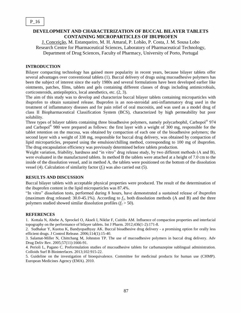
87
DEVELOPMENT AND CHARACTERIZATION OF BUCCAL BILAYER TABLETS
CONTAINING MICROPARTICLES OF IBUPROFEN
J. Conceição, M. Estanqueiro, M. H. Amaral, P. Lobão, P. Costa, J. M. Sousa Lobo
Research Centre for Pharmaceutical Sciences, Laboratory of Pharmaceutical Technology,
Department of Drug Sciences, Faculty of Pharmacy, University of Porto, Portugal
INTRODUCTION
Bilayer compacting technology has gained more popularity in recent years, because bilayer tablets offer
several advantages over conventional tablets (1). Buccal delivery of drugs using mucoadhesive polymers has
been the subject of interest since the early 1980s and several formulations have been developed earlier like
ointments, patches, films, tablets and gels containing different classes of drugs including antimicrobials,
corticosteroids, antiepileptics, local anesthetics, etc. (2, 3).
The aim of this study was to develop and characterize buccal bilayer tablets containing microparticles with
ibuprofen to obtain sustained release. Ibuprofen is an non-steroidal anti-inflammatory drug used in the
treatment of inflammatory diseases and for pain relief of oral mucositis, and was used as a model drug of
class II Biopharmaceutical Classification System (BCS), characterized by high permeability but poor
solubility.
Three types of bilayer tablets containing three bioadhesive polymers, namely polycarbophil, Carbopol® 974
and Carbopol® 980 were prepared as follows: the first layer with a weight of 300 mg, responsible for the
tablet retention on the mucosa, was obtained by compaction of each one of the bioadhesive polymers; the
second layer with a weight of 338 mg, responsible for buccal drug delivery, was obtained by compaction of
lipid microparticles, prepared using the emulsion/chilling method, corresponding to 100 mg of ibuprofen.
The drug encapsulation efficiency was previously determined before tablets production.
Weight variation, friability, hardness and “in vitro” drug release study, by two different methods (A and B),
were evaluated in the manufactured tablets. In method B the tablets were attached at a height of 7.0 cm to the
inside of the dissolution vessel, and in method A, the tablets were positioned on the bottom of the dissolution
vessel (4). Calculation of similarity factor (f2) was also carried out (5).
RESULTS AND DISCUSSION
Buccal bilayer tablets with acceptable physical properties were produced. The result of the determination of
the ibuprofen content in the lipid microparticles was 87.4%.
“In vitro” dissolution tests, performed during 8 hours, have demonstrated a sustained release of ibuprofen
(maximum drug released: 30.0-45.1%). According to f2, both dissolution methods (A and B) and the three
polymers studied showed similar dissolution profiles (f2 > 50).
REFERENCES
1. Kottala N, Abebe A, Sprockel O, Akseli I, Nikfar F, Cuitiño AM. Influence of compaction properties and interfacial
topography on the performance of bilayer tablets. Int J Pharm. 2012;436(1-2):171-8.
2. Sudhakar Y, Kuotsu K, Bandyopadhyay AK. Buccal bioadhesive drug delivery - a promising option for orally less
efficient drugs. J Control Release. 2006;114(1):15-40.
3. Salamat-Miller N, Chittchang M, Johnston TP. The use of mucoadhesive polymers in buccal drug delivery. Adv
Drug Deliv Rev. 2005;57(11):1666-91.
4. Perioli L, Pagano C. Preformulation studies of mucoadhesive tablets for carbamazepine sublingual administration.
Colloids Surf B Biointerfaces. 2013;102:915-22.
5. Guideline on the investigation of bioequivalence. Committee for medicinal products for human use (CHMP).
European Medicines Agency (EMA). 2010.
P_16

88
HUMAN SERUM ALBUMINE LOADED MICROPARTICLES TO MODULATE
DEXAMETHASONE DRUG RELEASE
Javier Rodríguez 1, Manuel Guzmán
1, Irene Bravo
2, Irene Molina
2, Rocio Herrero
2
1Departamento de Ciencias Biomédicas, Unidad docente de Farmacia y Tecnología Farmacéutica,
Universidad de Alcalá (UAH) Ctra Madrid-Barcelona (A2) Km. 33,600 28871 Alcalá de Henares
(Madrid) 2Departamento de Farmacia y Tecnología Farmacéutica, Universidad Complutense de Madrid.
Plaza Ramón y Cajal 28040 Madrid.
INTRODUCTION
Biodegradable and biocompatible polymeric microparticles are of special interest to avoid
the inconvenients related to surgical insertion of implants1. Among the methods employed to
prepare microparticles, the emulsion solvent extraction-evaporation technique is one of the most
employed. PLGA (poly lactide-co-glycolide) is widely used to prepare biodegradable drug delivery
systems. Many researchers have investigated the modulation of the drug release profiles by
different strategies. One of the most widely employed is to incorporate additives into the
formulation2-3
.
RESULTS AND DISCUSSION
A family of dexamethasone loaded microparticles have been obtained using a double
emulsion S/O/W extraction-evaporation method4. The additives employed in this study have been
vitamin E (Vit E) and Human Serum Albumine (HSA). Physico-chemical characterization of the
different formulations has been accomplished. Dexamethasone – vitamin E - human serum
albumine (HSA) microparticles show no difference in encapsulation efficiency assays compared to
those only including dexamethasone. In order to evaluate the influence of the different additives,
release assays of dexamethasone from the different formulations were performed in PBS buffer.
The release of the drug resulted slower in the vitamin E-HSA microparticles compared with
dexamethasone and dexamethasone-vitamin E microparticles. The initial phase characterized by a
burst effect that was clearly higher in the dexamethasone loaded microparticles. The profile
obteined in both formulations show the typical shape of PLGA systems.
Acknowledgements- UCM Research Group 920415 and MAT 2010- 18242
REFERENCES
1. Alarcon, R., Martinez, M. (2006). Intraocular drug delivery systems (Sistemas de cesión controlada por vía
intraocular). Arch. Soc. Esp. Oftalmol. 81 (2), 57–59
2. Herrero-Vanrell, R., Ramirez, L., Fernandez-Carballido, A., Refojo, M. F., (2000) Biodegradable PLGA
microspheres loaded with ganciclovir for intraocular administration. Encapsulation technique, in vitro release profiles,
and sterilization process. Pharm Res 17(10): p. 1323-8
3. Barcia E, Herradón C., Herrero-Vanrell R. (2005). Biodegradable Additives Modulate Ganciclovir Release Rate from
PLGA Microspheres destined to Intraocular Administration. Letters in Drug Design and Discovery. 2: 184-193
4. Herrero-Vanrell R and Refojo MF “Intravitreal microsphere drug delivery and method of preparation”U.S. Patent #
5,718,922
P_17
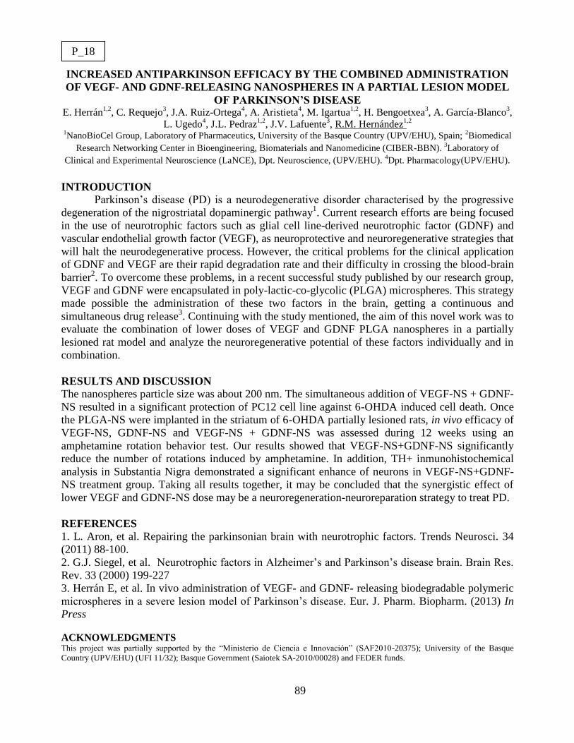
89
INCREASED ANTIPARKINSON EFFICACY BY THE COMBINED ADMINISTRATION
OF VEGF- AND GDNF-RELEASING NANOSPHERES IN A PARTIAL LESION MODEL
OF PARKINSON’S DISEASE E. Herrán
1,2, C. Requejo
3, J.A. Ruiz-Ortega
4, A. Aristieta
4, M. Igartua
1,2, H. Bengoetxea
3, A. García-Blanco
3,
L. Ugedo4, J.L. Pedraz
1,2, J.V. Lafuente
3, R.M. Hernández
1,2
1NanoBioCel Group, Laboratory of Pharmaceutics, University of the Basque Country (UPV/EHU), Spain;
2Biomedical
Research Networking Center in Bioengineering, Biomaterials and Nanomedicine (CIBER-BBN). 3Laboratory of
Clinical and Experimental Neuroscience (LaNCE), Dpt. Neuroscience, (UPV/EHU). 4Dpt. Pharmacology(UPV/EHU).
INTRODUCTION
Parkinson’s disease (PD) is a neurodegenerative disorder characterised by the progressive
degeneration of the nigrostriatal dopaminergic pathway1. Current research efforts are being focused
in the use of neurotrophic factors such as glial cell line-derived neurotrophic factor (GDNF) and
vascular endothelial growth factor (VEGF), as neuroprotective and neuroregenerative strategies that
will halt the neurodegenerative process. However, the critical problems for the clinical application
of GDNF and VEGF are their rapid degradation rate and their difficulty in crossing the blood-brain
barrier2. To overcome these problems, in a recent successful study published by our research group,
VEGF and GDNF were encapsulated in poly-lactic-co-glycolic (PLGA) microspheres. This strategy
made possible the administration of these two factors in the brain, getting a continuous and
simultaneous drug release3. Continuing with the study mentioned, the aim of this novel work was to
evaluate the combination of lower doses of VEGF and GDNF PLGA nanospheres in a partially
lesioned rat model and analyze the neuroregenerative potential of these factors individually and in
combination.
RESULTS AND DISCUSSION
The nanospheres particle size was about 200 nm. The simultaneous addition of VEGF-NS + GDNF-
NS resulted in a significant protection of PC12 cell line against 6-OHDA induced cell death. Once
the PLGA-NS were implanted in the striatum of 6-OHDA partially lesioned rats, in vivo efficacy of
VEGF-NS, GDNF-NS and VEGF-NS + GDNF-NS was assessed during 12 weeks using an
amphetamine rotation behavior test. Our results showed that VEGF-NS+GDNF-NS significantly
reduce the number of rotations induced by amphetamine. In addition, TH+ inmunohistochemical
analysis in Substantia Nigra demonstrated a significant enhance of neurons in VEGF-NS+GDNF-
NS treatment group. Taking all results together, it may be concluded that the synergistic effect of
lower VEGF and GDNF-NS dose may be a neuroregeneration-neuroreparation strategy to treat PD.
REFERENCES
1. L. Aron, et al. Repairing the parkinsonian brain with neurotrophic factors. Trends Neurosci. 34
(2011) 88-100.
2. G.J. Siegel, et al. Neurotrophic factors in Alzheimer’s and Parkinson’s disease brain. Brain Res.
Rev. 33 (2000) 199-227
3. Herrán E, et al. In vivo administration of VEGF- and GDNF- releasing biodegradable polymeric
microspheres in a severe lesion model of Parkinson’s disease. Eur. J. Pharm. Biopharm. (2013) In
Press
ACKNOWLEDGMENTS This project was partially supported by the “Ministerio de Ciencia e Innovación” (SAF2010-20375); University of the Basque
Country (UPV/EHU) (UFI 11/32); Basque Government (Saiotek SA-2010/00028) and FEDER funds.
P_18

90
IN VITRO DISSOLUTION/PERMEABILITY ASSAYS WITH NIMESULIDE
NANOPARTICLES
C. Huerta, M.R. Aberturas, J. Molpeceres
Departamento de Ciencias Biomédicas. Unidad Docente Farmacia y Tecnología Farmacéutica,
Facultad de Farmacia, Universidad de Alcalá, 28871 Alcalá de Henares, Madrid.
INTRODUCTION
Nimesulide (NS) is a non-steroidal anti-inflammatory drug with preferential inhibition of the COX-
2 isoform and less renal and gastrointestinal adverse effects than other non-selective NSAIDs (1). It
has been shown that the COX-2 isoform is over-expressed in different epithelial cancers such as the
prostate (2) and several in vivo experiments have shown nimesulide is a promising lead compound
for anti-cancer drug discovery (3). However, the nimesulide concentrations used in these studies are
ranged from 200 to 500 mM, which greatly exceed the concentration necessary to inhibit COX-2
activity. These facts suggest that nimesulide inhibits cancer cell growth and induces cancer cell
apoptosis independent of COX-2. Although intratumoral administration is a promising approach for
the treatment of various solid tumors by achieving high local drug concentrations with minimal
systemic toxicity, its efficacy is strongly dependent on the timing and frequency of the drug
injections because of its rapid clearance from the tumor site. To achieve this goal we have
formulated poly lactide-co-glycolide (PLGA) nanoparticles (NPs) loaded with NS. In recent studies,
surface coating by hydrophilic polymers such as chitosan (CS) has been also evaluated (4). This
study focused on the in vitro release and permeation of the NS loaded in PLGA NPs which were
prepared by emulsification-solvent evaporation method.
RESULTS AND DISCUSSION
NPs showed average sizes of 378.7 ± 58.9 and 392.9 ± 66.1 nm when using CS as an external
coating. Free NS in solution passed through a simulated biological membrane (adapted PAMPA)
following apparent zero order kinetics (0.42 %/min). NS in suspension at concentrations
comparable to NP formulations showed non linear kinetics with a first faster step lasting about 100
min followed by a slower permeation rate. In all cases NPs retarded the permeability of NS from 50
to 100 min. CS-coated NP did not modify the apparent zero order kinetics but NS permeability rates
decreased to 0.15 and 0.11 %/min for 1 and 0.25% PVA, respectively. Uncoated NP showed
biphasic permeability curves like the one obtained with drug crystals but the initial release and
permeability rates were slower. However, these initial release rates were faster than the ones
achieved with CS coated NP (30% vs. 10% released in 200 min).
These experiments would simulate the situation occurring after intraprostatic injection of NSNP. At
first, a deposit of NS (non-sink conditions) will be formed but, later, the initially released drug
could be swept along by the blood flow causing additional NS dissolving from the NP and
permeating through biological membranes.
REFERENCES 1. Singla, A.K. et al. Nimesulide: Some pharmaceutical and Pharmacological Aspects - An Update. J. Pharm.
Pharmacol. (2000), 52, 467 - 486.
2. Soslow, R. et al. COX-2 Is Expressed Human Pulmonary, Colonic, and Mammary Tumors. Cancer (2000), 89, 2637-
2645.
3. Zhong, B. et al. From COX-2 inhibitor nimesulide to potent anti-cancer agent: Synthesis, in vitro, in vivo and
pharmacokinetic evaluation. European Journal of Medicinal Chemistry (2012), 47, 432-444.
4. Parveen, S., Sahoo S.K. Long circulating chitosan/PEG blended PLGA nanoparticle for tumor drug delivery,
European Journal of Pharmacology, November (2011), 670, 2–3 (30), 372–383.
P_19

91
PHARMACOKINETICS AND PHARMACODYNAMICS OF HYDROPHOBIC GENTAMICIN-
LOADED NANOPARTICLES IN BRUCELLA-INFECTED MICE Edurne Imbuluzqueta
1, Carlos Gamazo
2, Hugo Lana
1, Miguel Ángel Campanero
3, David Salas
4, Ana Gloria Gil
4,
Elisa Elizondo5,6
, Nora Ventosa5,6
, Jaume Veciana5,6
, María J. Blanco-Prieto1
1Department of Pharmacy and Pharmaceutical Technology,
2Department of Microbiology, University of Navarra,
Pamplona, Spain. 3Clinical Pharmacology Service, Clínica Universidad de Navarra, Pamplona, Spain.
4Department
of Nutritional Sciences, Physiology and Toxicology, University of Navarra, Pamplona, Spain. 5Department of
Molecular Nanoscience and Organic Materials, Institut de Ciencia de Materials de Barcelona,6CIBER de
Bioingenieria, Biomateriales y Nanociencia, Bellaterra, Spain.
INTRODUCTION
Brucellosis is a worldwide zoonosis that remains widespread and neglected in many areas despite
notable advances in science, technology, and management. Specific therapy is based on long-lasting
combined drug treatments that generate toxicity and are difficult to achieve, therefore, relapse currently
presents a significant problem1. On the other hand, the intracellular location of the pathogen in
phagocytic cells is also involved in therapeutic failure2. As an alternative to current therapy, the use of
biodegradable and biocompatible poly(lactic-co-glycolic acid) (PLGA) nanoparticles (NPs) have been
studied for the intracellular targeting of a hydrophobic gentamicin (GEN-AOT)3. A sustained release of
the antibiotic at the site of infection would make it possible a higher accumulation of the drug in the
infected tissues, thus improving the effectiveness of the antibiotic, and may make it possible to reduce
both the required number of doses and the frequency of administration, and thus the treatment-associated
toxicity.
RESULTS AND DISCUSSION
GEN-AOT PLGA NPs prepared by the single emulsion-solvent evaporation method presented mean
diameters of 300 nm, 100% of encapsulation efficiency and drug loadings of 24 μg of GEN/mg of NP.
When tested in Brucella melitensis-infected human macrophages GEN-AOT NPs significantly improved
the efficacy of the free drug at clinically relevant concentrations (>2-log10 unit reduction at 18 μg/mL).
On the other hand, pharmacokinetics studies were performed after the administration of a single
intraperitoneal dose equivalent to 5 mg/kg of GEN to Balb/c mice. The concentrations of the antibiotic in
the liver and spleen (target organs of Brucella) and kidneys (as GEN is nephrotoxic) were determined. It
was observed that while no GEN was detected in the liver or in the spleen, NPs efficiently targeted the
drug at both tissues and maintained therapeutic antibiotic concentrations for up to 4 days, allowing the
design of a therapeutic schedule for NPs with an extended dosing interval. Finally, mice were infected
with B. melitensis and treated for 14 days with daily GEN (5 mg/kg), the NPs every 4 days (5 mg/kg) and
the classical combined therapy of daily GEN (5 mg/kg) and doxycycline (10 mg/kg). Importantly, 3
weeks after the end of the treatment the splenic infection of mice treated with the NPs was reduced by 3.2
logs, and 50% of the infected mice were cured, with no evidence of adverse toxic effect. In contrast, the
classical combined therapy of GEN and doxycycline was associated with nephrotoxicity and with a rapid
re-emergence of the infection. In summary, encapsulation of gentamicin in PLGA nanoparticles
improved the antibiotic therapeutic index and allowed to reduce the required dosing frequency and the
treatment-associated side effects, presenting a great therapeutic potential.
REFERENCES 1 Ersoy, Y. et al. (2005). Comparison of three different combination therapies in the treatment of human brucellosis. Trop
Doct 35, 210.
2. Gamazo, C. et al. (2006). Chemical and biological factors in the control of Brucella and brucellosis. Curr Drug Deliv 3, 359.
3. Imbuluzqueta, E. et al. (2012). Cellular pharmacokinetics and intracellular activity against Listeria monocytogenes and
Staphylococcus aureus of chemically modified and nanoencapsulated gentamicin. J Antimicrob Chemother 67,2158.
P_20
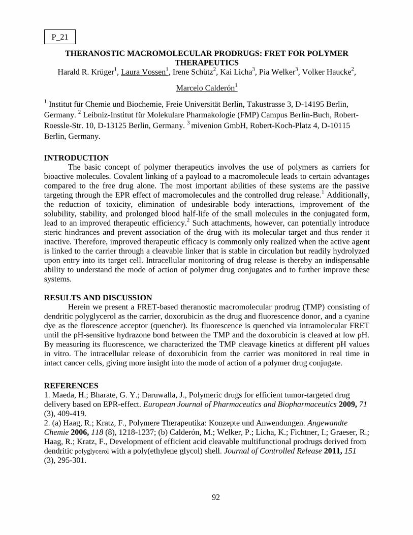
92
THERANOSTIC MACROMOLECULAR PRODRUGS: FRET FOR POLYMER
THERAPEUTICS
Harald R. Krüger1, Laura Vossen
1, Irene Schütz
2, Kai Licha
3, Pia Welker
3, Volker Haucke
2,
Marcelo Calderón1
1 Institut für Chemie und Biochemie, Freie Universität Berlin, Takustrasse 3, D-14195 Berlin,
Germany. 2 Leibniz-Institut für Molekulare Pharmakologie (FMP) Campus Berlin-Buch, Robert-
Roessle-Str. 10, D-13125 Berlin, Germany. 3
mivenion GmbH, Robert-Koch-Platz 4, D-10115
Berlin, Germany.
INTRODUCTION
The basic concept of polymer therapeutics involves the use of polymers as carriers for
bioactive molecules. Covalent linking of a payload to a macromolecule leads to certain advantages
compared to the free drug alone. The most important abilities of these systems are the passive
targeting through the EPR effect of macromolecules and the controlled drug release.1 Additionally,
the reduction of toxicity, elimination of undesirable body interactions, improvement of the
solubility, stability, and prolonged blood half-life of the small molecules in the conjugated form,
lead to an improved therapeutic efficiency.2 Such attachments, however, can potentially introduce
steric hindrances and prevent association of the drug with its molecular target and thus render it
inactive. Therefore, improved therapeutic efficacy is commonly only realized when the active agent
is linked to the carrier through a cleavable linker that is stable in circulation but readily hydrolyzed
upon entry into its target cell. Intracellular monitoring of drug release is thereby an indispensable
ability to understand the mode of action of polymer drug conjugates and to further improve these
systems.
RESULTS AND DISCUSSION
Herein we present a FRET-based theranostic macromolecular prodrug (TMP) consisting of
dendritic polyglycerol as the carrier, doxorubicin as the drug and fluorescence donor, and a cyanine
dye as the florescence acceptor (quencher). Its fluorescence is quenched via intramolecular FRET
until the pH-sensitive hydrazone bond between the TMP and the doxorubicin is cleaved at low pH.
By measuring its fluorescence, we characterized the TMP cleavage kinetics at different pH values
in vitro. The intracellular release of doxorubicin from the carrier was monitored in real time in
intact cancer cells, giving more insight into the mode of action of a polymer drug conjugate.
REFERENCES
1. Maeda, H.; Bharate, G. Y.; Daruwalla, J., Polymeric drugs for efficient tumor-targeted drug
delivery based on EPR-effect. European Journal of Pharmaceutics and Biopharmaceutics 2009, 71
(3), 409-419.
2. (a) Haag, R.; Kratz, F., Polymere Therapeutika: Konzepte und Anwendungen. Angewandte
Chemie 2006, 118 (8), 1218-1237; (b) Calderón, M.; Welker, P.; Licha, K.; Fichtner, I.; Graeser, R.;
Haag, R.; Kratz, F., Development of efficient acid cleavable multifunctional prodrugs derived from
dendritic polyglycerol with a poly(ethylene glycol) shell. Journal of Controlled Release 2011, 151
(3), 295-301.
P_21

93
DESIGN OF NEW NANOCARRIERS OF HISTONE DEACETYLASE
M.López-Viota1, J.A. Vidal
2, F. R. Rodriguez
1, J. Martin, Mª A. Ruiz
1, J. L.Viota
3.
1Dpto of Pharmacy and Tech. Pharma. University of Granada. Granada, Spain.
2Dpto Chemical-Physics. University of Granada. Granada, Spain.
3Vircell microbiologist. Granada. Spain
INTRODUCTION
The treatment of Leishmaniasis is a complex procedure due to the different clinical forms and the
influence on the immune status of the host. In addition, the drugs used in this treatment present
major problems including the high toxicity and many side effects to which must be added the
emergence of resistant strains. Nowadays, none of them eliminate completely the parasite so the
host, end up suffering from a chronic infection and remains infectious for sandflies. Nevertheless
the treatments commercially available achieve a reduction/elimination of the clinical signs of
disease. One of the paths aimed in this work is focused in the inhibition of histone deacetylase
(HDAC). In the present work we will explore this therapeutic strategy in combination with gold
nanoparticles as drug delivery systems.
RESULT AND DISCUSSION
In the present work, 20 nm gold nanoparticles synthesized following the citrate thermal reduction
method (1), were functionalized with histone deacetylase inhibitors. The adsorption of HDAC is
proven by dynamic light scattering measurements, electrophoretic mobility measurements and
optical absorbance. The adsorption of HDAC on the gold cores, produce an increase of the size up
to 150 nm. In addition, the electrophoretic mobility of the particles tends, in absolute value, to
decrease to more positive. By optical absorbance measurements the loading capacity of HDAC by
the gold nanoparticles was determined, and it was found that the amount adsorbed is below 10 %. In
addition, the release of HDAC was followed up in culture medium at 37 º C, and it was found that
the capacity of release is very effective, being the amount released higher than 80 %. Finally,
preliminary in vitro test shows that the functionalization of gold nanoparticles with HDAC, is
effective.
REFERENCES
1. J. Turkevich, P.C. Stevenson, J. Hillier, Discuss. Trans. Faraday Soc. 11 (1951) 55
P_22

94
OPTIMISED POLYMERIC NANOPARTICLES LOADED WITH
ANTIINFLAMATORY DRUG. Mireia Mallandrich
1, Araceli Barreto
1, Alexander Parra
2, Francisco Fernández-Campos
1,
Nora Provenza1, M. Antonia Egea
2, Maria Luisa García
2, Ana C. Calpena
1,3
1Department of Pharmacy and Pharmaceutical Technology. Faculty of Pharmacy. University of Barcelona.
Spain. e-mail: [email protected], [email protected] 2Department of Physical Chemistry. Faculty of Pharmacy. University of Barcelona. Spain.
3Institut de Nanociència i Nanotecnologia. IN2UB. Barcelona. Spain.
INTRODUCTION
Biodegradable Polymeric Nanoparticles (NPs) are widely used to enhance the bioavailability of
topically administered drugs. Poly lactic-co-glycolic acid (PLGA) is the most extensive
biodegradable polymer used due to its biocompatibility, mucoadhesiveness and nonantigenic nature
(1,2,3). In addition, PLGA-NPs are mainly absorbed in inflamed areas by the reticulo-endothelial
system (RES), resulting in an increased concentration of NPs in the aforementioned areas.
Ketorolac Tromethamine (KT) is a non-steroidal anti-inflammatory drug (NSAID) with a potent
and moderate non-opioid analgesic activity. It is currently administered orally and intramuscularly
for pre- and postoperative pain treatment (4).
The purpose of this work was to study the release behaviour of developed NPs of KT formulations
for ophthalmic administration.
RESULTS AND DISCUSSION
PLGA nanoparticles (6.25 mg/ml, 185 nm, PI 0,040, pH 5.09 and EE 99.72%) were tested in Franz
diffusion cell, using a KT solution as a reference formulation. Samples were analyzed by a
validated HPLC method with UV detector, and data analysis was carried out by non-linear
regression software (WinNonLin) applying mathematical modelling (zeroorder, first order, Weibull,
Higuchi and Korsmeyer-Peppas) in order to depict release behaviour. Best fitting profile was
selected based on the lowest AIC (Akaike Information Criteria) value, CV% and good-of-fitness.
According to these criteria, Weibull was the best model to describe the release profile for both
formulations. In this regard, td from solution and NPs were compared (tdNPs > tdsolution) and
analysed by statistical software (GraphPad) which demonstrated significant differences (p<0.05).
Thus, NPs showed a sustained release in comparison to the solution.
According to the results obtained, and taking into account higher mucoadhesiveness of polymeric
NPs, PLGA-NPs loading KT should present a major antiinflammatory local effect.
REFERENCES 1. dos Santos KC, da Silva MFG, Pereira-Filho ER, Fernandes JB, Polikarpov I, Forim MR. Polymeric
nanoparticles loaded with the 3,5,3 ´-triiodothyroacetic acid (Triac), a thyroid hormone: Factorial design,
characterization, and release kinetics. Nanotechnology, Science and Applications 2012;5(1):37-48.
2. Danhier F, Ansorena E, Silva JM, Coco R, Le Breton A, Préat V. PLGA-based nanoparticles: An
overview of biomedical applications. J Controlled Release 2012;161(2):505-522.
3. Nagarwal RC, Kant S, Singh PN, Maiti P, Pandit JK. Polymeric nanoparticulate system: A potential
approach for ocular drug delivery. J Controlled Release 2009;136(1):2-13.
4. El‐ Setouhy DA, El‐ Ashmony A, Mohy S. Ketorolac trometamol topical formulations: release behaviour,
physical characterization, skin permeation, efficacy and gastric safety. J Pharm Pharmacol 2010;62(1):25-34.
Acknowledgements: This research has been supported by the Ministry of Science and Innovation of Spain,
MATT2011-26994.
P_23
95
9-THC-BASED NANOACARRIERS FOR ORAL ADMINISTRATION: ANTICANCER IN
VITRO EVALUATION
I. Muñoz-Rubio1, L. Martín-Banderas
1, Josefa Álvarez-Fuentes
1, J.M. Calderón-Montaño
2, M.
López-Lázaro2, Mª Ángeles Holgado
1, M. Fernández-Arévalo
1
1Dept.of Pharmacy and Pharmaceutical Technology,
2Dept.of Pharmacology. Faculty of Pharmacy,
University of Seville. c/ Profesor García González nº 2, 41012 Seville, Spain
INTRODUCTION
9-Tetrahydrocannabinol (THC) exhibits antitumor effects on various cancer cell types and in
animal models of cancer. Nevertheless, the route of administration for THC requires special
attention due to the physicochemical characteristics of drug1. Hence, the objective of the present
work is to develop biocompatible nanoparticles (NPs) based on PLGA, in order to improve its oral
absorption.
PLGA-NPs were produced by the nanoprecipitation technique2. To produce surface modified
nanoparticles, NPs were incubated in a chitosan (CS) and/or in a polyethylenglycol (PEG) solution.
Main physicochemical characteristics, in vitro release, hemocompatibility, protein adsorption of
NPs and uptake in Caco-2 cell line have been studied. Anticancer activity in A549 cell line (human
lung carcinoma) and in non-malignant lung cell line (MRC-5) was also studied.
RESULTS AND DISCUSSION
Plain PLGA NPs presented a size of 320-420 nm with narrow size distribution and negative zeta
potential (ZP) (-35.6 mV). Modification with CS turned ZP strongly to positive values (+70 mV).
However, PEG-NPs presented neutral ZP (+ 2.90 mV). In all cases, high encapsulation efficiency
values were obtained (≈ 80%).
In order to determine protein adsorption for each formulation, THC-loaded NPs were incubated in a
400 μg/mL BSA solution. After this, changes in ZP values were evaluated. For coated-PEG
nanoparticles, ZP changes were less than 10 units, meaning a lower NPs clearance in the body3.
In hemocompatibility studies, all formulations presented high safety in vivo behavior.
Hemocompatibility of nanoparticles was studied evaluating the potential effects of nanoparticles on
different blood component values, such as erythrocyte lysis.
A higher uptake in Caco-2 cell line was obtained in PEG-CS-PLGA, due to mucoadhesive
properties of CS. After 6 h incubation, 75% of PEG-CS-PLGA-NPs were uptaked in cells.
NPs cytotoxicity was studied in A549 human lung cancer cells and in MRC-5 human non-malignant
lung cells. Statistical significances were found among cell lines at a 50 µM THC concentration
which indicate selectivity for cancer human lung cells A549. In A549 cell line, the cytotoxicity was
higher for loaded PEG-PLGA NPs than for free THC and the other kind of NPs (PEG-PLGA >
PEG-CS-PLGA > PLGA). In MRC-5: THC = PEG-PLGA > PEG-CS-PLGA > PLGA.
REFERENCES 1. Velasco G.; Sánchez C. andGuzman M. (2012). Towards the use of cannabinoids as antitumour agents Nat. Rev.
Cancer, 12, 436-444
2. Álvarez-Fuentes J , Martín-Banderas L., Muñoz-Rubio I. (2012) Development and Validation of an RP-HPLC
Method forCB13 Evaluation in Several PLGA Nanoparticle Systems TSWJ Article ID 737526
3. Gref M. Lück , P. Quellec, (2000) Stealth’ corona-core nanoparticles surface modified by polyethylene glycol (PEG):
influences of the corona (PEG chain length and surface density) and of the core composition on phagocytic uptake and
plasma protein adsorption Colloids Surf. B Biointerfaces, 18, 301-313
P_24
96
POLYETHYLENIMINE NANOCONJUTES AS MAGNETIC RESPONSIVE NONVIRAL
VECTORS FOR TARGETED GEN DELIVERY
R. Megías Iglesias, M. López-Viota, Mª. A. Ruiz, J. L. Arias. 1Dpto of Pharmacy and Tech. Pharma. University of Granada. Granada, Spain.
INTRODUCTION
Last years, gen therapy has emerged as a promising novel strategy, which can give answers and
better therapeutic options to a high number of diseases. Specially focused on hereditary diseases at
the beginning, its aim has reached many others diseases such as cancer or vascular diseases1. The
study and application of this new therapeutic strategy require new delivery agents know as
“vectors”. First vectors were based on viral structures but some problems like autoimmune response
suggested the development of another carrying agents. This new vectors were called non-viral
vectors and try to solve the main problem of viral vectors. In this area, many polymers have been
proposed as possible non-viral vectors, one of them is polyethylene imine (PEI). This polymer has
been described as promising gen carrier despise his high transfection efficiency and many other
advantages like simple manipulation or functionalization properties2. In this work we describe the
characterization of magnetite-PEI complexes and we describe the pros and contras of their utility as
gene delivery vehicles.
RESULT AND DISCUSSION
Magnetite-PEI complexes were prepared at different ratios of magnetite:PEI. The follow up of the
functionalization of the magnetite surfaces was carried out by means of electrophoretic mobility and
dynamic light scattering measurements. The consecutive characterization of the non-viral vector
obtained as described has shown that the polymeric coating its effective and his size its small
enough to work with it as an injectable system. The zeta potential measurements was made by laser
Doppler electrophoresis and shown a result of 66,8 ± 8,6 mV for the selected synthesis method. The
particle size measurements was made by photon correlation spectroscopy (PCS) and shown a
diameter of 110,1 ± 21 nm for the selected synthesis method.
REFERENCES
1. Wysocki PJ, Mackiewicz-Wysocka M, Mackiewicz A. Cancer gene therapy – state-of-the-art. Rep Prac Oncol
Radiother. 2002; 7 (4): 149–155.
2. Lungwitz U, Breunig M, Blunk T, Göpferich A. Polyethylenimine-based non-viral gene delivery systems. Eur J of
Pharm Biopharm. 2005; 60 (2): 247-266.
3. Massart R. Preparation of Aqueous Magnetic Liquids in Alkaline and Acidic Media. IEEE Trans. Magn. 1981; 17
(2): 1247-1248.
P_25

97
NANOPRECIPITATION SYNTHESIS OF ALL GLYCEROL THERMO-RESPONSIVE
NANOGELS FOR ENCAPSULATION AND RELEASE OF DOXORUBICIN
Maria Molina Solera, Michael Giulbudagian
a, Stefanie Wedepohl
a, Marcelo Calderón
a
a Institut für Chemie und Biochemie, Freie Universität Berlin, Takustrasse 3, 14195 Berlin.
INTRODUCTION
Thermo-responsive nanogels are water soluble/dispersible networks that are able to shrink or
swell upon a thermal trigger. Their chemical structure is based on linear thermo-responsive
polymers connected to cross-linking points, forming a tridimensional lattice. The linear polymers
undergo a process of shrinkage or expansion upon a thermal trigger in aqueous environments,
providing a responsive modality to the whole network. In such way, they enable the encapsulation
of bioactive molecules with high loading capacities and their release at the site of action after an
external thermal trigger [1].
In this work we describe a methodology for the synthesis of a thermo-responsive nanogels based on
glycerol trough nanoprecipitation polymerization. A systematic study of the conditions and the
polymer composition was performed. The sizes, thermo-responsive properties, encapsulation, and
release studies were investigated.
RESULTS AND DISCUSSION
For the synthesis of the nanogels poly(glycidyl methyl ether-co-ethyl glycidyl ether)
(poly(GME-co-EGE)) and hyperbranched polyglycerol (hPG) as crosslinker were copolymerized.
Nanogels were prepared by nanoprecipitation while the cross-linking points were generated by (1)
copper catalized azide alkyne cycloaddition (CuAAC) [2] or (2) strain promoted azide alkyne
cycloaddition (SPAAC) [3].
The size of the particles could be finely tuned from 150 to 400 nm by changing the concentration of
the polymers in the synthesis, and the transition temperature could be controlled between 18 to
55°C by varying the composition of the co-polymer and the molecular weight of the
thermoresponsive polymer.
In order to use these nanoparticles as carriers, the encapsulation of different molecules was studied.
Due to the synthetic method, hydrophobic molecules could be in situ encapsulated, as for example
nile red. On the other hand amphiphilic drugs could be encapsulated post synthesis (doxorubicin,
DOXO). Besides, DOXO release was studied and showed temperature responsive release above the
cloud point.
These results suggest that these thermoresponsive nanogels are promising as nanocarriers for
biomedical applications and therefore cellular experiments are currently ongoing.
We gratefully acknowledge financial support from the Bundesministerium für Bildung und
Forschung (BMBF) through the NanoMatFutur award (13N12561).
REFERENCES 1. M. Asadian-Birjand, A. Sousa-Herves, D. Steinhilber, J.C. Cuggino and M. Calderon, Curr. Med. Chem. 2012; 19,
5029.
2. D. Steinhilber, M. Witting, X. Zhang, M. Staegemann, W. Friess, S. Küchler, and R.Haag, J. Controlled Release.
Epub doi: 10.1016/j.jconrel.2012.12.008
3. J. Dommerholt, S. Schmidt, R. Temming, L. J. A. Hendriks, F. P. J. T. Rutjes, J. C. M. van Hest, D. J. Lefeber, P.
Friedl, F. L. van Delft, Angew. Chem. Int. Ed. 2010, 49, 9422.
P_26

98
PREPARATION AND CHARACTERIZATION OF POLYMERIC MICROPARTICLES
CONTAINING CIPROFLOXACIN
Begoña Muñoz1, Mª Teresa Marín
1
1 Department of Pharmacy andPharmaceutical Thechnology, School of Pharmacy, University of
Granada
INTRODUCTION
The two major types ofso called inflammatory bowel disease (IBD),Crohn's disease (CD) and
ulcerative colitis (UC), are characterizedby chronic, intermittent inflammation of the
gastrointestinaltract (both the large and small bowel). The distinction between CD and UC isbased
largely on the distribution of inflammation. Quinolonesare used in therapy directed against
mycobacteria (one of main causes ofIBD) because are effectives against aerobic gram-positive
bacteria inhibiting DNA synthesis by affecting the DNA gyrase1.Reducing the size of drug delivery
systems to micro- ornanometre scale might increase colonic residence time. A size dependent
accumulation of micro- and nanoparticles (µP and NP) can be observedspecifically in the inflamed
intestinal regions.An increase mucus productionis observed in CD, making mucoadhesion a strategy
toincrease targeting and retention of drug delivery systems. Dendritic cells and macrophages are
highly activated and increased in numbers in inflamed areas,showing increased phagocytotic
activity. As a consequence of inflammation a disruption of the intestinalepithelial structures was
found, a phenomenon comparable to the enhanced permeability and retentioneffect. This offers
therapeutic potentialas nanoformulations maytarget to the inflamed areas2.So microparticles with
Ciprofloxacin and Eudragit® RS (PO) were prepared in two different proportion drug:polymer and
by two different methods, spray drying3 and solvent evaporation
4. The aim of our study is to
compare the differences on particle size and drug delivery profiles of the prepared microparticles.
RESULTS AND DISCUSSION
For the twostudieddrug:polymer proportions(2:1 and 1:1), microparticles obtained by spray drying
method (SDµP) were smaller and than those obtained by emulsion-solvent evaporation (SEµP).
Drug delivery studies at pH 7.4 (PBS) showed that at the same drug:polymer proportion, drug
release was slower for SDµp than SEµp. For all formulations the percent of drug released in 24
hours was 80 % whereas in the 2sp samples (1:1 drug:polymer prepared by spray dryer) was only
60 %.
Release data were fitted to zero order, Higuchi, Peppas-Ritger and Higuchi-zero order kinetic
equations. The best fit was for the latter equation and in all cases the KH values were higher than Ko,
suggesting that the drug release corresponds to a diffusion mechanism. This is confirmed by the
high r2 values in Higuchi equation and the n values in Peppas-Ritger equation (n<0.5).
REFERENCES 1. K. Hulten, a. almashhrawi, F.A.K. El-Zaatari, D. Y. Graham.Antibacterial therapy for Crohn´s disease: a review
emphasizing therapy directed against mycobacteria. Digest. Dis. & Sci.45 (3) (2000) 445-456.
2. E. Collnot, H. Ali, C. Lehr. Nano- and microparticulate drug carriers for targeting of the inflamed intestinal mucosa.
J. Control. Rel. 161 (2012) 235–246
3. E. Esposito, F. Cervellati E. Menegatti, C. Nastruzzi, R. Cortesi. Spray dried Eudragitmicroparticles as encapsulation
devices for vitamin C. Int. J. Pharm. 242 (2002) 329–334.
4. I. Biswal, A. Dinda, D. Das, S. Si, K. A. Chowdary. Encapsulation protocol for highly hydrophilic drug using non-
biodegradable polymer.Int. J. Pharm. & Pharm. Sci. 3 (2) (2011) 256-259.
P_27
99
NOVEL ZWITTERIONIC SYSTEMS FOR CYTOSOLIC DELIVERY A. Nino
1, C. Scholz
2, P. Kos
2, E. Wagner
2, M.J. Vicent
1
1Centro de Investigación Príncipe Felipe, Polymer Therapeutics Lab., E-46012 Valencia, Spain
2Department of Pharmacy, Pharmaceutical Biotechnology, Ludwig-Maximilians-University, D-81377 Munich,
Germany
INTRODUCTION Current research focuses on the synthesis of a non-viral gene transfer carrier that will be able to perform
successfully the cytosolic transport of siRNA or pDNA via endocytosis. Typically, the encapsulation in liposomal
envelopes and the complexation of the negatively charged siRNA or pDNA with cationic polymers and peptides
(the so called polyplexes [1]) are applied to form non-viral gene transfer vectors [2]. However, up to now almost
no conjugates with negatively charged surface delivery system (with completely different behaviour in the
pharmacokinetic properties with respect to the cationic carriers) have been postulated.
Polyglutamic acid, PGA, is a negatively charged polypeptide with multiple favorable qualities for drug
delivery [3]. It is a multifunctional, biodegradable polymer already in Phase III trials through Opaxio®[4]. Its
valuable features make it a good candidate for therapy in chronic diseases or those requiring high doses of drug,
among other interesting applications.
The aim of this study was to formulate and to characterize several compounds formed by a well defined
structure of PGA (synthesized in Dr. Vicent´s lab) and different percentages of different oligo(ethaneamino)amides
developed in Prof. Wagner’s lab[5] . These substances are used to produce efficient polymeric nucleic acid
carriers with the help of solid-phase assisted synthesis. In our case, the main function of these artificial oligoamino
acids is to complex the nucleic acid and to provide an endosomolytic activity through the proton sponge effect. The
resulting zwitterionic molecule will then be complexed with the siRNA or pDNA.
RESULTS AND DISCUSSION In our work, for nucleic acid binding, 20% of W-Stp5 was incorporated in the PGA structure by
EDC/sulfo-NHS activation. The primary and secondary amines of this polyamidoamine are partially protonated
under physiological conditions and therefore, are able to bind nucleic acids by means of electrostatic interactions.
The uptake into the cells is the mediated through endocytosis. Gel shift assays showed that the modification of PGA with W-Stp5 allows DNA complexation. Conjugates
formed by PGA-WStp5 20% are capable to form complex with DNA in contrast to both compounds separately.
Therefore this union improves the DNA binding ability of W-Stp5. Moreover, PGA-WStp5 20% efficiently
mediated gene transfer. An increasing luciferase expression with increasing N/P values was observed. The
obtained RLU values at the highest N/P ratio exceeded those of the “gold standard” linPEI (w/w 0,8) for more
than 1 log unit. On the other hand, MTT assay showed no significant reduction in cell viability.
REFERENCES 1. Duncan, R. (2003). The dawning era of polymer therapeutics. Nat Rev Drug Discov 2(5): 347-360.
2. Scholz, C. and E. Wagner (2012). Therapeutic plasmid DNA versus siRNA delivery: Common and different
tasks for synthetic carriers. J. ControL Rel 161(2): 554-565.
3. a) Barz M, Luxenhofer R, Zentel R, Vicent MJ. Overcoming the PEG-addiction: well-defined alternatives to
PEG, from structure-property relationships to better defined therapeutics. Polymer Chemistry; 2: 1900-
1918;b) Canal, F., J. Sanchis, et al. (2011). Polymer–drug conjugates as nano-sized medicines. Curr Opin
BiotechnoL 22(6): 894-900.
4. Singer, J. W.; Shaffer, S.; Baker, B.; Bernareggi, A.; Stromatt, S.; et al. Paclitaxel poliglumex
(XYOTAX.;CT-2103): an intracellularly targeted taxane. Anti-Cancer Drugs 2005, 16, 243-254.
5. Fröhlich, T., D. Edinger, et al. (2012). Structure–activity relationships of siRNA carriers based on sequence-
defined oligo (ethane amino) amides. J ControL Rel 160(3): 532-541.
P_28
100
Cτ AS A WAIVER FOR STEADY-STATE STUDIES ON ORAL EXTENDED-RELEASE
DRUG PRODUCTS
Paulo Paixão 1,2
, Luís F.Gouveia 1, José A.G. Morais
1,2
1 iMed.UL – Faculdade de Farmácia, Universidade de Lisboa, Portugal;
2 INFARMED – National Authority of Medicines and Health Products, Portugal
INTRODUCTION
It is widely accepted that single-dose studies are generally more sensitive than multiple-dose studies
to differences in the rate of drug substance absorption, and therefore, the requirements for the use of
steady-state studies have been reduced on the new EMA guideline for bioequivalence on oral
immediate release drug products [1]. The same rational supports the FDA decision to currently not
require multiple-dose studies for the determination of bioequivalence between innovator and
generic MR products [2]. However, under the European regulations, its use for the purpose of
bioequivalence has been mandatory for these type of drug products [3].The objective of this work is
to use a computer simulation in order to test the two approaches and evaluate the possibility of
waiving the steady-state studies.
RESULTS AND DISCUSSION
Three pharmacokinetic models, representing different release mechanisms, were considered, and
Monte Carlo simulations with intra- and inter-individual variabilities were performed. Five different
bioequivalence protocols were used and a new pharmacokinetic metric – Cτ, the concentration at
the end of the intended dosing interval obtained in the single-dose study – is proposed in order to
avoid the need for steady-state studies while keeping the ability to detect differences between
formulations. Results have shown that the European requirements are more capable to discriminate
between two potentially different formulations but at the cost of the multiple-dose study and with an
increased number of subjects when compared to the FDA requirements. However, the use of Cmax
and AUC0–t obtained on a single-dose study with the added Cτ metric equals the discriminatory
ability of the current EMA requirements, without the need of a multiple-dose study. This proposed
approach results in the reduction in the number of studies and volunteers enrolled in clinical
bioequivalence trials, without compromising the quality assurance of a new extended-release oral
formulation [4].
REFERENCES
1 - J.A. Morais, R. Lobato Mdo, The new European medicines agency guideline on the investigation of bioequivalence,
Basic Clin. Pharmacol. Toxicol. 106 (2010) 221–225.
2 - FDA, Bioavailability and bioequivalence studies for orally administered drug products – general considerations,
Guidance for Industry, FDA, CDER, Washington DC, 2003.
3 - EMA, Note for guidance on modified release oral and transdermal dosage forms: section II (pharmacokinetic and
clinical evaluation) (CPMP/EWP/280/96 Corr), European Medicines Agency, London, 1999.
4 - Paixão, P; Gouveia, L.F.; Morais, J.A.G.; An alternative single dose parameter to avoid the need for steady-state
studies on oral extended-release drug products. European Journal of Pharmaceutics and Biopharmaceutics, v. 80, n. 2, p.
410-417, 2012
P_29

101
DIALYSIS OF MEMBRANE OF FREEZE DRYING NANOPARTICLES
CARPROFEN
Parra A1, Fernández F1. Mallandrich M1, Abrego G1. Alvarado H1, González E23, García
ML23 and Calpena AC13 1Department of Pharmacy and Pharmaceutical Technology, Faculty of Pharmacy, University of Barcelona,
Spain, e-mail: [email protected] [email protected]. 2Department of Physical Chemistry, Faculty of Pharmacy, University of Barcelona, Spain.
3Institut de
Nanociència i Nanotecnologia IN2UB, Barcelona, Spain
INTRODUCTION
Carprofen (CP) is non-steroidal anti-inflammatory drug (NSAID), which is used for the treatment of
inflammatory diseases due to COX-2 inhibition [1]. CP is a racemic mixture of the R and S and it is widely
used in veterinary medicine where it has been investigated in several species [2]. Nowadays only CP solution
for intravenous. Administration is commercially available. Due to its half-life the drug should be
administrated frequently; hence it was decided to introduce CP into polymeric nanoparticles, in order to
increase the drug residence time into the animal. Poly (D,L lactic-co-glycolic acid) (PLGA) nanoparticles (NPs) has been widely used because of its
biocompatibility and the ability of deliver the drug in a controlled release pattern. Unfortunately, the
nanoparticles in solution could lead into short stability of the drug delivery. One approximation performed
by researchers in order to increase the stability is the NPs freeze-drying, using different tipes of
cryoprotectants. Thus, the aim of this research is to study the biopharmaceutical profile of CP NPs after
freeze-drying stabilization.
RESULTS AND DISCUSSION
Optimized NPs with a matrix structure containing CP (0.75mg/ml) were prepared by the solvent
displacement technique described by Vega et al. [3]. The mean particle size was 189 ± 0.03 nm, the
polydispersion index (PDI) was 0.03 ± 0.007 and the zeta potential (ZP) was - 20 0.001 mV. These results
suggest a good stability of the produced NPs. Good values of encapsulation efficiency were obtained
(74.7%). CP-loaded NPs were freeze-dried in the presence of 5% (w/v) HP CD used as cryoprotectant,
according to previous investigations [4]. The final concentration of the cryoprotectant used was chosen in
order to maintain the NSs suspension around 300 mOsm/K. Due to the nanoparticles would be administrared
intravenous or intraarticular route a -irradiation sterilization was performed using 60Co as the irradiation
source at dose of 25 KGy, according to Eur. Ph.
Sample reconstitution was performed by adding 3 mL of MilliQ water pH 3.5 to the dried cake followed by
manual shaking. The macroscopic appearance was similar and the particle size, PDI and ZP remained almost
constant after reconstitution (p<0.05), demonstrating that the sterilization and the freeze-dried process did
not affect the NPs phisico-chemical properties. Resuspended NPs underwent into a release experiment versus
free CP solution using PBS pH 7.4 which ensure that sink conditions were kept along the study. Both
formulations exhibit a first-order kinetic release, typical for the PLGA NPs. Solution release was faster than
NPs release (K=1.68 h-1 versus 0.15 h-1, respectively) and the maximum percentage achieved at the end of
the experiment was 73.37 % versus 45.8%, respectively, demonstration the ability of NPs to controlled the
release of CP.
REFERENCES
1. P Delatour, R Foot, AP Foster, D Baggot and P Lees. British Veterinary Journal 152, 183-198, 1996.
2. P. Lees, FS. Aliabadi, M F. Landoni. J. Vet. Pharmacol. Therap. 25, 433–448, 2002.
3. E. Vega, et al. Journal of Pharmaceutical Sciences 97, Nº 12, 5306-5317. 2008.
4. W. Abdelwahed, et al. Advanced Drug Delivery Reviews 58 (2006) 1688–1713.
Acknowledgments: MAT2011-26994 of the Ministry of Science and Innovation of Spain.
P_30
102
DEVELOPMENT OF A NEW COMBINED FORMULATION FOR OPHTHALMIC
ADMINISTRATION: ACETAZOLAMIDE AND TIMOLOL LOADED
TRANSETHOSOMES
L. Cabanzo, C. Pazmiño, M.J. Cózar-Bernal, M.L. González-Rodríguez, A.M. Rabasco Department
of Pharmacy and Pharmaceutical Technology. C/ Profesor García González 2, 41012 Seville, Spain.
Faculty of Pharmacy, University of Seville.
INTRODUCTION
Glaucoma is the second cause of blindness in the world. Acetazolamide is used orally at high doses
to reduce intraocular pressure in glaucomatous patients. However, chronic administration causes
important side effects in patients. So, an alternative to minimize these side effects consists in topical
drug administration in the eye. Moreover, timolol is considered a first-line drug for the treatment of
glaucoma. Nevertheless, after instillation of a solution of timolol to the eye, about 80% of the
instilled dose being lost, either through leakage or due to draining towards the nasolacrimal duct,
causing cardiovascular and respiratory side effects.
This study aims to design, develop and characterize transethosomes of acetazolamide and timolol,
in order to provide a controlled release system, which is suitable for topical administration
improving the ocular bioavailability of these drugs.
RESULTS AND DISCUSSION
Phosphatidylcholine, cholesterol, 1.4% ethanol, buffer Hepes pH 7.2 and sodium deoxycholate as
edge activator have been used to produce transethosomes. Different techniques to make them, such
as thin layer evaporation (TLE), freezing and thawing (FAT), reverse phase evaporation (REV) and
sonication (SON), have been employed. Transethosomes characterization was performed according
to morphological analysis (optical microscopy and transmission electron microscopy), entrapment
efficiency (EE%), size and zeta potential. In vitro permeation properties were evaluated by using
artificial membranes from gels incorporating drug transethosomal dispersions. In vitro release was
determined using the membrane diffusion technique. Additionally, a solubility assay of timolol and
thermal analysis of the components of the formulation were realized.
TLE was the technique that provided the most efficient encapsulation of both drugs and better
physical stability. The inclusion of acetazolamide in the aqueous phase and timolol in the oil phase
of the vesicles has shown the best results of EE%.
The permeation of drugs was primarily influenced by the size of the vesicles rather than the method
used for its preparation. In vitro release study was clearly highlighted the modulatory effect of all
transethosomal formulations. Drugs were released from transethosomes by a diffusion mechanism.
Thermal analysis of the components of the vesicles disclosed a significantly interaction between
drugs and amphiphilic compounds such as phosphatidylcholine and sodium deoxycholate.
REFERENCES
1. Aggarwal D, Kaur IP (2005) Improved pharmacodynamics of timolol maleate from a
mucoadhesive niosomal ophthalmic drug delivery system. Int J Pharm 290 (1-2): 155-159.
2. Gan L et al. (2013) Recent advances in topical ophthalmic drug delivery with lipid-based
nanocarriers. Drug Discov Today 18(5): 290-297.
3. Kaur IP, Smitha R, Aggarwal D, Kapil M (2002) Acetazolamide: future perspective in topical
glaucoma therapeutics. Int J Pharm 248: 1-14.
P_31
103
TIMOLOL-LOADED TRANSFERSOMES FOR OPHTHALMIC ADMINISTRATION:
DEFORMABILITY STUDIES FROM EXPERIMENTAL DESIGN
C. Arroyo, M.J. Cózar-Bernal, A.M. Rabasco, M.L. González-Rodríguez
Department of Pharmacy and Pharmaceutical Technology. C/ Profesor García González 2, 41012 Seville,
Spain. Faculty of Pharmacy, University of Seville.
INTRODUCTION
Timolol is a non-selective beta-adrenergic receptor antagonist widely used in ophthalmic pathologies to
reduce the intraocular pressure in patients suffering from glaucoma.
Topical route is preferred for administration of these drugs. However, this route has certain drawbacks due to
the impermeability of ocular barriers. Actually, vesicular systems, in particular, transfersomes, have been
developed as drug delivery systems. These lipid vesicles include in their formulation one or more “edge
activator” surfactant, showing a highly deformable membrane that allows the intact penetration of liposomes
through the membrane.
The technological development of transfersomes involves many factors, which will significantly affect
properties such as size, encapsulation efficiency and deformability index of vesicles.
In this work, transfersomes composed of different surfactants (Tween® 20 and sodium deoxycholate) with
cholesterol (20 or 27 molar) and stearylamine amount (0-5 mg) were formulated.
These factors were selected as variables and their levels were introduced into L16 orthogonal array. The aim
was to optimize the manufacturing conditions by applying Taguchi methodology. Response variables were
vesicle size, polydispersity index, zeta potential, encapsulation efficiency, phosphate groups and
deformability index. The latest parameter was determined from the difference of vesicle size before and after
the extrusion process. Also, the influence of the extrusion on these parameters was evaluated. Finally,
formulations were optimized by applying the marginal means methodology.
RESULTS AND DISCUSSION
Once analyzed data obtained from the experimental matrix, Taguchi method concluded that all factors
exerted a significant influence on the responses. With respect to vesicular size and polydispersity index, as
logical, the extruded batches showed lower values compared to non-extruded. In relation to zeta potential
and phosphate groups, no significant variation after extrusion was detected, so it can be considered that
transfersomes are quite stable during the process.
According to encapsulation efficiency and deformability index data, it was observed that extruded batches
which had higher deformability index values, showed higher encapsulation efficiency. This may be related
with the ability of liposomes to pass intact through the membrane, so maintaining their integrity.
After analysis, the optimization process was carried out. This optimization methodology of the individual
responses, allowed us to select the factor levels which theoretically reach maximum values for the
encapsulation efficiency, deformability index and phosphate groups and minimum values for size,
polydispersity index and zeta potential. The optimized conditions were checked with the confirmatory tests.
However, results of this assay were not satisfactory, because it is a multivariate analysis and it is more
difficult to reach only one conclusion. In this sense, we are currently working in the application of alternative
optimization techniques to obtain the most advantageous formulation.
REFERENCES 1. Badran M, Shalaby K, Al-Omrani A (2012) Influence of the flexible liposomes on the skin deposition of a
hydrophilic model drug, carboxyfluorescein: dependency on their composition. The Sci. World J., Article ID 134876,
pp 1-9, doi: 10.11./2012/134876.
2. Bhardwaj U, Burgess DJ (2010) Physicochemical properties of extruded and non-extruded liposomes containing the
hydrophobic drug dexamethasone. Int. J. Pharm. 388, 181-189.
3. González-Rodríguez ML, Mouram I, Cózar-Bernal MJ, Villasmil S, Rabasco AM (2012) Applying the Taguchi
method to optimize sumatriptan succinate niosomes as drug carriers for skin delivery. J. Pharm. Sci. 101(10), 3845-
3863.
P_32
104
ROLE OF ENDOCYTIC UPTAKE IN TRANSFECTION EFFICIENCY OF SOLID LIPID
NANOPARTICLES-BASED VECTORS
Aritz Perez Ruiz de Garibay1, Marian Solinís Aspiazu
1, Alicia Rodríguez Gascón
1, Renate Fuchs
2.
1 Pharmacokinetic, Nanotechnology and Gene Therapy Group (PharmaNanoGene). Faculty of
Pharmacy. Centro de investigación Lascaray ikergunea. University of the Basque Country
UPV/EHU. Vitoria-Gasteiz (Spain) 2 Department of Pathophysiology, Medical University of Vienna, Währinger Gürtel 18-20, A-1090,
Vienna, Austria.
INTRODUCTION
When developing vectors for efficient gene therapy it has to be kept in mind that the entry routes
will influence the overall transfection process. The mode the vector is internalized affects its uptake
quantitatively, whereas its intracellular trafficking and final destination (e.g. recycling
compartments, late endosomes, lysosomes) will lastly define the efficiency of penetration into the
nucleus and finally gene expression. In addition, the components of the formulation itself may have
particular features that facilitate the transfection process. Currently, 9 different endocytic routes can
be differentiated based on the coat material, lipid microdomains and the requirements for dynamin
and accessory molecules1. Experimentally, distinct entry pathways can be blocked using
pharmacological drugs (so-called endocytosis inhibitors). Therefore, the aim of this work was to
explore the cellular uptake mechanisms of three different vectors based on SLN in HeLa cells. We
prepared three vectors based on SLN2 bearing the plasmid that encodes the green fluorescent
protein (EGFP): without protamine, with protamine, and with protamine and dextran. The uptake,
the percent of transfected cells and the EGFP production were analyzed in the presence or absence
of different endocytosis inhibitors (including Pitstop2®, EIPA, dynasore, chlorpromazine and
filipin). In addition, colocalization studies using lysosomal markers were carried out in order to
determine the influence of the trafficking to late endosomal compartments on the transfection
capacity of the vectors.
RESULTS AND DISCUSSION
The uptake and transfection of each vector was differently affected by each endocytosis inhibitors.
EIPA did not affect the uptake of the DNA-SLN vector, whereas all the inhibitors affected the
transfection. In the case of protamine-DNA-SLN and dextran-protamine-DNA-SLN vectors EIPA
affected the uptake, and dynasore did not decreased the transfection. In summary, DNA-SLN vector
seems to enter productively by multiple pathways in HeLa cells. In contrast, dynamin does not seem
to be essential in the productive entry of protamine containing vectors. In addition, the enhancement
of the macropinocytic route increases the EGFP production when dextran is added to the vector.
Finally, dextran-protamine-DNA-SLN vector needs to be routed within the late
endosomes/lysosomes in order to produce EGFP.
REFERENCES
1 Mercer J, et al. (2010). Virus entry by endocytosis. Annu Rev Biochem. 79: 803-833.
2. del Pozo-Rodriguez A, et al. (2007). Solid lipid nanoparticles: formulation factors affecting cell
transfection capacity. Int J Pharm. 339: 261-268.
P_33

105
RELEASE STUDIES OF DOXEPIN FROM MUCOADHESIVES FILMS
Mª Adolfina Ruiz, Herminia Castán, Beatriz Clares and Mª Encarnación Morales.
Department of Pharmacy and Pharmaceutical Technology, Faculty of Pharmacy, University of
Granada, 18071 Granada, Spain
INTRODUCTION
Toothache is one of the most important causes of the increased demand of drugs for pain
treatment. Another alternative to analgesics are tricyclic antidepressants, as doxepin hydrochloride
(DH), since there are studies reporting that they may have analgesic local effect due to its
biochemical action, as they are able to increase the amount of noradrenaline and serotonin in the
synaptic space. Oral administration of DH has also side effects related to its main function as
antidepressant, especially when it is systemically administered (e.g. sublingual administration).
However, delivery of DH directly to the oral cavity could be an interesting alternative for toothache
due to its analgesic local effect1.
The objective of this study was to develop new DH-based mucoadhesive films indicated for
dental pain treatment. For this purpose, we tested three different polymers: chitosan, sodium
hydroxypropylmethylcellulose and sodium carboxymethylcellulose. The DH release profiles of
different formulations were compared using Franz-type diffusion cells.
RESULTS AND DISCUSSION
As a previous step to the drug release study, membrane selection processes were studied in
order to arrive at a reliable assessment of the influence of the films in doxepin hydrochloride
release. It is clear that the nylon membrane is that which offers least resistance, allowing a greater
quantity of the drug to pass through it.
The percentages of the quantities of released doxepin hydrochloride past the first 6 hours of
the test are similar for SCMC and HPMC films being 46.4%, however the release continues for 24
hours with percentages of 63.5% and 58.2% respectively. These two results are very similar, and
after being subjected to statistical analysis (ANOVA), the release profiles among two formulations
did not show any statistically significant differences (P >0.05). On the other hand, chitosan film
release maximum yields 57.8% of the drug to 3 hours of starting the test and this value does not
change during the same. This behaviour can be explained by the fact that SCMC and HPMC
polymers form hydrophilic matrices that forms viscous liquids when hydrated with aqueous media,
as confirmed by previous swelling studies2-4
. It is therefore possible to make an accurate assessment
of the influence of the swelling behaviour on released amounts. On completion of the test, it can be
seen that chitosan film are not dissolved and release the drug more slowly.
REFERENCES 1. Micó J.A, Ardid D., Berrocoso E. and Eschalier A. (2006). Antidepressants and pain. TRENDS in Pharmacological
Sciences 27(7), 348.
2. Avachat A. M., Gujar K.N. and Wagh K.V. (2012). Development and evaluation of tamarind seed xyloglucan based
mucoadhesive buccal films of rizatriptan benzoate. Carbohydrate Polymers, 91(2), 537.
3. Mehera J.G., Tarai M., Yadav N.P., Patnaik A., Mishra P., Yadav K.S. (2013). Development and characterization of
cellulose–polymethacrylate mucoadhesive film for buccal delivery of carvedilol. Carbohydrate Polymers 96, 172.
4. Diaz del Consuelo I., Falson F., Guy R.H. and Jacques Y. (2007). Ex vivo evaluation of bioadhesive films for buccal
delivery of fentanyl. J. Control. Rel., 122(2), 135.
P_34

106
EVALUATION OF A LIPOSOMAL ACTIVE IN THE FORMULATION OF ANTI-
AGEING CREAM: PRELIMINARY STUDY "IN VITRO"
JP Sánchez-Rivera1, M. Abad
2, A. Melero
1, R. Talens Visconti
1, TM Garrigues
1
1Department Pharmacy and Pharmaceutical Technology,Faculty of Pharmacy, University of
Valencia.Street Vicente Andrés S/N, 46100, Valencia, Spain. 2NIRVEL, S.L. Pol. Ind. Cotes Baixes C/ F Nº 9, 03804, Alcoy, Alicante, Spain
INTRODUCTION
The aim of this project was the evaluation of Emortal ®, a registered liposomal product as active in
a cosmetic cream anti-aging. This product is a mixture of peptides derived from pea, whose main
claim is to activate epidermal stem cells. The EM has ability to increase the number of fibroblasts in
vitro. This would allow to get insight into the actual ability of liposomes to diffuse along the
stratum corneum.
RESULTS AND DISCUSSION
The sizes of EM-liposomes were measured be means of a Malvern Z-sizer (U.K.). The
measurements were performed at room temperature (25°C) and measurement angle of 173º. A
sample of each of the three available batches was measured and fixed to 611.5±127.4 nm. A slightly
high degree of polydispersity (PDI) has been measured (around 0.2). To ensure that this was not due
to agglomerations of liposomes in the starting solution, serial 1:10 dilutions of each sample were
assayed. The last dilution has a very low particle concentration, and yet, the polydispersity remains.
This fact shows that liposomes are quite heterogeneous in terms of size and the presence of
agglomeration cannot be discarded.
To have an insight in the proteic composition of EM, in order to identify the active, determination
by MALDI / TOF mass spectrometry was used at the Proteomics Central Service of U.V. This
equipment allows proteins identification by disrupting the amino acid sequence, which can be
identified with the help of database. Once identified, a quantification method can be established. To
simplify this step, only actives that can permeate through skin are analyzed. For doing so, the Franz
diffusion cell set-up was used with human heat separated epidermis as membrane. Samples of
permeated active were analyzed. For the analysis a trap column (Column nanoLC, 3C18-CL,
75umx15cm; Eksigen) via an isocratic flow of 0.1% TFA at 2 uL / min for 10 min, a guard column
(LC Column, 3C18-CL, 75umx25cm) were used with a gradient 5 to 50% acetonitrile in aqueous
0.1% in 150 min. The mass spectrometer device was a nanoESIQqTOF (5600 TripleTOF,
ABSCIEX). The results obtained show that no active permeated through skin from EM liposome
solution after 24 h of contact. In this case, we can conclude that liposomes do not cross the
epidermal barrier. On the other hand taking into account that the actives must diffuse to dermis to
produce an effect “in vivo” on fibroblasts this formulation is not suitable for cosmetic use, even if
there is “in vitro” evidence of activity in the fibroblasts culture. However, it is well known that for
transdermal permeability of drugs, the vehicle in which the actives are formulated can influence
their permeability at a high extent. Therefore, the use of these active is a great challenge that will
need to be faced for to the use active for cosmetic purposes.
REFERENCES 1. OECD (2004b). Test Guideline 428: Skin Absorption: in vitro Method.
2. Gold MH, Goldman MP, Biron J. J Drugs Dermatol. 2007;6(10):1018-23.
P_35
107
MULTIFUNCTIONAL DENDRITIC POLYGLYCEROL SULFATE AS A NOVEL
PLATFORM FOR PACLITAXEL DELIVERY
Ana Sousa-Herves,1 Pia Welker,
2 Patrick Würfel,
2 Kai Licha,
2 Jayant Khandare,
1 Marcelo
Calderón1 and Rainer Haag
1
1Institut f r Chemie und iochemie. reie niversit t erlin. Takustrasse 3, 14195 Berlin,
Germany. 2 mivenion GmbH. Robert-Koch-Platz 4, 10115 Berlin, Germany
INTRODUCTION
In the last years, polymer-drug conjugates have emerged as promising platforms to improve the
therapeutic value of many low molecular weight drug candidates by increasing their water
solubility, bioavailability, and their blood circulation time1.
Recently, increased attention has been paid to dendrimers as drug carriers because of their
monodispersity, multiple sites of attachment and controllable, well-defined size and structure2.
Thus, the covalent attachment of drugs to dendritic scaffolds is a promising route for controlling the
loading and release of the active species. In addition, the inherent multivalency of dendrimers
allows the simultaneous incorporation of different molecules of interest, such as imaging agents,
targeting ligands, or biocompatible molecules.
In particular, we wanted to explore the potential of dendritic polyglycerol sulfate (dPGS) as a
suitable platform for the delivery of the anticancer drug paclitaxel. dPGS is a highly anionic
sulfated compound that presented extremely high affinities towards inflammation targets
(glycoproteins L- and P- selectins)3. In addition, in vivo studies showed that dPGS displayed and
efficient anti-inflammatory efficacy in a mouse dermatitis model3 and could be employed for the
molecular imaging of inflammatory diseases4.
RESULTS AND DISCUSSION
Polymer–drug conjugates derived from biocompatible dendritic polyglycerol (dPG) and paclitaxel
have been efficiently synthesized. More specifically, neutral dPG and anionic dPGS were linked to
paclitaxel through an ester bond, which could be potentially cleaved at acidic pHs such as those
typically found in solid tumors and in the intracellular compartments endosomes/lysosomes. The
conjugates were labelled with a fluorescent dye, which allowed the study of their cellular uptake by
confocal microscopy. The release of paclitaxel could be evaluated at different pHs and in human
plasma by HPLC. Finally, the in vitro activity of paclitaxel was monitored for both dPGS- and dPG-
conjugates and compared with the activity of the free drug.
REFERENCES 1. Haag, R.; Kratz, F. (2006) Polymer Therapeutics: Concepts and Applications. Angew. Chem. Int. Ed. 45(8), 1198.
2. Tekade, R. K.; Kumar, P. V.; Jain, N. K. (2009) Dendrimers in Oncology: An Expanding Horizon. Chem. Rev.
109(1), 49.
3. Dernedde, J.; Rausch, A.; Weinhart, M.; Enders, S.; Tauber, R.; Licha, K.; Schirner, M.; Zügel, U.; von Bonin, A.;
Haag, R, (2010) Dendritic polyglycerol sulfates as multivalent inhibitors of inflammation Proc. Natl. Acad. Sci. U.S.A.
107(46), 19679.
4. Licha, K.; Welker, P.; Weinhart, M.; Wegner, N.; Kern, S.; Reichert, S.; Gemeinhardt, I.; Weissbach, C.; Ebert, B.;
Haag, R.; Schirner, M. (2011) Fluorescence Imaging with Multifunctional Polyglycerol Sulfates: Novel Polymeric near-
IR Probes Targeting Inflammation. Bioconjugate Chem. 22(12), 2453.
P_36
108
PGA-BASED COMBINATION THERAPY FOR THE TREATMENT OF BREAST CANCER
C. Deladriere1, J.J. Arroyo
1, E. Masiá
2, A. Paul
3, A. Armiñán
1 and M. J. Vicent
1
1Centro de Investigación Príncipe Felipe. Polymer Therapeutics Laboratory.
2Screening Platform. Av. Autopista del Saler,16,
46012 Valencia, Spain. 3Cardiff University.
INTRODUCTION
Polymer-drug conjugates are drug delivery technologies in which a bioactive agent is covalently bound to
a polymeric carrier, normally via a biodegradable linker. The first generation of polymer conjugates
already achieved clinical proof and more sophisticated second-generation polymer conjugates are already
being developed1. The use of polymer-drug conjugates in combination therapy is seen as an important
opportunity to enhance tumour response rates and to reduce the severe side-effects2. By conjugating two
different drugs covalently to a single polymer chain, the simultaneous or controlled delivery of both
drugs can be accomplished. The combination of endocrine therapy with chemotherapeutic agent by
simultaneous binding to the polymer could bring significant advantage versus single treatments. We have
previously reported the first conjugate of this type: an HPMA copolymer carrying the combination of
endocrine therapy (the aromatase inhibitor aminoglutethimide (AGM)) and chemotherapy (Dox), HPMA
copolymer-AGM-Dox conjugate3. In vivo proof of concept for the combination conjugate has already
been achieved in an orthotopic 4T1 murine metastatic breast cancer model and its molecular mechanism
of action studied4.
In order to further improve this construct, we now propose the use of a biodegradable and multivalent
carrier poly(L-glutamic acid) (PGA) being able to increase conjugate molecular weight enhancing
therefore its tumour targeting by EPR effect.
RESULTS AND DISCUSSION
A novel PGA-AGM-DOX conjugate family has been done synthesised via carbodiimide coupling
reaction. Different polymer drug linkers have been explored looking at a possible structure-activity
relationship. Conjugate identity by different techniques, (NMR, HPLC, etc) solution conformation by
Small Angle Neutron Scattering (SANS), and drug release kinetics in presence of the lysosomal enzyme
cathepsin B have been carried out in order to identify the design features required to achieve drug
synergism.
Biological evaluation of combination conjugates in comparison with the single counterparts has been also
performed in 4T1 and MCF-7ca cell lines looking at drug synergism. The best conjugates were selected
for in vivo evaluation in the 4T1 model and PGA-G-AGM10-Dox5 has been selected as the best
candidate to move forward. Importantly, we have demonstrated with this PGA conjugate family that
conjugate solution conformation is a key feature ruling biological performance, in vitro as well as in vivo.
REFERENCES
1. Canal, F. Sanchis, J. Vicent, M.J., Curr. Opin. Biotech (2011): 22, 894-900.
2. Greco F, Vicent M.J, Adv. Drug. Deliv. Rev. (2009) 12;61(13):1203-13.
3. Greco, .F, and Vicent, M.J., Gee,S., Jones,A.T., Gee, J., Nicholson, R.I., and Ducan, R. (2007), J. Control Rel: 13,
459-470;
4. M.J. Vicent, F. Greco, A. Paul, P.C. Griffiths, R.I. Nicholson, R. Duncan, Angew. Chem. Int. Ed. 44 (2005) 4061–
4066.
P_37

109
AUTHOR INDEX
No PAG
Abad, M 106 Aberturas, M.R 90 Abizanda, G 82 Abrego, G 101
Agirre, X 54 Agüeros, M 61 Aguirre, J.J 48 Alarcão, A 38 Albericio, F 47 Albiasu, E 82 Alonso, M.J 22,50,55 Alvarado, H 101 Álvarez-Fuentes, J 95 Amaral, M.H 87 Apaolaza, P.S 45 Aparicio, J 49,72 Arias, J.L 80,96 Aristieta, A 89 Armiñán, A 37,65,73,108 Arroyo, C 103 Arroyo, I 83 Arroyo, J.J 108 Artolazabal, M 75 Asadian-Birjand, M 77
Aznar, M.A 54 Balaguer, C 76 Barcia, E 60,74,75 Barreto, A 94 Barry, S.T 66
Bastos, V 26 Benavides, C 57 Bengoetxea, H 89 Bernard, R 64 Bertonceli, V 43 Biedma-Ortiz, R 80
Blanco-Prieto, M.J 54,57,82,91 Boeva, L 75

110
Bonacucina, G 84 Bonillo, A.D 59 Borrajo, E 46 Botella, P 18 Bravo, I 21,88 Bronte, V 55 Burke, K.A 41 Cadete, A 50 Calatayud, M.A 76 Calderon, M 25,77,92,97,107 Calderón-Montaño, J.M 95 Calles, J.A 78 Calpena, A.C 56,64,79,94,101 Calvo, A 63 Campaner, M.A 91 Carbajo, D 47 Carbanzo, L 85,102 Carrola, J 26 Carvalho, L 38 Casettari, L 84
Castán, H 105 Cervantes, A 24 Cespi, M 84
Checa-Casalengua, P 21 Clares, B 56, 79,80,105 Conceiçao, J 87 Conejos-Sanchez, I 27,51 Contreras, A.M 39 Córdoba-Díaz, D 83,84 Córdoba-Díaz, M 83,84 Costa, P 87 Cózar-Bernal, M.J 85,86,102,103 Crecente-Campo, J 46 Csaba, N 22,52 De la Fuente, M 46,52 Del Pozo-Rodríguez, A 45 Del Rosario, C 42 Deladriere, C 37,73,108 Delgado, A 42 Delgado, D 45 Delgado-Charro, M.B 76

111
Diebold, Y 78 Domenech, O 64 Duarte, I.F 26 Durán-Lobato, M 40 Duro-Castano, A 27,51 Egea, M.A 94 Egusquiaguirre, S.P 47 Elizondo, E 91 England, R.M 27,51 Esparza, I 61 Espinosa-Oliva, A.M 40
Espuelas, S 53 Estanqueiro, M 87
Estella-Hermoso, A 54 Evora, C 42 Fernández-Arévalo, M 40,95 Fernández-Campos, F 56,94,101 Fernández-Carballido, A 60,74,75,81 Ferreira, T 38 Fisa, R 79 Fonseca, N 38 Formiga, F.R 57 Fráguas, A.I 81 Fuchs, R 104 Gainza, G 48 Gálvez-Martín, P 56 Gamazo, C 91 Garbayo, E 82 García, E 42 García, M.L 94,101 García, V 83,84 García-Blanco, A 89 García-Caballero, C 21 García-Esteban, E 86 García-Fuentes, M 46,50,52,55 García-Quintanilla, A 40 Garrido, M.J 39 Garrigues, T.M 62,106 Gaspar, R 30 Gavira, J.J 82 Gil, A.G 91
112
Gil, A.M 26 Girbés, T 84 Giulbudagian, M 97 Gómez-Serranillos, P 49 González, E 49,101 González-Fernández, A 29 Gonzalez-Rodriguez, M.L 85,86,102,103 Gouveia, L.F 100 Grama, C.N 57 Granero, G.E 78
Guada, M 54 Gutierrez, F.B 48 Guzmán, M 88
Haag, R 107 Hare, J 66 Haucke, V 92 He, B 53 Herklotz, M 41 Hernán, D 31 Hernández, R.M 47,48,63,89 Hernández-Borell, J 64 Hernández-Pastor, P 79 Herrán, E 89
Herrero-Vanrell, R 21,88 Holgado, M.A 95 Hrkach, J 23
Huarte, J 53 Huerta, C 90 Igartua, M 47,48,89 Imbuluzqueta, E 57 Irache, J.M 53,61 Jiménez, P 84 Kaplan, D.L 41
Khandare, J 107 Kocbek, P 43 Kos, P 99 Kristl, J 43 Krüger, H.R 92 Kumar, M.N.V.R 57 Ladeirinha, A 38 Lafuente, J.V. 89
113
Lai, Y 53
Lana, H 54,57,91 Lara-Villoslada, F 58 Larzabal, L 63
Lasa-Saracíbar, B 54 Licha, K 92,107 Lobão, P 87 Longui, M.R 78 López, A 76 López-García, A 78 López-Lázaro, M 95 López-Viota, M 93,96 Lucas, R 37
Luo, M 22 Maitz, Manfred F 41
Malheiro, E 26 Mallandrich, M 94,101 Manguán-García, C 47 Marcianes, P 60,74 Margarit, M.V 59 Marigo, I 55 Martín, J 93 Martín, M.T 98
Martín-Banderas, L 40,95 Martínez-Ledo, A 55 Martínez-Máñez, R 20 Martins, S 44 Martín-Villena, M.J 56,58 Masiá, E 37,108 Masimirembwa, C 13 Matveeva, E 62 Mayer, L 12 McGlone, A 55 Megías, R 96
Melero, A 106 Merino, V 76 Micó-Albiñana, T 62 Molina, M 88,97 Molina-Martínez, I 21 Molpeceres, J 90 Montejo, C 74

114
Mora, M.J 78 Morais, J.A.G 100 Morales, M.E 58,105 Morcillo, M.A 51 Moreira, J.N. 38 Moura, V 38 Munóz, B 98 Muñóz-Rubio, I 95 Nebot, V.J 27 Negro, S 60,74,75 Nino, A 99
Oliveira, H 26
Oliveira, J.M 26 Orive, G 63 Ortíz-de-Solorzano, C 57 Oteo, M 51 Paixão, P 100 Palma, S.D 78 Palmieri, G.F 84 Palomino-Schätzlein, M 65,73 Parra, A 56,94,101 Pastor, E 62 Pastor, M 48 Patricia Horcajada 19 Paul, A 108 Pazmino, C 85,102 Pedraz, J.L 47,48,63,89
Pedrosa, T 26 Pelacho, B 57,82 Pelipenko, J 43
Peñalva, R 53,61
Perez, A 104 Perinelli, D.R 84 Perona, R 47 Pineda-Lucena, A 65,73 Pintado-Berniches, L 47 Pinto, J 22 Prieto, L 49 Prosper, F 14,54,57,82
Provenza, N 94 Puri, S 66
115
Rabasco, A.M 85,86,102,103 Razola, D 51 Redondo, L 84 Reimóndez, S 52 Requejo, C 89 Reyes, R 42 Riera, C 79 Rincón, E 61 Rodríguez, A 59 Rodriguez, F.R 93 Rodríguez, I.C 59 Rodríguez, J 88 Rodríguez-Évora, M 42 Rodriguez-Gascón, A 45,104 Romero, E 51 Royo, M 47 Ruiz, M.A 58,93,96,105 Ruiz-Ortega, J.A 89 Sáez-Fernández, E 80 Salama, A 75 Salas, D 91 Sánchez-Rivera, J.P 62,106 Santos, C 26 Santos, E 63 Sasso, M.S 55
Scholz, C 99 Schûtz, I 92 Sebastián, M 76 Seib, F.P 41 Silva, A.L.D 26 Simões, S 38 Simón, T 57 Solinis, M.A 45,104 Sousa, J.M 87
Sousa-Herves, A 107 Storm, G 66
Talens Visconti, R 106 Tamayo, E 57 Tormo, D 28 Torres, A. I 49,72,81 Torres, D 50

116
Tucker, B 21 Ugedo, L 89 Valério-Fernandes, A 38 Vallés, E.M 78 Vázquez-González, M.L 65 Veciana, J 91 Venero, J.L 40 Ventosa, N 91 Vera, M 60,74 Vicent, MJ 27,37,51,65,73,99,108 Vidal, A 46 Vidal, J.A 93
Vilas-Zornosa, A 54 Viota, J.L 93 Vossen, L 92
Wagner, E 99 Wedepohl, S 97 Welker, P 92,107 Werner, C 41
Würfel, P 107 Young, M.J 21 Zalba, S 39

117
PARTICIPANT CONTACT DETAILS
Alegre Gil, Mª Esther Univ Complutense Madrid [email protected]
Almela Navarro, Joaquim Farmaprojects SA, Spain [email protected]
Alonso, Mª Jose Univ Santiago de Compostela [email protected]
Alquezar Moncho, Renato Univ Politecnica Cataluña [email protected]
Apaolaza Gallegos, Paola UPV/EHU Vitoria-Gasteiz [email protected]
Aparicio Blanco, Juan Univ Complutense Madrid [email protected]
Armiñan, Ana CIPF, Valencia [email protected]
Arroyo, Juanjo CIPF, Valencia [email protected]
Ashford Marianne AstraZeneca, UK [email protected]
Barcia, Emilia Univ Complutense Madrid [email protected]
Blanco Prieto, Maria Univ Navarra [email protected]
Bonillo Martínez, Ana Univ Granada [email protected]
Botella, Pablo UPV-CSIC, Valencia [email protected]
Cadete, Ana Univ Santiago de Compostela [email protected]
Calatayud Pascual, María Aracely CEU, Valencia [email protected]
Calderon, Marcelo Freie Univ. Berlin, Germany [email protected]
Calles, Javier Adrian CONICET, Argentina [email protected]
Calpena Campmany Ana Cristina Univ Barcelona [email protected]
Cervantes, Andres INCLIVA Health Research Institute, Spain [email protected]
Charbonnier, David CIPF, Valencia [email protected]
118
Clares Naveros, Beatriz Univ Granada [email protected]
Conceiçao, Jaime Univ Porto, Portugal [email protected]
Crecente Campo, Jose Univ Santiago de Compostela [email protected]
Duarte, Iola CICECO, Univ Aveiro Portugal [email protected]
Duncan, Ruth Univ Cardiff, UK [email protected]
Duro, Aroa CIPF, Valencia [email protected]
Egusquiaguirre Martin, Susana UPV/EHU Vitoria-Gasteiz [email protected]
Evora, Carmen Univ La Laguna, Tenerife [email protected]
Ferrandis, Mª Helena CIPF, Valencia [email protected]
Fraguas Sáchez, Ana Isabel Univ Complutense Madrid [email protected]
Gainza, Garazi UPV/EHU Vitoria-Gasteiz [email protected]
Garbayo, Elisa Univ Navarra [email protected]
García Recio, Verónica Univ Complutense Madrid [email protected]
Gaspar, Rogerio Univ Lisbon, Portugal [email protected]
Gonzalez, Africa Univ Vigo [email protected]
González-Rodríguez, Mª Luisa Univ Sevilla [email protected]
Guzmán Navarro, Manuel Univ Alcalá, Madrid [email protected]
Hare, Jennifer AstraZeneca, UK [email protected]
Hernan, Dolores EMA [email protected]
Hernandez, Rosa UPV/EHU Vitoria-Gasteiz [email protected]
Herrero-Vanrell, Rocio Univ Complutense Madrid [email protected]
Horcajada, Patricia Univ Versailles, France [email protected]

119
Hrach, Jeff BIND Bioscences, USA [email protected]
Huarte Ciganda, Judit Univ Navarra [email protected]
Huerta Villadangos, Concepción Univ Alcalá, Madrid [email protected]
Igartua, Manoli UPV/EHU Vitoria-Gasteiz [email protected]
Imbuluzqueta, Edurne Univ Navarra [email protected]
Irache, Juan Manuel Univ Navarra [email protected]
Kocbek, Petra Univ Ljubljana, Slovenia [email protected]
Lasa Saracívar, Beatriz Univ Navarra [email protected]
Lopez Viota, Margarita Univ Granada [email protected]
Mallandrich Miret, Mireia Univ Barcelona [email protected]
Marcianes Moreno, Patricia Univ Complutense Madrid [email protected]
Marques Martins, Susana A. Univ Porto, Portugal [email protected]
Martin Banderas Lucia Univ Sevilla [email protected]
Martínez-Ledo, Adriana Univ Santiago de Compostela [email protected]
Martinez-Mañez, Ramon UPV, Valencia [email protected]
Martín-Villena, María José Univ Granada [email protected]
Masimirembwa, Collen African Institute of Biomedical Science & Technology, Zimbabwe [email protected]
Mayer, Lawrence Celator Pharmaceuticals Inc., Canada [email protected]]
Molina Soler, Maria Freie Univ. Berlin, Germany [email protected]
Moreira, Joao Nuno Univ. Coimbra, Portugal [email protected]
Movellan, Julie CIPF, Valencia [email protected]
Muñoz, Begoña Univ Granada [email protected]

120
Nebot, Vicent Polypetide Therapeutic Solutions SL [email protected]
Niño, Amaya CIPF, Valencia [email protected]
Paixao Paulo UL/INFARMED Lisbon [email protected]
Palacin, Javier EVONIK [email protected]
Palomino Martina CIPF, Valencia [email protected]
Palop, María BioxPharma [email protected]
Parra, Alexander Univ Barcelona [email protected]
Peñalva, Rebeca Univ Navarra [email protected]
Pineda-Lucena, Antonio CIPF, Valencia [email protected]
Prosper, Felipe Univ Navarra [email protected]
Rabasco Álvarez, Antonio M Univ Sevilla [email protected]
Reimondez Troitiño, Sonia Univ Santiago de Compostela [email protected]
Rodriguez Gascon, Alicia UPV/EHU Vitoria-Gasteiz [email protected]
Rodríguez Villanueva, Javier Univ Alcalá, Madrid [email protected]
Ruiz Martínez, Adolfina Univ Granada [email protected]
Sadurni Nuria Pierre Fabre Ibérica, Barcelona [email protected]
Salamaña, Joan BioxPharma joan.salamañ[email protected]
San Roman, Julio ICTP CSIC, Madrid [email protected]
Sánchez, Juan Pablo Univ Valencia [email protected]
Santos Vizcaína, Edorta UPV/EHU Vitoria-Gasteiz [email protected]

121
Sebastian Morello, Maria CEU, Valencia [email protected]
Seib, Philipp Friedich Univ Stratclyde, UK [email protected]
Simon Yarza, Teresa Univ Navarra [email protected]
Sousa-Herves, Ana Freie Univ. Berlin, Germany [email protected]
Tormo, Damia BiOncotech Pharmaceuticals SL, Spain [email protected]
Torres Suárez, Ana Isabel Univ Complutense Madrid [email protected]
Torres, Dolores Univ Santiago de Compostela [email protected]
Vázquez Glez, Martha Leticia Univ Barcelona [email protected]
Vera Sarríá, María Univ Complutense Madrid [email protected]
Vicent, Maria J. CIPF, Valencia [email protected]
Villena, Maria J. Univ Granada [email protected]
Zalba Oteiza, Sara Univ Navarra [email protected]

122