xerostomia therapy due to ionized radiation using ......embedding agent, the liquid paraffin....
TRANSCRIPT

Xerostomia Therapy Due to Ionized Radiation Mulyani et al.THIEME
238 Original Article
Xerostomia Therapy Due to Ionized Radiation Using Preconditioned Bone Marrow-Derived Mesenchymal Stem CellsSri Wigati Mardi Mulyani1 Eha Renwi Astuti1 Otty Ratna Wahyuni1 Diah Savitri Ernawati2 Nastiti Faradilla Ramadhani1
1Department of Dentomaxillofacial Radiology, Faculty of Dental Medicine, Airlangga University, Surabaya, Indonesia
2Department of Oral Medicine, Faculty of Dental Medicine, Airlangga University, Surabaya, Indonesia
Address for correspondence Sri Wigati Mardi Mulyani, Department of Dentomaxillofacial Radiology, Faculty of Dental Medicine Universitas Airlangga, Mayjen Prof. Dr. Moestopo Street 47, Surabaya 60132, Indonesia (e-mail: [email protected]).
Objectives The aim of this study was to describe the process of regeneration of dam-aged salivary glands due to ionizing radiations by bone marrow mesenchymal stem cells (BM-MSCs) transplantation that have been given hypoxic preconditioning with 1% O2 concentration.Materials and Methods Stem cell culture was performed under normoxic (O2: 21%) and hypoxic conditions by incubating the cells for 48 hours in a low oxygen tension chamber consisting of 95% N2, 5% CO2, and 1% O2. Thirty male Wistar rats were divided into four groups: two groups of control and two groups of treatment. A single dose of 15 Gy radiation was provided to the ventral region of the neck in all treatment groups, damaging the salivary glands. BM-MSCs transplantation was performed in the treat-ment groups for normoxia and hypoxia 24-hour postradiation.Statistical Analysis Statistical analysis was done using normality test, followed by MANOVA test (p < 0.05).Results There was a significant difference in the expression of binding SDF1-CXCR4, Bcl-2 (p < 0.05) and also the activity of the enzyme α-amylase in all groups of hypoxia.Conclusion BM-MSCs transplantation with hypoxic precondition increases the expression of binding SDF1-CXCR4, Bcl-2 that contributes to cell migration, cell sur-vival, and cell differentiation.
Abstract
Keywords ► bone marrow mesen-chymal stem cells ► hypoxic precondition ► salivary gland defect ► SDF1-CXCR4 ► Bcl-2 ► α-amylase
DOI https://doi.org/ 10.1055/s-0039-1694697 ISSN 1305-7456.
©2019 Dental Investigation Society
Introduction
Salivary gland is one of the normal tissues frequently affected by the head and neck radiation therapy. Irreversible salivary gland defect frequently occurs due to the radiation exposure. The salivary gland damage results in the decrease of saliva production and in a very severe condition called xerostomia.1 Following irradiation-induced, irreversible hyposalivation often occur because the stem cell sterilization of the prim-itive salivary glands.2 It is required to apply an alternative approach to treat the severe damage of glands and the left tissue. One of the alternative approaches for this purpose is the stem cell therapy.
Some factors that affect the success of stem cell therapy include the following: stem cells strongly attach and sur-vive in the defect area and they can integrate with the sur-rounding microenvironment.3 However, considering the low amount of survived cells, which shows the low viability of the transplanted stem cells, the effectiveness of stem cell therapy decreases. The low rate of viability is probably due to the conventional method of culture, which was done under normoxic condition with 21% O2. It is contrary to the in vitro environment, which is a hypoxic condition with 1 to 7% O2,4 depending on the stem cell location and type. Therefore, it is assumed that the same condition as the micro environment is necessary, to get a desirable result regarding the viability
Eur J Dent 2019;13:238–242
Published online: 2019-09-11

239Xerostomia Therapy Due to Ionized Radiation Mulyani et al.
European Journal of Dentistry Vol. 13 No. 2/2019
of the transplanted stem cells in the injured tissue and is expected to allow the stem cells proliferation and differen-tiation into the origin-like cells.5 In addition to the appro-priate microenvironment, there are others factors that also play a role in the success of stem cell therapy, the factors that can induce stem cells to migrate into the defect area. One of the mediators that plays an important role in the migration process into the defect area is stromal derived-cell factor 1 (SDF1) through binding with CXCR4 receptor.6
The purpose of this study was to explain the mechanism of regeneration of salivary gland defect due to ionizing radi-ations by BM-MSCs transplantation that has been given hypoxic preconditioning with 1% of O2 concentration.
Materials and MethodsEthical ApprovalAll animal studies were performed via a protocol approved by the Institutional Animal Care and Use Committee of Fac-ulty of Veterinary Medicine, Universitas Airlangga, and com-plied with the National Research Council’s guidelines (366-KE) through ethical seminar.
Salivary Gland Damage in Animal ModelDamage to salivary glands in a healthy male Wistar rat was induced by single dose of 15 Gy radiation in the ventral region of the neck. Rats used in this study were 3 to 4 months old and each with 250 to 300 g weight. Rats were kept in an individual plastic cage in the laboratory for Experimental Animal of Institute of Tropical Disease, Universitas Airlangga with adequate ventilation.
TreatmentThis research was a true experimental posttest control group design. The bone marrow mesenchymal stem cells (BM-MSCs) were isolated from the femur of Wistar male rats. Stem cell culture was divided into two conditions: normoxic (O2: 21%) and hypoxic conditions for 48 hours of incubation in a low oxygen tension chamber consisting of 95% N2, 5% CO2, and 1% O2. Forty male Wistar rats were divided into four groups: two groups of control and two groups with damage, each with 10 replicates. They are as follows:
• The negative control group (T0–): rats with normal salivary glands (not irradiated) and without MSCs treatment.
• The positive control group (T0+): rats with damaged salivary glands and without MSCs treatment.
• The treatment Group 1 (T1): rats with damaged salivary glands, given MSCs transplant under normoxia 24-hour postradiation.
• The treatment Group 2 (T2): rats with damaged salivary glands, given MSCs transplant under hypoxia 24-hour postradiation.
The regeneration process of salivary gland damage was determined after 4 weeks post BM-MSCs transplantation by studying the expression of several chemokines and proteins,
such as binding of SDF1-CXCR4, Bcl-2 on the tissue by immu-nohistochemistry methods and the activity of enzyme α-amylase produced by acinar cells through enzyme-linked immunosorbent assay (ELISA) activity as a marker of salivary gland regeneration process.
Statistical AnalysisData were analyzed statistically with normality test and MANOVA test using Statistical Package for the Social Sciences (SPSS) 17.0 (IBMTM, Chicago, Illinois, United States).
Immunohistochemical Methods for Observation of SDF1-CXCR4 and Bcl-2Immunohistochemistry (IHC) was performed to deter-mine the expression of SDF1-CXCR4 and Bcl-2.7 First, sali-vary glands were transversely incised from paraffin blocks. Monoclonal antibodies, namely anti-SDF1-CXCR4 and anti-Bcl-2, were used in this technique. Samples were observed under a light microscope at a magnification of 200× to check SDF1-CXCR4 and Bcl-2 expression. The expression of each variable was described by the number of cells with brown discoloration due to diaminobenzidine (DAB-chromogen) in each incision.8
Histological Observation of Salivary GlandsLight microscopy examination was performed to study sal-ivary gland histology and regenerate acinar cells. After that, samples were prepared for histology in the following steps: submandibular gland of rats was fixed in 10% formalin buffer, followed by dehydration using a series of alcohol, from 70, 80, 90, to 96% (absolute). Gland tissues of rats were cleared in a xylene solution. The tissues were infiltrated with an embedding agent, the liquid paraffin. Microtome was set with a distance at 4 to 6 µ for sectioning, and the sections were placed on a slide. The embedding process must be reversed to get the paraffin wax out of the tissue and allow water soluble dyes to penetrate the sections. Therefore, the slides are “deparaffinized” by washing them through xylenes to alcohols to water before any staining can be done. The rou-tine H&E staining was performed. After that, Canada balsam was used to mount the stained section and covered with a coverslip. Submandibular gland and acinar cell regenerations are observed and identified based on the histological mea-sures of that of the normal tissue.
ResultsThe IHC examination results showed that in the hypoxic group the expression of binding SDF1-CXCR4 and Bcl-2, and α-am-ylase enzyme increased significantly than in the normoxia group. The mean of SDF-1 and CXCR4 expression significant-ly expressed in hypoxic preconditioning group than in the normoxic group. A significant difference (p < 0.05) between normoxia and hypoxia group with SDF1-CXCR4 binding also showed in MANOVA test (►Table 1). It can be seen from com-parison of CXCR4 (brown chromogen) expression between treatments that hypoxic group in acute conditions showed

240
European Journal of Dentistry Vol. 13 No. 2/2019
Xerostomia Therapy Due to Ionized Radiation Mulyani et al.
mainly CXCR4 expression (that was stronger than the expres-sion in the normoxia group (►Fig. 1). It also can be seen from the green color of the microscopic image of salivary gland tis-sue that occupies most of the ductal basal membrane. The dis-tribution of BM-MSCs, as depicted by labeled PKH 26, shows that green coloration is stronger in hypoxic groups than in the normoxic group (►Fig. 2). The mean of Bcl-2 expression was significantly expressed in hypoxic preconditioning group than in the normoxic group; the MANOVA test results indicated a significant difference (p < 0.05; ►Table 2). It can be seen from a comparison of the expression of Bcl-2 (brown chromogen) between treatments that groups with acute hypoxia showed stronger Bcl-2 expression compared with the acute normoxia
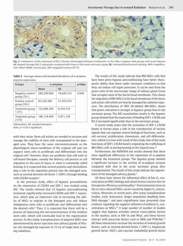
group (►Fig. 3). Furthermore, there were significant differ-ences in MANOVA test (p < 0.05) of α-amylase expression between the treatment groups. The acute hypoxia group showed a significant increase in α-amylase enzyme activity compared with that in the acute normoxia group (►Table 3).
DiscussionStem cell transplantation is one of the promising therapies that is predicted to restore the function of the salivary gland damage by regenerating acinar cells. Stem cell therapy can be successful in addition to the need for adaptive stem cells and when transplanted stem cells are attached and integrated
Table 1 Average value and standard deviation (SD) expres-sion of SDF1-CXCR4
Groups SDF1-CXCR4 p-Value
Mean SD
Negative control group (T0–) 1.640 0.555 0.000
Positive control group (T0+) 1.160 0.219
Treatment group 1 (T1) 2.920 0.540
Treatment group 2 (T2) 3.720 1.825
Abbreviation: SD, standard deviation.
Fig. 1 Comparison of CXCR4 (brown chromogen) expression between treatments. In the slide it appears that hypoxic group in acute conditions showed mainly CXCR4 expression (A) that was stronger than that in the normoxic group (B). Immunohistochemical staining, 400× magnifica-tion; Nikon H600L microscope; 300-megapixel camera DS Fi2.
Fig. 2 Microscopic images of BM-MSCs labeled using PKH 26. The green light in the image shows the distribution of labeled BM-MSCs. Hypoxic condition (A), normoxic condition (B).
Table 2 Average values and standard deviations of expres-sion of Bcl-2
Groups Bcl-2 p-Value
Mean SD
Negative control group (T0–) 23.600 5.176 0.000
Positive control group (T0+) 4.000 1.870
Treatment group 1 (T1) 5.400 1.140
Treatment group 2 (T2) 11.800 1.303
Abbreviation: SD, standard deviation.Note: p < 0.05 is significant.
241Xerostomia Therapy Due to Ionized Radiation Mulyani et al.
European Journal of Dentistry Vol. 13 No. 2/2019
Fig. 3 Comparison of the expression of Bcl-2 (brown chromogen) between treatments. In the slide, it appears that groups with acute hypoxia (A) showed stronger Bcl-2 expression compared with those in the acute normoxic group (B). Immunohistochemical staining, 400× magnifica-tion; Nikon H600L microscope; 300-megapixel camera DS Fi2.
Table 3 Average values and standard deviations of α-amylase enzyme expression
Groups α-amylase p-Value
Mean SD
Negative control group (T0–)
289,259.000 18,645.313 0.000
Positive control group (T0+)
95,330.400 31,503.973
Treatment group 1 (T1)
152,088.200 6,434.510
Treatment group 2 (T2)
186,118.400 5,971.156
Abbreviation: SD, standard deviation.Note: p < 0.05 is significant.
with their niche. Stem cell niches are needed to increase and manage the viability of stem cells transplanted in the dam-aged area. They have the same microenvironment as the physiological micro-conditions of the original cell and can support stem cells to proliferate and differentiate into the original cell.4 However, there are problems that still exist in cell-based therapies, namely the delivery cell process or cell migration to the area of injury or what is commonly called homing. It is suspected that several proteins and chemokines play a role in the migration process into the damaged area, such as stromal derived-cell factor 1 (SDF1) through binding with CXCR4 receptor.1
In the previous study, effect of hypoxic preconditions on the expression of CXCR4 and SDF-1 was studied using IHC. The results showed that 1% hypoxic preconditioning treatment significantly increased CXCR4 and SDF-1 expres-sion, which play an important role in increasing the abil-ity of MSCs to migrate to the damaged area and induce endogenous stem cells to proliferate and differentiate into the desired cells.9 Thus, it is expected that there will be a process of improving the microenvironment of the resident stem cells, which will eventually lead to the regeneration process. In this study, transplantation of adaptive MSCs was administered by direct injection into salivary glands of Wis-tar rats damaged by exposure to 15 Gy of single dose ioniz-ing radiations.
The results of this study indicate that BM-MSCs cells that have been given hypoxic preconditioning have better thera-peutic ability than those under normoxic conditions so that they can induce cell repair processes. It can be seen from the green color of the microscopic image of salivary gland tissue that occupies most of the ductal basal membrane. This shows the migration of BM-MSCs in the basal membrane of the ducts and acinar cells which are heavily damaged by radiation expo-sure. The distribution of PKH 26-labeled BM-MSCs shows that green coloration is stronger in hypoxic group than in the normoxic group. The IHC examination results in the hypoxic group showed that the expression of binding SDF1-CXCR4 and Bcl-2 increased significantly than in the normoxic group.
A recent study states that the activation of SDF-1-CXCR4 bonds in tissues plays a role in the transduction of various signals that can regulate several biological functions, such as cell survival, proliferation, chemotaxis, and cell differentia-tion, which is in line with result of this study. One of the main functions of SDF1-CXCR4 bond is regulating the trafficking of BM-MSCs cells in during homing in the injured area.10
Furthermore, the MANOVA test results showed that there were significant differences in the expression of α-amylase between the treatment groups. The hypoxia group showed a significant increase in the activity of α-amylase enzyme compared with that in the acute normoxic group after transplantation. The results of this study indicate the regener-ation of the damaged salivary glands.11
All these have shown the influential effect of low O2 con-centration on MSCs biology and raised serious concern over its therapeutic efficiency and biosafety.12 Environmental stress to the in vitro cultured MSCs can be caused by higher O2 concen-tration. Moreover, in recent years after many transplantation studies, early senescence, longer population doubling time, DNA damage,13 and poor engraftment have presented clear evidence regarding the negative influence of ambient O2 con-centration on MSCs.14 A large number of number of studies suggest that hypoxia activates several transcription factors in the nucleus, such as HIF-1α and NFκβ, and these factors interact with paracrine factors such as MEK and PI3K/Akt.15 All these interactions increase the secretion of several growth factors, such as stromal-derived factor 1 (SDF-1), hepatocyte growth factor (HGF), and vascular endothelial growth factor
242
European Journal of Dentistry Vol. 13 No. 2/2019
Xerostomia Therapy Due to Ionized Radiation Mulyani et al.
(VEGF) with increased expression of each receptor such as CXCR4, increased secretion of some anti apoptotic proteins, such as Bcl-2 and Bcl-xL as a survival factor.16
ConclusionThis study concluded that (1) BM-MSCs transplantation with hypoxic preconditioning of 1% O2 increases the expression of SDF1-CXCR4 and Bcl-2 that contribute to cell migration and cell survival. (2) BM-MSCs can improve regeneration process of the damaged salivary glands by increasing the activity of the α-amylase as a marker of regeneration process in the sal-ivary gland tissue.
Conflict of InterestNone declared.
AcknowledgmentsThe authors would like to thank Universitas Airlangga, Institute of Tropical Disease, Stem Cell Division, Faculty of Dental Medicine and Stem Cell and Tissue Bank Center RSUD Dr. Soetomo, Surabaya, Indonesia.
References
1 Chambers MS, Garden AS, Kies MS, Martin JW. Radiation-in-duced xerostomia in patients with head and neck cancer: pathogenesis, impact on quality of life, and management. Head Neck 2004;26(9):796–807
2 Lombaert IM, Brunsting JF, Wierenga PK, et al. Rescue of sali-vary gland function after stem cell transplantation in irradiat-ed glands. PLoS One 2008;3(4):e2063
3 Ponnaiyan D, Jegadeesan V. Comparison of phenotype and dif-ferentiation marker gene expression profiles in human dental pulp and bone marrow mesenchymal stem cells. Eur J Dent 2014;8(3):307–313
4 Sohni A, Verfaillie CM. Mesenchymal stem cells migration homing and tracking. Stem Cells Int 2013;13:1–10
5 Estrada JC, Albo C, Benguría A, et al. Culture of human mes-enchymal stem cells at low oxygen tension improves growth
and genetic stability by activating glycolysis. Cell Death Differ 2012;19(5):743–755
6 Lin CY, Chang FH, Chen CY, et al. Cell therapy for salivary gland regeneration. J Dent Res 2011;90(3):341–346
7 Rantam FA, Ferdiansyah M, Purwati A, Stem Cell Mesenchy-mal, Hematopoetik dan Model Aplikasi. 2nd ed. Surabaya, Indonesia: Airlangga University Press; 2014;145–155
8 Crosby K, Simendinger J, Grange C, Ferrante M, Bernier T, Stanen C, Immunohistochemistry protocol for paraffin-embedded tis-sue section - advertisement. Cell Signal Technol 2016. Available at: https://www.jove.com/video/5064/immunohistochemistry-protocol-for-paraffin-embedded- tissue-sections. Accessed on June 15, 2016
9 Mulyani SWM, Ernawati DS, Astuti ER, Rantam FA. Hypoxic preconditioning effect on stromal cells derived factor-1 and C-X-C chemokine receptor type 4 expression in Wistar rat’s (Rattus norvegicus) bone marrow mesenchymal stem cells (in vitro study). Vet World 2018;11(7):965–970
10 Haque N, Rahman MT, Abu Kasim NH, Alabsi AM. Hypoxic culture conditions as a solution for mesenchymal stem cell based regenerative therapy. ScientificWorldJournal 2013;2013:632972
11 Lim JY, Ra JC, Shin IS, et al. Systemic transplantation of human adipose tissue-derived mesenchymal stem cells for the regen-eration of irradiation-induced salivary gland damage. PLoS One 2013;8(8):e71167
12 Feng J, van der Zwaag M, Stokman MA, van Os R, Coppes RP. Isolation and characterization of human salivary gland cells for stem cell transplantation to reduce radiation-induced hyposalivation. Radiother Oncol 2009;92(3):466–471
13 Mohamadnejad M, Pournasr B, Bagheri M, et al. Transplanta-tion of allogeneic bone marrow mesenchymal stromal cell-de-rived hepatocyte-like cells in homozygous familial hypercho-lesterolemia. Cytotherapy 2010;12(4):566–568
14 Greijer AE, van der Wall E. The role of hypoxia inducible factor 1 (HIF-1) in hypoxia induced apoptosis. J Clin Pathol 2004;57(10):1009–1014
15 Alcarez TM, Naranjo SS, Jimenez C. Hypoxic induces the activa-tion of the PI3K/Akt cell survival pathway in PC12 cells – pro-tective role in apoptosis. J Biol Chem 2001;276:22368–22374
16 Kagami H, Wang S, Hai B. Restoring the function of salivary glands. Oral Dis 2008;14(1):15–24