x-ray photoelectron spectroscopy - kfupm · x-ray photoelectron spectroscopy. elemental + chemical...
TRANSCRIPT

Tour in
Techniques

ContentsMedical: Quick Impressions Why Surface?One detailed example : XPS
• Conditions, information, • Ingredients:
1-Source 2-Surface 3-Spectrom• Extensions
XPD, ARPES, SRPES, PEEMOther techniquesConclusion

Medical: quick impressionsalmost alwaysLiving/ moving tissues
Nondestructive testing
Volumes to treat, image in 3D or project to 2D
mm level imaging (nm with STM

Why surface is interesting?Interesting physics:– half the volume is missing: laws are different – Oxidation and gas association phenomena– Layers growth, thin films
The place where atoms are manipulated (STM)
Important Applications– electronics industry: chips are in surface realm
• Silicon, germanium…
– catalysis -corrosion

XPS:
X-ray
Photoelectron
Spectroscopy


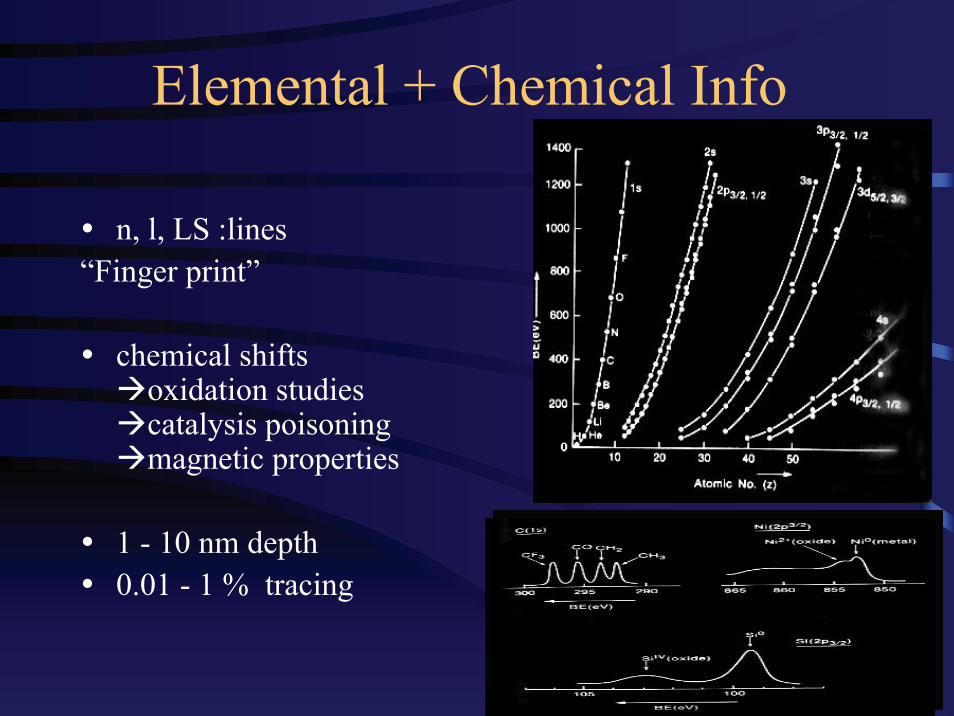
Elemental + Chemical Info
n, l, LS :lines“Finger print”
chemical shifts oxidation studies catalysis poisoning magnetic properties
1 - 10 nm depth0.01 - 1 % tracing

22 24 26 28 30 32 34 36 38 400
5
10
15
20
25
30
35
40
45
50
55
60
1.3 (min.)
2.0 (min.)
2.7 (min.)
3.3 (min.)
5.0 (min.)
10 (min.)
18 (min.)
35 (min.)
95 (min.)
322 (min.)
599 (min.)
1604 (min.)
2620 (min.)
3728 (min.)
0 (min.)
380oC, 1atm, air
I Ge3
d (a
rb. u
nits
)
Binding Energy (eV)

Extended techniques

X-ray Photoelectron Diffraction (XPD)
Track angular variation of a certain peak intensity. It varies only if it belongs to the second layer
Consideration: preparation, propped depth, time->surface structure + enhanced surface sensitivity

Photoelectron Emission Microscopy (PEEM)
Focus x-rays image– collimating (signal↓ -> synchrotron)– x-ray optics (under development)
Advantage: Element specific, chemical status
Applications:• diffusion, segregation, Shottky barrier

Auger process
3-e process, not f (hν)X-ray or e-beam inducedMicroscopy (SAM)
fluorescence (by x-ray as TRXF)1 in 109-1012

Other Surface Techniques

Scanning Probe Microscopy
STM, AFM …
high (or even atomic) resolution atomically sharp tip raster a surfacevary/fix current voltage, move tactictip material
www.park.com

NIST
(left) single atomic zig-zag chain of Cs (red) on GaAs(110) surface.
(right) substitutional Cr impurities (small bumps) in Fe(001) surface

Proton-induced X-ray emission (PIXE)
micro PIXE– images (now: 5 x 5 µm)3 MeV protons (accelerator + focus) inner vacancy – florescence
trace analysis of elemental compositionsimultaneous multi-element (NDT!)multilayer --- not a surface technique
Mineralogy, Geochemistry & Materials Science


Conclusion
Microscopy with Spectroscopy feature
Medical: interest, restrictions, importance
Material science: diversity