wright, cody leforge. evaluation of absorption and post
TRANSCRIPT

ABSTRACT
Wright, Cody LeForge. Evaluation of Absorption and Post-Absorptive Metabolism of
Inorganic and Organic Zinc Sources. (Under the direction of Dr. Jerry W. Spears)
Experiments were conducted to compare the absorption and metabolism of Zn
from inorganic or organic sources. In Experiment 1, Holstein bull calves were
supplemented with 0 or 20 mg Zn/kg diet as ZnSO4, Zn proteinate (ZnProt) or a mixture
of ZnSO4 and ZnProt (ZnMix) for 98 d. From d 98 to 112, supplemental Zn levels for
half of the bulls in each treatment were increased to 500 mg Zn/kg. Zinc concentrations
in plasma and duodenal, liver, kidney, and muscle samples were greater in bulls
supplemented with high levels of ZnProt or ZnMix than ZnSO4. At high levels, ZnSO4
increased skin Zn concentrations relative to ZnProt. Skin and hoof wall Zn concentrations
were greater in bulls supplemented with low levels of ZnMix compared to ZnSO4.
Experiments 2 and 3 compared the uptake and transport of Zn from ZnSO4 and ZnProt or
Zn propionate (ZnProp) by ruminal and omasal epithelium using parabiotic chambers.
Uptake of Zn by omasal epithelium was negligible and Zn transport by ruminal and
omasal epithelium was non-detectible in all experiments. Zinc uptake by ruminal
epithelium increased as incubation time increased and was greater and tended to be
greater from ZnProt and ZnProp, respectively, than from ZnSO4. In concentration-
dependent experiments, Zn uptake was affected by a concentration × source interaction.
More Zn was absorbed from ZnProt than from ZnSO4 when added at 200, but not 10 µM.
Concentration-dependent uptake of ZnSO4 and ZnProp was unaffected by source.
Following ruminal digestion, solubility of Zn from ZnSO4 and ZnProt was influenced and
uptake tended to be influenced by a source × concentration interaction with both being

greater from ZnProt than from ZnSO4. Solubility Zn from ZnSO4 and ZnProp was
influenced by a concentration × source interaction with Zn from ZnSO4 being more
soluble than Zn from ZnProp at 10, but not 200 µM. Zinc uptake from ZnSO4 and
ZnProp following ruminal digestion was affected by a source × concentration × time
interaction. At lower Zn levels, uptake was unaffected by source; however, at higher
concentrations, uptake increased over time and was greater from ZnSO4 than ZnProp.
Experiments 4 and 5 compared the uptake and transport of Zn from ZnSO4 and ZnProt or
ZnProp using Caco-2 cell monolayers. In time- or concentration-dependent experiments,
Zn uptake and transport increased as incubation time and Zn concentration increased;
however, neither was affected by Zn source. Addition of inositol hexaphosphate (IP6) and
Ca reduced solubility, uptake and transport of Zn. In the presence of IP6 and Ca, Zn
uptake from ZnSO4 and ZnProp tended to be affected by a source × concentration
interaction with uptake being greater from ZnSO4 than from ZnProp at 200, but not 10
µM. Following ruminal and intestinal digestions, Zn uptake from ZnProp tended to be
greater than from ZnSO4; whereas, uptake from ZnSO4 and ZnProt were similar.
Following intestinal digestion alone, Zn uptake from ZnSO4 and ZnProt was affected and
uptake from ZnSO4 or ZnProp tended to be affected by a source × concentration
interaction with uptake being greater from ZnProt or ZnProp than from ZnSO4 when
added at 200, but not 10 µM. Zinc solubility following intestinal digestion was affected
by a source × concentration interaction. Zinc from ZnSO4 was more soluble than Zn from
ZnProp when added to digestions at 10; however, at 200 µM Zn, Zn from ZnProp was
more soluble than from ZnSO4. Results suggest that at low concentrations or in the
absence of antagonists uptake and transport of Zn from inorganic and organic Zn sources

are similar. However, following simulated digestion, uptake of Zn from organic Zn
sources appears to be greater than from inorganic source.

EVALUATION OF ABSORPTION AND POST-ABSORPTIVE METABOLISM OF INORGANIC
AND ORGANIC ZINC SOURCES
by
CODY LEFORGE WRIGHT
A dissertation submitted to the Graduate Faculty of North Carolina State University
in partial fulfillment for the requirements for the Degree of
Doctor of Philosophy
ANIMAL SCIENCE
Raleigh
2000
APPROVED BY:
______________________________ ______________________________ Dr. Jerry W. Spears Dr. Vivek Fellner Chair of Advisory Committee
______________________________ ______________________________ Dr. Gerald B. Huntington Dr. Matthew H. Poore

ii
DEDICATION
This dissertation is dedicated…
to my loving wife Stephanie for her unending support, and commitment and
devotion to me and my education, without her by my side, this would have
been impossible;
to my parents Larry and Jeane Wright, for their compassion and encouragement in
all of my endevors, their love and pride in me means everything;
and to the memory of my beloved grandparents Warren and Eldora Wright, for
their love and for instilling in me at a tender age a love and appreciation
for agriculture and fine people in agriculture, I miss you.

iii
BIOGRAPHY
Cody LeForge Wright was born in Sioux Falls, South Dakota, on December 21,
1971, to Larry and Jeane Wright. Cody, along with his sister Karli, and twin brothers
Casey and Kelly, atteneded elementary school in Valley Springs, South Dakota and
junior and senior high school in Brandon, South Dakota. Upon graduation in 1990, Cody
enrolled at South Dakota State University as a Mechanical Engineering major. However,
following a humbling semester in the Engineering curriculum, Cody came to his senses
and changed his major to Animal Science. In May of 1994, Cody graduated With Honor
with a Bachelor of Science degree in Animal Science. Cody and his wife, Stephanie,
were married on December 10, 1994. After their marriage, he and Stephanie moved to
Manhattan, Kansas, where Cody began pursuit of his Master of Science degree at Kansas
State University. In 1996, Cody earned his Master of Science degree in Animal Science
under the direction of Dr. Larry Corah. He and Stephanie then moved to Raleigh, North
Carolina, where Cody began pursuit of his Doctor of Philosophy degree in Animal
Science at North Carolina State University under the direction of Dr. Jerry Spears. While
at North Carolina State University, Cody also coached the Livestock Judging Team.
Upon completion of his Doctor of Philosophy degree, he and Stephanie will be moving to
Brookings, South Dakota, where Cody has accepted an Assistant Professor – Beef
Extension Specialist position at South Dakota State University.

iv
ACKNOWLEDGEMENTS
I owe a tremendous debt of gratitude to Dr. Jerry Spears for his willingness to
accept me as a graduate student, for the tremendous wealth of knowledge he has shared,
and for the personal and professional advice and guidance he has provided over the past
four years. His patience and understanding are greatly appreciated. My sincere thanks are
also extended to Drs. Vivek Fellner, Gerald Huntington and Matt Poore for their
willingness to serve as members of my advisory committee, and for their keen
perspectives and counsel with respect to my education and career. I would be remiss not
to acknowledge the contribution of Dr. Mark Failla to my education and professional
development. Dr. Failla not only taught me new research techniques, but also to view
research from a new perspectives, “to think outside the box” as he would say. His insight,
patience and friendship provided a tremendous environment for me to grow as a scientist.
My fellow graduate students and great friends Todd Armstrong, Terry Engle, Greg
Schaeffer, and Mark Tiffany have provided me with support, encouragement and
countless episodes of uncontrollable laughter for which I am incredibly grateful. My
collegues and I have also been blessed to have technicians like Karen “Missy” Lloyd,
Sarah McLeod, and Marcia Corns. Their dedication, kindness, friendship and sense of
humor remain unmatched. I appreciate the dedication and friendship of Robin Hopkins
and Maureen Searcy in Dr. Fialla’s laboratory. Without their assistance, much of my data
collection would have been nearly impossible. Those of us who are involved in ruminant
nutrition are absolutely spoiled by “The Crew” at the Butner Beef Cattle Field
Laboratory. Dean Askew, David Cheek, Rodney Willette, and Jay Woodlief unselfishly
contribute their time, talent, and good humor, making our jobs much easier. My parents,

v
Larry and Jeane Wright, and siblings, Karli, Casey, and Kelly have given me
unconditional love and support throughout my life. In good times and bad, success and
failure, they have been there for me and I love them dearly. My wife and best friend
Stephanie deserves more credit for my achievements than anyone. Her absolute love,
support, and devotion are the strength I have relied upon repeatedly throughout this
endeavour. The strength of the love and commitment we share for each other can
overcome any trial or obstacle that may come to bear. Above all, I thank the Lord God
and His Son Jesus Christ for guiding my life. Without Their love none of this would have
been possible.

vi
TABLE OF CONTENTS
LIST OF FIGURES ................................................................................................................ viii LIST OF TABLES ...................................................................................................................xi LITERATURE REVIEW ........................................................................................................... 1
Introduction..................................................................................................................... 1 Zinc Absorption .............................................................................................................. 1 Peptide and Amino Acid Absorption in Ruminants ....................................................... 3 Propionate Absorption in Ruminants .............................................................................. 4 Bioavailability of Organic Zinc Sources......................................................................... 5 Literature Cited ............................................................................................................. 15
CHAPTER 1. EFFECT OF ZINC SOURCE AND DIETARY LEVEL ON ZINC METABOLISM IN
HOLSTEIN BULLS .................................................................................................... 21 Introduction................................................................................................................... 24 Materials and Methods.................................................................................................. 25 Results ........................................................................................................................... 29 Discussion..................................................................................................................... 32 Implications ................................................................................................................... 38 References Cited ........................................................................................................... 39
CHAPTER 2. UPTAKE AND TRANSPORT OF ZINC FROM ZINC SULFATE AND ZINC PROTEINATE
BY RUMINAL AND OMASA L EPITHELIUM ................................................................. 51 Introduction................................................................................................................... 54 Materials and Methods.................................................................................................. 55 Results ........................................................................................................................... 58 Discussion..................................................................................................................... 59 Implications ................................................................................................................... 63 Literature Cited ............................................................................................................. 64
CHAPTER 3. UPTAKE AND TRANSPORT OF ZINC FROM ZINC SULFATE AND ZINC PROPIONATE
BY RUMINAL AND OMASA L EPITHELIUM ................................................................. 70 Introduction................................................................................................................... 73 Materials and Methods.................................................................................................. 74 Results ........................................................................................................................... 77 Discussion..................................................................................................................... 78 Implications ................................................................................................................... 81 Literature Cited ............................................................................................................. 82

vii
CHAPTER 4. UPTAKE AND TRANSPORT OF ZINC FROM ZINC SULFATE AND ZINC PROTEINATE BY CACO-2 CELLS .................................................................................................. 88
Introduction................................................................................................................... 91 Materials and Methods.................................................................................................. 92 Results ........................................................................................................................... 99 Discussion................................................................................................................... 103 Implications ................................................................................................................. 108 Literature Cited ........................................................................................................... 110
CHAPTER 5. UPTAKE AND TRANSPORT OF ZINC FROM ZINC SULFATE AND ZINC PROPIONATE
BY CACO-2 CELLS ................................................................................................ 126 Introduction................................................................................................................. 129 Materials and Methods................................................................................................ 130 Results ......................................................................................................................... 137 Discussion................................................................................................................... 140 Implications ................................................................................................................. 145 Literature Cited ........................................................................................................... 146
SUMMARY........................................................................................................................ 163

viii
LIST OF FIGURES
Chapter 2 Figure 1. Uptake of Zn by rumen epithelium from mucosal buffer containing
20 µM Zn as ZnSO4 or ZnProt during a 240 min incubation at 39°C……... 67 Figure 2. Uptake of Zn by rumen epithelium from mucosal buffer containing
10 or 200 µM Zn as ZnSO4 or ZnProt during a 240 min incubation at 39°C……………………………………………………………………... 68
Figure 3. Uptake of Zn by rumen epithelium from aqueous fractions of
simulated rumen digestions containing 10 or 200 µM added Zn as ZnSO4 or ZnProt during a 240 min incubation at 39°C……………………. 69
Chapter 3 Figure 1. Uptake of Zn by rumen epithelium from mucosal buffer containing
20 µM Zn as ZnSO4 or ZnProp during a 240 min incubation at 39°C…….. 85 Figure 2. Uptake of Zn by rumen epithelium from mucosal buffer containing
10 or 200 µM Zn as ZnSO4 or ZnProp during a 240 min incubation at 39°C……………………………………………………………………... 86
Figure 3. Uptake of Zn by rumen epithelium from aqueous fractions of
simulated rumen digestions containing 10 or 200 µM Zn as ZnSO4 or ZnProp during a 240 min incubation at 39°C……………………………… 87
Chapter 4 Figure 1. Uptake of Zn from buffer containing 20 µM Zn as ZnSO4 or ZnProt
incubated for increasing times at 37°C…………………………………….. 115 Figure 2. Uptake of Zn from buffer containing increasing Zn concentrations as
ZnSO4 or ZnProt, during a 60 min incubation at 37°C…………………….. 116 Figure 3. Uptake of Zn from buffers containing increasing concentrations of Zn
as ZnSO4 or ZnProt, in the presence or absence of 200 µM IP6 and µM Ca. Cells were incubated for 60 min at 37°C……………………...………. 117
Figure 4. Solubility of increasing concentrations of Zn as ZnSO4 or ZnProt in
the presence of 200 µM IP6 and µM Ca………………………...…………. 118 Figure 5. Uptake and solubility of Zn from aqueous fractions of ruminal and
intestinal digestions containing 10 or 200 µM added Zn as ZnSO4 or

ix
ZnProt……………………………………………………………………… 119 Figure 6. Uptake and solubility of Zn from aqueous fractions of intestinal
digestions containing 10 or 200 µM added Zn as ZnSO4 or ZnProt……….. 120 Figure 7. Uptake and transport of Zn from buffer containing 20 µM Zn as
ZnSO4 or ZnProt incubated for increasing times at 37°C………………….. 121 Figure 8. Uptake and transport of Zn from buffer containing increasing Zn
concentrations as ZnSO4 or ZnProt, during a 120 min incubation at 37°C... 122 Figure 9. Uptake of Zn from buffers containing 10 or 200 µM Zn as ZnSO4
or ZnProt, in the presence or absence of 200 µM IP6 and µM Ca……...….. 123
Figure 10. Transport of Zn from buffers containing 10 or 200 µM Zn as ZnSO4 or ZnProt, in the presence or absence of 200 µM IP6 and µM Ca……...….. 124
Figure 11. Uptake and transport of Zn from aqueous fractions of ruminal and
intestinal digestions containing 10 or 200 µM added Zn as ZnSO4 or ZnProt……………………………………………………………………… 125
Figure 12. Uptake and transport of Zn from aqueous fractions of intestinal
digestions containing 10 or 200 µM added Zn as ZnSO4 or ZnProt………. 126 Chapter 5 Figure 1. Uptake of Zn from buffer containing 20 µM Zn as ZnSO4 or ZnProp incubated for increasing times at 37°C…………………………………….. 151 Figure 2. Uptake of Zn from buffer containing increasing Zn concentrations as
ZnSO4 or ZnProp, during a 60 min incubation at 37°C……………………. 152 Figure 3. Uptake of Zn from buffers containing increasing concentrations of Zn
as ZnSO4 or ZnProp, in the presence or absence of 200 µM IP6 and µM Ca……………………………………………………………...…………… 153
Figure 4. Solubility of increasing concentrations of Zn as ZnSO4 or ZnProp in
the presence of 200 µM IP6 and µM Ca……………………...……………. 154 Figure 5. Uptake and solubility of Zn from aqueous fractions of ruminal and
intestinal digestions containing 10 or 200 µM added Zn as ZnSO4 or ZnProp……………………………………………………………………... 155
Figure 6. Uptake and solubility of Zn from aqueous fractions of intestinal
digestions containing 10 or 200 µM added Zn as ZnSO4 or ZnProp………. 156

x
Figure 7. Uptake and transport of Zn from buffer containing 20 µM Zn as
ZnSO4 or ZnProp incubated for increasing times at 37°C…………………. 157 Figure 8. Uptake and transport of Zn from buffer containing increasing Zn
concentrations as ZnSO4 or ZnProp, during a 120 min incubation at 37°C………………………………………………………………………... 158
Figure 9. Uptake of Zn from buffers containing 10 or 200 µM Zn as ZnSO4
or ZnProp, in the presence or absence of 200 µM IP6 and µM Ca…...……. 159 Figure 10. Transport of Zn from buffers containing 10 or 200 µM Zn as ZnSO4
or ZnProp, in the presence or absence of 200 µM IP6 and µM Ca……….... 160 Figure 11. Uptake and transport of Zn from aqueous fractions of ruminal and intestinal digestions containing 10 or 200 µM added Zn as ZnSO4 or
ZnProp……………………………………………………………………... 161 Figure 12. Uptake and transport of Zn from aqueous fractions of intestinal
digestions containing 10 or 200 µM added Zn as ZnSO4 or ZnProp…….… 162

xi
LIST OF TABLES
Chapter 1 Table 1. Diet composition………………………………………………………….. 44 Table 2. Effect of Zn source and level on performance……………………………. 45 Table 3. Effect of time on plasma Zn concentration and alkaline phosphatase
activity (Phase 1)……………………………..…………………………….. 46 Table 4. Effect of Zn source and level tissue homogenate Zn concentration…..….. 47 Table 5. Effect of Zn source and level tissue metallothionein concentration…..….. 48 Table 6. Effect of Zn source and level on plasma Zn concentrations and
alkaline phosphatase activity (Phase 2)……………………………………. 49 Table 7. Effect of Zn source and level on final tissue Zn concentrations
(Phase 2)……………………………………………………………………. 50 Table 8. Effect of Zn source and level on hair, hoof, and skin Zn
concentrations (Phase 2)…………………………………………………… 51 Chapter 2 Table 1. Final tissue Zn uptake………………………………………..…………… 70 Chapter 3 Table 1. Final tissue Zn uptake…………………………………………..………… 88

1
LITERATURE REVIEW
Introduction
Zinc is a group IIb transition metal with an atomic number of 30 and a molecular
weight of 65.39. Since Zn possesses a completed d subshell, it has only one oxidation
state. Zinc is known to be a component of enzymes that catalyze over 50 different
reactions and reside in all six major Enzyme Commission classes (Chesters, 1997). Some
major Zn-metalloenzymes found in mammalian tissue are alcohol dehydrogenase,
alkaline phosphatase, carbonic anhydrase, carboxypeptidases A and B, leucine
aminopeptidase, mannosidase, and superoxide dismutase.
Presence of Zn in animal tissues has been known since the end of the nineteenth
century; however, clear evidence of the biological function of Zn was not available until
1934 (Chesters, 1997). Todd et al. (1934) first determined that Zn was necessary for
growth and health in rats and mice. Shortly thereafter, Tucker and Salmon (1955)
discovered that Zn prevented and cured parakeratosis in pigs and Zn deficiency was
demonstrated in the chick (O’Dell and Savage, 1957). In ruminants, Zn deficiency is
uncommon; however, growth and health responses to supplemental Zn have been
observed in cattle and sheep grazing pastures containing 20 mg Zn/kg DM (Underwood
and Suttle, 1999).
Zinc Absorption
Zinc absorption occurs primarily from the small intestine in most species
(Underwood and Suttle, 1999) and is believed to be facilitated by a carrier-mediated
process, which has yet to be clearly defined (Cousins, 1996). In ruminants, Zn is
absorbed primarily from the small intestine (Miller and Cragle, 1965; Hampton et al.,

2
1976); however, Zn absorption from ruminal tissue has been demonstrated. Zinc uptake
and apparent absorption from rumen tissue has been clearly demonstrated in vivo using
65Zn dosed lambs (Arora et al., 1969) and wethers fitted with ruminal, abomasal and ileal
cannulas (Kennedy and Bunting, 1991, Kirk et al., 1994). Less than 2% of total Zn is
absorbed from the large intestine in ruminants (Hampton et al., 1976); however, when
intestinal absorption was impaired Zn was absorbed from the cecum and colon in rats
(Hara et al., 2000).
Specific Zn absorption mechanisms have yet to be clearly defined; however,
absorption kinetics have been examined in several systems. At least two components of
Zn uptake have been identified in isolated brush border membrane vesicles of rats
(Menard and Cousins, 1983) and pigs (Blakeborough and Salter, 1987). The first
component exibits saturation kinetics with a Km of 70-350 µM depending on the system
used (Chesters, 1997). The second component responded linearly to Zn concentrations up
to 2 mM (Lee et al., 1989). Zinc uptake is also know to be greater from the lumen of Zn-
deficient rats than from Zn-adequate controls, suggesting that Zn uptake is a major site of
homeostatic regulation (Chesters, 1997). Within the mucosa Zn is likely bound by one of
several potential intracellular ligands. Hempe and Cousins (1991, 1992) hypothesized
that at low Zn concentrations, absorbed Zn is bound by a cysteine-rich intestinal protein
(CRIP) that is not metallothionein (MT). The authors suggested that CRIP may function
as an intracellular Zn shuttle to translocate Zn to the basolateral membrane or to one of
many potential Zn metalloproteins within the mucosa. In contrast, when Zn was at high
concentrations, absorbed Zn induced synthesis of MT (Cousins, 1985) and subsequently a
large fraction of the absorbed Zn was bound by MT and sequestered in the mucosa

3
(Hempe and Cousins, 1992). The authors suggested that, at high Zn concentrations, MT
binds Zn to prevent absorption of potentially toxic levels of Zn; whereas, when Zn was at
low concentrations, CRIP transferred absorbed Zn to the basolateral membrane for
transport into circulation. Researchers have only recently defined zinc transporters. Four
distinct Zn transport proteins have been isolated; however only two, ZnT-1 and ZnT-2,
appear to function in the small intestine (McMahon and Cousins, 1998). Both of these
transport proteins seem to be localized to the basolateral membrane and function to
transport Zn out of the enterocyte into circulation (McMahon and Cousins, 1998).
Divalent cation transporter 1 (DCT1), a transporter of a number of cations, has recently
been cloned (Gunshin et al., 1997). Upregulation of DCT1 in response to low dietary iron
status but without additional iron in the intestinal lumen may cause more Zn to be
absorbed because the affinities for both cations are similar (Cousins and McMahon,
2000).
Peptide and Amino Acid Absorption in Ruminants
Ruminant protein digestion relies on the same complement of pancreatic and
intestinal proteases to effect breakdown of protein as do non-ruminants and absorb amino
acids and small peptides by similar mechanisms (Merchen, 1988). Amino acid absorption
in the small intestine is a Na+-dependent, carrier-mediated process, and can be described
as a type of secondary active transport (Armstrong and Hutton, 1975). The small intestine
has the capacity to transport between 65 and 80% of the total amino acid supply reaching
duodenum (Armstrong and Hutton, 1975). The small intestine has been found to be the
primary site of amino acid absorption in ruminants. Wilson and Webb (1990)
characterized the absorption of lysine and methionine in bovine ileal and jejunal brush

4
border membrane vesicles. Their observations suggest that amino acid absorption may
occur via mediated and non-mediated, and Na+-dependent and Na+-independent
components.
Di- and tri-peptides have also been found to be absorbed in the small intestine via
mechanisms driven by H+ and(or) Ca gradients (Webb et al., 1993). Evidence is strong
for the existence of multiple peptide transport systems, including one type that is
electrogenic in nature and cotransports two H+ for every peptide transported (Webb et al.,
1992). Furthermore, peptides may be absorbed more rapidly than amino acids and
subsequently partially hydrolyzed by intracellular peptidases (Webb et al., 1993).
Recently, researchers have demonstrated the ability of ovine ruminal and omasal
epithelium to absorb and translocate amino acids and di-peptides (Matthews and Webb,
1995). Free and peptide-bound amino acid absorption appeared to be non-saturable and
occurred to a greater extent from omasal epithelium than from ruminal epithelium.
Furthermore, carnosine was absorbed across both tissues without hydrolysis, whereas
methionylglycine was partially hydrolyzed in both tissues and to a greater extent by
omasal epithelium (Matthews and Webb, 1995). In sheep and lactating Holstein cows,
peptide transporter mRNA has been found in rumen, omasum, duodenum, jejunum, and
ileum (Chen et al., 1999). No hybridization was observed with mRNA from abomasum,
cecum, colon, liver, kidney, or semitendinosis and longissumus muscles.
Propionate Absorption in Ruminants
Though some volatile fatty acids (VFA) will leave the reticulo-rumen with the
digesta, flowing to the lower gastro-intestinal tract, the vast majority of the acids
produced by ruminal fermentation are absorbed from the reticulo-rumen (Merchen,

5
1998). To date there is no evidence for any active transport of VFA from the rumen.
Since the pKa of VFA is near 4.8, most VFA are dissociated at normal pH (6-7). Stevens
(1970) suggested that free (undissociated) acids may be absorbed via simple diffusion
whereas dissociated acids are likely absorbed in exchange for bicarbonate. In vitro
experiments sopport the hypothesis of Stevens. Kramer et al. (1996) used isolated ovine
ruminal epithelium in Ussing chambers to evaluate absorption of short-chain fatty acids
(SCFA). Their results suggest that SCFA anions either compete with Cl- for binding sites
on a common anion exchange mechanism or that SCFA anions are absorbed in exchange
with HCO3-.
Bioavailability of Organic Zinc Sources
Feed grade Zn is commercially available as inorganic salts, ZnSO4 or ZnO.
Organic Zn sources have become increasingly more popular in recent years as a result of
reported improvements in growth performance, feed efficiency, health, and carcass
characteristics in livestock. While the mode of action by which organic trace minerals
elicit beneficial responses remains to be elucidated, proposed mechanisms have included
enhanced solubility, greater absorption, prevention of antagonistic interactions, and
differences in post-absorptive metabolism. Ashmead et al. (1985) suggested that metal
ions may be transported in metal:amino acid or metal:peptide complexes. It is possible
that these complexes may be transported intact by amino acid and(or) peptide
transporters. The following is a brief review of research conducted with various organic
Zn sources.
Zinc amino acid chelates. Zinc amino acid chelates are defined as the products
resulting from the reaction of a metal ion from a soluble metal salt with a mole ratio of

6
one mole metal to one to three (preferably two) moles of amino acids to form coordinate
covalent bonds (Spears, 1996). The average weight of the hydrolyzed amino acids must
be approximately 150 and the resulting molecular weight of the chelate must not exceed
800. Zinc amino acid chelates (ZnAAC) have been compared to inorganic Zn sources in
chicks, dogs, lambs, and pigs.
Cao et al. (2000) compared numerous organic Zn sources to inorganic ZnSO4
using several in vitro techniques and broiler chicks and lambs in vivo. Chicks were fed
corn-soybean meal diets supplemented with 0, 200, 400, or 600 mg Zn/kg from ZnSO4 or
200 or 400 mg Zn/kg from ZnAAC or Zn proteinate (ZnProt). Feed intake and gain were
not different between organic Zn sources; however, both were greater in chicks fed
organic Zn relative to those fed inorganic Zn. Relative bioavailability values (%)
calculated based on bone Zn concentration were 104 and 116 in week 1, 100 and 135 in
week 2, and 83 and 139 in week 3 for ZnAAC and ZnProt, respectively. Lambs were
supplemented with 0, 700, 1400, or 2100 mg Zn/kg from ZnSO4, or 1400 mg Zn/kg from
ZnProt, ZnAAC, or ZnMet. Relative bioavailability calculations based on liver Zn and
metallothionein, kidney Zn and pancreas Zn concentrations were 130, 110, and 113% for
ZnProt, ZnAAC, and ZnMet respectively as compared to ZnSO4.
In three different experiments with dogs, Lowe et al. (1994a, 1994b, and 1998)
observed greater absorption and retention of Zn, and greater hair growth and Zn
concentration in dogs fed Zn as ZnAAC than those fed ZnO or Zn polysaccharide. In
contrast, Swinkels et al. (1996) fed Zn depleted pigs and pair-fed nondepleted pigs 45 mg
Zn/kg diet from ZnSO4 or ZnAAC for 36 d. Zinc concentrations of serum, liver,
pancreas, kidney, brain, and small intestine were not affected by Zn source.

7
Zinc amino acid complexes. Zinc amino acid complexes are defined as the
products resulting from the complexing of a soluble metal salt with an amino
acid(s)(Spears, 1996). Generally, Zn amino acid complexes refer to either Zn lysine
(ZnLys) or Zn methionine (ZnMet); however, in some cases multiple amino acids may
be complexed with the metal ion.
Malcolm-Callis et al. (2000) compared the effects of 30 mg supplemental Zn/kg
diet as ZnSO4, Zn amino acid complex or Zn polysaccharide on performance, carcass
characteristics and serum Zn concentrations of Brangus- and Angus-sired steers. The
authors reported no differences in performance or serum Zn concentrations with respect
to Zn source. Steers supplemented with organic Zn sources had greater s.c. fat thickness
and lower kidney, pelvic, and heart fat percentages than steers supplemented with ZnSO4.
Individual Zn amino acid complexes, ZnLys and ZnMet, have been researched
extensively in several species; however, for sake of brevity literature reviewed here will
focus on ruminants. Moreover, many early experiments compared responses of cattle fed
ZnMet to those of unsupplemented cattle. The lack of an inorganic Zn source for
comparison in these experiments precludes estimation of bioavailability and clouds
interpretation of the data. As such, only experiments that included an inorganic Zn
treatment will be reviewed.
Effects of ZnMet on growth performance, carcass characteristics, health and
immune function have been examined in growing and finishing cattle. Zinc methionine
supplementation has improved USDA Quality Grade and marbling score, and increased
external and kidney, pelvic, and heart fat in feedlot steers (Greene et al., 1988). Growth
performance of growing heifers (Spears, 1989) and feed efficiency of stressed steers

8
(Spears et al., 1991) tended to be greater in cattle supplemented with ZnMet than in those
supplemented with ZnO. Antibody titers to bovine herpes virus-1, but not parainfluenza-
3, were greater in stresses steers supplemented with ZnMet than in those supplemented
with ZnO (Spears et al., 1991). Lambs born to ewes fed 50 mg Zn per ewe daily were
weaned and supplemented with 25 mg Zn/kg diet from ZnMet or ZnO. Blastogenic
responses of unstimulated lymphocytes were greater from lambs supplemented with
ZnMet than from those supplemented with ZnO. However, when stimulated with
phytohemaglutinin, concanavalin A, or pokeweed mitogen, blastogenic responses were
unaffected by Zn source. Furthermore, lambs fed ZnMet had smaller welt diameters
following intradermal PHA injection than those fed ZnO. In contrast, Engle et al.
reported no difference in cell-mediated immune responses between heifers that were
repleted with 23 mg Zn/kg as ZnMet or ZnSO4; however, response values were lower in
heifers supplemented with ZnLys. Droke et al. (1998) observed no difference in immune
function in lambs supplemented with 25 mg Zn/kg diet from ZnMet or ZnSO4.
Bioavailabilities of ZnMet and ZnLys have been assessed using apparent
absorption and retention, and tissue and plasma Zn and metallothionein concentrations in
lambs (Rojas et al., 1995; Spears, 1989) and Holstein heifer calves (Kincaid et al., 1997).
Rojas et al. (1995) supplemented lambs with 360 mg Zn/kg diet for 3 wk, then the
treatments were withdrawn for 4 wk and subsequently resumed for another week. Zinc
accumulation and metallothionein concentrations in kidney, liver, and pancreas in lambs
fed ZnLys were greater than values from lambs fed ZnO, ZnSO4, ZnMet or
unsupplemented controls. Tissue Zn concentrations were not different between lambs fed
ZnSO4 and ZnMet. Spears (1989) reported similar apparant absorption of ZnMet and

9
ZnO in lambs fed a semi-purified diet; however, retention was higher for lambs fed
ZnMet than those fed ZnO. Similar responses were observed in Holstein heifer calves.
Kincaid et al. (1997) reported elevated serum and liver Zn concentrations in Holstein
heifer calves fed 300 mg Zn/kg diet as ZnMet or ZnLys than heifers fed ZnO. Serum Lys
and Met concentrations were unaffected by treatment. Collectively, these experiments
suggest that Zn from ZnMet or ZnLys is absorbed more effectively than Zn from
inorganic Zn sources. Moreover, once absorbed, metabolism of Zn from the organic
sources appears to be different than Zn from inorganic sources. The lack of changes in
serum Lys and Met suggests that the organic Zn sources may not be absorbed intact.
Rather, it is possible that Zn from the organic Zn sources may be transferred more
effectively and(or) utilized more effectively by tissues than Zn from inorganic sources.
However, specific differences in absorption and post-absorptive utilization of inorganic
and organic trace minerals remain to be elucidated.
Zinc polysaccharide complex. Metal polysaccharide complexes are defined as the
products resulting from the complexing of a soluble salt with a polysaccharide solution
(Spears, 1996). Very little research has been conducted to evaluate the bioavailability of
Zn polysaccharide complexes (ZnPoly) relative to inorganic Zn sources. Kennedy et al.
(1993) examined the distribution of ZnPoly and ZnO in ruminal contents. Six Holstein
steers were provided 208, 920, or 896 mg Zn/d from basal, ZnPoly, and ZnO diets,
respectively. On the day ruminal contents were sampled, steers were dosed with 800 mg
Zn from the appropriate supplement. Zinc concentrations in cell-free fluid, and fluid- and
particle-associated microbial and particulate fractions were higher from steers
supplemented with ZnPoly than unsupplemented controls or steers supplemented with

10
ZnO. Zinc from ZnPoly was also more soluble in a buffer solution than was Zn from
ZnO.
Malcolm-Callis et al. (2000) compared the effects of 30 mg supplemental Zn/kg
diet as ZnSO4, Zn amino acid complex or ZnPoly on performance, carcass characteristics
and serum Zn concentrations of Brangus- and Angus-sired steers. The authors reported
no differences in performance or serum Zn concentrations with respect to Zn source.
Steers supplemented with organic Zn sources had greater s.c. fat thickness and lower
kidney, pelvic, and heart fat percentages than steers supplemented with ZnSO4.
Zinc propionate. Zinc propionate is a complexed Zn product consisting of one
molecule of Zn and two molecules of propionic acid (Kemin Industries, 1995). Research
to establish the bioavailability of Zn propionate (ZnProp) in ruminants has not been
published; however, bioavailability has been examined in chicks and dogs. To investigate
the bioavailability of organic and inorganic Zn sources Kemin Industries (1995) fed
chicks a corn-soybean meal diet alone or supplemented with 10 or 20 mg Zn/kg diet from
ZnMet or ZnProp, or 10, 20, or 30 mg Zn/kg from ZnSO4 for 21 d. Chicks fed 20 mg
Zn/kg from ZnProp and ZnMet gained more weight than chicks fed 20 mg Zn/kg from
ZnSO4; however, feed efficiency was similar between treatments. Plasma Zn was greater
in chicks fed 20 mg Zn/kg from ZnProp and ZnMet than those fed ZnSO4. Tibia ash was
not affected by Zn source; however, had the experiment been conducted for a longer
time, differences may have become more prominent. Relative bioavailability calculations
based on performance measures, and plasma and tibia Zn concentration ranged from
100.3 to 128.2% for ZnProp as compared to ZnSO4.

11
Brinkhaus et al. (1998) used 10 adult Beagles to determine the bioavailability of
ZnProp relative to ZnO in dogs. Following a 24 h period of feed restriction, dogs were
given a gelatin capsule containing 5 mg Zn/kg BW. Plasma Zn concentrations from 0.5 to
6 h after dosing were greater in dogs dosed with ZnProp than in those dosed with ZnO.
Wedekind et al. (1998) fed 42 dogs a semi-purified diet for two wk prior to
administration of Zn treatments, then fed treatment diets for another three wk. Dogs were
supplemented with 40 mg Zn/kg diet as ZnProp or ZnO, in the absence or presence of
antagonists (Ca and(or) beet pulp). While Zn intake was greater for ZnO, plasma Zn
concentrations in the presence of Ca and beet pulp tended to be higher in dogs
supplemented with ZnProp. With increasing dietary Ca and beet pulp, Zn concentrations
of teeth and testes declined, regardless of Zn source. In the absence of antagonists,
calculated bioavailability of ZnProp was 60-80% greater than that of ZnO. In the
presence of Ca and beet pulp, bioavailability of both Zn sources declined; however,
ZnProp appeared to be more available than ZnO.
In vitro, Buetler et al. (1998) compared uptake of Zn from ZnCl2, ZnMet, and
ZnProp using cultured human intestine epithelial cells, monkey kidney fibroblasts, and
perfused mouse intestine. Steady-state uptake of Zn by human intestinal epithelial cells
and monkey kidney fibroblasts was not affected by Zn source. Furthermore, using gel
filtration chromatography, the authors reported that 65Zn elution profiles of the three Zn
sources were similar.
Zinc proteinate. Metal proteinates are defined as the products resulting from the
chelation of a soluble salt with amino acids and(or) partially hydrolyzed protein (Spears,
1996). Proteinate compounds are commercially available for copper, cobalt, iron,

12
manganese, and zinc (Spears, 1996). For sake of brevity, this review will focus
exclusively on ZnProt. Zinc proteinate has received minimal attention in published
literature to date. Experiments have been conducted to compare the bioavailability and
retention of ZnProt relative to inorganic Zn in lambs (Cao et al., 2000; Lardy et al., 1992)
and chicks (Cao et al., 2000). The effect of ZnProt supplementation on performance,
health, and carcass characteristics of beef steers (Engle et al., 1998; Spears and Kegley,
1994), hoof strength characteristics of feedlot heifers (Reiling et al., 1992), somatic cell
counts and incidences of mammary infection in lactating dairy cows (Spain et al., 1993)
has been compared to inorganic Zn sources. In vitro experiments have also been
conducted to compare the chemical characteristics of several organic Zn sources,
including three different ZnProt compounds, to ZnSO4 (Cao et al., 2000).
Lardy et al. (1992) fed fifteen lambs with either no supplemental Zn or Zn from
ZnO or ZnProt to determine Zn retention from each source. Lambs were housed in
stainless steel metabilism crates and fed a basal diet composed of 40% soyhulls, 45%
cornstarch, 15% casein, and 1% urea. Lambs were allowed to adapt to the treatment diets
for 14 d then feces and urine were collected for the next five days. Zinc intake was 18.3,
40.1, and 40.5 mg/d for the control, ZnO and ZnProt treatments, respectively. Apparent
Zn digestion was increased by ZnO and ZnProt relative to controls, but was not different
between the two Zn sources (16.4, 14.8, and 5.3, respectively). However, Zn retention
appeared to be greater from ZnProt than from ZnO or controls (6.4, 0.6, and -4.7 mg/d,
respectively).
In three different experiments Cao et al. (2000) utilized broiler chicks and
crossbred wether lambs to determine the bioavailability of organic Zn sources compared

13
to inorganic ZnSO4. In the first experiment, chicks were fed a corn-soybean meal diet
supplemented with 0, 200, 400, or 600 mg Zn/kg DM as reagent grade ZnSO4 or 200 or
400 mg Zn/kg DM as ZnAAC or ZnProt to determine bioavailability of each of the
organic Zn sources relative to ZnSO4. Diets were fed for three weeks and three chicks
from each treatment were sacrificed each week. In a second experiment, broiler chicks
were fed a corn-soybean meal diet supplemented with 0, 200, or 400 mg Zn/kg as reagent
grade ZnSO4 or 200 mg Zn/kg as ZnPoly, or two different ZnProt compounds. In both
experiments, relative bioavailability was calculated from Zn concentrations in bone, liver
and intestine, and liver and intestinal MT concentrations. In a third experiment, 42
crossbred wether lambs were used to compare the bioavailability of ZnProt, ZnAAC, and
ZnMet to ZnSO4. Bioavailability was estimated from the Zn concentrations in liver,
kidney and pancreas, and liver MT concentrations. In the first experiment, when ZnSO4
was assigned a value of 100% as the standard, ZnProt and ZnAAC were estimated to
have relative bioavailability values of 139 and 83, respectively. In the second experiment,
ZnPoly, and two different ZnProt compounds were found to have relative bioavailability
values of 94, 99, and 108, respectively. Finally, using lambs, ZnProt, ZnAAC, and ZnMet
were estimated to have relative bioavailability values of 130, 110 and 113 relative to
ZnSO4. Collectively, these experiments suggest that Zn from ZnProt is more available to
ruminants and non-ruminants than Zn from ZnSO4.
Spears and Kegley (1994) used 60 Angus and Angus × Hereford steers to
compare the effects of ZnO and two forms of ZnProt on performance and carcass
characteristics of growing and finishing steers. Cattle were individually fed a corn silage-
based diet in the growing phase and a 90% ground corn diet in the finishing phase. Diets
14
were supplemented with 25 mg Zn/kg DM regardless of Zn source. In the finishing phase
and when the growing and finishing phases were combined, steers supplemented with
ZnProt tended to gain faster and more efficiently than steers supplemented with ZnO.
Soluble Zn concentrations in rumen fluid were greater in steers supplemented with
ZnProt than those fed ZnO. Hot carcass weights and dressing percentage tended to be
higher in steers fed ZnProt than those fed ZnO. Quality grades, yield grades, marbling
and back fat were increased by Zn supplementation, but was not affected by Zn source.
Plasma Zn, and measures of humoral and cell-mediated immunity were not influenced by
Zn supplementation (Spears and Kegley, unpublished data).
Reiling et al. (1992) examined the effect of supplementing 180 mg Zn/kg diet as
ZnSO4 or ZnProt on hoof durability in feedlot heifers. Heifers received treatment diets for
45, 60, or 75 d. Hooves from heifers that received ZnProt tended to require greater force
for shearing and appeared to be more elastic than hooves from heifers supplemented with
ZnSO4.
Spain et al. (1993) supplemented lactating dairy cows with 800 mg Zn/kg diet as
ZnO or a 50:50 mixture of ZnO and ZnProt for 20 weeks. Somatic cell counts and milk
production were not affected by Zn source; however, cows supplemented with the 50:50
mixture experienced fewer mammary infections than cows supplemented with ZnO
alone. Plasma and tissue Zn values were not reported.

15
Literature Cited
Armstrong, D. G., and K. Hutton. 1975. In: I. W. McDonald and A. C. I. Warner (eds.)
Digestion and Metabolism in the Ruminant. pp 432-447. University of New
England Publishing Unit, Armidale, Australia.
Arora, S. P., E. E. Hatfield, U. S. Garrigus, T. G. Lohman, and B. B. Doane. 1969. Zinc-
65 uptake by rumen tissue. J. Nutr. 97:25-28.
Ashmead, H. D, D. J. Graff, and H. H. Ashmead. 1985. Intestinal absorption of metal
ions and chelates. Charles C. Thomas, Springfield, IL.
Beutler, K. T., O. Pankewycz, and D. L. Brautigan. 1998. Equivalent uptake of organic
and inorganic zinc by monkey kidney fibroblasts, human intestinal epithelial cells,
and perfused mouse intestine. Biol. Tr. Elem. Res. 61:19-31.
Blakeborough, P., and D. Salter. 1987. The intestinal transport of zinc studied using
brush border membrane vesicles from the piglet. Br. J. Nutr. 57:45-55.
Brinkhaus, F., J. Mann, C. Zorich, and J. Greaves. 1998. Bioavailability of zinc
propionate in dogs. J. Nutr. 128:2596S-2597S.
Cao, J., P. R. Henry, R. Guo, R. A. Holwerda, J. P. Troth, R. C. Littell, R. D. Miles, and
C. B. Ammerman. 2000. Chemical characteristics and relative bioavailability of
supplemental organic zinc sources for poultry and ruminants. J. Anim. Sci.
78:2039-2054.
Chen, H., E. A. Wong, and K. E. Webb, Jr. 1999. Tissue distribution of a peptide
transporter mRNA in sheep, dairy cows, pigs, and chickens. J. Anim. Sci.
77:1277-1283.

16
Chesters, J. K. 1997. Zinc. In: B. L. O’Dell and R. A. Sunde (eds.) Handbook of
Nutritionally Essential Trace Elements. pp 185-230. Marcel Dekker, Inc. New
York, NY.
Cousins, R. J. 1985. Absorption, transport, and hepatic metabolism of copper and zinc:
special reference to metallothionein and ceruloplasmin. Physiol. Rev. 65:238-309.
Cousins, R. J. 1996. Zinc. In: L. J. Filer and E. E. Ziegler (eds.) Present Knowledge in
Nutrition, 7th Edition. pp 293-306. International Life Science Institute-Nutrition
Foundation, Washington, DC.
Cousins, R. J., and R. J. McMahon. 2000. Integrative aspects of zinc transporters. J. Nutr.
130:1384S-1387S.
Droke, E. A., G. P. Gengelbach, and J. W. Spears. 1998. Influence of level and source
(inorganic vs organic) of zinc supplementation on immune function in growing
lambs. Asian-Austrail. J. Anim. Sci. 11:139-144.
Greene, L. W., D. K. Lunt, F. M. Byers, N. K. Chirase, C. E. Richmond, R. E. Knutson,
and G. T. Schnelling. 1988. Performance and carcass quality of steers
supplemented with zinc oxide or zinc methionine. J. Anim. Sci. 66:1818-1823.
Gunshin, H., B. Mackenzie, U. V. Berger, Y. Gunshin, M. F. Romero, W. F. Boron, S.
Nussberger, J. L. Gollan, and M. A. Hediger. 1997. Cloning and characterization
of a mammalian proton-coupled metal-ion transporter. Nature 388:482-488.
Hampton, D. L., W. J. Miller, M. W. Neathery, R. L. Kincaid, D. M. Blackmon, and R. P.
Gentry. 1976. Absorption of zinc from the small and large intestine of calves. J.
Dairy Sci. 59:1963-1966.

17
Hara, H., A. Konishi, and T. Kasai. 2000. Contribution of the cecum and colon to zinc
absorption in the rat. J. Nutr. 130:83-89.
Hempe, J. M., and R. J. Cousins. 1991. Cysteine-rich intestinal protein binds zinc during
transmucosal zinc transport. Proc. Natl. Acad. Sci. 88:9671-9674.
Hempe, J. W., and R. J. Cousins. 1992. Cysteine-rich intestinal protein and intestinal
metallothionein: an inverse relationship as a conceptual model for zinc absorption
in rats. J. Nutr. 122:89-95.
Kemin Industries. 1995. Bioavailability of zinc from organic and inorganic sources using
the chick bioassay technique. Tech. Bull. No. 11575. Kemin Industries, Des
Moines, IA.
Kennedy, D. W., and L. D. Bunting. 1991. Alterations in ruminal utilization of
magnesium and zinc in lambs fed different ratios of concentrate:forage. Internat.
J. Vit. Nutr. Res. 61:67-71.
Kincaid, R. L., B. P. Chew, and J. D. Cronrath. 1997. Zinc oxide and amino acids as
sources of dietary zinc for calves: effects on uptake and immunity. J. Dairy Sci.
80:1381-1388.
Kirk, D. J., J. P. Fontenot, and S. Rahnema. 1994. Effects of feeding lasalocid and
monensin on digestive tract flow and partial absorption of minerals in sheep. J.
Anim. Sci. 72:1029-1037.
Kramer, T., T. Michelberger, H. Gurtler, and G. Gabel. 1996. Absorption of short-chain
fatty acids across ruminal epithelium of sheep. J. Comp. Physiol. 166:262-269.
Lardy, G., M. S. Kerley and J. A. Patterson. 1992. Retention of metal proteinates by
lambs. J. Anim. Sci. 70(suppl. 1):314(abstr.).

18
Lee, H. H., A. S. Prasad, G. J. Brewer, and C. Owyang. 1989. Zinc absorption in human
small intestine. Am. J. Physiol. 256:G87-G91.
Lowe, J. A., and J. Wiseman. 1998. A comparison of the bioavailability of three dietary
zinc sources using four different physiologic parameters in dogs. J. Nutr.
128:2809S-2811S.
Lowe, J. A., J. Wiseman, and D. J. A. Cole. 1994a. Absorption and retention of zinc
when administered as an amino acid chelate in the dog. J. Nutr. 124:2572S-
2574S.
Lowe, J. A., J. Wiseman, and D. J. A. Cole. 1994b. Zinc source influences zinc retention
in hair and hair growth in the dog. J. Nutr. 124:2575S-2576S.
Malcolm-Callis, K. J., G. C. Duff, S. A. Gunter, E. B. Kegley, and D. A. Vermeire. 2000.
Effects of supplemental zinc concentration and source on performance, carcass
characteistics, and serum values in finishing steers. J. Anim. Sci. 78:2801-2808.
Matthews, J. C., and K. E. Webb, Jr. 1995. Absorption of L-carnosine, L-methionine, and
L-methionylglycine by isolated sheep ruminal and omasal epithelium. J. Anim.
Sci. 73:3464-3475.
McMahon, R. J., and R. J. Cousins. 1998. Mammalian zinc transporters. J. Nutr. 128:667-
670.
Menard, M. P, and R. J. Cousins. 1983. Zinc transport by brush border membrane
vesicles from rat intestine. J. Nutr. 113:1434-1442.
Merchen, N. R. 1988. Digestion, absorption, and excretion in ruminants. In: D. C. Church
(ed.) The Ruminant Animal: Digestive Physiology and Nutrition. pp 172-201.
Prentice-Hall, Inc., Englewood Cliffs, NJ.

19
Miller, J. K., and R. G. Cragle. 1965. Gastrointestinal sites of absorption and endogenous
secretion of zinc in dairy cattle. J. Dairy Sci. 48:370-373.
O’Dell, B. L., and J. E. Savage. 1957. Symptoms of zinc deficiency in the chick. Fed.
Proc. 16:1394.
Reiling, B. A., L.L. Berger, G. L. Riskowski and R. E. Rompala. 1992. Effects of zinc
proteinate on hoof durability in feedlot heifers. J. Anim. Sci. 70(suppl.
1):313(abstr.).
Rojas, L. X., L. R. McDowell, R. J. Cousins, F. G. Martin, N. S. Wilkinson, A. B.
Johnson, and J. B. Velasquez. 1995. Relative bioavailability of two organic and
two inorganic zinc sources fed to sheep. J. Anim. Sci. 73:1202-1207.
Spain, J. N., D. Hardin, B. Steevens, and J. Thorne. 1993. Effect of organic zinc
supplementation on milk somatic cell count and incidence of mammary gland
infections of lactating dairy cows. J. Dairy Sci. 76(suppl. 1):354(abstr.).
Spears, J. W. 1989. Zinc methionine for ruminants: relative bioavailability of zinc in
lambs and effects of growth and performance of growing heifers. J. Anim. Sci.
67:835-843.
Spears, J. W. 1996. Organic trace minerals in ruminant nutrition. Anim. Feed Sci.
Technol. 58:151-163.
Spears, J. W. and E. B. Kegley. 1994. Influence of zinc proteinate on performance and
carcass characteristics of steers. J. Anim. Sci. 72(suppl. 1):(abstr.).
Spears, J. W., R. W. Harvey, and T. T. Brown, Jr. 1991. Effects of zinc methionine and
zinc oxide on performance, blood characteristics, and antibody titer response to

20
viral vaccination in stressed feeder calves. J. Am. Vet. Med. Assoc. 199:1731-
1733.
Stevens, C. E. 1970. In: A. T. Phillipson (ed.) Physiology of Digestion and Metabolism in
the Ruminant. pp 101-112. Oriel Press Ltd., Newcastle upon Tyne, UK.
Swinkels, J. W. G. M., E. T. Kornegay, W. Zhou, M. D. Lindemann, K. E. Webb, Jr., and
M. W. A. Verstegen. 1996. Effectiveness of a zinc amino acid chelate and zinc
sulfate in restoring serum and soft tissue zinc concentrations when fed to zinc-
depleted pigs. J. Anim. Sci. 74:2420-2430.
Todd, W. R., C. A. Elvehjem, and E. B. Hart. 1934. Zinc in the nutrition of the rat. Am. J.
Physiol. 107:146-156.
Tucker, H. F., and W. D. Salmon. 1955. Parakeratosis or zinc deficiency disease in the
pig. Proc. Soc. Exp. Biol. Med. 88:613-616.
Underwood, E. J. and N. F. Suttle. 1999. The Mineral Nutrition of Livestock. 3rd ed.
CABI Publishing, New York, NY.
Webb, K. E., Jr., D. B. DiRienzo, and J. C. Matthews. 1993. Recent developments in
gastrointestinal absorption and tissue utilization of peptides: a review. 76:351-
361.
Webb, K. E., Jr., J. C. Matthews, and D. B. DiRienzo. 1992. Peptide absorption: a review
of current concepts and future perspectives. J. Anim. Sci. 70:3248-3257.
Wedekind, K. J., and S. R. Lowry. 1998. Are organic zinc source efficacious in puppies?
J. Nutr. 128:2593S-2595S.
Wilson, J. W., and K. E. Webb, Jr. 1990. Lysine and methionine transport by bovine
jejunal and ileal brush border membrane vesicles. J. Anim. Sci. 68:504-514.

21
CHAPTER 1
Effect of zinc source and dietary level on zinc metabolism in Holstein bulls1,2
Cody. L. Wright3 and Jerry. W. Spears4
Department of Animal Science and Interdepartmental Nutrition Program, North Carolina
State University, Raleigh 27695-7621
Phone: (919) 515-4008
Fax: (919) 515-4463
E-mail: [email protected]
1 Supported in part by a gift from Chelated Minerals Corporation, Salt Lake City, UT.
2 Use of trade names in this production does not imply endorsement by the North
Carolina Agricultural Research Service or criticism of similar products not mentioned.
3 Current address: Department of Animal Science, South Dakota State University,
Brookings, SD 57007.
4 To whom correspondence should be addressed.

22
ABSTRACT: Forty-eight Holstein bull calves were stratified by origin and weight, and
randomly assigned to one of four treatment groups. Dietary treatments were administered
in two phases. In Phase 1, treatment groups received no supplemental Zn (Con), 20 mg
Zn/kg DM as ZnS04 (ZnS) or ZnProt (ZnP) or 20 mg Zn/kg DM with 50% of the Zn
supplied from each source (ZnM). In Phase 2, calves continued to receive the same Zn
source fed in Phase 1; however, half of the calves in each treatment group were randomly
selected to receive 500 mg Zn/kg DM (HiZnS, HiZnP, HiZnM). Calf ADFI, ADG and
feed efficiency were not affected by treatment in either phase of the experiment.
Treatment had no effect on plasma Zn concentration or Alp activity in Phase 1, but liver
Zn concentration was greater (P < 0.05) in bulls fed ZnS than those fed ZnP. In Phase 2,
plasma Zn was greater (P < 0.01) in bulls fed HiZnP and HiZnM than in those fed
HiZnS. In Phase 2, liver Zn was greater (P < 0.05) in bulls fed HiZnP than in those fed
HiZnS. Duodenal Zn concentrations were greater (P < 0.01) in bulls supplemented with
HiZnP and HiZnM than those supplemented with HiZnS. Liver MT was not affected by
Zn source. Rumen metallothionein (MT) concentrations tended (P < 0.10) to be greater
in bulls fed ZnP compared to ZnS. Omasal MT tended to be greater (P < 0.10) in calves
fed ZnM than in those fed ZnS. Duodenal MT concentrations were not affected by Zn
source or concentration. Bulls fed HiZnP and HiZnM had higher (P < 0.05) kidney Zn
concentrations than those fed HiZnS. Muscle Zn concentration tended (P < 0.10) to be
greater in bulls fed HiZnM than bulls fed HiZnS. Heart, spleen, testicular, and bone Zn
concentrations were not affected by Zn source. Bulls fed ZnM or HiZnS had greater (P <
0.05) skin Zn concentrations than bulls fed ZnS or HiZnP, respectively. Hoof wall
samples contained nearly three-fold greater (P < 0.01) Zn concentrations than hoof sole.

23
Bulls fed ZnS had greater (P < 0.05) Zn concentration in hoof wall samples than bulls fed
ZnM. Hoof sole Zn concentration was not affected by Zn source or concentration. At low
dietary levels, Zn bioavailability from ZnSO4 and ZnProt was similar; however, at high
levels, Zn from ZnSO4 and ZnProt appeared to be absorbed and metabolized differently.
Key Words : zinc, proteinate, sulfate, bioavailability, cattle
24
Introduction
Supplementation of ruminant diets with organic trace minerals has become
increasingly popular in recent years. While evidence suggests that organic trace mineral
sources can, under certain conditions, enhance performance, and improve health and
reproduction, specific mechanisms underlying observed responses remain unclear
(Spears, 1996).
Previously, Zn proteinate (ZnProt) improved performance and carcass
characteristics in feedlot steers (Spears and Kegley, 1994), increased force required for
shearing of hooves in heifers (Reiling et al., 1992), and increased Zn retention in lambs
(Lardy et al., 1992) relative to inorganic Zn sources (ZnSO4 or ZnO). Observed
differences in bioavailability are suggested to result from enhanced absorption of organic
relative to inorganic Zn sources. Zinc uptake and apparent absorption from rumen tissue
has been clearly demonstrated in vivo using 65Zn dosed lambs (Arora et al., 1969) and
wethers fitted with ruminal, abomasal and ileal cannulas (Kennedy and Bunting, 1991;
Kirk et al., 1994). Absorption of metal-peptide complexes may occur in the rumen
and(or) the omasum via peptide transporters described by Matthews and Webb (1995).
Absorption from the forestomach could significantly reduce the potential for ionization or
digestion of the complex during the digestive process. Metal-peptide complexes may also
be transported directly into the intestinal mucosa intact via peptide transport mechanisms
(Ashmead et al., 1985).
This experiment was conducted to determine the effect of Zn source and level on
plasma Zn concentration and alkaline phosphatase activity (Alp), and tissue Zn and
metallothionein (MT) concentrations in Holstein bull calves.

25
Materials and Methods
Care and handling of the animals and sampling procedures described herein were
approved by the North Carolina State University Animal Care and Use Committee.
Forty-eight Holstein bulls were obtained from research dairies in the North
Carolina Department of Agriculture and North Carolina State University systems. Due to
age differences, one group of bull calves (n = 16; average BW = 183.5 kg) began
receiving experimental diets on d 0, while the remaining calves (n = 32; average BW =
148.1 kg) began receiving experimental diets 28 d later. Start date did not affect (P >
0.10) any measured variables, thus data were pooled for analysis and will be referred to
as one experiment.
Bulls were stratified by origin and weight, and randomly assigned to one of four
treatment groups. Dietary treatments were administered in two phases. In Phase 1 (d 0 to
98), treatment groups received no supplemental Zn (Con), 20 mg Zn/kg DM as ZnS04
(ZnS) or ZnProt (ZnP; Chelated Minerals Corporation, Salt Lake City, UT) or 20 mg
Zn/kg DM with 50% of the Zn supplied from each source (ZnM). In Phase 2 (d 99 to
112) cattle continued to receive the same Zn source fed in Phase 1, however half of the
calves in each treatment group were randomly selected to receive 500 mg Zn/kg DM
(HiZnS, HiZnP, HiZnM). Bulls were fed a corn-soybean meal-cottonseed hull basal diet
(28.0 mg Zn/kg DM; Table 1), and were housed two per pen in covered, slotted-floor
pens (4 × 4 m). Weights were taken prior to feeding on d –1, 0, 28, 56, 84, 98, 111 and
112. On d 112, all bulls were transported to a commercial abattoir for slaughter.
Blood samples were collected on d 0, 28, 56, 84, 98 and 112 via jugular
venipuncture in heparinized-trace mineral free tubes (Vacutainer, Becton Dickinson,

26
Franklin Lakes, NJ). Whole blood was centrifuged at 1760 × g for 15 min at 4° C, then
plasma was aspirated into 5-mL polyethylene tubes (Elkay Products Incorporated,
Shrewsbury, MA) and frozen at -20°C until analysis. Plasma Alp activity was determined
by kinetic assay (ALP 20, Sigma Diagnostics Incorporated, St. Louis, MO).
Liver biopsies were collected on d 0, 56, and 98. Biopsy sites were clipped of
hair, scrubbed three times with betadine (Purdue Frederick, Norwalk, CT) and 70% ethyl
alcohol. A small incision was made between the 11th and 12th rib, on a line from the
tubercoxae to the point of the shoulder. Liver tissue was removed using a Jamshide bone
marrow punch (0.4 cm in diameter × 10 cm in length; Allegiance Healthcare Corp.,
McGaw Park, IL) while applying suction with a 10 mL syringe (Becton Dickinson,
Franklin Lakes, NJ). Liver samples were immediately rinsed with 0.01 mol/L PBS (pH
7.4) and drained to remove contaminating blood. Samples were then transferred to acid-
washed 5-mL polyethylene tubes, capped, and placed on ice for transport to the
laboratory, where they were stored at -20°C until analysis.
Hair samples were collected by clipping a 4 × 4 cm area 56 d prior to and the day
before slaughter. Samples were stored in plastic bags (Whirl-Pak, Nasco, Fort Atkinson,
WI) at room temperature until analysis. Hair samples were washed with 0.1 M SDS
solution and rinsed repeatedly with deionized water prior to analysis. Heart, kidney, liver,
omasum, rumen, spleen, duodenum samples and the right front leg (below the knee) of
each calf were collected at the abattoir, transported on ice to the laboratory where they
were stored at -20° C. Duodenal segments (7 to 10 cm in length) were excised from an
area approximately 30 to 60 cm from the pyloris, immediately rinsed and flushed with
ice-cold 0.85% saline, and placed into ice-cold saline for transport to the laboratory.
27
Upon arrival, segments were cut longitudinally to expose the mucosa and rinsed again
with ice-cold saline to remove remaining digesta. Mucosal cells were then removed by
scraping the tissue with a glass microscope slide. Cell scrapings were transferred into pre-
weighed 50-mL centrifuge tubes (Sorvall, Kendro Laboratory Products, Newtown, CT),
weighed and diluted 1:4 (wt:vol) with glycine buffer containing 0.2 mmol
phenylmethylsulfonyl fluoride, 0.6 mg leupeptin, 0.9 mg pepstatin A, and 0.2 sodium
azide/L (pH 8.6) to inhibit proteolysis. Samples were then homogenized (Polytron,
Brinkmann Instruments, Westbury, NY), and centrifuged at 20,000 × g for 30 min
(Sorvall RC5C, Kendro Laboratory Products, Newtown, CT). Supernatant fractions were
collected, heated for 2 min at 100°C, and centrifuged again at 20,000 × g for 30 min.
Final supernatant fractions were then transferred to 15-mL screw-top tubes (Falcon,
Becton Dickinson, Franklin Lakes, NJ) and stored at -20°C.
Tissue MT concentration was determined on liver, rumen, omasum, and small
intestinal homogenates using a non-radioactive Ag binding assay procedure (Lee et al.,
1989) as modified by Carlson et al. (1999). Briefly, approximately 2 g tissue (liver, and
ruminal and omasal epithelium) were diluted 1:4 (wt:vol) with 0.5 mol/L glycine buffer
(pH 8.3), homogenized, and heated for 2 min at 100°C. Samples were then centrifuged at
25,000 × g for 2 min. Silver concentrations in each supernatant fraction were determined
by flame atomic absorption spectroscopy (AA-6701F, Shimadzu, Kyoto, Japan). The
amount of silver present was assumed to be proportional to the MT concentration of the
sample.
Bone, hoof, skin, and muscle samples were harvested from the right front leg of
each calf. Hoof tissue was collected by first removing a thin layer of tissue to remove

28
contamination. Then a 0.5 cm slice was cut from each digit parallel to the sole of the
hoof. Each hoof sample was washed with 1.0 M SDS, rinsed repeatedly with deionized
water, and separated by location on the hoof (wall or sole). Bone (metacarpal), muscle
(deep and superficial digital flexors), and skin samples were removed from 1.0 cm slices
cut perpendicular to the longitudinal axis of the leg, both in the center of the shaft and
approximately 5 cm from the distal end of the metacarpal bone. Hair was shaved from
each skin sample and visible connective tissue was removed from both the skin and
muscle samples.
Bone samples were processed as described by Armstrong et al. (2000). Briefly,
bone cross-sections were weighed and dried for 48 h at 100°C then weighed again to
determine dry matter. Samples were then wrapped in filter paper (p8, 09-795D, Fisher
Scientific, Pittsburgh, PA) placed in a side-arm Soxhlet extraction apparatus, extracted
with petroleum ether for 48 h and allowed to air dry under a hood for 48 h. After lipid
extraction, bone sections were dried at 100°C for 18 h and weighed. Percentage of bone
ash was calculated after heating the cross-sections of bone in a muffle furnace at 700°C
for 48 h. Bone ash was dissolved in 10 mL 6 N HCl and brought to 25 mL with deionized
water for Zn analysis.
Feed and tissue samples were dried at 100° C for 48 h, weighed, and wet ashed
using a microwave digestion (Model MDS-81D, CEM, Matthews, NC) procedure
described by Genglebach et al. (1994). Plasma and tissue homogenates (rumen, omasum,
and duodenum) were diluted 1:4 with 5% HNO3 and centrifuged at 1760 × g for 15 min.
Ashed tissue and feed samples and plasma and tissue homogenate supernatant fractions
were analyzed for Zn content by flame atomic absorption spectroscopy.

29
Statistical Analysis. Statistical analysis of liver and plasma Zn concentrations and
plasma Alp activity in Phase 1 were analyzed as repeated measures using the Mixed
procedure of SAS (Version 6.12, SAS Institute Inc., Cary, NC) as described by Littel et
al. (1998). Animal was considered the experimental unit and animal within treatment was
used as a random error term. The model included treatment, time and treatment × time
interaction. Single degree of freedom contrasts were used to compare means between
unsupplemented (-Zn) vs Zn supplemented treatments (+Zn), ZnS vs ZnP, and ZnS vs
ZnM.
Statistical analysis of performance data from Phase 1 and all Phase 2 data were
performed by ANOVA using the General Linear Model procedure of SAS. Pen was
considered the experimental unit for ADFI, ADG, and feed efficiency (G:F) data. Animal
was considered the experimental unit for plasma Zn and ALP, and tissue Zn and MT
data. Single degree of freedom contrasts were used to compare means. Comparisons
made were: -Zn vs +Zn, high vs low supplemental Zn, ZnS vs ZnP, HiZnS vs HiZnP,
ZnS vs ZnM, and HiZnS vs HiZnM. Significance was declared at P ≤ 0.05 and a trend
declared at P ≤ 0.10.
Results
Phase 1. Average daily feed intake, ADG, and G:F were not affected by dietary
treatment (Table 2). Treatment had no effect on plasma Zn concentration or Alp activity.
Plasma Zn was higher (P < 0.08) on d 84 and d 98 than on d 0 and 28 (Table 3). Plasma
Alp activity increased (P < 0.01) by d 56 and then increased subsequently on d 84 and 98
(Table 3). Dietary treatment tended (P < 0.07) to influence liver Zn concentration. Liver
Zn concentration was greater in bulls fed ZnS (156.8 mg/kg) than those fed ZnP (133.3

30
mg/kg), but was not different between bulls fed ZnS and those fed ZnM (139.7 mg/kg).
Zinc supplemented bulls tended (P < 0.10) to have greater liver Zn concentration than –
Zn bulls.
Phase 2. Average daily feed intake, ADG, and G:F were not affected by dietary
treatment. Zinc concentration in duodenal mucosal scrapings from bulls supplemented
with HiZnP and HiZnM were greater (P < 0.01) than those supplemented with HiZnS
(Table 4).
Zinc concentrations were greater in duodenal homogenates from +Zn than in
those from –Zn bulls (P < 0.05) and in bulls supplemented with high relative to low
dietary Zn levels (P < 0.01; Table 4). Homogenates of ruminal and omasal epithelium
were not affected by Zn source or concentration.
Rumen MT concentration tended to be greater (P < 0.10) in rumen epithelial
homogenates from bulls fed low supplemental Zn relative to those from bulls fed higher
levels (Table 5). Metallothionein concentrations in rumen epithelial homogenates from
bulls fed ZnS were 25.3% less (P < 0.10) than those from bulls fed ZnP (Table 5).
Omasal MT concentrations were greater (P < 0.10) from bulls fed ZnM than from those
fed ZnS. Metallothionein concentrations in duodenal homogenates were not affected by
Zn source or concentration.
Plasma Zn concentrations were greater (P < 0.01) in bulls that received HiZnP
and HiZnM than in those fed HiZnS and in +Zn relative to –Zn bulls (Table 6). Bulls
supplemented with high dietary Zn levels had higher (P < 0.01) plasma Zn concentrations
than those fed lower Zn levels (Table 6).

31
Liver Zn was greater (P < 0.01) in bulls fed high compared to low dietary Zn and
tended (P < 0.10) to be greater in +Zn than in –Zn bulls (Table 6). Bulls fed HiZnP had
greater (P < 0.05) liver Zn concentrations than bulls fed HiZnS (Table 6). Liver MT
concentrations were increased (P < 0.01) by Zn supplementation and were nearly 50%
greater (P < 0.01) in bulls fed higher dietary Zn levels relative to those fed lower levels
(Table 6).
Bulls supplemented with HiZnP and HiZnM had higher (P <0.01 and P < 0.05,
respectively) kidney Zn concentrations than those fed HiZnS, and bulls fed HiZnM
tended (P < 0.10) to have greater muscle Zn concentrations than bulls fed HiZnS (Table
7). Zinc supplemented bulls had greater kidney (P < 0.01) and muscle (P < 0.05) Zn
concentrations, but tended to have lower spleen (P < 0.10) Zn concentrations than
unsupplemented controls (Table 7). Bulls fed higher dietary Zn levels had greater (P <
0.01) kidney and muscle Zn concentrations than those supplemented with low Zn. Heart
and testicular Zn concentrations were not affected by Zn source or concentration. Bone
Zn concentration varied by location on the bone shaft. Zinc concentrations in bone slices
harvested from the distal endplate region of the bone were 84.3% greater (P < 0.01) than
slices harvested from the center of the shaft. Bone shaft slices from bulls supplemented
with high dietary Zn had higher (P < 0.05) Zn concentrations than bone from bulls
supplemented at lower levels; however, bone Zn was not affected by Zn source.
At low Zn concentration, bulls fed ZnM had greater (P < 0.05) skin Zn
concentrations than bulls fed ZnS (Table 8). When fed high dietary Zn, bulls fed HiZnS
had greater (P < 0.05) skin Zn concentration than bulls fed HiZnP (Table 8). As with
bone Zn, hoof Zn concentration varied by location. Samples collected from the hoof wall

32
contained nearly three-fold greater (P < 0.01) Zn concentrations than samples taken from
sole of the hoof (Table 8). Bulls supplemented with ZnS had greater (P < 0.05) Zn
concentration in hoof wall samples than bulls supplemented with ZnM. Zinc
concentration of hoof sole tissue was not affected by Zn source or concentration.
Discussion
In the current experiment, performance was not different between cattle
supplemented with ZnSO4 and ZnProt. In contrast, steers fed a finishing diet
supplemented 25 mg/kg Zn with ZnProt tended to gain faster and more efficiently than
steers supplemented with ZnO or unsupplemented controls (Spears and Kegley, 1994).
The effect of Zn source on performance has also been examined using Zn amino acid
complexes, Zn polysaccharide complexes and Zn methionine. Malcolm-Callis et al.
(2000) observed differences in performance of feedlot steers supplemented with 30 mg
Zn/kg as ZnSO4, Zn amino acid complex, or Zn polysaccharide complex; however
responses were inconsistent between Zn sources. Supplementation of growing cattle with
Zn methionine has improved (Spears, 1989) or had no effect on ADG and G:F (Greene et
al., 1988) relative to controls supplemented with inorganic Zn.
Doudenal homogenate Zn concentrations were greater in intestinal segments from
bulls supplemented with HiZnP and HiZnM than in samples from bulls supplemented
with HiZnS treatment; however, duodenal MT concentrations were unaffected by Zn
source. These observations suggest that, when supplemented at high levels, Zn from
ZnProt accumulates in duodenal mucosa to a greater extent than Zn from ZnSO4, but
does not induce MT synthesis. It is possible that Zn from ZnProt was more soluble in the
duodenal lumen than Zn from ZnSO4 and hence was taken up more effectively. The lack

33
of difference in MT concentration suggests that Zn from ZnProt was either undissociated
or was associated with another intracellular ligand preventing the induction of mucosal
MT. All or part of the supplemental ZnProt may have remained intact through the
digestion process and been absorbed as a chelate, preventing interaction with dietary
antagonists. Ashmead et al. (1985) suggested that metal ions may be absorbed as part of a
metal:peptide complex, thereby facilitating absorption of Zn via intestinal transport
mechanisms distinct from inorganic Zn.
Rumen and omasal homogenate Zn concentrations were not influenced by Zn
source or concentration; however, MT concentrations tended to be greater in ruminal
epithelium of bulls supplemented with ZnP compared to ZnS, and in omasal epithelium
of bulls supplemented with ZnM compared to ZnS. The ability of rumen tissue to take up
Zn has been demonstrated in vivo using 65Zn dosed lambs (Arora et al., 1969) and
apparent absorption of Zn from the reticulo-rumen has been observed in wethers fitted
with ruminal, abomasal and ileal cannulas (Kennedy and Bunting, 1991; Kirk et al.,
1994). Previously, when animals were supplemented with high levels of Zn, excess Zn
accumulated with MT in various tissues. Whanger et al. (1981) observed Zn
accumulation with MT in liver, kidney, pancreas, and small and large intestine, but not
with MT in heart, testes, rumen papillae, abomasal mucosa or choroid plexus when cattle
and sheep were fed 2000 mg Zn/kg. In the present experiment, high dietary Zn
supplementation resulted in higher MT concentrations in liver but not in small intestine.
Hempe and Cousins (1991) observed that more Zn was associated with MT in the cytosol
of intestinal tissues from rats fed 180 µg Zn/g diet than those fed 1 µg Zn/g diet. It is
possible that MT may have been induced by one of any number of dietary or

34
physiological stimuli (Cousins, 1985); however, supplemental Zn levels were not high
enough to cause Zn accumulation with MT. Current theories of intestinal Zn absorption
state that when dietary Zn is low, a large portion of the absorbed Zn is bound by
intracellular species that are not MT. Intestinal mucosa has been found to contain a
cysteine-rich intestinal protein (CRIP) that is not MT (Hempe and Cousins, 1991).
Specific interrelationships that exist between CRIP and MT remain to be elucidated.
However, it has been hypothesized that when low Zn levels are fed, Zn that enters the
enterocyte is bound by CRIP and is subsequently shuttled to the basolateral membrane
where it is transferred into the plasma (Hempe and Cousins, 1992). When high Zn levels
are fed, MT binds Zn, perhaps to prevent the absorption of toxic levels of the metal;
whereas, only a small portion of the absorbed Zn is bound by CRIP (Hempe and Cousins,
1992). The reason for the tendency toward lower MT concentrations in ruminal
epithelium from bulls fed high supplemental Zn is unclear.
Plasma Zn and Alp were not affected by Zn source in Phase 1; however, in Phase
2, bulls supplemented with HiZnP and HiZnM had greater plasma Zn concentrations than
bulls supplemented with HiZnS. As expected, plasma Zn was greater in bulls
supplemented with high relative to low dietary Zn levels. The lack of responses with
respect to Zn source in Phase 1 is consistent with previous research with growing and
finishing steers supplemented with 25 mg Zn/kg as ZnProt or ZnSO4. (Spears and
Kegley, 1994). The effect of ZnProt supplemented at high levels on plasma Zn and(or)
Alp has not been previously evaluated. However, observations in the current experiment
are consistent with those of Kincaid et al. (1997) who reported greater plasma Zn

35
concentrations in Holstein heifer calves supplemented with 300 mg Zn/kg as Zn
methionine or Zn lysine than in those receiving ZnO.
In Phase 1, liver Zn was greater in bulls fed ZnS than in those fed ZnP; however,
in Phase 2, bulls fed HiZnP had higher liver Zn concentrations than those fed HiZnS.
This response correlates well with the observed increase in plasma Zn in bulls fed HiZnP
or HiZnM and suggests that liver Zn may have increased in response to higher plasma Zn
concentrations in the bulls supplemented with ZnProt. Alternatively, increased liver Zn
may indicate that Zn from ZnProt is retained more effectively than Zn from ZnSO4.
Greater retention of Zn from organic Zn has been previously demonstrated in sheep
supplemented with ZnProt (Lardy et al., 1992) and Zn methionine (Spears, 1989). Liver
MT was not affected by Zn source. Recently, Cao et al. (2000) reported higher Zn
concentrations in liver, kidney and pancreas and higher liver MT concentrations in lambs
supplemented with 700, 1400, or 2100 mg Zn/mg for 21 d as ZnProt than in those fed
ZnSO4. In Phase 1, dietary Zn levels may have been insufficient to allow delineation of
differences between Zn sources; however, liver Zn concentrations in Phase 2 agree
strongly with the findings of Cao et al. (2000). Increased liver Zn in the absence of an
increase in liver MT suggests that Zn from ZnProt may be metabolized differently by
hepatocytes than Zn from inorganic Zn. It is possible that Zn from the complex is bound
to intracellular ligands other than MT. Alternatively, if intestinal transport indeed
occurred via paracellular transport or simple diffusion, it is possible that the chelate was
delivered to the liver intact and was metabolized and stored as such. Spears (1989)
observed similar absorption of ZnSO4 and Zn methionine; however, based on greater Zn

36
retention in lambs fed Zn methionine, the author suggested that post-absorptive
differences in metabolism might exist.
Zinc concentrations in other organs varied by tissue analyzed. Kidney Zn
concentrations were greater in bulls fed HiZnP and HiZnM than in bulls fed HiZnS.
Since it has been established that kidney tissue contains MT induced by high circulating
Zn concentrations (Cousins, 1985), kidney Zn concentrations likely increase in response
to high dietary Zn. Bone and muscle are the primary Zn storage organs, and bone
degeneration and(or) deformation is a clinical sign of Zn deficiency (Underwood and
Suttle, 1999). Muscle Zn concentration was greater in bulls fed HiZnM than in those fed
HiZnS, while HiZnP was intermediate. These data parallel liver Zn concentration, further
supporting the hypothesis that, at high dietary levels, Zn from ZnProt is absorbed and(or)
retained more effectively than Zn from inorganic sources. Furthermore, this observation
lends credence to the suggestion that ZnProt may be absorbed and metabolized
differently by tissues than inorganic Zn. Bone Zn concentrations were unaffected by
source, however bulls that received high dietary Zn had greater bone Zn concentrations
than those that were fed lower levels. Diets containing higher Zn levels were only fed for
14 d in the current experiment. Supplementing the bulls for a longer time period may
have allowed for greater differences to become apparent.
Zinc is essential for proper keratinization of epithelial tissue (Mills et al., 1967).
As such, hair and wool Zn concentrations reflect dietary intakes in all species studied, but
individual variability is high and there is significant variation with age, breed, sampling
site, and seasonal conditions (Underwood and Suttle, 1999). However, since Zn is an
integral component of several metalloproteins and functions in gene expression, it is

37
reasonable to assume that it would be essential for rapidly growing epithelial tissues such
as skin, hair, and hoof. In the current experiment, skin samples from bulls fed HiZnS
contained more Zn than samples from bulls fed HiZnP. Furthermore, bulls fed ZnM had
greater skin Zn and lower hoof wall Zn concentrations than bulls fed ZnS.
Supplementing growing and finishing cattle with 620 mg Zn/kg as ZnO increased
hair Zn concentration relative to cattle supplemented at lower levels (Beeson et al., 1977).
Supplementation of Zn amino acid chelate has increased hair growth rate and hair Zn
concentration relative to ZnO or Zn polysaccharide complex in dogs (Lowe et al., 1994).
Greater hoof elasticity and required shear force were observed in heifers supplemented
with 180 mg Zn/hd/d as Zn proteinate compared to heifers supplemented with ZnSO4
(Reiling et al., 1992). Hoof strength characteristics were not evaluated in the current
experiment, however several observations were made with respect to Zn concentration.
Interestingly, Zn concentration in hoof wall tissue was three-fold greater than in samples
from the sole of the hoof. Since the hoof wall is a significantly harder tissue, more Zn
may be required to facilitate keratinization. Also, more Zn accumulated in the hoof wall
of heifers fed ZnSO4 relative to heifers fed Zn proteinate. Smith et al. (1999) observed no
difference in hoof Zn concentration in lactating Holstein cows supplemented with ZnSO4
and Zn methionine
In summary, plasma and tissue Zn concentrations were generally not affected by
Zn source when Zn was supplemented at 20 mg Zn/kg to a basal diet that contained 28
mg Zn/kg. However, when supplemented at 500 mg Zn/kg, Zn from ZnProt appears to be
absorbed and(or) retained more effectively than Zn from ZnSO4.

38
Implications
While chronic Zn deficiency is not a common occurrence in the beef industry,
certain management practices or nutritional interactions may cause an acute Zn
deficiency to develop. These data suggest that the Zn status of growing bull calves
receiving a basal diet supplemented with low levels of Zn is not affected by Zn source.
However, short-term supplementation of ZnProt at high levels may more effectively
elevate Zn status than ZnSO4. Given the large disparity in Zn concentrations and limited
supplementation time used in this experiment, more research is warranted to evaluate
ZnSO4 and ZnProt supplementation at various concentrations and for various lengths of
time.

39
References Cited
Armstrong, T. A., J. W. Spears, T. D. Crenshaw, and F. H. Nielsen. 2000. Boron
supplementation of a semipurified diet for weanling pigs improves feed efficiency
and bone strength characteristics and alters plasma lipid metabolites. J. Nutr.
139:2575-2581.
Arora, S. P., E. E. Hatfield, U. S. Garrigus, T. G. Lohman, and B. B. Doane. 1969. Zinc-
65 uptake by rumen tissue. J. Nutr. 97:25-28.
Ashmead, H. D, D. J. Graff, and H. H. Ashmead. 1985. Intestinal absorption of metal
ions and chelates. Charles C. Thomas, Springfield, IL.
Beeson, W. M., T. W. Perry, and T. D. Zurcher. 1977. Effect of supplemental zinc on
growth and on hair and blood serum levels of beef cattle. J. Anim. Sci. 45:160-
165.
Cao, J., P. R. Henry, R. Guo, R. A. Holwerda, J. P. Troth, R. C. Littell, R. D. Miles, and
C. B. Ammerman. 2000. Chemical characteristics and relative bioavailability of
supplemental organic zinc sources for poultry and ruminants. J. Anim. Sci.
78:2039-2054.
Carlson, M. S., G. M. Hill, and J. E. Link. 1999. Early- and traditionally-weaned nursery
pigs benefit from phase-feeding pharmacological concentrations of zinc oxide:
effect on metallothionein and mineral concentrations. J. Anim. Sci. 77:1199-1207.
Cousins, R. J. 1985. Absorption, transport, and hepatic metabolism of copper and zinc:
special reference to metallothionein and ceruloplasmin. Physiol. Rev. 65:238-309.

40
Eckert, G. E., L. W. Greene, G. E. Carstens, and W. S. Ramsey. 1999. Copper status of
ewes fed increasing amounts of copper from copper sulfate or copper proteinate.
J. Anim. Sci. 77:244-249.
Greene, L. W., D. K. Lunt, F. M. Byers, N. K. Chirase, C. E. Richmond, R. E. Knutson,
and G. T. Schnelling. 1988. Performance and carcass quality of steers
supplemented with zinc oxide or zinc methionine. J. Anim. Sci. 66:1818-1823.
Hempe, J. M., and R. J. Cousins. 1991. Cysteine-rich intestinal protein binds zinc during
transmucosal zinc transport. Proc. Natl. Acad. Sci. 88:9671-9674.
Hempe, J. M., and R. J. Cousins. 1992. Cysteine-rich intestinal protein and intestinal
metallothionein: an inverse relationship as a conceptual model for zinc
absorption in rats. J. Nutr. 122:89-95.
Kennedy, D. W., and L. D. Bunting. 1991. Alterations in ruminal utilization of
magnesium and zinc in lambs fed different ratios of concentrate:forage. Internat.
J. Vit. Nutr. Res. 61:67-71.
Kincaid, R. L., B. P. Chew, and J. D. Cronrath. 1997. Zinc oxide and amino acids as
sources of dietary zinc for calves: effects on uptake and immunity. J. Dairy Sci.
80:1381-1388.
Kirk, D. J., J. P. Fontenot, and S. Rahnema. 1994. Effects of feeding lasalocid and
monensin on digestive tract flow and partial absorption of minerals in sheep. J.
Anim. Sci. 72:1029-1037.
Lardy, G., M. S. Kerley and J. A. Patterson. 1992. Retention of metal proteinates by
lambs. J. Anim. Sci. 70(suppl. 1):314 (abstr.).

41
Lee, D-Y., G. J. Brewer, and Y. Wang. 1989. Treatment of Wilson’s disease with zinc.
VII. Protection of the liver from copper toxicity by zinc-induced metallothionein
in a rat model. J. Lab. Clin. Med. 114:639-646.
Littel, R. C., P. R. Henry, and C. B. Ammerman. 1998. Statistical analysis of repeated
measures data using SAS procedures. J. Anim. Sci. 76:1216-1231.
Lowe, J. A., J. Wiseman, and D. J. A. Cole. 1994. Zinc source influences zinc retention
in hair and hair growth in the dog. J. Nutr. 124:2575S-2576S.
Matthews, J. C., and K. E. Webb, Jr. 1991. Absorption of L-carnosine, L-methionine, and
L-methionylglycine by isolated sheep ruminal and omasal epithelium. J. Anim.
Sci. 73:3464-3475.
Malcolm-Callis, K. J., G. C. Duff, S. A. Gunter, E. B. Kegley, and D. A. Vermeire. 2000.
Effects of supplemental zinc concentration and source on performance, carcass
characteistics, and serum values in finishing steers. J. Anim. Sci. 78:2801-2808.
Mills, C. F., A. C. Dalgarno, R. B. Williams, and J. Quarterman. 1967. Zinc deficiency
and the zinc requirements of calves and lambs. Br. J. Nutr. 21:751.
Reiling, B. A., L.L. Berger, G. L. Riskowski and R. E. Rompala. 1992. Effects of zinc
proteinate on hoof durability in feedlot heifers. J. Anim. Sci. 70(suppl. 1):313
(abstr.).
Richards, M .P. 1989. Recent developments in trace element metabolism and function:
role of metallothionein in copper and zinc metabolism. J. Nutr. 119:1062-1070.
Smith, M. B., H. E. Amos, and M. A. Froetschel. 1999. Influence of ruminally
undegraded protein and zinc methionine on milk production, hoof growth and
composition, and selected plasma metabolites. Prof. Anim. Sci. 15:268-277.

42
Spears, J. W. 1989. Zinc methionine for ruminants: relative bioavailability of zinc in
lambs and effects of growth and performance of growing heifers. J. Anim. Sci.
67:835-843.
Spears, J. W. 1996. Organic trace minerals in ruminant nutrition. Anim. Feed Sci.
Technol. 58:151-163.
Spears, J. W. and E. B. Kegley. 1994. Influence of zinc proteinate on performance and
carcass characteristics of steers. J. Anim. Sci. (suppl. 1):(abstr.).
Underwood, E. J. and N. F. Suttle. 1999. The Mineral Nutrition of Livestock. 3rd ed.
CABI Publishing, New York, NY.
Ward, J. D., J. W. Spears, and E. B. Kegley. 1996. Bioavailability of copper proteinate
and copper carbonate relative to copper sulfate in cattle. J. Dairy Sci. 79:127-132.
Whanger, P. D., S. OH, and J. T. Deagen. 1981. Ovine and bovine metallothioneins:
accumulation and depletion of zinc in various tissues. J. Nutr. 111:1196-1206.

43
Table 1. Diet composition.
Ingredient % of diet DM
Cottonseed hulls 40.00
Cracked corn 45.10
Soybean meal 11.18
CaCO3 1.40
Urea 1.00
CaSO4 0.60
Dicalcium phosphate 0.50
NaCl 0.20
Vitamin premixa 0.02
Mineral premixb +
a Vitamin premix contained: 6,600,000 IU vitamin A, 4,400,000 IU vitamin D, and 2,200
IU vitamin E/kg.
b Trace minerals provided in mg/kg of diet: 25 mg Fe as FeSO4, 20 mg Mn as MnSO4, 10
mg Cu as CuSO4, 0.5 mg I as Ca(IO3)2«H2O, 0.1 mg Co as CoCO3, and 0.1 mg Se as
NaSeO3.

44
Table 2. Effect of Zn source and level on performance.
Treatmenta
Item Con ZnS ZnP ZnM HiZnS HiZnP HiZnM SEM Contrastsb
---------------------------------------------- kg ----------------------------------------------
Phase 1
ADFI 7.4 7.0 7.1 7.0 --- --- --- 0.4 NS
ADG 1.4 1.3 1.3 1.3 --- --- --- 0.1 NS
G:Fc 0.19 0.19 0.18 0.18 --- --- --- 0.01 NS
Phase 2
ADFI 9.2 7.0 8.7 7.8 8.8 8.1 8.9 0.7 NS
ADG 1.5 0.9 1.0 1.3 1.5 1.2 1.5 0.3 NS
G:F 0.16 0.13 0.15 0.12 0.18 0.19 0.13 0.02 NS
Total
ADFI 7.6 7.1 7.6 6.9 7.0 7.0 7.4 0.5 NS
ADG 1.4 1.3 1.2 1.3 1.3 1.3 1.2 0.1 NS
G:F 0.18 0.18 0.17 0.18 0.19 0.18 0.17 0.01 NS
a Treatments consisted of 0 (Con), 20 or 500 mg supplemental Zn/kg DM as ZnSO4 (ZnS
and HiZnS), Zn proteinate (ZnP and HiZnP) or a mixture supplying 50% of supplemental
Zn from each source (ZnM and HiZnM).
b NS = not significant.
c Gain:feed.

45
Table 3. Effect of time on plasma Zn concentration and alkaline phosphatase (Alp)
activity (Phase 1).
Item d 0 d 28 d 56 d 84 d 98 SEM
Plasma Zn (mg/L)a 1.04 1.02 1.05 1.14 1.12 0.04
Plasma Alp (U/L)b 84.8 89.2 98.1 109.0 118.9 3.62
a Time effect (P < 0.08).
b Time effect (P < 0.01).

46
Table 4. Effect of Zn source and level tissue homogenate Zn concentration.
Treatmenta
Item Con ZnS ZnP ZnM HiZnS HiZnP HiZnM SEM Contrastsb,c
-------------------------------------------- mg/L -------------------------------------------
Duodenum 0.8 1.0 0.7 0.9 0.7 2.6 2.0 0.3 A*, B**, C**, D**
Rumen 8.8 8.2 10.3 9.1 10.3 9.1 9.0 0.9 NS
Omasum 10.7 10.5 10.5 10.3 10.5 11.8 10.5 0.9 NS
a Treatments consisted of 0 (Con), 20 or 500 mg supplemental Zn/kg DM as ZnSO4 (ZnS
and HiZnS), Zn proteinate (ZnP and HiZnP) or a mixture supplying 50% of supplemental
Zn from each source (ZnM and HiZnM).
b Single degree of freedom contrasts: A = no Zn vs Zn, B = low Zn vs high Zn, C =
HiZnS vs HiZnP, D = HiZnS vs HiZnM.
c NS = not significant.
* P < 0.05.
** P < 0.01.

47
Table 5. Effect of Zn source and level tissue metallothionein (MT) concentration.
Treatmenta
Item Con ZnS ZnP ZnM HiZnS HiZnP HiZnM SEM Contrastsb,c
------------------------------------ µg MT/g wet tissue ------------------------------------
Duodenum 106.8 100.6 111.8 88.7 106.9 113.0 118.9 14.3 NS
Rumen 188.8 186.7 233.9 180.3 185.8 168.2 168.4 18.5 A†, B†
Omasum 26.7 23.4 26.4 27.2 27.8 27.1 26.2 1.4 C†
a Treatments consisted of 0 (Con), 20 or 500 mg supplemental Zn/kg DM as ZnSO4 (ZnS
and HiZnS), Zn proteinate (ZnP and HiZnP) or a mixture supplying 50% of supplemental
Zn from each source (ZnM and HiZnM).
b Single degree of freedom contrasts: A = low Zn vs high Zn, B = ZnS vs ZnP, C = ZnS
vs ZnM
c NS = not significant.
† P < 0.10.

48
Table 6. Effect of Zn source and level on plasma Zn concentrations and alkaline
phosphatase (Alp) activity (Phase 2).
Treatmenta
Item Con ZnS ZnP ZnM HiZnS HiZnP HiZnM SEM Contrastsb,c
Plasma Zn
(mg/L)
1.1 1.2 1.2 1.1 1.8 2.3 2.3 0.1 A**, B**, C**,
D**
Liver Zn
(mg/kg)
158.2 138.0 152.8 163.9 237.1 378.0 293.3 42.9 A†, B**, C*
Liver MT
(µg/g tissue)
334.8 425.4 418.8 377.1 616.1 683.6 668.8 56.3 A** B**
a Treatments consisted of 0 (Con), 20 or 500 mg supplemental Zn/kg DM as ZnSO4 (ZnS
and HiZnS), Zn proteinate (ZnP and HiZnP) or a mixture supplying 50% of supplemental
Zn from each source (ZnM and HiZnM).
b Single degree of freedom contrasts: A = no Zn vs Zn, B = low Zn vs high Zn, C =
HiZnS vs HiZnP, D = HiZnS vs HiZnM.
c NS = not significant.
† P < 0.10.
* P < 0.05.
** P < 0.01.

49
Table 7. Effect of Zn source and level on final tissue Zn concentrations (Phase 2).
Treatmenta
Item Con ZnS ZnP ZnM HiZnS HiZnP HiZnM SEM Contrastsb,c
------------------------------------------- mg/kg -------------------------------------------
Boned
Shaft 49.7 45.6 44.6 49.5 55.2 53.8 59.5 4.7 B*
Endplate 84.1 86.6 87.1 109.7 85.9 89.2 116.8 21.0 NS
Heart 102.8 80.1 85.3 89.6 90.2 90.7 102.1 12.1 NS
Kidney 95.8 97.2 89.7 114.3 183.1 356.7 299.5 33.5 A**, B**, C**, D*
Muscle 13.3 16.2 11.0 12.0 18.1 21.5 24.2 2.2 A*, B**, D†
Spleen 112.5 97.6 97.6 92.8 105.2 99.3 99.3 9.0 A†
Testicle 98.3 83.7 77.5 69.4 84.1 83.2 104.9 11.4 NS
a Treatments consisted of 0 (Con), 20 or 500 mg supplemental Zn/kg DM as ZnSO4 (ZnS
and HiZnS), Zn proteinate (ZnP and HiZnP) or a mixture supplying 50% of supplemental
Zn from each source (ZnM and HiZnM).
b Single degree of freedom contrasts: A = no Zn vs Zn, B = low Zn vs high Zn, C =
HiZnS vs HiZnP, D = HiZnS vs HiZnM.
c NS = not significant.
d Location effect (P < 0.01).
† P < 0.10.
* P < 0.05.
** P < 0.01.

50
Table 8. Effect of Zn source and level on hair, hoof, and skin Zn concentrations (Phase
2).
Treatmenta
Item Con ZnS ZnP ZnM HiZnS HiZnP HiZnM SEM Contrastsb,c
------------------------------------------- mg/kg -------------------------------------------
Hair 155.9 156.6 174.4 157.1 172.0 172.7 160.9 7.3 NS
Hoof d
Sole 61.6 58.7 66.7 54.3 51.5 54.9 51.3 5.5 NS
Wall 159.7 175.6 185.0 149.5 157.5 174.4 171.7 9.0 B*
Skin 18.5 14.9 14.2 21.1 23.8 15.3 20.6 2.2 A*, B*
a Treatments consisted of 0 (Con), 20 or 500 mg supplemental Zn/kg DM as ZnSO4 (ZnS
and HiZnS), Zn proteinate (ZnP and HiZnP) or a mixture supplying 50% of supplemental
Zn from each source (ZnM and HiZnM).
b Single degree of freedom contrasts: A = HiZnS vs HiZnP, B = ZnS vs ZnM.
c NS = not significant.
d Location effect (P < 0.01).
* P < 0.05

51
CHAPTER 2
Uptake and transport of zinc from zinc sulfate and zinc proteinate by ruminal and omasal
epithelium1
Cody L. Wright2 and Jerry W. Spears3
Department of Animal Science and Interdepartmental Nutrition Program, North Carolina
State University, Raleigh, NC 27695-7621
Phone: 919/515-4008
Fax: 919/515-4463
E-mail: [email protected]
1 Supported in part by a gift from Chelated Minerals Corporation, Salt Lake City, UT.
2 Current address: Department of Animal Science, South Dakota State University,
Brookings, SD 57007.
3 To whom correspondence should be addressed.

52
ABSTRACT: Uptake and transport of Zn from ZnSO4 and Zn proteinate (ZnProt) by
ruminal and omasal epithelium were examined using a parabiotic chamber system and
65Zn-labelled ZnSO4 and ZnProt. Time-dependent experiments were conducted for 4 h
with mucosal buffer (pH 6.0, Krebs-Ringer phosphate) containing 20 µM Zn as ZnSO4 or
ZnProt. Concentration-dependent experiments were conducted for 4 hr with 10 or 200
µM Zn as ZnSO4 or ZnProt in the mucosal buffer. Zinc uptake and transport were also
evaluated following simulated ruminal digestion. Digestion solutions contained substrate
and 10 or 200 µM added Zn as ZnSO4 or ZnProt. Zinc uptake and transport by omasal
tissue were negligible, thus remaining experiments were conducted with rumen
epithelium only. In time-dependent experiments, Zn uptake increased (P < 0.01) as
incubation time increased, but was unaffected by Zn source. Zinc uptake in
concentration-dependent experiments, was affected by a source × concentration
interaction (P < 0.02). At 10 µM Zn, uptake was not influenced by Zn source, whereas
when 200 µM Zn was added, Zn uptake from ZnProt was greater than from ZnSO4. Zinc
uptake in concentration-dependent experiments was also affected by a concentration ×
time interaction (P < 0.01). Increasing incubation time increased Zn uptake at 200 µM
Zn; however at 10 µM, Zn uptake increased to 30 min then remained constant over time.
Following simulated ruminal digestion, Zn solubility was influenced by a source ×
concentration interaction (P < 0.03). When digestion solutions contained 10 µM added
Zn, solubility was not different between sources; however when 200 µM Zn was added,
Zn from ZnProt was more soluble than from ZnSO4 and tended to be taken up by rumen
epithelium to a greater extent. Zinc uptake from digestions containing 200 µM added Zn
was greater (P < 0.01) than from those containing 10 µM added Zn. Zinc transport was

53
non-detectable in every experiment. Tissue Zn concentration was not affected by Zn
source or incubation time in any experiment; however, in concentration-dependent
experiments and following ruminal digestion, tissue Zn concentration was greater at high
than at low added Zn concentrations. In the absence of dietary inhibitors and with 200
µM Zn in the uptake buffer, Zn uptake was greater from ZnProt than from ZnSO4. With
dietary antagonists present, solubility of Zn at the high Zn concentration was greater from
ZnProt than from ZnSO4, and uptake was numerically greater from ZnProt than from
ZnSO4.
Key Words: rumen, omasum, parabiotic, proteinate, zinc

54
Introduction
Zinc bioavailability from a variety of inorganic and organic sources has been
evaluated in numerous in vivo experiments (Spears, 1996). However, little research to
date has compared zinc proteinate (ZnProt) to inorganic Zn sources. Previously, ZnProt
has improved performance and carcass characteristics in feedlot steers (Spears and
Kegley, 1994), increased the force required to shear hoof tissue in heifers (Reiling et al.,
1992), and increased Zn retention in lambs (Lardy et al., 1992) relative to inorganic Zn
sources (ZnSO4 or ZnO). While the mechanism(s) responsible for these observed
differences remains unclear, it has been hypothesized that organic Zn is more soluble
and(or) more bioavailable for absorption.
Zinc uptake and apparent absorption from rumen tissue has been demonstrated in
vivo using 65Zn-dosed lambs (Arora et al., 1969) and apparent absorption of Zn from the
reticulo-rumen has been observed in wethers fitted with ruminal, abomasal and ileal
cannulas (Kennedy and Bunting, 1991; Kirk et al., 1994). Ashmead et al. (1985)
suggested that metal-peptide complexes might be transported directly into the intestinal
mucosa intact via peptide transport mechanisms. Amino acid- or peptide-bound Zn may
be absorbed from the rumen or omasum via peptide transport mechanisms described by
Matthews and Webb (1995). Absorption from the forestomach could significantly reduce
the potential for ionization or digestion of the complex during the digestive process.
A series of experiments was conducted to determine the effects of time, Zn
concentration, and simulated ruminal digestion on uptake and transport of Zn from 65Zn-
labelled ZnSO4 and ZnProt by ruminal epithelium.

55
Materials and Methods
Parabiotic chamber experiments. Uptake and transport of Zn by ovine ruminal
and omasal epithelium were examined in a parabiotic chamber system described by
Matthews and Webb (1995). Serosal (pH 7.4; Krebs-Ringer phosphate + 10 mM glucose)
and mucosal (pH 6.0; Krebs-Ringer phosphate + 10 mM mannitol and 500 µM phenol
red) buffers and 0.85% saline were prepared the day prior to each experiment, and
warmed overnight in a 39°C water bath. On the day of each experiment, serosal and
mucosal buffers were oxygenated by bubbling an O2:CO2 (95:5) gas mixture through
each solution for one hour. Treatment solutions were prepared by adding the appropriate
amount of Zn (10, 20 or 200 µM) from ZnSO4 or Zn proteinate (ZnProt; Chelated
Minerals Corporation, Salt Lake City, UT) and 7.4 kBq 65Zn/mL to a volume of mucosal
buffer. One Dorset or Katahdin wether was stunned by captive bolt and killed by
exsanguination. The entire rumen and omasum were excised, digesta was removed, and
tissues were rinsed with warm tap water and pre-warmed (39°C) 0.85% saline. Tissues
were transported to the laboratory in pre-warmed (39°C), oxygenated serosal buffer. At
the laboratory, eighteen parabiotic chambers were prepared as described by Matthews
and Webb (1995). Two lambs were used to provide eight replicate chambers per
treatment in time- and concentration-dependent experiments. One lamb was used to
provide four replicate chambers per treatment in experiments following simulated
ruminal digestion.
Preliminary experiments determined that Zn uptake from omasal tissue was
negligible, thus all experiments were subsequently conducted solely with ruminal
epithelium. Furthermore, Zn transport was not influenced by time, Zn source or

56
concentration, or the presence of 2.5 mg/mL BSA in the serosal buffer. As such, only Zn
uptake, as calculated by decreasing specific radioactivity of mucosal buffer, is reported
herein. Initial time-dependent experiments conducted for 6 h determined that minimal Zn
uptake occurred beyond 4 h, thus all experiments were conducted during 4 h incubations
at 39°C. Concentration-dependent transport was assessed with either 10 or 200 µM Zn as
ZnSO4 or ZnProt in the mucosal buffer. Zinc uptake by ruminal epithelium was also
determined following a simulated ruminal digestion (described below). Aqueous fractions
from each digestion were pooled prior to being placed into mucosal side of each
chamber. In all experiments, aliquots from each test solution were withdrawn from both
the serosal and mucosal sides of each chamber at 0, 0.5, 1.0, 1.5, 2.0, 3.0, and 4.0 h after
inoculation and placed in microcentrifuge tubes for storage. Aliquots (0.2 mL) from each
sample were transferred into 5-mL polyethylene tubes for gamma counting (Cobra II,
Packard Instrument Company, Meriden, CT). At the conclusion of the experiments,
excess buffer was discarded and tissue exposed to buffer was excised, washed 3 times in
ice-cold 5 mM EDTA solution, dried and weighed. Tissue samples were transferred to 5-
mL polyethylene tubes for gamma counting.
Simulated ruminal digestion. Buffered mineral medium (McDougall, 1948) was
prepared fresh and warmed (39°C) prior to each experiment. Rumen fluid was collected
from fistulated steers and strained through 8 layers of cheesecloth into a pre-warmed
(39°C), insulated container for transport to the laboratory. Upon arrival, rumen inoculum
and mineral medium were combined in a 2:1 ratio and anaerobic conditions were
maintained by bubbling CO2 through the mixture. Urea was added to provide a final
concentration of 0.05% and the solution was mixed well under CO2. Aliquots (10 mL)

57
were transferred into 50 mL centrifuge tubes containing 0.118 g substrate (74% corn,
11% soybean meal, 10% oat straw, and 5% CaCl2⋅2H2O). Zinc sulfate or ZnProt and
65Zn-labelled ZnSO4 and ZnProt were combined prior to the experiment as described
above and added to provide either 10 or 200 µM Zn and 18.5 kBq 65Zn/mL of final
digestion solution. Four replicate digestions were maintained for each source ×
concentration combination. Tubes were mixed gently, blanketed in CO2, and stoppers
with one-way valves were installed to maintain an anaerobic environment. Tubes were
then incubated in a waterbath for 18 h at 39°C, and were mixed gently periodically
during the incubation. After incubation, 1-mL aliquots of the complete digestion solution
in each digestion tube were transferred into 5-mL polyethylene tubes for gamma
counting. Digestion tubes were then centrifuged at 25,000 × g for 30 min. Following
centrifugation, a 0.2-mL aliquot from the aqueous fraction of each digestion solution was
transferred into a 5-mL polyethylene tube for gamma counting. The remaining aqueous
fractions from the four replicates of each Zn source × concentration combination were
pooled prior to being added directly to the mucosal side of each parabiotic chamber.
Solubility of each Zn source was calculated from the specific activity of the digestion
solution before centrifugation and the aqueous fraction of each digestion solution after
centrifugation.
Statistical analysis. Analysis of repeated measures data was conducted by
ANOVA using the Mixed procedure of SAS (Version 6.12, SAS Institute Inc., Cary, NC)
as described by Littell et al. (1998). Chamber was considered the experimental unit and
chamber within source was included as a random error term. Eight replicate chambers
were included in analysis of time- and concentration–dependent experiments. Four

58
replicate chambers were included in analysis of data from experiments following
simulated digestion. The models included concentration (in concentration-dependent
experiments), source, time, and all appropriate interactions. Analysis of final tissue Zn
concentration was conducted by ANOVA using the General Linear Model procedure of
SAS. Chamber was considered the experimental unit, and the models included source,
concentration and concentration × source (in concentration-dependent experiments).
Results
In time-dependent experiments, Zn uptake increased (P < 0.01) at a decreasing
rate as incubation time increased; however, uptake was not affected by Zn source (Figure
1).
In concentration-dependent experiments, Zn uptake was affected by a source ×
concentration interaction (P < 0.02; Figure 2). When 10 µM Zn was added to the mucosal
buffer, uptake was not influenced by Zn source, whereas when 200 µM Zn was added, Zn
uptake from ZnProt was greater than from ZnSO4. Zinc uptake in concentration-
dependent experiments was also affected by a concentration × time interaction (P < 0.01).
Increasing incubation time resulted in increased Zn uptake when mucosal buffer
contained 200 µM Zn; however, when mucosal buffer contained 10 µM, Zn uptake did
not change after 30 min.
Following simulated ruminal digestion, Zn solubility was influenced by a source
× concentration interaction (P < 0.03). When digestion solutions contained 10 µM added
Zn, solubility was not different between ZnSO4 and ZnProt (39.7 and 14.7%,
respectively). However, when 200 µM Zn was added, Zn from ZnProt was more soluble
than from ZnSO4 (41.5 and 8.8%, respectively). Zinc uptake from aqueous fractions of
59
simulated rumen digestions containing 200 µM added Zn was greater (P < 0.01) than
from those containing 10 µM added Zn (Figure 3). Uptake of Zn from ZnProt tended to
be greater than ZnSO4 for ruminal digestions containing 200 µM added Zn, but not those
containing 10 µM of Zn.
Final tissue specific activity in time-dependent experiments was not affected by
Zn source or incubation time (Table 1). Tissue specific activities in concentration-
dependent experiments and experiments following simulated ruminal digestion were
greater when 200 µM Zn was added compared to 10 µM added Zn (Table 1). Tissue
specific activity was unaffected by Zn source in any experiment.
Discussion
At 10 and 20 µM Zn concentrations uptake by ruminal epithelial cells was not
affected by Zn source. However at 200 µM Zn, Zn uptake by ruminal epithelial tissue
was greater from ZnProt than from ZnSO4. These data suggest that, in the absence of
dietary antagonists, uptake of Zn from ZnSO4 and ZnProt may be absorbed via different
mechanisms depending on concentration. Inorganic ZnSO4 is known to dissociate in
solution, whereas the dissociation of chelated Zn depends heavily upon the specific
chelation chemistry of the compound. Using various in vitro techniques, Cao et al. (2000)
investigated the chemical characteristics and bioavailability of several organic Zn sources
relative to ZnSO4. In gel filtration experiments Zn from all of the organic Zn products
eluted in the same range of fractions as Zn from ZnSO4 when examined at pH 2 and pH
5. However, in deionized H2O, small peaks appeared for each organic Zn source before
that of ZnSO4 indicating that a small percentage (ranging from 2.2 to 11.7%) of the
organic products remained chelated or complexed. Values for three different ZnProt

60
compounds examined ranged from 10.2 to 11.7% and were three of the four highest
values suggesting that ZnProt compounds remain intact, at least to some extent, in
solution. Given the observations of Cao et al. (2000), it is reasonable to assume that
uptake of Zn from the dissociated fractions would be similar between inorganic and
organic Zn sources. However, if approximately 10% of the Zn from ZnProt remained
chelated, it is possible that ZnProt may have been transported into the rumen epithelium
intact via amino acid and(or) peptide transport mechanisms. Ashmead et al. (1985)
suggested that it may be possible for metal ions to be transported into the intestinal
mucosa as part of metal:peptide complexes via mechanisms distinct from ionic Zn.
Furthermore, researchers recently demonstrated the ability of ruminal and omasal tissue
to effectively absorb and translocate methionine and the dipeptides carnosine and
methionylglycine (Matthews and Webb, 1995). Alternatively, Zn from ZnSO4 may have
interacted with phosphates contained in the KRP buffer used for time- and concentration-
dependent experiments. This suggestion is also consistent that a fraction of the added
ZnProt remained intact in solution, thereby preventing the antagonism.
Following simulated ruminal digestion, Zn uptake was not significantly
influenced by Zn source; however, when 200 µM Zn was added to digestion solutions Zn
uptake was numerically greater from ZnProt (10.3 nmol Zn/mg dry tissue) than from
ZnSO4 (7.9 nmol Zn/mg dry tissue). Enhanced uptake following simulated ruminal
digestion may at least partially be explained by greater Zn solubility from ZnProt than
from ZnSO4 at high Zn concentrations. Greater solubility and uptake of ZnProt at high
concentration supports that hypothesis that a fraction of ZnProt remains chelated in
solution preventing potential interactions with dietary antagonists. Ionic Zn from ZnSO4

61
or ZnProt was likely bound by one of many potential dietary ligands, many of which may
negatively impact solubility and uptake. In contrast, intact ZnProt would remain soluble
and available for uptake by reducing or eliminating interactions with dietary antagonists.
Matthews and Webb (1995) clearly demonstrated the uptake and transport of
amino acids and dipeptides across ruminal and omasal epithelium. Thus, the lack of Zn
uptake by omasal tissue and the lack of Zn transport in both tissues is puzzling. Amino
acid and peptide transporters found in ruminal and omasal tissue may differ in such a way
as to allow uptake of amino acid- and(or) peptide-bound Zn in the rumen, and preclude
such uptake in the omasum. Binding of Zn by intracellular ligands such as
metallothionein (MT) may also partially explain the lack of Zn transport. In vivo, excess
Zn accumulated with MT in liver, kidney, pancreas, and small and large intestine, but not
with MT in heart, testes, rumen papillae, abomasal mucosa or choroid plexus in cattle and
sheep fed 2000 mg Zn/kg (Whanger et al.,1981). Hempe and Cousins (1991) observed
that more Zn was associated with MT in the cytosol of intestinal tissues from rats fed 180
µg Zn/g diet than those fed 1 µg Zn/g diet. While MT was not quantified in the current
experiments, in vivo rumen MT concentration was not affected by Zn source or
concentration (Wright and Spears, unpublished data). Zinc may have entered the
epithelium and been bound to MT that was already present. While ZnProt may have been
taken up intact as suggested above, cytosolic MT may have bound Zn from both Zn
sources. Metallothionein is known to have a high affinity for Zn (Cousins, 1985), thus
MT may have removed Zn from the ZnProt chelate.
Alternatively, presence of cytosolic peptidases in rumen tissue may also be
responsible for hydrolysis of ZnProt compounds and subsequent release of bound Zn.
62
Though cytosolic peptidases in rumen tissue have not been isolated experimentally,
presence of peptides in rumen fluid and peptide transporters in ruminal and epithelial
tissue supports their existence. Partial hydrolysis of methionylglycine but not carnosine
by rumen tissue (Matthews and Webb, 1995) suggests that peptidases are specific and as
such hydrolysis of ZnProt would depend on the specific amino acid composition of the
compound.
The lack of Zn transport into the basolateral buffer may have several potential
explanations. Addition of 5 mM EDTA to the wash solution may have removed loosely
bound Zn. Since EDTA is known to be a strong Zn chelator, Zn may have been removed
from the epithelial tissue potentially eliminating any measurable differences in Zn that
may have occurred. Alternatively, translocation of Zn from the apical to basolateral
buffers may require more time than the 4 h allowed in the current experiment. Finally,
transport of Zn across the basolateral membrane may require some factor(s) found in the
plasma that was not present in the basolateral buffer used in vitro.
Results of the current experiments suggest that in the absence of dietary
inhibitors, Zn uptake by ruminal tissue is greater from ZnProt than from ZnSO4. In the
presence of dietary antagonists, Zn solubility was greater and Zn uptake tended to be
greater from aqueous fractions of simulated ruminal digestions containing 200 µM added
Zn as ZnProt than from ZnSO4. When 10 µM Zn was added to simulated ruminal
digestions, Zn solubility was greater from ZnSO4 than from ZnProt; however, uptake of
Zn was unaffected by Zn source.

63
Implications
Numerous organic Zn sources are available commercially and while they have
been touted as being more available or more effectively utilized by livestock, specific
mechanisms remain to be elucidated. These data suggest that uptake of Zn from ZnProt
and ZnSO4 may occur via distinct mechanisms and that Zn from ZnProt appears to be
more available for uptake by ruminal epithelium than Zn from ZnSO4. More research is
required to further define differences in absorption between inorganic and organic Zn
sources.

64
Literature Cited
Arora, S. P., E. E. Hatfield, U. S. Garrigus, T. G. Lohman, and B. B. Doane. 1969. Zinc-
65 uptake by rumen tissue. J. Nutr. 97:25-28.
Ashmead, H. D, D. J. Graff, and H. H. Ashmead. 1985. Intestinal absorption of metal
ions and chelates. Charles C. Thomas, Springfield, IL.
Cao, J., P. R. Henry, R. Guo, R. A. Holwerda, J. P. Toth, R. C. Littell, R. D. Miles, and
C. B. Ammerman. 2000. Chemical characteristics and relative bioavailability of
supplemental organic zinc sources for poultry and ruminants. J. Anim. Sci.
78:2039-2054.
Cousins, R. J. 1985. Absorption, transport, and hepatic metabolism of copper and zinc:
special reference to metallothionein and ceruloplasmin. Physiol. Rev. 65:238-309.
Hempe, J. W., and R. J. Cousins. 1992. Cysteine-rich intestinal protein and intestinal
metallothionein: an inverse relationship as a conceptual model for zinc absorption
in rats. J. Nutr. 122:89-95.
Lardy, G., M. S. Kerley and J. A. Patterson. 1992. Retention of metal proteinates by
lambs. J. Anim. Sci. 70(suppl. 1):314 (abstr.).
Littell, R. C., P. R. Henry, and C. B. Ammerman. 1998. Statistical analysis of repeated
measures data using SAS procedures. J. Anim. Sci. 76:1216-1231.
Kennedy, D. W., and L. D. Bunting. 1991. Alterations in ruminal utilization of
magnesium and zinc in lambs fed different ratios of concentrate:forage. Internat.
J. Vit. Nutr. Res. 61:67-71.

65
Kirk, D. J., J. P. Fontenot, and S. Rahnema. 1994. Effects of feeding lasalocid and
monensin on digestive tract flow and partial absorption of minerals in sheep. J.
Anim. Sci. 72:1029-1037.
Matthews, J. C., and K. E. Webb, Jr. 1995. Absorption of L-carnosine, L-methionine, and
L-methionylglycine by isolated sheep ruminal and omasal epithelium. J. Anim.
Sci. 73:3464-3475.
McDougall, E. I. 1948. Studies on ruminant saliva. I. The composition and output of
sheep saliva. Biochem. J. 3:15-18.
Reiling, B. A., L.L. Berger, G. L. Riskowski and R. E. Rompala. 1992. Effects of zinc
proteinate on hoof durability in feedlot heifers. J. Anim. Sci. 70(suppl. 1):313
(abstr.).
Spears, J. W. 1996. Organic trace minerals in ruminant nutrition. Anim. Feed Sci.
Technol. 58:151-163.
Spears, J. W. and E. B. Kegley. 1994. Influence of zinc proteinate on performance and
carcass characteristics of steers. J. Anim. Sci. 72(suppl. 1):(abstr.).
Whanger, P. D., S. OH, and J. T. Deagen. 1981. Ovine and bovine metallothioneins:
accumulation and depletion of zinc in various tissues. J. Nutr. 111:1196-1206.

66
Figure 1. Uptake of Zn by rumen epithelium from mucosal buffer containing 20 µM Zn
as ZnSO4 or ZnProt during a 240 min incubation at 39°C. Time effect (P < 0.01).
0.0
0.5
1.0
1.5
2.0
2.5
0 30 60 90 120 150 180 210 240
Time, minutes
Up
take
, nm
ol Z
n/m
g d
ry ti
ssu
e
ZnSO4ZnProt

67
Figure 2. Uptake of Zn by rumen epithelium from mucosal buffer containing 10 or 200
µM Zn as ZnSO4 or ZnProt during a 240 min incubation at 39°C. Concentration effect (P
< 0.01), Source effect (P < 0.01), Time effect (P < 0.01), Concentration × source
interaction (P < 0.03), Concentration × time interaction (P < 0.01). Data lines for ZnSO4
and ZnProt at 10 µM are superimposed upon each other.
0
2
4
6
8
10
12
14
16
18
20
0 30 60 90 120 150 180 210 240
Time, minutes
10 ZnSO4
10 ZnProt200 ZnSO4200 ZnProt

68
Figure 3. Uptake of Zn by rumen epithelium from aqueous fractions of simulated rumen
digestions containing 10 or 200 µM added Zn as ZnSO4 or ZnProt during a 240 min
incubation at 39°C. Concentration effect (P < 0.01).
0
5
10
15
20
25
0 30 60 90 120 150 180 210 240
Time, minutes
10 ZnSO4
10 ZnProt200 ZnSO4200 ZnProt

69
Table 1. Final tissue Zn uptake.
Mucosal buffer Zn concentration
Experiment 10 20 200 SEM
------------------------- nmol Zn/mg dry tissue -------------------------
Time-dependent uptake
ZnSO4 --- 1.81 --- 0.33
ZnProt --- 2.07 --- 0.33
Concentration-dependent uptake a
ZnSO4 1.00 --- 7.09 0.59
ZnProt 1.01 --- 8.93 0.60
Uptake following simulated ruminal digestion a
ZnSO4 0.06 --- 0.82 0.16
ZnProt 0.04 --- 0.34 0.16
a Concentration effect (P < 0.01).

70
CHAPTER 3
Uptake and transport of zinc from zinc sulfate and zinc propionate by ruminal and omasal
epithelium1
Cody L. Wright2 and Jerry W. Spears3
Department of Animal Science and Interdepartmental Nutrition Program, North Carolina
State University, Raleigh, NC 27695-7621
Phone: 919/515-4008
Fax: 919/515-4463
E-mail: [email protected]
1 Supported in part by a gift from Kemin Industries, Des Moines, IA.
2 Current address: Department of Animal Science, South Dakota State University,
Brookings, SD 57007.
3 To whom correspondence should be addressed.
71
ABSTRACT: Uptake and transport of Zn from ZnSO4 and Zn propionate (ZnProp) by
ruminal and omasal epithelium was examined using a parabiotic chamber system and
65Zn-labelled ZnSO4 and ZnProp. Time-dependent experiments were conducted for 4 h
with mucosal buffer (pH 6.0, Krebs-Ringer phosphate) containing 20 µM Zn as ZnSO4 or
ZnProp. Concentration-dependent experiments were conducted for 4 h with 10 or 200
µM Zn as ZnSO4 or ZnProp in the mucosal buffer. The effect of dietary antagonists was
evaluated following simulated ruminal digestion. Digestion solutions contained substrate
and 10 or 200 µM added Zn as ZnSO4 or ZnProp. Zinc uptake and transport by omasal
tissue was negligible, and Zn transport was non-detectable in both ruminal and omasal
tissue. In time-dependent experiments, Zn uptake increased (P < 0.01) with increasing
incubation time and tended to be greater (P < 0.10) from ZnProp than from ZnSO4. In
concentration-dependent experiments Zn uptake was affected by a concentration × time
interaction (P < 0.01). With 10 µM Zn in the mucosal buffer, Zn uptake did not increase
over time, whereas with 200 µM Zn in the mucosal buffer, Zn uptake increased as
incubation time increased. Zinc uptake was not affected by Zn source in concentration-
dependent experiments. Following ruminal digestion, Zn uptake was affected by a source
× concentration × time interaction (P < 0.03). Zinc uptake from digestion solutions
containing 10 µM added Zn was not affected by Zn source or increasing incubation time.
However, when digestion solutions contained 200 µM added Zn, uptake increased over
time and was greater from digestions containing ZnSO4 than those containing ZnProp.
Zinc solubility after digestion was affected by a concentration × source interaction (P <
0.01). At low concentrations of added Zn, Zn from ZnSO4 was more soluble than from
ZnProp; however, at high concentrations, solubility was not affected by source. Final

72
tissue specific activity in time-dependent experiments was not affected by Zn source or
incubation time. Tissue specific activity in concentration-dependent experiments and
following simulated ruminal digestion increased (P < 0.01) as Zn concentration in the
mucosal buffer increased from 10 to 200 µM Zn, but was unaffected by Zn source. In the
absence of dietary antagonists, Zn uptake was similar between Zn sources. However,
following simulated ruminal digestion Zn uptake appeared to be greater from ZnSO4 than
from ZnProp.
Key Words: zinc, propionate, rumen, omasum, parabiotic

73
Introduction
Bioavailability of Zn from organic and inorganic sources has been the subject of
numerous in vivo experiments (Spears, 1996). However, minimal research to date has
specifically addressed absorption characteristics of organic and inorganic Zn sources.
Relatively little research has been conducted to evaluate the bioavailability of Zn
propionate (ZnProp) relative to inorganic Zn. To date, ZnProp has been demonstrated to
be more bioavailable in dogs (Brinkhaus et al., 1998; Wedekind and Lowry, 1998), and
chicks (Kemin Industries, 1995). In contrast, Zn uptake by kidney fibroblasts (CV-1) and
epithelial cells (T-84) in vitro was not different between three different Zn sources
(ZnCl2, ZnProp, and Zn methionine; Buetler et al., 1998). Observed differences in
bioavailability have been hypothesized to be the result of enhanced absorption and(or)
solubility of organic relative to inorganic sources. Zinc uptake by rumen tissue has been
clearly demonstrated in vivo using 65Zn-dosed lambs (Arora et al., 1969). Apparent
absorption of Zn from the rumen has also been observed in experiments using
indigestible markers and wethers fitted with ruminal, abomasal and ileal cannulas
(Kennedy and Bunting, 1991; Kirk et al., 1994). Since propionate is readily absorbed
from the rumen (Merchen, 1988), it is possible that ZnProp may be absorbed from the
rumen intact.
A series of experiments was conducted to examine the effects of incubation time,
Zn concentration and simulated ruminal digestion on the uptake and transport of Zn from
ZnSO4 and ZnProp by ruminal and omasal epithelium.

74
Materials and Methods
Parabiotic chamber experiments. Uptake and transport of Zn by ovine ruminal
and omasal epithelium were examined in a parabiotic chamber system described by
Matthews and Webb (1995). Serosal (pH 7.4; Krebs-Ringer phosphate + 10 mM glucose)
and mucosal (pH 6.0; Krebs-Ringer phosphate + 10 mM mannitol and 500 µM phenol
red) buffers and 0.85% saline were prepared the day prior to each experiment, and
warmed overnight in a 39°C water bath. On the day of each experiment, serosal and
mucosal buffers were oxygenated by bubbling an O2:CO2 (95:5) gas mixture through
each solution for one hour. Treatment solutions were prepared by adding the appropriate
amount of Zn (10, 20 or 200 µM) from ZnSO4 or ZnProp (Kemin Industries, Des Moines,
IA) and 7.4 kBq 65Zn/mL to a volume of mucosal buffer. One Dorset or Katahdin wether
was stunned by captive bolt and killed by exsanguination. The entire rumen and omasum
were excised, digesta was removed, and tissues were rinsed with warm tap water and pre-
warmed (39°C) 0.85% saline. Tissues were transported to the laboratory in pre-warmed
(39°C), oxygenated serosal buffer. At the laboratory, eighteen parabiotic chambers were
prepared as described by Matthews and Webb (1995). Two lambs were used to provide
eight replicate chambers per treatment in time- and concentration-dependent experiments.
One lamb was used to provide four replicate chambers per treatment in experiments
following simulated ruminal digestion.
Preliminary experiments determined that Zn uptake from omasal tissue was
negligible, thus all experiments were subsequently conducted solely with ruminal
epithelium. Furthermore, Zn transport was not influenced by time, Zn source or
concentration, or the presence of 2.5 mg/mL BSA in the serosal buffer. As such, only Zn

75
uptake, as calculated by decreasing specific radioactivity of mucosal buffer, is reported
herein. Initial time-dependent experiments conducted for 6 h determined that minimal Zn
uptake occurred beyond 4 h, thus all experiments were conducted during 4 h incubations
at 39°C. Concentration-dependent transport was assessed with either 10 or 200 µM Zn as
ZnSO4 or ZnProp in the mucosal buffer. Zinc uptake by ruminal epithelium was also
determined following a simulated ruminal digestion (described below). Aqueous fractions
from each digestion were pooled prior to being placed into mucosal side of each
chamber. In all experiments, aliquots from each test solution were withdrawn from both
the serosal and mucosal sides of each chamber at 0, 0.5, 1.0, 1.5, 2.0, 3.0, and 4.0 h after
inoculation and placed in microcentrifuge tubes for storage. Aliquots (0.2 mL) from each
sample were transferred into 5-mL polyethylene tubes for gamma counting (Cobra II,
Packard Instrument Company, Meriden, CT). At the conclusion of the experiments,
excess buffer was discarded and tissue exposed to buffer was excised, washed 3 times in
ice-cold 5 mM EDTA solution, dried and weighed. Tissue samples were transferred to 5-
mL polyethylene tubes for gamma counting.
Simulated ruminal digestion. Buffered mineral medium (McDougall, 1948) was
prepared fresh and warmed (39°C) prior to each experiment. Rumen fluid was collected
from fistulated steers and strained through 8 layers of cheesecloth into a pre-warmed
(39°C), insulated container for transport to the laboratory. Upon arrival, rumen inoculum
and mineral medium were combined in a 2:1 ratio and anaerobic conditions were
maintained by bubbling CO2 through the mixture. Urea was added to provide a final
concentration of 0.05% and the solution was mixed well under CO2. Aliquots (10 mL)
were transferred into 50 mL centrifuge tubes containing 0.118 g substrate (74% corn,

76
11% soybean meal, 10% oat straw, and 5% CaCl2⋅2H2O). Zinc sulfate or ZnProp and
65Zn-labelled ZnSO4 and ZnProp were combined prior to the experiment as described
above and added to provide either 10 or 200 µM Zn and 18.5 kBq/mL of final digestion
solution. Four replicate digestions were maintained for each source × concentration
combination. Tubes were mixed gently, blanketed in CO2, and stoppers with one-way
valves were installed to maintain an anaerobic environment. Tubes were then incubated
in a waterbath for 18 h at 39°C, and were mixed gently periodically during the
incubation. After incubation, 1-mL aliquots of the complete digestion solution in each
digestion tube were transferred into 5-mL polyethylene tubes for gamma counting.
Digestion tubes were then centrifuged at 25,000 × g for 30 min. Following centrifugation,
a 0.2-mL aliquot from the aqueous fraction of each digestion solution was transferred into
a 5-mL polyethylene tube for gamma counting. The remaining aqueous fractions from the
four replicates of each Zn source × concentration combination were pooled prior to being
added directly to the mucosal side of each parabiotic chamber. Solubility of each Zn
source was calculated from the specific activity of the digestion solution before
centrifugation and the aqueous fraction of each digestion solution after centrifugation.
Statistical analysis. Analysis of repeated measures data was conducted by
ANOVA using the Mixed procedure of SAS (Version 6.12, SAS Institute Inc., Cary, NC)
as described by Littell et al. (1998). Chamber was considered the experimental unit and
chamber within source was included as a random error term. Eight replicate chambers
were included in analysis of time- and concentration–dependent experiments. Four
replicate chambers were included in analysis of data from experiments following
simulated digestion. The models included concentration (in concentration-dependent

77
experiments), source, time, and all appropriate interactions. Analysis of final tissue Zn
concentration was conducted by ANOVA using the General Linear Model procedure of
SAS. Chamber was considered the experimental unit, and the models included source,
concentration and concentration × source (in concentration-dependent experiments).
Results
In time-dependent experiments, Zn uptake increased (P < 0.01) as incubation time
increased from 0 to 240 min, and tended to be 26.3% greater (P < 0.10) from ZnProp than
from ZnSO4 (Figure 1). Final tissue specific activity in time-dependent experiments was
not affected by Zn source or incubation time (Table 1).
Concentration-dependent experiments were affected by a concentration × time
interaction (P < 0.01; Figure 2). With 10 µM Zn in the mucosal buffer, Zn uptake did not
increase over time, whereas with 200 µM Zn in the mucosal buffer, Zn uptake increased
as incubation time increased from 0 to 240 min. Zinc uptake was not affected by Zn
source in concentration-dependent experiments. Tissue specific activity in concentration-
dependent experiments increased as Zn concentration in the mucosal buffer increased
from 10 to 200 µM Zn, but was unaffected by Zn source.
Zinc solubility following simulated ruminal digestion was affected by a
concentration × source interaction (P < 0.01). When added to digestion solutions at low
concentration, Zn from ZnSO4 was more soluble than Zn from ZnProp (5.3 and 2.4%,
respectively); however, when added at high concentration, Zn solubility was not different
between ZnSO4 and ZnProp (2.3 and 1.7%, respectively). Uptake of Zn from aqueous
fractions of simulated ruminal digestions was affected by a source × concentration × time
interaction (P < 0.03; Figure 3). Zinc uptake from aqueous fractions of digestion

78
solutions containing 10 µM added Zn was not affected by Zn source or increasing
incubation time. However, when digestion solutions contained 200 µM added Zn, uptake
increased over time and was greater from aqueous fractions of digestion solutions
containing ZnSO4 than those containing ZnProp. Final tissue Zn specific activity
following simulated ruminal digestion was greater (P < 0.01) when exposed to aqueous
fractions of digestions containing 200 µM added Zn than those containing 10 µM.
Discussion
In time-dependent experiments, Zn uptake increased at a decreasing rate as
incubation time increased, and tended to be greater from ZnProp than from ZnSO4. Zinc
uptake in concentration-dependent experiments increased when Zn concentration was
increased from 10 to 200 µM, but was not affected by Zn source. These observations are
consistent with those of Buetler et al. (1998), who observed no difference in uptake of
65Zn by cultured monkey kidney fibroblasts (CV-1) or intestinal epithelial cells (T-84)
when supplied as ZnProp, ZnCl2, or Zn methionine. The reason for inconsistent Zn
uptake with respect to Zn source between time- and concentration-dependent experiments
is unclear. Inorganic ZnSO4 is known to completely dissociate in solution, whereas the
dissociation of complexed Zn depends heavily upon the specific chelation chemistry of
the compound. Using gel filtration chromatography, Buetler et al. (1998) reported similar
elution 65Zn peaks for ZnCl2, and ZnProp, suggesting that if indeed complexes were
formed, they readily dissociate in solution. However, if a small fraction of the Zn from
ZnProp remained complexed, it is possible that ZnProp may have been transported into
the rumen epithelium intact via a different mechanism than ZnSO4.

79
Research suggests that propionic acid/propionate is absorbed from the rumen as
an undissociated acid via passive diffusion or as an anion by a short-chain fatty acid
anion/HCO3- exchanger (Merchen, 1988; Kramer et al., 1996). The curvilinear response
in Zn uptake with low concentration in the uptake buffer suggests that Zn uptake is either
saturable or Zn contained in the mucosal buffer was completely taken up by rumen tissue.
In most chambers, regardless of Zn source, the total amount of Zn removed from the
mucosal buffer was at or near the amount of Zn added. When Zn was added at high
concentration Zn uptake was also curvilinear; however, total Zn uptake was only 50-60%
of the Zn present in the mucosal buffer. The curvilinear response suggests the presence of
two different uptake mechanisms for Zn. One that rapidly takes up Zn, and another that
takes up Zn more slowly and is saturable at high Zn concentration.
The complete lack of Zn transport was unexpected. Binding of Zn by intracellular
ligands such as metallothionein (MT) may partially explain this observation. In vivo,
excess Zn accumulates with MT in liver, kidney, pancreas, and small and large intestine,
but not with MT in heart, testes, rumen papillae, abomasal mucosa or choroid plexus in
cattle and sheep fed 2000 mg Zn/kg (Whanger et al., 1981). In the current experiments
rumen MT concentrations, measured in companion experiments (data not shown), were
not affected by Zn source or concentration; however, Zn may have entered the epithelium
and been bound to MT that was already present. Alternatively, translocation of Zn from
the apical to basolateral buffers may require more time than the 4 h allowed in the current
experiment. Transport of Zn across the basolateral membrane may also require some
factor(s) found in the plasma that was not present in the basolateral buffer used in vitro.

80
In vivo, ZnProp has been reported to be more bioavailable than ZnO in dogs
(Brinkhaus et al., 1998; Wedekind and Lowry, 1998) and ZnSO4 in chicks (Kemin
Industries, 1995); however, numerous physiological and dietary factors may influence Zn
absorption in vivo. As components of the diet are degraded, Zn is presented to
enterocytes as smaller Zn-binding ligands, primarily peptides, amino acids, and
nucleotides, and perhaps as free Zn (Cousins, 1996). Depending on their chemical nature
and complexation constants, organic chelators may affect bioavailability either positively
or negatively (Baker and Ammerman, 1997). The effect of other dietary ligands, such as
phytate, citrate, cysteine, EDTA, histidine, and picolinate on Zn absorption has depended
on experimental conditions (Wapnir, 1989). In the current experiment, Zn uptake from
aqueous fractions of simulated ruminal digestions containing 200 µM ZnSO4 was greater
than from those containing ZnProp. Zinc solubility following ruminal digestion was
greater from ZnSO4 than from ZnProp when added at 10 µM. When added at 200 µM, Zn
solubility was not statistically different between Zn sources; however, numerically Zn
from ZnSO4 was more soluble than Zn from ZnProp. Greater solubility of Zn from
ZnSO4 may explain the greater uptake by rumen epithelial tissue. Lower solubility of Zn
from ZnProp than from ZnSO4 was also unexpected. Perhaps Zn from ZnProp exchanged
with dietary ligands more readily or at a faster rate than Zn from ZnSO4. As discussed
above, a number of potential ligands may depress Zn solubility and(or) Zn availability.
Tissue Zn accumulation increased as Zn concentration increased; however, the
specific activity of tissue samples could not account for all of the Zn removed from the
mucosal buffer. It is possible that lack of differences in tissue specific activity may result
from the addition of 5 mM EDTA to the wash solution. Since EDTA is known to be a

81
strong Zn chelator, loosely bound Zn may have been removed from the epithelial tissue,
potentially eliminating any measurable differences in Zn that may have occurred,
whereas tightly bound Zn remained in the tissue.
Results of the current experiments suggest that, in the absence of dietary
inhibitors, Zn from ZnProp is taken up more readily by ruminal epithelial tissue than Zn
from ZnSO4 when at 20 µM but not at 10 or 200 µM added Zn. However, following
simulated ruminal digestion, Zn from ZnSO4 appears to be more soluble and taken up to a
greater extent than Zn from ZnProp. Furthermore, in the current experimental conditions,
Zn was not taken up by omasal epithelium and was not transported into the serosal buffer
by either ruminal or omasal epithelium.
Implications
Numerous organic Zn sources, available commercially have been touted as being
more available or more effectively utilized by livestock. Specific mechanisms responsible
for observed effects of organic Zn sources on performance, reproduction, and health
remain to be elucidated. Data from the current experiment suggest that in the absence of
dietary antagonists, uptake of Zn from ZnProp appears to be greater than from ZnSO4. In
the presence of potential dietary antagonists, Zn from ZnSO4 appears to be more soluble
and be taken up more readily than Zn from ZnProp. Given the myriad of dietary and
physiological factors that may influence Zn absorption in vivo, more research is required
to clearly define differences in absorption between inorganic and organic Zn sources in
various systems.

82
Literature Cited
Arora, S. P., E. E. Hatfield, U. S. Garrigus, T. G. Lohman, and B. B. Doane. 1969. Zinc-
65 uptake by rumen tissue. J. Nutr. 97:25-28.
Baker, D. H., and C. B. Ammerman. 1995. Zinc bioavailability. In: Bioavailability of
Nutrients for Animals: Amino Acids, Minerals, and Vitamins. pp 367-398.
Beutler, K. T., O. Pankewycz, and D. L. Brautigan. 1998. Equivalent uptake of organic
and inorganic zinc by monkey kidney fibroblasts, human intestinal epithelial cells,
and perfused mouse intestine. Biol. Tr. Elem. Res. 61:19-31.
Brinkhaus, F., J. Mann, C. Zorich, and J. Greaves. 1998. Bioavailability of zinc
propionate in dogs. J. Nutr. 128:2596S-2597S.
Cousins, R. J. 1996. Zinc. In: L. J. Filer and E. E. Ziegler (eds.) Present Knowledge in
Nutrition, 7th Edition. pp 293-306. International Life Science Institute-Nutrition
Foundation, Washington, DC.
Kennedy, D. W., and L. D. Bunting. 1991. Alterations in ruminal utilization of
magnesium and zinc in lambs fed different ratios of concentrate:forage. Internat.
J. Vit. Nutr. Res. 61:67-71.
Kirk, D. J., J. P. Fontenot, and S. Rahnema. 1994. Effects of feeding lasalocid and
monensin on digestive tract flow and partial absorption of minerals in sheep. J.
Anim. Sci. 72:1029-1037.
Kemin Industries. 1995. Bioavailability of zinc from organic and inorganic sources using
the chick bioassay technique. Tech. Bull. No. 11575. Kemin Industries, Des
Moines, IA.
83
Kramer, T., T. Michelberger, H. Gurtler, and G. Gabel. 1996. Absorption of short-chain
fatty acids across ruminal epithelium of sheep. J. Comp. Physiol. 166:262-269.
Littell, R. C., P. R. Henry, and C. B. Ammerman. 1998. Statistical analysis of repeated
measures data using SAS procedures. J. Anim. Sci. 76:1216-1231.
Matthews, J. C., and K. E. Webb, Jr. 1995. Absorption of L-carnosine, L-methionine, and
L-methionylglycine by isolated sheep ruminal and omasal epithelium. J. Anim.
Sci. 73:3464-3475.
McDougall, E. I. 1948. Studies on ruminant saliva. I. The composition and output of
sheep saliva. Biochem. J. 3:15-18.
Merchen, N. R. 1988. Digestion, absorption, and excretion in ruminants. In: D. C. Church
(ed.) The Ruminant Animal: Digestive Physiology and Nutrition. p 172. Prentice-
Hall, Inc., Englewood Cliffs, NJ.
Spears, J. W. 1996. Organic trace minerals in ruminant nutrition. Anim. Feed Sci.
Technol. 58:151-163.
Wedekind, K. J., and S. R. Lowry. 1998. Are organic zinc source efficacious in puppies?
J. Nutr. 128:2593S-2595S.
Whanger, P. D., S. OH, and J. T. Deagen. 1981. Ovine and bovine metallothioneins:
accumulation and depletion of zinc in various tissues. J. Nutr. 111:1196-1206.

84
Figure 1. Uptake of Zn by rumen epithelium from mucosal buffer containing 20 µM Zn
as ZnSO4 or ZnProp during a 240 min incubation at 39°C. Time effect (P < 0.01), Source
effect (P < 0.10).
0.0
0.5
1.0
1.5
2.0
2.5
3.0
0 30 60 90 120 150 180 210 240
Time, minutes
Up
take
, nm
ol Z
n/m
g d
ry ti
ssu
e
ZnSO4ZnProp

85
Figure 2. Uptake of Zn by rumen epithelium from mucosal buffer containing 10 or 200
µM Zn as ZnSO4 or ZnProp during a 240 min incubation at 39°C. Concentration effect (P
< 0.01), Time effect (P < 0.01), Concentration × time (P < 0.01).
0
2
4
6
8
10
12
14
16
18
20
0 30 60 90 120 150 180 210 240
Time, minutes
10 ZnSO4
10 ZnProp200 ZnSO4200 ZnProp

86
Figure 3. Uptake of Zn by rumen epithelium from aqueous fractions of simulated rumen
digestions containing 10 or 200 µM Zn as ZnSO4 or ZnProp during a 240 min incubation
at 39°C. Uptake of Zn beyond 90 min remained constant, thus only values for 0, 30, 60,
and 90 min are presented. Concentration effect (P < 0.01), Source effect (P < 0.08), Time
effect (P < 0.01), Concentration × time interaction (P < 0.01), Source × time interaction
(P < 0.01), Concentration × source × time interaction (P < 0.03).
0
5
10
15
20
25
0 30 60 90
Time, minutes
10 ZnSO4
10 ZnProp
200 ZnSO4
200 ZnProp

87
Table 1. Final tissue zinc uptake.
Added Zn concentration
10 20 200 SEM
------------------------- nmol Zn/mg dry tissue -------------------------
Time-dependent uptake
ZnSO4 --- 3.84 --- 0.35
ZnProp --- 3.89 --- 0.40
Concentration-dependent uptake a
ZnSO4 1.58 --- 7.56 0.54
ZnProp 1.30 --- 8.16 0.54
Uptake following simulated digestion a
ZnSO4 0.29 --- 2.82 0.64
ZnProp 0.17 --- 2.41 0.64
a Concentration effect (P < 0.01).

88
CHAPTER 4
Uptake and transport of zinc from zinc sulfate and zinc proteinate by Caco-2 cells1,2
Cody L. Wright3, Jerry W. Spears4, and Mark L. Failla*,5
Department of Animal Science and Interdepartmental Nutrition Program, North Carolina
State University, Raleigh, NC 27695-7621 and *Department of Nutrition and Food
Service Systems, University of North Carolina at Greensboro, Greensboro, NC 27402-
6170
Phone: 919/515-4008
Fax: 919/515-4463
E-mail: [email protected]
1 Supported in part by a gift from Chelated Minerals Corporation, Salt Lake City, UT.
2 The authors extend sincere appreciation to Ms. Maureen Searcy for technical assistance.
3 Current address: Department of Animal Science, South Dakota State University,
Brookings, SD 57007.
4 To whom correspondence should be addressed.
5 Current address: Department of Human Nutrition and Food Management, The Ohio
State University, Columbus, OH 43210.

89
ABSTRACT: Experiments were conducted to evaluate effects of time, Zn concentration,
inositol hexaphosphate (IP6) addition, and simulated ruminal and intestinal digestion on
the uptake and transport of Zn from ZnSO4 and Zn proteinate (ZnProt) by Caco-2 cells.
Experiments were conducted with cells grown on plastic (uptake only) or on membrane
inserts (uptake and transport). Reported data refer to both systems unless stated
otherwise. Increasing incubation time up to 120 min and Zn concentration up to 200 µM
increased (P < 0.01) Zn uptake and transport. In the absence of antagonists, Zn uptake
and transport were not affected by Zn source. Addition of 200 µM IP6 and 200 µM Ca, to
uptake buffer resulted in a concentration × IP6 interaction (P < 0.05) for Zn uptake and
transport by cells. In the absence of antagonists, uptake and transport increased with
increasing Zn concentration; however, when antagonists were added, uptake increased up
to 20 µM added Zn then remained constant and Zn transport was lower at 200 µM added
Zn than at 10 µM. Zinc solubility in the presence of IP6 and Ca was affected by a
concentration × IP6 interaction (P < 0.01). In the absence of IP6 and Ca, Zn solubility
from both Zn sources was nearly 100%; however, when the antagonists were added,
solubility declined as Zn concentration increased. Zinc source did not affect Zn solubility
in the presence of IP6 and Ca. Following ruminal and intestinal digestions, Zn uptake and
transport were greater from digestions containing 200 µM added Zn than from those
containing 10 µM. Following intestinal digestion alone, uptake by monolayers grown on
plastic was affected by a concentration × source interaction (P < 0.04). Uptake from
digestions containing 10 µM added Zn was unaffected by Zn source; however, uptake
from digestions containing 200 µM added Zn was greater from ZnProt than from ZnSO4.
Zinc solubility following intestinal digestion alone was greater (P < 0.05) when

90
digestions contained 200 µM relative to 10 µM added Zn, but was not affected by Zn
source. Uptake and transport of Zn by cells grown on inserts was greater (P < 0.01) from
intestinal digestions containing 200 µM relative to 10 µM added Zn, but neither was
influenced by Zn source. In the absence of antagonists, uptake and transport of Zn from
ZnSO4 and ZnProt was similar. However, following simulated digestion, uptake of Zn
from ZnProt was greater than from ZnSO4 by Caco-2 cells when added at 200 µM.
Key Words: zinc, proteinate, Caco-2, uptake, transport

91
Introduction
Bioavailability of Zn from organic and inorganic sources has been the subject of
numerous in vivo experiments (Spears, 1996). Zinc proteinate (ZnProt) is an organic Zn
compound formed from the chelation of Zn from a soluble Zn salt with amino acids
and(or) partially hydrolyzed protein (Spears, 1996). Previously, ZnProt has improved
performance in feedlot steers (Spears and Kegley, 1994), increased Zn retention in lambs
(Lardy et al., 1992), and increased the force required to shear hoof tissue in heifers
(Reiling et al., 1992) relative to inorganic Zn sources (ZnSO4 or ZnO). Observed
differences in bioavailability are hypothesized to be the result of enhanced absorption of
organic relative to inorganic Zn sources. Minimal research to date has specifically
compared absorption characteristics of organic and inorganic Zn sources. Metal-peptide
complexes may be transported directly into the intestinal mucosa intact via peptide
transport mechanisms (Ashmead et al., 1985). However, if complexes are degraded or
ionized during the digestive process, metal uptake may be similar to that found with
inorganic salts.
Caco-2, a transformed human colonic cell line, has been used as an in vitro model
to evaluate nutrient and drug metabolism by intestinal absorptive epithelium (Han et al.,
1994). Monolayers of Caco-2 cells spontaneously differentiate into highly polarized cells
that possess many of the properties of mature intestinal absorptive epithelial cells
(Hidalgo et al., 1989). Characteristics of Ca (Giuliano and Wood, 1991), Cu (Reeves et
al., 1996), Fe (Alverez-Hernandez et al., 1991; Halleux and Schneider, 1991), Mn (Finley
and Monroe, 1997) and Zn (Raffaniello and Wapnir, 1991; Raffaniello et al., 1992)
transport have been evaluated using Caco-2 cells.

92
A series of experiments was conducted to determine the effects of time, Zn
concentration, inositol hexaphosphate (IP6) and simulated digestion on uptake and
transport of Zn from ZnSO4 and ZnProt by Caco-2 cells.
Materials and Methods
Materials. Dulbecco’s modified Eagle medium (DMEM ), fetal calf serum (FCS),
medium supplements except antibiotics and test reagents were purchased from Sigma
Chemical Company (St. Louis, MO). Gentamicin and fungizone were purchased from
GIBCO (Grand Island, NY). 65Zinc (185 GBq/g) was obtained from New England
Nuclear (Boston, MA). All other supplies were purchased from Fisher Scientific
Company (Pittsburgh, PA) unless specifically stated otherwise.
Caco-2 cells. The TC-7 clone of the Caco-2 human colonic adenocarcinoma cell
line (HTB-37) was generously donated by Dr. Monique Rousset (INSERM, Cedex,
France). Cells were grown in 75-cm2 plastic flasks (Falcon, Becton Dickinson &
Company, Franklin Lakes, NJ) in high glucose complete Dulbecco’s modified minimal
essential medium (cDMEM ; pH 7.0) containing DMEM (D7777), heat inactivated fetal
calf serum (FCS; 10%, v/v), NaHCO3 (44 mM), HEPES (15 mM), non-essential amino
acids (1%), L-glutamine (4 mM), and antibiotics (50 mg gentamicin and 0.5 mg
fungizone/L) in an atmosphere of air:CO2 (95:5) at 37°C and 90% humidity. Stock
cultures were seeded at a density of 5333 cells/cm2, and harvested by treatment with
Hank’s balanced salt solution without Ca2+ or Mg2+ and containing trypsin (0.25%) and
EDTA (2 mM) for re-seeding when the monolayer was 70-80% confluent. Suspended
cells were transferred either to 12-well cluster dishes (Falcon, Becton Dickinson &
Company, Franklin Lakes, NJ) to assess uptake alone or track-etched polyethylene

93
teraphthalate membrane filters (0.4 µm pore size, Falcon, Becton Dickinson & Company,
Franklin Lakes, NJ) in 6-well cluster dishes (Falcon, Becton Dickinson & Company,
Lincoln Park, NJ) to examine Zn uptake and transport together. Medium was changed
every second day and the day before cultures were used for experiments. Differentiated
cultures were used 10 to 14 d after reaching confluency between passages 79 and 91
(Ellwood et al., 1993).
Uptake experiments. Preliminary experiments were conducted to determine
solubility of the Zn sources in the uptake buffer and to determine the optimum pH for
uptake and transport. Results indicated no difference in solubility between the Zn sources
and that Zn uptake was maximal at pH 6.0 and declined steadily as pH was either reduced
to 5.0 or increased to 7.5. Test solutions were prepared and utilized in an identical
manner for uptake experiments alone and uptake and transport experiments together.
Solutions were prepared fresh prior to the experiment by adding an aliquot of ZnSO4 or
ZnProt (SoluKey, Chelated Minerals Corporation, Salt Lake City, UT) stock solution and
65Zn to a volume of uptake buffer (pH 6.0; 130 mM NaCl, 10 mM KCl, 10 mM 2-[N-
morpholino]ethanesulfonic acid, 5 mM glucose, and 1 mM MgSO4) to provide the
appropriate Zn concentration(s) and 9.25 kBq 65Zn/mL. To determine the effect of dietary
inhibitors on Zn uptake, 200 µM IP6 and 200 µM Ca were added to uptake solutions.
Stock solutions of IP6 (1000 µM) and Ca (1000 µM Ca as CaCl2⋅2H2O) were prepared in
uptake buffer, then appropriate aliquots were added to test solutions following addition of
Zn treatments and a 15 min incubation period. Final test solutions were allowed to
equilibrate for 15 min at 37°C prior to the experiment. Spent medium was aspirated and
each well was washed twice with 1 mL uptake buffer at 37°C. Test solutions (1 mL) were

94
added to each well and cultures were incubated at 37°C for 5, 10, 15, 30, 60 or 120 min
in time-dependent uptake experiments or 60 min in other uptake experiments. After
incubation, test solutions were removed and each well was washed three times with ice-
cold wash solution (pH 7.0; 150 mM NaCl, 10 mM HEPES, and 5 mM tetrasodium
EDTA). Preliminary experiments comparing the effectiveness of multiple washes on
removal of non-specifically bound 65Zn determined that more than 3 washes resulted in
no further reduction in the 65Zn content of cell suspensions. Cellular material was
collected by adding 0.5 mL harvest solution (pH 7.0; 1.7 mM SDS and 1.0 mM
tetrasodium EDTA) to each well, scraping the surface with a rubber policeman, and
transferring the material to microfuge tubes for solication and storage. The process was
then repeated. Cellular material was homogenized by sonication (50 Sonic
Dismembrator, Fisher Scientific, Pittsburgh, PA) for 2 to 3 sec. Aliqouts of each cell
suspension (0.4 mL) and test solution (0.1 mL) were transferred to 5 mL-polypropylene
tubes (Sarstedt, Newton, NC) for gamma counting (Cobra II, Packard Instrument
Company, Meriden, CT). The quantity of Zn taken up by the cells was calculated from
the specific radioactivity of each test solution. Total protein content of each cell
suspension was determined by colorimetric assay (Bio-Rad Laboratories Incorporated,
Hercules, CA) using a plate reader (PowerWaveX, Bio-Tek Instruments, Winooski, VT).
Cell suspensions were diluted 1:5 with harvest solution and aliqouts (0.01 mL) of each
dilution and BSA standard were pipetted into 96-well dishes (Costar, Corning
Incorporated, Corning, NY) and combined with assay reagent. Data were expressed as
nmol Zn/mg protein to normalize data between samples.

95
Transport experiments. Test solutions were prepared as described above,
however phenol red (500 µM) was added to the uptake buffer to facilitate a measure of
monolayer integrity. Phenol red is not metabolized or transported by Caco-2 cells, thus
appearance of phenol red in the basolateral buffer would be indicative of either
paracellular transport or compromised monolayer integrity. A preliminary experiment
determined that basal Dulbecco’s modified minimal essential medium (bDMEM ; pH 7.0,
containing 44 mM NaHCO3 and 15 mM HEPES) and cDMEM maintained monolayer
integrity more effectively than uptake buffer during a 120 min incubation at 37°C. Since
cDMEM contains several potential Zn ligands (FCS and glutamine), bDMEM was
selected as the basolateral media and BSA (2.5 mg/mL) was added to provide a single
ligand for Zn transported across the basolateral membrane. Previous research has
demonstrated that apical-to-basolateral Zn transport was markedly increased by the
addition of BSA to the basolateral media up to 2.5 mg/mL (Fleet et al., 1993). Spent
medium was aspirated and each insert and well was washed twice with uptake buffer (1
or 2 mL, respectively) at 37°C. Test solution was added to each insert (1 mL) and
bDMEM + BSA was added to each well (2 mL). After incubation, inserts were removed
and placed into new 6-well cluster dishes containing 2 mL ice-cold wash solution per
well. Aliquots of each basolateral buffer were transferred to 5-mL polypropylene tubes
for gamma counting (1 mL) and to 96-well dishes for determination of phenol red content
(150 µL). Phenol red content of basolateral buffers was determined colorimetrically after
the addition of 20 µL 1 M NaOH. Collection and gamma counting of cellular material
and determination of total protein were determined as described above.

96
Solubility experiments. Test solutions were prepared fresh prior to each
experiment by adding an aliquot of ZnSO4 or ZnProt stock solution and 65Zn to a volume
of uptake buffer to provide appropriate Zn concentrations and 9.25 kBq 65Zn/mL. To
determine the effect of dietary inhibitors on Zn solubility, stock solutions of IP6 and Ca
were added to the test solutions to provide 200 µM IP6 and 200 µM Ca and test solutions
were allowed to equilibrate for 15 min. Test solutions were then vortexed and an aliquot
was transferred to 5-mL polyethylene tubes for gamma counting. Test solutions were
then allowed to equilibratre at room temperature for 1 hr and centrifuged at 8,160 × g for
5 min (5415C, Eppendorf Scientific Incorporated, Westbury, NY). Following
centrifugation, a second aliquot was transferred to 5-mL polyethylene tubes for gamma
counting. Solubility was determined from the specific radioactivity of aliquots collected
before and after centrifugation.
Simulated ruminal digestion. Buffered mineral medium (McDougall, 1948) was
prepared fresh prior to the experiment. Rumen fluid was collected from fistulated steers
and strained through 8 layers of cheesecloth into a pre-warmed (39°C), insulated
container for transport to the laboratory. Upon arrival, pH was measured and rumen fluid
was strained again through 8 layers of cheesecloth. Rumen inoculum and mineral
medium were combined in a 2:1 ratio and anaerobic conditions were maintained by
bubbling CO2 through the mixture. Urea was added to provide a final concentration of
0.05% and the solution was mixed well under CO2. Aliquots (10 mL) were transferred
into 50 mL centrifuge tubes containing 0.118 g substrate (74% corn, 11% soybean meal,
10% oat straw, and 5% CaCl2⋅2H2O). Zinc sulfate or ZnProt and 65Zn were combined
prior to the experiment as described above and added to provide either 10 or 200 µM Zn

97
and 18.5 kBq/mL in the final intestinal digestion (only 8 mL from each ruminal digestion
solution was required for each intestinal digestion solution). Tubes were mixed gently,
blanketed in CO2, and stoppers with one-way valves were installed to preserve an
anaerobic environment. Tubes were then incubated in a waterbath for 18 h at 39°C, and
were mixed gently periodically during the incubation. After incubation, digestion
mixtures were immediately transferred to 15-mL tubes (Sarstedt, Newton, NC) and used
in a simulated intestinal digestion procedure described below.
Simulated intestinal digestion. Two grams of substrate (85% corn, 10% soybean
meal, and 5% CaCl2⋅2H2O) were diluted in 160 mL of saline (120 mM NaCl and 150 µM
butylated hydroxytoluene) and then homogenized for 3 min at 80 rpm (Tissumizer,
Tekmar Company, Cincinnati, OH). The pH of the solution was lowered to 2.1 using 1 M
HCl, and porcine pepsin was added to provide a final concentration of 2 mg/mL. The
solution was homogenized for 3 min at 80 rpm and aliquots (8 mL) were transferred into
15 mL-tubes (Sarstedt, Newton, NC). Zinc sulfate or ZnProt and 65Zn were combined
prior to the experiment as described above and added to provide either 10 or 200 µM Zn
and 18.5 kBq/mL in the final digestion volume (10 mL). Digestion solutions from
simulated ruminal digestions entered the intestinal phase of the digestion process with the
addition of porcine pepsin and reduction in pH. From that point all tubes, regardless of
origin (ruminal or intestinal) were treated in an identical manner. Three replicate
digestion tubes were maintained for each treatment combination. Solubility of
endogenous Zn and the contribution of added ZnSO4 and ZnProt to soluble Zn
concentration were determined in parallel digestion tubes. Endogenous Zn was
determined in triplicate digestion tubes containing the meal alone with no added Zn. The

98
contribution of added Zn was determined by adding either 10 or 200 µM Zn from each
source to triplicate digestion tubes without 65Zn. Tubes were wrapped in laboratory film
(Parafilm, American National Can, Chicago, IL) and incubated in a shaking water bath
(95 rpm; YB-531, American Scientific) for 60 min at 37°C. After incubation, the pH was
raised to 6.0 with 1 M NaHCO3, and bile extract, pancreatin and pancreatic lipase (all of
porcine origin) were added to each tube to provide final concentrations of 2.4, 0.4, and
0.2 mg/mL, respectively. The pH was then adjusted to 6.9 with 1 M NaHCO3 and the
final volume was brought to 10 mL with saline. Tubes were blanketed with nitrogen gas,
wrapped in laboratory film and incubated in a shaking water bath (95 rpm) for 120 min at
37°C.
Following the final incubation, aliquots (4.5 mL each) from each digestion were
transferred into ultracentrifuge tubes (Beckman Quick-Seal, Beckman Coulter
Incorporated, Fullerton, CA). Tubes were then capped and centrifuged at 142,646 × g for
40 min at 4°C (Beckman L7-65, 50.3 Ti rotor, Beckman Coulter Incorporated, Fullerton,
CA). Aqueous fractions were collected by puncturing the side of the tubes with 18 ga
needles (Precision Glide, Becton Dickinson and Company, Franklin Lakes, NJ) attached
to 10 mL syringes (Becton Dickinson and Company, Franklin Lakes, NJ) and filtered
through syringe filters (0.2 µm pores, polysulfone membrane, Acrodisc, Pall Gelman
Laboratory, Ann Arbor, MI) into 5-mL polypropylene tubes. Aliquots (0.1 mL) of each
test solution prior to and after ultracentrifugation were transferred into 5-mL
polypropylene tubes for quantification of 65Zn. Solubility of Zn in samples containing
65Zn was determined from the specific radioactivity of each sample prior to and after
ultracentrifugation. Solubility of endogenous Zn and of Zn contributed by ZnSO4 and

99
ZnProt was assessed by determining the Zn concentrations in non-radioactive digestion
solutions prior to and after ultracentrifugation. Zinc concentrations were determined by
flame atomic absorption spectophotometry (AA-6701F, Shimadzu, Kyoto, Japan).
Remaining aqueous fractions were pooled among replicate digestions from each
concentration × source combination. Pooled aqueous fractions were used as is for each
experiment, and were placed directly onto cell monolayers.
Statistical analysis. All variables were tested in at least triplicate and each
experiment was repeated twice. Results are presented as pooled means between
replicates. Time-dependent data were analyzed by ANOVA using the Mixed procedure of
SAS (Version 6.12, SAS Institute Inc., Cary, NC) as described by Littell et al. (1998).
The model for time-dependent data included time, Zn source and time × Zn source
interaction. Remaining data were analyzed by ANOVA using the General Linear Models
procedure of SAS. The models included Zn source, Zn concentration, and Zn source × Zn
concentration interaction. All data are presented as means ± SE, and significance was
declared at P < 0.05.
Results
Uptake Experiments. The effect of time on zinc uptake was examined by
incubating cultures with uptake buffer containing 20 µM Zn from each source while
increasing incubation times from 5 to 120 min. Incremental increases in incubation time
resulted in corresponding increases (P < 0.01) in Zn uptake (Figure 1). To determine the
effect of Zn concentration on Zn uptake, cultures were incubated for 60 min at 37°C with
uptake buffer containing incremental increases in Zn concentrations from 10 to 200 µM
Zn from each source. Zinc uptake increased (P < 0.01) accordingly as Zn concentration

100
increased from 10 to 150 µM Zn; however, increasing the Zn concentration to 200 µM
failed to further increase uptake (Figure 2). Time- and concentration-dependent uptake
were unaffected by Zn source.
Since interactions with IP6 and Ca are known to decrease Zn bioavailability,
solubility and uptake of Zn from both sources at concentrations ranging from 10 to 200
µM Zn were examined in the presence of 200 µM IP6 and 200 µM Ca. Cultures were
incubated for 60 min at 37°C. Uptake was affected by a concentration × IP6 interaction (P
< 0.01), but was not affected by Zn source (Figure 3). In the absence of IP6 and Ca, Zn
uptake uptake increased with increasing Zn concentration. However, when IP6 and Ca
were present, uptake increased with increasing Zn concentration up to 20 µM, but
remained constant as Zn concentration increased further. Zinc solubility was affected by
a concentration × IP6 interaction (P < 0.01; Figure 4). In the absence of antagonists, Zn
solubility was nearly 100% from both Zn sources; however, when antagonists were
added, Zn solubility declined as Zn concentration increased.
Complete diets contain several ligands that are potentially beneficial or
detrimental to Zn absorption. Solubility and uptake of Zn from each source were
evaluated following simulated ruminal and intestinal digestion processes in series.
Digestion solutions contained either 10 or 200 µM Zn. Cultures were incubated with the
aqueous fractions of each digestion solution for 60 min at 37°C. Zinc uptake was 11-fold
greater (P < 0.01) from supernatant fractions digested with 200 µM Zn relative to 10 µM
Zn, but was unaffected by Zn source (Figure 5). Zinc solubility following simulated
ruminal and intestinal digestion was not affected by Zn source or concentration.

101
Zinc uptake following intestinal digestion alone was affected by a source ×
concentration interaction (P < 0.04; Figure 6). Uptake from aqueous fractions of
digestions containing 10 µM Zn was not affected by source; however, uptake was greater
from aqueous fractions from digestions containing 200 µM ZnProt than those containing
200 µM ZnSO4. Solubility of Zn from both sources declined (P < 0.05) from 44.0 to
22.7% as the concentration of Zn added to digestion solutions increased from 10 to 200
µM.
Transport experiments. Preliminary experiments determined that minimal
transport of Zn into the basolateral buffer occurred prior to 30 min. Thus, the effect of
increasing incubation times from 30 to 120 min was investigated with 20 µM Zn in the
uptake buffer. Uptake and transport of Zn increased with increasing incubation time, but
were not influenced by Zn source. Uptake of Zn increased nearly two-fold between 30 to
60 min and again between 90 and 120 min; however, Zn uptake did not increase between
60 and 90 min of incubation (Figure 7). Increasing incubation times from 30 to 60 min
and from 60 to 90 min resulted in two-fold increases in Zinc transport. Further increasing
incubation time to 120 min resulted in a three-fold increase in Zn transport over that at 90
min.
Effects of increasing Zn concentration in the uptake buffer from 10 to 200 µM Zn
on uptake into and transport across cell monolayers were evaluated by incubating cultures
for 120 min at 37°C. Zinc uptake and transport were greater (P < 0.01) when cultures
were incubated with 200 µM Zn than when incubated with 10 µM Zn (Figure 8),
however, uptake and transport were not influenced by Zn source.

102
Zinc bioavailability in the presence of dietary inhibitors was examined by adding
200 µM IP6 and Ca to the uptake buffer and measuring uptake into and transport of Zn
across monolayers following incubation at 37°C for 120 min. In the presence of IP6 and
Ca, Zn uptake was affected by a concentration × IP6 interaction (P < 0.05; Figure 9). In
the absence of IP6 and Ca, Zn uptake increased as Zn concentration of the uptake buffer
increased; however, following addition of the inhibitors, Zn uptake was greater with 10
µM Zn in the uptake buffer than with 200 µM. Zinc transport was also influenced by a
concentration × IP6 interaction (P < 0.01; Figure 10). In the absence of inhibitors, Zn
transport at 10 µM Zn was 41.6% of transport at 200 µM Zn. The amount of IP6 and Ca
added nearly completely attenuated Zn transport at both concentrations. Neither uptake
nor transport of Zn in the presence of IP6 and Ca were affected by Zn source.
Bioavailability of Zn was also evaluated following simulated ruminal and
intestinal digestions. Uptake of Zn from supernatant fractions following ruminal and
intestinal digestion was affected by a source × concentration interaction (P < 0.03; Figure
11). When 10 µM Zn was added to the digestion, subsequent uptake was not different
between Zn sources; however, when 200 µM Zn was added, Zn uptake from ZnProt was
65% greater than from ZnSO4. Zinc transport was 25-fold greater (P < 0.05) from
digestions containing 200 µM added Zn compared to 10 µM, but was not influenced by
Zn source during the 120 min incubation.
Zinc uptake and transport were greater (P < 0.01; 16-fold and 12-fold,
respectively) from aqueous fractions of digestions containing 200 µM added Zn relative
to those containing 10 µM (Figure 12). However, neither uptake nor transport was

103
affected by Zn source. Uptake of Zn from the aqueous fractions of digestions containing
200 µM added Zn was numerically greater (P < 0.18) from ZnProt than from ZnSO4.
Discussion
Uptake and transport of Zn by Caco-2 cell monolayers increased as incubation
time and Zn concentration in the uptake buffer increased. These observations agree with
those of Raffaniello et al. (1992) and Finley et al. (1995) who observed linear increases in
Zn uptake during incubations of 50 min and 25 h, respectively. The lack of a linear
response in Zn uptake from 60 to 90 min by monolayers grown on inserts and subsequent
increases in both Zn uptake and transport may indicate that transport of Zn from the
cytosol to the basolateral buffer may be necessary to allow for continued Zn uptake from
the apical compartment. Zinc transport responded to increasing incubation time in a
similar manner to that reported by Raffaniello et al. (1992). Transport increased at an
increasing rate as incubation times increased, suggesting a lag in Zn transport following
initial Zn uptake into the cell. Binding of Zn to one of several potential intracellular
ligands likely accounts for the lag in Zn transport. Previously, Fleet et al. (1993) observed
a curvilinear response in Zn transport as Zn concentration of the uptake buffer increased
from 10 to 1000 µM, suggesting the presence of both saturable and non-saturable
transport mechanisms. The saturable component was calculated to have a Kt of 226 µM.
Two distinct components of Zn absorption have also been identified in rat brush border
membrane vesicles (BBMV) and isolated doubly perfused rat intestine. One component
exhibits saturation kinetics with a Km of 70-350 µM in BBMV and 30-55 µM in perfused
rat intestine, while the second responds linearly to Zn concentrations up to 2 mM Zn
(Chesters, 1999). Only two Zn concentrations were used in the current transport
104
experiments, 10 and 200 µM, precluding examination of transport kinetics; however,
based on the data of Fleet et al. (1993) saturation of any potential transport mechanism
was unlikely at the highest concentration used in the present study.
The lack of differences in Zn uptake and transport in time- and concentration-
dependent experiments with respect to Zn source is inconsistent with the observations of
Matsui and Yano (1998). Using Caco-2 cells as a model, Matsui and Yano (1998) found
that uptake of Zn from inorganic sources was greater than from a Zn amino acid chelate;
however, Zn transport into basolateral buffers was greater from Zn amino acid chelate
than from inorganic Zn sources. The authors suggested that MT was sequestering Zn
from inorganic sources in the enterocyte; whereas, Zn from the Zn chelate was protected
from interacting with MT allowing it to traverse the enterocyte more rapidly. Caco-2 cells
have been reported to possess MT (Raffaniello and Wapnir, 1991), and MT has been
suggested to interfere with intracellular Zn transport (Hempe and Cousins, 1992). Data
from the current experiments suggest that, in the absence of dietary inhibitors, Zn from
ZnSO4 and ZnProt are absorbed at a similar rate possibly via similar mechanisms.
In solution, inorganic Zn sources dissociate, thus the potential exists for Zn to be
absorbed as is or to bind to one of several potential ligands in the intestinal lumen.
Similar absorption characteristics of the Zn sources used herein may be partially
explained by dissociation of both Zn sources. Cao et al. (2000) recently used gel filtration
chromatography to evaluate the chelation effectiveness of several feed grade sources of
Zn, including three different ZnProt compounds. Soluble fractions from each source in
three different media (deionized water, and pH 2 and 5 buffers) were analyzed. In pH 2
and 5 buffers, all Zn from organic products eluted in the same range of fractions as Zn

105
from ZnSO4, suggesting that none of the material remained chelated in the given
conditions. However, when soluble fractions of the Zn sources in deionized water were
analyzed, the percent of total Zn that remained chelated ranged from 2.2% to 12.2%.
Percentages for three ZnProt compounds ranged from 10.2% to 11.7%. The current
experiments were conducted at pH 6.0, thus it is possible that a fraction of the ZnProt was
dissociated and as such utilized a similar absorption mechanism as Zn from ZnSO4. The
undissociated fraction of ZnProt may have been absorbed as well, albeit via a distinct
mechanism from the ionic Zn mentioned above.
Zinc proteinate may be absorbed intact via peptide transporters. Caco-2 cells have
been shown to contain two distinct types of peptide transport proteins than are highly
homologous and similar to those found in human and rat small intestine (Erickson et al.,
1995). Ashmead et al. (1985) suggested that complexing metal ions with di- and tri-
peptides may facilitate uptake of the metal ions into intestinal mucosal cells via peptide
transporters. This hypothesis has yet to be examined or proven conclusively. Using pig
isolated BBMV, Tacnet et al. (1990) determined that Zn uptake could be facilitated by a
Zn receptor (transporter), chelated Zn, or endocytotic co-transport with an anion.
Subsequent experiments provided experimental evidence against anion co-transport;
however, active transport as a peptide complex using a peptide carrier system remains a
possibility (Tacnet et al., 1993). Zinc may also be transported by multi-element transport
mechanisms; however, these have not been well defined experimentally (Gunshin et al.,
1997; McMahon and Cousins, 1998).
Phytate is known to reduce absorption and utilization of Zn in several
monogastric species (Baker and Ammerman, 1995), thus prevention of antagonistic

106
interactions with phytate would clearly be beneficial. Chelation of Zn with an organic
ligand has been hypothesized to reduce or prevent interactions with dietary antagonists.
In the current experiment, solubility, uptake and transport of Zn were reduced by addition
of 200 µM IP6 and 200 µM Ca; but were not affected by Zn source. Reduced Zn
solubility in the presence of IP6 and Ca observed in the current experiment agrees with
those of Han et al. (1994). Zinc uptake by Caco-2 cells from a buffer containing 10 µM
Zn as a Zn:citrate complex (1:2) was 96% attenuated by the addition of 100 µM IP6 (Han
et al., 1994). Reduced Zn bioavailability in the presence of phytate and Ca is likely a
consequence of reduced Zn solubility. In animals, molar phytate:Zn ratios above 12 to 15
have been found to be detrimental to Zn status when dietary Zn was near the required
level (Baker and Ammerman, 1997). The current experiment included 200 µM IP6 and
200 µM Ca in uptake buffer containing Zn concentrations ranging from 10 to 200 µM,
representing IP6:Zn molar ratios ranging from 20:1 to 1:1. Monolayers used by Han et al.
(1994) were exposed to uptake buffer containing a 10:1 IP6:Zn ratio. Han et al. (1994)
reported a 92% reduction in Zn solubility following the addition of a 10-fold molar
excess of IP6 to a Ca-free uptake buffer. In the current experiment, Zn solubility in the
presence of both IP6 and Ca declined from 90% to 10% as Zn concentration in the uptake
buffer increased from 10 to 200 µM.
Reduced Zn solubility in the presence of IP6 and Ca and increasing Zn
concentration seems counterintuitive given the depressed Zn bioavailability associated
with high phytate:Zn ratios in vivo. Lonnerdahl et al. (1984) suggested that Ca might
block the phytate binding sites, leaving Zn available for absorption. Given this suggestion
and the 1:1 phytate:Ca molar ratio used in the current experiment, it is reasonable to

107
assume that competition for binding sites prevents Zn from associating with phytate at
low Zn concentrations, allowing greater Zn solubility. As Zn concentration increases, Zn
would become increasingly competitive for binding sites, thereby reducing Zn solubility.
Lack of differences in Zn solubility, between sources suggests that either both sources
dissociated in the given conditions or that the amount of IP6 and Ca added was simply too
overwhelming for the concentration of Zn present, resulting in a significant reduction in
Zn solubility from both Zn sources. Reduced uptake and transport of Zn are likely a result
of depressed Zn solubility. In contrast to the current data, Hansen et al. (1996) observed a
beneficial response in Zn binding and uptake by Caco-2 cells following the addition of
casein phosphopeptides. The authors reported a 79% reduction in Zn binding and uptake
by Caco-2 cells from solutions containing 4.6 µM Zn and 415 µM phytate. Addition of
14 µM casein phosphopeptides increased uptake to 94%, whereas 36 and 72 µM casein
phosphopeptides depressed zinc binding and uptake (75 and 39%, respectively). It is
possible that small peptides function as ligands that bind Zn and aid in facilitation of the
absorption process when antagonists are present.
As components of the diet are degraded, Zn is presented to enterocytes as smaller
Zn-binding ligands, primarily peptides, amino acids, and nucleotides, and perhaps as free
Zn (Cousins, 1996). Depending on their chemical nature and complexation constants,
organic chelators may affect bioavailability either positively or negatively (Baker and
Ammerman, 1997). The effect of dietary ligands, such as citrate, cysteine, EDTA,
histidine, and picolinate on Zn absorption has varied depending on experimental
conditions (Wapnir, 1989). When 10 µM Zn was added to simulated ruminal and(or)
intestinal digestions, Zn uptake and transport were not affected by Zn source; however,

108
when 200 µM Zn was added, uptake of Zn from ZnProt was greater than from ZnSO4.
Solubility of added Zn following ruminal and(or) intestinal digestions was not affected by
source. These observations suggest that at high concentrations, and in the presence of
dietary antagonists, Zn from organic Zn sources may be taken up more effectively than
Zn from inorganic sources. This suggestion is consistent with increased plasma and tissue
Zn concentrations observed in growing Holstein bull calves supplemented with 500 mg
Zn/kg diet as ZnProt relative to those supplemented with 500 mg Zn/kg diet as ZnSO4
(Wright and Spears, unpublished data). Since solubility was not different between Zn
sources, uptake of all or part of the Zn from ZnProt may have occurred via a mechanism
distinct from that used for uptake of inorganic Zn.
The current series of experiments suggest that, in the absence of dietary inhibitors,
uptake and transport of Zn from ZnSO4 and ZnProt by Caco-2 cells were not different.
Zinc uptake and transport by Caco-2 cell monolayers increased with increasing
incubation time and Zn concentration in the uptake buffer. In the presence of IP6 and Ca,
uptake, transport, and solubility of Zn were reduced, but were unaffected by Zn source.
When supplemental Zn was added to ruminal and(or) intestinal digestions at 10 µM,
uptake, transport, and solubility of Zn were not affected by Zn source. However, when
200 µM Zn was added to digestion solutions, uptake and transport tended to be greater
from ZnProt than from ZnSO4. Furthermore, these data suggest that solubility of Zn from
ZnSO4 and ZnProt is similar in the digestion conditions used.
Implications
Commercially available organic Zn sources have been touted as being more
bioavailable than inorganic sources. Enhanced absorption has been proposed to be a

109
primary mechanism by which organic Zn sources elicit responses in growth,
reproduction, and health in various species. Results of the current experiment suggest
that, following simulated ruminal and(or) intestinal digestion processes, Zn from ZnProt
may be more soluble and may be taken up by intestinal mucosa cells to a greater extent
than Zn from ZnSO4. Potential differences in post-absorptive metabolism certainly
warrant further investigation in an attempt to explain responses to organic Zn
supplementation observed in vivo.

110
Literature Cited
Alvarez-Hernandez, X., G. M. Nichols, and J. Glass. 1991. Caco-2 cell line: a system for
studying intestinal iron transport across epithelial cell monolayers. Biochim.
Biophys. Atca 1070:205-208.
Ammerman, C. B., D. H. Baker, and A. J. Lewis. 1995. Bioavailability of Nutrients for
Animals: Amino Acids, Minerals, and Vitamins. Academic Press, San Diego, CA.
Ashmead, H. D., D. J. Graff and H. H. Ashmead. 1985. Intestinal absorption of metal
ions and chelates. Charles C. Thomas, Springfield, IL.
Baker, D. H., and C. B. Ammerman. 1995. Zinc bioavailability. In: Bioavailability of
Nutrients for Animals: Amino Acids, Minerals, and Vitamins. pp 367-398.
Academic Press, San Diego, CA.
Cao, J., P. R. Henry, R. Guo, R. A. Holwerda, J. P. Toth, R. C. Littell, R. D. Miles, and
C. B. Ammerman. 2000. Chemical characteristics and relative bioavailability of
supplemental organic zinc sources for poultry and ruminants. J. Anim. Sci.
78:2039-2054.
Chesters, J. K. 1997. Zinc. In: B. L. O’Dell and R. A. Sunde (eds.) Handbook of
Nutritionally Essential Trace Elements. pp 185-230. Marcel Dekker, Inc. New
York, NY.
Cousins, R. J. 1996. Zinc. In: L. J. Filer and E. E. Ziegler (eds.) Present Knowledge in
Nutrition, 7th Edition. pp 293-306. International Life Science Institute-Nutrition
Foundation, Washington, DC.
Ellwood, K. C., C. Chatzidakis, and M. L. Failla. 1993. Fructose utilization by the human
intestinal epithelial cell line, Caco-2. Proc. Soc. Exp. Biol. Med. 202:440-446.

111
Erickson, R. H., J. R. Gum, Jr., M. M. Lindstrom, D. McKean, and Y. S. Kim. 1995.
Regional expression and dietary regulation of rat small intestinal peptide and
amino acid transporter mRNAs. Biochim. Biophys. Res. Comm. 216:249-257.
Finley, J. W., M. Briske-Anderson, P. G. Reeves and L. K. Johnson. 1995. Zinc uptake
and transcellular movement by Caco-2 cells: studies with media containing fetal
bovine serum. J. Nutr. Biochem. 6:137-144.
Finley, J. W., and P. Monroe. 1997. Mn absorption: the use of Caco-2 cells as a model of
the intestinal epithelia. Nutr. Biochem. 8:92-101.
Fleet, J. C., A. J. Turnbull, M. Bourcier, and R. J. Wood. 1993. Vitamin D-sensitive and
quinacrine-sensitive zinc transport in human intestinal cell line Caco-2. Am. J.
Physiol. 264:G1037-G1045.
Giuliano, A. R., R. T. Franceschi, and R. J. Wood. 1991. Characterization of the vitamin
D receptor from the Caco-2 human colon carcinoma cell line: effect of cellular
differentiation. Arch. Biochem. Biophys. 285:261-269.
Giuliano, A. R., and R. J. Wood. 1991. Vitamin D-regulated calcium transport in Caco-2
cells: unique in vitro model. Am. J. Physiol. 260:G207-G212.
Gunshin, H., B. Mackenzie, U. V. Berger, Y. Gunshin, M. F. Romero, W. F. Boron, S.
Nussberger, J. L. Gollan, and M. A. Hediger. 1997. Cloning and characterization
of a mammalian proton-coupled metal-ion transporter. Nature 388:482-488.
Halleux, C., and Y. Schneider. 1991. Iron absorption by intestinal epithelial cells. 1.
Caco-2 cultivated in serum-free medium, in polyethyleneterephthalate
microporous membrane, as an in vitro model. In Vitro Cell. & Dev. Biol. 27:293-
302.

112
Han, O., M. L. Failla, A. D. Hill, E. R. Morris, and J. C. Smith. 1994. Inositol phosphates
inhibit uptake and transport of iron and zinc by a human intestinal cell line. J.
Nutr. 124:580-587.
Hansen, M., B. Sandstrom, and B. Lonnerdal. 1996. The effect of casein phosphopeptides
on zinc and calcium absorption from high phytate infant diets assessed in rat pups
and Caco-2 cells. Ped. Res. 40:547-552.
Hempe, J. W., and R. J. Cousins. 1992. Cysteine-rich intestinal protein and intestinal
metallothionein: an inverse relationship as a conceptual model for zinc absorption
in rats. J. Nutr. 122:89-95.
Hidalgo, I. J., T. J. Raub, and R. T. Borchart. 1989. Characterization of the human colon
carcinoma cell line (Caco-2) as a model system for intestinal epithelial
permeability. Gastroenterology 96:736-749.
Littell, R. C., P. R. Henry, and C. B. Ammerman. 1998. Statistical analysis of repeated
measures data using SAS procedures. J. Anim. Sci. 76:1216-1231.
Lonnerdahl, B., A. Cederblad, L. Davidson, and B. Sandstrom. 1984. The effect of
individual components of soy formulas and cow’s milk formula on zinc
bioavailability. Am. J. Clin. Nutr. 40:1064-1070.
Matsui, T., and H. Yano. 1998. Formulation of low pollution feeds for animal pollution.
In: Proceedings of the 8th World Conference on Animal Production: Symposium
Series 1, Seoul, Korea. pp 110-119.
McDougall, E. I. 1948. Studies on ruminant saliva. I. The composition and output of
sheep saliva. Biochem. J. 3:15-18.

113
McMahon, R. J., and R. J. Cousins. 1998. Mammalian zinc transporters. J. Nutr. 128:667-
670.
Raffaniello, R. D., and R. A. Wapnir. 1991. Zinc-induced metallothionein synthesis by
Caco-2 cells. Biochem. Med. Metab. Biol. 45:101-107.
Raffaniello, R. D., S. Lee, S. Teichberg, and R. A. Wapnir. 1992. Distinct mechanisms of
zinc uptake at the apical and basolateral membranes of Caco-2 cells. J. Cell.
Physiol. 152:356-361.
Reeves, P. G., M. Briske-Anderson, and S. M. Newman, Jr. 1996. High zinc
concentrations in culture media affect copper uptake and transport in
differentiated human colon adenocarcinoma cells. J. Nutr. 126:1701-1712.
Spears, J. W. 1996. Organic trace minerals in ruminant nutrition. Anim. Feed Sci.
Technol. 58:151-163.
Tacnet, F., D. W. Watkins, and P. Ripoche. 1990. Studies of Zn transport into brush-
border membrane vesicles isolated from pig small intestine. Biochim. Biophys
Acta 1024:323-330.
Tacnet, F., F. Lauthier, and P. Ripoche. 1993. Mechanisms of zinc transport into pig
small intestine brush-border membrane vesicles. J. Physiol. 465:57-72.

114
Figure 1. Uptake of Zn from buffer containing 20 µM Zn as ZnSO4 or ZnProt incubated
for increasing times at 37°C. Time effect (P < 0.01).
0.0
0.5
1.0
1.5
2.0
2.5
3.0
0 30 60 90 120
Time, minutes
ZnSO4ZnProt

115
Figure 2. Uptake of Zn from buffer containing increasing Zn concentrations as ZnSO4 or
ZnProt, during a 60 min incubation at 37°C. Concentration effect (P < 0.01).
0.0
0.2
0.4
0.6
0.8
1.0
1.2
0 50 100 150 200
Zn concentration, uM
ZnSO4ZnProt

116
Figure 3. Uptake of Zn from buffers containing increasing concentrations of Zn as ZnSO4
or ZnProt, in the presence or absence of 200 µM IP6 and µM Ca. Cells were incubated for
60 min at 37°C. Concentration effect (P < 0.01), IP6 effect (P < 0.01), Concentration ×
IP6 interaction (P < 0.01).
0.0
1.0
2.0
3.0
4.0
5.0
6.0
7.0
0 50 100 150 200
Zn concentration, uM
ZnSO4 -IP6
ZnProt -IP6ZnSO4 +IP6ZnProt +IP6

117
Figure 4. Solubility of increasing concentrations of Zn as ZnSO4 or ZnProt in the absence
or presence of 200 µM IP6 and µM Ca. Concentration effect (P < 0.01), IP6 effect (P <
0.01), Concentration × IP6 interaction (P < 0.01).
0
10
20
30
40
50
60
70
80
90
100
0 50 100 150 200
Zn concentration, uM
Sol
ubili
ty, % ZnSO4 + IP6
ZnProt + IP6ZnSO4 +IP6ZnProt +IP6

118
Figure 5. Uptake and solubility of Zn from aqueous fractions of ruminal and intestinal
digestions containing 10 or 200 µM added Zn as ZnSO4 or ZnProt. Cells were incubated
for 60 min at 37°C. Uptake: Concentration effect (P < 0.01).
0
2
4
6
8
10
12
10 ZnSO4 10 ZnProt 200 ZnSO4 200 ZnProt
Zn concentration and source, uM
Uptake, nmol Zn/mg protein Solubility, %

119
Figure 6. Uptake and solubility of Zn from aqueous fractions of intestinal digestions
containing 10 or 200 µM added Zn as ZnSO4 or ZnProt. Cells were incubated for 60 min
at 37°C. Uptake: Concentration effect (P < 0.01), Source effect (P < 0.04), Concentration
× source interaction (P < 0.04). Solubility: Concentration effect (P < 0.05).
0
10
20
30
40
50
60
10 ZnSO4 10 ZnProt 200 ZnSO4 200 ZnProt
Zn concentration and source, uM
Uptake, nmol Zn/mg protein Solubility, %

120
Figure 7. Uptake and transport of Zn from buffer containing 20 µM Zn as ZnSO4 or
ZnProt incubated for increasing times at 37°C. Uptake: Time effect (P < 0.01). Transport:
Time effect (P < 0.01).
0.0
0.2
0.4
0.6
0.8
1.0
1.2
1.4
1.6
1.8
2.0
0 30 60 90 120
Time, minutes
ZnSO4 UptakeZnProt Uptake
ZnSO4 TransportZnProt Transport
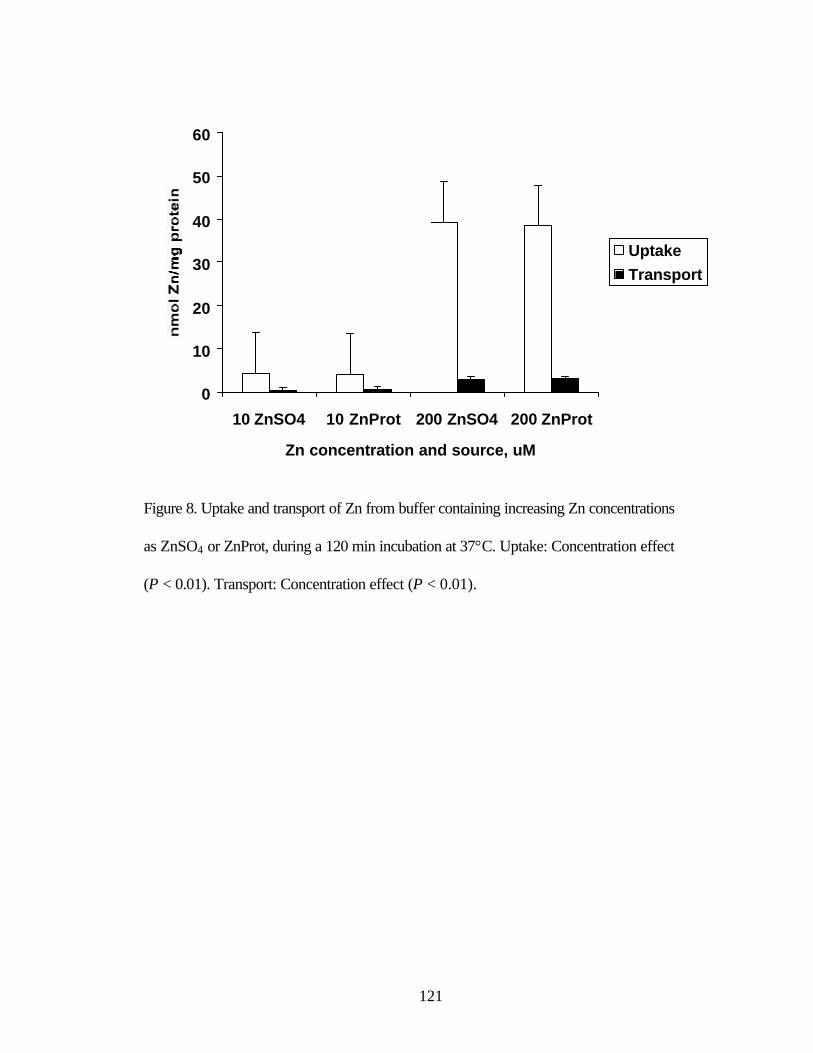
121
Figure 8. Uptake and transport of Zn from buffer containing increasing Zn concentrations
as ZnSO4 or ZnProt, during a 120 min incubation at 37°C. Uptake: Concentration effect
(P < 0.01). Transport: Concentration effect (P < 0.01).
0
10
20
30
40
50
60
10 ZnSO4 10 ZnProt 200 ZnSO4 200 ZnProt
Zn concentration and source, uM
UptakeTransport
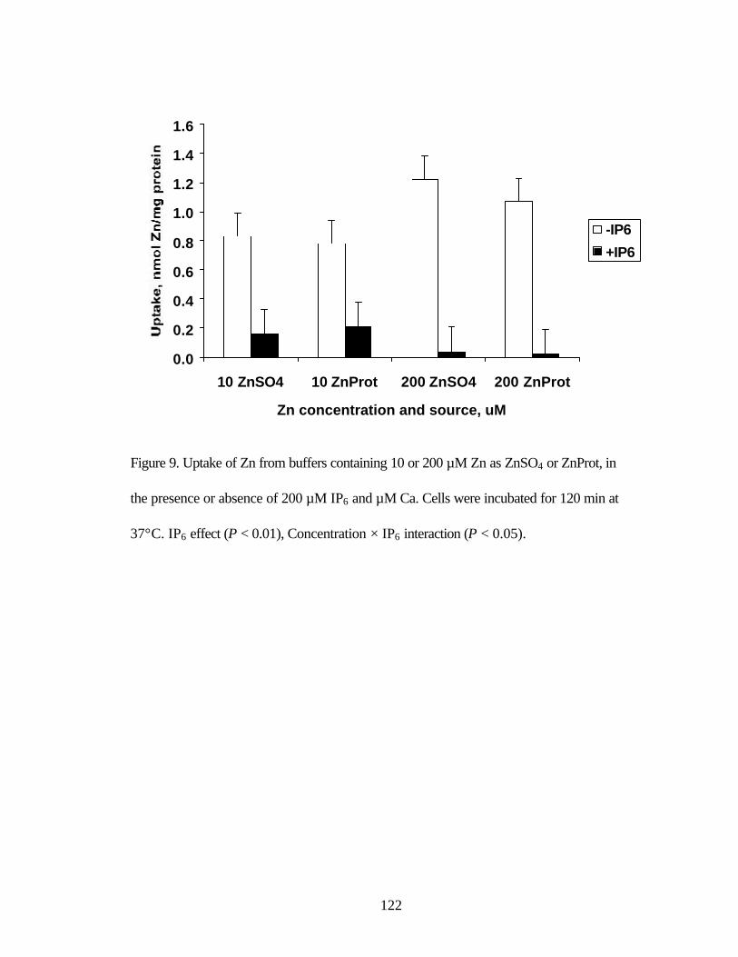
122
Figure 9. Uptake of Zn from buffers containing 10 or 200 µM Zn as ZnSO4 or ZnProt, in
the presence or absence of 200 µM IP6 and µM Ca. Cells were incubated for 120 min at
37°C. IP6 effect (P < 0.01), Concentration × IP6 interaction (P < 0.05).
0.0
0.2
0.4
0.6
0.8
1.0
1.2
1.4
1.6
10 ZnSO4 10 ZnProt 200 ZnSO4 200 ZnProt
Zn concentration and source, uM
-IP6
+IP6

123
Figure 10. Transport of Zn from buffers containing 10 or 200 µM Zn as ZnSO4 or
ZnProt, in the presence or absence of 200 µM IP6 and µM Ca. Cells were incubated for
120 min at 37°C. Concentration effect (P < 0.01), IP6 effect (P < 0.01), Concentration ×
IP6 interaction (P < 0.01).
0.0
0.2
0.4
0.6
0.8
1.0
1.2
10 ZnSO4 10 ZnProt 200 ZnSO4 200 ZnProt
Zn concentration and source, uM
-IP6+IP6

124
Figure 11. Uptake and transport of Zn from aqueous fractions of ruminal and intestinal
digestions containing 10 or 200 µM added Zn as ZnSO4 or ZnProt. Cells were incubated
for 120 min at 37°C. Uptake: Concentration effect (P < 0.01), Source effect (P < 0.05),
Concentration × source interaction (P < 0.03). Transport: Concentration effect (P < 0.05).
0.0
0.5
1.0
1.5
2.0
2.5
10 ZnSO4 10 ZnProt 200 ZnSO4 200 ZnProt
Zn concentration and source, uM
Uptake
Transport
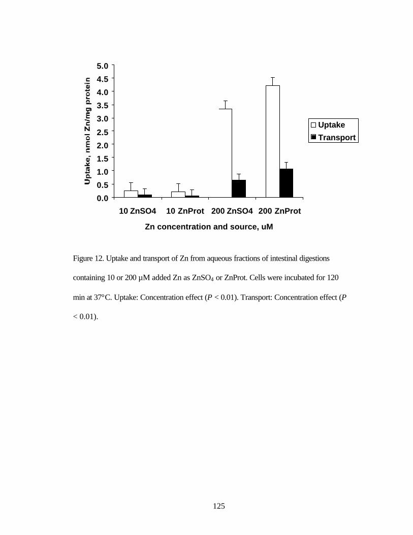
125
Figure 12. Uptake and transport of Zn from aqueous fractions of intestinal digestions
containing 10 or 200 µM added Zn as ZnSO4 or ZnProt. Cells were incubated for 120
min at 37°C. Uptake: Concentration effect (P < 0.01). Transport: Concentration effect (P
< 0.01).
0.0
0.5
1.0
1.5
2.0
2.5
3.0
3.5
4.0
4.5
5.0
10 ZnSO4 10 ZnProt 200 ZnSO4 200 ZnProt
Zn concentration and source, uM
Uptake
Transport

126
CHAPTER 5
Uptake and transport of zinc from zinc sulfate and zinc propionate by Caco-2 cells1,2
Cody L. Wright3, Jerry W. Spears4, and Mark L. Failla*,5
Department of Animal Science and Interdepartmental Nutrition Program, North Carolina
State University, Raleigh, NC 27695-7621 and *Department of Nutrition and Food
Service Systems, University of North Carolina at Greensboro, Greensboro, NC 27402-
6170
Phone: 919/515-4008
Fax: 919/515-4463
E-mail: [email protected]
1 Supported in part by a gift from Kemin Industries, Des Moines, IA.
2 The authors extend sincere appreciation to Ms. Maureen Searcy for technical assistance.
3 Current address: Department of Animal Science, South Dakota State University,
Brookings, SD 57007.
4 To whom correspondence should be addressed.
5 Current address: Department of Human Nutrition and Food Management, The Ohio
State University, Columbus, OH 43210.
127
ABSTRACT: Experiments were conducted to evaluate effects of time, Zn concentration,
inositol hexaphosphate (IP6) addition, and simulated ruminal and intestinal digestion on
the uptake and transport of Zn from ZnSO4 and Zn propionate (ZnProp) by Caco-2 cells
grown on plastic (uptake only) or on membrane inserts (uptake and transport). Increasing
incubation time from 2 to 120 min and Zn concentration from 10 to 200 µM increased (P
< 0.01) Zn uptake and transport. In the presence of IP6 and Ca, Zn uptake and transport
were affected by a concentration × IP6 interaction (P < 0.01). In the absence of
antagonists, uptake and transport increased as Zn concentration increased; however when
antagonists were added, uptake and transport were nearly completely attenuated. Zinc
uptake by cells grown on inserts tended to be influenced by a source × concentration
interaction (P < 0.07). At 10 µM Zn, uptake was not different; however, at 200 µM,
uptake from ZnSO4 was greater than from ZnProp. Following simulated ruminal and
intestinal digestions, Zn uptake was greater from digestions containing 200 µM added Zn
than from those containing 10 µM. In cells grown on plastic, Zn uptake following
ruminal and intestinal digestion tended (P < 0.10) to be greater from ZnProp than from
ZnSO4. Transport and solubility of Zn following ruminal and intestinal digestion were
not affected by Zn source or concentration. Following intestinal digestion alone, Zn
uptake by cells grown on inserts tended to be affected by a source × concentration
interaction (P < 0.09). Zinc uptake from digestions containing 10 µM added Zn was not
affected by Zn source; however, when 200 µM Zn was added, uptake was greater from
ZnSO4 than from ZnProp. Zinc uptake by cells grown on plastic and Zn transport by cells
grown on inserts were greater (P < 0.01) from digestions containing 200 µM added Zn
than from those containing 10 µM added Zn, but were not affected by Zn source.

128
Solubility of Zn following intestinal digestion was affected by a source × concentration
interaction (P < 0.01). When 10 µM Zn was added to digestions, Zn from ZnSO4 was
more soluble than from ZnProp; however, when 200 µM Zn was added, Zn from ZnProp
was more soluble than from ZnSO4. In the absence of inhibitors or in the presence of 200
µM IP6 and Ca, uptake and transport of Zn from ZnSO4 and ZnProp were similar;
however, following simulated ruminal digestions, Zn solubility and uptake tended to be
greater from ZnProp than from ZnSO4.
Key Words: zinc, propionate, Caco-2, uptake, transport

129
Introduction
Bioavailability of Zn from organic and inorganic sources has been the subject of
numerous in vivo experiments (Spears, 1996). The organic Zn compound Zn propionate
(ZnProp) is a complex formed by combining 1 mol Zn from a soluble Zn salt with 2 mol
propionate. Zinc propionate has been demonstrated to be more bioavailable in dogs
(Brinkhaus et al., 1998; Wedekind and Lowry, 1998), and chicks (Kemin Industries,
1995) than inorganic Zn. However, in vitro, Zn uptake by monkey kidney fibroblasts
(CV-1) and epithelial cells (T-84) was not different between inorganic and organic Zn
sources (Buetler et al., 1998). Observed differences have been hypothesized to result
from enhanced absorption and(or) solubility of organic relative to inorganic Zn sources.
If Zn complexes are degraded or ionized during the digestive process, uptake would
likely be similar to that found with inorganic salts. Minimal research to date has
specifically addressed absorption characteristics of organic and inorganic Zn sources.
Caco-2, a transformed human colonic cell line, has been used as an in vitro model
to evaluate nutrient and drug metabolism by intestinal absorptive epithelium (Han et al.,
1994). Monolayers of Caco-2 cells spontaneously differentiate into highly polarized cells
that possess many of the properties of mature intestinal absorptive epithelial cells
(Hidalgo et al., 1989). Characteristics of Ca (Giuliano and Wood, 1991), Cu (Reeves et
al., 1996), Fe (Alverez-Hernandez et al., 1991; Halleux and Schneider, 1991), Mn (Finley
and Monroe, 1997) and Zn (Raffaniello and Wapnir, 1991; Raffaniello et al., 1992)
uptake and transport have been evaluated using Caco-2 cells.
A series of experiments were conducted to compare the uptake and transport of
Zn from ZnSO4 and ZnProp by Caco-2 cells.

130
Materials and Methods
Materials. Dulbecco’s modified Eagle medium (DMEM ), fetal calf serum (FCS),
medium supplements except antibiotics and test reagents were purchased from Sigma
Chemical Company (St. Louis, MO). Gentamicin and fungizone were purchased from
GIBCO (Grand Island, NY). 65Zinc (185 GBq/g) was obtained from New England
Nuclear (Boston, MA). All other supplies were purchased from Fisher Scientific
Company (Pittsburgh, PA) unless specifically stated otherwise.
Caco-2 cells. The TC-7 clone of the Caco-2 human colonic adenocarcinoma cell
line (HTB-37) was generously donated by Dr. Monique Rousset (INSERM, Cedex,
France). Cells were grown in 75-cm2 plastic flasks (Falcon, Becton Dickinson &
Company, Franklin Lakes, NJ) in high glucose complete Dulbecco’s modified minimal
essential medium (cDMEM ; pH 7.0) containing DMEM (D7777), heat inactivated fetal
calf serum (FCS; 10%, v/v), NaHCO3 (44 mM), HEPES (15 mM), non-essential amino
acids (1%), L-glutamine (4 mM), and antibiotics (50 mg gentamicin and 0.5 mg
fungizone/L) in an atmosphere of air:CO2 (95:5) at 37°C and 90% humidity. Stock
cultures were seeded at a density of 5,333 cells/cm2, and harvested by treatment with
Hank’s balanced salt solution without Ca or Mg and containing trypsin (0.25%) and
EDTA (2 mM) for re-seeding when the monolayer was 70-80% confluent. Suspended
cells were transferred either to 12-well cluster dishes (Falcon, Becton Dickinson &
Company, Franklin Lakes, NJ) to assess uptake alone or track-etched polyethylene
teraphthalate membrane filters (0.4 µm pore size, Falcon, Becton Dickinson & Company,
Franklin Lakes, NJ) in 6-well cluster dishes (Falcon, Becton Dickinson & Company,
Lincoln Park, NJ) to examine Zn uptake and transport together. Medium was changed

131
every second day and the day before cultures were used for experiments. Differentiated
cultures were used 10 to 14 d after reaching confluency between passages 79 and 91
(Ellwood et al., 1993).
Uptake experiments. Preliminary experiments were conducted to determine
solubility of the Zn sources in the uptake buffer and to determine the optimum pH for
uptake and transport. Results indicated no difference in solubility between the Zn sources
and that solubility of both sources was nearly 100%. Zinc uptake was maximal at pH 6.0
and declined steadily as pH was either reduced to 5.0 or increased to 7.5. Test solutions
were prepared and utilized in an identical manner for uptake experiments alone and
uptake and transport experiments together. Solutions were prepared fresh prior to the
experiment by adding an aliquot of ZnSO4 or ZnProp (Kemin Industries, Des Moines,
IA) stock solution and 65Zn to a volume of uptake buffer (pH 6.0; 130 mM NaCl, 10 mM
KCl, 10 mM 2-[N-morpholino]ethanesulfonic acid, 5 mM glucose, and 1 mM MgSO4) to
provide the appropriate Zn concentration(s) and 9.25 kBq 65Zn/mL. To determine the
effect of dietary inhibitors on Zn uptake, 200 µM IP6 and 200 µM Ca were added to
uptake solutions. Stock solutions of IP6 (1000 µM) and Ca (1000 µM Ca as CaCl2⋅2H2O)
were prepared in uptake buffer, and then appropriate aliquots were added to test solutions
following addition of Zn treatments and a 15 min incubation period. Final test solutions
were allowed to equilibrate for 15 min at 37°C prior to the experiment. Spent medium
was aspirated and each well was washed twice with 1 mL uptake buffer at 37°C. Test
solutions (1 mL) were added to each well and cultures were incubated at 37°C for the
appropriate time. After incubation, test solutions were removed and each well was
washed three times with ice-cold wash solution (pH 7.0; 150 mM NaCl, 10 mM HEPES,

132
and 5 mM tetrasodium EDTA). Preliminary experiments comparing the effectiveness of
multiple washes on removal of non-specifically bound 65Zn determined that more than 3
washes resulted in no further reduction in the 65Zn content of cell suspensions. Cellular
material was collected by adding 0.5 mL harvest solution (pH 7.0; 1.7 mM SDS and 1.0
mM tetrasodium EDTA) to each well, scraping the surface with a rubber policeman, and
transferring the material to microfuge tubes for solication and storage. The process was
then repeated. Cellular material was homogenized by sonication (50 Sonic
Dismembrator, Fisher Scientific, Pittsburgh, PA) for 2 to 3 sec. Aliqouts of each cell
suspension (0.4 mL) and test solution (0.1 mL) were transferred to 5 mL-polypropylene
tubes (Sarstedt, Newton, NC) for gamma counting (Cobra II, Packard Instrument
Company, Meriden, CT). The quantity of Zn taken up by the cells was determined by
first expressing specific radioactivity of the cell suspensions as a percent of the total
specific radioactivity in the uptake buffer. Then the percent of the specific radioactivity
taken up by the cells was multiplied by the non-radioactive Zn concentration in the
uptake buffer. The ratio of radioactive Zn to non-radioactive Zn was assumed to remain
constant. Total protein content of each cell suspension was determined by colorimetric
assay (Bio-Rad Laboratories Incorporated, Hercules, CA) using a plate reader
(PowerWaveX, Bio-Tek Instruments, Winooski, VT). Cell suspensions were diluted 1:5
with harvest solution and aliqouts (0.01 mL) of each dilution and BSA standard were
pipetted into 96-well dishes (Costar, Corning Incorporated, Corning, NY) and combined
with assay reagent. Data were expressed as nmol Zn/mg protein to normalize data among
samples.

133
Transport experiments. Test solutions were prepared as described above,
however phenol red (500 µM) was added to the uptake buffer to facilitate a measure of
monolayer integrity. Phenol red is not metabolized or transported by Caco-2 cells, thus
appearance of phenol red in the basolateral buffer would be indicative of either
paracellular transport or compromised monolayer integrity. A preliminary experiment
determined that basal Dulbecco’s modified minimal essential medium (bDMEM ; pH 7.0,
containing 44 mM NaHCO3 and 15 mM HEPES) and cDMEM maintained monolayer
integrity more effectively than uptake buffer during a 120 min incubation at 37°C. Since
cDMEM contains several potential Zn ligands (FCS and glutamine), bDMEM was
selected as the basolateral media and BSA (2.5 mg/mL) was added to provide a single
ligand for Zn transported across the basolateral membrane. Previous research has
demonstrated that apical-to-basolateral Zn transport was markedly increased by the
addition of BSA to the basolateral media up to 2.5 mg/mL (Fleet et al., 1993). Spent
medium was aspirated and each insert and well was washed twice with uptake buffer (1
or 2 mL, respectively) at 37°C. Test solution was added to each insert (1 mL) and
bDMEM + BSA was added to each well (2 mL). After incubation, inserts were removed
and placed into new 6-well cluster dishes containing 2 mL ice-cold wash solution per
well. Aliquots of each basolateral buffer were transferred to 5-mL polypropylene tubes
for gamma counting (1 mL) and to 96-well dishes for determination of phenol red content
(150 µL). Phenol red content of basolateral buffers was determined colorimetrically after
the addition of 20 µL 1 M NaOH. Collection and gamma counting of cellular material
and determination of total protein were determined as described for the uptake
experiments.

134
Solubility experiments. Test solutions were prepared fresh prior to each
experiment by adding an aliquot of ZnSO4 or ZnProp stock solution and 65Zn to a volume
of uptake buffer to provide appropriate Zn concentrations and 9.25 kBq 65Zn/mL. To
determine the effect of dietary inhibitors on Zn solubility, stock solutions of IP6 and Ca
were added to the test solutions to provide 200 µM IP6 and 200 µM Ca and test solutions
were allowed to equilibrate for 15 min. Test solutions were then vortexed and an aliquot
was transferred to 5-mL polyethylene tubes for gamma counting. Test solutions were
then allowed to equilibrate at room temperature for 1 hr and centrifuged at 8,160 × g for 5
min (5415C, Eppendorf Scientific Incorporated, Westbury, NY). Following
centrifugation, a second aliquot was transferred to 5-mL polyethylene tubes for gamma
counting. Solubility was determined from the specific radioactivity of aliquots collected
before and after centrifugation.
Simulated ruminal digestion. Buffered mineral medium (McDougall, 1948) was
prepared fresh prior to the experiment. Rumen fluid was collected from fistulated steers
and strained through 8 layers of cheesecloth into a pre-warmed (39°C), insulated
container for transport to the laboratory. Upon arrival, pH was measured and rumen fluid
was strained again through 8 layers of cheesecloth. Rumen inoculum and mineral
medium were combined in a 2:1 ratio and anaerobic conditions were maintained by
bubbling CO2 through the mixture. Urea was added to provide a final concentration of
0.05% and the solution was mixed well under CO2. Aliquots (10 mL) were transferred
into 50 mL centrifuge tubes containing 0.118 g substrate (74% corn, 11% soybean meal,
10% oat straw, and 5% CaCl2⋅2H2O). Zinc sulfate or ZnProp and 65Zn were combined
prior to the experiment as described above and added to provide either 10 or 200 µM Zn
135
and 18.5 kBq/mL in the final intestinal digestion (only 8 mL from each ruminal digestion
solution was required for each intestinal digestion solution). Tubes were mixed gently,
blanketed in CO2, and stoppers with one-way valves were installed to preserve an
anaerobic environment. Tubes were then incubated in a waterbath for 18 h at 39°C, and
were mixed gently periodically during the incubation. After incubation, digestion
mixtures were immediately transferred to 15-mL tubes (Sarstedt, Newton, NC) and used
in a simulated intestinal digestion procedure described below.
Simulated intestinal digestion. Two grams of substrate (85% corn, 10% soybean
meal, and 5% CaCl2⋅2H2O) were diluted in 160 mL of saline (120 mM NaCl and 150 µM
butylated hydroxytoluene) and then homogenized for 3 min at 80 rpm (Tissumizer,
Tekmar Company, Cincinnati, OH). The pH of the solution was lowered to 2.1 using 1 M
HCl, and porcine pepsin was added to provide a final concentration of 2 mg/mL. The
solution was homogenized for 3 min at 80 rpm and aliquots (8 mL) were transferred into
15 mL-tubes (Sarstedt, Newton, NC). Zinc sulfate or ZnProp and 65Zn were combined
prior to the experiment as described above and added to provide either 10 or 200 µM Zn
and 18.5 kBq/mL in the final digestion volume (10 mL). Digestion solutions from
simulated ruminal digestions entered the intestinal phase of the digestion process with the
addition of porcine pepsin and reduction in pH. From that point all tubes, regardless of
origin (ruminal or intestinal) were treated in an identical manner. Three replicate
digestion tubes were maintained for each treatment combination. Solubility of
endogenous Zn and the contribution of added ZnSO4 and ZnProp to soluble Zn
concentration were determined in parallel digestion tubes. Endogenous Zn was
determined in triplicate digestion tubes containing the meal alone with no added Zn. The

136
contribution of added Zn was determined by adding either 10 or 200 µM Zn from each
source to triplicate digestion tubes without 65Zn. Tubes were wrapped in laboratory film
(Parafilm, American National Can, Chicago, IL) and incubated in a shaking water bath
(95 rpm; YB-531, American Scientific) for 60 min at 37°C. After incubation, the pH was
raised to 6.0 with 1 M NaHCO3, and bile extract, pancreatin and pancreatic lipase (all of
porcine origin) were added to each tube to provide final concentrations of 2.4, 0.4, and
0.2 mg/mL, respectively. The pH was then adjusted to 6.9 with 1 M NaHCO3 and the
final volume was brought to 10 mL with saline. Tubes were blanketed with nitrogen gas,
wrapped in laboratory film and incubated in a shaking water bath (95 rpm) for 120 min at
37°C.
Following the final incubation, aliquots (4.5 mL each) from each digestion were
transferred into ultracentrifuge tubes (Beckman Quick-Seal, Beckman Coulter
Incorporated, Fullerton, CA). Tubes were then capped and centrifuged at 142,646 × g for
40 min at 4°C (Beckman L7-65, 50.3 Ti rotor, Beckman Coulter Incorporated, Fullerton,
CA). Aqueous fractions were collected by puncturing the side of the tubes with 18 ga
needles (Precision Glide, Becton Dickinson and Company, Franklin Lakes, NJ) attached
to 10 mL syringes (Becton Dickinson and Company, Franklin Lakes, NJ) and filtered
through syringe filters (0.2 µm pores, polysulfone membrane, Acrodisc, Pall Gelman
Laboratory, Ann Arbor, MI) into 5-mL polypropylene tubes. Aliquots (0.1 mL) of each
test solution prior to and after ultracentrifugation were transferred into 5-mL
polypropylene tubes for quantification of 65Zn. Solubility of Zn in samples containing
65Zn was determined from the specific radioactivity of each sample prior to and after
ultracentrifugation. Solubility of endogenous Zn and Zn contributed by added ZnSO4 and
137
ZnProp was determined were analyzed by flame atomic absorption spectophotometry
(AA-6701F, Shimadzu, Kyoto, Japan). Remaining aqueous fractions were pooled among
replicate digestions from each concentration × source combination. Pooled aqueous
fractions were used as is for each experiment, and were placed directly onto cell
monolayers.
Statistical analysis. All variables were tested in at least triplicate and each
experiment was repeated twice. Results are presented as pooled means between
replicates. Time-dependent data were analyzed by ANOVA using the Mixed procedure of
SAS (Version 6.12, SAS Institute Inc., Cary, NC) as described by Littell et al. (1998).
The model for time-dependent data included time, Zn source and time × Zn source
interaction. Remaining data were analyzed by ANOVA using the General Linear Models
procedure of SAS. The models included Zn source, Zn concentration, and Zn source × Zn
concentration interaction. All data are presented as means ± SE, and significance was
declared at P < 0.05.
Results
Uptake experiments. The effect of time on Zn uptake was examined by
incubating cultures with uptake buffer containing 20 µM Zn as ZnSO4 or ZnProp while
increasing incubation times from 5 to 120 min. Increasing incubation times resulted in
corresponding increases (P < 0.01) in Zn uptake (Figure 1). To determine the effect of Zn
concentration on Zn uptake, cultures were incubated for 60 min with uptake buffer
containing incremental increases in Zn concentration from 10 to 200 µM Zn as ZnSO4 or
ZnProp. Zinc uptake increased (P < 0.01) at a decreasing rate as Zn concentration
increased from 10 to 50 µM Zn, then as Zn concentration increased from 50 to 200 µM,
138
uptake was nearly linear (Figure 2). Zinc uptake in time- and concentration-dependent
experiments was not affected by Zn source.
Interactions with IP6 and Ca are known to decrease Zn bioavailability, thus
solubility and uptake of Zn from ZnSO4 and ZnProp at concentrations ranging from 10 to
200 µM were examined in the presence of 200 µM IP6 and 200 µM Ca. Zinc uptake was
affected by a concentration × IP6 interaction (P < 0.01; Figure 3). In the absence of
inhibitors, Zn uptake increased with increasing Zn concentration in the uptake buffer.
Addition of IP6 and Ca reduced uptake at all Zn concentrations; however reductions were
more pronounced as Zn concentration increased. Zinc solubility in the presence of IP6
and Ca was affected by a concentration × IP6 interaction (P < 0.01). In the presence of IP6
and Ca, Zn solubility diminished as Zn concentration increased (Figure 4); however in
the absence of antagonists, Zn solubility was near 100% for both Zn sources. Uptake and
solubility of Zn were not different between ZnSO4 and ZnProp.
Complete diets contain several potential ligands that may positively or negatively
influence Zn absorption. Solubility and uptake of Zn from each source were evaluated
following ruminal and intestinal digestions in series and intestinal digestion alone with 10
or 200 µM added Zn as ZnSO4 or ZnProp in the digestion solutions. Following ruminal
and intestinal digestion, Zn uptake was 6-fold greater (P < 0.01) from digestions
containing 200 relative to 10 µM Zn and numerically (P < 0.10) was 37.5% greater from
digestions containing ZnProp than those containing ZnSO4 (Figure 5). When exposed to
the intestinal digestion alone, uptake was 15-fold greater (P < 0.01) from aqueous
fractions of digestions containing 200 µM Zn than digestions containing 10 µM added
Zn; however, Zn uptake was not affected by source (Figure 6). Zinc solubility was

139
affected by a concentration × source interaction (P < 0.01; Figure 6). When 10 µM Zn
was added to digestion solutions, Zn from ZnSO4 was 12.9% more soluble than Zn from
ZnProp; however when 200 µM Zn was added, Zn from ZnProp was 7.8% more soluble
than Zn from ZnSO4.
Transport experiments. Preliminary experiments determined that transport of Zn
into the basolateral buffer from 20 µM Zn was minimal prior to 30 min. Thus, the effect
of incremental increases in incubation time from 30 to 120 min was investigated with 20
µM Zn in the uptake buffer. Main effect means for Zn uptake increased (P < 0.01) with
incremental increases in incubation time from 30 to 90 min; however, increasing
incubation time from 90 to 120 min resulted in no further increase in uptake (Figure 7).
Increasing incubation time from 30 to 60 min increased (P < 0.01) Zn transport; however,
subsequent increases in incubation time did not affect transport. The effect of increasing
Zn concentration in the uptake buffer from 10 to 200 µM Zn on uptake into and transport
across cell monolayers was evaluated by incubating cultures for 120 min. Zinc uptake
and transport were 5- and 16-fold greater (P < 0.01), respectively, when cultures were
incubated with 200 µM Zn than with 10 µM Zn (Figure 8). Uptake and transport of Zn in
time- and concentration-dependent experiments were not significantly affected by Zn
source.
Zinc bioavailability in the presence of dietary inhibitors was examined by adding
200 µM IP6 and Ca to the uptake buffer and measuring uptake into and transport of Zn
across monolayers following incubation for 120 min. Uptake (Figure 9) and transport
(Figure 10) of Zn were affected by a concentration × IP6 interaction (P < 0.01). In the
absence of inhibitors, Zn uptake and transport were 5- and 19-fold greater with 200 µM

140
Zn in the uptake buffer than 10 µM Zn. However, in the presence of IP6 and Ca, uptake
and transport of Zn were greatly reduced, but were not affected by Zn concentration. Zinc
uptake also tended to be influenced by a source × concentration (P < 0.07) interaction.
With 10 µM Zn in the uptake buffer, uptake was not different, however with 200 µM Zn
in the uptake buffer, Zn uptake from ZnSO4 was 11.8% greater than from ZnProp. Zinc
transport was not affected by Zn source.
Bioavailability of Zn was estimated following simulated ruminal and intestinal
digestions processes. Uptake of Zn from aqueous fractions of digestions containing 200
µM added Zn was 9-fold greater (P < 0.01) than those containing 10 µM added Zn, but
was not different between ZnSO4 and ZnProp (Figure 11). Zinc transport following
simulated ruminal and intestinal digestions was not affected by Zn source or
concentration. Zinc uptake following intestinal digestion alone tended to be influenced by
a source × concentration interaction (P < 0.09). When incubated with aqueous fractions
from digestion solutions containing 10 µM added Zn, uptake of Zn was not different;
however, when 200 µM Zn was added, uptake was 12.5% greater from ZnSO4 than from
ZnProp (Figure 12). Transport of Zn was greater from aqueous fractions of digestions
containing 200 µM added Zn than from those containing 10 µM added Zn, but was not
affected by Zn source.
Discussion
Zinc uptake and transport by Caco-2 cell monolayers increased as incubation time
and Zn concentration in the uptake buffer increased; however, neither was affected by Zn
source. Uptake in time-dependent experiments was nearly linear as incubation time
increased, while uptake in concentration-dependent experiments displayed characteristics

141
indicative of both saturable and non-saturable components of Zn absorption. These
observations are consistent with those of Raffaniello et al. (1992) and Finley et al. (1995)
who observed linear increases in Zn uptake during incubations of 50 min and 25 h,
respectively. Zinc transport responded to increasing incubation time in a manner similar
to that reported by Raffaniello et al. (1992). Transport increased at an increasing rate as
incubation times increased, suggesting a lag in Zn transport with respect to Zn uptake.
Binding of Zn to one of several potential intracellular ligands likely accounts for the lag
in Zn transport. In contrast, Fleet et al. (1993) observed a curvilinear response in Zn
transport as Zn concentration of the uptake buffer increased from 10 to 1000 µM,
indicating the presence of both saturable and non-saturable transport mechanisms. The
saturable component was calculated to have a Kt of 226 µM. Only two Zn concentrations
were used in the current transport experiments, 10 and 200 µM, precluding examination
of transport kinetics; however, based on the data of Fleet et al. (1993) saturation of any
potential transport mechanism was unlikely. Lack of differences in uptake with respect to
Zn source is consistent with the findings of Buetler et al. (1998). The authors observed no
difference in uptake of 65Zn by cultured monkey kidney fibroblasts (CV-1) or intestinal
epithelial cells (T-84) when supplied as ZnCl2, Zn methionine, or ZnProp. Furthermore,
using gel filtration chromatography, Buetler et al. (1998) reported similar elution 65Zn
peaks for ZnCl2, reagent Zn methionine and ZnProp, suggesting that if indeed complexes
were formed, they readily dissociate in solution.
Phytate is a common component of plant seeds and is particularly high in oilseeds
and cereal grains (Baker and Ammerman, 1995). The effect of IP6 and Ca on Zn
solubility and bioavailability has been investigated extensively. Reduced absorption of

142
Zn in the presence of IP6 has been demonstrated in several monogastric species (Baker
and Ammerman, 1995), and is generally accepted as the mechanism by which Zn
bioavailability is attenuated. In the current experiments, Zn solubility the presence of IP6
and Ca declined from 70% to 5% as Zn concentration in the uptake buffer increased
incrementally from 10 to 200 µM. This observation agrees with Han et al. (1994) who
reported a 92% reduction in Zn solubility following the addition of a 10-fold molar
excess of IP6 to a Ca-free uptake buffer. The current experiment included 200 µM IP6 and
200 µM Ca representing IP6:Zn molar ratios ranging from 20:1 to 1:1. In vivo, molar
phytate:Zn ratios above 12 to 15 are considered detrimental to Zn status when Zn is near
the required level (Baker and Ammerman, 1997). Depressed Zn bioavailability observed
in the presence of phytate and Ca, and the associated reduction in solubility proposed to
be responsible, makes the results of the current experiments intriguing. Lonnerdahl et al.
(1984) suggested that Ca might block the phytate binding sites, leaving Zn available for
absorption. Given this suggestion and the 1:1 phytate:Ca molar ratio used in the current
experiment, it is reasonable to assume that competition for binding sites prevents Zn from
associating with phytate at low Zn concentrations, allowing greater Zn solubility. As Zn
concentration increases, Zn would become increasingly competitive for binding sites,
thereby reducing Zn solubility. Zinc uptake by monolayers grown on inserts in the
presence of IP6 and Ca was greater from ZnSO4 than from ZnProp, at 200 but not 10 µM
added Zn. Since Zn solubility in the presence of IP6 and Ca was not influenced by Zn
source and Zn uptake in the absence of inhibitors was similar between sources, the reason
for differences in Zn uptake is unclear. It is possible that a portion of the Zn from ZnProp
bound to IP6 and Ca remained soluble, but was unavailable for uptake.

143
As components of the diet are degraded, Zn is presented to enterocytes as smaller
Zn-binding ligands, primarily peptides, amino acids, and nucleotides, and perhaps as free
Zn (Cousins, 1995). Depending on their chemical nature and complexation constants,
organic chelators may affect bioavailability either positively or negatively (Baker and
Ammerman, 1997). The effect of dietary ligands, such as citrate, cysteine, EDTA,
histidine, and picolinate on Zn absorption has varied depending on experimental
conditions (Wapnir, 1989). Uptake of Zn by cells grown on plastic following ruminal and
intestinal digestions was greater from digestions containing ZnProp than from those
containing ZnSO4. Zinc solubility following ruminal and intestinal digestion was not
statistically different between Zn sources; however, numerically Zn from ZnProp
appeared to be more soluble that Zn from ZnSO4. This small difference in solubility may
account for greater uptake of Zn from digestions containing ZnProp. Zinc uptake and
transport by cells grown on inserts was not affected by Zn source. Since identical
digestion processes and substrates were used in both systems, differences in Zn uptake
with respect to Zn source are difficult to explain. When uptake was assessed alone, cells
were incubated for 60 min as compared to 120 min for uptake and transport experiments.
It is possible that Zn from ZnProp is absorbed more rapidly than Zn from ZnSO4, then, as
incubation time increases, differences in absorption become insignificant. Fundamental
differences in the cell systems used (i.e. cells grown on plastic for uptake and on semi-
permeable membranes for uptake and transport experiments) may also account for
observed differences. Following intestinal digestion alone, Zn uptake by cells grown on
plastic was not affected by Zn source; however, Zn uptake by cells grown on inserts
tended to be greater from ZnSO4 than from ZnProp. Transport of Zn was not significantly

144
affected by Zn source; however, numerically, Zn transport appeared to be greater from
ZnProp. Zinc solubility at 10 µM added Zn was greater from ZnSO4 than from ZnProp;
however, when 200 µM Zn was added, ZnProp was more soluble. Tendencies for lower
uptake and greater transport of Zn from ZnProp than from ZnSO4 suggests that different
uptake and transport mechanisms may exist. Given the greater solubility of Zn from
ZnProp at high Zn concentrations, it is possible that all or part of the Zn from ZnProp is
translocated via paracellular transport or simple diffusion. As described above,
differences in cell systems and incubation times may also partially explain observed
differences in uptake and transport of Zn following intestinal digestion. In vivo
observations suggest that ZnProp may be more bioavailable than ZnO in dogs (Brinkhaus
et al., 1998; Wedekind and Lowry, 1998) and ZnSO4 in chicks (Kemin Industries, 1995).
Numerous physiological and dietary factors in vivo are likely responsible for the
differences between in vitro and in vivo models.
These data suggest that increasing incubation time and Zn concentration increase
Zn uptake and transport by Caco-2 cells. Solubility, uptake and transport are clearly
reduced by the addition of IP6 and Ca, and depression in Zn solubility is more severe as
Zn concentration increases. In the absence of antagonists or in the presence of a limited
number of antagonists at high concentration, Zn source appears to have a minor effect on
solubility, uptake and transport. However, following simulated ruminal and(or) intestinal
digestion processes, solubility and uptake of Zn from ZnProp appeared to be greater than
from ZnSO4 when each were added at high concentration.

145
Implications
Commercially available organic Zn sources have been touted as being more
bioavailable than inorganic sources. Enhanced absorption has been proposed to be a
mechanism by which organic Zn sources may elicit responses in growth, reproduction, or
health in various species. Results of the current experiment suggest that in the absence of
dietary antagonists, inorganic and organic Zn sources are absorbed to a similar extent and
by a similar, if not identical, mechanism. However, in the presence of potential inhibitors,
solubility, uptake and transport of Zn from ZnProp may be greater than from ZnSO4.
Potential differences in post-absorptive metabolism also warrant further investigation in
an attempt to explain physiological responses to organic Zn supplementation.

146
Literature Cited
Alvarez-Hernandez, X., G. M. Nichols, and J. Glass. 1991. Caco-2 cell line: a system for
studying intestinal iron transport across epithelial cell monolayers. Biochim.
Biophys. Atca 1070:205-208.
Ammerman, C. B., D. H. Baker, and A. J. Lewis. 1995. Bioavailability of Nutrients for
Animals: Amino Acids, Minerals, and Vitamins. Academic Press, San Diego, CA.
Baker, D. H., and C. B. Ammerman. 1995. Zinc bioavailability. In: Bioavailability of
Nutrients for Animals: Amino Acids, Minerals, and Vitamins. pp 367-398.
Academic Press, San Diego, CA.
Beutler, K. T., O. Pankewycz, and D. L. Brautigan. 1998. Equivalent uptake of organic
and inorganic zinc by monkey kidney fibroblasts, human intestinal epithelial cells,
and perfused mouse intestine. Biol. Tr. Elem. Res. 61:19-31.
Brinkhaus, F., J. Mann, C. Zorich, and J. Greaves. 1998. Bioavailability of zinc
propionate in dogs. J. Nutr. 128:2596S-2597S.
Cao, J., P. R. Henry, R. Guo, R. A. Holwerda, J. P. Toth, R. C. Littell, R. D. Miles, and
C. B. Ammerman. 2000. Chemical characteristics and relative bioavailability of
supplemental organic zinc sources for poultry and ruminants. J. Anim. Sci.
78:2039-2054.
Chesters, J. K. 1997. Zinc. In: B. L. O’Dell and R. A. Sunde (eds.) Handbook of
Nutritionally Essential Trace Elements. pp 185-230. Marcel Dekker, Inc. New
York, NY.

147
Cousins, R. J. 1996. Zinc. In: L. J. Filer and E. E. Ziegler (eds.) Present Knowledge in
Nutrition, 7th Edition. pp 293-306. International Life Science Institute-Nutrition
Foundation, Washington, DC.
Ellwood, K. C., C. Chatzidakis, and M. L. Failla. 1993. Fructose utilization by the human
intestinal epithelial cell line, Caco-2. Proc. Soc. Exp. Biol. Med. 202:440-446.
Erickson, R. H., J. R. Gum, Jr., M. M. Lindstrom, D. McKean, and Y. S. Kim. 1995.
Regional expression and dietary regulation of rat small intestinal peptide and
amino acid transporter mRNAs. Biochim. Biophys. Res. Comm. 216:249-257.
Finley, J. W., M. Briske-Anderson, P. G. Reeves and L. K. Johnson. 1995. Zinc uptake
and transcellular movement by Caco-2 cells: studies with media containing fetal
bovine serum. J. Nutr. Biochem. 6:137-144.
Finley, J. W., and P. Monroe. 1997. Mn absorption: the use of Caco-2 cells as a model of
the intestinal epithelia. Nutr. Biochem. 8:92-101.
Fleet, J. C., A. J. Turnbull, M. Bourcier, and R. J. Wood. 1993. Vitamin D-sensitive and
quinacrine-sensitive zinc transport in human intestinal cell line Caco-2. Am. J.
Physiol. 264:G1037-G1045.
Giuliano, A. R., R. T. Franceschi, and R. J. Wood. 1991. Characterization of the vitamin
D receptor from the Caco-2 human colon carcinoma cell line: effect of cellular
differentiation. Arch. Biochem. Biophys. 285:261-269.
Giuliano, A. R., and R. J. Wood. 1991. Vitamin D-regulated calcium transport in Caco-2
cells: unique in vitro model. Am. J. Physiol. 260:G207-G212.

148
Gunshin, H., B. Mackenzie, U. V. Berger, Y. Gunshin, M. F. Romero, W. F. Boron, S.
Nussberger, J. L. Gollan, and M. A. Hediger. 1997. Cloning and characterization
of a mammalian proton-coupled metal-ion transporter. Nature 388:482-488.
Halleux, C., and Y. Schneider. 1991. Iron absorption by intestinal epithelial cells. 1.
Caco-2 cultivated in serum-free medium, in polyethyleneterephthalate
microporous membrane, as an in vitro model. In Vitro Cell. & Dev. Biol. 27:293-
302.
Han, O., M. L. Failla, A. D. Hill, E. R. Morris, and J. C. Smith. 1994. Inositol phosphates
inhibit uptake and transport of iron and zinc by a human intestinal cell line. J.
Nutr. 124:580-587.
Hempe, J. W., and R. J. Cousins. 1992. Cysteine-rich intestinal protein and intestinal
metallothionein: an inverse relationship as a conceptual model for zinc absorption
in rats. J. Nutr. 122:89-95.
Hidalgo, I. J., T. J. Raub, and R. T. Borchart. 1989. Characterization of the human colon
carcinoma cell line (Caco-2) as a model system for intestinal epithelial
permeability. Gastroenterology 96:736-749.
Kemin Industries. 1995. Bioavailability of zinc from organic and inorganic sources using
the chick bioassay technique. Tech. Bull. No. 11575. Kemin Industries, Des
Moines, IA.
Littell, R. C., P. R. Henry, and C. B. Ammerman. 1998. Statistical analysis of repeated
measures data using SAS procedures. J. Anim. Sci. 76:1216-1231.
149
Lonnerdahl, B., A. Cederblad, L. Davidson, and B. Sandstrom. 1984. The effect of
individual components of soy formulas and cow’s milk formula on zinc
bioavailability. Am. J. Clin. Nutr. 40:1064-1070.
Matsui, T., and H. Yano. 1998. Formulation of low pollution feeds for animal pollution.
In: Proceedings of the 8th World Conference on Animal Production: Symposium
Series 1, Seoul, Korea. pp 110-119.
McDougall, E. I. 1948. Studies on ruminant saliva. I. The composition and output of
sheep saliva. Biochem. J. 3:15-18.
McMahon, R. J., and R. J. Cousins. 1998. Mammalian zinc transporters. J. Nutr. 128:667-
670.
Raffaniello, R. D., S. Lee, S. Teichberg, and R. A. Wapnir. 1992. Distinct mechanisms of
zinc uptake at the apical and basolateral membranes of Caco-2 cells. J. Cell.
Physiol. 152:356-361.
Raffaniello, R. D., and R. A. Wapnir. 1991. Zinc-induced metallothionein synthesis by
Caco-2 cells. Biochem. Med. Metab. Biol. 45:101-107.
Reeves, P. G., M. Briske-Anderson, and S. M. Newman, Jr. 1996. High zinc
concentrations in culture media affect copper uptake and transport in
differentiated human colon adenocarcinoma cells. J. Nutr. 126:1701-1712.
Spears, J. W. 1996. Organic trace minerals in ruminant nutrition. Anim. Feed Sci.
Technol. 58:151-163.
Tacnet, F., D. W. Watkins, and P. Ripoche. 1990. Studies of Zn transport into brush-
border membrane vesicles isolated from pig small intestine. Biochim. Biophys
Acta 1024:323-330.

150
Tacnet, F., F. Lauthier, and P. Ripoche. 1993. Mechanisms of zinc transport into pig
small intestine brush-border membrane vesicles. J. Physiol. 465:57-72.
Wedekind, K. J., and S. R. Lowry. 1998. Are organic zinc source efficacious in puppies?
J. Nutr. 128:2593S-2595S.

151
Figure 1. Uptake of Zn from buffer containing 20 µM Zn as ZnSO4 or ZnProp incubated
for increasing times at 37°C. Time effect (P < 0.01).
0.0
1.0
2.0
3.0
4.0
5.0
6.0
7.0
0 30 60 90 120
Time, minutes
ZnSO4ZnProp

152
Figure 2. Uptake of Zn from buffer containing increasing Zn concentrations as ZnSO4 or
ZnProp, during a 60 min incubation at 37°C. Concentration effect (P < 0.01).
0.0
1.0
2.0
3.0
4.0
5.0
6.0
7.0
8.0
9.0
0 50 100 150 200
Zn concentration, uM
ZnSO4
ZnProp
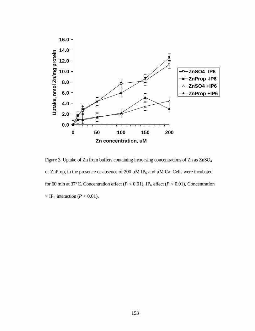
153
Figure 3. Uptake of Zn from buffers containing increasing concentrations of Zn as ZnSO4
or ZnProp, in the presence or absence of 200 µM IP6 and µM Ca. Cells were incubated
for 60 min at 37°C. Concentration effect (P < 0.01), IP6 effect (P < 0.01), Concentration
× IP6 interaction (P < 0.01).
0.0
2.0
4.0
6.0
8.0
10.0
12.0
14.0
16.0
0 50 100 150 200
Zn concentration, uM
Up
take
, nm
ol Z
n/m
g p
rote
in
ZnSO4 -IP6ZnProp -IP6ZnSO4 +IP6ZnProp +IP6

154
Figure 4. Solubility of increasing concentrations of Zn as ZnSO4 or ZnProp in the
absence or presence of 200 µM IP6 and µM Ca. Concentration effect (P < 0.01), IP6
effect (P < 0.01), Concentration × IP6 interaction (P < 0.01).
0
10
20
30
40
50
60
70
80
90
100
0 50 100 150 200
Zn concentration, uM
Sol
ubili
ty, % % ZnSO4 - IP6
% ZnProp - IP6% ZnSO4 + IP6% ZnProp + IP6
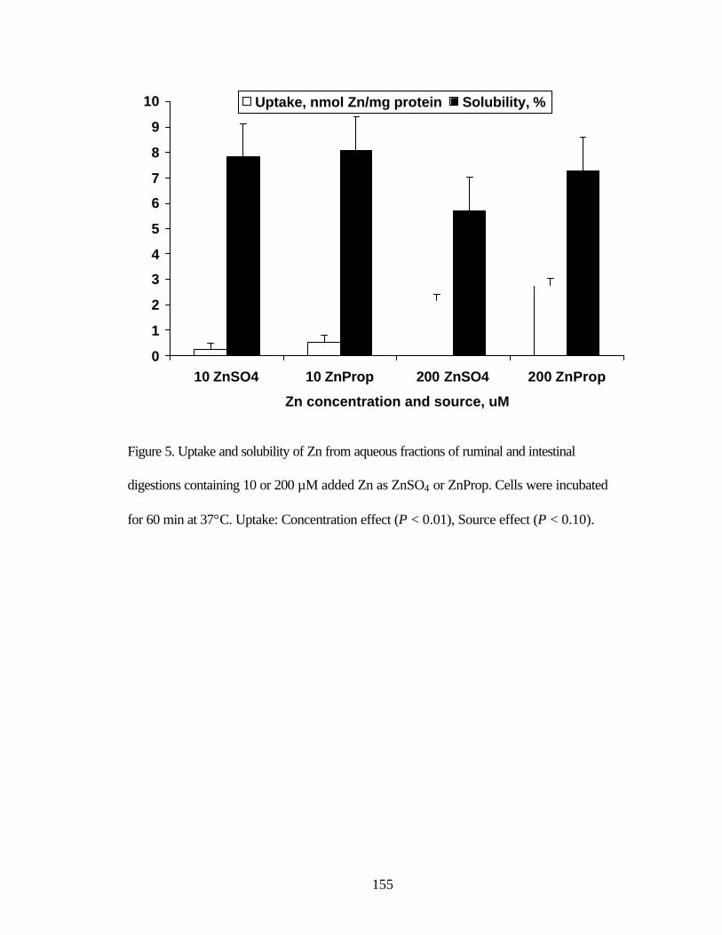
155
Figure 5. Uptake and solubility of Zn from aqueous fractions of ruminal and intestinal
digestions containing 10 or 200 µM added Zn as ZnSO4 or ZnProp. Cells were incubated
for 60 min at 37°C. Uptake: Concentration effect (P < 0.01), Source effect (P < 0.10).
0
1
2
3
4
5
6
7
8
9
10
10 ZnSO4 10 ZnProp 200 ZnSO4 200 ZnProp
Zn concentration and source, uM
Uptake, nmol Zn/mg protein Solubility, %

156
Figure 6. Uptake and solubility of Zn from aqueous fractions of intestinal digestions
containing 10 or 200 µM added Zn as ZnSO4 or ZnProp. Cells were incubated for 60 min
at 37°C. Uptake: Concentration effect (P < 0.01). Solubility: Concentration effect (P <
0.01), Concentration × source interaction (P < 0.01).
0
5
10
15
20
25
10 ZnSO4 10 ZnProp 200 ZnSO4 200 ZnProp
Zn concentration and source, uM
Uptake, nmol Zn/mg protein Solubility, %

157
Figure 7. Uptake and transport of Zn from buffer containing 20 µM Zn as ZnSO4 or
ZnProp incubated for increasing times at 37°C. Uptake: Time effect (P < 0.01).
Transport: Time effect (P < 0.01).
0.0
0.5
1.0
1.5
2.0
2.5
0 30 60 90 120
Time, minutes
ZnSO4 Uptake
ZnProp UptakeZnSO4 TransportZnProp Transport

158
Figure 8. Uptake and transport of Zn from buffer containing increasing Zn concentrations
as ZnSO4 or ZnProp, during a 120 min incubation at 37°C. Uptake: Concentration effect
(P < 0.01). Transport: Concentration effect (P < 0.01).
0.0
1.0
2.0
3.0
4.0
5.0
6.0
7.0
10 ZnSO4 10 ZnProp 200 ZnSO4 200 ZnProp
Zn concentration and source, uM
UptakeTransport

159
Figure 9. Uptake of Zn from buffers containing 10 or 200 µM Zn as ZnSO4 or ZnProp, in
the presence or absence of 200 µM IP6 and µM Ca. Cells were incubated for 120 min at
37°C. Concentration effect (P < 0.01), IP6 effect (P <0.01), Source effect (P < 0.01),
Concentration × IP6 interaction (P <0.01) Concentration × source interaction (P <0.07).
0.0
1.0
2.0
3.0
4.0
5.0
6.0
7.0
10 ZnSO4 10 ZnProp 200 ZnSO4 200 ZnProp
Zn concentration and source, uM
Up
take
, nm
ol Z
n/m
g p
rote
in
-IP6+IP6

160
Figure 10. Transport of Zn from buffers containing 10 or 200 µM Zn as ZnSO4 or
ZnProp, in the presence or absence of 200 µM IP6 and µM Ca. Cells were incubated for
120 min at 37°C. Concentration effect (P < 0.01), IP6 effect (P < 0.01), Concentration ×
IP6 interaction (P < 0.01).
0.0
1.0
2.0
3.0
4.0
5.0
6.0
7.0
10 ZnSO4 10 ZnProp 200 ZnSO4 200 ZnProp
Zn concentration and source, uM
Up
take
, nm
ol Z
n/m
g p
rote
in
-IP6+IP6

161
Figure 11. Uptake and transport of Zn from aqueous fractions of ruminal and intestinal
digestions containing 10 or 200 µM added Zn as ZnSO4 or ZnProp. Cells were incubated
for 120 min at 37°C. Uptake: Concentration effect (P < 0.01).
0.0
0.5
1.0
1.5
2.0
2.5
10 ZnSO4 10 ZnProp 200 ZnSO4 200 ZnProp
Zn concentration and source, uM
Up
take
, nm
ol Z
n/m
g p
rote
in
Uptake Transport

162
Figure 12. Uptake and transport of Zn from aqueous fractions of intestinal digestions
containing 10 or 200 µM added Zn as ZnSO4 or ZnProp. Cells were incubated for 120
min at 37°C. Uptake: Concentration effect (P < 0.01), Concentration × source interaction
(P < 0.09). Transport: Concentration effect (P < 0.01).
0.0
0.5
1.0
1.5
2.0
2.5
3.0
3.5
10 ZnSO4 10 ZnProp 200 ZnSO4 200 ZnProp
Zn concentration and source, uM
Up
take
, nm
ol Z
n/m
g p
rote
in
Uptake Transport

163
SUMMARY
A series of experiments was conducted to examine the absorption and post-
absorptive metabolism of zinc (Zn) from inorganic and organic sources. Results of an in
vivo experiment, using Holstein bull calves as models, suggested that, when Zn was
supplemented at 20 mg Zn/kg DM for 98 d, Zn source had minimal impact on plasma or
tissue Zn concentrations. However, when Zn was supplemented at 500 mg Zn/kg DM for
12 d, plasma and tissue Zn concentrations were greater in bulls that received ZnProt than
in those that received ZnSO4. These data suggested that when supplemented at high
levels to a diet containing adequate Zn levels, Zn from ZnProt was absorbed and(or)
retained to a greater extent than Zn from ZnSO4. Subsequently, in vitro experiments were
conducted to determine the uptake and transport of Zn from ZnSO4, ZnProt or ZnProp by
ruminal and omasal epithelium and Caco-2 cell monolayers. Results of these experiments
suggested that when Zn was included in the mucosal buffer at low concentrations, or in
the absence of antagonists, uptake and transport of Zn from inorganic and organic Zn
sources were similar. However, following simulated ruminal and(or) intestinal digestion,
uptake of Zn from the aqueous fractions of simulated digestion solutions appeared to be
greater from organic than from inorganic Zn sources when Zn was included in the
digestion solutions at 200 but not at 10 µM. Collectively these data suggest that, in the
absence of antagonists, or in the presence of IP6 and Ca only, uptake and transport of Zn
from inorganic and organic Zn sources were similar. However, when added at high
concentration in the presence of dietary antagonists, Zn uptake from organic sources
appeared to be greater than from inorganic sources. Further research is certainly

164
warranted to more clearly define differences in Zn absorption that may exist between
inorganic and organic Zn sources at different dietary levels.